Embed Size (px)
Citation preview
© The Authors, 2017. Journal compilation © Australian Museum, Sydney, 2017Records of the Australian Museum (2017) Vol. 69, issue number 4, pp. 259–275. ISSN 0067-1975 (print), ISSN 2201-4349 (online)https://doi.org/10.3853/j.2201-4349.69.2017.1679urn:lsid:zoobank.org:pub:BD74A0FE-CB17-4D7C-8595-3912F0406AA7Shane T. Ahyong orcid.org/0000-0002-2820-4158Martin Schwentner orcid.org/0000-0002-1373-456XStefan Richter orcid.org/0000-0002-2865-2751
The Tasmanian Lake Shrimps, Paranaspides Smith, 1908 (Crustacea, Syncarida, Anaspidesidae)
Shane T. Ahyong1*, Martin Schwentner2,3 and Stefan Richter2
1 Australian Museum Research Institute, Australian Museum, 1 William Street, Sydney NSW 2010, Australia, and
School of Biological, Earth & Environmental Sciences, University of New South Wales NSW 2052, Australia
2 Institut für Biowissenschaften, Allgemeine und Spezielle Zoologie, Universität Rostock, Rostock, Germany
3 current address: Centrum für Naturkunde, Universität Hamburg, Hamburg, Germany
Abstract. The Tasmanian Lake Shrimps of the genus Paranaspides Smith, 1908 (Syncarida: Anaspidesidae) are endemic to lakes on the eastern Central Plateau, Tasmania, namely Great Lake, Shannon Lagoon, Penstock Lagoon, Arthurs Lake and Woods Lake. Prior to the present study, only the type species, P. lacustris Smith, 1908, was recognized. Reconsideration of Paranaspides from throughout its range, however, showed that Paranaspides from Arthurs Lake and Woods Lakes are referrable to a new species, P. williamsi sp. nov. Morphometric differences in the uropodal exopod and maxilliped, and subtle differences in the morphology of the male pleopods 1 and 2, and colour-in-life distinguish the two species. Genetic divergence (p-distance) between the two species exceeds 10% in mitochondrial COI and 3% in 16S. Both species are described and illustrated, and a lectotype fixed for P. lacustris. Although P. lacustris and P. williamsi occur in relatively close proximity, they occupy different drainages. The Great Lake-Shannon Lagoon-Penstock Lagoon system drains to the southeast, and the Arthurs Lake-Woods Lake system to the northeast. The distributions of P. lacustris and P. williamsi precisely parallel those of a cognate pair of galaxiid fishes, Paragalaxias eleotroides and Paragalaxias mesotes. Given the geological history of the Central Plateau and molecular divergence estimates for Paragalaxias, Paranaspides may also have diverged prior to the Pleistocene glaciations. Species of Paranaspides are dependent on their shallow water algal bed habitat, making them highly susceptible to sudden or significant fluctuations in lake water levels as a result of hydroelectric operations. Both species of Paranaspides have small areas of occupancy and are prone to the effects of hydroelectric activities on their lake habitats; under IUCN Red List criteria, their conservation status corresponds to Vulnerable (D2).
Keywords. Crustacea; Anaspidacea; Paranaspides; Tasmania; freshwater; shrimp.
Ahyong, Shane T., Martin Schwentner and Stefan Richter. 2017. The Tasmanian Lake Shrimps, Paranaspides Smith, 1908 (Crustacea, Syncarida, Anaspidesidae). Records of the Australian Museum 69(4): 259–275. https://doi.org/10.3853/j.2201-4349.69.2017.1679
* author for correspondence urn:lsid:zoobank.org:pub:BD74A0FE-CB17-4D7C-8595-3912F0406AA7

260 Records of the Australian Museum (2017) Vol. 69
The Tasmanian syncarid shrimp family Anaspidesidae comprises three genera: Anaspides Thomson, 1894, Paranaspides Smith, 1908, and Allanaspides Swain, Wilson, Hickman & Ong, 1970. Although these genera were formerly placed in Anaspididae Thomson, 1893, Ahyong & Alonso-Zarazaga (2017) showed Thomson’s family name to be preoccupied, warranting creation of the new family, Anaspidesidae. Anaspidesids are notable for their sometimes relatively large size and usual occupation of epigean habitats; though some are subterranean or pholeteric, none are interstitial like most other extant syncarids. As a result, they show little structural reduction and have a near complete complement of appendages and associated rami. Anaspidesid taxonomy has received little attention since the 1970s. Ahyong (2015, 2016) recently revised Anaspides, and Paranaspides is treated herein.
The type species of Paranaspides, P. lacustris Smith, 1908, was described from Great Lake on the eastern part of the Tasmanian Central Plateau, hence its common name, the Great Lake Shrimp. Paranaspides lacustris was sub-sequent ly discovered in neighbouring Shannon and Penstock Lagoons at the south end of the lake (Evans, 1942, Nicholls, 1947), and later reported from Arthurs and Woods Lakes (Fulton, 1982, 1983). Reconsideration of Paranaspides from throughout its range based on morphological and molecular data shows that populations from Arthurs and Woods Lakes represent an undescribed species. The new species is formally described, and P. lacustris redescribed and figured based on type and topotypic material.
Materials and methodsMorphological terminology follows Ahyong (2016). The two species of Paranaspides are very similar, sharing most morphological features. Therefore, a diagnosis and extended description are given at genus level, and a shorter description provided for each species. Measurements of specimens are of total body length, measured from the apex of the rostrum to the tip of the telson. Abbreviations: above sea-level (asl); indeterminate (indet); juvenile (juv.). Specimens are deposited in the collections of the Australian Museum, Sydney (AM); Museum Victoria, Melbourne (NMV); Oxford University Museum of Natural History (OUMNH); Queen Victoria Museum and Art Gallery, Launceston (QVM); Tasmanian Museum and Art Gallery, Hobart (TMAG); National Museum of Natural History, Smithsonian Institution, Washington DC (USNM); Western Australian Museum (WAM); and Zoological Collection, Universität Rostock (ZSRO).
To assess inter- and intraspecific genetic variation between the two species, mitochondrial cytochrome oxidase subunit I (COI) and 16S ribosomal RNA (16S) markers were sequenced for selected series of mostly freshly collected individuals from Great Lake (Swan Bay, 3 specimens; Tods Corner, 4 specimens) and Arthurs Lake (11 specimens). Sequenced specimens are deposited in the ZSRO (P. lacustris: ZSRO CR21–22); P. williamsi: ZRSO CR23, ZSRO CR392). DNA was extracted either following the HotSHOT protocol (modified after Montero-Pau et al., 2008; Schwentner et al., 2014) with a final volume of 60 µl or the DNeasy Blood and Tissue kit (Qiagen), following
the manufacturer’s instructions. The PCR reactions had a total volume of 30 µl and contained 3 µl of each primer (each 10 µM), 3 µl 10× Buffer (Molzym), 3 µl dNTP mix (2 mM, Fermentas), 1.05 µl MgCl2 (50 mM), 0.15 µl MolTaq polymerase (Molzym), 12.3 µl ultrapure water and 4.5 µl of the DNA extract. COI primers were LCO2 (5' TCN ACH AAY CAT AAA GAY ATT GGA AC 3') (Schwentner et al., 2011) and HCO2198 (5' TAA ACT TCA GGG TGA CCA AAA AAT CA 3') (Folmer et al., 1994) and 16S primers were 16Sa (5' CGC CTG TTT ATC AAA AAC AT 3') (Xiong & Kocher, 1991) and 16sb (5' CTC CGG TTT GAA CTC AGA TCA 3') (Xiong & Kocher, 1991). PCR amplifications programs comprised an initial denaturation step at 95°C for 4 min, followed by 40 amplification cycles of 95°C for 30 s, 48°C for 30 s, 72°C for 1:30 min and a finial elongation step at 72°C for 5 min. PCR products were visualized by gel electrophoresis, using 5 µl of the PCR product on a 1.5% agarose/TAE gel stained with 0.01% ethidium bromide. PCR products were cleaned with paramagnetic beads (Agencourt AMPure XP, Beckman Coulter) following the manu fac-turer’s instructions with a final volume of 30 µl. Sequencing reactions were performed with the Big Dye Terminator Cycle Sequencing Kit (Applied Biosystems) using PCR primers. Sequencing with the respective forward and reverse primers was conducted on an ABI 3110 xl (Applied Biosystems). The resulting chromatograms were checked and adjusted with Geneious 8.1.4 (Biomatters Limited) or Bioedit (Hall, 1999); new sequences are deposited in GenBank. The only available 16S Paranaspides sequence on GenBank derived from a specimen from Shannon Lagoon (accession number AF133682; Jarman & Elliot, 2000) was included in the subsequent genetic distance analysis. All sequences were aligned with ClustalW (Thompson et al., 1994) implemented in Bioedit and uncorrected p-distances were calculated in MEGA7 (Kumar et al., 2015).
Results
Molecular dataThirty-six new sequences were generated from 12 speci-mens of P. williamsi from Arthurs Lake (COI: KX923369, KX923370, KX923380–923383, MF158850–158852, MF593633, MF593634; 16S: KX923497, KX923498, KX923511–923515, MF158858–158860) and seven specimens of P. lacustris from Swan Bay, Great Lake (3 specimens; COI: MF158844–158846; 16S: MF158853) and Tods Corner, Great Lake (5 specimens; COI: MF158847–158849, MF593631, MF593632; 16S: MF158854–158857). Sequence alignment was unambiguous; the COI alignment was free of stop-codons. Maximum intraspecific uncorrected p-distances were 1.5% for COI and 0.2% for 16S, while interspecific distances were 10.4–11.8% for COI and 3.2–3.7% for 16S. All three studied populations of P. lacustris had identical 16S sequences, including the specimen sequenced by Jarman & Elliott (2000) from Shannon Lagoon. This pattern of genetic distances supports the delimitation of these two species and does not suggest further cryptic species within the genetically studied populations.

Ahyong et al.: Tasmanian lake shrimps 261
Systematics
Syncarida Packard, 1885Anaspidacea Calman, 1904
Anaspidesidae Ahyong & Alonso-Zarazaga, 2017
Paranaspides Smith, 1908Paranaspides Smith, 1908: 470 (type species: Paranaspides
lacustris, 1908, by monotypy).
Diagnosis. Rostrum prominent, well-developed. Cephalo-thorax without fenestra dorsalis. Body subcylindrical in cross-section, with prominent obtusely angled flexure at pleonite 1, appearing obtusely bent in lateral view. Free pereonites length subequal, shorter than pleonites. Pleonites 1–5 lower midlateral surface with vertical or near vertical row of minute spines; pleonite 6 lower mid-lateral surface with arcuate row of prominent spines. Pleonite 6 longer than twice length of pleonite 5. Telson dorsoventrally compressed; longer than wide, subquadrate; posterior margin and posterior half of lateral margin spinose. Antennal peduncles unarmed. Scaphocerite with lateral spine. Thoracopod 1 (maxilliped) with epipods. Thoracopod 7 with exopod. Uropodal endopod about two-thirds length of exopod; exopod with row of fixed spines proximal to diaraesis.Description. Body subcylindrical in cross-section; prominent, obtuse flexure at pleonite 1. Rostrum triangular, apex blunt, slightly deflexed ventrally; few distal setae, arising submarginally. Head (cephalothorax) comprising fused cephalon and pereonite 1; cervical groove distinct; dorsal organ present on dorsal midline anterior to cervical groove; midlateral surface posterior to cervical groove with shallow diagonal groove. Pereonites 2–8 length slightly increasing posteriorly, subparallel, shorter than pleonites. Female gonopore (spermatheca) on pereonite 8 sternum between coxae; bulbous, directed anteriorly, anterior surface with genital orifice as narrow transverse slit.
Pleonite 1 enlarged, wedge-shaped in lateral view, dorsal margin rounded, forming prominent, obtuse flexure; longer than pleonite 2. Pleonites 2–5 length subequal; subparallel, dorsal margin straight. Pleonites 1–5 lower lateral tergal surface with vertical row of minute, close-set spines. Pleura 1–5 rounded; pleuron 1 margin unarmed, those of 2–5 posteriorly multispinose. Pleonites 1–2 upper posterior tergal margins unarmed, of pleonites 3–5 multispinose. Pleonite 6 longer than twice length of pleonite 5; lower midlateral surface of integument with arcuate row of prominent, well-spaced posteriorly directed spines, extending from slightly below mid-height almost to ventral surface; upper posterior margin multispinose; posterolateral angle spinose; posteroventral angle anterior to uropod articulation multispinose. Pleonal sternites 3–5 with low, broadly curved to truncate median processes between pleopod bases.
Telson elongate, subquadrate, dorsoventrally compressed, with low, broad median prominence proximally; posterior margin truncate to slightly concave; posterior margin and posterior half of lateral margins prominently spinose, lengths uneven.
Eyes pedunculate; cornea well-developed, rounded, distinctly wider than peduncle, dorsoventrally compressed; peduncle slightly longer than cornea, distally divergent.
Antennular peduncle 3-articulate, unarmed, dorso ventrally
compressed; article 1 with statocyst, longer than article 2; article 2 longer than article 3, with rounded distomesial lappet; biflagellate, mesial (= accessory) flagellum shorter than lateral, similar in both sexes.
Antenna uniflagellate, flagellum slightly shorter than lateral antennular flagellum; protopod 2-articulate, coxa with splayed row of spines on lateral margin, basis with 2 lateral spines; exopod (scaphocerite) laminar, longer than wide, subovate, reaching end of antennular peduncle, distinct lateral spine, mesial and distal margin setose to base of lateral spine; endopod peduncle 2-articulate, unarmed, proximal article longer than distal article.
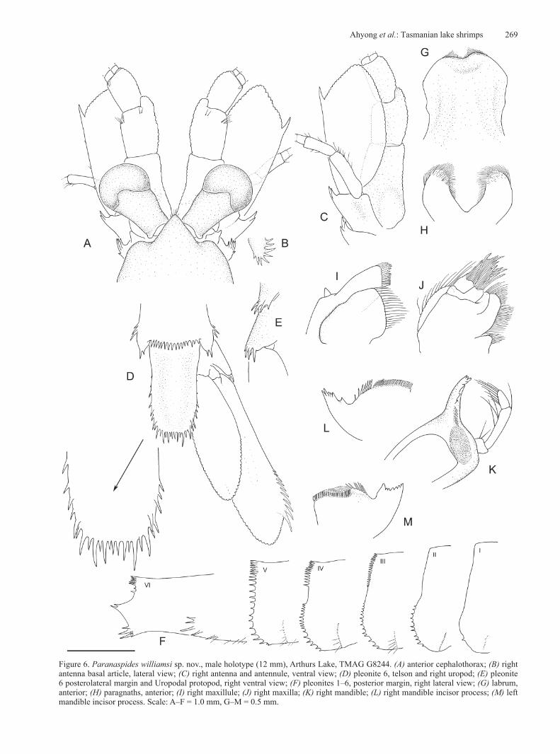
Labrum with shallow proximal constriction; distal margin slightly concave, finely setose.
Mandibular corpus (apophysis) robust; molar process and incisor process well-developed; molar with elongate, ovate, triturating surface, surrounded by spiniform setae; incisor process diagonal to axis of mandibular corpus. Left incisor process with 8 triangular teeth in sinuous row, proximal tooth largest; proximally with spine row between proximal incisor tooth and molar process. Right incisor process similar to left except with 6 triangular teeth, proximalmost tooth usually apically bifid, widely separated from adjacent tooth. Palp 3-articulate, setose, article 1 short, subquadrate, with 2 setae, article 2 slender, longer than articles 1 and 3.
Paragnaths widely separated by deep V-shaped incision, without lobes, distal half finely setose, especially mesially.
Maxillule with 2 endites; proximal endite distally setose; distal endite spinose distally, lateral surface with small conical palp.
Maxilla with 4 endites, proximal 2 endites with plumose setae, distal 2 endites densely arrayed with serrulate setae.
Thoracopods 1–8 protopod with coxa, basis, preischium, ischium, merus, carpus, propodus and dactylus; flexure at carpus-merus articulation.
Thoracopod 1 (maxilliped) coxa mesial margin with setose coxal endites, lateral margin with 2 lamellar epipods, proximal wider than distal; basis with slender, flattened, liguliform exopod; coxa-basis demarcation often ill-defined; preischium rectangular, more than quadruple length of quadrate ischium, expanded mesially, projecting beyond mesial margin of ischium; merus slightly tapering distally, distinctly longer than ischium; carpus triangular, longer than high, half length of merus; propodus slender, as long as merus; dactylus short, terminating in slender claw, with 2 slender movable spines on either side.
Thoracopods 2–8 (pereopods) as ambulatory legs. Thoracopods 2–6 structurally similar, distal 4 articles with tufts of setae, primarily along flexor margins, dactylus strongly setose; thoracopods 4–5 longest; coxa lateral margin with 2 ovate, lamelliform epipods, proximal epipod more pointed distally than distal epipod; coxa mesial margin in adult females with setose endite; basis short, partially fused with preischium; exopod articulating with lateral margin of basis, with elongate basal article and setose multi-annulate flagellum; ischium about as long as basis-preischium; merus elongate, slightly tapering distally, about twice ischium length; carpus triangular, longer than high, about half merus length or slightly less; propodus elongate, slender, shorter than merus; dactylus short, terminating in long, slender claw, with slender movable spine on lateral side, 2 movable spines on mesial side. Thoracopod 7 similar to thoracopods 2–6 except epipods proportionally more slender; exopod
262 Records of the Australian Museum (2017) Vol. 69
a single narrow lamella; dactylus with movable spine on either side. Thoracopod 8 structurally similar to preceding thoracopods but lacking epipods or exopod; basis and preischium indistinguishably fused; longer than thoracopod 7; dactylus with movable spine on either side.
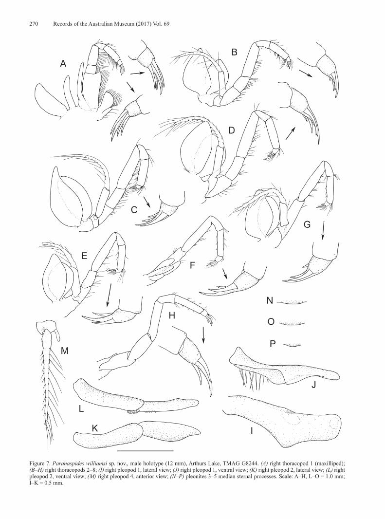
Pleopods 1–5 exopod long, slender, setose, multi-annulate. Pleopods 1–2 endopod always present; unmodified endopod ovate, lamellar, short, length subequal to first exopod annulation in females and juvenile males; endopod always present on pleopods 3, present or absent on pleopods 4–5; adult male pleopods 1–2 endopod modified as copulatory structures (petasma). Adult male pleopod 1 elongate, directed anteriorly, reaching beyond thoracopod 8 coxa; slender proximally, expanded distally, hollowed mesially, deepest near midlength; distally bluntly rounded to subtruncate, lateral margin thin, lamellate; dorsomesial margin with short row of retinacula near midlength and distally; proximo-mesial surface with long scattered setae and spinules; midventral margin bluntly triangular, incurved mesially; left and right pleopods united by retinacula, together forming scoop-like structure. Male pleopod 2 endopod of 2 articles, slightly longer than pleopod 1, directed anteriorly, reaching to thoracopod 8 coxa; proximal article slightly longer than distal article, mesial proximal margin with row of retinacula; distal article straight, mesially hollowed, with short scattered spinules, apex blunt.
Uropods forming tail-fan with telson. Uropodal exopod elongate, spatulate, distolateral spine row of fixed graded spines; indistinct, partial diaeresis extending from base of distalmost spine; spine row preceded by straight margin; mesial margin and lateral margin distal to spine row setose. Uropodal endopod distinctly shorter than exopod, reaching to distal two-thirds of exopod; mesial margin to distal half of lateral margin setose.
Remarks. Paranaspides is readily distinguished from Anaspides and Allanspides by the distinct flexure at pleonite 1, the presence of a vertical row of minute spines on the lower midlateral surface of pleonites 1–5, an arcuate row of prominent spines on the midlateral surface of pleonite 6, a spinose basal antennal article, a more pronounced distomesial lobe on the maxilliped ischium, absence of sexually dimorphic antennules, a proportionally larger, more elongate scaphocerite, presence of the triangular ventromesial lobe of the male pleopod 1 endopod, a rectangular versus polygonal or linguiform telson, and more elongate uropods. In other respects, Anaspides and Paranaspides closely agree. Note that Ahyong (2016) inadvertently described Anaspides as having a 3-articulate antennal protopod; both Anaspides and Paranaspides have only two free protopod articles. Like Anaspides, Paranaspides further differs from Allanaspides by lacking the fenestra dorsalis on the cephalothorax.
When first described, Paranaspides immediately attracted controversy with its apparent possession of a biramous mandibular palp, a feature otherwise unknown in malacostracans (Smith 1908, 1909b; Hansen, 1925). Gordon (1961), however, showed Smith’s mandibular observations to be based on an aberrant specimen; the palp of P. lacustris is uniramous and 3-articulate as in Anaspides. Phylogenetic
analyses indicate a close relationship between Anaspides and Paranaspides, as sister groups or with the latter possibly even nested within the former (Jarman & Elliott, 2000).
Species of Paranaspides differ ecologically from those of Anaspides and Allanaspides, which are epibenthic and pholeteric, respectively. Instead, species of Paranaspides are frequently natatory and occur only in association with aquatic vegetation, amongst and above which they freely swim. Some morphological differences between Paranaspides and other anaspidesid genera, such as the enlarged scaphocerite and more elongated uropods may relate to the primarily pelagic or natatory rather than benthic habits of other anaspidesids.
Subsequent to Smith’s (1908, 1909b) accounts of Paranaspides lacustris, other studies have examined pleonal musculature (Daniel, 1931), functional morphology and excretion (Cannon & Manton, 1929; Manton, 1930, 1931), internal reproductive organs and the alimentary canal (Nicholls & Spargo, 1932), ommatidial structure (Richter, 1999) and cuticular sclerites (Kutschera et al., 2015).
Paranaspides lacustris Smith, 1908
Figs 1–4, 9A, 10Paranaspides lacustris Smith, 1908: 470–471, fig. 3–6;
1909a: 63, 71; 1909b: 492, 497, 506, 560–562, fig. 1, 4, 8, 10, 13, 16, 19, 22, 24–29, 49, pl. 11: fig. 2. —Manton, 1930, pl. 1. —Nicholls & Spargo, 1932: 153–155. —Nicholls, 1947: 9, 14. —Riek, 1959: 251. —Gordon, 1961: 214–221, fig. 1–5. —Williams, 1965: 95, 96, 99–105, 122, 123, 125, tab. 5. —Mayrat, 1966: 1542. —Hewer, 1967: 1. —Goede, 1967: 83. —Swain et al., 1970: 6. —Lake & Knott, 1973: 96. —Williams, 1974: 80, tab. 4.1. —Knott, 1975: 157, 177, 183, 184. —Silvey, 1980: 72. —Fulton, 1982: 23–25, fig. 1, 2 [Great Lake, Shannon Lagoon, Penstock Lagoon only]; 1983: tab. 1 [Great Lake]. —Wells et al., 1983: xliii, 275, 277, 278. —Schram, 1984: 191. —Schram & Hessler, 1984: 194. —Michaelis, 1985: 6. —Zeidler, 1985: 75. —Campbell et al., 1986: 92. —Davies & Fulton, 1987: 2, 3, 4 (unnumbered fig.), 9. —Horwitz, 1990: 65–67. —Jarman & Elliott, 2000: 625, 626, 631, 632, fig. 1, tab. 1, 2. —Ovenden et al., 1993: 227. —Richter, 1999: 171, fig. 20. —Lake et al. 2002: 12. —Camacho et al., 2002: tab. 1, fig. 1. —Serov, 2002: 8, 15, 16, fig. 46. —Bonham, 2006: 3. —Camacho, 2006: 6. —Driessen & Mallick, 2007: 1173. —Schram, 2008: 131. —Boxshall & Jaume, 2009: 245. —Coineau & Camacho, 2013: 377, 436, figs 50.6C, 50.8A, 50.11A, 50.19A–C, 50.24, 50.33. —Kutschera et al., 2015: 3–4, 17, 24, figs 1N, 3C–E, tab. 3, 4. —Richards et al., 2015: 61, 62, tab. 1. —Ahyong, 2016: 316.
Lectotype: AM P100400, male (11 mm), Great Lake, coll. G. W. Smith, 1907–1908. Paralectotypes: OUMNH 5403, 1♂ (11 mm), 12 juvenile ♂♂ (6–10 mm), 1♀ (TL 11 mm), 11 juvenile ♀♀ (6–10 mm), 1 partial cephalothorax, Great Lake, coll. G. W. Smith, 1907–1908; AM P100401, 1♀ (11 mm), Great Lake, coll. G. W. Smith, 1907–1908.
Ahyong et al.: Tasmanian lake shrimps 263
Other material examined. Great Lake: QVM 10:8080, 4♂♂ (12–13 mm), Great Lake, coll. Evans, 1939; USNM 29140, 1♂ (15 mm), Great Lake, coll. F. R. Schram, 26 May 1980; USNM 60112, 2♂♂ (13–15 mm), 4♀♀ (12–18 mm), Great Lake, coll. W. M. Tattersall, 1914; AM P8766, 2♂♂ (14–15 mm), 1♀ (16 mm), Great Lake, pres. J. J. Flynn; AM P56372, 1♂ (15 mm), 1♀ (20 mm), 1 juvenile ♀ (10 mm), Great Lake, #327, coll. J. W. Evans; WAM C58159, 11♂♂ (12–14 mm), 46 juvenile ♂♂ (9–11 mm), 2♀♀ (13 mm), 102 juvenile ♀♀ (9–13 mm), N end Great Lake, dredged near old shore line, coll. G. Nicholls, 25 Jan 1947; WAM C58163, 2♂♂ (11–12 mm), Great Lake, coll. G. Nicholls; WAM C11776, 1♀ (11 mm), NW corner Great Lake, coll. “H.D.”, 1 Feb 1945; WAM C11777, 1♂ (11 mm), Brandum Bay [41°48.0'S 146°41.0'E], coll. G. Nicholls, 26 Jan 1947; WAM C11795, 1♂ (11 mm), 1♀ (11 mm), Brownie Bay, Brandum Bay [41°49.84'S 146°41.09'E], coll. G. Nicholls, 26 Jan 1947; WAM C11779, 1♂ (18 mm), 3♀♀ (17–22 mm), N end Breona [41°47'S 146°42'E], near old shore line, coll. G. Nicholls, 25 Jan 1947; WAM C11778, 4 juvenile ♂♂ (10–11 mm), 1♀ (22 mm), 10 juvenile ♀ (9–11 mm), Brownie Bay [41°49.84'S 146°41.09'E], dredged, 12–15 feet, coll. G. Nicholls, 26 Jan 1947; QVM 10:49146, 1♀ (6 mm), Brandum Bay [41°48.0'S 146°41.0'E], level 2, coll. W. Fulton, 4 Dec 1976; QVM 10:49147, 1♂ (13 mm), Brandum Bay [41°48.0'S 146°41.0'E], level 2, coll. W. Fulton, 31 Mar 1975; QVM 10:49148, 1♂ (17 mm), Brandum Bay, level 2, [41°48.0'S 146°41.0'E], coll. W. Fulton, 2 Jun 1975; QVM 10:49149, 1♀ (c. 8 mm, poor condition), Brandum Bay, level 2, [41°48.0'S 146°41.0'E], coll. W. Fulton, 27 Jan 1975; WAM C58155, 1♂ (10 mm), 1 juvenile ♀ (9 mm), Beckett Bay, S end of Great Lake [41°58.06'S146°44.80'E], 1933; QVM 10:49150, 2♂♂ (14–17 mm), Swan Bay [41°58.43'S 146°41.56'E], level 2, coll. W. Fulton, 31 Mar 1975; QVM 10:49151, 1♂ (17 mm), 1♀ (21 mm), Swan Bay [41°58.28'S 146°41.55'E], from weed on anchor (Chara sp.), 30 ft, coll. W. Fulton, 7 Nov 1975; AM P100405, 2♀♀ (16–20 mm), Swan Bay, #2, from Nitella bed, 6.3–6.7 m, 41°56'16"S 146°41'12"E, on SCUBA, coll. M. Reinhardt & C. Hoepel, 8 March 2017; ZSRO CR20, 2♀♀ (16–17 mm), Swan Bay, #2, from Nitella bed, 6.3–6.7 m, 41°56'16"S 146°41'12"E, on SCUBA, coll. M. Reinhardt & C. Hoepel, 8 March 2017; AM P100406, 2♀♀ (13–16 mm), Swan Bay, #3, from Nitella & Chara bed, 7.0–7.3 m, 41°59'14"S 146°41'16"E, on SCUBA, 8 March 2017; ZSRO CR21, 3♀♀, Swan Bay, #3, from Nitella & Chara bed, 7.0–7.3 m, 41°59'14"S 146°41'16"E, on SCUBA, 8 March 2017; AM P100408, 2♂♂ (14–15 mm), 1♀ (15 mm), Tods Corner, #1, off northeastern shore, 41°57'24"S 146°47'18"E, 6.3–7 m, sparse Chara beds, 11 March 2017; AM P100410, 1♀ (18 mm), Tods Corner, #2, off western shore, 41°57'30"S 146°47'02"E, 3–4 m, dense Chara beds, 11 March 2017; AM P100411, 2♂♂ (15 mm), 5♀♀ (14–17 mm), Tods Corner, #2, off western shore, 41°57'30"S 146°47'02"E, 3–4 m, dense Chara beds, 11 March 2017; ZSRO CR22, 4♂♂, 3♀♀, Tods Corner, #2, off western shore, 41°57'30"S 146°47'02"E, 3–4 m, dense Chara beds, 11 March 2017.
Shannon Lagoon: AM P11898, 3♂♂ (20–22 mm), Shannon Lagoon at Miena, [41°59.25'S 146°44.04'E], coll. J. Waterhouse; QVM 10:49160, 8♂♂ (12–15 mm), 9♀♀ (14–16 mm), Shannon Lagoon, [42°00.86'S 146°44.30'E], coll. W. Fulton, 3 Mar 1973; QVM 10:49060, 3♂♂ (c. 9–10
mm, poor condition), 5♀♀ (c. 7–10 mm, poor condition), Shannon Lagoon, [41°59.50'S 146°44.33'E], coll. J. H. Wilson, 30 Jan 1965; AM P99513, 1♀ (19 mm), Shannon Lagoon, 41°59'10.53"S 146°44'16.9"E, 3 m, weeds, 1015 m asl, coll. S. Jarman; NMV J37892, 7♂♂ (16–20 mm), 5♀♀ (18–20 mm), Shannon Lagoon, 42°S 146°E, coll. B. Knott, 16 November 1972.
Description. Pleonite 6 lower midlateral surface with arcuate row of 2–5 (usually 4) prominent, well-spaced spines; posterolateral angle bispinous (rarely unispinous); posteroventral angle anterior to uropod articulation with cluster of 7–13 spines.
Antennule mesial (accessory) flagellum about 0.2–0.3 × body length (24 articles in figured male); lateral flagellum 0.5–0.7 × body length (58 articles in figured male).
Antennal flagellum 0.5–0.7 × body length (47 articles in figured male); protopod coxa with splayed row of 6–8 spines on lateral margin, basis with 2 (rarely 3) lateral spines.
Labrum anterior proximal surface swollen medially, usually with median point.
Thoracopod 1 (maxilliped) merus length 2.5–3.0 × width.Pleopod 3 endopod always present; pleopod 4 endopod
usually present; pleopod 5 endopod absent. Adult male pleopod 1 margin of dorsodistal half concave. Male pleopod 2 endopod distal article with straight distoventral surface.
Uropodal protopod with cluster of 2 or 3 posterolateral spines. Uropodal exopod elongate, spatulate; lateral margin between incurved anterolateral margin and distolateral spine row, straight or faintly or faintly concave, with 0–6 minute widely spaced setae; spine row of 8–10 fixed graded spines; spine row length 0.3 × length of straight portion of preceding exopod margin.
Colour in life (Fig. 9A). Body transparent, covered in dull red and brown chromatophores forming diffuse transverse bands across pereon and pleon; cephalothorax with red brown patch on lateral surface behind cervical groove. Antennular peduncle article 1 transparent with scattered spots and dark midline; article 2 transparent with longitudinal brown patches; article 3 solid dark brown. Scaphocerite transparent. Eyestalks red-brown. Pereopods and pleopods translucent pale brown. Tailfan transparent with scattered brown spots, densest distally.
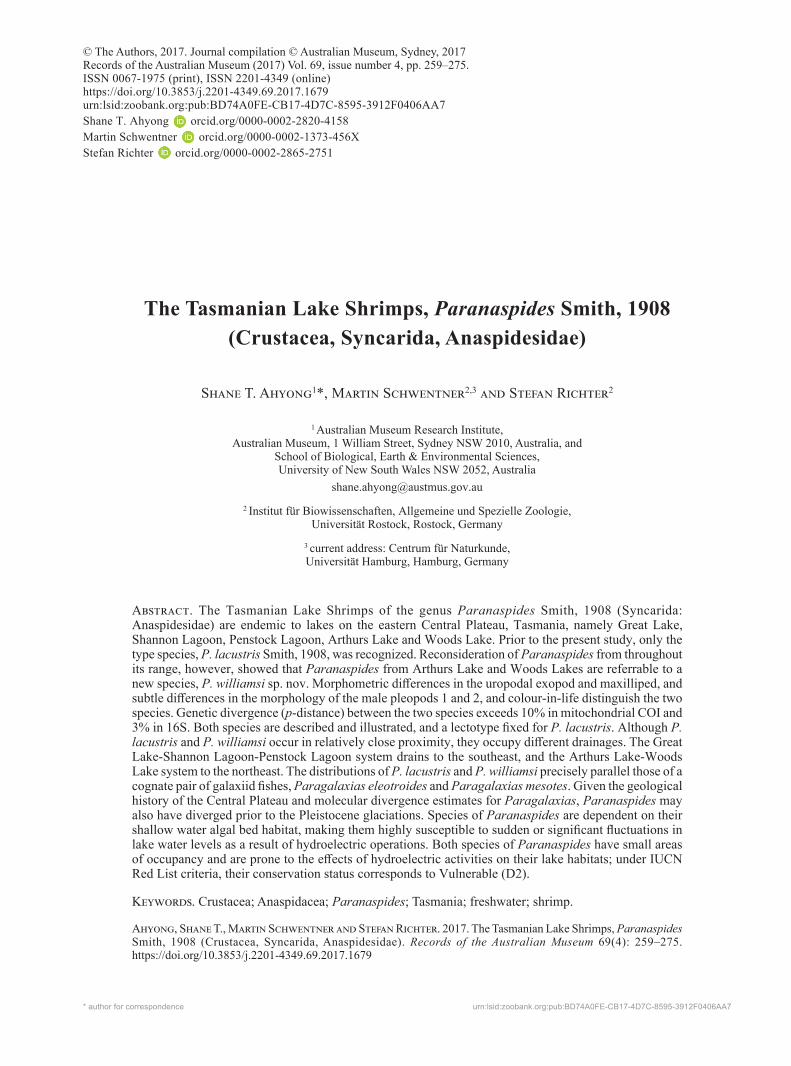
Figure 1. Paranaspides lacustris Smith, 1908, male (17 mm), Swan Bay, Great Lake, QVM 10:49151, right habitus. Scale = 1.0 mm.
264 Records of the Australian Museum (2017) Vol. 69
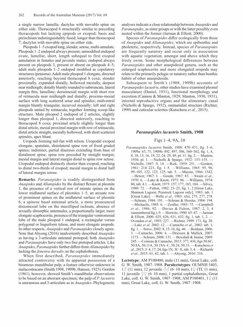
Figure 2. Paranaspides lacustris Smith, 1908, male (17 mm), Swan Bay, Great Lake, QVM 10:49151. (A) anterior cephalothorax; (B) right antenna basal article, lateral view; (C) right antenna and antennule, ventral view; (D) pleonite 6, telson and right uropod; (E) pleonite 6 posterolateral margin and Uropodal protopod, right ventral view; (F) pleonites 1–6, posterior margin, right lateral view; (G) labrum, anterior; (H) paragnaths, anterior; (I) right maxillule; (J) right maxilla; (K) right mandible; (L) right mandible incisor process; (M) left mandible incisor process. Scale: A–F = 1.0 mm, G–M = 0.5 mm.

Ahyong et al.: Tasmanian lake shrimps 265
Figure 3. Paranaspides lacustris Smith, 1908, male (17 mm), Swan Bay, Great Lake, QVM 10:49151. (A) right thoracopod 1 (maxilliped); (B–H) right thoracopods 2–8; (I) right pleopod 1, lateral view; (J) right pleopod 1, ventral view; (K) right pleopod 2, lateral view; (L) pleopod 1 & 2, in-situ, ventral view; (M) right pleopod 3, anterior view; (N–P) pleonites 3–5 median sternal processes. Scale: A–H, L–O = 1.0 mm; I–K = 0.5 mm.
266 Records of the Australian Museum (2017) Vol. 69
Measurements. Male (n = 121) 6–22 mm; female (n = 183) 6–22 mm. Fulton (1982) reported specimens up to 25 mm body length.
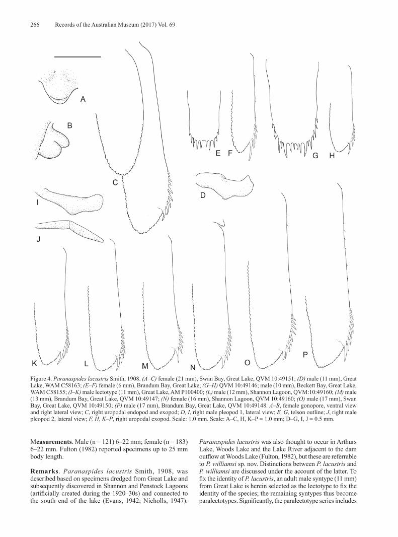
Remarks. Paranaspides lacustris Smith, 1908, was described based on specimens dredged from Great Lake and subsequently discovered in Shannon and Penstock Lagoons (artificially created during the 1920–30s) and connected to the south end of the lake (Evans, 1942; Nicholls, 1947).
Paranaspides lacustris was also thought to occur in Arthurs Lake, Woods Lake and the Lake River adjacent to the dam outflow at Woods Lake (Fulton, 1982), but these are referrable to P. williamsi sp. nov. Distinctions between P. lacustris and P. williamsi are discussed under the account of the latter. To fix the identity of P. lacustris, an adult male syntype (11 mm) from Great Lake is herein selected as the lectotype to fix the identity of the species; the remaining syntypes thus become paralectotypes. Significantly, the paralectotype series includes
Figure 4. Paranaspides lacustris Smith, 1908. (A–C) female (21 mm), Swan Bay, Great Lake, QVM 10:49151; (D) male (11 mm), Great Lake, WAM C58163; (E–F) female (6 mm), Brandum Bay, Great Lake; (G–H) QVM 10:49146; male (10 mm), Beckett Bay, Great Lake, WAM C58155; (I–K) male lectotype (11 mm), Great Lake, AM P100400; (L) male (12 mm), Shannon Lagoon, QVM:10:49160; (M) male (13 mm), Brandum Bay, Great Lake, QVM 10:49147; (N) female (16 mm), Shannon Lagoon, QVM 10:49160; (O) male (17 mm), Swan Bay, Great Lake, QVM 10:49150; (P) male (17 mm), Brandum Bay, Great Lake, QVM 10:49148. A–B, female gonopore, ventral view and right lateral view; C, right uropodal endopod and exopod; D, I, right male pleopod 1, lateral view; E, G, telson outline; J, right male pleopod 2, lateral view; F, H, K–P, right uropodal exopod. Scale: 1.0 mm. Scale: A–C, H, K–P = 1.0 mm; D–G, I, J = 0.5 mm.

Ahyong et al.: Tasmanian lake shrimps 267
an 8 mm juvenile of Anaspides richardsoni Ahyong, 2016, evidently overlooked by Smith (1908), but representing the first confirmed record of the genus from Great Lake. The occurrence of Anaspides in Great Lake itself has often been questioned (e.g., Nicholls, 1947; Williams, 1965; O’Brien, 1990) so the present specimen of A. richardsoni, collected together with P. lacustris, demonstrates that Anaspides was at least a transient resident of the lake. Whether Anaspides still occurs there remains to be determined.
Morphological variation in P. lacustris is minor; meristic differences in spination usually vary allometrically, with the smallest specimens having fewest spines. The smallest juveniles (c. 6 mm) lack lateral spines on the telson, the pleopod 1–2 endopods are present as tiny buds and the pleopod 3–5 endopods are absent. By c. 8 mm, the lateral telson spines are present, the pleopod 1–2 endopods are evident (albeit as yet unmodified in males) and the pleopod 3–4 endopods appear. The relative length of the spine row on the uropodal exopod is stable across the size range, though the number of spines and relative length of the distal spine changes with body size: five spines are present at 6 mm body length, with the distal spine overreaching the apex of the exopod; by 9 mm, eight or more spines are present with the distal spine reaching the end of the exopod; and above 9 mm, the distal spine distinctly falls short of the end of the exopod. Maturity in both sexes appears to be reached at 10–11 mm body length. The adult male pleopod 1 endopod of P. lacustris is concave on the upper margin of the distal half, usually with a bluntly rounded apex. In some males, however, the distal pleopod 1 endopod margin is produced to a triangular lobe. The adult complement of pleopod endopods is variable and overlaps that of P. williamsi, being always present on pleopod 3, variable on pleopod 4 and always absent on pleopod 5. In P. williamsi, the pleopod 3–4 endopods are present, but variable on pleopod 5.
Although yet to be studied in detail, the life cycle of P. lacustris is apparently univoltine, with a single reproductive event and little overlap between year classes. Spawning is believed to take place in summer and hatching in winter, with individuals living for up to 18 months (Williams, 1965; Fulton, 1982).
Distribution. Known only from Great Lake, Shannon Lagoon and Penstock Lagoon; 0.2–10 m depth; 1040 m above sea level.
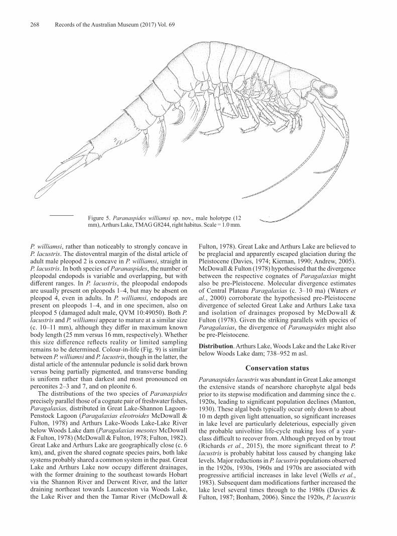
Paranaspides williamsi sp. nov.urn:lsid:zoobank.org:act:613B9B3D-7EE8-4A00-8DD2-A4411956B170
Figs 4–8, 9B, 10Paranaspides lacustris. —Fulton, 1982: 23, 25, fig. 1 [Woods
Lake, Arthurs Lake, Lake River only]; 1983: 776, tab. 1 [Arthurs Lake].
Holotype: TMAG G8244, male (12 mm), Arthurs Lake, Pumphouse Bay, near pumping station, 41°59'16.3"S 146°51'44.8"E, netted from weeds, < 1 m, coll. S. Richter & C. Wirkner, 28 February 2006. Paratypes: AM P99963, 3♂♂ (11–12 mm), 6♀♀ (13–14 mm), collected with holotype; AM P100414, 3♀♀ (11–12 mm), Pumphouse Bay, near pumping station, 41°59'15"S 146°51'42"E, Nitella beds, 0.7 m, light trap, coll. S. Ahyong, C. Hoepel, M. Reinhardt, S. Richter, 10 March 2017; QVM 10:49058, 1♀ (15 mm), southern East Lake, Arthurs Lake [41°59.30'S 146°57.19'E], coll. W. Fulton, 17 May 1977.
Other material examined. Arthurs Lake: AM P100413, 4♂♂ (11 mm), 1♀ (11 mm), Pumphouse Bay, near pumping station, 41°59'15"S 146°51'42"E, Nitella beds, 0.2–0.7 m, hand net, coll. S. Ahyong, C. Hoepel, M. Reinhardt, S. Richter, 8 March 2017; ZSRO CR23, 3♂♂ (12–13 mm), 2♀♀ (11–12 mm), Pumphouse Bay, near pumping station, 41°59'15"S 146°51'42"E, Nitella beds, 0.2–0.7 m, netted, coll. S. Ahyong, C. Hoepel, M. Reinhardt, S. Richter, 8 March 2017; QVM:10:49059, 1 damaged ♂, 1♀ (15 mm), 1 exoskeleton, Sand Lake, Arthurs Lake [41°56.46'S 146°57.88'E], coll. W. Fulton, 29 June 1977; QVM 10: 49057, 1♂ (16 mm), 2 damaged ♀♀, southern East Lake, Arthurs Lake [41°59.30'S 146°57.19'E], coll. W. Fulton, 19 April 1977.
Woods Lake: QVM 10:49161, 10♂♂ (14–16 mm), 10♀♀ (13–16 mm), west shore Woods Lake, [42°05.50'S 146°59.82'E], coll. W. Fulton, 22 July 1977.
Description. Pleonite 6 lower mid-lateral surface with arcuate row of 3–5 (usually 4) prominent, well-spaced spines; posterolateral angle bispinous (rarely unispinous); posteroventral angle anterior to uropod articulation with cluster of 6–14 spines.
Antennule mesial (accessory) flagellum 0.1–0.2 × body length (17 articles in holotype); lateral flagellum 0.4 × body length (42 articles in holotype).
Antennal flagellum 0.3–0.4 × body length (40 articles in holotype); protopod coxa with splayed row of 6–9 spines on lateral margin, basis with 2 lateral spines.
Labrum anterior proximal surface swollen medially, without median point.
Thoracopod 1 (maxilliped) merus slightly tapering distally, length twice length of ischium.
Pleopods 3–4 endopod always present in adults; pleopod 5 endopod rarely present. Adult male pleopod 1 margin of dorsodistal half straight to faintly concave. Male pleopod 2 endopod distal article with distoventral surface broadly concave.
Uropodal protopod with cluster of 1–4 posterolateral spines. Uropodal exopod elongate, spatulate; lateral margin between incurved anterolateral margin and distolateral spine row, straight or faintly or faintly concave, with 0–7 minute, widely spaced setae; distolateral spine row of 9–14 fixed graded spines; spine row length 0.4–0.7 × length of straight portion of preceding exopod margin.
Colour in life (Fig. 9B). Body transparent, covered in dull red and brown chromatophores forming diffuse transverse bands across pereon and pleon, most pronounced and darkest across pereonites 2–3, 7 and anterior half of pleonite 6; with dark-brown; cephalothorax with red brown patch on lateral surface behind cervical groove. Antennular peduncle article 1 transparent with scattered spots and dark midline; article 2 transparent with longitudinal brown midline and brown mesial margin; article 3 transparent, with partial pigmentation. Scaphocerite transparent. Eyestalks red brown. Pereopods and pleopods translucent pale brown. Tailfan transparent with scattered brown spots, densest distally.
Measurements. Male (n = 23) 11–16 mm; female (n = 26) 11–16 mm.
Remarks. Paranaspides williamsi sp. nov. differs from P. lacustris in the following features: the proportionally shorter merus of the maxilliped (length twice the width in the new species versus 2.5–3 times length in P. lacustris), the proportionally longer spine row on the uropodal exopod (about half or longer versus one-third length of the preceding straight, unarmed lateral margin), and subtle differences in the adult male pleopods 1 and 2. The adult male pleopod 1 distodorsal margin is straight or, at most, faintly concave in
268 Records of the Australian Museum (2017) Vol. 69
Figure 5. Paranaspides williamsi sp. nov., male holotype (12 mm), Arthurs Lake, TMAG G8244, right habitus. Scale = 1.0 mm.
P. williamsi, rather than noticeably to strongly concave in P. lacustris. The distoventral margin of the distal article of adult male pleopod 2 is concave in P. williamsi, straight in P. lacustris. In both species of Paranaspides, the number of pleopodal endopods is variable and overlapping, but with different ranges. In P. lacustris, the pleopodal endopods are usually present on pleopods 1–4, but may be absent on pleopod 4, even in adults. In P. williamsi, endopods are present on pleopods 1–4, and in one specimen, also on pleopod 5 (damaged adult male, QVM 10:49050). Both P. lacustris and P. williamsi appear to mature at a similar size (c. 10–11 mm), although they differ in maximum known body length (25 mm versus 16 mm, respectively). Whether this size difference reflects reality or limited sampling remains to be determined. Colour-in-life (Fig. 9) is similar between P. williamsi and P. lacustris, though in the latter, the distal article of the antennular peduncle is solid dark brown versus being partially pigmented, and transverse banding is uniform rather than darkest and most pronounced on pereonites 2–3 and 7, and on pleonite 6.
The distributions of the two species of Paranaspides precisely parallel those of a cognate pair of freshwater fishes, Paragalaxias, distributed in Great Lake-Shannon Lagoon-Penstock Lagoon (Paragalaxias eleotroides McDowall & Fulton, 1978) and Arthurs Lake-Woods Lake-Lake River below Woods Lake dam (Paragalaxias mesotes McDowall & Fulton, 1978) (McDowall & Fulton, 1978; Fulton, 1982). Great Lake and Arthurs Lake are geographically close (c. 6 km), and, given the shared cognate species pairs, both lake systems probably shared a common system in the past. Great Lake and Arthurs Lake now occupy different drainages, with the former draining to the southeast towards Hobart via the Shannon River and Derwent River, and the latter draining northeast towards Launceston via Woods Lake, the Lake River and then the Tamar River (McDowall &
Fulton, 1978). Great Lake and Arthurs Lake are believed to be preglacial and apparently escaped glaciation during the Pleistocene (Davies, 1974; Kiernan, 1990; Andrew, 2005). McDowall & Fulton (1978) hypothesised that the divergence between the respective cognates of Paragalaxias might also be pre-Pleistocene. Molecular divergence estimates of Central Plateau Paragalaxias (c. 3–10 ma) (Waters et al., 2000) corroborate the hypothesised pre-Pleistocene divergence of selected Great Lake and Arthurs Lake taxa and isolation of drainages proposed by McDowall & Fulton (1978). Given the striking parallels with species of Paragalaxias, the divergence of Paranaspides might also be pre-Pleistocene.
Distribution. Arthurs Lake, Woods Lake and the Lake River below Woods Lake dam; 738–952 m asl.
Conservation statusParanaspides lacustris was abundant in Great Lake amongst the extensive stands of nearshore charophyte algal beds prior to its stepwise modification and damming since the c. 1920s, leading to significant population declines (Manton, 1930). These algal beds typically occur only down to about 10 m depth given light attenuation, so significant increases in lake level are particularly deleterious, especially given the probable univoltine life-cycle making loss of a year-class difficult to recover from. Although preyed on by trout (Richards et al., 2015), the more significant threat to P. lacustris is probably habitat loss caused by changing lake levels. Major reductions in P. lacustris populations observed in the 1920s, 1930s, 1960s and 1970s are associated with progressive artificial increases in lake level (Wells et al., 1983). Subsequent dam modifications further increased the lake level several times through to the 1980s (Davies & Fulton, 1987; Bonham, 2006). Since the 1920s, P. lacustris
Ahyong et al.: Tasmanian lake shrimps 269
Figure 6. Paranaspides williamsi sp. nov., male holotype (12 mm), Arthurs Lake, TMAG G8244. (A) anterior cephalothorax; (B) right antenna basal article, lateral view; (C) right antenna and antennule, ventral view; (D) pleonite 6, telson and right uropod; (E) pleonite 6 posterolateral margin and Uropodal protopod, right ventral view; (F) pleonites 1–6, posterior margin, right lateral view; (G) labrum, anterior; (H) paragnaths, anterior; (I) right maxillule; (J) right maxilla; (K) right mandible; (L) right mandible incisor process; (M) left mandible incisor process. Scale: A–F = 1.0 mm, G–M = 0.5 mm.
270 Records of the Australian Museum (2017) Vol. 69
Figure 7. Paranaspides williamsi sp. nov., male holotype (12 mm), Arthurs Lake, TMAG G8244. (A) right thoracopod 1 (maxilliped); (B–H) right thoracopods 2–8; (I) right pleopod 1, lateral view; (J) right pleopod 1, ventral view; (K) right pleopod 2, lateral view; (L) right pleopod 2, ventral view; (M) right pleopod 4, anterior view; (N–P) pleonites 3–5 median sternal processes. Scale: A–H, L–O = 1.0 mm; I–K = 0.5 mm.

Ahyong et al.: Tasmanian lake shrimps 271
Figure 8. Paranaspides williamsi sp. nov. (A–C) female paratype (14 mm), Arthurs Lake, AM P99963; (D) male (14 mm), Woods Lake, QVM 10:149161; (E) female (15 mm), Sand Lake, Arthurs Lake, QVM 10:49059; (F) female (15 mm), East Lake, Arthurs Lake, QVM 10:49058; (G) male (16 mm), Arthurs Lake, QVM 10:49057. A, B, female gonopore, ventral and right lateral views. C, right uropod; D–G, right uropodal exopod. Scale = 1.0 mm.
Figure 9. Colour in life. (A) Paranaspides lacustris Smith, 1908, female, 18 mm, Tods Corner, Great Lake, AM P100410; (B) P. williamsi sp. nov., paratype female, 12 mm, Arthurs Lake, AM P100414.

272 Records of the Australian Museum (2017) Vol. 69
Figure 10. Distribution of Paranaspides. * Record from Penstock Lagoon, based on Williams (1965).
Table 1. Uncorrected p-distances between species and populations of Paranaspides. Intraspecific or intrapopulation distances shown on the diagonal (COI distances above, 16S distances below).
P. williamsi P. lacustris P. lacustris P. lacustris (Arthurs (Shannon (Great Lake, (Great Lake, Lake) Lagoon) Swan Bay) Tods Corner)
P. williamsi (Arthurs Lake) COI 0.2–1.5 — 10.4–11.7 11.0–11.8 16S 0.0–0.2 3.2–3.3 3.2–3.3 3.2–3.7 P. lacustris (Shannon Lagoon) COI — — — 16S — 0.0 0.0 P. lacustris (Great Lake, Swan Bay) COI 0.0–0.2 0.0–0.3 16S — 0.0 P. lacustris (Great Lake, Tods Corner) COI 0.0–0.2 16S 0.0
Ahyong et al.: Tasmanian lake shrimps 273
has seldom been found in significant numbers in Great Lake, with the frequent raising and lowering of lake levels for hydroelectric operations believed to retard establishment of the littoral vegetation essential as habitat (Horwitz, 1990).
Little is known of the current population size and dynamics of either species of Paranaspides, so conserv-ation assess ments have relied largely on area of occupancy and the limited number of locations at which either species occurs. Paranaspides lacustris is currently assessed by the IUCN Red List of Threatened Species as Vulnerable (D2) (Inland Water Crustacean Specialist Group, 1996) based on its limited area of occupancy, few known locations, and in being prone to the effects of hydroelectric operations. With Arthurs and Woods Lakes now excluded from the range of P. lacustris, the area of occupancy is reduced, though the Vulnerable D2 assessment would remain applicable. Paranaspides williamsi, being restricted to Arthurs and Woods Lakes (and the Lake River immediately below the Woods Lake dam) has a limited area of occupancy and occurrence at no more than three locations. The dependence of P. williamsi on aquatic vegetation (charophyte and macrophyte beds) indicates an area of occupancy in Arthurs Lake of 0.63–8.3 km2 depending on water level (Lobdale, 2011). Although the proportion of vegetated habitat of Woods Lake is not known, the total surface area is approximately 1.2 km2 so the total area of occupancy of P williamsi (both lakes combined) would not exceed 9.5 km2. Like P. lacustris, P williamsi is also subject to artificial lake level fluctuations and stochastic events given its very narrow range. As such, the conservation status of P. williamsi under IUCN Red List categories would also correspond to Vulnerable D2. Our efforts to sample P. williamsi in Woods Lake in March 2017, however, were unsuccessful and no other recent collections are presently available. Therefore, establishing the population status of P. williamsi in Woods Lake should be prioritized, especially given the sharp decline in Paragalaxias mesotes observed in Woods Lake over the past two decades (TSSC, 2016). If the Woods Lake population of P. williamsi has also significantly declined, it might require a higher level of protection.
Neither species of Paranaspides is currently listed on either the Commonwealth Environment Protection and Biodiversity Conservation Act 1999 or the Tasmanian Threatened Species Protection Act 1995 (Bonham, 2006). It is noteworthy, however, that the galaxiid fishes Paragalaxias eleotroides and Paragalaxias mesotes, under both the Commonwealth Environment Protection and Biodiversity Conservation Act 1999 and the Tasmanian Threatened Species Protection Act 1995, are currently assessed as vulnerable and endangered, respectively (TSS, 2006). Given that these species of Paragalaxias parallel the species of Paranaspides in distribution, habitat requirements and in similar threats, they may warrant a similar conservation status under Tasmanian and Commonwealth jurisdictions. Since key proposed conservation priorities for Paragalaxias emphasize mitigating habitat deterioration and loss (TSSC, 2014, 2016), their adoption could also benefit Paranaspides.
Acknowledgments. Thanks are due to Sammy De Grave and (OUMNH), Andrew Hosie (WAM), Kirrily Moore (TMAG), Judy Rainbird (QVMAG), and Jo Taylor (NMV) for the loan of specimens. Karen Reed and Rafael Lemaitre are thanked for their hospitality at the USNM in 2016, and Mark Carnley and Sammy De Grave (OUMNH) are thanked for facilitating the transfer of type material of P. lacustris to the Australian Museum. Likewise, D. Christopher Rogers and Rachael Peart are thanked for constructive reviews of the manuscript. For our fieldwork in 2017, we gratefully acknowledge the assistance of Alastair Richardson (University of Tasmania), Mike Driessen (Department of Primary Industries, Parks, Water and Environment, Tasmania), and Stefan Eberhard (Subterranean Ecology Pty Ltd) for facilitating our activities in Tasmania, and Simon Talbot (Institute for Marine and Antarctic Studies, University of Tasmania) for the loan of diving equipment and air compressor; specimens were collected under permit no. TFA 17038 granted by the Tasmanian Department of Primary Industries, Parks, Water and Environment. We also thank Christian Wirkner, Marian Reinhardt and Christoph Hoepel (Universität Rostock) for assistance in the field and Jessica O’Donnell for creating Fig. 10. This study was partially funded by a grant from the Australian Biological Resources Study. Collecting in 2017 was funded by the German Science Foundation (DFG RI 837/22-1). This is a contribution from the Australian Museum Research Institute.
ReferencesAhyong, S. T. 2015. Preliminary diagnoses of three new species
of Tasmanian mountain shrimps, Anaspides Thomson, 1894 (Syncarida, Anaspidacea, Anaspididae). Zootaxa 3957: 596–599.
https://doi.org/10.11646/zootaxa.3957.5.8
Ahyong, S. T. 2016. The Tasmanian mountain shrimps, Anaspides Thomson, 1894 (Crustacea, Syncarida, Anaspididae). Records of the Australian Museum 68(7): 313–364.
https://doi.org/10.3853/j.2201-4349.68.2016.1669
Ahyong, S. T., and M. A. Alonso-Zarazaga. 2017. Anaspidesidae, a new family for syncarid crustaceans formerly placed in Anaspididae Thomson, 1894. Records of the Australian Museum 69(4): 257–258.
https://doi.org/10.3853/j.2201-4349.69.2017.1680
Andrew, J. 2005. Biogeography and Systematics of the Tasmanian Mountain Shrimps of the Family Anaspididae (Crustacea: Syncarida). Unpublished M.Sc. thesis. University of Tasmania, Hobart, 105 pp.
Bonham, K. 2006. Great Lake Crustaceans. Threatened Species Section 2006 Listing Statement. Conservation Branch, Department of Primary Industries and Water, Hobart, Tasmania, 6 pp.
Boxshall, G. A., and D. Jaume. 2009. Exopodites, epipodites and gills in crustaceans. Arthropod Systematics and Phylogeny 67: 229–254.
Camacho, A. I. 2006. An annotated checklist of the Syncarida (Crustacea, Malacostraca) of the world. Zootaxa 1374: 1–54.
Camacho, A. I., I. Rey, B. A. Dorda, A. Machordom, and A. G. Valdecasas. 2002. A note on the systematic position of the Bathynellacea (Crustacea, Malacostraca) using molecular evidence. Contributions to Zoology 71: 123–129.
Campbell, I. C., M. E. McKaige, and P. S. Lake. 1986. The fauna of Australian high mountain streams: ecology, zoogeography and evolution. In Flora and Fauna of Alpine Australasia, Ages and Origins, ed. B. A. Barlow. Melbourne: CSIRO Publishing, pp. 83–104.
Cannon, H. G., and S. A. Manton. 1929. On the feeding mechanism of the syncarid Crustacea. Transactions of the Royal Society of Edinburgh 56 Part 1: 175–189.
https://doi.org/10.1017/S0080456800027782

274 Records of the Australian Museum (2017) Vol. 69
Coineau, N., and A. I. Camacho. 2013. Superorder Syncarida Packard, 1885. In Treatise on Zoology – Anatomy, Taxonomy, Biology: The Crustacea, ed. F. R. Schram and J. C. Von Vaupel Klein. Leiden: Brill, pp. 357–449.
Daniel, R. J. 1931. The abdominal musculature system of Paranaspides lacustris Smith. Proceedings and Transactions of the Liverpool Biological Society 46: 26–45.
Davies, J. L. 1974. Geomorphology and quaternary environments. In Biogeography and Ecology in Tasmania, Monographiae Biologicae vol. 25, ed. W. D. Williams. The Hague: D. W. Junk, pp. 17–27.
https://doi.org/10.1007/978-94-010-2337-5_3
Davies, P., and W. Fulton. 1987. The Great Lake trout fishery. Inland Fisheries Commission Newsletter 16: 1–14.
Driessen, M. M., and S. A. Mallick. 2007. The life history of Allanaspides hickmani Swain, Wilson & Ong, 1971 (Syncarida, Anaspididae). Crustaceana 80: 1171–1192.
https://doi.org/10.1163/156854007782321173
Evans, J. W. 1942. The Food of Trout in Tasmania. Salmon and Freshwater Fisheries Commission Hobart.
Folmer, O., M. Black, W. Hoeh, R. Lutz, and R. Vrijenhoek. 1994. DNA primers for amplification of mitochondrial cytochrome oxidase subunit I from diverse metazoan invertebrates. Molecular Marine Biology and Biotechnology 3: 294–299.
Fulton, W. 1982. Notes on the distribution and life cycle of Paranaspides lacustris Smith (Crustacea: Syncarida). Bulletin of the Australian Society of Limnology 8: 23–25.
Fulton, W. 1983. Macrobenthic fauna of Great Lake, Arthurs Lake and Lake Sorell, Tasmania. Australian Journal of Marine and Freshwater Research 34: 775–785.
https://doi.org/10.1071/MF9830775
Goede, A. 1967. Tasmanian cave fauna: character and distribution. Helictite 5: 71–86, pl. 1–3.
Gordon, I. 1961. On the mandible of Paranaspides lacustris Smith—a correction. Crustaceana 2: 213–222.
https://doi.org/10.1163/156854061X00185
Hall, T. A. 1999. BioEdit: a user-friendly biological sequence alignment editor and analysis program for Windows 95/98/NT. Nucleic Acids Symposium Series 41: 95–98.
Hansen, H. J. 1925. On the comparative morphology of the appendages in the Arthropoda. A. Crustacea. Studies on Arthropoda 2: 1–176, pl. i–viii. Copenhagen: Gyldendalske Boghandel.
Hewer, A. M. 1967. Anaspides tasmaniae—notes on its discovery and distribution. The Tasmanian Naturalist 8: 1–2.
Horwitz, P. 1990. The conservation status of Australian freshwater Crustacea with a provisional list of threatened species, habitats and potentially threatening processes. Australian National Parks and Wildlife Service, Report Series 14: 1–121.
Inland Water Crustacean Specialist Group. 1996. Paranaspides lacustris. The IUCN Red List of Threatened Species 1996: e.T16137A5408118.
Jarman, S. N., and N. G. Elliott. 2000. DNA evidence for morphological and cryptic Cenozoic speciations in the Anaspididae, “living fossils” from the Triassic. Journal of Evolutionary Biology 13: 624–633.
https://doi.org/10.1046/j.1420-9101.2000.00207.x
Kiernan, K. 1990. The Extent of Late Cenozoic Glaciation in the Central Highlands of Tasmania, Australia. Arctic and Alpine Research 22: 341–354.
https://doi.org/10.2307/1551459
Knott, B. 1975. Systematic studies on the Phreatoicidea (Order Isopoda) with a discussion on the phylogeny and zoogeography of other freshwater malacostracan crustaceans from Australia and Tasmania. Hobart: University of Tasmania, 344 pp.
Kumar, S., G. Stecher, and K. Tamiura. 2015. MEGA7: Molecular Evolutionary Genetics Analysis version 7.0 for bigger datasets. Molecular Biology and Evolution 33: 1870–1874.
https://doi.org/10.1093/molbev/msw054
Kutschera, V., A. Maas, G. Gayer, and D. Waloszek. 2015. Calcitic sclerites at base of malacostracan pleopods (Crustacea)—part of a coxa. BMC Evolutionary Biology 15: 117.
https://doi.org/10.1186/s12862-015-0357-6
Lake, P. S., and B. Knott. 1973. On the freshwater crustaceans of the Central Plateau. In The Lake Country of Tasmania., ed. M. R. Banks. Hobart: Royal Society of Tasmania, pp. 95–99.
Lake, P. S., G. C. B. Poore, and H. M. Lew Ton. 2002. Order: Anaspidacea Calman, 1904. In Crustacea: Malacostraca: Syncarida, Peracarida: Isopoda, Tanaidacea, Mictacea, Thermosbaenacea, Spelaeogriphacea. Zoological Catalogue of Australia, volume 19.2A, ed. G. C. B. Poore. Melbourne: CSIRO Publishing, pp. 9–18.
Lobdale, S. 2011. Arthurs Lake—Habitat Mapping 301982.1. Entura Hydro Tasmania, 21 pp.
Manton, S. A. 1930. Notes on the habits and feeding mechanisms of Anaspides and Paranaspides (Crustacea, Syncarida). Proceedings of the Zoological Society of London Part 3: 791–800, pl. 1–4.
Manton, S. A. 1931. Notes on the segmental excretory organs of Crustacea.—V. On the maxillary glands of the Syncarida. Journal of the Linnean Society (Zoology) 37: 467–472.
https://doi.org/10.1111/j.1096-3642.1931.tb00471.x
Mayrat, A. 1966. Yeux, centres optiques et glandes endocrines du pédoncule oculaire des Anaspidacés. Comptes Rendus Hebdomadaires des Séances de l’Académie des Sciences, série D, Sciences naturelles 262: 1542–1545.
McDowall, R. M., and W. Fulton. 1978. A revision of the genus Paragalaxias Scott (Salmoniformes: Galaxiidae). Australian Journal of Marine and Freshwater Research 29: 93–108.
https://doi.org/10.1071/MF9780093
Michaelis, F. B. 1985. Rare or threatened species from inland waters of Tasmania, Australia. Records of the Queen Victoria Museum 87: 1–14.
Montero-Pau, J., A. Gómez, and J. Muñoz. 2008. Application of an inexpensive and high-throughput genomic DNA extraction method for the molecular ecology of zooplanktonic diapausing eggs. Limnology and Oceanography: Methods 6: 218–222.
https://doi.org/10.4319/lom.2008.6.218
Nicholls, G. E. 1947. Tasmanian Syncarida. Records of the Queen Victoria Museum 2: 9–16.
Nicholls, G. E., and D. Spargo. 1932. XVII.—Notes on the internal anatomy of the Anaspidacea. Annals and Magazine of Natural History (series 10) 10: 153–166.
O’Brien, D. P. 1990. The conservation status of the mountain shrimp (Anaspides tasmaniae and Anaspides spinulae): a report on its distribution, ecology and taxonomy, including recommendations for management. Department of Parks, Wildlife and Heritage, Tasmania, 46 pp.
Ovenden, J. R., R. W. G. White, and M. Adams. 1993. Mito-chondrial and allozyme genetics of two Tasmanian galaxiids (Galaxias auratus and G. tanycephalus, Pisces: Galaxiidae) with restricted lacustrine distributions. Heredity 70: 223–230.
https://doi.org/10.1038/hdy.1993.33
Packard, A. S. 1885. The Syncarida, a group of Carboniferous Crustacea. American Naturalist 19: 700–703.
Richards, K., C. P. Spencer, L. Barmuta, and J. Allen. 2015. Insights into the diet of trout in Tasmania’s central highlands lakes—an angler’s contribution to science. The Tasmanian Naturalist 137: 60–69.
Richter, S. 1999. The structure of the ommatidia of the Malacostraca—a phylogenetic approach. Verhandlungen des naturwissenschaftlichen Vereins Hamburg (NF) 38: 161–204.
Riek, E. F. 1959. The Australian freshwater Crustacea. In Biogeography and ecology in Australia, ed. F. S. Bodenheimer, and W. W. Weisbach. Den Haag: Dr W. Junk, pp. 246–258.
https://doi.org/10.1007/978-94-017-6295-3_14
Schram, F. R. 1984. Fossil Syncarida. Transactions of the San Diego Society of Natural History 20: 189–246.
https://doi.org/10.5962/bhl.part.29006

Ahyong et al.: Tasmanian lake shrimps 275
Schram, F. R. 2008. Does biogeography have a future in a globalized world with globalized faunas? Contributions to Zoology 77: 127–133.
Schram, F. R., and R. R. Hessler 1984. Anaspidid Syncarida. In Living Fossils, ed. N. Eldredge and S. M. Stanley. New York: Springer, pp. 192–195.
https://doi.org/10.1007/978-1-4613-8271-3_22
Schwentner, M., B. V. Timms, and S. Richter. 2011. An integrative approach to species delineation incorporating different species concepts: a case study of Limnadopsis (Branchiopoda: Spinicaudata). Biological Journal of the Linnean Society 104: 575–599.
https://doi.org/10.1111/j.1095-8312.2011.01746.x
Schwentner, M., B. V. Timms, and S. Richter. 2014. Evolutionary systematics of the Australian Eocyzicus fauna (Crustacea: Branchiopoda: Spinicaudata) reveals hidden diversity and phylogeographic structure. Journal of Zoological Systematics and Evolutionary Research 52: 15–31.
https://doi.org/10.1111/jzs.12038
Serov, P. A. 2002. A preliminary identification of Australian Syncarida (Crustacea). Cooperative Research Centre for Freshwater Ecology Identification and Ecology Guide 44: 1–30.
Silvey, G. E. 1980. Shrimps in high places. Australian Natural History 20: 71–74.
Smith, G. W. 1908. Preliminary account of the habits and structure of the Anaspididae, with remarks on some other fresh-water Crustacea from Tasmania. Proceedings of the Royal Society of London B 80: 465–473, pl. 13.
Smith, G. W. 1909a. The freshwater Crustacea of Tasmania, with remarks on their geographical distribution. Transactions of the Linnean Society of London (2, Zoology) 11: 61–92, pl. 12–18.
Smith, G. W. 1909b. On the Anaspidacea, living and fossil. Quarterly Journal of Microscopical Science 53: 489–578.
Swain, R. L., I. S. Wilson, J. L. Hickman, and J. E. Ong. 1970. Allanaspides helonomus gen. et sp. nov. (Crustacea: Syncarida) from Tasmania. Records of the Queen Victoria Museum 35: 1–13.
Thompson, J. D., D. G. Higgins, and T. J. Gibson. 1994. Clustal-W—improving the sensitivity of progressive multiple sequence alignment through sequence weighting, position specific gap penalties and weight matrix choice. Nucleic Acids Research 22: 4673–4680.
https://doi.org/10.1093/nar/22.22.4673
Thomson, G. M. 1894. On a freshwater schizopod from Tasmania. Transactions of the Linnean Society of London (2, Zoology) 6: 285–303, pl. 224–226.
Manuscript submitted 2 June 2017, revised 4 August 2017, and accepted 4 August 2017. The authors are:
Shane T. Ahyong orcid.org/0000-0002-2820-4158Martin Schwentner orcid.org/0000-0002-1373-456X
Stefan Richter orcid.org/0000-0002-2865-2751
Threatened Species Scientific Committee (TSSC). 2014. Approved conservation advice for Paragalaxias eleotroides (Great Lake paragalaxias). Canberra, Department of the Environment, 4 pp.
Threatened Species Scientific Committee (TSSC). 2016. Approved conservation advice for Paragalaxias mesotes (Arthurs paragalaxias). Canberra, Department of the Environment and Energy, 6 pp.
Threatened Species Section (TSS). 2006. Recovery Plan: Tasmanian Galaxiidae 2006–2010. Hobart, Department of Primary Industries, Water, 85 pp.
Waters, J. M., J. Andrés López, and G. P. Wallis. 2000. Molecular phylogenetics and biogeography of galaxiid fishes (Osteichthyes: Galaxiidae): dispersal, vicariance, and the position of Lepidogalaxias salamandroides. Systematic Biology 49: 777–795.
https://doi.org/10.1080/106351500750049824
Wells, S. M., R. M. Pyle, and N. M. Collins. 1983. The IUCN Invertebrate Red Data Book. Gland, Switzerland: International Union for the Conservation of Nature.
Williams, W. D. 1965. Ecological notes on Tasmanian Syncarida (Crustacea: Malacostraca), with a description of a new species of Anaspides. Internationale Revue der Gesamten Hydrobiologie 50: 95–126.
https://doi.org/10.1002/iroh.19650500109
Williams, W. D. 1974. Freshwater Crustacea. In Biogeography and Ecology in Tasmania, ed. W. D. Williams. The Hague: Dr W. Junk, pp. 63–112.
https://doi.org/10.1007/978-94-010-2337-5_5
Xiong, B., and T. D. Kocher. 1991. Comparison of mitochondrial DNA sequences of seven morphospecies of black flies (Diptera: Simuliidae). Genome 34: 306–311.
https://doi.org/10.1139/g91-050
Zeidler, W. 1985. A new species of Crustacean (Syncarida: Anaspidacea: Koonungidae), from sinkholes and caves in the south-east of South Australia. Transactions of the Royal Society of South Australia 109: 63–75.


