Embed Size (px)
Citation preview
The Smallpox Vaccine Induces an Early Neutralizing IgMResponse
Juan E. Moyron-Quiroz, Megan M. McCausland, Robin Kageyama, Alessandro Sette, andShane Crotty*Division of Vaccine Discovery, La Jolla Institute for Allergy and Immunology (LIAI), La Jolla, CA92037, USA
AbstractThe antibody response elicited after immunization with vaccinia virus (VacV) is known to besufficient to confer host protection against VacV or smallpox. In humans it has been shown that suchanti-VacV antibody production can be sustained for decades. Nevertheless, little is known about thekinetics and the role in protection of the early antibody response after vaccination. In this study weidentify VacV neutralizing IgM antibodies as early as four days after infection of C57BL/6 mice.Most of this IgM production is T cell dependent and predominantly independent of the germinalcenter reaction (SAP / SH2D1A independent). Importantly, the IgM neutralized both infectious formsof VacV: the intracellular mature virion (MV, IMV) and the extracellular enveloped virion (EV,EEV). Moreover, in mice primed with MHCII restricted peptides, an increase in the total VacVneutralizing antibody titers was seen, a large component of which was neutralizing IgM against thesame protein from which the priming peptide was derived. To further demonstrate the biologicalrelevance of this early neutralizing response, we examined anti-VacV antibodies in humans aftervaccination. Human subjects could be divided into two groups early after immunization: IgGhi andIgGlo. VacV IgM neutralizing antibodies were detected in the IgGlo group. Taken together theseresults indicate that both in a small animal model and in humans an early neutralizing IgM responseafter VacV immunization is present and likely contributes to control of the infection prior to thedevelopment of a robust IgG response.
KeywordsSmallpox vaccine; Vaccinia virus; IgM; Neutralizing antibodies
IntroductionImmunization with the smallpox vaccine results in long term protection against VacV itselfand other orthopoxviruses, most importantly variola (smallpox). The protective activity ismainly due to anti-VacV neutralizing antibodies [1], which are continuously produced fordecades after vaccination [2-5]. Many studies have described roles of different innate andadaptive immune response cell types in the control of a primary VacV infection or vaccination
© 2009 Elsevier Ltd. All rights reserved.* Corresponding author. Mailing address: La Jolla Institute for Allergy and Immunology, 9420 Athena Circle, La Jolla, CA 92037. Phone:(858) 752-6816. Fax: (858) 752-6993. [email protected]'s Disclaimer: This is a PDF file of an unedited manuscript that has been accepted for publication. As a service to our customerswe are providing this early version of the manuscript. The manuscript will undergo copyediting, typesetting, and review of the resultingproof before it is published in its final citable form. Please note that during the production process errors may be discovered which couldaffect the content, and all legal disclaimers that apply to the journal pertain.
NIH Public AccessAuthor ManuscriptVaccine. Author manuscript; available in PMC 2010 December 10.
Published in final edited form as:Vaccine. 2009 December 10; 28(1): 140–147. doi:10.1016/j.vaccine.2009.09.086.
NIH
-PA Author Manuscript
NIH
-PA Author Manuscript
NIH
-PA Author Manuscript
[1,6,7]. However, while B cells and antibodies are crucial for the long term protection, the rolesof B cells and antibodies in the primary response to the smallpox vaccine remain less clear[1,8].
Mice deficient of B cells (μMT/μMT) have an increased susceptibility to VacV infection, andhigher viral titers are found in μMT mice compared to wild-type (WT) controls [6], suggestingthat the early antibody production has an important role in the control of the infection.Furthermore, antibodies are essential for control and clearance of the related poxvirus,ectromelia (mousepox) [9-11], while T cell responses are also critical for control of a primaryectromelia infection [9,12]. VacV has two different infectious forms, the intracellular maturevirion (IMV, MV) thought to be primarily responsible from host to host infection, and theextracellular enveloped virion (EEV, EV), responsible for much of the viral disseminationwithin the host [13]. Studies have demonstrated that antibodies directed against either virionform are protective in mice [14-17], and we have recently demonstrated that B5-specific anti-EV antibodies can efficiently clear virally infected cells, illustrating that the protective valueof anti-VacV antibodies is not limited to free virions [18]. Moreover, in the rhesus macaquemodel it has been shown that B cells but not CD4+ nor CD8+ T cells are essential for protectionagainst a lethal challenge with monkeypox virus [19]. All of these reports highlight theimportance of antibodies in the control of VacV and related poxviruses.
While it is clear that neutralizing antibodies are a very potent mechanism for protection againstVacV, monkeypox, and variola, the neutralizing IgG response is relatively slow to developafter a primary infection or immunization. In vaccinated humans, neutralizing IgG is reportedat 2-3 weeks after vaccination [1,20,21], after the resolution of the primary infection and lesion.In mice, significant anti-VacV IgG titers have not been reported earlier than 8 days post-infection (i.p.), which is after the bulk of the infection has been cleared. Therefore, it appearsthat the neutralizing IgG response develops too late in humans and mice to be of significantvalue in controlling a primary VacV infection/immunization. Given that, is there any role forneutralizing antibodies in control of the primary VacV infection?
The study reported here demonstrates for the first time that there is an early induction ofneutralizing IgM after VacV immunization of mice and humans, and implicates a protectiverole for IgM during the primary response.
Materials and MethodsMouse Procedures
C57BL/6J (B6 or WT) and C57BL/6J Iab-Ea−/− (MHC class II−/−) mice were purchased fromThe Jackson Laboratory. SAP−/− mice [22] back-crossed into C57BL/6J [23] were bred in-house.
Mice were infected with the vaccinia virus Western Reserve strain (VacV) by bilateralintraperitoneal (i.p.) injection of 2×10⁶ PFU total with standard purified VacV stocks. Forpeptide immunizations, 30 μg of peptide was emulsified in complete Freund adjuvant (CFA)and injected subcutaneously between the scapula, and 11–13 days after peptide immunizationwere infected with VacV. Serum was obtained by retro-orbital bleed at determined time pointspost-infection. To determine the kinetics of the appearance of neutralizing antibodies, twogroups of WT mice were infected with VacV, one group was bled on odd days and the otherwas bled on even days. All mice were maintained in an accredited facility at LIAI, and all theexperiments were conducted in accordance with approved animal protocols.
Moyron-Quiroz et al. Page 2
Vaccine. Author manuscript; available in PMC 2010 December 10.
NIH
-PA Author Manuscript
NIH
-PA Author Manuscript
NIH
-PA Author Manuscript
Human study subjectsA cohort of 14 normal healthy volunteer donors were immunized for the first time with thesmallpox vaccine (Dryvax) and blood samples were obtained at the indicated time post-vaccination. The gender distribution of the cohort was□□~50:50, within an age range from23-60 years.
Plasma and serumPlasma samples from the human subjects and sera from VacV infected mice was stored asaliquots at −80°C. To eliminate IgM antibodies, aliquots were treated with an equal volume of0.1M 2-mercaptoethanol (2-ME) in PBS for 1 h at room temperature, as described [24-26].
MV productionVacV MV stocks were produced as described in [27]. Briefly, MV were in grown on HeLacells in D-10 (Dulbecco's modified Eagle medium [DMEM] plus 10% heat inactivated fetalcalf serum [FCS] plus penicillin/streptomycin/glutamine) in T175 flasks (Falcon; BectonDickinson), infecting at a multiplicity of infection (MOI) of 0.1 to 0.5. Cells were harvestedat 2.5 to 3 days, and virus was isolated by rapidly freeze-thawing the cell pellet three times ina volume of 2.3 ml DMEM or RPMI supplemented with 1% heat inactivated FCS. Cell debriswas removed by centrifugation (700×g, 8 min). Clarified supernatant was frozen at −80°C asvirus stock. Titers of VacV stocks were determined with VeroE6 cells (~2×10⁸ PFU/ml).Purified VacV stocks were made by sonication of the VacV stock (40 s) using a water sonicator(Branson Ultrasonics, CT) and layered over 36% sucrose in TM buffer (10 mM Tris-HCl [pH7.4], 5 mM MgCl2). VacV was centrifuged (SW28 rotor) at 13,500 rpm (33,000×g) for 80 minat 4°C. The VacV pellet was resuspended in 1 ml TM buffer and then brought up to 10 ml withDMEM medium supplemented with 1% of heat inactivated FCS. Purified VacV was stored at−80°C.
EV productionVacV EV was prepared as described elsewhere [18]. Briefly, HeLa cells were cultured in D-10in T75 flasks (Falcon, Becton Dickinson) at 90% confluence and infected with VacV at a MOIof 0.5. The medium containing EV was harvested at 2 days, and virus was isolated bycentrifugating twice (450 × g, 8 min) to remove cells and debris. Clarified supernatant wasused immediately or stored at 4 °C for a maximum of 3 weeks. EV VacV stocks were titratedon VeroE6 cells (~5 × 10⁵ PFU/ml).
MV neutralization assayTitration of VacV MV neutralizing antibodies, was performed according to Newman et al.[28]. Briefly, VeroE6 cells were seeded at 2×10⁵ cells/well into 24-well Costar plates (Corning,Inc., Corning, NY) and used the following day (75 to 90% confluence). Total or IgM depletedplasma or serum was incubated overnight with 50 μl of freshly sonicated VacV (104 PFU/ml)at 37°C with 5% CO2. Plasma from non-vaccinated human individuals or sera from naive micewere treated under the same conditions and used as negative controls. Multiple wells of VacV-alone controls were always used. Medium was aspirated, and the samples were added andallowed to adsorb for 60 min at 37°C. Then the cells were rinsed with warm phosphate-bufferedsaline (PBS) and 1 ml of D-10 medium was added. Plates were incubated for 40 to 48 h andthen fixed and stained with 0.1% crystal violet in 25% reagent alcohol (90% ethanol, 5%methanol, 5% isopropanol) to count viral plaques.
Moyron-Quiroz et al. Page 3
Vaccine. Author manuscript; available in PMC 2010 December 10.
NIH
-PA Author Manuscript
NIH
-PA Author Manuscript
NIH
-PA Author Manuscript
EV neutralization assayVacV EV neutralization assay was described elsewhere [18]. Briefly, all the samples and FCSwere heat inactivated (56°C, 30-60 min) prior to use to eliminate complement function. VeroE6cells were prepared in 24-well Costar plates as described. The samples were incubated for 30min at 37°C with an equal volume (50 μl) of EV stock (1:100 - 1:400 dilution) supplementedwith 10% (final concentration) sterile baby rabbit complement (Cedarlane Laboratories,Ontario, Canada) and rabbit anti-L1 (1:25-1:100 final). Anti-L1 was used to neutralize the MVpresent in the EV stock [18,29]. EV supplemented with anti-L1 antibody alone was regularlyused in each assay +/− baby rabbit complement as negative controls. Medium from 24-wellplate wells was aspirated and samples were added and allowed to adsorb for 45 minutes at 37°C. After this step the plates were treated as in the MV neutralization assay.
In both neutralization assays the PRNT50 was defined as the reciprocal of the last dilution ofthe plasma that reduced the average number of plaques by 50% compared to the mean numberof VacV-alone plaques.
VacV proteome microarrayProduction and use of protein microarrays is described elsewhere [17,27,30]. Briefly, VacVproteome arrays were probed with the sera or plasma samples and bound antibodies weredetected with a Cy3-conjugated goat anti-mouse IgG (heavy and light chains) secondaryantibody (Jackson ImmunoResearch). The arrays were examined in a GSI LumonicsScanArray 4000 confocal glass slide scanner and intensities were quantified using QuantArraysoftware. Results were quantified as relative fluorescent units (RU) over background. Signalsfrom three negative control spots were averaged, and that background signal was subtractedfrom all spots on the array to give RU.
ELISAFor whole VacV enzyme-linked immunosorbent assay (ELISA), the antigen preparation wasmade by incubating unpurified VacV (108 PFU/ml) in 0.1% bovine serum albumin with 10μg/ml trioxsalen-psoralen (4’ aminomethyl-trioxsalen HCl; Calbiochem) for 10 min at RT[31]. VacV was then UV inactivated with 2.25 J/cm2 (Stratalinker 1800; Stratagene, CA). Thisresulted in a >108-fold reduction in PFU. The UV-inactivated virus was then used at a 1:25dilution in PBS to coat Nunc Polysorp flat-bottomed 96-well plates. Plates were washed andsamples were added in PBS plus 0.05% Tween-20 plus 10% FCS (PBS-T+FCS). Horseradishperoxidase (HRP)-conjugated goat anti-mouse IgG (Caltag) or biotinylated anti-human IgG(BD-Pharmingen) followed by HRP-strepatvidin (Vector labs) diluted in PBS-T+FCS wereused for detection of murine and human IgG respectively. For human IgM detection,biotinylated anti-human IgM (BD-Pharmingen) followed by HRP-strepatvidin diluted in PBS-T+FCS was used. The plates were developed using o-phenylenediamine and the optical density(OD) at 490 nm was read on a SpectraMax 250 (Molecular Devices).
For B5 ELISA, the NUNC Polysorp flat-bottomed 96-well plates were coated overnight with50 μl of a 2 μg/ml solution of recombinant B5 protein in PBS and processed as described.
Flow cytometrySurface staining of mouse splenocytes was done as previously described [23]. Intracellularstaining was done as previously described [32,33]. Briefly, DCs were generated bysubcutaneous implantation of Flt3L-producing B16 cells and harvesting CD11c+ DCs fromspleen at day 12 after implantation with CD11c paramagnetic beads (MACS Miltenyi).Splenocytes from B546-60-immunized mice (11-13 days after immunization) were incubatedwith peptide-pulsed CD11c+ DC for 1 hr prior to addition of brefeldin A. After 5 hr, cells were
Moyron-Quiroz et al. Page 4
Vaccine. Author manuscript; available in PMC 2010 December 10.
NIH
-PA Author Manuscript
NIH
-PA Author Manuscript
NIH
-PA Author Manuscript
surface stained for CD62L and CD4, followed by intracellular staining for CD40L and IFN-γ or CD40L and IL-2. Cells expressing CD40L and producing IFN-γ or IL-2 were determinedby gating lymphocytes on FSC/SSC and then gating on CD62Llo/CD4+ T cells. All antibodiesfor flow cytometry were purchased from eBiosciences or BD PharMingen.
Statistical analysisTests were performed using Prism 5.0 (GraphPad, San Diego, CA). Statistics were done usingtwo-tailed, unpaired T test with 95% confidence bounds. Error bars are ± SEM.
ResultsVacV infected mice produce IgM neutralizing antibodies before the production of IgG
Intraperitoneal VacV infection of mice results in a potent adaptive immune response that leadsto virus clearance from most tissues within seven days. Using VacV proteome microarrays,we were able to identify the first IgG specificities elicited by the virus infection [17,33,34].While IgG responses were detected in most mice, a low percentage of mice exhibited very lowor undetectable IgG titers at day 8 post-infection (Figure 1A), while all mice exhibitedconsistently high IgG titers by day 15 after infection. However, we observed that even day 8serum samples from mice with very low anti-VacV IgG titers (Figure 1A) still consistentlyneutralized VacV in vitro (Figure 1B). This observation led us to hypothesize that a substantialearly anti-VacV IgM neutralizing antibody response was generated after immunization withVacV, before an IgG response, and this early response contributed to host protection. To testour hypothesis, we then proceeded to determine the kinetics of the appearance of anti-VacVantibodies after infection and the effects of IgM depletion on VacV neutralization.
Two groups of B6 mice were infected i.p. with VacV and bled at different days to determineneutralizing antibodies in serum (Figure 2). Virus neutralization was first detected at day 4post-infection; after that, the neutralizing antibody titers increased daily. IgM, but not IgG, canbe fully inactivated by treatment with 0.1M 2-mercaptoethanol [24-26]. At days 4 and 5 post-infection all the VacV neutralization was due to IgM antibodies, as the elimination of IgMresulted in no detectable neutralizing activity (Day 4, P = 0.0153. Day 5, P = 0.0338. Figure2). Day 6 was the earliest time point that VacV neutralizing IgG antibodies were detected(Figure 2). These results prove that the earliest anti-VacV neutralizing antibodies are of theIgM isotype.
Production of anti-VacV neutralizing IgM depends on CD4 T cell help but is independent ofgerminal centers
Previously we have shown that almost all of the anti-VacV IgG response is T cell helpdependent (TD) [33]. Nevertheless, others have detected the presence of some T cellindependent (TI) anti-VacV antibodies in the sera of infected mice [6,26]. Therefore, we usedCD4 T cell deficient mice (MHCII−/−) to evaluate the T cell help requirement for productionof anti-VacV neutralizing IgM. No anti-VacV IgG was found in MHCII−/− mice (Figure 3A).In addition there was very low VacV neutralization capacity of sera from class II−/− micecompared to WT mice (Figure 3B). By depleting IgM we determined that 91% of the VacVneutralizing activity in the WT mice was due to IgM antibodies (P = 0.0039), similarly to whatwe observed before (Figure 2). The small amount of anti-VacV neutralizing antibody detectedin the sera of MHCII−/− mice dropped below the limit of detection of the neutralization testafter IgM depletion (Figure 3B), indicating that the few neutralizing antibodies generated inMHCII−/− mice were IgM.
To confirm that the treatment of the sera with 2-ME only inactivated the IgM, leaving the IgGantibodies intact, we measured VacV neutralization in sera from WT mice 64 days after VacV
Moyron-Quiroz et al. Page 5
Vaccine. Author manuscript; available in PMC 2010 December 10.
NIH
-PA Author Manuscript
NIH
-PA Author Manuscript
NIH
-PA Author Manuscript
infection, with or without IgM inactivation (2-ME treated). At memory time points, high levelsof anti-VacV IgG are present and no anti-VacV IgM is detectable (data not shown). Asexpected, the IgM inactivation resulted in no significant reduction in neutralizing antibodies(Figure 3C. P >>0.05). Altogether, these results show that the majority of the early anti-VacVIgM neutralizing antibody production, and all of the IgG neutralizing antibody production, isthe result of cognate T cell help to VacV-specific B lymphocytes.
Once the B lymphocytes receive T cell help in vivo there are two possible outcomes. One is arapid B lymphocyte differentiation to short lived plasma cells at an extrafollicular site, and theother is the initiation of a germinal center (GC) reaction [35-38]. Given the kinetics, it wasunlikely that the anti-VacV IgM was GC-dependent. To formally dissect the nature of the earlyanti-VacV IgM response, we utilized SAP−/− (sh2d1a−/−) mice. SAP-deficient mice lackgerminal center CD4 T cell help and no germinal center reaction occurs [23,39,40], butextrafollilcular T-dependent antibody responses can still occur [39]. When SAP−/− mice wereinfected with VacV, IgG antibodies were detected at day 8 post-infection. This IgG productionfaded with time (Figure 4A). In contrast, the anti-VacV IgG titers in the WT mice increasedwith time(Figure 4A). These results are in agreement with previous reports of SAP−/− miceinfected with LCMV [39,41], or immunized with SRBC [42]. There was no statisticaldifference in VacV neutralizing antibodies between WT and SAP−/− mice at day 8 (Figure 4B).IgM depletion resulted in a pronounced diminution of the VacV neutralizing titers in both cases(~70%, WT, P = 0.0178. SAP−/−, P = 0.0388, Figure 4B). SAP−/− mice make no germinalcenters after infection with VacV, in contrast to the robust germinal center response observedin WT mice (Fig. 4C-D. WT vs. SAP−/−, P < 0.0001). These results demonstrate that the VacV-neutralizing IgM response is CD4+ T cell dependent but precedes the germinal center reaction.
Peptide immunization primes an increased VacV neutralizing IgM responseIn addition to VacV neutralizing anti-MV IgG antibodies, immunized mice also makeneutralizing anti-EV IgG antibodies. Having shown that mice make anti-MV VacVneutralizing IgM responses (Figures 2-4), we then tested whether immunized mice make anti-EV neutralizing IgM responses. Using a complement-dependent “physiological EVneutralization” assay [18], we detected a clear anti-EV VacV neutralizing antibody responseat day 7 after VacV infection (Figure 5A), and the response was demonstrated to bepredominantly IgM (Figure 5A).
Previously we have shown that VacV-specific CD4 T cells preferentially provide help to Bcells of paired protein specificity [33]. Since the anti-VacV IgM response is dependent on Tcell help, we wanted to determine if priming the CD4 T cell response induced an increasedanti-VacV IgM response. Mice were immunized subcutaneously with the class II restrictedpeptide B546-60, derived from the EV-specific protein B5. After 12 days, we observed apopulation of B5 specific CD4 T cells expressing CD40L, IFN-γ, and IL-2 (Figure 5B). Groupsof peptide primed or mock primed mice were then infected i.p. with VacV and bled at day 7post-infection. Sera was used to determine anti-B5 IgM and IgG antibodies by ELISA and tomeasure VacV neutralizing activity. The physiological EV neutralization assay was utilized[18]. Mice with B546-60 specific CD4 T cells developed higher titers of anti-B5 IgG and IgMantibodies than the control group (Figure 5C, P < 0.0008 and P < 0.0002). Importantly, micepre-immunized to develop B546-60 specific CD4 T cells showed a significant increase in VacVneutralizing antibody titers compared with the CFA only group (P = 0.0148, Figure 5D). Thisdifference was maintained even after IgM inactivation (P = 0.0009, Figure 5D), demonstratingthat both IgM and IgG neutralizing antibodies were boosted by preexisting VacV-specific CD4T cells. The bulk of the EV neutralizing activity at day 8 was due to IgM antibodies, as IgMdepletion resulted in >70% reduction in VacV neutralizing titers (B546-60 = 73%, CFA only =74%). These results suggest that in the extrafollicular environment, as well as the germinal
Moyron-Quiroz et al. Page 6
Vaccine. Author manuscript; available in PMC 2010 December 10.
NIH
-PA Author Manuscript
NIH
-PA Author Manuscript
NIH
-PA Author Manuscript
center, the virus-specific B cells preferentially interact with T cells of the exact same proteinspecificity.
Early neutralizing IgM in vaccinated humansIs an early anti-VacV neutralizing IgM response produced in humans after vaccination? Todetermine the time needed for the appearance of VacV neutralizing antibodies, and the role ofIgM antibodies in such neutralization, a group of 14 healthy human subjects were immunizedwith Dryvax, the licensed US smallpox vaccine. Blood samples were obtained between days12 to 18 post-vaccination. Based on anti-VacV IgG titers, we were able to divide the vaccineesin two groups. One group presented an anti-VacV IgG response (IgGhi), and consisted of fivevaccinees, 4 that were bled at day 14 post-vaccination and one bled at day 18 post-vaccination(Figure 6A). The other group showed a poor or absent anti-VacV IgG response (IgGlo), andconsisted of nine individuals bled primarily at earlier time points: days 12 (n = 1), 13 (n = 5),14 (n = 2), or 15 (n = 1) post-vaccination (Figure 6A). The IgGhi group had 2/5 vaccinees thatpresented IgM titers above background, meanwhile, the IgGlo group had 7/9 vaccinees withan IgM response above background (Figure 6B). When the samples were tested for neutralizingantibodies, both groups (IgGhi and IgGlo) showed significant VacV-neutralizing titers.Importantly, the IgGhi group did not show a significant reduction in VacV neutralization titerswhen the plasma was IgM depleted (P >> 0.05. Figure 6C). In contrast, when the plasma ofthe IgGlo group was depleted of IgM, a statistically significant 77% decrease in neutralizingantibody titer was observed (P = 0.0095, Figure 6D), showing that IgM was a substantialcontributor to the VacV neutralization antibody titer.
DiscussionMost of the smallpox vaccine antibody research has been focused on IgG and particularly thelong-lived IgG response, as these antibodies are responsible for the long-term protectionagainst smallpox, VacV, and related orthopoxvirus infections [1]. While antibodies aftervaccination or passive transfer are very effective at protecting mice [14,17,18,43,44], primates[19,45], and humans [46-50] [51] against subsequent poxvirus infections, the role of antibodiesin control and clearance of a primary VacV immunization is less clear. VacV infection in miceis well under control by the time IgG is present at day 8. A human subject immunized withVacV will not develop circulating anti-VacV IgG until day 14 or later in our study (Fig. 6),with a strong IgG response present by day 30 [1,20,52,53]. This is well past the point whenthe primary lesion has begun to be controlled, and in many vaccinees the lesion is completelyresolved by this time point. Therefore, do neutralizing antibodies play a role in control of aprimary vaccinia infection or immunization?
This study demonstrates the presence of early IgM antibodies after vaccination, capable ofneutralizing VacV. Importantly, the IgM response was capable of neutralizing both infectiousforms of the VacV, MV and EV. Anti-VacV neutralizing IgM was found in the sera ofimmunized mice and human subjects. This neutralizing IgM response is detectable as early asday four post-vaccination in mice (Figure 2), and suggests an important role for the first waveof anti-VacV IgM antibodies in the control of the infection. The IgM EV neutralization activitywas complement dependent, consistent with our previous EV neutralization studies using IgG[18]. Germinal center independence was consistent with a rapid T-cell dependentextrafollicular B cell differentiation to plasma cells (Figure 4). Extending our previous work[33], the linkage found in which CD4 T cells preferentially provide help to B cells of the samevirion protein specificity is also seen in the extrafollicular IgM response (Figure 5).
IgM can be detected prior to IgG after monkeypox infection, as early as 5 days after rashformation (likely ~13 days after infection, based on known kinetics of vaccinia and smallpoxinfections in humans) [54]. There are three reports in the literature that implicate neutralizing
Moyron-Quiroz et al. Page 7
Vaccine. Author manuscript; available in PMC 2010 December 10.
NIH
-PA Author Manuscript
NIH
-PA Author Manuscript
NIH
-PA Author Manuscript
IgM in the control of human poxvirus infections. There is one report of a severe smallpoxinfection in a patient with selective IgM deficiency [55]. Interestingly, this person had beenpreviously vaccinated multiple times with VacV, and the only evidence of immundeficiencyin the patient was the severe reduction in circulating IgM. Surprisingly, the patient was reportedto have “adequate levels” of anti-VacV IgG. A second case involved generalized non-progressive vaccinia after vaccination of a child with IgM deficiency [56]. A third case involveda 16 year old female with disseminated molluscum contagiosum [57]. Again the only evidentimmunodeficiency in each case was IgM, all other serum immunoglobulin levels were normal.Human IgM deficiency is a rare and poorly understood condition without an identified geneticcause. As such, it is difficult to put IgM-deficient patients into a clear immunological context.Nevertheless, these reports of severe poxvirus infections in humans with IgM deficiency isconsistent with our findings of very early neutralizing IgM to VacV in immunized mice andhumans. We suggest that this neutralizing IgM response likely contributes to early control ofviral spread.
AcknowledgmentsThis work was supported in part by NIH NIAID AI63107, NIH NIAID AI077953, NIH NIAID AI072543, a PewScholar Award, and a Cancer Research Institute Award to SC.
References1. Amanna IJ, Slifka MK, Crotty S. Immunity and immunological memory following smallpox
vaccination. Immunol Rev Jun 1;2006 211:320–37. [PubMed: 16824139]2. Pütz MM, Alberini I, Midgley CM, Manini I, Montomoli E, Smith GL. Prevalence of antibodies to
Vaccinia virus after smallpox vaccination in Italy. J Gen Virol Nov 1;2005 86(Pt 11):2955–60.[PubMed: 16227216]
3. Crotty S, Felgner P, Davies H, Glidewell J, Villarreal L, Ahmed R. Cutting edge: long-term B cellmemory in humans after smallpox vaccination. J Immunol Nov 15;2003 171(10):4969–73. [PubMed:14607890]
4. Hammarlund E, Lewis MW, Hansen SG, Strelow LI, Nelson JA, Sexton GJ, et al. Duration of antiviralimmunity after smallpox vaccination. Nat Med Sep;2003 9(9):1131–7. [PubMed: 12925846]
5. Taub DD, Ershler WB, Janowski M, Artz A, Key ML, McKelvey J, et al. Immunity from smallpoxvaccine persists for decades: a longitudinal study. Am J Med Dec 1;2008 121(12):1058–64. [PubMed:19028201]
6. Xu R, Johnson AJ, Liggitt D, Bevan MJ. Cellular and humoral immunity against vaccinia virus infectionof mice. J Immunol May 15;2004 172(10):6265–71. [PubMed: 15128815]
7. Fischer MA, Norbury CC. Initiation of primary anti-vaccinia virus immunity in vivo. Immunol Res2007;37(2):113–33. [PubMed: 17695247]
8. Panchanathan V, Chaudhri G, Karupiah G. Correlates of protective immunity in poxvirus infection:where does antibody stand? Immunol Cell Biol Jan 1;2008 86(1):80–6. [PubMed: 17923850]
9. Fang M, Sigal LJ. Antibodies and CD8+ T cells are complementary and essential for natural resistanceto a highly lethal cytopathic virus. J Immunol Nov 15;2005 175(10):6829–36. [PubMed: 16272340]
10. Chaudhri G, Panchanathan V, Bluethmann H, Karupiah G. Obligatory requirement for antibody inrecovery from a primary poxvirus infection. Journal of Virology Jul 1;2006 80(13):6339–44.[PubMed: 16775322]
11. Panchanathan V, Chaudhri G, Karupiah G. Protective immunity against secondary poxvirus infectionis dependent on antibody but not on CD4 or CD8 T-cell function. Journal of Virology Jul 1;2006 80(13):6333–8. [PubMed: 16775321]
12. Chaudhri G, Panchanathan V, Buller RM, van den Eertwegh AJ, Claassen E, Zhou J, et al. Polarizedtype 1 cytokine response and cell-mediated immunity determine genetic resistance to mousepox.Proc Natl Acad Sci USA Jun 15;2004 101(24):9057–62. [PubMed: 15184649]
13. Moss B. Poxvirus entry and membrane fusion. Virology Jan 5;2006 344(1):48–54. [PubMed:16364735]
Moyron-Quiroz et al. Page 8
Vaccine. Author manuscript; available in PMC 2010 December 10.
NIH
-PA Author Manuscript
NIH
-PA Author Manuscript
NIH
-PA Author Manuscript
14. Lustig S, Fogg C, Whitbeck JC, Eisenberg RJ, Cohen GH, Moss B. Combinations of polyclonal ormonoclonal antibodies to proteins of the outer membranes of the two infectious forms of vacciniavirus protect mice against a lethal respiratory challenge. Journal of Virology Nov 1;2005 79(21):13454–62. [PubMed: 16227266]
15. Fogg CN, Americo JL, Earl PL, Resch W, Aldaz-Carroll L, Eisenberg RJ, et al. Disparity betweenlevels of in vitro neutralization of vaccinia virus by antibody to the A27 protein and protection ofmice against intranasal challenge. Journal of Virology Aug 1;2008 82(16):8022–9. [PubMed:18524827]
16. Chen Z, Earl P, Americo J, Damon I, Smith SK, Yu F, et al. Characterization of chimpanzee/humanmonoclonal antibodies to vaccinia virus A33 glycoprotein and its variola virus homolog in vitro andin a vaccinia virus mouse protection model. Journal of Virology Sep 1;2007 81(17):8989–95.[PubMed: 17581986]
17. Davies DH, McCausland MM, Valdez C, Huynh D, Hernandez JE, Mu Y, et al. Vaccinia virus H3Lenvelope protein is a major target of neutralizing antibodies in humans and elicits protection againstlethal challenge in mice. Journal of Virology Sep 1;2005 79(18):11724–33. [PubMed: 16140750]
18. Benhnia MR, McCausland MM, Moyron J, Laudenslager J, Granger S, Rickert S, et al. Vaccinia virusextracellular enveloped virion neutralization in vitro and protection in vivo depend on complement.Journal of Virology Feb 1;2009 83(3):1201–15. [PubMed: 19019965]
19. Edghill-Smith Y, Golding H, Manischewitz J, King LR, Scott D, Bray M, et al. Smallpox vaccine-induced antibodies are necessary and sufficient for protection against monkeypox virus. Nat Med Jul1;2005 11(7):740–7. [PubMed: 15951823]
20. Frey SE, Newman FK, Yan L, Lottenbach KR, Belshe RB. Response to smallpox vaccine in personsimmunized in the distant past. JAMA Jun 25;2003 289(24):3295–9. [PubMed: 12824212]
21. McClain DJ, Harrison S, Yeager CL, Cruz J, Ennis FA, Gibbs P, et al. Immunologic responses tovaccinia vaccines administered by different parenteral routes. J Infect Dis Apr;1997 175(4):756–63.[PubMed: 9086127]
22. Czar MJ, Kersh EN, Mijares LA, Lanier G, Lewis J, Yap G, et al. Altered lymphocyte responses andcytokine production in mice deficient in the X-linked lymphoproliferative disease gene SH2D1A/DSHP/SAP. Proc Natl Acad Sci U S A Jun 19;2001 98(13):7449–54. [PubMed: 11404475]
23. McCausland MM, Yusuf I, Tran H, Ono N, Yanagi Y, Crotty S. SAP regulation of follicular helperCD4 T cell development and humoral immunity is independent of SLAM and Fyn kinase. J ImmunolJan 15;2007 178(2):817–28. [PubMed: 17202343]
24. Hosono M, Muramatsu S. Use of 2-mercaptoethanol for distinguishing between IgM and IgGantibody-producing cells of mice immunized with bovine globulin. J Immunol Oct 1;1972 109(4):857–63. [PubMed: 4116361]
25. Okuno T, Kondelis N. Evaluation of dithiothreitol (DTT) for inactivation of IgM antibodies. J ClinPathol Dec 1;1978 31(12):1152–5. [PubMed: 34632]
26. Ochsenbein AF, Pinschewer DD, Odermatt B, Carroll MC, Hengartner H, Zinkernagel RM. ProtectiveT cell-independent antiviral antibody responses are dependent on complement. J Exp Med Oct18;1999 190(8):1165–74. [PubMed: 10523614]
27. Benhnia MR, McCausland MM, Su HP, Singh K, Hoffmann J, Davies DH, et al. Redundancy andplasticity of neutralizing antibody responses are cornerstone attributes of the human immune responseto the smallpox vaccine. Journal of Virology Apr 1;2008 82(7):3751–68. [PubMed: 18234801]
28. Newman FK, Frey SE, Blevins TP, Mandava M, Bonifacio A, Yan L, et al. Improved assay to detectneutralizing antibody following vaccination with diluted or undiluted vaccinia (Dryvax) vaccine. JClin Microbiol Jul 1;2003 41(7):3154–7. [PubMed: 12843056]
29. Lustig S, Fogg C, Whitbeck JC, Moss B. Synergistic neutralizing activities of antibodies to outermembrane proteins of the two infectious forms of vaccinia virus in the presence of complement.Virology Oct 10;2004 328(1):30–5. [PubMed: 15380355]
30. Davies DH, Molina D, Wrammert J, Miller J, Hirst S, Mu Y, et al. Proteome-wide analysis of theserological response to vaccinia and smallpox. Proteomics May 1;2007 7(10):1678–86. [PubMed:17443847]
Moyron-Quiroz et al. Page 9
Vaccine. Author manuscript; available in PMC 2010 December 10.
NIH
-PA Author Manuscript
NIH
-PA Author Manuscript
NIH
-PA Author Manuscript
31. Tsung K, Yim JH, Marti W, Buller RM, Norton JA. Gene expression and cytopathic effect of vacciniavirus inactivated by psoralen and long-wave UV light. J Virol Jan;1996 70(1):165–71. [PubMed:8523521]
32. Moutaftsi M, Bui H, Peters B, Sidney J, Salek-Ardakani S, Oseroff C, et al. Vaccinia virus-specificCD4+ T cell responses target a set of antigens largely distinct from those targeted by CD8+ T cellresponses. J Immunol Jun 1;2007 178(11):6814–20. [PubMed: 17513729]
33. Sette A, Moutaftsi M, Moyron-Quiroz J, McCausland MM, Davies DH, Johnston RJ, et al. SelectiveCD4+ T cell help for antibody responses to a large viral pathogen: deterministic linkage ofspecificities. Immunity Jun 1;2008 28(6):847–58. [PubMed: 18549802]
34. Davies DH, Liang X, Hernandez JE, Randall A, Hirst S, Mu Y, et al. Profiling the humoral immuneresponse to infection by using proteome microarrays: high-throughput vaccine and diagnostic antigendiscovery. Proc Natl Acad Sci USA Jan 18;2005 102(3):547–52. [PubMed: 15647345]
35. Odegard JM, Marks BR, Diplacido LD, Poholek AC, Kono DH, Dong C, et al. ICOS-dependentextrafollicular helper T cells elicit IgG production via IL-21 in systemic autoimmunity. The Journalof Experimental Medicine. Nov 3;2008
36. Okada T, Cyster JG. B cell migration and interactions in the early phase of antibody responses. CurrOpin Immunol Jun 1;2006 18(3):278–85. [PubMed: 16516453]
37. Schoenberger SP, Crotty S. Immunologic Memory. Fundamental Immunology 2009:862–97.38. MacLennan IC. Germinal centers. Annu Rev Immunol 1994;12:117–39. [PubMed: 8011279]39. Crotty S, Kersh EN, Cannons J, Schwartzberg PL, Ahmed R. SAP is required for generating long-
term humoral immunity. Nature Jan 16;2003 421(6920):282–7. [PubMed: 12529646]40. Schwartzberg PL, Mueller KL, Qi H, Cannons JL. SLAM receptors and SAP influence lymphocyte
interactions, development and function. Nat Rev Immunol Jan 1;2009 9(1):39–46. [PubMed:19079134]
41. Crotty S, McCausland MM, Aubert RD, Wherry EJ, Ahmed R. Hypogammaglobulinemia andexacerbated CD8 T-cell-mediated immunopathology in SAP-deficient mice with chronic LCMVinfection mimics human XLP disease. Blood Nov 1;2006 108(9):3085–93. [PubMed: 16788096]
42. Cannons JL, Yu LJ, Jankovic D, Crotty S, Horai R, Kirby M, et al. SAP regulates T cell-mediatedhelp for humoral immunity by a mechanism distinct from cytokine regulation. J Exp Med Jun 12;2006203(6):1551–65. [PubMed: 16754717]
43. Law M, Pütz MM, Smith GL. An investigation of the therapeutic value of vaccinia-immune IgG ina mouse pneumonia model. J Gen Virol Apr 1;2005 86(Pt 4):991–1000. [PubMed: 15784892]
44. Galmiche MC, Goenaga J, Wittek R, Rindisbacher L. Neutralizing and protective antibodies directedagainst vaccinia virus envelope antigens. Virology Feb 1;1999 254(1):71–80. [PubMed: 9927575]
45. Fogg CN, Americo JL, Lustig S, Huggins JW, Smith SK, Damon I, et al. Adjuvant-enhanced antibodyresponses to recombinant proteins correlates with protection of mice and monkeys to orthopoxviruschallenges. Vaccine Apr 12;2007 25(15):2787–99. [PubMed: 17229505]
46. Fenner F, Henderson DA, Arita I, Jezek z, Ladnyi i. Smallpox and its Eradication Oct 17;2001 :4.47. Hopkins RJ, Lane JM. Clinical efficacy of intramuscular vaccinia immune globulin: a literature
review. Clin Infect Dis Sep 15;2004 39(6):819–26. [PubMed: 15472814]48. Kempe CH, Berge TO, England B. Hyperimmune vaccinial gamma globulin. Pediatrics 1956;18:177–
88. [PubMed: 13349330]49. Kempe CH, Bowles C, Meiklejohn G, Berge TO, St. Vincent L, Sundara Babu BV, et al. The use of
vaccinia hyperimmune gamma-globulin in the prophylaxis of smallpox. Bulletin of the World HealthOrganization 1961;25:41–8. [PubMed: 14455084]
50. Marennikova SS. The use of hyperimmune antivaccinia gamma-globulin for the prevention andtreatment of smallpox. Bull World Health Organ 1962;27:325–30. [PubMed: 13932966]
51. Hobday TL. Antivaccinial gamma-globulin in the control of smallpox. Lancet Apr 28;1962 1:907–8. [PubMed: 13907883]
52. Lawrence SJ, Lottenbach KR, Newman FK, Buller RM, Bellone CJ, Chen JJ, et al. Antibody responsesto vaccinia membrane proteins after smallpox vaccination. J Infect Dis Jul 15;2007 196(2):220–9.[PubMed: 17570109]
Moyron-Quiroz et al. Page 10
Vaccine. Author manuscript; available in PMC 2010 December 10.
NIH
-PA Author Manuscript
NIH
-PA Author Manuscript
NIH
-PA Author Manuscript
53. Pütz MM, Midgley CM, Law M, Smith GL. Quantification of antibody responses against multipleantigens of the two infectious forms of Vaccinia virus provides a benchmark for smallpoxvaccination. Nat Med Nov 1;2006 12(11):1310–5. [PubMed: 17086190]
54. Karem KL, Reynolds M, Braden Z, Lou G, Bernard N, Patton J, et al. characterization of acute-phasehumoral immunity to monkeypox: use of immunoglobulin M enzyme-linked immunosorbent assayfor detection of monkeypox infection during the 2003 North American outbreak. Clin Diagn LabImmunol Jul;2005 12(7):867–72. [PubMed: 16002637]
55. Brilliant LB, Nakano JH, Kitamura T, Hodakevic LN, Bharucha PB. Occupationally-acquiredsmallpox in an IgM-deficient health worker. Bull World Health Organ Jan 1;1981 59(1):99–106.[PubMed: 7020974]
56. Chandra RK, Kaveramma B, Soothill JF. Generalised non-progressive vaccinia associated with IgMdeficiency. Lancet Apr 5;1969 1(7597):687–9. [PubMed: 4182649]
57. Mayumi M, Yamaoka K, Tsutsui T, Mizue H, Doi A, Matsuyama M, et al. Selective immunoglobulinM deficiency associated with disseminated molluscum contagiosum. Eur J Pediatr Apr 1;1986 145(12):99–103. [PubMed: 3089801]
Moyron-Quiroz et al. Page 11
Vaccine. Author manuscript; available in PMC 2010 December 10.
NIH
-PA Author Manuscript
NIH
-PA Author Manuscript
NIH
-PA Author Manuscript
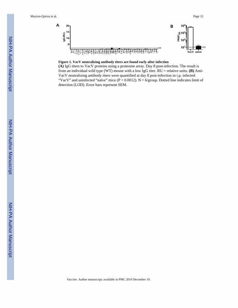
Figure 1. VacV neutralizing antibody titers are found early after infection(A) IgG titers to VacV proteins using a proteome array. Day 8 post-infection. The result isfrom an individual wild type (WT) mouse with a low IgG titer. RU = relative units. (B) Anti-VacV neutralizing antibody titers were quantified at day 8 post-infection in i.p. infected“VacV” and uninfected “naïve” mice (P = 0.0012). N = 6/group. Dotted line indicates limit ofdetection (LOD). Error bars represent SEM.
Moyron-Quiroz et al. Page 12
Vaccine. Author manuscript; available in PMC 2010 December 10.
NIH
-PA Author Manuscript
NIH
-PA Author Manuscript
NIH
-PA Author Manuscript
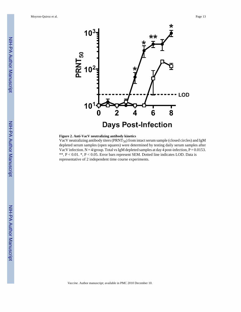
Figure 2. Anti-VacV neutralizing antibody kineticsVacV neutralizing antibody titers (PRNT50) from intact serum sample (closed circles) and IgMdepleted serum samples (open squares) were determined by testing daily serum samples afterVacV infection. N = 4/group. Total vs IgM depleted samples at day 4 post-infection, P = 0.0153.**, P < 0.01. *, P < 0.05. Error bars represent SEM. Dotted line indicates LOD. Data isrepresentative of 2 independent time course experiments.
Moyron-Quiroz et al. Page 13
Vaccine. Author manuscript; available in PMC 2010 December 10.
NIH
-PA Author Manuscript
NIH
-PA Author Manuscript
NIH
-PA Author Manuscript
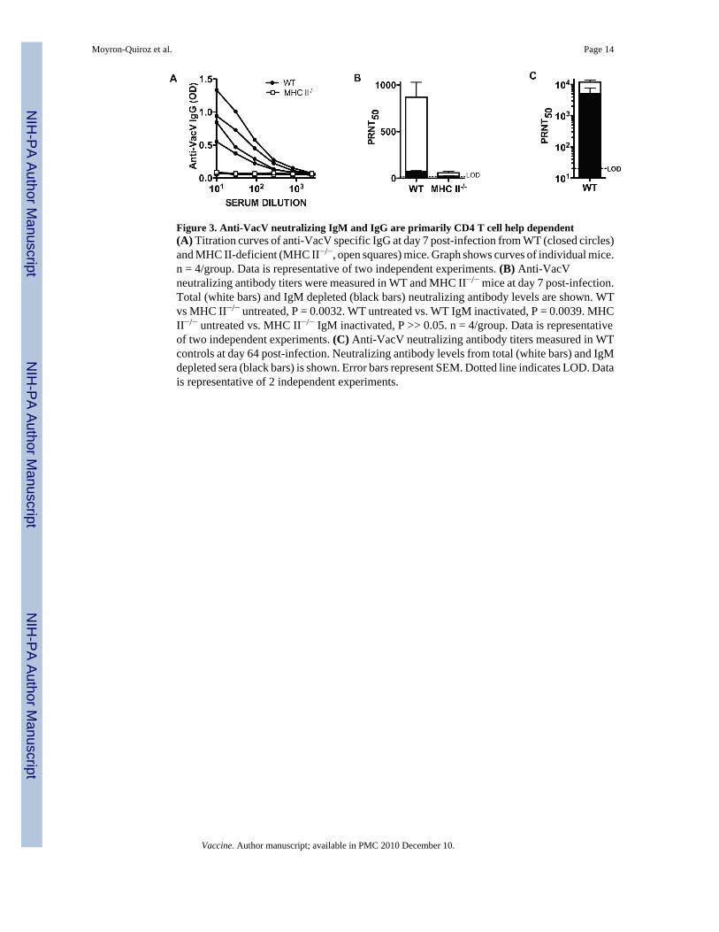
Figure 3. Anti-VacV neutralizing IgM and IgG are primarily CD4 T cell help dependent(A) Titration curves of anti-VacV specific IgG at day 7 post-infection from WT (closed circles)and MHC II-deficient (MHC II−/−, open squares) mice. Graph shows curves of individual mice.n = 4/group. Data is representative of two independent experiments. (B) Anti-VacVneutralizing antibody titers were measured in WT and MHC II−/− mice at day 7 post-infection.Total (white bars) and IgM depleted (black bars) neutralizing antibody levels are shown. WTvs MHC II−/− untreated, P = 0.0032. WT untreated vs. WT IgM inactivated, P = 0.0039. MHCII−/− untreated vs. MHC II−/− IgM inactivated, P >> 0.05. n = 4/group. Data is representativeof two independent experiments. (C) Anti-VacV neutralizing antibody titers measured in WTcontrols at day 64 post-infection. Neutralizing antibody levels from total (white bars) and IgMdepleted sera (black bars) is shown. Error bars represent SEM. Dotted line indicates LOD. Datais representative of 2 independent experiments.
Moyron-Quiroz et al. Page 14
Vaccine. Author manuscript; available in PMC 2010 December 10.
NIH
-PA Author Manuscript
NIH
-PA Author Manuscript
NIH
-PA Author Manuscript
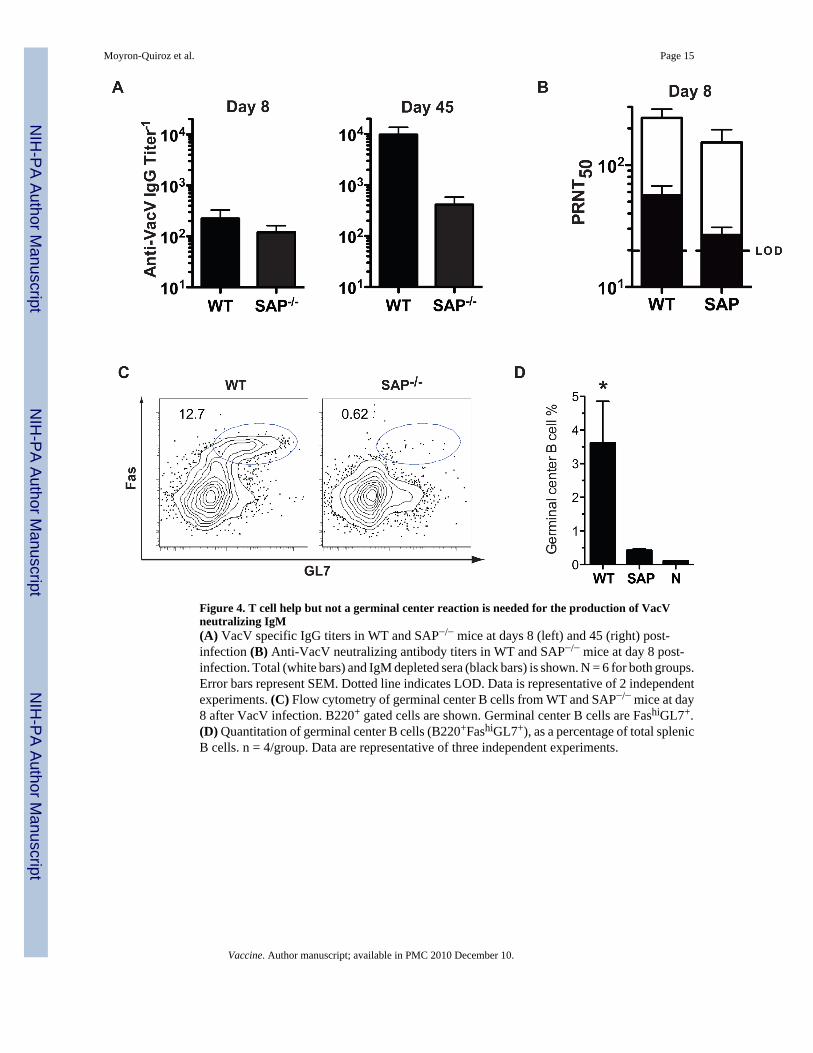
Figure 4. T cell help but not a germinal center reaction is needed for the production of VacVneutralizing IgM(A) VacV specific IgG titers in WT and SAP−/− mice at days 8 (left) and 45 (right) post-infection (B) Anti-VacV neutralizing antibody titers in WT and SAP−/− mice at day 8 post-infection. Total (white bars) and IgM depleted sera (black bars) is shown. N = 6 for both groups.Error bars represent SEM. Dotted line indicates LOD. Data is representative of 2 independentexperiments. (C) Flow cytometry of germinal center B cells from WT and SAP−/− mice at day8 after VacV infection. B220+ gated cells are shown. Germinal center B cells are FashiGL7+.(D) Quantitation of germinal center B cells (B220+FashiGL7+), as a percentage of total splenicB cells. n = 4/group. Data are representative of three independent experiments.
Moyron-Quiroz et al. Page 15
Vaccine. Author manuscript; available in PMC 2010 December 10.
NIH
-PA Author Manuscript
NIH
-PA Author Manuscript
NIH
-PA Author Manuscript
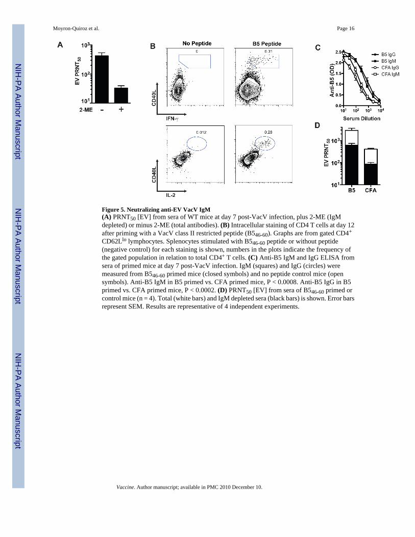
Figure 5. Neutralizing anti-EV VacV IgM(A) PRNT50 [EV] from sera of WT mice at day 7 post-VacV infection, plus 2-ME (IgMdepleted) or minus 2-ME (total antibodies). (B) Intracellular staining of CD4 T cells at day 12after priming with a VacV class II restricted peptide (B546-60). Graphs are from gated CD4+
CD62Llo lymphocytes. Splenocytes stimulated with B546-60 peptide or without peptide(negative control) for each staining is shown, numbers in the plots indicate the frequency ofthe gated population in relation to total CD4+ T cells. (C) Anti-B5 IgM and IgG ELISA fromsera of primed mice at day 7 post-VacV infection. IgM (squares) and IgG (circles) weremeasured from B546-60 primed mice (closed symbols) and no peptide control mice (opensymbols). Anti-B5 IgM in B5 primed vs. CFA primed mice, P < 0.0008. Anti-B5 IgG in B5primed vs. CFA primed mice, P < 0.0002. (D) PRNT50 [EV] from sera of B546-60 primed orcontrol mice (n = 4). Total (white bars) and IgM depleted sera (black bars) is shown. Error barsrepresent SEM. Results are representative of 4 independent experiments.
Moyron-Quiroz et al. Page 16
Vaccine. Author manuscript; available in PMC 2010 December 10.
NIH
-PA Author Manuscript
NIH
-PA Author Manuscript
NIH
-PA Author Manuscript
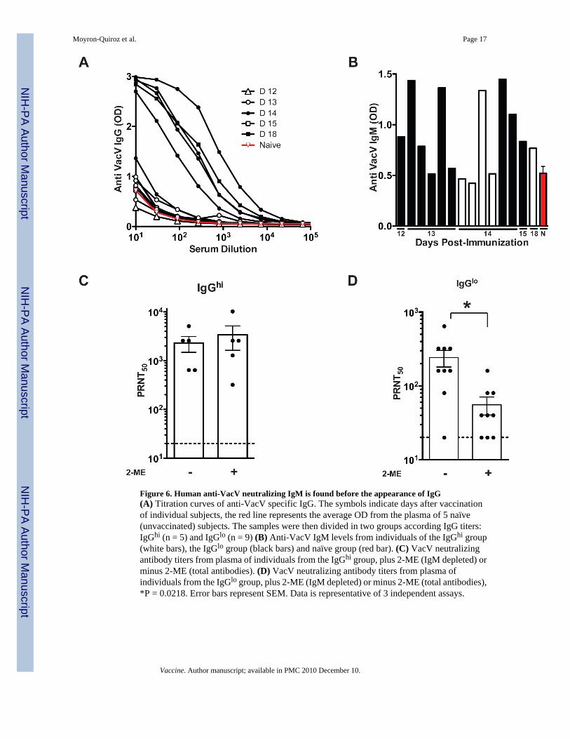
Figure 6. Human anti-VacV neutralizing IgM is found before the appearance of IgG(A) Titration curves of anti-VacV specific IgG. The symbols indicate days after vaccinationof individual subjects, the red line represents the average OD from the plasma of 5 naïve(unvaccinated) subjects. The samples were then divided in two groups according IgG titers:IgGhi (n = 5) and IgGlo (n = 9) (B) Anti-VacV IgM levels from individuals of the IgGhi group(white bars), the IgGlo group (black bars) and naïve group (red bar). (C) VacV neutralizingantibody titers from plasma of individuals from the IgGhi group, plus 2-ME (IgM depleted) orminus 2-ME (total antibodies). (D) VacV neutralizing antibody titers from plasma ofindividuals from the IgGlo group, plus 2-ME (IgM depleted) or minus 2-ME (total antibodies),*P = 0.0218. Error bars represent SEM. Data is representative of 3 independent assays.
Moyron-Quiroz et al. Page 17
Vaccine. Author manuscript; available in PMC 2010 December 10.
NIH
-PA Author Manuscript
NIH
-PA Author Manuscript
NIH
-PA Author Manuscript


![[The journey of the vaccine against smallpox: one expedition, two oceans, three continents, and thousands of children]](https://img.dokumen.tips/doc/110x75/63536dc46ff1b55f420e5651/the-journey-of-the-vaccine-against-smallpox-one-expedition-two-oceans-three.jpg)


![IGM janfeb11-Ipatov[1]](https://img.dokumen.tips/doc/110x75/63457d2b596bdb97a908f42a/igm-janfeb11-ipatov1.jpg)


![[Smallpox in Ferrara in the nineteenth century]](https://img.dokumen.tips/doc/110x75/6335d9e164d291d2a302a581/smallpox-in-ferrara-in-the-nineteenth-century.jpg)
