Embed Size (px)
Citation preview

Human Antibodies 11 (2002) 131–142 131IOS Press
Nature’s best weapons to fight cancer.Revival of human monoclonal IgM antibodies
H. Peter Vollmers∗ and Stephanie BrandleinInstitute of Pathology, University of Wurzburg, Josef-Schneider-Str. 2, 97080 Wurzburg, Germany
Abstract. The unique features of monoclonal antibodies (specificity, effectiveness, purity and unlimited reproducibility) makethem ideal tools for the specific treatment of all kind of diseases. The third generation of monoclonal antibodies for the treatmentof human diseases will be, after murine and “humanised’ murine immunoglobulins, fully human antibodies. The best source ofhuman monoclonal antibodies are the antibody pools of cancer patients themselves with the best technique for generating thembeing conventional human hybridoma technology. This technique, will generate human monoclonal antibodies which will notonly define important new targets on cancerous tissue, but will also provide the necessary therapeutic human antibodies in thefight against cancer.
Keywords: Natural immunity, human monoclonal IgM antibodies, cancer therapy
Abbreviations: HAMA, human-anti-mouse-antibody; DAF, decay accelerating factor; CFR-1, cysteine-rich fibroblast growthfactor receptor 1
1. Introduction
Hybridoma technology began mid-60’s after Little-field described a selection method for fused fibroblastcells [1,2]. Approximately ten years later, Kohler andMilstein used this somatic hybridisation and selectiontechnique and fused a lymphocyte from an immunisedmouse to a myeloma cell to produce the first murineantibody of defined specificity [3]. This milestone ex-periment has had enormous consequences for all fieldsof scientific and commercial activity [4,5].
Monoclonal antibodies are now widely accepted assupportive, or even alternative, therapeutic agents forthe treatment of specific human medical disorders, withapproximately 75 antibodies either in clinical trials orhaving already received approval for human use [6–12]. However, a closer inspection of the current pre-clinical and clinical trials with monoclonal antibodies,
∗Corresponding address: Prof. H.P. Vollmers, Institut furPathologie, Universitat Wurzburg, Josef-Schneider-Str. 2, D-97080Wurzburg, Germany. Fax.: +49 931 20147798; E-mail: [email protected].
reveals three striking facts. First, only a small frac-tion (about 20%) of the trials concern antibodies di-rected against malignancies. Moreover, only four an-tibodies have so far been approved for human ther-apeutic use (Campath�, Herceptin�, Rituximab�,Gemtuzumab�), in contrast to eight antibodies forother diseases [6,10].
Second, most of the targets for anti-cancer antibodiesare differentiation antigens, like CD20, CD52, EGF-receptor, which are not really tumour-specific [6,7,10,13,14].
Third, in cancer studies none of the antibodies are ofhuman origin, compared to about 5% human antibodiesin other clinical trials [6,10].
Hybridoma technology, like no other modern ap-proach (including vaccination and gene therapy), holdsthe highest expectations for cancer treatment, with theadded belief that human antibodies will become ofgreater significance in cancer therapy. Bearing thispoint in mind, it is surprising that after 25 years of insti-tutional and industrial research that progress in devel-oping better therapeutic antibodies for tumour-specifictargets has been so slow.
ISSN 1093-2607/02/$8.00 2002 – IOS Press. All rights reserved

132 H. Peter Vollmers and S. Brandlein / Nature’s best weapons to fight cancer. Revival of human monoclonal IgM antibodies
2. The IgM trauma
In 1977, two years after Kohler and Milstein pro-duced the first murine hybridoma antibody, the firsthuman immunoglobulin was generated by infection ofa human B-cell with Epstein-Barr-virus [15]. Threeyears later the first human-human hybridoma secretinga human immunoglobulin was described [16].
Compared to the progress achieved with murine an-tibodies, human hybridoma technology developed onlyslowly. However after the initial technical problemshad been solved, like the limited availability of properfusion partners and human lymphocytes, a series ofpromising advances were made, including the genera-tion of a huge panel of human antibodies against ma-lignancies and other diseases [17–30].
Interestingly, almost 100% of the tumour-reactingantibodies, which were isolated from cancer patients,were pentameric IgM antibodies. These IgM antibod-ies also exhibited characteristics common to conven-tional antisera, like cross-reactivity or poly-specificityand low affinity. The antibodies also reacted not onlywith membrane-bound structures, but also with cyto-plasmic and nuclear proteins, and with carbohydratestructures and glycolipids [31–36]. Subsequent se-quence analysis revealed that these human IgM anti-bodies with anti-tumour activity were part of the innate(natural) immunity, germ-line coded and not affinitymaturated [37–39].
Per definition, monoclonal antibodies had to bemono-specific, with the epitope representing a definedprotein. Immunity was equivalent to adaptation, affin-ity maturation, mutation and memory and, in contrastto mono-specific, mature antibodies generated by im-munisation of mice, these cross-reacting “natural” IgMantibodies were termed “non-specific”. In addition tothe numerous intellectual and technical problems, ex-perimental data also suggested that IgM molecules donot pass the blood-tissue endothelial barrier and cannotpenetrate tumour tissue [40,41].
The unfortunate consequence of the problems whichsurrounded human antibody technology was that withbeginning of the 90’s the interest in the classical humanhybridoma-technology decreased dramatically. Onlya handful of research groups continued to work withhuman monoclonal antibodies,almost all of which wereisolated directly from cancer patients [31,42–48].
It was therefore not surprising that interest in the useof murine antibodies as therapeutic agents was againfavoured by many research groups. However, in anattempt to decrease the immunogenicity of the murine
antibodies in patients, a lot of effort was invested indeveloping approaches to make murine antibodies ap-pear more ‘human-like’ to a patient’s immune system.To achieve this, murine antibodies were ‘humanised’through the exchanging of murine immunoglobulin se-quences with those of human origin [49–51].
Importantly, interest in the use of fully human mon-oclonal antibodies as therapeutic agents did not disap-pear completely. However, advances in scientific tech-nology meant that instead of following the classic ap-proach of immortalising human lymphocytes for theproduction of human antibodies, more sophisticatedmethods were established. Genes for human antibod-ies were now being cloned and expressed in bacte-ria (phage display libraries) or transfected into mice(HuMAbmouse or Xenomouse) [52–57]. Through thein vitro screening of the phage display libraries and theimmunisation of transgenic mice, it should be possibleto produce not only human antibodies of high speci-ficity and affinity,but it should also be possible to definenew antibody specificities.
3. State of affairs
The donor species and specificity of monoclonal an-tibodies are important for the success of clinical tri-als. Murine antibodies are both antigenic and immuno-geneic in humans and therefore induce a strong human-anti-mouse-antibody (HAMA) immune response. TheHAMA effect decreases the half-life of murine antibod-ies in humans which necessitates the need for increaseddoses and concentrations of murine antibody to achievetherapeutic levels. In an attempt to improve murine an-tibodies to either lessen or overcome the HAMA effect,genetically engineered antibodies comprising portionsof murine and human antibody gene fragments, referredto as “chimeric” and “humanised” antibodies, were de-veloped. Such chimeric (30% murine) or humanised(under 10% murine) antibodies are more “human-like”,but they still retain varying amounts of mouse anti-body protein sequence and, accordingly, may continueto trigger the HAMA response. Once the process of“humanisation” of murine antibodies is complete, thegenetically engineered and remodeled antibody genemust first be expressed in a recombinant cell line appro-priate for antibody manufacture. However, the combi-nation of murine and human antibody gene fragmentscan result in a final chimeric or humanised antibodyproduct the structure of which differs from the original

H. Peter Vollmers and S. Brandlein / Nature’s best weapons to fight cancer. Revival of human monoclonal IgM antibodies133
murine antibody, leading to a decrease in specificity ora loss in affinity [50,58].
By using phage display techniques, human singlechain antibodies or antibody fragments are coupled tophages and expressed in bacteria. The resulting li-braries are screened with specific antigens and the pos-itive reacting antibody chains are then converted (engi-neered) to produce a complete immunoglobulin. Theengineered immunoglobulins are then inserted into astandard immunoglobulin expression vector and “ful-ly” human antibodies, which are then ready for assess-ment as therapeutic tools, can be produced [52,53,57,59].
Using the Xenomouse technology, a substantial ma-jority of human antibody genes have been introducedinto murine hosts, the advantage being that murine an-tibody genes remain silent. In this approach geneti-cally reconstituted mice are first immunised after whichmurine lymphocytes are immortalised, and human im-munoglobulin producing hybrids selected. “Human”antibodies of murine origin are produced without theneed for subsequent engineering, unlike for the phagedisplay technique described above [54–56].
All these techniques have one thing in common inthat they offer ideal opportunities for the productionof humanised or human monoclonal IgG antibodies ofhigh specificity and affinity and which minimise ad-verse patient immune responses (HAMA) to the mon-oclonal antibody.
Surprisingly, however, the majority of antibodies thatare either approved, or are in late stage development,for use against solid human tumours are all of murineorigin. The antibodies, either chimeric or human-ised murine antibodies, are nearly all directed againstmolecules of the same EGF-R family, or structurallyrelated tyrosin kinase receptors, known as erbB recep-tors: Abgenix and Genentech with Herceptin� anti-HER2/neu for mammary carcinoma [60], ImCloneand Merck with Cetuximab� (anti-EGF R) for carci-noma of the colon, head and neck [61] and Medarexwith MDX-447� (bi-specific anti-CD64 and EGF-R) and MDX-H210� (bi-specifc anti-CD64 and anti-HER2/neu) for carcinoma of the head, neck, prostateand kidney [62–64]. An additional phase III trial witha humanised murine antibody (OvaRex�) directedagainst CA 125 [65], a receptor detected 20 years ago,is being conducted by Altarex as a therapeutic agentfor ovarian cancer [66,67].
Despite the availability of new techniques whichhave improved the antibodies for therapeutic useagainst human carcinomas the search for new targets
for antibodies in the fight against cancer has been slow.After 25 years of xeno-immunisations with all mannerof tumour cells and extracts, we have now hundredsof murine antibodies against hundreds of differentia-tion antigens, but only a handful of antibodies againststructures which are really tumour-specific.
Furthermore, the immunisation of “humanised”mice, or the screening of human antibody phage dis-play libraries with whole cells or cell extracts gener-ate immune responses against the major differentiationantigens whereas minor modifications, for example incarbohydrates structures, are not detected. These tech-niques described above are ideal for the generation ofhuman antibodies against defined viral and bacterialantigens, and probably auto-antigens, but not in thesearch for new antibodies against important, and as yetundetected, human carcinoma epitopes.
4. Nature’s defence
“The major question is not why cancer occurs, butwhy it occurs so infrequently?” (J.D. Watson) [68]:The immunity of an organism can be divided into aninnate (natural) and an adapted (acquired) response,or into an existing immunity and one that has to betrained [69–71]. The innate immunity (also referredto as “unspecific immunity”) consists of natural killer(NK) cells, dendritic and mast cells, macrophages andantibody producing B-cells [69,72–78]. This systemcan distinguish between self and non-self and is respon-sible for the first specific immune response directedagainst bacteria, viruses and also malignant cells [79–81]. To guarantee a fast reaction, the response is T-cellindependent, which means that antigen-presentation byT-cells is not required [82,83]. This innate immunesystem has enough genetic variability to cover a verybroad spectrum of foreign antigens which ensures thatwe are not overcome by the multitude of infections ormalignant cells that challenge the average human bodyover the period of a standard lifetime.
The cells which are involved in innate immunity rec-ognize specific pattern instead of specific single struc-tures. These conservative pattern are expressed inde-pendently from mutational events [79]. To recognizesuch pattern, the immune-competent cells use specificsets of germ-line coded receptors which belong to dis-tinct protein families [70,74,79]. Based on and initiatedby the innate immunity, the acquired or “specific” im-munity then becomes involved. This T-cell dependentdefence requires a period of in which specifically tai-

134 H. Peter Vollmers and S. Brandlein / Nature’s best weapons to fight cancer. Revival of human monoclonal IgM antibodies
Human Hybridoma Technology
Tumour Lymphnode
Tests onautologous
tissue
Tissueculture
Tissuesection
Monoclonalantibodies
Cell-fusion
Triom
Receptor analysisSpecificity analysis
Preclinical and Clinical trials
Singlecells
Fig. 1. Conventional human hybridoma technology.
Transmembranecomponent(unknown)
PO4
PO4 PO4
PO4
Glucosamin
Glycan
Ethanolamin
GPI-anchor
SCR1
SCR2
SCR3
SCR4
N-CHO Anti body SC-1
-O-CHO
-O-CHO
-O-CHOCHO-O-CHO-O-CHO-O-
CHO-O-
CD55
src-kinasese.g. P56lck , p59fyn
Plasmamembrane
- GPI (Glycos yl-phosph atidyl-inosi tol )-ancho red memb rane pro tein
- 82 kDa molecu lar weight
- a new variant of CD55 (DAF, decayaccelerat ing factor)
DAF/SC-1 Receptor
Fig. 2. DAF/SC-1 Receptor.
lored weapons are developed to combat the challengeand reaches a maximum response several days after theinitial contact with the antigen. The result of the T-cell dependent immune response is at the humoral levelwith mature B-cells secreting mutated and highly spe-cific IgG and long-lasting memory cells [84,85]. The“natural B-cell” pool, consisting of CD5+ positive lym-phocytes, produces antibodies that principally belongto the IgM class [86–88]. These IgM antibodies aregerm-line coded and do not increase their variability bymutational events and maturation [37–39]. The num-ber of possible binding sites is therefore limited andeach antibody has to cover a broader spectrum of dif-
ferent specific antigens such as specific pattern of car-bohydrates etc. [31–35,47,48,88]. Natural antibodiesare therefore “oligo-specific” and not “non-specific”,as they are often referred to in the literature. However,the price for oligo-reactivity is a lower affinity to eachspecific antigen.
Reflecting all data on humoral response to malignantcells, there are striking similarities between humoraldefence against bacterial and malignant cells suggest-ing that nature’s defence against transformed cells isbased on innate immune mechanisms and is not theresult of an antigen-induced maturation process.

H. Peter Vollmers and S. Brandlein / Nature’s best weapons to fight cancer. Revival of human monoclonal IgM antibodies135
CFR-1/PAM-1 Receptor
- Integr al membrane glycopr otei n
- 130 kDa molecular weigh t
- a new variant of CFR-1 (cystein e-richfibroblast growth factor receptor 1)
- structural homologous to rat Golgi MG160and human myeloid adhesi on moleculeESL-1
Membrane
Extracell ular Domain(1142 amino acids)
16 Cysteinerich repeats
Possibl e N-Glyco-
sylation sites
Possibl e N-Glyco-
sylation site s
FGF bin dingdomain
FGF bin dingdomain
Transmemb rane Domain (21 amino acids )
(13 amino acids ) Intracell ular Domain
Anti body PAM-1
Fig. 3. CFR-1/PAM-1 Receptor.
5. Real human antibodies
Antibodies that define new tumour-related moleculescan be readily isolated from the sera of cancer pa-tients [89,90]. Whilst the antisera have only limitedtherapeutical advantages, they can however be usedto isolate and characterise new tumour-related struc-tures. The tumour-reactive antibodies can be purifiedand then used for the construction or isolation of humanantibodies using techniques like phage display or theHuMAbmouse systems, described above.
The classic or conventional approach to hybridomatechnology of immortalising human lymphocytes bysomatic hybridisation solves the problem of speciesand specificity of monoclonal antibodies for therapeuticuse. In this technique, the first screening following im-mortalisation of the human lymphocyte is made againstthe autologous tumour and in the second step, the corre-sponding epitope on the tumour cells is identified withthe same tumour-specific antibody (see Fig. 1).
By using this approach, we have been able to charac-terise a series of human monoclonal antibodies, whichare useful for therapy and diagnosis, in addition to twonew receptors. Figure 2 shows the receptor identifiedby the human monoclonal antibody SC-1 [31,37,47,91,92]. The structure identified on the membrane ofstomach carcinoma cells is a modified version of DAF(Decay acceleration factor), also known as CD55.
Figure 3 shows the receptor for the human mon-oclonal antibody PAM-1 (formerly 103/51). Thismolecule belongs to the family of cysteine-rich FGF
receptors and is a variant of CFR-1 (cysteine-rich fi-broblast growth factor receptor 1) [38,45,48]. CFR-1/PAM-1 is expressed on nearly all epithelial cancercells and on the precursor cells of neoplasms.
It is therefore possible, to produce useful humanmonoclonal IgM antibodies which define new tumour-related structures with a single experimental approach,making this technique superior to phage display orhumAbmice
6. IgM handling
Cross-reactivity of germ-line IgMs is commonly ob-served in biochemical and immunohistochemical pro-cedures [31,33–35]. Therefore, the handling of naturalIgMs in experimental systems requires specific adap-tations to compensate for their characteristics. Firstof all, the primary selection of human antibodies fromcancer patients is a pre-requisite for success. The initialsearch for membrane epitopes always begin with bind-ing assays with living cells, (e.g. FACS analysis, func-tional assays). This approach guarantees the recogni-tion of a membrane-bound epitope on the tumour cellsand staining on autologous tissue enables selection fortumour-specificity.
Specificity analysis of antibodies in immunohisto-chemical studies provides vital information, in addi-tion to the specific reactivity of the antibody, regardingcross-reactivities with repetitive structures such as mus-cle or connective, glandular tissue (mucine in parietal

136 H. Peter Vollmers and S. Brandlein / Nature’s best weapons to fight cancer. Revival of human monoclonal IgM antibodies
Fig. 4. Immunohistochemical staining of antibody SC-1 on gastric signet ring carcinoma. Paraffin sections were stained with A: isotype matchedcontrol antibody Chrompure IgM, B: positive control antibody CK8, C: negative control (second antibody alone) and D: antibody SC-1. (Originalmagnification,×100).
cells) and squamous epithelial cells. Background stain-ing levels following binding of the second anti-humanantibodies to immunoglobulins in the tissue, in additionto non-specific binding or stickiness of the first anti-body over the Fc-part,can be reduced by pre-incubationof the sections with proteins or antibodies. Monoclonaland polyclonal human IgMs should be used in each ex-periment as controls and their reactivity should be sub-stracted from the study antibody to provide the specificactivity (see Figs 4 and 5).
Receptor analysis using western blots with proteinextracts usually identifies several reactive proteins,membranous, cytoplasmic and nuclear structures [31,32,48]. Fractionation of the extracts and saturationwith large amounts of protein can reduce non-specificbinding and provide specific bands.
In general, it is important to determine the impactof antibody cross-reactivities with regards to the studygoal. However, should a membrane reacting IgM an-tibody also react with an epitope in the cytoplasm ornucleus, this should, in general, not limit its use as alikely therapeutic agent. However, thorough acute ani-mal toxicity studies should be conducted to exclude thepossibility of antibody cross-reactivity posing a dangerfor therapeutic use.
7. Breaking the dogma
The ideal mouse tumour model should mimic thepathobiologic, genetic, aetiologic and therapeutic char-acteristics of its human counterpart and faithfully repro-duce all aspects of tumourgenesis. Validation param-eters should not only include histopathology, molecu-lar/genetic changes and stages of progression, but alsotherapeutic responses [93,94]. Mouse cancer mod-els, in which the murine host has been transplantedwith human tumours, have been useful in screeningsome potential chemotherapeutic and chemopreventiveagents. Recently, mouse models with spontaneous tu-mours have been “constructed” in an attempt to mimicmore closely human tumourgenesis but even with thisapproach, no mouse tumour model will mimic everyaspect of its human counterpart and there remains con-siderable scepticisms about the value of such modelsfor drug development [95–98].
In addition, human proteins induce a humoral im-mune response in all mice, regardless of whether themurine species exhibits wild-type characteristics or ismutated, e.g. athymic mice [99–101]. This leads notonly to a rapid and unpredictable clearance of humanantibodies in the murine host, but also to a high vari-ability in experimental evaluations. The only reliable

H. Peter Vollmers and S. Brandlein / Nature’s best weapons to fight cancer. Revival of human monoclonal IgM antibodies137
Fig. 5. Immunohistochemical staining of antibody PAM-1 on different gastric mucosae. Paraffin sections were stained with A, C, E: proliferationmarker Ki67 and B, D, F: antibody PAM-1. A and B: gastric tissue with inflammation, C and D: gastric dysplasia, E and F: gastric adenocarcinoma.(Original magnification,×100).
and reproducible investigation in murine hosts involv-ing either “humanised” or human antibodies is the acutetoxicity assay. All other experimental approaches maygive a hint to antibody activity in patients, but they arenot a serious method for quality and quantity analysisof human antibodies.
Despite this, animal experiments are still used forthe evaluation of human antibodies. However, one ofthe most prominent arguments against the use of nativeIgM molecules for therapy is that a pentameric IgMmolecule with a mass of approximately 1 Mill kDa can-not pass the blood-tissue endothelial barrier and doesnot penetrate surrounding tissue [40,41]. This dogmais mainly based on observations in experimental animalsystems, where mice, or other animals, were inoculatedwith human tumour cells, mostly via the subcutanousroute, and the subsequent diffusion and penetration pat-terns of radiolabeled antibodies was measured. Gener-ally, a highly specific tumour targeting was only possi-
ble with small antibody fragments, whereas moleculeswith a size of monomeric immunoglobulins remainedpredominantly in the bloodstream, did not cross the en-dothelial barriers, and were rapidly cleared [102–105].
However, morphological and structural analysis ofhuman, and even experimentally induced solid mousetumours, demonstrate that the blood vessels in tumoursare a mix of normal and abnormal vessels and are notcomparable to vessels in healthy tissue [106,107]. Thevascular permeability of tumour tissue has been shownto be heterogeneous and the ultrastructure is dynamicwhich can be modulated by the microenvironment, e.g.inflammatory processes [108–110]. Tumour microves-sels are in general thin walled, chaotically arranged,and lack innervation [105] with a greater permeabil-ity for macromolecules than the normal vessels [106,111–115].
After having left the blood vessels, macromoleculeshave to pass the extracellular matrix and tumour inter-
138 H. Peter Vollmers and S. Brandlein / Nature’s best weapons to fight cancer. Revival of human monoclonal IgM antibodies
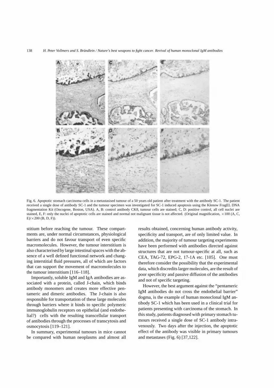
Fig. 6. Apoptotic stomach carcinoma cells in a metastasised tumour of a 50 years old patient after treatment with the antibody SC-1. The patientreceived a single dose of antibody SC-1 and the tumour specimen was investigated for SC-1 induced apoptosis using the Klenow FragEL DNAfragmentation Kit (Oncogene, Boston, USA). A, B: control antibody CK8, tumour cells are stained; C, D: positive control, all cell nuclei arestained, E, F: only the nuclei of apoptotic cells are stained and normal not malignant tissue is not affected. (Original magnification,×100 (A, C,E)/×200 (B, D, F)).
stitium before reaching the tumour. These compart-ments are, under normal circumstances, physiologicalbarriers and do not favour transport of even specificmacromolecules. However, the tumour interstitium isalso characterised by large intestinal spaces with the ab-sence of a well defined functional network and chang-ing interstitial fluid pressures, all of which are factorsthat can support the movement of macromolecules tothe tumour interstitium [116–118].
Importantly, soluble IgM and IgA antibodies are as-sociated with a protein, called J-chain, which bindsantibody monomers and creates more effective pen-tameric and dimeric antibodies. The J-chain is alsoresponsible for transportation of these large moleculesthrough barriers where it binds to specific polymericimmunoglobulin receptors on epithelial (and endothe-lial?) cells with the resulting transcellular transportof antibodies through the processes of transcytosis andosmocytosis [119–121].
In summary, experimental tumours in mice cannotbe compared with human neoplasms and almost all
results obtained, concerning human antibody activity,specificity and transport, are of only limited value. Inaddition, the majority of tumour targeting experimentshave been performed with antibodies directed againststructures that are not tumour-specific at all, such asCEA, TAG-72, EPG-2, 17-1A etc. [105]. One musttherefore consider the possibility that the experimentaldata, which discredits larger molecules, are the result ofpoor specificity and passive diffusion of the antibodiesand not of specific targeting.
However, the best argument against the “pentamericIgM antibodies do not cross the endothelial barrier”dogma, is the example of human monoclonal IgM an-tibody SC-1 which has been used in a clinical trial forpatients presenting with carcinoma of the stomach. Inthis study, patients diagnosed with primary stomach tu-mours received a single dose of SC-1 antibody intra-venously. Two days after the injection, the apoptoticeffect of the antibody was visible in primary tumoursand metastases (Fig. 6) [37,122].

H. Peter Vollmers and S. Brandlein / Nature’s best weapons to fight cancer. Revival of human monoclonal IgM antibodies139
This proves that pentameric IgM molecules are ableto leave the circulation, cross the endothelia and ma-trices to reach the interstitium and the tumour, and tospecifically kill tumour cells in vivo (see also [91,92,123]).
8. Denouement
It is widely accepted that third generation, fully hu-man antibodies are likely to offer the most effectivetreatment for numerous human diseases for the nearfuture. These state-of-the-art antibodies, produced ei-ther by phage technology or transgenic mice will beused for the treatment of both immune and infectiousdiseases. However, for the treatment of cancer, the best(and only?) source of fully human antibodies are thecancer patients themselves.
After 15 years of experience with human antibodytechnology and human IgMs, the majority of thetechnical problems concerning low affinity and cross-reactivity of human IgM antibodies have been solved.It is now possible to use these human monoclonal IgMantibodies for both diagnostic and therapeutic studies.Furthermore, the use of human antibodies, producedusing conventional hybridoma technology, is the bestand most rapid method for the definition of new tu-mour targets. IgM antibodies may prove to be the bestweapon against malignancies that nature itself created.However, if we are unable to overcome our prejudicesagainst the use of IgM antibodies as therapeutic agents,it is likely that we will have overlooked one of the bestweapons in the fight against cancer.
Acknowledgements
We thank Mrs. E. Wozniak, Mrs. T. Pohle andMrs. N. Ruoff for excellent technical assistant, Mr. E.Schmitt for preparing the artwork and Dr. L. Dunsterfor improving the manuscript.
References
[1] J.W. Littlefield, Selection of hybrids from matings of fobrob-lasts in vitro and their presumed recombinants,Science145(1964), 709–711.
[2] J.W. Littlefield, The use of drug-resistant markers to study thehybridization of mouse fibroblasts,Exp. Cell Res.41 (1966),190–196.
[3] G. Kohler and C. Milstein, Continuous cultures of fusedcells secreting antibody of predefined specificity,Nature256(1975), 495–497.
[4] S. Dickman, Antibodies stage a comeback in cancer treat-ment,Science280 (1998), 1196–1197.
[5] P. Holliger and H. Hoogenboom, Antibodies come back fromthe brink,Nat. Biotechnol.16 (1998), 1015–1016.
[6] P. Carter, Improving the efficacy of antibody-based cancertherapies,Nature Rev. Cancer1 (2001), 118–129.
[7] L.M. Weiner, An overview of monoclonal antibody therapyof cancer,Semin. Oncol.26 (1999), 41–50.
[8] M. von Mehren and L.M. Weiner, Monoclonal antibody-based therapy,Curr. Opin. Oncol.8 (1996), 493–498.
[9] F.C. Breedveld, Therapeutic monoclonal antibodies,Lancet355 (2000), 735–740.
[10] M.J. Glennie and P.W. Johnson, Clinical trials of antibodytherapy,Immunol. Today21 (2000), 403–410.
[11] A.N. Houghton and D.A. Scheinberg, Monoclonal antibodytherapies-a ‘constant’ threat to cancer,Nat. Med.6 (2000),373–374.
[12] E. Drewe and R.J. Powell, Clinically useful monoclonal an-tibodies in treatment,J. Clin. Pathol.55 (2002), 81–85.
[13] S. Matzku, Monoclonal antibodies in tumor therapy,RecentResults Cancer Res.141 (1996), 1–8.
[14] H.J. Ditzel, Human antibodies in cancer and autoimmunedisease,Immunol. Res.21 (2000), 185–193.
[15] M. Steinitz, G. Klein, S. Koskimies and O. Makel, EB virus-induced B lymphocyte cell lines producing specific antibody,Nature269 (1977), 420–422.
[16] L. Olsson and H.S. Kaplan, Human-human hybridomas pro-ducing monoclonal antibodies of predefined antigenic speci-ficity, Proc .Natl. Acad. Sci.USA 77 (1980), 5429–5431.
[17] J. Schlom, D. Wunderlich and Y.A. Teramoto, Generationof human monoclonal antibodies reactive with human mam-mary carcinoma cells,Proc. Natl. Acad. Sci. USA77 (1980),6841–6845.
[18] K. Sikora, T. Alderson, J. Ellis, J. Phillips and J. Watson,Human hybridomas from patients with malignant disease,Br.J. Cancer47 (1983), 135–145.
[19] M.V. Haspel, R.P. McCabe, N. Pomato, N.J. Janesch, J.V.Knowlton, L.C. Peters, H.C. Hoover, Jr and M.G. Hanna,Jr Generation of tumor cell-reactive human monoclonal an-tibodies using peripheral blood lymphocytes from activelyimmunized colorectal carcinoma patients,Cancer Res45(1985), 3951–3961.
[20] J. Kan-Mitchell, A. Imam, R.A. Kempf, C.R. Taylor and M.S.Mitchell, Human monoclonal antibodies directed againstmelanoma tumor-associated antigens,Cancer Res.46 (1986),2490–2496.
[21] H. Yamaguchi, K. Furukawa, S.R. Fortunato, P.O. Liv-ingston, K.O. Lloyd, H.F. Oettgen and L.J. Old, Cell-surfaceantigens of melanoma recognized by human monoclonal an-tibodies,Proc. Natl. Acad. Sci.USA 84 (1987), 2416–2420.
[22] K. Furukawa, H. Yamaguchi, H.F. Oettgen, L.J. Old and K.O.Lloyd, Two human monoclonal antibodies reacting with themajor gangliosides of human melanomas and comparisonwith corresponding mouse monoclonal antibodies,CancerRes.49 (1989), 191–196.
[23] R.J. Cote, D.M. Morrissey, A.N. Houghton, T.M. Thomson,M.E. Daly, H.F. Oettgen and L.J. Old, Specificity analysis ofhuman monoclonal antibodies reactive with cell surface andintracellular antigens,Proc. Natl. Acad. Sci.USA 83 (1986),2959–2963.

140 H. Peter Vollmers and S. Brandlein / Nature’s best weapons to fight cancer. Revival of human monoclonal IgM antibodies
[24] M.C. Glassy, Immortalization of human lymphocytes from atumor-involved lymph node,Cancer Res.47 (1987), 5181–5188.
[25] J.W. Larrick and J.M. Bourla, Prospects for the therapeuticuse of human monoclonal antibodies,J. Biol. Response Mod.5 (1986), 379–393.
[26] K. Sikora, Human monoclonal antibodies,Br. Med. Bull.40(1984), 209–212.
[27] D.A. Carson and B.D. Freimark, Human lymphocyte hy-bridomas and monoclonal antibodies,Adv. Immunol.38(1986), 275–311.
[28] S.P. Cole, B.G. Campling, I.H. Louwman, D. Kozbor and J.C.Roder, A strategy for the production of human monoclonalantibodies reactive with lung tumor cell lines,Cancer Res.44 (1984), 2750–2753.
[29] M. Glassy, Creating hybridomas by electrofusion,Nature333 (1988), 579–580.
[30] K. James and G.T. Bell, Human monoclonal antibody produc-tion: Current status and future prospects,J.Immunol.Methods100 (1987), 5–40.
[31] H.P. Vollmers, R. O’Connor, J. Muller, T. Kirchner and H.K.Muller-Hermelink, SC-1, a functional human monoclonal an-tibody against autologous stomach carcinoma cells,CancerRes.49 (1989), 2471–2476.
[32] M. Pfaff, R. O’Connor, H.P. Vollmers and H.K. Muller-Hermelink, Human monoclonal antibody against a tissuepolypeptide antigen-related protein from a patient with asignet-ring cell carcinoma of the stomach,Cancer Res.50(1990), 5192–5198.
[33] A.M. Campbell, P. Whitford and R.E. Leake, Human mono-clonal antibodies and monoclonal antibody multispecificity,Br. J. Cancer56 (1987), 709–713.
[34] K.O. Lloyd and L.J. Old, Human monoclonal antibodies toglycolipids and other carbohydrate antigens: dissection ofthe humoral immune response in cancer patients,Cancer Res.49 (1989), 3445–3451.
[35] M.E. McKnight, K. Koda, K. DeBoer and M.C. Glassy, Hu-man monoclonal antibodies to nuclear antigens,Hum. Anti-bodies Hybridomas1 (1990), 77–82.
[36] S. Mukerjee, M. Nasoff, M. McKnight and M. Glassy, Char-acterization of human IgG1 monoclonal antibody againstgangliosides expressed on tumor cells,Hybridoma17 (1998),133–142.
[37] H.P. Vollmers, F. Hensel, R. Hermann, J. Dammrich, E. Woz-niak, P. Gessner, B. Herrmann, U. Zimmermann and H.K.Muller-Hermelink, Tumor-specific apoptosis by the humanmonoclonal antibody SC-1: A new therapeutical approachfor stomach cancer,Oncology Reports5 (1998), 35–40.
[38] F. Hensel, C. Knorr, R. Hermann, V. Krenn, H.K. Muller-Hermelink and H.P. Vollmers, Mitogenic autoantibodiesin Helicobacter pylori-associated stomach cancerogenesis,Int.J.Cancer81 (1999), 229–235.
[39] S. Brandlein, J. Lorenz, N. Ruoff, F. Hensel, I. Beyer,J. Muller, K. Neukam, M. Eck, B. Illert, H.K. Muller-Hermelink and H.P. Vollmers, Human monoclonal IgM anti-bodies with apoptotic activity isolated from cancer patients,Hum. Antibodies(2003), in press.
[40] R.K. Jain and L.T. Baxter, Mechanisms of heterogeneousdistribution of monoclonal antibodies and other macro-molecules in tumors: significance of elevated interstitialpressure,Cancer Res.48 (1988), 7022–7032.
[41] G.P. Adams and R. Schier, Generating improved single-chainFv molecules for tumor targeting,J. Immunol. Methods231(1999), 249–260.
[42] M.C. Glassy, Production methods for generating human mon-oclonal antibodies,Hum Antibodies Hybridomas4 (1993),154–165.
[43] H.R. Chang, K. Koda, S. Chang and S. Baird, AgSK1, anovel carcinoma associated antigen,Cancer Res.53 (1993),1122–1127.
[44] K. Koda, M.C. Glassy, M.E. McKnight, J. Yasutomi, N.Saito, M. Dan and N. Nakajima, Immunotherapy for recur-rent colorectal cancers with human monoclonal antibody SK-1, Anticancer Res.21 (2001), 621–627.
[45] H.P. Vollmers, J. Dammrich, H. Ribbert, S. Grassel, S. De-bus, J. Heesemannn, and H.K. Muller-Hermelink, Humanmonoclonal antibodies from stomach carcinoma patients re-act with Helicobacter pylori and stimulate stomach cancercells in vitro,Cancer74 (1994), 1525–1532.
[46] H.P. Vollmers, E. Wozniak, E. Stepien-Botsch, U. Zimmer-mann and H.K. Muller Hermelink, A rapid method for pu-rification of monoclonal human IgM from mass culture,Hum.Antibodies.Hybridomas.7 (1996), 37–41.
[47] F. Hensel, R. Hermann, C. Schubert, N. Abe, K.Schmidt, A. Franke, A. Shevchenko, M. Mann, H.K.Muller-Hermelink and H.P. Vollmers, Characterisation ofglycosylphosphatidylinositol-linked molecule CD55/Decay-accelerating factor as the receptor for antibody SC-1-inducedapoptosis,Cancer Res.59 (1999), 5299–5306.
[48] F. Hensel, S. Brandlein, M. Eck, K. Schmidt, V. Krenn, A.Kloetzer, A. Bachi, M. Mann, H.K. Muller-Hermelink andH.P. Vollmers, A novel proliferation-associated variant ofCFR-1 defined by a human monoclonal antibody,Lab. Invest.81 (2001), 1097–1108.
[49] D.R. Burton, Human monoclonal antibodies: achievementand potential,Hosp. Pract.(Off Ed) 27 (1992), 67–74.
[50] M. Clark, Chimeric and humanised–misunderstood,Lancet355 (2000), 1557.
[51] J.S. Huston and A.J. George, Engineered antibodies takecenter stage,Hum. Antibodies10 (2001), 127–142.
[52] K.D. Wittrup, Phage on display,Trends Biotechnol.17(1999), 423–424.
[53] E. Boel, S. Verlaan, M.J. Poppelier, N.A. Westerdaal, J.A.Van Strijp and T. Logtenberg, Functional human monoclonalantibodies of all isotypes constructed from phage displaylibrary-derived single-chain Fv antibody fragments,J. Im-munol. Methods239 (2000), 153–166.
[54] M. Neuberger and M. Bruggemann, Monoclonal antibodies,Mice perform a human repertoire,Nature386 (1997), 25–26.
[55] C.G. Davis, M.L. Gallo and J.R. Corvalan, Transgenic miceas a source of fully human antibodies for the treatment ofcancer,Cancer Metastasis Rev.18 (1999), 421–425.
[56] L.L. Green, Antibody engineering via genetic engineering ofthe mouse: XenoMouse strains are a vehicle for the facilegeneration of therapeutic human monoclonal antibodies,J.Immunol. Methods231 (1999), 11–23.
[57] M. Little, M. Welschof, M. Braunagel, L. Hermes, C. Christ,A. Keller, P. Rohrbach, T. Kurschner, S. Schmidt, C. Kleistand P. Terness, Generation of a large complex antibody li-brary from multiple donors,J. Immunol. Methods231 (1999),3–9.
[58] G. Winter and W.J. Harris, Humanized antibodies,Immunol.Today14 (1993), 243–246.
[59] H.R. Hoogenboom and P. Chames, Natural and designerbinding sites made by phage display technology,Immunol.Today21 (2000), 371–378.
[60] B. Leyland-Jones, Trastuzumab: hopes and realities,LancetOncol.3 (2002), 137–144.

H. Peter Vollmers and S. Brandlein / Nature’s best weapons to fight cancer. Revival of human monoclonal IgM antibodies141
[61] J. Baselga, The EGFR as a target for anticancer therapy–focuson cetuximab,Eur. J. Cancer37(Suppl 4) (2001), 16–22.
[62] A. Ullrich, L. Coussens, J.S. Hayflick, T.J. Dull, A. Gray,A.W. Tam, J. Lee, Y. Yarden, T.A. Libermann, J. Schlessingerand et al., Human epidermal growth factor receptor cDNAsequence and aberrant expression of the amplified gene inA431 epidermoid carcinoma cells,Nature309 (1984), 418–425.
[63] J.S. de Bono and E.K. Rowinsky, The ErbB receptor family:a therapeutic target for cancer,Trends Mol. Med.8 (2002),19–26.
[64] R.T. Curnow, Clinical experience with CD64-directed im-munotherapy,An overview. Cancer Immunol. Immunother.45 (1997), 210–215.
[65] A.A. Noujaim, B.C. Schultes, R.P. Baum and R.Madiyalakan, Induction of CA125-specific B and T cell re-sponses in patients injected with MAb-B43.13–evidence forantibody-mediated antigen-processing and presentation ofCA125 in vivo, Cancer Biother. Radiopharm.16 (2001),187–203.
[66] R.C. Bast, Jr, M. Feeney, H. Lazarus, L.M. Nadler, R.B.Colvin and R.C. Knapp, Reactivity of a monoclonal antibodywith human ovarian carcinoma,J. Clin. Invest.68 (1998),11331–1337.
[67] P.A. Canney, M. Moore, P.M. Wilkinson and R.D. James,Ovarian cancer antigen CA125: a prospective clinical assess-ment of its role as a tumour marker,Br. J. Cancer50 (1984),765–769.
[68] J.D. Watson, Cancer, in:Molekular biology of the cell, thirdedition, M. Robertson, R. Adams, S.M. Cobert, D. Goertzen,eds, Garland publishing, Inc.: New York, 1994, pp. 1255–1291.
[69] A.F. Ochsenbein and R.M. Zinkernagel, Natural antibod-ies and complement link innate and acquired immunity,Im-munol. Today21 (2000), 624–630.
[70] R. Medzhitov and C. Janeway, Jr. Innate immunity,N. Engl.J. Med.343 (2000), 338–344.
[71] D.A. Kimbrell and B. Beutler, The evolution and genetics ofinnate immunity,Nat. Rev. Genet.,2 (2001), 256–267.
[72] L. Moretta, R. Biassoni, C. Bottino, M.C. Mingari and A.Moretta, Natural killer cells: a mystery no more,Scand. J.Immunol.55 (2002), 229–232.
[73] F. Martin, A.M. Oliver and J.F. Kearney, Marginal zone andB1 B cells unite in the early response against T-independentblood-borne particulate antigens,Immunity14 (2001), 617–629.
[74] R. Medzhitov, Toll-like receptors and innate immunity,Na-ture Rev. Immunol.,1 (2001), 135–145.
[75] L. Zitvogel, Dendritic and natural killer cells cooperate in thecontrol/switch of innate immunity,J. Exp. Med.195 (2002),F9–14.
[76] F. Martin and J.F. Kearney, B1 cells: similarities and dif-ferences with other B cell subsets,Curr. Opin. Immunol.13(2001), 195–201.
[77] S. Greenberg and S. Grinstein, Phagocytosis and innate im-munity,Curr. Opin. Immunol.,14 (2002), 136–145.
[78] Y.A. Mekori and D.D. Metcalfe, Mast cells in innate immu-nity, Immunol. Rev.173 (2000), 131–140.
[79] C.A. Janeway, Jr How the immune system works to protectthe host from infection: a personal view,Proc. Natl. Acad.Sci.USA 98 (2001), 7461–7468.
[80] D.V. Cramer, Natural antibodies and the host immune re-sponses to xenografts,Xenotransplantation7 (2000), 83–92.
[81] I.D. Davis, An overview of cancer immunotherapy,Immunol.Cell Biol. 78 (2000), 179–195.
[82] N. Baumgarth, A two-phase model of B-cell activation,Im-munol. Rev.176 (2000), 171–180.
[83] S. Fagarasan and T. Honjo, T-Independent immune response:new aspects of B cell biology,Science290 (2000), 89–92.
[84] G. MacPherson, N. Kushnir and M. Wykes, Dendritic cells,B cells and the regulation of antibody synthesis,Immunol.Rev.172 (1999), 325–334.
[85] J. Parkin and B. Cohen, An overview of the immune system,Lancet357 (2001), 1777–1789.
[86] J. Bohn, Are natural antibodies involved in tumour defence?Immunol. Lett.69 (1999), 317–320.
[87] M. Boes, Role of natural and immune IgM antibodies inimmune responses,Mol. Immunol.37 (2000), 1141–1149.
[88] P. Casali and A.L. Notkins, CD5+ B lymphocytes, polyre-active antibodies and the human B-cell repertoire,Im-munol.Today10 (1989), 364–368.
[89] Y.T. Chen, M.J. Scanlan, U. Sahin, O. Tureci, A.O. Gure,S. Tsang, B. Williamson, E. Stockert, M. Pfreundschuh andL.J. Old, A testicular antigen aberrantly expressed in humancancers detected by autologous antibody screening,Proc.Natl. Acad. Sci.USA 94 (1997), 1914–1918.
[90] Y.T. Chen, A.D. Boyer, C.S. Viars, S. Tsang, L.J. Old andK.C. Arden, Genomic cloning and localization of CTAG,a gene encoding an autoimmunogenic cancer-testis antigenNY-ESO-1, to human chromosome Xq28,Cytogenet. Cell.Genet.79 (1997), 237–240.
[91] H.P. Vollmers, J. Dammrich, H. Ribbert, E. Wozniak andH.K. Muller-Hermelink, Apoptosis of stomach carcinomacells induced by a human monoclonal antibody,Cancer76(1995), 550–558.
[92] F. Hensel, R. Hermann, S. Brandlein, V. Krenn, B.Schmausser, S. Geis, H.K. Muller-Hermelink and H.P.Vollmers, Regulation of the New Coexpressed CD55 (Decay-Accelerating Factor) Receptor on Stomach Carcinoma CellsInvolved in Antibody SC-1-Induced Apoptosis,Lab. Invest.81 (2001), 1553–1563.
[93] J. Jonkers and A. Berns, Conditional mouse models of spo-radic cancer,Nature Rev. Cancer2 (2002), 251–265.
[94] R.K. Jain, L.L. Munn and D. Fukumura, Dissecting tumourpathophysiology using intravital microscopy,Nature Rev.Cancer2 (2002), 266–276.
[95] B. Hann and A. Balmain, Building validated mouse modelsof human cancer,Curr. Opin. Cell. Biol.13 (2001), 778–784.
[96] R.D. Klausner, Studying cancer in the mouse,Oncogene18(1999), 5249–5252.
[97] P. Guilford, J. Hopkins, J. Harraway, M. McLeod, N.McLeod, P. Harawira, H. Taite, R. Scoular, A. Miller andA.E. Reeve, E-cadherin germline mutations in familial gas-tric cancer,Nature392 (1998), 402–405.
[98] R. Meuwissen, J. Jonkers and A. Berns, Mouse models forsporadic cancer,Exp. Cell Res.264 (2001), 100–110.
[99] M. Pelleitier and S. Montplaisir, The nude mouse: a modelof deficient T-cell function,Methods Achiev. Exp. Pathol.7(1975), 149–166.
[100] K. Lindroth, M. Troye-Blomberg, M. Singh, F. Dieli, J.Ivanyi and C. Fernandez, The humoral response in TCRalpha-/- mice. Can gammadelta-T cells support the humoralimmune response?Scand. J. Immunol.55 (2002), 256–263.
[101] L. Wen, S.J. Roberts, J.L. Viney, F.S. Wong, C. Mallick, R.C.Findly, Q. Peng, J.E. Craft, M.J. Owen and A.C. Hayday,Immunoglobulin synthesis and generalized autoimmunity in

142 H. Peter Vollmers and S. Brandlein / Nature’s best weapons to fight cancer. Revival of human monoclonal IgM antibodies
mice congenitally deficient in alpha beta(+) T cells,Nature369 (1994), 654–658.
[102] D. Colcher, R. Bird, M. Roselli, K.D. Hardman, S. John-son, S. Pope, S.W. Dodd, M.W. Pantoliano, D.E. Milenicand J. Schlom, In vivo tumor targeting of a recombinantsingle-chain antigen-binding protein,J. Natl. Cancer Inst.82(1990), 1191–1197.
[103] D.E. Milenic, T. Yokota, D.R. Filpula, M.A. Finkelman, S.W.Dodd, J.F. Wood, M. Whitlow, P. Snoy and J. Schlom, Con-struction, binding properties, metabolism, and tumor target-ing of a single-chain Fv derived from the pancarcinoma mon-oclonal antibody CC49,Cancer Res.51 (1991), 6363–6371.
[104] G.P. Adams, J.E. McCartney, M.S. Tai, H. Oppermann, J.S.Huston, W.F. Stafford, 3rd, M.A. Bookman, I. Fand, L.L.Houston and L.M. Weiner, Highly specific in vivo tumortargeting by monovalent and divalent forms of 741F8 anti-c-erbB-2 single-chain Fv,Cancer Res.53 (1993), 4026–4034.
[105] A. Todorovska, R.C. Roovers, O. Dolezal, A.A. Kortt, H.R.Hoogenboom and P.J. Hudson, Design and application ofdiabodies, triabodies and tetrabodies for cancer targeting,J.Immunol. Methods248 (2001), 47–66.
[106] H.I. Peterson, Tumor blood circulation: angiogenesis, vas-cular morphology and blood flow of experimental and hu-man tumors, in:The vaskular morphology of tumors, B.A.Warren, ed., CRC Press: Boca Raton, FL, 1979, pp. 1–47.
[107] H.F. Dvorak, J.A. Nagy, J.T. Dvorak and A.M. Dvorak, Iden-tification and characterization of the blood vessels of solidtumors that are leaky to circulating macromolecules,Am. J.Pathol.133 (1988), 95–109.
[108] S. Malik and J. Waxman, Cytokines and cancer,BMJ 305(1992), 265–267.
[109] P.K. Lala and C. Chakraborty, Role of nitric oxide in carcino-genesis and tumour progression,Lancet Oncol.,2 (2001),149–156.
[110] L. Schwartz, J. Balosso, F. Baillet, B. Brun, J.P. Amman andA.J. Sasco, Cancer: the role of extracellular disease,Med.Hypotheses58 (2002), 340–346.
[111] H.F. Dvorak, J.A. Nagy and A.M. Dvorak, Structure of solidtumors and their vasculature: implications for therapy withmonoclonal antibodies,Cancer Cells3 (1991), 77–85.
[112] L.T. Baxter and R.K. Jain, Transport of fluid and macro-molecules in tumors, I. Role of interstitial pressure and con-vection,Microvasc. Res.37 (1989), 77–104.
[113] R.K. Jain, Transport of molecules in the tumor interstitium:a review,Cancer Res.47 (1987), 3039–3051.
[114] L.F. Brown, B. Berse, R.W. Jackman, K. Tognazzi, E.J.Manseau, H.F. Dvorak and D.R. Senger, Increased expressionof vascular permeability factor (vascular endothelial growthfactor) and its receptors in kidney and bladder carcinomas,Am. J. Pathol.143 (1993), 1255–1262.
[115] J. Denekamp, Vascular endothelium as the vulnerable ele-ment in tumours,Acta Radiol. Oncol.23 (1984), 217–225.
[116] H.F. Dvorak, Leaky tumor vessels: consequences for tumorstroma generation and for solid tumor therapy,Prog. Clin.Biol. Res.354A (1990), 317–330.
[117] R.K. Jain, Delivery of molecular medicine to solid tumors:lessons from in vivo imaging of gene expression and function,J. Control Release74 (2001), 7–25.
[118] P. Vaupel and W. Muller-Klieser, Interstitieller Raum undMikromilieu in malignen Tumoren,Mikrozirk. Forsch. Klein.2 (1983), 78–90.
[119] K.E. Mostov, Transepithelial transport of immunoglobulins,Annu. Rev. Immunol.12 (1994), 63–84.
[120] C.C. Michel, Transport of macromolecules through mi-crovascular walls,Cardiovasc. Res.,32 (1996), 644–653.
[121] F.E. Johansen, R. Braathen and P. Brandtzaeg, Role of J chainin secretory immunoglobulin formation,Scand. J. Immunol.52 (2000), 240–248.
[122] H.P. Vollmers, U. Zimmermann, V. Krenn, W. Timmermann,B. Illert, F. Hensel, R. Hermann, A. Thiede, M. Wilhelm,H. Ruckle-Lanz, L. Reindl and H.K. Muller-Hermelink, Ad-juvant therapy for gastric adenocarcinoma with the apopto-sisinducing human monoclonal antibody SC-1: first clinicaland histopathological results,Oncol.Rep.5 (1998), 549–552.
[123] H.P. Vollmers, F. Hensel, R. Hermann, J. Dammrich, U. Zim-mermann and H.K. Muller-Hermelink, Specific apoptotic ac-tivity of the human monoclonal antibody SC-1 on the humanstomach cancer cells in vitro,Immunobiology179 (1997),394.


