Embed Size (px)
Citation preview

The EMBO Journal Vol.18 No.20 pp.5761–5777, 1999
The RNA export factor Gle1p is located on thecytoplasmic fibrils of the NPC and physicallyinteracts with the FG-nucleoporin Rip1p, theDEAD-box protein Rat8p/Dbp5p and a new proteinYmr255p
Yvan Strahm, Birthe Fahrenkrog1,Daniel Zenklusen, Elizabeth Rychner,Julia Kantor2, Michael Rosbash2 andFrancoise Stutz3
Microbiology Institute, CHUV, Bugnon 44, 1011 Lausanne,1M.E.Muller Institute for Structural Biology, Biozentrum, University ofBasel, CH-4056 Basel, Switzerland and2Howard Hughes MedicalInstitute, Biology Department, Brandeis University, Waltham,MA 02254, USA3Corresponding authore-mail: [email protected]
Y.Strahm and B.Fahrenkrog contributed equally to this work
Gle1p is an essential, nuclear pore complex (NPC)-associated RNA export factor. In a screen for highcopy suppressors of aGLE1mutant strain, we identifiedthe FG-nucleoporin Rip1p and the DEAD-box proteinRat8p/Dbp5p, both of which have roles in RNA export;we also found Ymr255p/Gfd1p, a novel inessentialprotein. All three high copy suppressors interact withthe C-terminal domain of Gle1p; immunoelectronmicroscopy localizations indicate that Gle1p, Rip1pand Rat8p/Dbp5p are present on the NPC cytoplasmicfibrils; Rip1p was also found within the nucleoplasmand on the nuclear baskets.In vivo localizations supportthe hypothesis that Rip1p contributes to the associationof Gle1p with the pore and that Gle1p, in turn, providesa binding site for Rat8p/Dbp5p at the NPC. These dataare consistent with the view that Gle1p, Rip1p, Rat8p/Dbp5p and Ymr255p/Gfd1p associate on the cyto-plasmic side of the NPC to act in a terminal step ofRNA export. We also describe a human functionalhomologue of Rip1p, called hCG1, which rescues Rip1pfunction in yeast, consistent with the evolutionaryconservation of this NPC-associated protein.Keywords: DEAD-box protein/FG-nucleoporin/nuclearpore complex/RNA export/yeast
Introduction
Transport through nuclear pores requires a concertedinteraction between the structural components of thenuclear pore complex (NPC) and the soluble transportfactors that bind specific cargoes and shuttle between thenuclear and cytoplasmic compartments. Transport is asignal-mediated and energy-dependent process. Localiz-ation signals within the transported cargoes are recognizedby transport receptors, which mediate either import orexport. These receptors (importins or exportins) are mem-bers of a family of proteins related to the import receptorimportin-β. They all share the functional characteristic ofbinding to phenylalanine–glycine (FG) repeat domains of
© European Molecular Biology Organization 5761
a family of NPC proteins called FG-nucleoporins, as wellas the small GTPase Ran/Gsp1p (reviewed in Mattaj andEnglmeier, 1998). Ran acts as a molecular switch thatregulates the association of transport receptors with theircargoes (reviewed in Dahlberg and Lund, 1998). The firsttransport receptor identified was importin-β, the importfactor for nuclear proteins containing a ‘classical’ nuclearlocalization signal (NLS) (reviewed in Go¨rlich, 1998).
Export receptors were identified more recently, and amajor contributor to the understanding of export wasthe human immunodeficiency virus type 1 (HIV-1) Revprotein. Rev is a shuttling RNA-binding protein whoserole is to promote the export of partially spliced orunspliced viral transcripts. Rev was the first protein inwhich a nuclear export signal (NES) was identified. Rev-NES was shown to function in a variety of organismsincluding yeast, indicating that the export machinerytargeted by Rev was evolutionarily conserved (reviewedin Pollard and Malim, 1998). A yeast two-hybrid screeninitially identified the yeast FG-nucleoporin Rip1p andthe non-homologous mammalian protein hRip/RAB aspotential targets for Rev-NES at the nuclear pore (Bogerdet al., 1995; Fritzet al., 1995; Stutzet al., 1995). However,the export factor Crm1/Xpo1p was subsequently shownto interact directly with Rev-NES in a Ran-GTP-dependentmanner and to promote the export of Rev and its associatedRNA, presumably through an interaction with componentsof the NPC (reviewed in Mattaj and Englmeier, 1998;Stutz and Rosbash, 1998). As the initially described Rev–Rip1p two-hybrid interaction was compromised in aCRM1mutant background, it was proposed that Crm1p bridgesthis association; Crm1p interacts with Rip1pin vitro (Floerand Blobel, 1999) but also with many other FG-repeatdomains in the two-hybrid assay (Nevilleet al., 1997).However, most of these domains can be deleted individu-ally without affecting viability (Fabre and Hurt, 1997),indicating extensive functional redundancy, and it isunclear at present which FG-repeat regions are relevantto Crm1p functionin vivo.
Despite the identification of Crm1p and the elucidationof its role in Rev-mediated RNA export, our understandingof cellular RNA export is still limited. Newly synthesizedRNAs are exported as ribonucleoprotein complexes(RNPs), which harbour multiple signals recognized by theexport machinery. The export of the different classes ofRNAs is dependent on nuclear Ran-GTP, suggesting theinvolvement of exportins in these processes (Izaurraldeet al., 1997). There is strong evidence that Crm1p exportsUsnRNAs and 5S RNAs in vertebrate systems, but theexact contribution of Crm1p to mRNA export is still underdebate (Neville and Rosbash, 1999; reviewed in Stutz andRosbash, 1998).
Several shuttling hnRNP proteins (hnRNPA1, hnRNPK)

Y.Strahm et al.
or the yeast hnRNP-like proteins, Npl3p and Hrp1p/Nab4p, have been proposed to participate in mRNA export(reviewed in Nakielnyet al., 1997; Izaurralde and Adam,1998; Stutz and Rosbash, 1998). However, no clearphysical connection has yet been established betweenthese RNA-binding proteins and a specific export factoror component of the NPC.
The understanding of RNP translocation through thenuclear pore relies in part on the functional characterizationof the NPC components involved in that process.Numerous nucleoporins have been identifed in yeast, anumber of which assemble in discrete NPC sub-complexes.The potential role of these sub-complexes has been elucid-ated mainly by determining how deletion or mutation ofindividual members affect NPC function (e.g. RNA export,protein import) or biogenesis (reviewed in Doye and Hurt,1997; Fabre and Hurt, 1997). Nup159p and Nup84p definetwo sub-complexes with primary roles in RNA export(Siniossoglouet al., 1996; Teixeiraet al., 1997; Belgarehet al., 1998; Hurwitzet al., 1998; and see Discussion).
Several additional factors essential for poly(A)1 RNAexport have been found in association with the NPC. Oneconsists of the yeast Mex67p/Mtr2p protein complex; thetwo proteins are present on both sides of the NPC,suggesting that their localization may be dynamic (Santos-Rosaet al., 1998). TAP, the human homologue of Mex67p,is the cellular factor recruited by the CTE (constitutivetransport element) of type D retroviruses to exportunspliced viral RNAs (Gru¨ter et al., 1998). TAP shuttlesbetween nucleus and cytoplasm, and both Mex67p andTAP bind RNA and interact with FG-nucleoporins(Katahira et al., 1999). TAP and Mex67p may promotemRNA export by mediating the interaction of mRNPswith FG-nucleoporins at the pore. These properties defineTAP and Mex67p as new types of export receptors, notrelated to the importin-β family of transporters (de Castilliaand Rout, 1999).
The DEAD-box protein Rat8p/Dbp5p is also an essentialplayer in mRNA export. Rat8p/Dpb5p was detected in thecytoplasm as well as in association with NPCs. The RNA-dependent ATPase and ATP-dependent RNA-unwindingactivities of Rat8p/Dpb5p were proposed to participate inRNA export by restructuring RNP complexes duringtranslocation through and release from the NPC. In thecytoplasm, the energy generated by ATP hydrolysis mayalso be used to dissociate shuttling RNA-binding proteinsand promote their recycling back into the nucleus (Snay-Hodgeet al., 1998; Tsenget al., 1998; Schmittet al., 1999).
Finally, Gle1p is another NPC-associated componentessential for RNA export; it was identified initially in ascreen for mutations synthetically lethal with aNUP100disruption (Murphy and Wente, 1996) and as a high copysuppressor of therat7-1/nup159-1temperature-sensitiveallele (Del Prioreet al., 1996). Gle1p was also found ina screen for cold-sensitive mutants exhibiting a poly(A)1
RNA export defect (Noble and Guthrie, 1996) and in a∆RIP1 synthetic lethal screen (Stutzet al., 1997); addi-tional genetic interactions have been described betweenGle1p and NPC or NPC-associated components, most ofwhich have a defined role in RNA export (Snay-Hodgeet al., 1998). Gle1p was proposed to contain a Rev-likeNES essential for poly(A)1 RNA export. In support of aconnection with the NES export pathway, Gle1p was
5762
shown to interact with Rip1p in two-hybrid andin vitroassays and proposed to promote poly(A)1 RNA export ina process similar to Rev-mediated RNA export (Murphyand Wente, 1996). There is no evidence, however, thatGle1p shuttles or binds RNA.
Our studies on Rip1p and Gle1p have pointed to adifferent functional relationship between these two pro-teins. We isolatedGLE1 alleles in a screen for mutationssynthetically lethal with aRIP1deletion, supporting a rolefor Rip1p in poly(A)1 RNA export. Rip1p contains twodomains: an N-terminal FG-repeat region, which interactswith Crm1p and Rev (presumably through bridging byCrm1p) in the two-hybrid assay (Nevilleet al., 1997),and a unique C-terminus (referred to as the C-domain).The C-domain is sufficient to rescue synthetic lethality ofGLE1mutants, indicating a functional interaction betweenthis region and Gle1p. Rip1p also becomes essential underheat shock conditions, where it is required for the efficientexport of heat-induced transcripts; the short C-domain ofRip1p is both necessary and sufficient for this activity(Saavedraet al., 1997; Stutzet al., 1997). These datasuggest that the C-domain of Rip1p performs a similarfunction in RNA export under stress or normal conditions.
Here we further characterize the functional relationshipbetween Gle1p and Rip1p and show that the physicalinteraction between these two proteins is distinct from theRev–Rip1p interaction. Through a high copy suppressorscreen of aGLE1 mutant strain, we identified Rip1p andtwo additional Gle1p partners: the DEAD-box proteinRat8p/Dbp5p and an unknown protein, Ymr255p.Immunoelectron microscopy localizations indicate thatthese interactions are likely to take place on the cyto-plasmic side of the NPC and may therefore contribute toa late step of RNA export. We also describe the identific-ation of a human homologue of Rip1p, termed hCG1,which recapitulates several aspects of Rip1p functionin yeast.
Results
hCG1, the human homologue ofRip1p/Nup42pAs earlier data implicated Rip1p in RNA export, we askedwhether this protein was evolutionarily conserved. Giventhe functional importance of the Rip1p unique C-terminus(Stutz et al., 1997), database searches were performedwith the 66 C-terminal residues. A single sequence wasidentified (DDBJ/EMBL/GenBank accession No.U97198), corresponding to a complete human cDNA of1778 bp named hCG1, previously described in an unrelatedapproach (Van Laeret al., 1997). hCG1 mRNA encodesa 423 amino acid protein. ClustalW alignment of theRip1p and hCG1 proteins revealed a 55% homology overthe entire length and a 35% identity over the C-terminal40 amino acids. These data indicated a significant conser-vation of the region important for Rip1p function(Figure 1A). hCG1 also contains multiple FG-dipeptidesin its N-terminal domain and therefore presents a domainorganization similar to that of Rip1p. These observationstogether with the functional data presented below supportthat hCG1 corresponds to the human homologue of Rip1p/Nup42p. Further BLAST searches identified several mouseexpressed sequence tags (ESTs) encoding a protein with

Gle1p interactions on the cytoplasmic fibrils of the NPC
Fig. 1. The functional homologues of Rip1p/Nup42p are mostly conserved in the C-terminal domain. (A) Sequence comparison of Rip1p and hCG1.ClustalW sequence alignment revealed 20% perfect matches (*), 15% high similarity (:) and 20% low similarity (.) residues. The boxed regionhighlights the 40 highly conserved C-terminal amino acids (35% identity, 30% high similarity). The FG-dipeptides scattered through the N-terminaldomains of Rip1p and hCG1 are indicated in bold. (B) ClustalW multiple alignment of the C-terminal domains from Rip1p, hCG1, and putativemouse,S.pombeandB.mori homologues. Identical and similar residues are shown in dark and light grey boxes, respectively. The derived consensussequence corresponds to residues identical in at least four species.
extensive homology to hCG1; finally,Bombyx moriandSchizosaccharomyces pombesequences were identifiedwith C-terminal domains highly related to the correspond-ing region in Rip1p/hCG1 (Figure 1B). The comparisonof the Rip1p C-terminal domains revealed a series ofhighly conserved residues with a spacing strictly main-tained among the five species. While this manuscript wasin preparation, hCG1 was also identified in two-hybridscreens with the human TAP and HIV-1 Rev proteins(Farjot et al., 1999; Katahiraet al., 1999).
hCG1 can substitute for Rip1p function in yeastRip1p is not essential under normal growth conditions butis required under heat shock for the efficient export ofheat-induced transcripts. Rip1p also becomes essential inthe presence of mutations in the RNA export factor Gle1p,indicating a contribution of Rip1p to non-heat shock RNAexport. Earlier work showed that the unique C-domain ofRip1p was sufficient to restore heat shock RNA export at42°C in a∆RIP1 strain and to rescue synthetic lethalityin GLE1 mutant strains (Stutzet al., 1997). To testwhether hCG1 was functionally homologous to Rip1p, weexamined the ability of its C-terminal domain to rescueRip1p function. For this purpose, the C-terminal regions
5763
of Rip1p and hCG1 were expressed in yeast as LexAfusions by cloning into the two-hybrid bait vector pEG202(HIS3, 2µ). Fusion to LexA allowed good expression ofthese short peptides when assayed by Western blotting(data not shown), and the same fusions were used in two-hybrid interaction assays (see below).
First, we tested whether the LexA fusions restored heatshock RNA export at 42°C in a∆RIP1strain. Heat shockprotein synthesis at 42°C was used as an assay forheat shock RNA export (Figure 2A). For comparison,[35S]methionine incorporation at 25 or 42°C was monitoredin a W303 wild-type strain in which heat shock proteinsare strongly induced at 42°C (Figure 2A, lanes 1 and 2).In contrast, no heat shock proteins were synthesized at42°C in a∆RIP1strain transformed with an empty vector,due to the potent block of heat shock RNA export in theabsence of Rip1p (compare lanes 2 and 4); heat shockprotein synthesis was restored in∆RIP1 with a plasmidexpressing wild-type Rip1p (RIP1; lane 5). More import-antly, heat shock protein synthesis was rescued efficientlyby constructs expressing LexA fused to the last 66 or 38amino acids of Rip1p (LexA–RIP1-C66 and LexA–RIP1-C38, lanes 8 and 9) or to the last 43 residues of hCG1(LexA–hCG1-C43, lane 10). These results show that the

Y.Strahm et al.
Fig. 2. hCG1 and Rip1p C-terminal domains are functionallyhomologous. (A) hCG1 and Rip1p C-termini rescue heat shock RNAexport at 42°C in a∆RIP1 strain. Heat shock RNA export wasassessed by examining heat shock protein synthesis at 25 or 42°C. The∆RIP1 strain was transformed with a LEU2/CEN vector (lanes 3 and4) or plasmids expressing full-length Rip1p (RIP1, pFS398), LexA(pEG202), LexA–Rip1p-FG (pFS476), LexA–Rip1p-C66 (pFS1031),LexA–Rip1p-C38 (pFS1033), LexA–hCG1-C43 (pFS1035), ProtA–Rip1p (pFS829) or ProtA–Rip1p-C66 (pFS923) (lanes 5–14). TheW303 wild-type strain, transformed with vector (LEU2/CEN), wasanalysed in parallel (lanes 1 and 2). Cultures of transformed strainswere pre-heated for 15 min and subsequently labelled with[35S]methionine for 15 min at the indicated temperatures. Total proteinextracts were fractionated by SDS–PAGE and autoradiographed.(B) hCG1 and Rip1p C-termini rescue the synthetic lethality betweenthe gle1-8mutant and aRIP1 disruption. Thegle1-8synthetic lethalstrain covered by a RIP1 plasmid (gle1-8, rip1::KANR/pFS652 URA/CEN) was transformed with the plasmids described in (A). Rescue ofsynthetic lethality was assessed by streaking the transformants onmedium containing 5-FOA.
last 38 amino acids of Rip1p, which are most highlyconserved, are sufficient for function, and that theC-domains of hCG1 and Rip1p have the same functionalproperties. Finally, plasmids expressing protein A fusedto full-length Rip1p or to the last 66 amino acids of Rip1palso efficiently rescued heat shock RNA export in the∆RIP1strain (ProtA–RIP1 and ProtA–RIP1-C66; lanes 12and 14). No heat shock protein synthesis was observedwith LexA alone (LexA; lane 6) nor with LexA fused tothe FG-repeat domain of Rip1p (LexA–RIP1-FG; lane 7).
Secondly, we tested whether the same constructs couldrescue the∆RIP1 synthetic lethal phenotype of theGLE1mutant strain FSY38 (Figure 2B). This strain (gle1-8,rip1::KANR, pURA-RIP1) was isolated earlier in a∆RIP1synthetic lethal screen (see below); it contains thegle1-8allele on the chromosome, aRIP1 deletion and a wild-type RIP1 plasmid (pFS652; URA3, CEN) to cover the
5764
synthetic lethality. The LexA–hCG1-C43 and LexA–RIP1-C66 constructs complemented synthetic lethality andallowed strain FSY38 to grow on 5-fluoro-orotic acid(5-FOA) as efficiently as the wild-typeRIP1 plasmid(pFS398). The LexA–RIP1-C38 construct also rescuedthe gle1-8 synthetic lethality, albeit less efficiently (datanot shown), and no growth was detected on 5-FOA in thepresence of LexA–RIP1-FG or an empty vector. Finally,the ProtA–RIP1 and ProtA–RIP1-C66 constructs similarlyrescued thegle1-8synthetic lethal mutation, showing thatthese fusions also function under normal growth conditions(Figure 2B and see below).
These data taken together demonstrate that the43 C-terminal amino acids of hCG1 can substitute for thecorresponding region of Rip1p under stress or normalgrowth conditions and support that hCG1 and Rip1p/Nup42p represent functional homologues.
Identification of extragenic high copy suppressorsof a GLE1 temperature-sensitive mutantThe genetic interactions between Rip1p and Gle1p andthe clear involvement of these two proteins in RNA exportprompted us to explore further the genetic space aroundGle1p. Thegle1-8temperature-sensitive (ts) allele, identi-fied as one of severalGLE1 alleles in an earlier∆RIP1synthetic lethal screen (Stutzet al., 1997), was usedto screen for high copy extragenic suppressors. Gle1pcomprises a non-essential N-terminal region (amino acids1–112), an essential predicted coiled-coil domain (aminoacids 113–255) and an essential C-terminal domain (aminoacids 256–538), which contains the proposed Rev-likeNES (Del Prioreet al., 1997; Watkinset al., 1998; andFigure 3A). Thegle1-8allele contains a T21A substitutionin the N-terminal domain and an E340K change (upstreamof the proposed NES) in the C-terminal domain likely tobe responsible for the ts phenotype. Interestingly, all theGLE1alleles isolated in the∆RIP1synthetic lethal screencontained mutations within the essential C-terminaldomain, indicating a role for this region in the functionalinteraction with Rip1p.
High copy suppressors of thegle1-8mutant strain wereidentified by transformation of a yeast genomic librarycloned into a 2µ-based high copy vector and selection oftransformants at 37°C. BesidesGLE1, the genes foundmost frequently and exhibiting the strongest suppressionphenotype wereRIP1, RAT8/DBP5, which encodes aDEAD-box protein essential for RNA export, and theuncharacterized open reading frame (ORF)YMR255w.This ORF has also been identified in a screen for highcopy suppressors of aRAT8/DBP5mutant allele and wasnamedGFD1 (Hodgeet al., 1999). High level expressionof Rip1p, Rat8p/Dbp5p or Gfd1p was not able to bypassa GLE1 null allele (data not shown).
To determine how well these three genes in high copysuppressed the growth defect ofgle1-8 cells, the growthat 37°C of thegle1-8 transformants was examined inliquid cultures or on plates (Figure 3B). Overexpressionof Rip1p, Rat8p/Dbp5p and Gfd1p substantially, but notcompletely, rescued thegle1-8 ts, since the strains trans-formed with high copyRIP1, RAT8/DBP5and GFD1plasmids did not grow as fast as that transformed with wild-type GLE1. The rescue appeared more potent on plates.
As the C-domain of Rip1p was able to complement the

Gle1p interactions on the cytoplasmic fibrils of the NPC
synthetic lethality of a∆RIP1-gle1-8 strain (Figure 2B),we tested whether this domain would also exhibit highcopy suppression of thegle1-8 ts phenotype. TheC-terminal 66 amino acids of Rip1p were expressed as aGal-inducible GST fusion from a 2µ plasmid. Thegle1-8growth defect was rescued substantially at 37°C uponexpression of GST–Rip1p-C66 both in liquid and on plates.This phenotype was dependent on galactose (Figure 3C).
Mutations in Gle1p induce poly(A)1 RNA exportdefects. We next examined the ability of the high copysuppressors to restore poly(A)1 RNA export in therss1-37 (gle1)ts strain (Saavedraet al., 1997; and see below)by in situ hybridization with an oligo(dT) probe(Figure 3D). Therss1-37 strain (FSY292) transformedwith an empty vector exhibited a strong nuclear poly(A)1
RNA signal and very weak cytoplasmic staining after a30 min incubation at 37°C (panel a). The RNA exportdefect was eliminated in the presence of a wild-typeGLE1plasmid (panel b). High copy plasmids expressing Rip1pand Rat8p/Dbp5p significantly rescued poly(A)1 RNAexport, as all the cells exhibited substantial cytoplasmicstaining and less frequent or weaker nuclear signal (panelsc and d). Overexpression of Gfd1p had a detectable, butmore modest effect (panel e). The incomplete restorationof poly(A)1 RNA export in therss1-37mutant at 37°Cby overexpression of Rip1p, Rat8p/Dbp5p or Gfd1p paral-lels the partial suppression of thegle1-8 and rss1-37growth phenotypes (Figure 3B and data not shown).
To determine whether the localization of gle1-8p wasaltered in the presence of the high copy suppressors,we chromosomally tagged the mutant gene with greenfluorescent protein (GFP). The distribution of the gle1-8p–GFP fusion was examined in living cells at 25°C orafter a shift to the non-permissive temperature in thepresence of an empty vector or 2µ plasmids expressingRip1p or Rat8p/Dbp5p (Figure 3E). The distribution ofgle1-8p–GFP was compared with that of Gle1p–GFP ina chromosomally tagged wild-type strain. Like wild-typeGle1p–GFP, gle1-8p–GFP exhibited a typical nuclear rimstaining. After a 4 hshift to the non-permissive temperaturein the presence of an empty vector, the gle1-8p–GFPsignal became modestly weaker than at 25°C or than inwild-type Gle1p–GFP cells grown at 37°C. Overexpressionof Rip1p or Rat8p/Dbp5p marginally increased the fractionof gle1-8p–GFP present at the nuclear envelope at 37°C(Figure 3E). Western blot analysis showed that the steady-state levels of gle1-8p–GFP at 25°C were identical tothose of wild-type Gle1p–GFP (Figure 3F, compare lanes 1and 6); the gle1-8p–GFP levels remained stable over a6 h incubation of the cells at 37°C and were not affected byoverexpression of Rip1p, Rat8p/Dbp5p or Gfd1p (comparelanes 1–5). These data indicate that the high copy sup-pressors do not act by stabilizing the mutant protein.As the localization of gle1-8p–GFP was only modestlyaffected by overexpression of Rip1p or Rat8p/Dbp5p, itis not clear whether the high copy suppressors enhancethe association of gle1-8p–GFP with the NPC, or whetherthey suppress the ts phenotype through a different process.
Physical interactions between Gle1p and the highcopy suppressor proteinsTo investigate the relationship between Gle1p and itshigh copy suppressors, we tested the physical interaction
5765
between these proteins in the two-hybrid assay (Table I).As full-length Gle1p two-hybrid fusions were poorlyexpressed, constructs were used that expressed theC-terminal domain of Gle1p (amino acids 257–538), whichcontains the proposed Rev-like NES (Figure 3A).
Consistent with the genetic observations, Gle1p baitstrongly interacted with preys containing full-length orthe C-terminal 66 or 38 residues of Rip1p but not withpreys containing the FG-repeat domain of Rip1p. Anidentical pattern of interaction was observed with hCG1,verifying the functional homology between Rip1p andhCG1. The reciprocal Rip1p/hCG1p bait and Gle1p preyconstructs gave the same results (Table I and data notshown). The interactions between Gle1p and theC-domains of Rip1p and hCG1 were weakened by thegle1-8mutation (E340K) and were completely abolishedin the presence of therss1-37 (gle1)ts allele. This latterGLE1 mutant, obtained by PCR mutagenesis (Saavedraet al., 1997), exhibits a stronger growth defect thangle1-8and contains several mutations in the C-terminal domain.Western blot analysis confirmed equal expression of wild-type and mutant Gle1p two-hybrid fusions (data notshown).
As Gle1p was proposed to share functional character-istics with HIV-1 Rev, the two proteins were compared inthese two-hybrid analyses. In contrast to Gle1p andconsistent with our earlier data, the HIV-1 Rev bait fusioninteracted with the FG-repeats of both Rip1p and hCG1(presumably through bridging by Crm1p) but not withtheir C-termini (Table I). Taken together with the absenceof an interaction between Gle1p and Crm1p, the dataindicate that Gle1p does not behave like a bona fide NES-containing protein in this assay.
Lastly, we tested the two-hybrid interaction betweenGle1p and prey fusions expressing full-length Rat8p/Dbp5p or Gfd1p prey fusions. The C-terminal domainof Gle1p strongly interacted with these two high copysuppressors, and both interactions were abolished by thegle1-8 and rss1-37 mutations. These results show thatthe three high-copy suppressors of thegle1-8 mutantphysically interact with the C-terminal region of Gle1p.In addition, we detected an interaction between Gfd1pand the FG-repeat domain of Rip1p (Table I) and betweenRat8p/Dbp5p and Gfd1p (data not shown).
Gle1p interacts with its high copy suppressorsin vitroTo test whether the two-hybrid interactions between Gle1pand the three suppressors may represent direct associations,the same proteins were tested for bindingin vitro. Full-length Gle1p or its C-terminal domain were prepared byin vitro translation in reticulocyte lysates in the presenceof [35S]methionine. The35S-labelled Gle1 proteins wereincubated with GST fusions produced inEscherichia coliand purified on glutathione beads (Figure 4). Full-lengthGle1p specifically interacted with the C-terminal domainsof Rip1p and hCG1 (GST–Rip1-C66 and GST–hCG1-C43), as well as with full-length Gfd1p and Rat8p/Dbp5p(GST–Gfd1 and GST–Rat8/Dbp5) (top panel, lanes 4–7).Gle1p did not interact significantly with the FG-repeatdomain of Rip1p as the binding was not greater than withGST alone (top panel, compare lanes 2 and 3). Consistentwith the two-hybrid data, the same pattern was obtained

Y.Strahm et al.
in the presence of the truncated Gle1p protein with nosubstantial loss in binding efficiency, suggesting thatthe C-terminal region of Gle1p is responsible for theseinteractions (top two panels, compare Gle1p input withthe amount of Gle1p bound to each GST fusion). Noneof the GST fusions interacted within vitro translatedluciferase (third panel), and Coomassie Blue staining of
5766
the polyacrylamide gels before autoradiography showedcomparable GST fusion input in each binding reaction(lower panel). These data strongly indicate that Gle1pinteracts directly with the three high copy suppressors;however, the possibility that some of these associationsare mediated by components in the reticulocyte lysatecannot be discarded.

Gle1p interactions on the cytoplasmic fibrils of the NPC
Poly(A)F RNA export is dependent on Rip1p in aGLE1 mutant backgroundThe genetic and physical relationships between Gle1p andRip1p strongly support the participation of Rip1p in RNAexport through an interaction with Gle1p. To demonstratefurther the importance of Rip1p in RNA export, wecompared poly(A)1 RNA distribution in two∆RIP1 syn-thetic lethal strains, FSY58 and FSY57, which containthe weak tsgle1-1allele (F381S substitution) and aRIP1disruption covered by a RIP1 plasmid. In FSY58, theRIP1 plasmid (pFS398) expresses normal levels of Rip1p(Rip1p normal); in FSY57, the RIP1 plasmid (pFS724)contains a truncatedRIP1 promoter which results in theproduction of 5–10 times less Rip1p (Rip1p low) (Stutzet al., 1997). Poly(A)1 RNA distribution was examinedin FSY58 and FSY57 byin situ hybridization with adigoxigenin-labelled oligo(dT) probe at 25°C or after a30 min shift to 37°C (Figure 5). Both strains exhibited anenhanced nuclear accumulation of poly(A)1 RNA at 37°C.However, the fraction of cells presenting an export defectwas significantly higher in the strain expressing low levelsof Rip1p both at 25°C (Figure 5, compare panels a ande) and 37°C (compare panels c and g). The enhancedpoly(A)1 RNA export defect exhibited by thegle1-1strain
Table I. Gle1p two-hybrid interactions
Preys
Baits Rip1p Rip1-FG Rip1-C66 Rip1-C38 hCG1 hCG1-FG hCG1-C43 Rat8p/ Ymr255p/ Gle1 RevDbp5p Gfd1p (257–538)
Gle1 (257–538) 11 – 111 111 111 1 111 111 111 – NDgle1-8 (257–538) 11 – 11 – 1/– – 1/– – – – NDrss1-37 (257–538) – – – – – – – – – – NDRip1-FG – – – – – – – – 11 – 111Rip1-C66 – – – – – – – – – 111 –Rev 111 11 – – 11 111 – – – – 111Crm1p 1 11 – – 111 111 – – – – 111
The indicated bait and prey constructs were transformed into the RFY206 and EGY48 strain, respectively, and containing theβ-galactosidasereporter construct pSH18-34. The transformants were mated on YEPD. Diploids were replica-plated on SD-Ura-His-Trp indicator plates containing3% galactose/1% raffinose and X-gal. The strength of two-hybrid interactions was estimated visually by the intensity of the blue colour after 2 daysat 30°C:111 very dark blue;11 blue; 1 light blue; – white; ND, not determined.
Fig. 3. Effects of theGLE high copy suppressors on growth, poly(A)1 RNA distribution and localization of gle1-8p–GFP. (A) Gle1p contains a non-essential N-terminus followed by a coiled-coil region (amino acids 108–256) and a C-terminal domain with a putative leucine-rich (LR) NES. Theamino acid changes present in thegle1-8allele are indicated. (B) The GLE1 high copy suppressors partially rescue the growth defect of thegle1-8strain at 37°C. Straingle1-8was transformed with control vectors (URA3 or TRP1, 2µ) or high copy plasmids (pFS998, pFS997, pFS999 orpFS952) expressing Gle1p, Rip1p, Rat8p/Dbp5p or Gfd1p, respectively. Liquid cultures of the transformed strains were diluted to OD600 5 0.03,grown for 2 h at25°C and shifted to 37°C. Growth was followed by measuring optical density at 600 nm over time (left). Thegle1-8 transformantswere also compared for growth on solid medium by spotting dilution series of saturated cultures on selective plates at 25 or 37°C (right). (C) TheRip1p C-terminus partially rescues the growth defect of thegle1-8strain at 37°C. Straingle1-8 wastransformed with the Gal-GST vector(pYGEX2T, URA/2µ) or the galactose-inducible GST–RIP1-C66 construct (pFS961) and growth was monitored at 37°C in SD-Ura mediumcontaining galactose (left); growth was also examined on SD-Ura plates containing galactose at 25 or 37°C (right), as described above. (D) TheGLE1 high copy suppressors partially rescue the poly(A)1 RNA export defect of the temperature-sensitive strainrss1-37(gle1) at 37°C. Strainrss1-37(FSY292) was transformed with empty vector or with high copy (2µ) plasmids expressing Gle1p, Rip1p, Rat8p/Dbp5p or Gfd1p.Transformants were grown to mid-log phase at 25°C and subjected to a 30 min incubation at 37°C before fixation andin situ hybridization with adigoxigenin-labelled oligo(dT) probe for localization of poly(A)1 RNA. Panels a–e show the fluorescence after the cells were probed withFITC-conjugated anti-digoxigenin antibody. Panels f–j show DAPI staining of the same cells, respectively, and indicate the position of the nuclei.(E) The NPC localization of gle1-8p–GFP is modestly enhanced at 37°C by high level expression of Rip1p or Rat8p/Dbp5p. The chromosomallytaggedgle1-8-GFP(FSY399) andGLE1-GFP(FSY398) strains were transformed with empty vector or high copy (2µ) plasmids expressing Rip1p orRat8p/Dbp5p. Cells grown to mid-log phase were kept at 25°C or shifted to 37°C for 4 h and examined directly under the microscope. (F) Thelevels of gle1-8p–GFP are stable and not affected by the high copy suppressors. The chromosomally taggedgle1-8-GFP(FSY399) andGLE1-GFP(FSY398) strains were transformed with an empty vector (V; lanes 1, 2, 6 and 7) or high copy plasmids expressing Rip1p, Rat8p/Dbp5p, Gfd1p(lanes 3–5) or Gle1p (lane 8). The transformants were grown at 25°C or incubated for 6 h at37°C as indicated. Protein extracts from equivalentnumber of cells were subjected to Western blot analysis with a rabbit polyclonal antibody against Gle1p. The positions of GFP-tagged and non-tagged Gle1p are indicated. The same blot was probed with an anti-Rna15p antibody as a control for loading.
5767
in the presence of limiting levels of Rip1p is consistentwith a role for Rip1p in mRNA export.
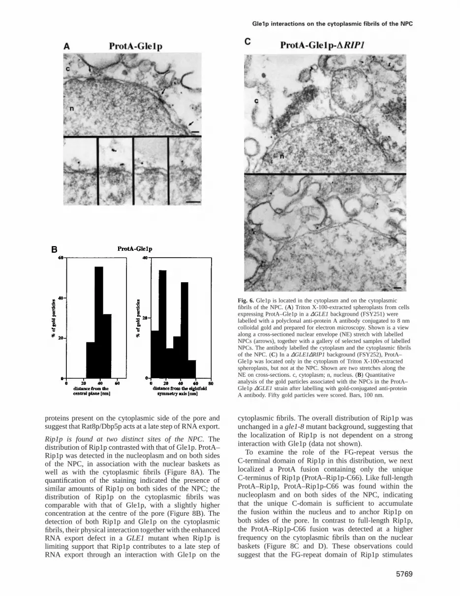
Localization of Gle1p, Rip1p and Rat8p/Dbp5p byimmunoelectron microscopyAlthough Gle1p, Rip1p and Rat8p are clearly involved inRNA export, their exact contribution to this process isunknown. Defining the precise localization of these pro-teins within the NPC three-dimensional structure may helpto assign them a role in specific steps of export. For thispurpose, the location of Gle1p, Rip1p and Rat8p wasdetermined at the ultrastructural level by performingpre-embedding labelling immunoelectron microscopy, aprocedure which yields structurally intact yeast NPCs(Fahrenkroget al., 1998). The proteins of interest weretagged with protein A and expressed in strains disruptedfor the wild-type gene. The tagged proteins were detectedwith a colloidal gold-conjugated anti-protein A antibody.The ProtA–Gle1p and ProtA–Rat8p fusions are functionalas they complement the corresponding lethal gene disrup-tion (Murphy and Wente, 1996; Tsenget al., 1998). Twoconstructs that encoded protein A fused to full-lengthRip1p (ProtA–Rip1p) or to the C-terminal 66 amino acidsof Rip1p (ProtA–Rip1p-C66) were used to localize Rip1p.

Y.Strahm et al.
Fig. 4. Gle1p interacts with Rip1p, hCG1, Dbp5p/Rat8p and Gfd1pin vitro. GST alone or GST fusions containing the FG-repeat orC-terminal domains of Rip1p, the C-domain of hCG1, as well asfull-length Gfd1p or Rat8p/Dbp5p were produced inE.coli andpurified on glutathione beads. GST fusions on beads were tested forinteraction within vitro translated and [35S]methionine-labelled full-length Gle1p, Gle1p C-terminal region (amino acids 257–538) orluciferase. After binding and washing, beads boiled in sample bufferwere analysed by SDS–PAGE and bound35S-labelled proteinsrevealed by autoradiography (three top panels, lanes 2–7). Input(lane 1) corresponds to the amount of [35S]protein added to eachbinding reaction. The amounts of GST fusions in each reaction werecompared by Coomassie staining of a gel before autoradiography(lower panel; each GST fusion is indicated by a dot). BSA was usedas non-specific competitor in the binding reactions and co-migrates onthese gels.
Fig. 5. Limiting levels of Rip1p enhance the poly(A)1 RNA export defect of aGLE1 mutant strain. Strains FSY58 (gle1-1, rip1::KANR/pFS398) andFSY57 (gle1-1, rip1::KANR/pFS724) expressing normal (wild-type) and low levels of Rip1p, respectively, were grown to mid-log phase at 25°C andeither kept at 25°C or subjected to a 30 min incubation at 37°C. Cells were processed forin situ hybridization as described in Figure 3D.(a, c, e andg) The distribution of poly(A)1 RNA; (b, d, f andh) DAPI staining of the same cells, respectively.
5768
These ProtA fusions are functional as well, since theyefficiently rescued phenotypes associated with theRIP1deletion (Figure 2).
Gle1p is associated with NPC cytoplasmic fibrils. Con-sistent with earlier immunofluorescence studies (Del Prioreet al., 1996; Murphy and Wente, 1996), Gle1p was detectedin the cytoplasm but a significant fraction was found inassociation with NPCs. Interestingly, all the gold particleslocalized on the cytoplasmic side of the NPC, ~40–50 nmfrom the central plane, indicating an association with thecytoplasmic fibrils. Quantitative analysis of the labellingwith respect to the centre of the 8-fold symmetry axisshowed that Gle1p distributes over the whole diameter ofthe pore (Figure 6A and B). Given the physical interactionbetween Gle1p and Rip1p, we examined ProtA–Gle1pdistribution in a ∆GLE1/∆RIP1 double deletion back-ground (Figure 6C). Surprisingly, the ProtA–Gle1p stain-ing was mostly lost from the NPCs, and gold particleswere only found in the cytoplasm. Rip1p being inessential,it is unlikely to represent a major binding site for Gle1pat the NPC. One explanation for the drastic effect of theRIP1 deletion on ProtA–Gle1p localization is that thisfusion is not fully functional and is more sensitive to theabsence of Rip1p; indeed, a strain expressing ProtA–Gle1p grows normally in the presence of Rip1p but istemperature sensitive whenRIP1 is deleted (data notshown). The data suggest that Rip1p contributes to theassociation of Gle1p with the NPC.
ProtA–Rat8p is cytoplasmic and associated with the cyto-plasmic fibrils. Indirect immunofluorescence experimentshave localized Rat8p/Dbp5p in the cytoplasm and inassociation with NPCs (Snay-Hodgeet al., 1998; Tsenget al., 1998). By immunogold localization, the ProtA–Rat8p fusion was detected in the cytoplasm and on thecytoplasmic fibrils, with an apparent exclusion from thenuclear compartment (Figure 7A and B). These data areconsistent with an association of Rat8p/Dbp5p with NPC
Gle1p interactions on the cytoplasmic fibrils of the NPC
Fig. 6. Gle1p is located in the cytoplasm and on the cytoplasmicfibrils of the NPC. (A) Triton X-100-extracted spheroplasts from cellsexpressing ProtA–Gle1p in a∆GLE1 background (FSY251) werelabelled with a polyclonal anti-protein A antibody conjugated to 8 nmcolloidal gold and prepared for electron microscopy. Shown is a viewalong a cross-sectioned nuclear envelope (NE) stretch with labelledNPCs (arrows), together with a gallery of selected samples of labelledNPCs. The antibody labelled the cytoplasm and the cytoplasmic fibrilsof the NPC. (C) In a ∆GLE1∆RIP1 background (FSY252), ProtA–Gle1p was located only in the cytoplasm of Triton X-100-extractedspheroplasts, but not at the NPC. Shown are two stretches along theNE on cross-sections. c, cytoplasm; n, nucleus. (B) Quantitativeanalysis of the gold particles associated with the NPCs in the ProtA–Gle1p∆GLE1 strain after labelling with gold-conjugated anti-proteinA antibody. Fifty gold particles were scored. Bars, 100 nm.
proteins present on the cytoplasmic side of the pore andsuggest that Rat8p/Dbp5p acts at a late step of RNA export.
Rip1p is found at two distinct sites of the NPC. Thedistribution of Rip1p contrasted with that of Gle1p. ProtA–Rip1p was detected in the nucleoplasm and on both sidesof the NPC, in association with the nuclear baskets aswell as with the cytoplasmic fibrils (Figure 8A). Thequantification of the staining indicated the presence ofsimilar amounts of Rip1p on both sides of the NPC; thedistribution of Rip1p on the cytoplasmic fibrils wascomparable with that of Gle1p, with a slightly higherconcentration at the centre of the pore (Figure 8B). Thedetection of both Rip1p and Gle1p on the cytoplasmicfibrils, their physical interaction together with the enhancedRNA export defect in aGLE1 mutant when Rip1p islimiting support that Rip1p contributes to a late step ofRNA export through an interaction with Gle1p on the
5769
cytoplasmic fibrils. The overall distribution of Rip1p wasunchanged in agle1-8mutant background, suggesting thatthe localization of Rip1p is not dependent on a stronginteraction with Gle1p (data not shown).
To examine the role of the FG-repeat versus theC-terminal domain of Rip1p in this distribution, we nextlocalized a ProtA fusion containing only the uniqueC-terminus of Rip1p (ProtA–Rip1p-C66). Like full-lengthProtA–Rip1p, ProtA–Rip1p-C66 was found within thenucleoplasm and on both sides of the NPC, indicatingthat the unique C-domain is sufficient to accumulatethe fusion within the nucleus and to anchor Rip1p onboth sides of the pore. In contrast to full-length Rip1p,the ProtA–Rip1p-C66 fusion was detected at a higherfrequency on the cytoplasmic fibrils than on the nuclearbaskets (Figure 8C and D). These observations couldsuggest that the FG-repeat domain of Rip1p stimulates

Y.Strahm et al.
Fig. 7. Rat8p/Dbp5p is located in the cytoplasm and on the cytoplasmic fibrils of the NPC. (A) Triton X-100-extracted spheroplasts from cellsexpressing ProtA–Rat8p/Dbp5p in a∆RAT8/DBP5background (FSY342) were prepared as described in Figure 6. Shown is a view of a cross-sectioned NE stretch with labelled NPCs (arrows) and a gallery of selected gold-labelled NPCs. c, cytoplasm; n, nucleus. (B) Distribution of the goldparticles associated with the NPCs. Fifty gold particles were scored. Bars, 100 nm.
the recruitment of Rip1p to the nuclear baskets. Altern-atively, if Rip1p was trafficking between the two sides ofthe pore, the FG-repeats could increase the recycling ofRip1p from the cytoplasmic to the nuclear side of theNPC. Finally, given the essential role of Rip1p under heatshock conditions, we examined the localization of ProtA–Rip1p in a ∆RIP1 background after a 10 min shift to42°C. The overall distribution of Rip1p at the NPCs wasnot substantially affected under these stress conditions(data not shown).
Since through pre-embedding labelling, some cyto-plasmic epitopes can be lost (the cells are treated withTriton X-100 to facilitate labelling on both sides of theNPC), we cannot exclude that a small fraction of Rip1pis also present in the cytoplasm.
In vivo localization of Gle1p–GFP andRat8p/Dbp5p–GFP in mutant backgroundsImmunoelectron microscopy localization of ProtA–Gle1pshowed that Gle1p was lost from the NPC in anRIP1deletion strain, suggesting a contribution of Rip1p to theassociation of Gle1p with the NPC (Figure 6C). Tosubstantiate this observation, we examined the distributionof a Gle1p–GFP fusion in living cells in a∆GLE1 strainor a ∆GLE1∆RIP1 double deletion strain (Figure 9A).Similarly to the ProtA–Gle1p fusion, the Gle1p–GPFfusion conferred a ts phenotype in the absence of Rip1p,indicating that Gle1p–GFP was not fully functional (datanot shown). Consistent with the immunoelectron micro-scopy data, the amount of Gle1p–GFP detected in associ-ation with the nuclear envelope after growing the cells at37°C for 4 h was considerably lower in the absence ofRip1p. The disappearance of Gle1p–GFP from the NPCdid not result from increased turnover or decreased syn-thesis, since the steady-state levels of Gle1p–GFP in the∆RIP1 strain were stable at 37°C over at least 6 h (datanot shown). The localization of Gle1p appeared to beaffected more drastically by the absence of Rip1p whenexamined by immunoelectron microscopy. This difference
5770
may be due to the fact that the electron microscopy dataare from the quantitation of gold particles in cell sections,while the intensity of GFP fluorescence is derived fromwhole cells.
An in vivo interaction between Gle1p and Rat8p/Dbp5pwas suggested by the high copy suppression and proteininteraction assays. To determine whether Gle1p contributesto the association of Rat8p/Dbp5p with the NPC, a Rat8p/Dbp5p–GFP fusion was localized in a strain containingthe rss1-37 (gle1)mutant allele (Figure 9B). Therss1-37mutation induces a strong poly(A)1 RNA export defectand abolishes the two-hybrid interaction between Gle1pand Rat8p/Dbp5p (Figure 3D and Table I). Two strainswere examined, both of which contained therss1-37alleleand expressed a Rat8p/Dbp5p–GFP fusion. In one case,the RAT8/DBP5 gene was tagged with GFP on thechromosome (rss1-37 RAT8/DBP5-GFP); in the other,genomicRAT8/DBP5was deleted and Rat8p/Dbp5p–GFPexpressed from a plasmid (rss1-37∆RAT8/DBP5pRAT8/DBP5-GFP; Figure 9B, right). Both strains behaved sim-ilarly. At 25°C, Rat8p/Dbp5p–GFP was detected in thecytoplasm as well as in association with the NPC, resultingin a strong nuclear rim staining as described (Snay-Hodgeet al., 1998). After 2 h at 37°C, the GFP staining at therim became weaker and more diffuse but did not disappear(even after prolonged exposure to 37°C); there was aconcomitant modest increase in the cytoplasmic signal.These data are consistent with the notion that Gle1prepresents a binding site for Rat8p/Dbp5p at the nuclearpore, but that other interactions are involved in therecruitment of this DEAD-box protein to the NPC.
Discussion
In this study, we used a high copy suppressor approachto identify proteins functionally or physically related toGle1p. The screen identified the NPC-associated nucleo-porin Rip1p, the DEAD-box protein Rat8p/Dbp5p and anew non-essential protein Ymr255p. This protein was

Gle1p interactions on the cytoplasmic fibrils of the NPC
Fig. 8. Rip1p resides in the nucleus and on both the cytoplasmic and the nuclear face of the NPC. (A) Cells expressing ProtA–Rip1p in a∆RIP1background (FSY221) were prepared as described in Figure 6. The anti-protein A antibody conjugated to 8 nm colloidal gold was found in thenucleus (overview, top), and in association with the cytoplasmic fibrils (middle) and the nuclear basket (bottom) of the NPC. This location of Rip1premained the same in the ProtA–Rip1p-C66∆RIP1 strain (FSY 329) (C). Arrows point to labelled NPCs in the overviews. c, cytoplasm; n, nucleus.The quantitative analysis of the gold particles associated with the NPCs (B andD) revealed an equal distribution on both sides of the NPC in theProtA–Rip1p∆RIP1 cells, whereas in the ProtA–Rip1p-C66∆RIP1 cells the cytoplasmic epitope was labelled significantly more frequently than thenuclear epitope of the NPC. Fifty gold particles were scored. Bars, 100 nm.
identified independently as a high copy suppressor ofRAT8/DBP5(Hodgeet al., 1999) and named Gfd1p. Ourtwo-hybrid andin vitro binding assays indicate that theessential C-terminal domain of Gle1p interacts directlywith Rip1p, Rat8p/Dbp5p and Gfd1p (Table I andFigure 4). These data together with the synthetic lethalinteractions between Gle1p, Rip1p and Rat8p/Dbp5p(Stutzet al., 1997; Snay-Hodgeet al., 1998) are consistentwith a functional association of these proteinsin vivo.
5771
Gle1p initially was proposed to contain a Rev-like NESin its essential C-terminal domain and to promote poly(A)1
RNA export through an NES-dependent export pathwayinvolving an interaction with Rip1p, analogous to thatdescribed between Rev-NES and the FG-repeat domainof Rip1p (Murphy and Wente, 1996). However, the NESis not conserved in the human homologue of Gle1p(Watkinset al., 1998), and the localization of Gle1p–GFPat the nuclear rim was not affected in thexpo1-1(crm1)

Y.Strahm et al.
Fig. 9. (A) The localization of Gle1p–GFP at the NPC is weakened inthe absence of Rip1p.∆GLE1 (FSY297) or∆GLE1-∆RIP1 (FSY298)strains expressing Gle1p–GFP from a centromeric plasmid (pFS1030)were grown at 25°C in selective medium to mid-log phase, shifted to37°C for 4 h and examined under the microscope. (B) Rat8p/Dbp5p–GFP is partially lost from the NPC in therss1-37 (gle1)mutantbackground. Rat8p/Dbp5p–GFP was localized in two strainscontaining therss1-37allele. The strain on the left (rss1-37 RAT8/DBP5-GFP; FSY400) contains aRAT8/DBP5gene chromosomallytagged with GFP. The strain on the right (rss1-37∆RAT8/DBP5pRAT8/DBP5-GFP; FSY401) contains aRAT8/DBP5deletion coveredby a plasmid (pCS835) expressing a Rat8p/Dbp5p–GFP fusion. Thestrains were grown in SD complete medium to mid-log phase at 25°C.They were then kept at 25°C or shifted to 37°C for 2 h and examinedunder the microscope. Two fields are shown for each strain at 25 and37°C.
ts strain (Stadeet al., 1997). The physical interactionbetween Gle1p and Rip1p further shows that Gle1p doesnot behave like a Rev-NES-containing protein. Indeed,the Gle1p essential C-domain interacts directly with theunique C-domain of Rip1p rather than its FG-repeat region(Table I and Figure 4); the association between these twoproteins is therefore clearly distinct from that betweenRev-NES and the Rip1p FG-repeats, which is bridged bythe NES export factor Crm1p (Nevilleet al., 1997);consistently, neither we nor others were able to detect aninteraction between Gle1p and Crm1p (Watkinset al.,1998) (Table I).
Although rapid movement of Gle1p in and out ofthe nucleus cannot be excluded, the immunoelectronmicroscopic detection of Gle1p uniquely in the cytoplasmand on the cytoplasmic fibrils further supports that Gle1pis not a shuttling protein; however its association with thepore may be dynamic (Figure 6). Like Gle1p, Rat8p/Dbp5p was localized in the cytoplasm and in associationwith the cytoplasmic fibrils by immunoelectron micro-scopy (Figure 7; Schmittet al., 1999). Gfd1p was located
5772
in the cytoplasm and at the nuclear rim by indirectimmunofluorescence, a distribution similar to that ofRat8p/Dbp5p (Hodgeet al., 1999). A substantial fractionof Rip1p was also found in association with the cyto-plasmic fibrils (Figure 8). These distributions, togetherwith the genetic and physical interaction data, are con-sistent with the view that Gle1p, Rip1p, Rat8p/Dbp5p andGfd1p functionally interact on the cytoplasmic face ofthe NPC and participate in one or more late steps ofRNA export.
These observations raise the question of whether Gle1pinteracts with its high copy suppressors simultaneously toform a single complex or whether some of these inter-actions are mutually exclusive. In this latter case, Gle1pmay combine with its partners to form different sub-complexes with redundant or complementary functions.Alternatively, the C-terminal domain of Gle1p mayundergo sequential interactions with Rip1p, Rat8p/Dbp5pand Gfd1p, which may be part of a series of eventscontributing to the release of an RNP from the NPC.
Based on the localization and interaction data, thesynthetic lethality between aRIP1 deletion andGLE1mutations is probably due to the absence of theRIP1C-terminal domain from the cytoplasmic fibrils. Thisis because synthetic lethality is rescued by the Rip1pC-terminus alone, and overexpression of this domain isable to suppress a temperature-sensitiveGLE1 mutation(Figures 2 and 3). A contribution of Rip1p to Gle1pfunction is also supported by the enhanced RNA exportdefect in aGLE1 mutant strain when Rip1p becomeslimiting (Figure 5). One interpretation of these observ-ations is that the binding of the C-domain of Rip1p toGle1p stimulates Gle1p activity, perhaps by optimizingthe association of Gle1p with some of its partners (e.g.Rat8p/Dbp5p or Gfd1p). This function of Rip1p becomesessential in aGLE1 mutant background or under stressconditions, where the overall efficiency of RNA exportis reduced.
Two biochemically defined NPC sub-complexes withprimary roles in poly(A)1 RNA export have beendescribed. One is composed of the essential nucleoporinsNup159p/Nup82p/Nsp1p (Belgarehet al., 1998; Hurwitzet al., 1998) and the other consists of Nup84p/Nup85p/Nup120p/C–Nup145/Seh1p/Sec13p (Siniossoglouet al.,1996; Teixeira et al., 1997). Members of both sub-complexes have been localized on the NPC cytoplasmicfibrils (Kraemer et al., 1995; Fahrenkroget al., 1998;Hurwitz et al., 1998; Katahiraet al., 1999; Stoffleret al.,1999). It is noteworthy that these distributions may notall be evolutionarily conserved, as components of ahuman NPC sub-complex, homologous to the Nup84psub-complex, have been located at the nuclear basket(Fontoura et al., 1999). The exact function of thesecomplexes in pore structure versus export function is stillpoorly defined. More specific roles have been attributedto individual components. Nup82p, for example, anchorsthe corresponding complex within the NPC (Belgarehet al., 1998), whereas Nup159p or components of theNup84p complex provide binding sites for shuttling pro-teins involved in more dynamic aspects of export (Santos-Rosaet al., 1998; Schmittet al., 1999; and see below).
No physical connections have yet been establishedbetween the Nup84p and Nup159p sub-complexes, but

Gle1p interactions on the cytoplasmic fibrils of the NPC
multiple genetic interactions functionally relate these twosets of proteins (reviewed in Fabre and Hurt, 1997).Similarly, multiple genetic interactions link Gle1p and itspartners to the two defined sub-complexes (Del Prioreet al., 1996; Stutzet al., 1997; Hurwitzet al., 1998; Snay-Hodge et al., 1998), suggesting that a number of theseNPC components have overlapping or redundant functions.Alternatively, these proteins may all belong to a singlemultifunctional complex participating in several sequentialsteps of RNA export.
The identification of the DEAD-box protein Rat8p/Dbp5p as a partner for Gle1p and the substantial decreaseof Rat8p/Dbp5p–GFP rim staining in therss1-37(gle1)temperature-sensitive mutant background suggest thatGle1p participates in the binding of Rat8p/Dbp5p to thenuclear pore (Figure 9B). However, the partial retentionof Rat8p/Dbp5p–GFP at the NPC in the mutant backgroundat 37°C is consistent with the existence of alternativebinding sites. Indeed, a direct interaction between Rat8p/Dbp5p and the N-terminal domain of Rat7p/Nup159p hasbeen identified (Hodgeet al., 1999; Schmittet al., 1999).These observations could explain the suppression of therat7-1/nup159-1temperature-sensitive mutation by highlevel expression of Gle1p (Del Prioreet al., 1996), i.e. anexcess of Gle1p may provide additional binding sitesfor Rat8p/Dbp5p when the mutant rat7-1p/nup159-1p isdegraded, and thereby partially restore poly(A)1 RNAexport.
The ProtA–Gle1p and Gle1p–GFP fusions are substan-tially lost from the NPC in the absence of Rip1p(Figures 6C and 9A); these strains are also temperaturesensitive for growth indicating that Rip1p contributes toa functionally relevant association of Gle1p with the NPC.As Rip1p is inessential, it is unlikely to represent a majorbinding site for Gle1p. Consistently, such a role for Rip1pis revealed only in the presence of the ProtA–Gle1p andGle1p–GFP fusions, which are not fully functional proteins(see Results). Rip1p may therefore strengthen the associ-ation of Gle1p with the nuclear pore, presumably bystabilizing Gle1p within a complex. The major anchoringsite for Gle1p at the pore is not known, but it may involvethe essential coiled-coil domain of Gle1p, by analogy toother NPC proteins (Belgarehet al., 1998).
The immunoelectron microscopic localizations detectedRip1p within the nucleoplasm and the NPC nuclear basketsand cytoplasmic fibrils, a distribution distinct from thatobserved for Gle1p and Rat8p/Dbp5p. The presence ofRip1p on both sides of the NPC suggests that this FG-nucleoporin may have a role in late as well as in earlysteps of export, i.e. the recruitment of export complexesfrom the nucleoplasm towards the baskets. Rip1p maythen move from one side to the other, perhaps in associationwith cargoes, and participate in their release on thecytoplasmic side of the NPC. The accumulation of Rip1pat two distinct NPC sites may reflect rate-limiting stepsduring that process. The vertebrate NPC-associated FG-nucleoporins Nup98 and Nup153 recently have beenproposed to move between the nuclear and cytoplasmiccompartments in association with exported cargoes(Nakielny et al., 1999; Zolotukhin and Felber, 1999)
An association of Rip1p with RNP cargo was suggestedby the identification of an interaction between the humanRNA export factor TAP and the FG-nucleoporins CAN/
5773
Nup214 and hCG1, the functional homologues ofNup159p and Rip1p, respectively (Katahiraet al., 1999;E.Izaurralde, personal communication; and Figure 2).These data support the view that TAP and its yeasthomologue Mex67p promote the export of bound RNPsthrough an interaction with one or more FG-nucleoporins(de Castillia and Rout, 1999). Consistent with the TAP–hCG1 interaction, we observed a homologous two-hybridinteraction between Mex67p and the FG-repeat domainof Rip1p (D.Zenklusen, unpublished results).
The association of Mex67p with the NPC is essentialfor RNA export, and this interaction is mediated byMtr2p. Nup85p, a component of the Nup84p sub-complex,represents one target of the Mex67p/Mtr2p complex atthe NPC (Santos-Rosaet al., 1998). Interestingly, weidentified severalNUP85 alleles in the∆RIP1 syntheticlethal screen that were only rescued by full-length Rip1p(our unpublished data). These genetic observationstogether with the Mex67p/Rip1p–FG two-hybrid inter-action suggest that the FG-repeat domain of Rip1p maycomplement Nup85p by contributing to the association ofMex67p with the pore. As the immunoelectron microscopicdistributions of Rip1p and Mex67p are comparable(Santos-Rosaet al., 1998; and Figure 8), Rip1p couldassociate with Mex67p within the nucleus and travelalong with the RNP complex through the NPC. On thecytoplasmic side, Rip1p is likely to interact with Gle1pthrough its C-terminus; this interaction may in turn stimu-late the association of Gle1p with the DEAD-box proteinRat8p/Dbp5p. ATP binding and hydrolysis may driveconformational rearrangements within associated RNPs,which ultimately will result in the release of the RNPfrom the NPC and/or the recycling of RNP componentstowards the nucleus. As the RNA-unwinding activity ofRat8p/Dbp5pin vitro is dependent on a co-factor (Tsenget al., 1998), Gle1p and/or its associated proteins couldbe candidates for such a function.
In summary, our data support the notion that Gle1p actsin a terminal step of RNA export through an interactionwith multiple partners. Further studies will address themolecular details of these interactions and possibly revealaspects of the dynamic rearrangements underlying export.The ability of hCG1 to rescue aRIP1deletion is consistentwith the functional conservation of Rip1p during evolution.Our data support that hCG1 represents the true homologueof Rip1p, which is distinct from the earlier describedhRIP/RAB1 protein (Bogerdet al., 1995; Fritz et al.,1995); the extent of conservation between all of thepartners of hCG1 in yeast and in vertebrate systems willbe investigated in the future.
Materials and methods
The DNA manipulations were performed according to standard methods.Yeast media and yeast transformations were performed with establishedprocedures (Ausubelet al., 1994). The strains and plasmids used in thisstudy are listed in Tables II and III.
Yeast plasmid constructionsProtA–RIP1 (pFS829) was obtained by introducing aSalI site after theATG of RIP1 in pFS398 to generate pFS800. A 400 bpSalI PCR productcontaining two IgG-binding domains from protein A was introducedinto the SalI site of pFS800 to generate pFS829. ProtA–RIP1-C66(pFS923) was generated by deleting the FG-repeat domain in pFS829;briefly, a 1.6 kbHindIII–XhoI PCR fragment extending from the 59

Y.Strahm et al.
Table II. Yeast strains used in this study
Name Description Genotype Source
W303 a, ade2, his3, leu2, trp1, ura3FSY17 W303∆RIP1 a, ade2, his3, leu2,trp1,ura3,rip1::KANR Stutzet al. (1997)FSY38 gle1-8SL α, ade2,ade3,leu2,ura3,his3,∆RIP1, gle1-8(pFS652, URA3/ADE3/CEN) this study; Stutzet al. (1997)FSY57 gle1-1Rip1p low α, ade2,ade3,leu2,ura3,his3,∆RIP1, gle1-1(pFS724, LEU2/CEN) Stutzet al. (1997)FSY58 gle1-1Rip1p high α, ade2,ade3,leu2,ura3,his3,∆RIP1, gle1-1(pFS398, LEU2/CEN) Stutzet al. (1997)FSY216 gle1-8 a, ade2,his3,leu2,trp1,ura3, gle1-8 this studyFSY221 ProtA–RIP1 a, ade2, his3, leu2,trp1,ura3, rip1::KANR (pFS829, LEU2/CEN) this studyFSY329 ProtA–RIP1-C66 a, ade2, his3, leu2,trp1,ura3,rip1::KANR (pFS923, LEU2/CEN) this studyFSY195 GLE1 shuffle a, ade2, his3, leu2,trp1,ura3, gle1::HIS3(pSW410, URA3/CEN) this study, Murphyet al.(1996)FSY201 GLE1 shuffle a, ade2, his3, leu2,trp1,ura3, gle1::HIS3, rip1::KANR (pSW410, URA3/CEN) this study, Murphyet al. (1996)FSY292 rss1-37 (gle1) a, ade2, his3, leu2,trp1,ura3, gle1::HIS3(pVDP29, LEU/CEN) this study, Saavedraet al. (1997)FSY251 ProtA–GLE1 a, ade2, his3, leu2,trp1,ura3, gle1::HIS3(pSW464, TRP1/CEN) this study, Murphyet al. (1996)FSY252 ProtA–GLE1∆RIP1 a, ade2, his3, leu2,trp1,ura3, gle1::HIS3, rip1::KANR(pSW464, TRP1/CEN) this study, Murphyet al. (1996)FSY297 GLE1–GFP a, ade2, his3, leu2,trp1,ura3, gle1::HIS3(pFS1030, LEU2/CEN) this studyFSY298 GLE1–GFP∆RIP1 a, ade2, his3, leu2,trp1,ura3, gle1::HIS3, rip1::KANR(pFS1030, LEU2/CEN) this studyCSY512 RAT8/DBP5 shuffle α, trp1∆63,leu2∆1,ura3-52, his3∆200,rat8::HIS3 (pRAT8/DBP5, URA3/CEN) Snay-Hodgeet al. (1998)CSY835 RAT8-GFP a, trp1∆63,leu2∆1,ura3-52, his3∆200, rat8::HIS3 (pCS835, LEU2/CEN) Snay-Hodgeet al. (1998)FSY342 ProtA–RAT8/DBP5 α, trp1∆63,leu2∆1,ura3-52, his3∆200, rat8::HIS3 (pCA5032, LEU2/CEN) this study, Tsenget al. (1998),
Snay-Hodgeet al. (1998)FSY398 W303GLE1–GFP a, ade2, his3, leu2, trp1, ura3, GLE1-GFP-KANR this studyFSY399 gle1-8-GFP a, ade2, his3, leu2, trp1, ura3, gle1-8-GFP-KANR this studyCH1462 α, ade2,ade3,leu2,ura3,his3 Holm (1993), Stutzet al. (1997)FSY249 rss1-37 (gle1) α, ade2,ade3,leu2,ura3,his3, rss1-37 this studyFSY400 rss1-37 RAT8-GFP α, ade2,ade3,leu2,ura3,his3, rss1-37, RAT8/DBP5-GFP this studyFSY401 rss1-37pRAT8-GFP ?, ade2,ade3,leu2,ura3,his3, rss1-37, rat8::HIS3 (pCS835, LEU2/CEN) this study, Snay-Hodgeet al. (1998)
Table III. Plasmids used in this studya
Name Description Source
pFS652 RIP1 HindIII genomic 3.5 kb fragment intoSalI of pCH1122 (URA3/CEN) withSalI Stutz et al. (1997)linkers
p366 YCP50-based vector in which URA3 was replaced with LEU2 Liao (1993)pFS398 RIP1 RIP1 HindIII genomic 3.5 kb fragment inserted into theHindIII site of p366 Stutzet al.(1997)pFS724 RIP1 low RIP1 HindIII 1.9 kb PCR fragment inserted into theHindIII site of p366; contains Stutzet al. (1997)
0.38 kbRIP1 59-flanking sequencespFS800 pFS398 with engineeredSalI site after the ATG this studypFS829 ProtA–RIP1 pFS800 with two IgG-binding domains (protein A) in the 59 SalI site this studypFS923 ProtA–RIP1-C66 pFS829 with a deletion of the FG-domain (codons 2–364) this studypSW464 ProtA–GLE1 GLE1 with five IgG-binding domains inserted at the 59 end (TRP1/CEN) Murphyet al. (1996)pCA5032 ProtA–RAT8 DBP5/RAT8with ProtA tag at the 39 end in pRS315 (LEU2/CEN) Tsenget al. (1998)pFS952 YMR255/GFD1cloned in theEagI site of YEp24 (URA3/2µ) C.Cole laboratory
(Dartmouth, VT)pFS997 RIP1 HindIII genomic 3.5 kb fragment cloned in YEPLac112 (TRP1/2µ) this studypFS998 GLE1 cloned as aSalI fragment from pFS802 into YEPLac112 this studypFS999 RAT8cloned as aSphI-BamHI fragment into YEPLac112 this studypYGEX2T Galactose-inducible GST fusion yeast expression vector (URA3/2µ) P.Silver laboratorypFS961 yGST–RIP1-C66 RIP1 C-terminal codons 365–430 cloned as aBamHI–HindIII fragment into pYGEX2Tthis studypFS802 GLE1 gene cloned as aSalI fragment into YCPLac111 (LEU2/CEN) this studypFS1029 GLE1 39NotI pFS802 with engineeredNotI site upstream ofGLE1 stop codon this studypFS1030 GLE1-GFP GLE1 SalI fragment with GFP inserted into the engineeredNotI site of pFS1029 this studypCS835 RAT8-GFP RAT8-GFPfusion cloned in YCPlac111 (LEU2/CEN) Snay-Hodgeet al. (1998)pVDP29 prss1-37 (gle1) rss1-37temperature-sensitiveGLE1 allele in YCP111 (LEU2/CEN) Saavedraet al. (1997)pFS410 GST–RIP1-FG RIP1 codons 121–230 cloned as anEcoRI–XhoI PCR fragment into pGex4T-1 this studypFS507 GST–RIP1-C66 RIP1 codons 364–430 cloned as anEcoRI–XhoI PCR fragment into pGex4T-1 this studypFS883 GST–hCG1-C43 hCG1codons 380–423 cloned as anEcoRI–XhoI PCR fragment into pGex4T-1 this studypFS955 GST–GFD1 GFD1 coding region clones as anEcoRI–XhoI PCR fragment into pGex4T-1 this studypFS956 GST–RAT8/DBP5RAT8/DBP5coding region cloned as anEcoRI fragment into pGex4T-1 this study
aSee Materials and methods for two-hybrid constructs.
HindIII site to the 39 end of the two protein A moieties of pFS829 wasligated to a 1.5 kbXhoI–HindIII PCR fragment containing the last 66codons ofRIP1 and 39 sequences down to the naturalHindIII site. Thetwo PCR fragments were ligated through theirXhoI ends and the productcloned back into theHindIII site of p366 (LEU2/CEN). ProtA–GLE1(pSW464) and ProtA–RAT8 (pCA5032) have been described. TheyGST–RIP1-C66 construct was obtained by cloning a 1.5 kbSalI–HindIII PCR fragment comprising the last 66 codons ofRIP1 and 39
5774
sequences down to the naturalHindIII site in-frame with the GSTsequence of pYGEX2T (URA3/2µ) to generate pFS961.
The pGLE1-GFP plasmid (LEU2/CEN) was obtained by cloning theGLE1gene as aSalI PCR fragment with 500 bp upstream and downstreamsequences into YCPLac111 (LEU2/CEN) to generate pFS802. PCR wasused to introduce aNotI site upstream of theGLE1 stop codon inpFS802 to generate pFS1029. GFP (S65T) was cloned as aNotI PCRfragment into pFS1029 to generate pFS1030.

Gle1p interactions on the cytoplasmic fibrils of the NPC
The high copy plasmid pFS997 was obtained by cloning the genomic3.5 kb RIP1 HindIII fragment into YEpLac112 (TRP1/2µ). pFS998was obtained by cloning theGLE1 SalI fragment from pFS802 intoYEpLac112. pFS999 was obtained by cloning theRAT8/DBP5gene asan SphI–BamHI fragment from pCS830 into the corresponding sites ofYEPLac112. PFS952 was made by cloning the coding region ofYMR255w/GFD1with 300 bp upstream and downstream sequences intovector YEp24 (URA3/2µ) as anEagI PCR fragment (Cole Laboratory,Dartmouth, VT).
Two-hybrid LexA bait and prey cloning, strains andinteraction assaysAll baits were expressed as LexA fusions by cloning into vector pEG202(HIS3/2µ); the prey constructs were obtained by cloning into pJG4-5(TRP1/2µ) (Ausubel et al., 1994). TheGLE1 bait or prey constructswere obtained by inserting aBamHI–SalI or an EcoRI–SalI PCRfragment corresponding to codons 257–538 ofGLE1 into pEG202 cutwith BamHI–XhoI or pJG4-5 cut withEcoRI–XhoI to generate pFS795and pFS935, respectively. The RIP1-C66 bait and prey constructs weremade by cloning a 500 bpEcoRI–SalI PCR fragment corresponding tocodons 365–430 and following 39 sequences ofRIP1 into pEG202 orpJG4-5 cut withEcoRI–XhoI to generate pFS1031 and pFS1032. TheRIP1-C38 bait and prey constructs were made by ligating a shorterEcoRI–SalI fragment containing the last 38 codons ofRIP1 into pEG202or pJG4-5 digested withEcoRI–XhoI to generate pFS1033 and pFS1034.The hCG1-C43 bait and prey constructs were obtained by inserting a255 bpEcoRI–XhoI PCR fragment containing the last 43 codons followedby 39 sequences into pEG202 and pJG4-5 cut withEcoRI–XhoI togenerate pFS1035 and pFS1036. The hCG1 and hCG1-FG prey constructswere made by cloningEcoRI–XhoI PCR fragments of the whole codingregion and codons 1–380, respectively, into pJG4-5 cut withEcoRI–XhoI to generate pFS1037 and pFS1038. The RIP1-FG bait constructpF5476 contains codons 121–130 ofRip1 as anEcoRI–XhoI fragmentin pEG202. The YMR255/GFD1 prey construct pFS1039 contains thewhole coding region as anEcoRI–XhoI PCR fragment in pJG4-5EcoRI–XhoI. The DBP5/RAT8 prey plasmid pFS1040 contains the whole codingregion as anEcoRI PCR fragment in pGJ4-5EcoRI. The CRM1 andRev bait as well as the Rev and RIP1-FG prey constructs have beendescribed (Stutzet al., 1995; Nevilleet al., 1997).
Strain EGY48 (α, trp1 ura3 leu2::plexop6-LEU2) containing the LacZreporter pSH18-34 on a URA3/2µ plasmid was transformed with theprey constructs. Strain RFY206 (a,his3 leu2 ura3 trp1 lys2) andcontaining pSH18-34 was transformed with the bait constructs. Two-hybrid interactions were examined by mating followed by replica-platingof the diploids on X-Gal indicator plates containing galactose as describedearlier (Stutzet al., 1995).
In vivo protein labelingYeast cultures grown at 25°C or shifted to 42°C were labelled with[35S]methionine as described (Stutzet al., 1997). Total cell lysates werefractionated on a 10% SDS–polyacrylamide gel. Gels were dried andautoradiographed.
High copy suppressor screenThe gle1-8 mutant allele had been identified earlier in a screen formutants synthetically lethal with aRIP1deletion. The mutant allele wasamplified by PCR (High Fidelity Expand PCR, Boehringer MA) fromgenomic DNA as aSalI fragment containing 500 bp on both sides ofthe coding region and cloned into YCPLac111 (LEU2/CEN) to generatepFS824. pFS824 was confirmed to contain thegle1-8 mutation(s) bytesting its synthetic lethality with aRIP1 deletion. The mutations in thegle1-8allele were identified by sequencing both strands of overlappingPCR fragments spanning the whole wild-type or mutant gene. Com-parison of the two sequences identified three mutations, two of whichinduced amino acid changes (T21A and E340K).
To construct the starting strain for the high copy suppressor screen,the mutantgle1-8allele was integrated into the genome of a W303 wild-type strain at theGLE1 locus by the pop-in/out replacement method togenerate FSY216. Briefly, theSalI insert of pFS824 was subcloned intotheXhoI site of the yeast integrating plasmid pRS406 (URA3; Stratagene)to generate pFS845. pFS845 was linearized withXhoI and transformedinto W303. URA1 colonies were collected, grown overnight in YEPDmedium and plated on 5-FOA. 5-FOA1 colonies were replica-plated toYEPD containing 7.5 mg/l of the vital dye erythrosin B (Sigma) andincubated at 37°C. The strains with thegle1-8 ts allele were identifiedbased on slow growth and purple colour.
The gle1-8 high copy suppressors were identified by transforming
5775
strain FSY216 with a yeast genomic library cloned in YEp13 (LEU2/2µ). A total of 5 3 104 transformants were grown at 25°C on SD-Leuplates, collected and replated on SD-Leu plates at 37°C. Library plasmidswere rescued from colonies growing at 37°C and retransformed intoFSY216 to verify the suppressor phenotype. The rescuing plasmids weresequenced from both sides of the insert. Individual ORFs were subclonedinto YEpLac112 vector (TRP1/2µ) as described above.
Growth curves were established by diluting exponentially growingcultures to an OD600 5 0.05 (~53 105 cells/ml). The diluted cultureswere incubated for 2 h at 25°C and then shifted to 37°C. Growth rateswere followed by measuring OD600 at various time intervals over 24 h.
Western blot analysisProtein extracts and Western blots were performed as described (Stutzet al., 1997). The anti-Gle1p and anti-Rna15p antibodies were used at1:2000 and 1:15 000 dilutions, respectively.
In vitro translation[35S]Methionine-labelled full-length Gle1p and Gle1p C-terminus wereobtained by coupled T7 transcription–translation in reticulocyte lysates(TnT kit, Promega). T7 templates were generated by PCR. The templatefor full-length Gle1p was obtained by amplifying wild-typeGLE1 withthe 59 primer OFS296 59-GGGCGAAATTAATACGACTCACTAT-AGGGACACCATGAGATTTGTGTTCGATGAGGTTTT-39, containingthe T7 RNA polymerase promoter and sequences complementary to the59 end of the GLE1 coding region, and the 39 primer OFS13859-CGTATTTTCTGCCATCCCTTGATATCGAGCCG-39 complement-ary to a region ofGLE1 downstream of the stop codon. The templatefor the Gle1p C-terminus was generated with the 59 primer OFS38559-GGGCGAAATTAATACGACTCACTATAGGGACACCATGGACA-AAATTGCTCAAATAAAGC-3 9, containing the T7 promoter andsequences complementary toGLE1 starting from codon 257, and withthe OFS138 39 primer. A 2 µg aliquot of each T7 PCR template wasused per 25µl transcription–translation reaction.
GST fusions and in vitro binding assayThe constructs used to produce GST fusions inE.coli were obtained bycloning PCR fragments into vector pGex-4T-1 (Pharmacia Biotech).GST–RIP1-FG and GST–RIP1-C66 were made by insertingEcoRI–XhoI fragments, corresponding to codons 121–230 and 364–430 ofRIP1,respectively, into pGex-4T-1EcoRI–XhoI to generate pFS410 andpFS507. GST–hCG1-C43 was obtained by inserting a 255 bpEcoRI–XhoIPCR fragment corresponding to the last 43 codons and 39-untranslatedsequences of hCG1 into pGex-4T-1EcoRI–XhoI to generate pFS883.The complete coding sequences ofDBP5/RAT8and YMR255w/GFD1were amplified asEcoRI and EcoRI–XhoI fragments, respectively, andcloned into pGex4T-1 cut with the corresponding enzymes to generatepFS955 and pFS956.
The GST fusion constructs were transformed intoE.coli strainBL21(DE3) and fusion protein synthesis was induced overnight at 16°Cin the presence of 0.5 mM isopropyl-β-D-thiogalactopyranoside (IPTG).After cell lysis, GST fusions were affinity purified on glutathione–agarose beads (Pharmacia) as described by the manufacturer.In vitrobinding reactions contained 10µg of GST fusion protein immobilizedon 25 µl of packed glutathione–agarose beads and 1/10 of anin vitrotranscription–translation reaction (2.5µl) in 100 µl of universal bindingbuffer [20 mM HEPES pH 7, 10% glycerol, 0.1% bovine serumalbumin (BSA), 0.1% Tween, 100 mM KOAc, 2 mM MgOAc, 5mMβ-mercaptoethanol and 1 tablet/50 ml of protease inhibitors (Boehringer)].Binding reactions were incubated for 1 h at 4°C on a turning wheel andwashed three times with 500µl of binding buffer. The beads wereresuspended in 20µl of 23 sample buffer, boiled and 1/4 of the bindingreaction (10 µl) was fractionated on 10% polyacrylamide gels. Toevaluate binding efficiency, 1/4 of thein vitro translated protein input(0.6 µl) was loaded in parallel. After Coomassie Blue staining, gelswere dried and autoradiographed.
In situ hybridizationStrain FSY292 (rss1-37) was obtained by transforming theGLE1shuffle strain FSY197 with pVDP29 (prss1-37,LEU2/CEN) followedby selection on 5-FOA. Strains FSY57 (gle1-1, Rip1p low) and FSY58(gle1-1, Rip1p normal) have been described (Stutzet al., 1997). Yeaststrains were grown to OD600 5 0.5 at 25°C. Half of each culture wasshifted to 37°C by the addition of 1 vol. of medium pre-heated to 49°Cand incubated for 30 min at 37°C. Cells were fixed and processed forin situ hybridization with digoxigenin-labelled oligo(dT) probes onmulti-well slides as described (Nevilleet al., 1997). Pictures were taken

Y.Strahm et al.
on a Zeiss axioplan 2 fluorescence microscope equipped with a cooledCCD camera and 1003 and 633 objective lenses. Identical exposuretimes were used for comparable images, and composites were preparedusing Adobe Photoshop.
Immunoelectron microscopy localizationsThe ProtA–Gle1p strains FSY251 and FSY252 were obtained bytransforming pSW464 (Murphy and Wente, 1996) into the GLE1 shufflestrains FSY195 or FSY201 followed by selection on 5-FOA. The ProtA–Rat8p/Dbp5p strain FSY342 was obtained by transforming pCA5032into the RAT8/DBP5 shuffle strain CSY512 followed by selection on 5-FOA. The ProtA–Rip1p strains FSY221 and FSY329 were obtained bytransforming pFS829 and pFS923 into FSY17 (W303∆RIP1).
The localization of the ProtA–fusions was determined by using a pre-embedding labelling protocol essentially as described (Fahrenkroget al.,1998). Briefly, the cells were spheroplasted, washed and treated withTriton X-100. Triton-extracted cells were washed, resuspended in 100µlof anti-protein A antibody labelled with 8 nm colloidal gold andincubated at 30°C. The cells were then washed, fixed, dehydrated, Eponembedded and prepared for electron microscopy as described. The pre-embedding labelling conditions were adapted for each strain analysed.ProtA–Rip1p in∆RIP1: 15 min zymolyase digest, 0.05% Triton X-100extraction, 2.5 h antibody labelling; ProtA–Rip1p-C66 in∆RIP1: 15 minzymolyase digest, 0.025% Triton X-100 extraction, 2.5 h labelling;ProtA–Rip1p in∆RIP1 at42°C: 10 min heat shock, 10 min zymolyasedigest, 0.025% Triton X-100, 1 h labelling. ProtA–Gle1p in∆GLE1 or∆GLE1∆RIP1 background: 20 min zymolyase digest, 0.05% TritonX-100, 2.5 h labelling; ProtA–Rat8p in∆RAT8 background: 15 minzymolyase, 0.025% Triton, 2.5 h labelling.
In vivo localizationsStrains FSY297 (∆GLE1 pGLE1-GFP) and FSY298 (∆GLE1∆RIP1pGLE1-GFP) were obtained by transforming pFS1030 into theGLE1shuffle strains FSY195 (gle1::HIS3, pGLE1 URA/CEN) or FSY201(gle1::HIS3, rip1::KANR pGLE1 URA/CEN), followed by selection on5-FOA. Cells were transformed with a pRSADE2 plasmid to minimizevacuolar fluorescence. Thegle1-8 allele in FSY216 and the wild-typeGLE1 gene in W303 were chromosomally tagged with GFP using thepFA6a-GFP(S65T)-KanMX6 module as described (Longtineet al., 1998)to generate FSY399 and FSY398, respectively. These strains weretransformed with high copy plasmids and pRSADE2. The temperature-sensitiverss1-37 (gle1)strain FSY249 was obtained by replacing wild-typeGLE1by therss1-37mutant allele (Saavedraet al., 1997) in strainCH1462 (Holm, 1993); the strategy was the same as the one used togenerate FSY216 (see above). Therss1-37 allele of strain FSY249was tagged with GFP using the pFA6a-GFP(S65T)-kanMX6 module(Longtine et al., 1998) to generate FSY400; FSY249 was also crossedwith strain CSY835 (rat8/dbp5::HIS3, pCS835) (Snay-Hodgeet al.,1998) to generate the haploid strain FSY401, arss1-37 rat8/dbp5::HIS3double mutant strain expressing Rat8p/Dbp5p–GFP from plasmidpCS835 (LEU2/CEN). All the strains were grown in selective or SDcomplete medium to early logarithmic phase, shifted for various timesto 37°C, spun for a short time and examined directly under a Zeissaxioplan 2 fluorescence microscope equipped with a 1003 objectivelens and a cooled CCD camera (Kappa). Pictures were taken as describedfor in situ hybridizations.
Acknowledgements
We are grateful to Chuck Cole for valuable discussions and for sharingdata prior to publication. We thank Chuck Cole, Tien-Hsien Chang,Pamela Silver and Susan Wente for plasmids and yeast strains, LauraDavis and Lionel Minvielle-Sebastia for antibodies, and Lut Van Laerand the UK HGMP resource centre for the hCG1 cDNA. We also thankElisa Izaurralde and Hildur V.Colot for critical reading of the manuscript.We acknowledge Verena Mu¨ller for technical support, and members ofthe Microbiology Institute for their help. B.F. was supported by theM.E.Mueller Foundation of Switzerland and by an HFSP grant awardedto Ueli Aebi. These studies were supported by a research grant(No. 049135.96/1) from the Swiss National Science Foundation to F.S.
References
Ausubel,F.M., Brent,R., Kingston,R.E., Moore,D.D., Seidman,J.G.,Smith,J.A. and Struhl,K. (1994)Current Protocols in MolecularBiology. J.Wiley & Sons and Greene Publishing Associates.
5776
Belgareh,N., Snay-Hodge,C., Pasteau,F., Dagher,S., Cole,C.N. andDoye,V. (1998) Functional characterization of a Nup159p-containingnuclear pore subcomplex.Mol. Biol. Cell, 9, 3475–3492.
Bogerd,H.P., Fridell,R.A., Madore,S. and Cullen,B.R. (1995)Identification of a novel cellular cofactor for the Rev/Rex class ofretroviral regulatory proteins.Cell, 82, 485–494.
Dahlberg,J.E. and Lund,E. (1998) Functions of the GTPase Ran in RNAexport from the nucleus.Curr. Opin. Cell Biol., 10, 400–408.
de Castillia,C.S. and Rout,M.P. (1999) TAPping into transport.NatureCell Biol., 1, 31–33.
Del Priore,V., Snay,C.A., Bahr,A. and Cole,C.N. (1996) The product ofthe Saccharomyces cerevisiae RSS1gene, identified as a high-copysuppressor of therat7-1 temperature-sensitive allele of theRAT7/NUP159 nucleoporin, is required for efficient mRNA export.Mol.Biol. Cell, 7, 1601–1621.
Del Priore,V., Heath,C.V., Snay,C.A., MacMillan,A., Gorsch,L.C.,Dagher,S. and Cole,C.N. (1997) A structure/function analysis ofRat7p/Nup159p, an essential nucleoporin ofSaccharomyces cerevisiae.J. Cell Sci., 110, 2897–2999.
Doye,V. and Hurt,E. (1997) From nucleoporins to nuclear porecomplexes.Curr. Opin. Cell Biol., 9, 401–411.
Fabre,E. and Hurt,E. (1997) Yeast genetics to dissect the nuclear porecomplex and nucleocytoplasmic trafficking.Annu. Rev. Genet., 31,277–313.
Fahrenkrog,B., Hurt,E.C., Aebi,U. and Pante´,N. (1998) Moleculararchitecture of the yeast nuclear pore complex: localization of Nsp1psubcomplexes.J. Cell Biol., 143, 577–588.
Farjot,G., Sergeant,A. and Mikae´lian,I. (1999) A new nucleoporin-likeprotein interacts with both HIV-1 Rev nuclear export signal andCRM-1. J. Biol. Chem., 274, 17309–17317.
Floer,M. and Blobel,G. (1999) Putative reaction intermediates in Crm1-mediated nuclear protein export.J. Biol. Chem., 274, 16279–16286.
Fontoura,B.M.A., Blobel,G. and Matunis,M.J. (1999) A conservedbiogenesis pathway for nucleoporins: proteolytic processing of a 186-kilodalton precursor generates Nup98 and the novel nucleoporin,Nup96.J. Cell Biol., 144, 1097–1112.
Fritz,C.C., Zapp,M.L. and Green,M.R. (1995) A human nucleoporin-like protein that specifically interacts with HIV Rev.Nature, 376,530–533.
Gorlich,D. (1998) Transport into and out of the cell nucleus.EMBO J.,17, 2721–2727.
Gruter,P., Tabernero,C., von Kobbe,C., Schmitt,C., Saavedra,C.,Bachi,A., Wilm,M., Felber,B.K. and Izaurralde,E. (1998) TAP, thehuman homolog of Mex67p, mediates CTE-dependent RNA exportfrom the nucleus.Mol. Cell, 1, 649–659.
Hodge,C.A., Colot,H.V., Stafford,P. and Cole,C.N. (1999) Rat8p/Dbp5is a shuttling transport factor that interacts with Rat7p/Nup159p andGle1p and suppresses the mRNA export defect ofxpo1-1cells.EMBOJ., 18, 5778–5788.
Holm,C. (1993) A functional approach to identifying yeast homologs ofgenes from other species.Methods, 5, 102–109.
Hurwitz,M.E., Strambio-de-Castillia,C. and Blobel,G. (1998) Two yeastnuclear pore complex proteins involved in mRNA export form acytoplasmically oriented subcomplex.Proc. Natl Acad. Sci. USA, 95,11241–11245.
Izaurralde,E. and Adam,S. (1998) Transport of macromolecules betweenthe nucleus and the cytoplasm.RNA, 4, 351–364.
Izaurralde,E., Kutay,U., von Kobbe,C., Mattaj,I.W. and Gorlich,D. (1997)The asymmetric distribution of the constituents of the Ran system isessential for transport into and out of the nucleus.EMBO J., 16,6535–6547.
Katahira,J., Stra¨sser,K., Podtelejnikov,A., Mann,M., Jung,J.U. andHurt,E. (1999) The Mex67p-mediated nuclear mRNA export pathwayis conserved from yeast to human.EMBO J., 18, 2593–2609.
Kraemer,D.M., Strambio-de-Castillia,C., Blobel,G. and Rout,M.P. (1995)The essential yeast nucleoporin NUP159 is located on the cytoplasmicside of the nuclear pore complex and serves in karyopherin-mediatedbinding of transport substrate.J. Biol. Chem., 270, 19017–19021.
Liao,X.C., Tang,J. and Rosbash,M. (1993) An enhancer screen identifiesa gene that encodes the yeast U1 snRNP A protein: implications forsnRNP protein function in pre-mRNA splicing.Genes Dev., 7,419–428.
Longtine,M.S., McKenzie,A., Demarini,D.J., Shah,N.G., Wach,A.,Brachat,A., Philippsen,P. and Pringle,J.R. (1998) Additional modulesfor versatile and economical PCR-based gene deletion and modificationin Saccharomyces cerevisiae. Yeast, 14, 953–961.

Gle1p interactions on the cytoplasmic fibrils of the NPC
Mattaj,I.W. and Englmeier,L. (1998) Nucleocytoplasmic transport: thesoluble phase.Annu. Rev. Biochem., 67, 265–306.
Murphy,R. and Wente,S.R. (1996) An RNA-export mediator with anessential nuclear export signal.Nature, 383, 357–360.
Nakielny,S., Fischer,U., Michael,W.M. and Dreyfuss,G. (1997) RNAtransport.Annu. Rev. Neurosci., 20, 269–301.
Nakielny,S., Shaikh,S., Burke,B. and Dreyfuss,G. (1999) Nup153 is anM9-containing mobile nucleoporin with a novel Ran-binding domain.EMBO J., 18, 1982–1995.
Neville,M. and Rosbash,M. (1999) The NES–Crm1p export pathway isnot a major mRNA export route inSaccharomyces cerevisiae.EMBO J., 18, 3746–3756.
Neville,M., Stutz,F., Lee,L., Davis,L.I. and Rosbash,M. (1997) Evidencethat the importin-beta family member Crm1p bridges the interactionbetween Rev and the nuclear pore complex during nuclear export inS.cerevisiae. Curr. Biol., 7, 767–775.
Noble,S.M. and Guthrie,C. (1996) Identification of novel genes requiredfor yeast pre-mRNA splicing by means of cold-sensitive mutations.Genetics, 143, 67–80.
Pollard,V.W. and Malim,M.H. (1998) The HIV-1 Rev protein.Annu.Rev. Microbiol., 52, 491–532.
Saavedra,C.A., Hammell,C.M., Heath,C.V. and Cole,C.N. (1997) Exportof heat shock mRNAs following stress inSaccharomyces cerevisiaeemploys a distinct pathway defined by Rip1p and also requires asubset of factors essential for export of poly(A)1 mRNA. Genes Dev.,11, 2845–2856.
Santos-Rosa,H., Moreno,H., Simos,G., Segref,A., Fahrenkrog,B.,Pante,N. and Hurt,E. (1998) Nuclear mRNA export requires complexformation between Mex67p and Mtr2p at the nuclear pores.Mol. Cell.Biol., 18, 6826–6838.
Schmitt,C.et al. (1999) Dbp5p, a DEAD box-protein required for mRNAexport, is recruited to the cytolasmic fibrils of nuclear pore complexvia a conserved interaction with CAN/Nup159p.EMBO J., 18,4332–4347.
Siniossoglou,S., Wimmer,C., Rieger,M., Doye,V., Tekotte,H., Weise,C.,Emig,S., Segref,A. and Hurt,E.C. (1996) A novel complex ofnucleoporins, which includes Sec13p and a Sec13p homolog, isessential for normal nuclear pores.Cell, 84, 265–275.
Snay-Hodge,C.A., Colot,H.V., Goldstein,A.L. and Cole,C.N. (1998)Dbp5p/Rat8p is a yeast nuclear pore-associated DEAD-box proteinessential for RNA export.EMBO J., 17, 2663–2676.
Stade,K., Ford,C.S., Guthrie,C. and Weis,K. (1997) Exportin 1 (Crm1p)is an essential nuclear export factor.Cell, 90, 1041–1050.
Stoffler,D., Fahrenkrog,B. and Aebi,U. (1999) The nuclear pore complex:from molecular architecture to functional dynamics.Curr. Opin. CellBiol., 11, 391–401.
Stutz,F. and Rosbash,M. (1998) Nuclear RNA export.Genes Dev., 12,3303–3319.
Stutz,F., Neville,M. and Rosbash,M. (1995) Identification of a novelnuclear pore-associated protein as a functional target of the HIV-1Rev protein in yeast.Cell, 82, 495–506.
Stutz,F., Kantor,J., Zhang,D., McCarthy,T., Neville,M. and Rosbash,M.(1997) The yeast nucleoporin Rip1p contributes to multiple exportpathways with no essential role for its FG-repeat region.Genes Dev.,11, 2857–2868.
Teixeira,M.T., Siniossoglou,S., Podtelejnikov,S., Be´nichou,J.C.,Mann,M., Dujon,B., Hurt,E. and Fabre,E. (1997) Two functionallydistinct domains generated byin vivo cleavage of Nup145p: a novelbiogenesis pathway for nucleoporins.EMBO J., 16, 5086–5097.
Tseng,S.I., Weaver,P.L., Liu,Y., Hitomi,M., Tartakoff,A.M. andChang,T.H. (1998) Dbp5p, a cytosolic RNA helicase, is required forpoly(A)1 RNA export.EMBO J., 17, 2651–2662.
Van Laer,L. et al. (1997) Refined mapping of a gene for autosomaldominant progressive sensorineural hearing loss (DFNA5) to a 2-cMregion, and exclusion of a candidate gene that is expressed in thecochlea.Eur. J. Hum. Genet., 5, 397–405.
Watkins,J.L., Murphy,R., Emtage,J.L. and Wente,S.R. (1998) The humanhomologue of Saccharomyces cerevisiaeGle1p is required forpoly(A)1 RNA export.Proc. Natl Acad. Sci. USA, 95, 6779–6784.
Zolotukhin,A.S. and Felber,B.K. (1999) Nucleoporins Nup98 andNup214 participate in nuclear export of human immunodeficiencyvirus type 1 Rev.J. Virol., 73, 120–127.
Received June 21, 1999; revised and accepted August 31, 1999
5777


