Embed Size (px)
Citation preview
The metallurgy of Roman medical instruments
Katherine E. Jakielskia,*, Michael R. Notisb
aCenter for Materials Research in Archaeology and Ethnology, Massachusetts Institute of Technology,
Cambridge, MA 02139, USAbDepartment of Materials Science, Lehigh University, Bethlehem, PA 18015, USA
Received 20 March 2000; accepted 31 May 2000
Abstract
A metallographic study was conducted to characterize the composition and manufacturing techniques of two
Roman medical instruments. The instruments, one, the typical form of an ear speculum or `̀ scoop'', and the other,
a spatula, are part of a set of nine Roman medical instruments. The exact provenance of the instruments is unclear,
but they are stylistically similar to Roman medical instruments dating from the 2nd to 4th century AD. The present
results are compared with the findings from a previous examination of one Roman medical olivary probe. This
analysis illustrates the variety of manufacturing techniques that ancient Roman metallurgists implemented in
medical instrument fabrication. D 2001 Elsevier Science Inc. All rights reserved.
Keywords: Archaeometallurgy; Ancient medicine; Metallurgy; Roman; Medical instruments
I would prefer a knowledgeable surgeon with a rusty
knife to a charlatan with fancy equipment.
Ð Lucian, 2nd century AD [1]
1. Introduction
Harnessing the forces of nature and surmounting
their formidable powers to prevent sickness and
death has preoccupied humankind across the world
and throughout the ages. To the modern reader,
descriptions of early Roman medical techniques, as
outlined in the works of Celsus and Galen, appear
inhumane and can make even the hardiest reader
queasy. However, primitive as they may appear, it is
the successes and failures of ancient and historic
medical scientific endeavors that have fused into the
state of modern medicine.
Although the medical sciences have advanced far
beyond the imagination of ancient practitioners (and
will continue to advance beyond anything that we
can presently imagine), healing still contains an
element of mysticism. Throughout the development
of more `̀ advanced'' societies, medicine appears to
move from a healing system that is based completely
on the supernatural rule of the gods to a system that
places more power into the hands of human sur-
geons. The line between religious leader and healer
was blurred and as medical science advanced, doc-
tors and surgeons were imbued with the divine right
to heal.
1044-5803/00/$ ± see front matter D 2001 Elsevier Science Inc. All rights reserved.
PII: S1 0 4 4 - 5 8 0 3 ( 0 0 ) 0 0 0 7 8 - 4
* Corresponding author. 1230 Royal Street #12, New
Orleans, LA 70116, USA. Tel.: +1-504-581-3209; fax: +1-
504-257-1482.
E-mail address: [email protected] (K.E. Jakielski).
Materials Characterization 45 (2000) 379± 389
Like a religious leader, a medical practitioner
would need sufficient tools and symbols to be estab-
lished as a healer. An effective surgical instrument
would have to incorporate functional design and
aesthetic appeal, as well as establish a ritualistic
connection. Functional aspects, such as strength to
prevent breakage during use, were manipulated by
choosing associated metallurgical manufacturing tech-
niques to control the mechanical properties. Surgical
instruments could be connected to ritualistic objects
based on alloy selection. The aesthetic appeal of a
surgeon's medical tool kit was an important means of
earning a patient's trust [2]. An impressive medical
tool kit composed of intricately manufactured, eye-
catching, shiny metal objects could assuage the fears
of new patients. Alloy selection, design, and manip-
ulation of mechanical properties by associated metal-
lurgical technologies would be expected to vary by
region and culture. Though we can never fully know
the motivations or nuances of the everyday life of past
cultures, the scientific materials analysis of objects can
give us some insight into the technological and social
organization employed. Materials analysis of ancient
objects can answer such questions as the way in which
materials were manipulated in terms of form and
function, and why some instruments were manufac-
tured in certain styles or metals and others were not.
When coupled with historical medical documents,
materials studies of surgical instruments can give us
a glimpse into the way in which our ancestors viewed
human life, the risks that they were willing to take, and
the integration of separate technologies (i.e., metal-
lurgy and surgery) to, in essence, save human life.
This paper examines one small block of time and
space in this evolution in surgical history: the Roman
period in the 1st to 4th century AD. The Roman time
is of particular interest because it was in that time that
their folk-based medicine developed into a more
scientific method based on principles adopted from
the Greeks [1,3,4]. In addition, their successful mili-
tary campaigns and widespread imperialism gave
them the power to disseminate their surgical techni-
ques and medical principles throughout the Western
world. This paper presents the results of a metallo-
graphic and microchemical analysis of three Roman
medical instruments. It is intended to provide insight
on the metallurgical technologies used and basic
principles of material selection and design implemen-
ted by Roman metallurgists in the manufacture of
Roman medical instruments.
2. Background
Previous studies of Roman medical instruments
have focused primarily on constructing typologies
and little scientific materials analysis has been
done [5±8]. A notable exception is the investiga-
tion of a set of 40 Roman medical instruments
from the British Museum that incorporated macro-
scopic visual examination, X-ray radiography,
XRF, and XRD analyses [9]. Though X-ray radio-
graphy and visual examination provide a general
idea of the method of manufacture on a macro-
scopic level, metallographic investigations can pro-
vide a much deeper level of understanding of the
metallurgical processes employed. Only one metal-
lographic study on Roman medical instruments has
been published to our knowledge [10]. There is a
cursory overview of another metallographic inves-
tigation, but no formal results from this study have
been published [11]. Metallographic studies, when
coupled with scanning electron microscopy (SEM)
and electron probe microanalysis (EPMA) can give
a thorough picture of the metallurgical history of
an object.
2.1. Roman medicine
Early Roman medicine relied heavily on herbal
folk remedies, magical incantations, and the will of
the gods. The plague of Rome in 295 BC forced
the Romans to enlist the aid of the more scienti-
fically based Greek medicine [1,3,4]. It was neither
an easy nor rapid amalgamation of Greek surgical
techniques with the Roman medical repertoire. The
Roman citizenry was reluctant to place their lives
in the hands of another person instead of an
omnipotent god (let alone a person brandishing
knives and probes in a time of no anesthesia!).
In addition, the general populace of Rome was
highly opposed to human dissection, which re-
sulted in a poor knowledge of human anatomy.
The general mistrust of the public coupled with an
inadequate anatomical knowledge base made it
imperative for an ancient surgeon to fully charac-
terize each patient's physical ailment before opera-
tion. A misdiagnosed condition or failed operation
could not only ruin the surgeon's reputation, but
also the trust of the public in the medical field as
a whole [1]. It follows then that a wide range of
probes, used to fully characterize the wound before
operation, were an essential part of the Roman
surgeon's tool kit.
Roman medicine and surgical instruments have
their origin by transmission from the Greeks, but, as
the Roman Empire grew, this body of knowledge and
technology increased tremendously [3,5,10]. The
dissemination and evolution of Roman medical tech-
niques and theories throughout the Roman Empire
can be attributed largely to the medical staff within
the Roman army [1,12]. Roman medicine had a
K.E. Jakielski, M.R. Notis / Materials Characterization 45 (2000) 379±389380
symbiotic relationship with the folk medicine prac-
ticed in the conquered territories; Roman doctors
could amass new medical knowledge from interaction
with local healers, concurrently, the local healers
gained knowledge of Roman techniques [1]. It is
likely that this exchange of information included
the introduction of Roman medical instruments and
the associated metallurgical technology used to man-
ufacture them.
2.2. The Roman surgical toolkit
Probes, spatulas, scoops, forceps, and scalpels
comprise the major typological groups found in the
basic Roman medical toolkit [5,8,9,13]. Most Roman
medical instruments were double-ended and multi-
functional; most scoops and spatulas served also as
probes. This multi-functionality extended beyond
basic instrument design to the decorative inlays and
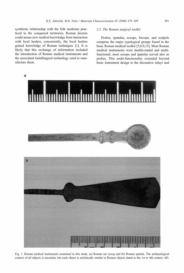
Fig. 1. Roman medical instruments examined in this study. (a) Roman ear scoop and (b) Roman spatula. The archaeological
context of all objects is uncertain, but each object is stylistically similar to Roman objects dated to the 1st to 4th century AD.
K.E. Jakielski, M.R. Notis / Materials Characterization 45 (2000) 379±389 381

ridges, which served the dual purpose of aesthetic
enhancement and the ergonomic role of ensuring a
firmer grip.
Ancient medical instruments functioned in ways
that are similar to the ways in which we employ their
modern counterparts. Spatulas were mainly used for
applying medicaments and ointments, and blunt dis-
section [5,9]. Probes were used to apply medicines
and characterize wounds to determine where to oper-
ate [14]. Ear scoops were used to dislodge wax or
foreign objects from the ear. They were also used to
apply liquid medicines to the ear by wrapping a piece
of wool around the handle of the scoop, dipping it in
the medicine, and letting the medicine drip down the
pointed end of the handle into the ear. Other sug-
gested uses of the ear scoop include curetting small
areas, pouring liquid medicine into the eyes, and
measuring medicines [5,9].
The materials used to manufacture Roman medi-
cal instruments included copper, bronze, brass, silver,
and gold. Roman smiths incorporated multiple types
of metals into a single instrument design such as the
typical scalpel, which consisted of an iron blade
inserted into a bronze handle. The majority of the
Roman medical instrument finds are composed of
bronze; Hippocrates prescribed the use of bronze for
manufacturing medical instruments in the 4th century
BC and the frequency of bronze instrument finds may
suggest adherence to this doctrine [4,10,12]. It was
essential that the instruments did not break during use
and therefore necessitated an understanding of mate-
rial properties and a high quality manufacturing
process. Functionality would be a key component in
the materials selection and design of medical instru-
ments, but not the only consideration. The metallur-
gist had to imbue the object with a sense of healing
power beyond the human realm. A religious connec-
tion could be established by incorporating elements
of ritual object design into the medical instruments.
For example, the alloy selection of bronze for medical
instruments suggested by Hippocrates may be due to
the long association of the alloy with healing cults
[10]. In a sense, these metal objects transformed the
ancient medical practitioner from an average human
being to a divine artist manipulating the most delicate
material of all Ð human life.
2.3. Roman surgical instrument finds
The largest documented finds of Roman medical
instruments are those found at the sites of Pompeii
and Herculaneum [5,8]. Additional Roman medical
instrument finds have been discovered in military
hospitals and grave sites throughout Britain [15,16].
Regrettably, a large majority of the Roman medical
surgical instrument finds are of unknown prove-
nance [6,7,9,17]. The most recent finding of surgi-
cal instruments and perhaps the most significant
were discovered in 1996 in Colchester, England in
a Doctor's grave at the Stanway site. The Stanway
set dates to the AD 50s and is the oldest known set
of medical instruments from Britain and throughout
the world [18,19].
2.4. The Lehigh University collection
There are a total of 15 Roman medical implements
currently held in the Department of Materials Science
and Engineering at Lehigh University. One (an oli-
vary probe) was purchased in 1995 from an antiqui-
ties dealer and of unknown provenance. Five
(spatulas, probes, and scoops) were purchased in
1997 from a private collection and reportedly found
in Egypt. The set examined here contains nine
instruments. Four of these instruments are scoop or
spoon type instruments and the other five are spatu-
las. This group was purchased in 1998 and reportedly
found along the Thames as part of a large group of
similar probes, scoops, and spatulas.
One scoop and one spatula from this collection
were chosen for metallographic investigations. Fig.
1 shows the objects chosen for investigation before
sectioning. Fig. 2 shows the other seven instruments
in this group not chosen for analysis. There is no
documented archaeological excavation of such a
hoard along the Thames. However, the instruments
in our collection are stylistically similar to Roman
instrument finds dated from the 1st to 4th century
AD [5,8]. In addition, during the metallographic
analyses, which will be discussed below, the depth
Fig. 2. Roman medical instruments in the Lehigh University
collection not examined in this study. This set was reported
to be found in hoard of medical instruments along the
Thames, 1st to 4th century AD.
K.E. Jakielski, M.R. Notis / Materials Characterization 45 (2000) 379±389382
of penetration of surface corrosion into the object's
base material could only be achieved after a sig-
nificant time of burial. Both of these factors, com-
bined with the alloy composition found by EPMA,
solidify the claim that these objects are from Roman
times. The results obtained for these two instruments
were then compared with a metallographic investi-
gation conducted on a Roman olivary probe by the
authors in 1996 [10].
3. Lab methodology
The objects were sectioned using a low-speed
diamond saw. Each sample was then mounted in an
epoxide resin, ground, and polished using silicon
carbide papers and then alumina powder to a 0.05-
mm finish. The as-polished microstructure was ob-
served using the light optical microscope. The
samples were then etched in a ferric nitrate solution
for times ranging from 2 to 5 s; photomicrographs
were taken in both the as-polished and etched state
using a Leitz metallograph or a Wild A420 Mak-
roskop low magnification metallograph. EPMA was
conducted using a JEOL 733 SuperProbe at the
Massachusetts Institute of Technology's Geology
Laboratory for the scoop and spatula instruments
reported here. EPMA was also conducted for the
previously reported olivary probe using a JEOL 733
SuperProbe at the Department of Materials Science
and Engineering at Lehigh University.
4. Results
4.1. Ear scoop
Macroscopic investigation revealed that the ear
scoop was manufactured as a single solid piece. It is
103 mm long with a 10-mm-diameter bowl (scoop)
contiguous with a cylindrical shaft that served as a
handle. The cross-sectional diameter of the handle is
3 mm and relatively constant throughout the entire
length. Two irregularly shaped 2-mm-wide decorative
bands separate the bowl from the handle. The corro-
sion patina on the handle was a relatively uniform
dark green/black color. The corrosion patina on the
inside of the bowl had patches of light brown and
white-colored corrosion on the dark green/black pa-
tina. Once sectioned, the scoop base metal appeared
to be a light gold/brassy color. The characteristic as-
polished microstructure is shown in Fig. 3. EPMA
results are shown in Table 1. The edge corrosion
forms a layer of relatively uniform thickness around
the entire surface area of the sample. There is no
preferred path of penetration of the edge corrosion
into the sample.
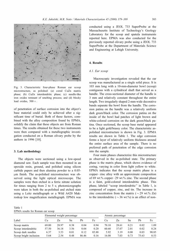
Four main phases characterize this microstructure
as observed in the as-polished state. The primary
phase is the matrix phase, which shows evidence of
coring, varying in color from light yellow to white.
EPMA indicates that the scoop matrix phase is a
copper±zinc alloy with an approximate composition
of 69 wt.% copper±29 wt.% zinc. The second phase
is a faint, gold-colored interdendritic phase. This
phase, labeled `̀ scoop interdendritic'' in Table 1, is
composed of copper, zinc, and tin. The increase in
zinc concentration from the matrix (� 29 wt.% Zn)
to the interdendritic (� 36 wt.%) is an effect of non-
Fig. 3. Characteristic four-phase Roman ear scoop
microstructure, as polished: (a) cored CuZn matrix
phase; (b) CuZn interdendritic phase; (c) needle-like
iron oxides remnant of smelting process; and (d) blocky
lead oxides, 500� .
Table 1
EPMA results for Roman ear scoop
Elemental weight percentage Atomic percentage
Label Cu Zn Sn Pb Fe Cu Zn Sn Pb Fe
Scoop matrix 69.00 28.97 0.38 0.04 0.94 70.10 28.61 0.21 0.02 1.09
Scoop interdendritic 57.50 36.18 3.56 0.04 0.20 60.60 37.07 2.01 0.02 0.24
Scoop dark needles 6.37 3.53 0.01 0.12 85.88 5.92 3.19 0.00 0.03 90.85
Scoop bright inclusion 5.64 2.54 0.48 86.46 0.16 16.08 7.03 0.73 75.63 0.53
K.E. Jakielski, M.R. Notis / Materials Characterization 45 (2000) 379±389 383
equilibrium cooling and the resultant solidification
phenomenon known as constitutional undercooling
[20]. The third phase consists of numerous tiny
blue-gray-colored dendrite-shaped inclusions (den-
drite arm length sizes range from less than 5±20
mm long), randomly distributed throughout the pri-
mary matrix phase. EPMA results (labeled `̀ scoop
dark needles'') show that this is an iron oxide and
could be a remnant of the original ores or slag from
the smelting process. The fourth phase is also blue-
gray in color but morphologically different from the
third phase. This phase consists of blocky and
globular inclusions (up to 5 mm in size) distributed
randomly throughout the matrix. These inclusions
appear very bright in the backscattered electron
(BSE) image and EPMA shows them to be lead
oxides (labeled `̀ scoop bright inclusion''). It should
be noted that the small amounts of copper and zinc
found in the EPMA readings for the third and fourth
phases could be attributed to signal overlap with the
matrix phase due to the small size of the inclusions
relative to the electron beam. Trace amounts of other
elements were found in each phase.
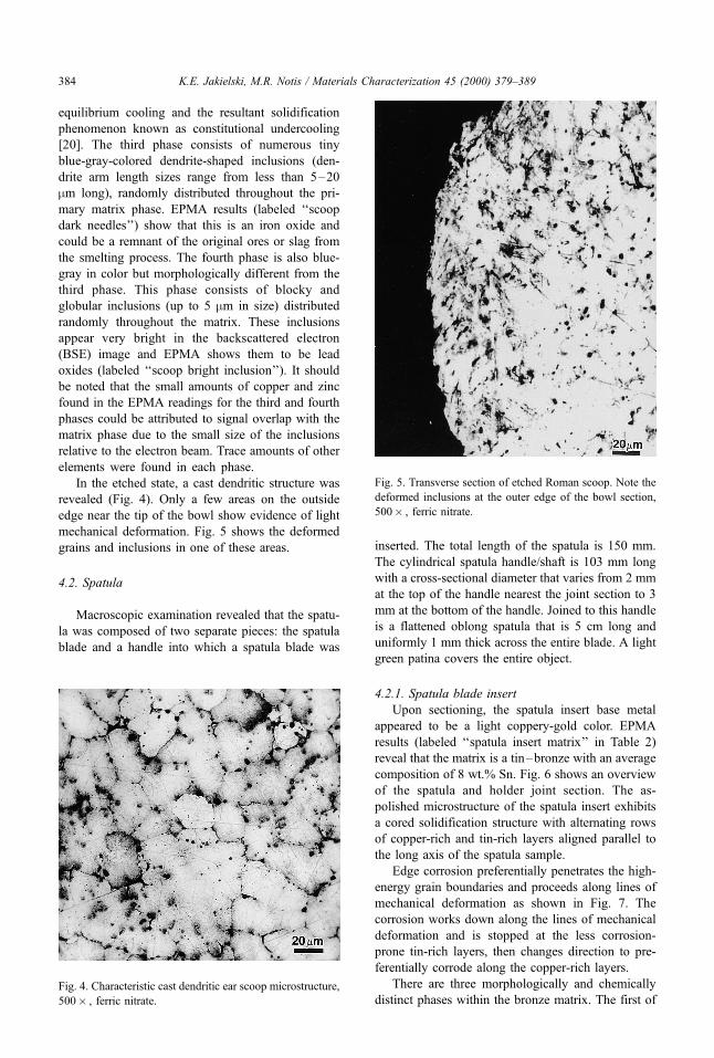
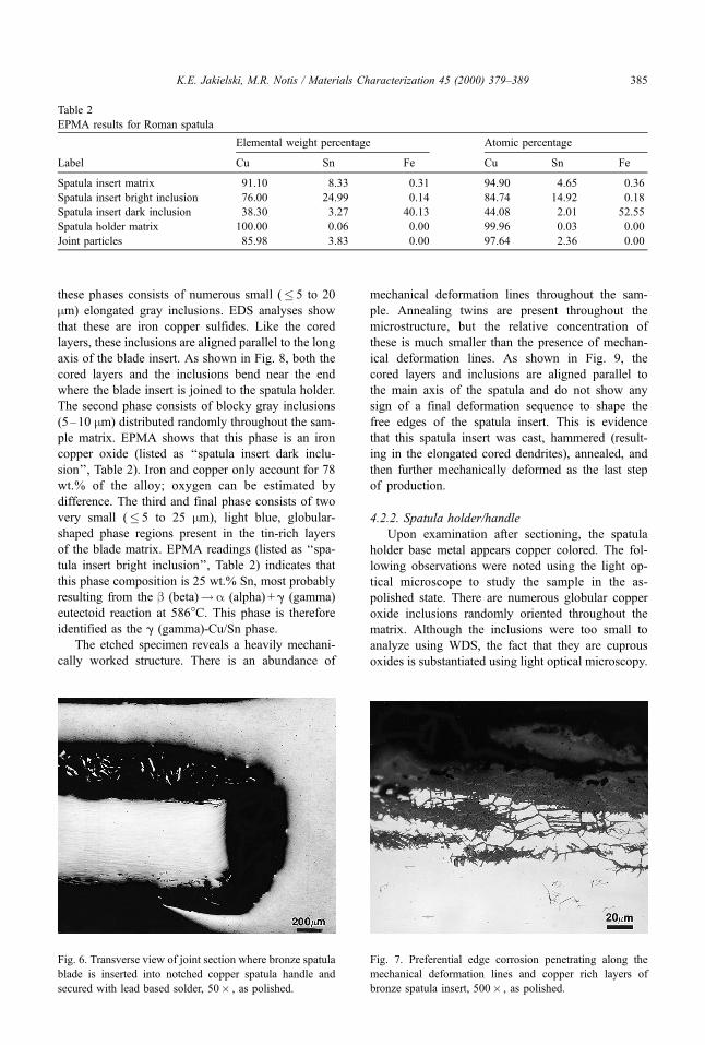
In the etched state, a cast dendritic structure was
revealed (Fig. 4). Only a few areas on the outside
edge near the tip of the bowl show evidence of light
mechanical deformation. Fig. 5 shows the deformed
grains and inclusions in one of these areas.
4.2. Spatula
Macroscopic examination revealed that the spatu-
la was composed of two separate pieces: the spatula
blade and a handle into which a spatula blade was
inserted. The total length of the spatula is 150 mm.
The cylindrical spatula handle/shaft is 103 mm long
with a cross-sectional diameter that varies from 2 mm
at the top of the handle nearest the joint section to 3
mm at the bottom of the handle. Joined to this handle
is a flattened oblong spatula that is 5 cm long and
uniformly 1 mm thick across the entire blade. A light
green patina covers the entire object.
4.2.1. Spatula blade insert
Upon sectioning, the spatula insert base metal
appeared to be a light coppery-gold color. EPMA
results (labeled `̀ spatula insert matrix'' in Table 2)
reveal that the matrix is a tin±bronze with an average
composition of 8 wt.% Sn. Fig. 6 shows an overview
of the spatula and holder joint section. The as-
polished microstructure of the spatula insert exhibits
a cored solidification structure with alternating rows
of copper-rich and tin-rich layers aligned parallel to
the long axis of the spatula sample.
Edge corrosion preferentially penetrates the high-
energy grain boundaries and proceeds along lines of
mechanical deformation as shown in Fig. 7. The
corrosion works down along the lines of mechanical
deformation and is stopped at the less corrosion-
prone tin-rich layers, then changes direction to pre-
ferentially corrode along the copper-rich layers.
There are three morphologically and chemically
distinct phases within the bronze matrix. The first ofFig. 4. Characteristic cast dendritic ear scoop microstructure,
500� , ferric nitrate.
Fig. 5. Transverse section of etched Roman scoop. Note the
deformed inclusions at the outer edge of the bowl section,
500� , ferric nitrate.
K.E. Jakielski, M.R. Notis / Materials Characterization 45 (2000) 379±389384
these phases consists of numerous small (� 5 to 20
mm) elongated gray inclusions. EDS analyses show
that these are iron copper sulfides. Like the cored
layers, these inclusions are aligned parallel to the long
axis of the blade insert. As shown in Fig. 8, both the
cored layers and the inclusions bend near the end
where the blade insert is joined to the spatula holder.
The second phase consists of blocky gray inclusions
(5±10 mm) distributed randomly throughout the sam-
ple matrix. EPMA shows that this phase is an iron
copper oxide (listed as `̀ spatula insert dark inclu-
sion'', Table 2). Iron and copper only account for 78
wt.% of the alloy; oxygen can be estimated by
difference. The third and final phase consists of two
very small (� 5 to 25 mm), light blue, globular-
shaped phase regions present in the tin-rich layers
of the blade matrix. EPMA readings (listed as `̀ spa-
tula insert bright inclusion'', Table 2) indicates that
this phase composition is 25 wt.% Sn, most probably
resulting from the b (beta)!a (alpha) + g (gamma)
eutectoid reaction at 586°C. This phase is therefore
identified as the g (gamma)-Cu/Sn phase.
The etched specimen reveals a heavily mechani-
cally worked structure. There is an abundance of
mechanical deformation lines throughout the sam-
ple. Annealing twins are present throughout the
microstructure, but the relative concentration of
these is much smaller than the presence of mechan-
ical deformation lines. As shown in Fig. 9, the
cored layers and inclusions are aligned parallel to
the main axis of the spatula and do not show any
sign of a final deformation sequence to shape the
free edges of the spatula insert. This is evidence
that this spatula insert was cast, hammered (result-
ing in the elongated cored dendrites), annealed, and
then further mechanically deformed as the last step
of production.
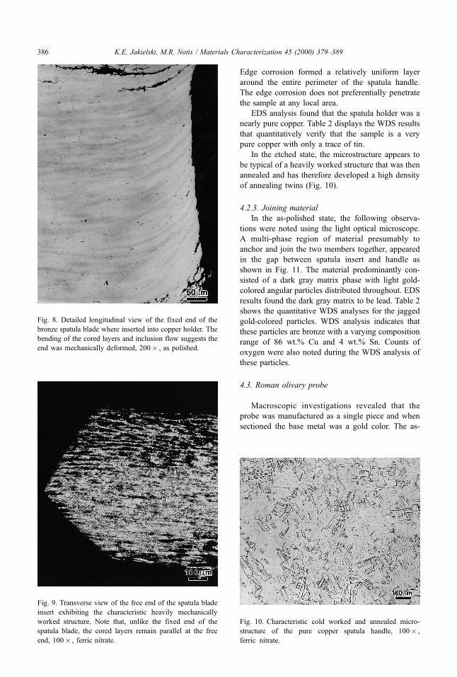
4.2.2. Spatula holder/handle
Upon examination after sectioning, the spatula
holder base metal appears copper colored. The fol-
lowing observations were noted using the light op-
tical microscope to study the sample in the as-
polished state. There are numerous globular copper
oxide inclusions randomly oriented throughout the
matrix. Although the inclusions were too small to
analyze using WDS, the fact that they are cuprous
oxides is substantiated using light optical microscopy.
Table 2
EPMA results for Roman spatula
Elemental weight percentage Atomic percentage
Label Cu Sn Fe Cu Sn Fe
Spatula insert matrix 91.10 8.33 0.31 94.90 4.65 0.36
Spatula insert bright inclusion 76.00 24.99 0.14 84.74 14.92 0.18
Spatula insert dark inclusion 38.30 3.27 40.13 44.08 2.01 52.55
Spatula holder matrix 100.00 0.06 0.00 99.96 0.03 0.00
Joint particles 85.98 3.83 0.00 97.64 2.36 0.00
Fig. 6. Transverse view of joint section where bronze spatula
blade is inserted into notched copper spatula handle and
secured with lead based solder, 50� , as polished.
Fig. 7. Preferential edge corrosion penetrating along the
mechanical deformation lines and copper rich layers of
bronze spatula insert, 500� , as polished.
K.E. Jakielski, M.R. Notis / Materials Characterization 45 (2000) 379±389 385
Edge corrosion formed a relatively uniform layer
around the entire perimeter of the spatula handle.
The edge corrosion does not preferentially penetrate
the sample at any local area.
EDS analysis found that the spatula holder was a
nearly pure copper. Table 2 displays the WDS results
that quantitatively verify that the sample is a very
pure copper with only a trace of tin.
In the etched state, the microstructure appears to
be typical of a heavily worked structure that was then
annealed and has therefore developed a high density
of annealing twins (Fig. 10).
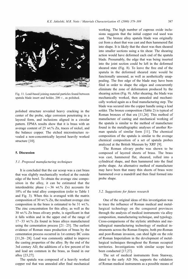
4.2.3. Joining material
In the as-polished state, the following observa-
tions were noted using the light optical microscope.
A multi-phase region of material presumably to
anchor and join the two members together, appeared
in the gap between spatula insert and handle as
shown in Fig. 11. The material predominantly con-
sisted of a dark gray matrix phase with light gold-
colored angular particles distributed throughout. EDS
results found the dark gray matrix to be lead. Table 2
shows the quantitative WDS analyses for the jagged
gold-colored particles. WDS analysis indicates that
these particles are bronze with a varying composition
range of 86 wt.% Cu and 4 wt.% Sn. Counts of
oxygen were also noted during the WDS analysis of
these particles.
4.3. Roman olivary probe
Macroscopic investigations revealed that the
probe was manufactured as a single piece and when
sectioned the base metal was a gold color. The as-
Fig. 8. Detailed longitudinal view of the fixed end of the
bronze spatula blade where inserted into copper holder. The
bending of the cored layers and inclusion flow suggests the
end was mechanically deformed, 200� , as polished.
Fig. 9. Transverse view of the free end of the spatula blade
insert exhibiting the characteristic heavily mechanically
worked structure. Note that, unlike the fixed end of the
spatula blade, the cored layers remain parallel at the free
end, 100� , ferric nitrate.
Fig. 10. Characteristic cold worked and annealed micro-
structure of the pure copper spatula handle, 100� ,
ferric nitrate.
K.E. Jakielski, M.R. Notis / Materials Characterization 45 (2000) 379±389386
polished structure revealed heavy cracking in the
center of the probe, edge corrosion penetrating in a
layered form, and inclusions aligned in a circular
pattern. EPMA results show that it is brass with an
average content of 25 wt.% Zn, traces of nickel, and
the balance copper. The etched microstructure re-
vealed a non-concentrically layered heavily worked
structure [10].
5. Discussion
5.1. Proposed manufacturing techniques
It is concluded that the ear scoop was a cast brass
that was slightly mechanically worked at the outside
edge of the bowl. To obtain the average zinc compo-
sition in the alloy, it can be estimated that the
interdendritic phase (� 36 wt.% Zn) accounts for
10% of the total alloy composition (refer to Table 1
and Fig. 3). When this is averaged with the matrix
composition of 30 wt.% Zn, the resultant average zinc
composition in the brass is estimated to be 31 wt.%.
The zinc concentration for this alloy, as well as the
30 wt.% Zn brass olivary probe, is significant in that
it falls within and in the upper end of the range of
17±33 wt.% Zn found in brass alloys manufactured
using the cementation process [21±25]. The earliest
evidence of Roman mass production of brass by the
cementation process occurred in 1st century BC coins
[11,26±28]. Lead was commonly added to improve
the casting properties of the alloy. By the end of the
3rd century AD, the additions of a few percent of tin
and lead are common in the brass, as found in our
alloy [23,27].
The spatula was composed of a heavily worked
copper rod that was annealed after final mechanical
working. The high number of cuprous oxide inclu-
sions suggests that the initial copper rod used was
cast. The bronze alloy spatula blade was originally
cut from a sheet that was cast and then hammered flat
into shape. It is likely that the sheet was then sheared
into smaller sections using a tin shear. The shearing
action would have deformed each end of the spatula
blade. Presumably, the edge that was being inserted
into the joint section could be left in the deformed
sheared state (Fig. 8). To leave the free end of the
spatula in the deformed sheared state would be
functionally unsound, as well as aesthetically unap-
pealing. The free edge of the blade may have been
filed in order to shape the edges and concurrently
eliminate the zone of deformation produced by the
shearing action (Fig. 9). After shearing, the blade was
mechanically worked, then annealed and mechani-
cally worked again as a final manufacturing step. The
blade was secured into the copper handle using a lead
solder. The bronze composition (Table 2) is typical of
Roman bronzes of that era [11,26]. This method of
manufacture of casting and mechanical working of
the spatula is similar to the method of manufacture
found in the metallographic analyses of another Ro-
man spatula of similar form [11]. The chemical
composition of the spatula is similar to the average
chemical composition of a set of spatula probes
analyzed at the British Museum by XRF [9].
The Roman olivary probe was shown to be
composed of layered sheets of brass. The brass
was cast, hammered flat, sheared, rolled into a
cylindrical shape, and then hammered into the final
probe shape. An alternative method of manufacture
may have been that many thin sheets of brass were
hammered over a mandrill and then final formed into
probe shape.
5.2. Suggestions for future research
One of the original ideas of this investigation was
to trace the influence of Roman medical and metal-
lurgical technology on the conquered provinces
through the analysis of medical instruments via alloy
composition, manufacturing technique, and typology.
Cross-comparisons of the stylistic attributes and me-
tallurgical manufacturing technology of medical in-
struments across the Roman Empire, both pre-Roman
and post-Roman invasion, can shed light on the role
of Roman Imperialism in the development of metal-
lurgical techniques throughout the Roman occupied
territories. Investigations with similar scope have
been conducted [17,27].
The set of medical instruments from Stanway,
dated to the early AD 50s, supports the validation
of Roman medical instruments as a possible means of
Fig. 11. Lead-based joining material particles found between
spatula blade insert and holder, 200� , as polished.
K.E. Jakielski, M.R. Notis / Materials Characterization 45 (2000) 379±389 387
tracing Roman influence on metallurgical technology.
The set consists of 13 instruments. One of the pieces,
a bronze scoop, is a definite Roman import; but what
is notable is that the rest of the set is slightly different
from the typical Roman instruments both stylistically
and metallurgically [19]. As Jackson points out, the
majority of the Stanway medical instruments are
single-piece iron instruments and contrast with the
Roman tendency to manufacture single piece bronze
instruments or composite bronze and iron instruments
[19]. It could be that this is a sign of a transition in
metallurgical technology in the area, with local black-
smiths slowly integrating Roman styles into their own
medical instrument design and manufacture. Little is
known of the metalsmiths who produced the Roman
surgical instruments for the militia, or if there even
was a system of centralized manufacture that speci-
fically catered to military supply. Evidence in the
Notitia Dignitatum shows that the Roman legions
were supplied with goods from various arsenals
throughout the Empire [29±31]. If military legions
were supplied with centrally manufactured surgical
instruments, we would expect the materials analysis
of medical instruments provenanced to military sites
to be stylistically, but more importantly, composition-
ally similar.
Though the unknown provenance of these ob-
jects severely limits the ability to make any clear
inferences from this data regarding metallurgical
technology in a specific area of the world, it is
significant to note that variation in surgical instru-
ment design via alloy composition and manufactur-
ing technique does exist. The olivary probe, spatula,
and scoop each have completely different manufac-
turing techniques that involve processes as diverse
as hammering, casting, shearing, rolling, and anneal-
ing. In addition, the alloy composition appears to be
coupled with a manufacturing technique that opti-
mizes the material properties for the specific in-
tended function. It has also been found that some
Roman medical instruments contain dissimilar metal
inlays, plating, and intricate decorative surface treat-
ments [11]. These variations in technique could
suggest localized production and regional specializa-
tion. The presence of Roman surgical instruments in
an area could reflect the level of local metallurgical
technologies as well as the level of integration of
local medicine with Roman techniques.
6. Conclusions
It is not assumed that analysis of a few medical
instruments as performed in the present work can
conclusively determine the level of technological
innovation and integration in the manufacture of
Roman medical instruments. Instead, it is hoped that
this work substantiates the importance of data gained
from metallographic investigations to provide a meth-
odological framework for future work in the field as
more finds become unearthed and are contributed to
the archaeological record.
References
[1] Jackson R. Doctors and Diseases in the Roman
Empire. Norman, OK: University of Oklahoma
Press, 1988.
[2] Bliquez LJ. Greek and Roman medicine. Archaeology
1981;34(2):10± 7.
[3] Majno G. The Healing Hand: Man and Wound in
the Ancient World. Cambridge, MA: Harvard Univ.
Press, 1975.
[4] Scarborough J. Roman Medicine. Ithaca, NY: Cornell
Univ. Press, 1976.
[5] Milne JS. Surgical Instruments in Greek and Roman
Times. Chicago, IL: Ares Publishers, 1907.
[6] Bliquez LJ. Roman surgical instruments in the Johns
Hopkins University Institute of the History of Medi-
cine. Bull Hist Med 1982;56:195±217.
[7] Bliquez LJ. Roman Surgical Instruments and Minor
Objects in the University of Mississippi. Goteberg: P.
Astrom Publishers, 1988.
[8] Bliquez LJ. Roman Surgical Instruments and other
Minor Objects in The National Archaeological Mu-
seum of Naples. Mainz, Germany: Verlag Phillipp
von Zabern Publishers, 1994.
[9] Jackson R. A set of Roman medical instruments from
Italy. Britannia 1986;17:68±113.
[10] Notis MR, Jakielski K. Ancient medical instruments:
an overview and case study. In: Vandiver PB, Druz-
nik JR, Merkel J, Stewart J, editors. Proc. Materials
Issues in Art and Archaeology V. Vol. 462. Materials
Research Society. Warrendale, PA: MRS, 1996 (Fall).
[11] Healy J. Mining and Metallurgy in the Greek and Ro-
man World. London: Thames and Hudson, 1978.
[12] Kirkup J. From flint to stainless steel: observations on
surgical instrument composition. Ann R Coll Surg
Engl 1993;75:365± 74.
[13] Kirkup J. The history and evolution of surgical instru-
ments: II. Origins: function: carriage: manufacture.
Ann R Coll Surg Engl 1982;64:125±32.
[14] Kirkup J. The history and evolution of surgical instru-
ments: IV. Probes and their allies. Ann R Coll Surg
Engl 1985;67(1):56 ±60.
[15] Richmond I. Hod Hill Excavations Volume Two Car-
ried out Between 1951 and 1958 for the Trustees of the
British Museum. London: The Trustees of the British
Museum, 1968.
[16] Dungworth D. Iron age and Roman copper alloys
from Northern Britain. Internet Archaeol 1997;2:12±
18. (http://intarch.ac.uk/journal/issue2/dungworth_
toc.html).
[17] Comstock MB, Vermeule CC, editors. Greek, Etrus-
can, and Roman Bronzes in the Museum of Fine
K.E. Jakielski, M.R. Notis / Materials Characterization 45 (2000) 379±389388

Arts, Boston. Greenwich, CT: New York Graphic
Society, 1971.
[18] Jackson R. The surgical instruments from Stanway,
Colchester. Colchester Archaeol 1997;11.
[19] Colchester Archaeological Trust, 1998, http://peipa.
essex.ac.uk/CAT/.
[20] Boyer HE, Gall TL, editors. Metals Handbook Desk
Edition. Metals Park, OH: American Society for Me-
tals, 1985.
[21] Werner O. The occurrence of zinc and lead in the
antiquity and in the Middle Ages. Erzmetall 1970;
23(6):259± 69.
[22] Haedecke K. Gleichgewichtsverhaltnisse bei der Mes-
singherstellung nach dem Galmeiverfahren. Erzmetall
1973;26:229±33.
[23] Craddock PT. The composition of the copper alloys
used by the Greek, Etruscan and Roman civilizations:
3. The origins and early use of brass. J Archaeol Sci
1978;5:1± 16.
[24] Craddock PT. Three thousand years of copper alloys:
from the bronze age to the industrial revolution. Ap-
plication of Science in Examination of Works of Art.
Boston: Museum of Fine Arts, 1983. pp. 59± 67.
[25] Pollard AM, Heron C. The chemical study of metals Ð
the European medieval and later brass industry. Ar-
chaeological Chemistry. Cambridge, UK: The Royal
Society of Chemistry, 1996. pp. 196± 238.
[26] Tylecote R. A History of Metallurgy. London, UK: The
Metals Society, 1976.
[27] Bayley J. The production of brass in antiquity with
particular reference to Roman Britain. In: Craddock
PT, editor. 2000 Years of Zinc and Brass. London,
UK: British Museum, 1990. pp. 7± 26.
[28] Craddock PT. Zinc in classical antiquity. In: Craddock
PT, editor. 2000 Years of Zinc and Brass. London, UK:
British Museum, 1990.
[29] Anonymous, Notitia Dignitatum. Translations and
Reprints from the Original Sources of European His-
tory. Department of History, University of Pennsylva-
nia, Philadelphia, PA, VI (4), pp. 1 ±40.
[30] Jones AHM. The later Roman empire. A Social, Eco-
nomic, and Administrative Survey. Norman, OK: Uni-
versity of Oklahoma Press, 1964. pp. 284±602.
[31] Boniardi M, Gariboldi E, Vedani M. Metallographic
studies on an ancient Roman nail. Metall Sci Technol
1992;10(1):28± 38.
K.E. Jakielski, M.R. Notis / Materials Characterization 45 (2000) 379±389 389