Embed Size (px)
Citation preview
Universidade de Lisboa Faculdade de Ciências
Secção Autónoma de História e Filosofia das Ciências
Università degli Studi di Milano Facoltà di Lettere e Filosofia
Dipartimento di Filosofia
!
!!
!!!
!!
Towards an Epistemology of Medical Imaging
Margherita Silvia Di Marco
Doutoramento em História e Filosofia das Ciências
Dottorato di Ricerca in Filosofia
2015 !!

! !
Universidade de Lisboa Faculdade de Ciências
Secção Autónoma de História e Filosofia das Ciências
Università degli Studi di Milano Facoltà di Lettere e Filosofia
Dipartimento di Filosofia
!
!!
!!!
!Towards an Epistemology of Medical Imaging
Margherita Silvia Di Marco
Tese orientada pela Prof.ª Dr.ª Olga Maria Pombo Martins e pelo Prof. Dr. Andrea Pinotti, especialmente elaborada para a obtenção do grau de doutor em História e Filosofia das Ciências e em Filosofia, em cotutela
entre a Universidade de Lisboa e a Università degli Studi di Milano
Tesi orientata dalla Prof. Olga Maria Pombo Martins e dal Prof. Andrea Pinotti, redatta specificamente per l’ottenimento del titolo di dottore di ricerca in Storia e Filosofia della Scienza e in Filosofia, in cotutela tra
Universidade de Lisboa e Università degli Studi di Milano
2015
!!

To the loving memory of Angela, my mother

The scientist [...] cuts open the visible body to look at its interior or catcheshidden objects by means of all sorts of sophisticated equipment that deprivesthem of their exterior properties through which they show themselves to our
natural senses.
H. Arendt – The Life of the Mind
Then he flung himself into his chair, and drew out his keepsake, his treasure,that consisted, this time, not of a few reddish-brown shavings, but a thin glass
plate, which must be held toward the light to see anything on it. It wasClavdia’s X-ray portrait, showing not her face, but the delicate bony structure
of the upper half of her body, and the organs of the thoracic cavity,surrounded by the pale, ghostlike envelop of flesh.
T. Mann – The Magic Mountain
What is the use of a book thought Alice, without pictures or conversations?
L. Carroll – Alice’s Adventures in Wonderland

Acknowledgments
I am grateful to the Fundação para a Ciência e a Tecnologia for giving financialsupport to this work through the doctoral grant SFRH/BD/64050/2009.
I also thank the Universidade de Lisboa and the Università degli Studi diMilano for providing the institutional framework necessary to the realization ofthis doctorate in co-supervision.
This dissertation was really an adventure, and this adventure would neverhave started without Olga Pombo. She offered me the opportunity to begranted a fellowship within the research project “Image in Science and Art”(PTDC/EAT/64201/2006), where the early ideas for this dissertation were in-cubated. She gave invaluable support in writing the doctoral proposal andaccompanied my work all throughout. I thank her for teaching me so muchduring all these years.
Andrea Pinotti also provided enormous support. I am particularly gratefulto him for having brought to my attention the importance of Peirce’s semioticsto the theory of photography, and for introducing me to Benjamin’s concept ofthe optical unconscious. His careful critiques and his encouragement, togetherwith his good humor, were much appreciated.
I am indebted to the librarians of the central library of the Faculdade deCiências for helping me in getting books from other libraries in Portugal andabroad. In particular, I am grateful to the chief librarian, Ana Fraga, for herhelpfulness and for answering my emails even when she was on holidays.
Thanks also to Chiara Tartarini, in Bologna, Pietro Conte and RosannaFeroldi, in Milan, Lorenza Moronetti and Cecilia Cotta-Ramusino, in Boston,for being so kind to take time to search the archives of their university librariesand send me copies of articles and book chapters that were too old to be foundonline, or too expensive to be bought.
The Centro de Filosofia das Ciências da Universidade de Lisboa offered meexcellent opportunities for study, research, and intellectual growth, and I metsome extraordinary people there. I am grateful to Zbigniew Kotowicz for read-ing this dissertation with a rigorous but sympathetic eye. His remarks andencouragement were precious. I thank Elena Casetta, for being the funniest
v
vi Acknowledgments
and smartest “desk-mate” ever; Marco Pina and Nathalie Gonthier, for theirfriendly support, especially in the early phases of my research; Nuno Melim, forhis kindness and black humor; Filipe Varela and Nuno Jerónimo, for being suchgood friends and such special guys.
I am particularly grateful to María de Paz, for letting me explain to her manyof my ideas before I wrote them down, for her uncanny ability to understandbetter than me what I was thinking and wanted to say, for reading what I wroteand giving suggestions, for having such a charming and contagious passion forphilosophy, and for being such a generous friend: thank you.
For making me feel at home since the first time I met them, and for mak-ing the Pensão Estrelinha such a welcoming and lively place, I wish to thankRosário, Manuel, and Tó Maneira.
Thanks to all my friends in Italy, for still being there after all these years,and through all these changes. Thanks to Fabio, for being so unrestful and forstopping over in Lisbon each time he flies to Africa; to Elena, for always findingtime to see me; to Anna, who made a very special little book; and to Milena,who magically appears when she is more needed.
My debt is immeasurable to my stubborn father, Antonio. I suspect I tookfrom him the passion for asking questions and questioning the answers, a lovefor reasoning and arguing, and a sometimes irritating inclination to check themeaning of words in the dictionary, or the internet, during mealtime.
I am forever grateful to Viviana, my sister, for her unconditional love andsupport, for being at my side in the hardest days, for her surreal sense of humorand for her smile. Thanks to Martina and Maia, for being Martina and Maia.And thanks to Stefano, for his patience, good heart, and for helping so much.
Finally, I want to thank José. For being the reason I came to Portugal inthe first place, for believing in my work even on those moments in which I wasat odds with it myself, and because his curiosity and intellectual honesty areinspiring, everyday.


Abstract
The objective of this dissertation is to contribute to the development of an epis-temology of medical imaging. My central thesis is that medical imaging doesnot merely produce more or less accurate pictures of the inner organs, it rathertransforms the living body into a scientific object by changing its very visibility.The imaging apparatus turns the body into a visual object that can be observedunder experimental conditions: unlike the real body, it can be filed, retrieved,shared, measured and manipulated in several ways. Alongside this main the-sis there are two others: firstly diagnostic images – like all scientific images –are actual cognitive instruments, epistemic objects inscribed within theoreticalcontexts and experimental practices. Secondly, an image of the inner body hasdiagnostic meaning and value only in the scope of a specific conceptualization ofthe body and its ailments. Accordingly, if we are to develop an epistemology ofmedical imaging, we cannot limit our analysis to diagnostic images qua images,we also have to understand them qua diagnostic instruments.
This is the reason why I take into examination the historical and conceptualconditions of possibility of radiography – the first medical imaging technology,invented in 1895 – in the first chapter of the dissertation. My aim is to under-stand which medical theories and practices had to be at work in the nineteenthcentury for those shadow-images produced by the X-ray apparatus, to be per-ceived and employed as diagnostic devices. I argue that the diagnostic relevanceof radiography is rooted in the conceptualization of body, disease and diagnosisput forward by clinical anatomy, as early as the end of the eighteenth century.I also defend the idea that the stethoscope, developed in 1816, was the ma-terial and intellectual predecessor of medical imaging, because it introduced aprimitive form of mediated perception in medical diagnosis, and allowed theclinician to explore the inner body of the living patient from the outside, ex-tracting from it signs of illness. The stethoscope was only the first of a vastarray of instruments invented in the nineteenth century to visualize differentaspects of the inner morphology and physiology of the living body. Each ofthese instruments fulfilled specific diagnostic aims and posed distinct epistemo-logical problems, but all of them shared some commonalities: they were meant
ix
x Abstract
to replace the subjective sensations of patients and doctors with objective in-dices of health and disease; they created visual records of the inner body thatcould be filed, retrieved and shared among physicians; they required the devel-opment of a specialized language agreed upon by a community of experts; theycreated a progressive physical separation between the body of the patient andthe body of the physician. It was in this complex scenario of medical practices,objects, images and ideas that radiography appeared and progressively acquiredits diagnostic function.
In the second chapter, I take into account the early developments of medicalphotography in order to understand how the early technology for the productionof mechanical images entered and influenced the domain of medicine. The maintheoretical references in this chapter are Charles Sanders Peirce’s semiotics (inparticular his classification of signs in indices, icons and symbols) and WalterBenjamin’s reflections on the photographic series (mechanical production andreproduction of an image and of the body it represents), on the intrinsic analyticand dissecting potential of photography (the photographer as a surgeon), andon the optical unconscious (photography as a prosthesis that enriches and trans-forms our sensorial experience). Drawing on these authors, and analyzing theworks of early physicians-photographers in psychiatry, dermatology, neurologyand physiology, I show that the photographic series collected in medical jour-nals, manuals and hospital archives produced a clinical gaze in the Foucauldiansense. I also argue that the photographic series was part of a larger experimentalapparatus, which encompassed the patient, the camera and the observer, andwhose aim was to turn the body and disease into a visual object available forscientific analysis.
In the third chapter, I discuss the problem of the invisible referent, that is,I analyze the processes whereby photographs that reveal invisible phenomenaare endowed with meaning. This is likely to be the fundamental problem of allscientific imaging. When the referent of a picture is invisible, the iconic modeof signification fails, because in this case the image produced by the mechanicalor electronic apparatus does not look like anything we already know, it resem-bles nothing. So, how do we know that the object we see in the photograph– e.g., a cell or a tubercular lesion – is really there and does really look likethat? Drawing on the theoretical analysis developed in the previous chapter, Imaintain that the visualization of the invisible entails a peculiar combination ofthe indexical, iconic and symbolic modes of signification. My reasoning opposesLorraine Daston and Peter Galison’s idea of mechanical objectivity, and demon-strates that their notion of mechanical objectivity as the moralizing suppressionof subjectivity is a caricature of the actual ideas and practices developed by the
Abstract xi
scientists of the nineteenth century to deal with the problem of visualizing theinvisible. The argument is articulated in three moments, corresponding to theanalysis of the problem of objectivity and image signification in microphotogra-phy, chronophotography, and radiography.
In the fourth chapter, I argue that images are cognitive tools and that repre-sentation and observation are never an act of automated repetition, they alwaysentail a creative component. As in the previous chapter, part of my discourseis built in contrast with Daston and Galison, challenging their claims about thepassive nature of representation. For these authors, up until the developmentof digital technologies for image manipulation, scientific images were mere re-presentations of the world, focused on copying nature. Computer images, onthe contrary, are presentations, because the observer can virtually manipulatethem so that they show the object in ever changing ways. I criticize this classi-fication of scientific images with historical and theoretical arguments. From thehistorical point of view, I show that at least since the sixteenth century therehave been attempts to create images that can be actually manipulated by theobserver. From the theoretical perspective, I draw on a variety of literaturespanning from art theory to neuroscience, to demonstrate that the very notionof a passive representation is unsustainable, because images always engage theobserver in an embodied act of perception, which elicits not only visual, but alsotactile sensations and motor reactions. Moreover, I argue that Daston and Gali-son’s emphasis on nanoimaging as the only technology that allows manipulatingthe object of study during the process of image production is misleading. Infact, even when they do not reach the peaks of technological sophistication thatcharacterizes nanoimages, scientific images are the result of some manipulationof the natural object they represent. A scientific image cannot be a passivecopy of nature, because it is part of an experimental praxis, whose goal is to un-derstand natural phenomena, not just to reproduce them. To corroborate thisidea I explore actual scientific practices of image signification, taking into ac-count written documents (semiotic analysis of a radiology article) and materialpractices (laboratory ethnography describing the interpretation of electrophore-sis images in a molecular biology laboratory, and description of an example ofsignification of electron microscopy pictures). From this analysis three remarkscan be put forward: (1) the process of signification of scientific images has adistributed character, because it can involve different persons, objects and ac-tivities; (2) scientific images can be considered experimental tools, in the sensethat scientists and physicians handle them in several forms in order to exploredifferent aspects of their object of study; (3) scientific images are to be under-
xii Abstract
stood as controlled, artificial phenomena produced with the aim of redefiningthe visibility of natural objects.
In order to clarify this latter idea, in the final chapter I introduce Gas-ton Bachelard’s concept of phenomenotechnique. Although the idea of phe-nomenotechnique cannot be directly applied to medical imaging, there are twocharacterizing elements of this concept that provide important insights for con-ceptualizing medical imaging. The first is the idea that in order to study anatural phenomenon, scientists must previously transform it into a scientific ob-ject. The second, closely related to the former, is that scientific experience is bynecessity mediated, and such mediation has both an intellectual and materialcharacter. This means that the development of instruments and new technolo-gies is not a second-order product of science, it is part and parcel of the scientificprocess. Technology is embedded into science, because our scientific graspingof the world is necessarily mediated by instruments; scientific instruments, inturn, are materializations of a vast body of scientific knowledge and practices(in the case of digital imaging this knowledge has an eminently mathematicalcharacter). Thus, science and technology are reciprocally constituted. On thesegrounds, I propose a description of medical imaging in terms of phenomenotech-nique, using this concept as a key word around which to reorganize the ideaspreviously discussed. Firstly, I resort to the concept of phenomenotechniqueto gain insight into how diagnostic images mediate the physician’s sensory andintellectual experience. Secondly, I give an account of diagnostic images as ar-tificial phenomena (visual reconfigurations of non-visual signals) that work assimulations of the patient’s body, and that reify different domains of knowledge(from medicine to physics and engineering). Finally, I argue that the properand efficient signification of a diagnostic image requires a phenomenotechniqueof the observer. To recognize the signs of disease in an image of the inner body,one has to master the explicit and implicit rules necessary to make sense of thenovel sensory domain produced by the technological apparatus. This impliesabandoning spontaneous modes of perception and signification to engage in aprocess of educated perception. The expert viewer goes through a formal andinformal training that deeply transforms natural vision, by placing the act ofwatching within a wide epistemic network that encompasses both theoreticaland practical knowledge.
Key-words: epistemology; history of medicine; image theory; medicalimaging; photography; radiography.
Resumo
O objetivo deste trabalho é o de contribuir para o desenvolvimento de umaepistemologia da imagiologia médica. A minha tese central é que a imagiologiamédica não produz meramente imagens mais ou menos precisas dos órgãos in-ternos, antes torna o corpo vivo num objeto científico ao modificar a sua própriavisibilidade. As tecnologias de imagiologia tornam o corpo num objeto visualque pode ser observado em condições experimentais: ao contrário do corpo real,pode ser arquivado, recuperado, partilhado, medido e manipulado de variadasformas. Esta tese é acompanhada por duas outras: em primeiro lugar, as ima-gens diagnósticas, como todas as imagens científicas, são efetivamente instru-mentos cognitivos, objetos epistémicos inscritos em contextos teórico-práticosespecíficos. Em segundo lugar, uma imagem do interior do organismo tem sig-nificado e valor diagnóstico apenas no âmbito de uma dada contextualização docorpo e da doença. Por conseguinte, se queremos desenvolver uma epistemo-logia da imagiologia médica, não podemos analisar as imagens de diagnósticosimplesmente como imagens, mas também como instrumentos médicos.
É por isso que no primeiro capítulo da dissertação tento compreender quaisforam as condições de possibilidade históricas e conceptuais da radiografia – aprimeira tecnologia de imagiologia médica, inventada em 1895. O meu obje-tivo é o de entender quais as teorias e práticas médicas que estavam em jogono século XIX, que permitiram que umas imagens que mostravam sombras dointerior do corpo fossem percecionadas e usadas como um instrumento clínico.Defendo que a relevância diagnóstica da radiografia está enraizada na concep-tualização de corpo, doença e diagnóstico estabelecida pela anatomia clínica jáem finais do século XVIII. Defendo também que o estetoscópio, desenvolvidoem 1816, foi o precursor material e intelectual da radiografia, pois introduziuuma forma primitiva de perceção mediada no diagnóstico médico, e permitiuao médico explorar a partir do exterior, o interior de um corpo vivo, extraindosinais de doença. O estetoscópio foi apenas o primeiro de um vasto conjuntode instrumentos inventados no século XIX para visualizar diferentes aspetostanto da morfologia como da fisiologia do corpo humano. Cada um desses ins-trumentos respondia a objetivos de diagnóstico específicos e punha problemas
xiii
xiv Resumo
epistemológicos distintos, mas todos partilhavam alguns aspetos comuns: elesdeveriam substituir as sensações subjetivas de pacientes e médicos por indica-dores objetivos de saúde e doença; criavam registos visuais do interior de umcorpo vivo; necessitavam do desenvolvimento de uma linguagem especializadafruto de um acordo da comunidade médico-científica; criaram uma progressivaseparação física entre o corpo do paciente e o corpo do médico. Foi neste cená-rio complexo de práticas, instrumentos, representações e ideias médicas, que aradiografia apareceu e progressivamente adquiriu a sua função diagnóstica.
No segundo capítulo examino o nascimento da fotografia de modo a enten-der como a primeira tecnologia de produção de imagens mecânicas entrou nasteorias e práticas médicas. A principal referência teórica neste capítulo é a se-miótica de Charles Sanders Pierce, em particular a sua classificação dos signosem índices, ícones e símbolos, e as reflexões de Walter Benjamin sobre as sériesfotográficas (produção e reprodução mecânica de uma imagem e do corpo que elarepresenta), sobre o intrínseco potencial analítico e de “dissecção” da fotografia(o fotógrafo como cirurgião), e sobre o inconsciente ótico (a fotografia como umaprótese que enriquece e transforma a nossa experiência sensorial). Baseando-me nestes autores e analisando os trabalhos dos primeiros médicos-fotógrafosem psiquiatria, dermatologia, neurologia e fisiologia, mostro que as séries fo-tográficas colecionadas em revistas médicas, manuais e arquivos de hospitais,produziram um “olhar clínico” no sentido Foucauldiano. Defendo também quea série fotográfica era parte de um dispositivo experimental mais vasto, queabrangia o paciente, a câmara fotográfica e o observador, e cujo objetivo eratornar o corpo e a doença num objeto visual, disponível para análise científica.
No terceiro capítulo discuto o problema do referente invisível, isto é, analisoos processos através dos quais é atribuído significado às fotografias que revelamfenómenos invisíveis. Este é provavelmente o problema fundamental de toda aimagiologia científica. Quando o referente de uma imagem é invisível, a modali-dade de significação icónica falha, porque neste caso a imagem produzida pelosinstrumentos (sejam eles mecânicos ou eletrónicos) não se parece com nada queconheçamos já. De facto, podemos dizer que não se parece com nada. En-tão, como podemos saber que o objeto que vemos na fotografia – por exemplo,uma célula ou uma lesão tubercular – está realmente lá e tem realmente o as-peto do que vemos? Baseando-me na análise teórica desenvolvida no capítuloanterior, defendo a ideia de que a visualização do invisível comporta uma com-binação peculiar das modalidades de significação de índice, ícone e símbolo. Aminha argumentação é construída em oposição à ideia de objetividade mecânicade Lorraine Daston e Peter Galison, e demonstra que a noção de objetividademecânica como a supressão moralizante da subjetividade, defendida por estes
Resumo xv
historiadores, é uma caricatura das ideias e práticas desenvolvidas pelos cien-tistas do século XIX para lidar com o problema de visualizar o invisível. Aargumentação é articulada em três momentos, correspondendo à análise do pro-blema da objetividade e significação das imagens na área da micro-fotografia,crono-fotografia e radiografia.
No quarto capítulo defendo que as imagens são instrumentos cognitivos (nosentido forte, não metafórico da palavra instrumento) e que representação eobservação nunca podem ser atos de repetição automática, porque comportamsempre uma componente criativa. Como no capítulo precedente, parte da argu-mentação é construída em contraste com Daston e Galison, desafiando as suasposições acerca da natureza passiva da representação. Para estes autores, atéao desenvolvimento das tecnologias digitais, as imagens científicas eram meras“re-apresentações” do mundo focadas em copiar a natureza. Com o desenvol-vimento das tecnologias digitais, porém, as imagens passaram a ser “apresen-tações”, porque através dessas imagens o observador pode visualizar o objetorepresentado de muitas maneiras, e manipulá-lo virtualmente. A minha críticaa esta posição é baseada em argumentos históricos e teóricos. Do ponto de vistahistórico, mostro que pelo menos desde o século XVI houve tentativas de criarimagens que podem ser de facto manipuladas pelo observador. Do ponto devista teórico, apoio-me numa vasta literatura que vai desde a teoria da arte àsneurociências, para demonstrar que a própria noção de representação passivaé insustentável, porque as imagens envolvem sempre o observador num ato deperceção corpórea, que provoca sensações não só visuais, mas também táteis,bem como reações motoras. Além disso, mostro que é enganadora a ênfase postapor Daston e Galison no nanoimaging como a única tecnologia que permite amanipulação do objeto de estudo durante o processo de produção da imagem.De facto, mesmo quando não atingem os picos de sofisticação tecnológica quecaracteriza as nano-imagens, as imagens científicas são o resultado de algumamanipulação do objeto natural que representam. Uma imagem científica nãopode ser uma cópia passiva da natureza, porque é parte de uma praxis experi-mental, cujo objetivo é o de aprender algo acerca dos fenómenos naturais, nãoapenas reproduzi-los. A fim de corroborar esta ideia, analiso algumas práticasconcretas de significação de imagens científicas, tomando em conta documen-tos escritos (análise semiótica de um artigo de radiologia) e práticas materiais(etnografia de laboratório sobre a interpretação de imagens de eletroforese embiologia molecular, e descrição de um caso de significação de imagens de micros-copia eletrónica). Esta análise permite fazer três observações: (1) O processode significação das imagens científicas é um processo distribuído, e pode incluirvárias pessoas, ações e instrumentos; (2) As imagens científicas podem ser con-
xvi Resumo
sideradas instrumentos de investigação, no sentido em que cientistas e médicosas manipulam de várias formas, para explorar aspetos diferentes dos seus obje-tos de estudo; (3) As imagens científicas devem ser entendidas como fenómenosartificiais controlados, produzidos com o intuito de redefinir a visibilidade dosobjetos naturais.
Para esclarecer e aprofundar esta última ideia, no capítulo final introduzoo conceito de fenomenotécnica de Gaston Bachelard. A ideia de fenomenoté-cnica não pode ser aplicada diretamente à imagiologia médica. Não obstante,há dois elementos caracterizantes deste conceito que fornecem ensinamentosimportantes para uma filosofia da tecnologia e, consequentemente, para umaepistemologia da imagiologia médica. O primeiro é a ideia de que, para se estu-dar um fenómeno natural, os cientistas devem previamente transformá-lo numobjeto científico. O segundo, estreitamente relacionado com o anterior, é o deque a experiência científica é necessariamente mediada, e que essa mediação temum caráter tanto intelectual como material. Isto significa que a construção deinstrumentos e o desenvolvimento de novas tecnologias não é um produto se-cundário da ciência, mas sim parte integrante do próprio processo científico. Atecnologia está integrada na ciência, porque o nosso entendimento científico domundo é necessariamente mediado por instrumentos; por outro lado, os instru-mentos científicos são materializações de um vasto conjunto de conhecimentose práticas científicas (no caso da imagiologia médica este conhecimento tem umcaráter eminentemente matemático). Portanto, ciência e tecnologia são cons-tituídas reciprocamente. Com bases nessas considerações apresento uma con-ceptualização da imagiologia médica em termos de fenomenotécnica, utilizandoeste conceito como palavra-chave que permite reorganizar as ideias desenvolvi-das anteriormente. Em primeiro lugar, recorro ao conceito de fenomenotécnicapara explicar como as imagens de diagnóstico medeiam a experiência sensoriale intelectual do médico. Em segundo lugar, descrevo as imagens de diagnósticocomo fenómenos artificiais (reconfiguração visual de sinais não visuais) que fun-cionam como simulações do corpo do paciente e que incorporam diferentes áreasdo conhecimento (da medicina à física e engenharia). Finalmente, defendo que asignificação correta e eficiente de uma imagem de diagnóstico requer uma feno-menotécnica do observador. Para reconhecer os sinais de doença numa imagemdo interior do corpo, o médico tem de dominar as regras implícitas e explí-citas necessárias para extrair um sentido do novo domínio sensório produzidopelo dispositivo tecnológico. Isto implica abandonar a perceção espontânea paraentrar num processo de educação da perceção-significação que molda as capaci-dades sensoriais do observador. O observador especializado é um observador que

Resumo xvii
tem feito um percurso formativo que transforma profundamente a visão natural,colocando o ato de olhar dentro de uma vasta rede epistémica.
Palavras chave: epistemologia; história da medicina; imagiologia médica;fotografia; radiografia; teoria da imagem.

xviii Resumo
Riassunto
L’obiettivo di questo lavoro è quello di contribuire allo sviluppo di un’epistemolo-gia dell’imaging medico, intendendo con questo termine sia le immagini utiliz-zate a fini diagnostici, sia le tecnologie che le producono. La mia tesi principaleè che le tecnologie di imaging medico non si limitano a produrre immagini piùo meno accurate degli organi interni e di alcuni processi fisiologici, ma piut-tosto trasformano il corpo in un oggetto scientifico, operando un cambiamentoprofondo della sua visibilità. Gli strumenti di imaging mutano il corpo in un og-getto visivo che può essere osservato in condizioni sperimentali. A differenza delcorpo reale, tale oggetto può essere archiviato, consultato, condiviso, misuratoe manipolato in varie maniere. Questa tesi di fondo è accompagnata da altredue: (1) Le immagini diagnostiche, come tutte le immagini scientifiche, sonoveri e propri strumenti cognitivi, strumenti epistemici integrati in un quadroteorico-pratico specifico; (2) Un’immagine che rivela l’interno dell’organismo hasignificato e valore diagnostico solo nell’ambito di una specifica concettualizza-zione del corpo e della malattia, di conseguenza uno studio sull’epistemologiadell’imaging medico non si potrà limitare a esaminare le immagini diagnostichein quanto immagini, ma dovrà analizzarle anche nella loro veste di strumenti didiagnosi medica.
Per questo motivo nel primo capitolo della dissertazione traccio le linee ge-nerali delle condizioni di possibilità storiche e concettuali della radiografia – laprima tecnologia di imaging medico – inventata nel 1895. Lo scopo è quello dicomprendere quali teorie e pratiche mediche dovessero essere vigenti alla fine delXIX secolo, affinché immagini che parevano ombre del corpo interno potesseroessere considerate strumenti diagnostici. La spiegazione da me proposta è chela rilevanza diagnostica della radiografia si fonda sulla concettualizzazione dicorpo, malattia e diagnosi resa operativa dall’anatomia clinica già alla fine delXVIII secolo. Seguendo e supportando questa linea di ragionamento mostroche lo stetoscopio, inventato nel 1816, può essere considerato il predecessoremateriale e intellettuale dell’imaging medico perché introdusse una primitivaforma di mediazione sensoriale nel campo della diagnostica e permise al medicodi esplorare dall’esterno le profondità del corpo del paziente, estraendone segni
xix
xx Riassunto
di malattia. Lo stetoscopio è solo il primo di una vasta famiglia di strumentiinventati nel XIX secolo per visualizzare diversi aspetti della morfologia internae della fisiologia del vivente. Sebbene ciascuno di questi strumenti rispondessea specifiche necessità diagnostiche e ponesse specifici problemi epistemologici,si possono identificare alcune caratteristiche comuni: tutti avevano come ob-biettivo quello di sostituire le sensazioni soggettive dei pazienti e dei medici conindici oggettivi di salute e malattia; tutti creavano registri visivi dell’interno delcorpo umano che potevano essere archiviati, recuperati e condivisi da diversimedici; tutti richiedevano la creazione di un linguaggio specializzato, condivisoda una comunità medico-scientifica; tutti creavano una progressiva separazionetra il corpo del paziente e il corpo del medico. È in questo complesso scenariodi pratiche, oggetti, raffigurazioni e idee che la radiografia fece la sua comparsae acquisì la sua funzione diagnostica.
Nel secondo capitolo prendo in esame la nascita della fotografia, al fine dicomprendere in che modo la prima tecnologia di produzione meccanica di im-magini influenzò la medicina. I principali riferimenti teorici di questo capitolosono dati dalla semiotica di Charles Sanders Peirce, in particolare la sua classifi-cazione dei segni in indici, icone e simboli, e dalla riflessione di Walter Benjaminsulla serie fotografica (produzione e riproduzione meccanica di un’immagine edel corpo in essa rappresentato), sull’intrinseco potenziale analitico e di dissezio-ne della fotografia (il fotografo come chirurgo), e sull’inconscio ottico (fotografiacome protesi che arricchisce e trasforma l’esperienza sensibile). Basandomi suquesti autori e esaminando i lavori dei primi medici-fotografi nell’ambito dellapsichiatria, dermatologia, fisiologia e neurologia, mostro che le serie fotograficheraccolte in riviste mediche, manuali di studio e archivi ospedalieri produsserouno sguardo clinico in senso foucauldiano. Sostengo, inoltre, che la serie fo-tografica faceva parte di un più ampio apparato sperimentale che includeva ilpaziente, la macchina fotografica e l’osservatore il cui scopo era trasformare ilcorpo e la malattia in oggetti visivi che potessero essere sottoposti ad analisiscientifica.
Nel terzo capitolo discuto il problema del referente invisibile, ossia analizzoi processi attraverso cui le immagini fotografiche di oggetti invisibili vengonodotate di significato. Probabilmente questo è il problema fondamentale di qua-lunque tipo di imaging scientifico. Quando il referente di una fotografia è invi-sibile, la modalità iconica di significazione non può essere messa in atto, perchénell’immagine prodotta dallo strumento (sia esso meccanico o elettronico) nonpossiamo riconoscere nessuna similitudine con l’oggetto rappresentato. Di fat-to, potremmo dire che in questi casi l’immagine non assomiglia a nulla. Comesappiamo, dunque, se l’oggetto che vediamo nella fotografia – per esempio una
Riassunto xxi
cellula o una lesione tubercolare – è davvero là, e possiede davvero l’aspettomostrato dall’immagine? Sulla scorta dell’analisi teorica sviluppata nel capitoloprecedente, difendo l’idea che la visualizzazione dell’invisibile richieda una pecu-liare combinazione delle modalità di significazione indicale, iconica e simbolica.La mia argomentazione è costruita in opposizione al concetto di oggettività mec-canica proposto da Lorraine Daston e Peter Galison. In particolare, dimostroche l’idea di oggettività meccanica come soppressione moralizzante del sogget-to proposta dai due storici è una caricatura delle idee e pratiche sviluppatedagli scienziati del XIX secolo per risolvere il problema della visualizzazionedell’invisibile. La mia argomentazione si articola in tre momenti, corrispondentiall’analisi del problema dell’oggettività e della significazione delle immagini intre diversi ambiti: microfotografia, cronofotografia e radiografia.
Nel quarto capitolo affronto il problema del valore cognitivo delle immagi-ni, sostenendo che le immagini sono strumenti epistemici (nel senso forte, nonmetaforico della parola strumento) e che rappresentazione e osservazione nonsono mai atti puramente automatici, perché richiedono sempre una componentecreativa. Come nel capitolo precedente, parte del mio discorso è una refutazionedella posizione di Daston e Galison, in particolare per quanto riguarda le loroaffermazioni sulla natura passiva di certe rappresentazioni visive. Secondo Da-ston e Galison, infatti, fino allo sviluppo delle tecnologie digitali, le immaginiscientifiche erano mere ri-presentazioni [re-presentations] del mondo, miranti acopiare la natura. Con la comparsa del digitale, invece, si è passati a un’epo-ca in cui le immagini sono presentazioni [presentations], perché attraverso diesse l’osservatore può visualizzare l’oggetto in mutevoli forme, manipolandolovirtualmente. La mia critica a questa posizione è basata su argomenti stori-ci e teorici. Sul piano storico mostro che i primi tentativi di creare immaginimediche manipolabili risalgono almeno al XVI secolo. Sul piano teorico, ricor-rendo alla letteratura prodotta in campi così diversi come la teoria dell’arte ele neuroscienze, dimostro che la nozione di ricezione passiva di un’immagine èinsostenibile, perché le immagini coinvolgono sempre l’osservatore in un attocorporeo di percezione che sollecita non solo sensazioni visive, ma anche sensa-zioni tattili e reazioni motorie. Inoltre, sostengo che l’enfasi posta da Dastone Galison sul nanoimaging come l’unica tecnologia che permette di manipolarel’oggetto durante la fase di produzione di un’immagine è fuorviante. Infatti,anche nei casi in cui non raggiungono le vette di sofisticazione tecnologica pro-prie delle nano-immagini, le immagini scientifiche sono sempre il risultato diuna manipolazione dell’oggetto naturale rappresentato. Un’immagine scientifi-ca non può essere una mera copia della natura, perché è sempre parte di unapraxis sperimentale il cui obiettivo è comprendere un fenomeno naturale, non
xxii Riassunto
solo riprodurlo. Per corroborare questa idea analizzo alcune pratiche concretedi significazione di immagini scientifiche, prendendo in esame documenti scritti(analisi semiotica di un articolo di radiologia) e pratiche materiali (etnografia dilaboratorio riguardante l’interpretazione di immagini di elettroforesi in biologiamolecolare e descrizione di un caso di significazione di immagini di microscopiaelettronica). Questa analisi permette di fare tre osservazioni: (1) Il processodi significazione delle immagini scientifiche è un processo distribuito; (2) Leimmagini scientifiche possono essere considerate strumenti di ricerca, nel sensoche scienziati e medici le manipolano in varie forme al fine di esplorare aspettidiversi del loro oggetto di studio; (3) Le immagini scientifiche vanno compresecome fenomeni artificiali controllati prodotti allo scopo di ridefinire la visibilitàdegli oggetti naturali.
Per approfondire meglio quest’ultima idea, nel capitolo finale introduco ilconcetto di fenomenotecnica sviluppato da Gaston Bachelard. La nozione difenomenotecnica non può essere applicata direttamente all’imaging medico, maalcuni degli elementi che caratterizzano il concetto bachelardiano offrono spuntiimportanti per pensare l’imaging medico. Il primo di questi elementi è l’idea cheper studiare un fenomeno naturale, lo scienziato deve innanzitutto trasformarloin un oggetto scientifico. Il secondo elemento, strettamente legato al primo, è chel’esperienza scientifica è necessariamente mediata, e tale mediazione ha un ca-rattere intellettuale e materiale. Questo significa che la costruzione di strumentie lo sviluppo di tecnologie non sono un prodotto della scienza, ma piuttosto unelemento interno al processo scientifico. La tecnologia è integrata nella scienza,perché la nostra apprensione? scientifica del mondo è necessariamente mediatada strumenti. Gli strumenti, a loro volta, sono materializzazioni di un vasto cor-po di conoscenze e pratiche scientifiche (nel caso dell’imaging digitale tale sapereha un carattere eminentemente matematico). Scienza e tecnologia, dunque, sicostituiscono reciprocamente. A partire da queste considerazioni propongo undescrizione dell’imaging medico in termini di fenomenotecnica, utilizzando taleconcetto come parola chiave attorno alla quale riorganizzare le idee discusse inprecedenza. In primo luogo ricorro al concetto di fenomenotecnica per spiegarecome le immagini diagnostiche mediano l’esperienza sensoriale e intellettualedel medico. Successivamente descrivo le immagini diagnostiche in termini difenomeni artificiali (riconfigurazione visiva di segnali non visivi) che funziona-no come simulazioni del corpo del paziente e che materializzano ambiti dellaconoscenza differenti (dalla medicina alla fisica, passando per l’ingegneria). In-fine, mostro che la significazione corretta ed efficace di un’immagine diagnosticarichiede una fenomenotecnica dell’osservatore. Per riconoscere i segni di malat-tia in un’immagine dell’interno del corpo è necessario padroneggiare le regole

Riassunto xxiii
implicite ed esplicite che permettono di dare senso al nuovo dominio sensoria-le prodotto dalla tecnologia. Ciò implica un abbandono dei modi spontanei dipercezione-significazione e il passaggio attraverso un processo educativo che mo-dula le capacità percettive. L’osservatore specializzato è un osservatore che hapreso parte a un processo di formazione che trasforma profondamente la visionenaturale, inserendo l’atto del guardare all’interno di una vasta rete epistemicache include conoscenze teoriche e pratiche concrete.
Parole chiave: epistemologia; imaging medico; fotografia; radiografia; sto-ria della medicina; teoria dell’immagine.

xxiv Riassunto
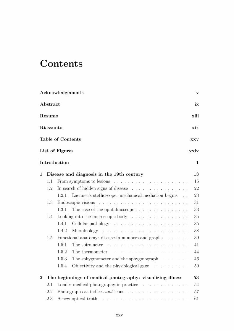
Contents
Acknowledgements v
Abstract ix
Resumo xiii
Riassunto xix
Table of Contents xxv
List of Figures xxix
Introduction 1
1 Disease and diagnosis in the 19th century 13
1.1 From symptoms to lesions . . . . . . . . . . . . . . . . . . . . . 151.2 In search of hidden signs of disease . . . . . . . . . . . . . . . . 22
1.2.1 Laennec’s stethoscope: mechanical mediation begins . . 231.3 Endoscopic visions . . . . . . . . . . . . . . . . . . . . . . . . . 31
1.3.1 The case of the ophtalmoscope . . . . . . . . . . . . . . . 331.4 Looking into the microscopic body . . . . . . . . . . . . . . . . 35
1.4.1 Cellular pathology . . . . . . . . . . . . . . . . . . . . . 351.4.2 Microbiology . . . . . . . . . . . . . . . . . . . . . . . . 38
1.5 Functional anatomy: disease in numbers and graphs . . . . . . 391.5.1 The spirometer . . . . . . . . . . . . . . . . . . . . . . . 411.5.2 The thermometer . . . . . . . . . . . . . . . . . . . . . 441.5.3 The sphygmometer and the sphygmograph . . . . . . . 461.5.4 Objectivity and the physiological gaze . . . . . . . . . . 50
2 The beginnings of medical photography: visualizing illness 53
2.1 Londe: medical photography in practice . . . . . . . . . . . . . 542.2 Photographs as indices and icons . . . . . . . . . . . . . . . . . 572.3 A new optical truth . . . . . . . . . . . . . . . . . . . . . . . . 61
xxv
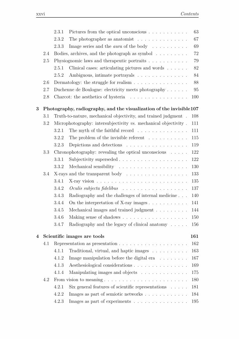
xxvi Contents
2.3.1 Pictures from the optical unconscious . . . . . . . . . . . 632.3.2 The photographer as anatomist . . . . . . . . . . . . . . 672.3.3 Image series and the aura of the body . . . . . . . . . . 69
2.4 Bodies, archives, and the photograph as symbol . . . . . . . . . 722.5 Physiognomic laws and therapeutic portraits . . . . . . . . . . . 79
2.5.1 Clinical cases: articulating pictures and words . . . . . . 822.5.2 Ambiguous, intimate portrayals . . . . . . . . . . . . . . 84
2.6 Dermatology: the struggle for realism . . . . . . . . . . . . . . . 882.7 Duchenne de Boulogne: electricity meets photography . . . . . . 952.8 Charcot: the aesthetics of hysteria . . . . . . . . . . . . . . . . 100
3 Photography, radiography, and the visualization of the invisible107
3.1 Truth-to-nature, mechanical objectivity, and trained judgment . 1083.2 Microphotography: intersubjectivity vs. mechanical objectivity . 111
3.2.1 The myth of the faithful record . . . . . . . . . . . . . . 1113.2.2 The problem of the invisible referent . . . . . . . . . . . 1153.2.3 Depictions and detections . . . . . . . . . . . . . . . . . 119
3.3 Chronophotography: revealing the optical unconscious . . . . . 1223.3.1 Subjectivity superseded . . . . . . . . . . . . . . . . . . . 1223.3.2 Mechanical sensibility . . . . . . . . . . . . . . . . . . . 130
3.4 X-rays and the transparent body . . . . . . . . . . . . . . . . . 1333.4.1 X-ray vision . . . . . . . . . . . . . . . . . . . . . . . . . 1353.4.2 Oculis subjecta fidelibus . . . . . . . . . . . . . . . . . . 1373.4.3 Radiography and the challenges of internal medicine . . . 1403.4.4 On the interpretation of X-ray images . . . . . . . . . . . 1413.4.5 Mechanical images and trained judgment . . . . . . . . . 1443.4.6 Making sense of shadows . . . . . . . . . . . . . . . . . . 1503.4.7 Radiography and the legacy of clinical anatomy . . . . . 156
4 Scientific images are tools 161
4.1 Representation as presentation . . . . . . . . . . . . . . . . . . . 1624.1.1 Traditional, virtual, and haptic images . . . . . . . . . . 1634.1.2 Image manipulation before the digital era . . . . . . . . 1674.1.3 Aesthesiological considerations . . . . . . . . . . . . . . . 1694.1.4 Manipulating images and objects . . . . . . . . . . . . . 175
4.2 From vision to meaning . . . . . . . . . . . . . . . . . . . . . . . 1804.2.1 Six general features of scientific representations . . . . . 1814.2.2 Images as part of semiotic networks . . . . . . . . . . . . 1844.2.3 Images as part of experiments . . . . . . . . . . . . . . . 195
Contents xxvii
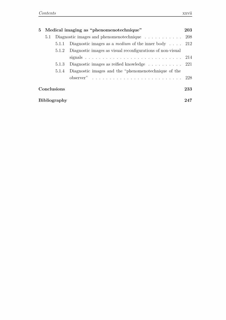
5 Medical imaging as “phenomenotechnique” 203
5.1 Diagnostic images and phenomenotechnique . . . . . . . . . . . 2085.1.1 Diagnostic images as a medium of the inner body . . . . 2125.1.2 Diagnostic images as visual reconfigurations of non-visual
signals . . . . . . . . . . . . . . . . . . . . . . . . . . . . 2145.1.3 Diagnostic images as reified knowledge . . . . . . . . . . 2215.1.4 Diagnostic images and the “phenomenotechnique of the
observer” . . . . . . . . . . . . . . . . . . . . . . . . . . 228
Conclusions 233
Bibliography 247

xxviii Contents
List of Figures
1.1 Figure representing different forms of tubercular matter and someof its effects. T. Laennec, Traité de l’auscultation mediate, 2ndEdition, 1826, Paris, Plate II, Fig. 2. . . . . . . . . . . . . . . . 25
1.2 Illustration of Virchow’s cell theory. Archiv fur PathologischeAnatomie und Physiologie (now Virchows Archive), 1847, firstissue. Wikimedia Commons. . . . . . . . . . . . . . . . . . . . . 37
1.3 Silhouette illustration of how to position the patient’s body inrelation to the spirometer in order to perform the respiratorytest. From J. Hutchinson, 1846, 236. . . . . . . . . . . . . . . . 42
1.4 An example of Hutchinson’s tables. Here vital capacity is pre-sented in relation to professional activity and stature. From J.Hutchinson, 1846, 156. . . . . . . . . . . . . . . . . . . . . . . . 43
1.5 An example of Hutchinson’s charts. From J. Hutchinson, 1846,155. . . . . . . . . . . . . . . . . . . . . . . . . . . . . . . . . . 43
1.6 A direct sphygmograph designed by Marey. E.-J. Marey, La cir-culation du sang à l’état physiologique et dans les maladies, Paris,1881, 214. . . . . . . . . . . . . . . . . . . . . . . . . . . . . . . 49
1.7 Example of sphygmograms. E.-J. Marey, La circulation du sangà l’état physiologique et dans les maladies, Paris, 1881, 216. . . . 50
2.1 Adiantum pedatum. Maidenhair fern, young rolled-up fronds en-larged 8 times. Photogravure after K. Blossfeld, Urformen derKunst, Berlin, 1928, Plate 55. . . . . . . . . . . . . . . . . . . . 64
2.2 A man walking. Photogravure after Eadweard Muybridge, 1887.Wellcome Library, London. . . . . . . . . . . . . . . . . . . . . . 65
2.3 Example of signaletic photograph, A. Bertillon, Instructions sig-nalétiques. Album, Melun, 1893. Wellcome Library, London.. . . . . . . . . . . . . . . . . . . . . . . . . . . . . . . . . . . . 74
2.4 Specimens of composite portraiture, F. Galton, Inquiries into Hu-man Faculty and It’s Development, London, 1883, Frontispieceillustration. Wellcome Library, London. . . . . . . . . . . . . . . 76
xxix
xxx List of Figures
2.5 Patient at Surrey Asylum, case identified as “religious melan-choly.” Calotype by H.W. Diamond, 1852. . . . . . . . . . . . . 81
2.6 Patient at Surrey Asylum, case identified as “melancholia passingon to mania.” Calotype by H.W. Diamond, 1852. . . . . . . . . 83
2.7 Patients at Surrey Asylum. Calotypes by H.W. Diamond, 1852. 862.8 Patient at Surrey Asylum. Calotype by H.W. Diamond, 1852. . 872.9 Female patients affected by impetigo (left) and pemphigus fo-
liaceus (right). A. Hardy and A. de Montméja, Clinique pho-tographique de l’Hôpital Saint-Louis, Paris, 1868. . . . . . . . . . 92
2.10 Syphilitic alopecia (left). Pigmentation probably caused by photo-sensitization due to a cosmetic product (right), F. Mehéux, ca.1884-1893. . . . . . . . . . . . . . . . . . . . . . . . . . . . . . 94
2.11 Muscular atrophy, Duchenne de Boulogne, Album de photogra-phies pathologiques, Paris, 1862, Figure 9. Public domain. . . . 97
2.12 The facial expression of profound attention on the human face be-ing induced by electrical current. Duchenne de Boulogne, Le Mé-canisme de la physionomie humaine, Paris, 1862, Plate 1, figure9. Wellcome Library, London. . . . . . . . . . . . . . . . . . . . 98
2.13 Passional attitudes (ecstasy). Photograph by Bourneville andP. Regnard, Iconographie photographique de la Salpêtrière, Paris,1878, Plate 23. . . . . . . . . . . . . . . . . . . . . . . . . . . . 103
2.14 Hystero-epileptic seizure. Photograph by Bourneville and P. Reg-nard, Iconographie photographique de la Salpêtrière, Paris, 1879-1880, Plate 3. . . . . . . . . . . . . . . . . . . . . . . . . . . . . 103
2.15 Hysterical woman yawning. Photograph by A. Londe, Nouvelleiconographie de la Salpêtrière, Paris, 1890. Wellcome Library,London. . . . . . . . . . . . . . . . . . . . . . . . . . . . . . . . 105
2.16 Episode of male hysteria. Photograph by A. Londe, Nouvelleiconographie de la Salpêtrière, Paris, 1888. . . . . . . . . . . . . 106
3.1 Blood corpuscles. Etchings produced after the original daguerreo-types. A. Donné and L. Foucault, 1845. . . . . . . . . . . . . . . 112
3.2 Examples of chronophotographs by E.-J. Marey. . . . . . . . . . 1243.3 Images of a runner reduced to a system of bright lines for rep-
resenting the positions of the limbs (geometrical chronophoto-graph). Photograph by E-J. Marey, 1886. . . . . . . . . . . . . . 125
3.4 Man with experimental shoe. E.-J. Marey, Le Movement, Paris,1894, Engl. translation, New York, 1895, Figure 4. . . . . . . . . 126
List of Figures xxxi
3.5 Diagram of the sequence and duration of the footfalls during fourdifferent forms of walking. E.-J. Marey, 1894. . . . . . . . . . . 127
3.6 Diagram of the sequence and duration of the footfalls of a horseat a full trot. E.-J. Marey, 1894. . . . . . . . . . . . . . . . . . . 128
3.7 Chronophotograph of a semicircular arc of polished brass rotatingaround a vertical axis. E.-J. Marey, 1894. . . . . . . . . . . . . . 132
3.8 The bones of a hand with a ring on one finger, viewed throughX-rays. Roentgen, W.K., 1895. Photoprint from radiograph.Wellcome Library, London. . . . . . . . . . . . . . . . . . . . . . 135
3.9 J.M.W. Turner (1775-1851), Rain, Steam and Speed – The GreatWestern Railway, 1844. National Gallery, London. . . . . . . . . 142
4.1 Examples of images produced using the Visible Human Projectdataset. . . . . . . . . . . . . . . . . . . . . . . . . . . . . . . . 164
4.2 Anatomical fugitive sheet bound at the end of Valverde, Vi-vae imagines partium corporis humani aereis formis expressae.Antwerp, 1566. Wellcome Library, London. . . . . . . . . . . . . 168
4.3 Table of correspondences between X-ray imaging and physicalexamination of the thorax. From J.F. Halls Dally, 1903. . . . . . 185
4.4 Table showing the range of mobility of the of the diaphragm.From J.F. Halls Dally, 1903. . . . . . . . . . . . . . . . . . . . . 188
4.5 Examples of Halls Dally’s diagrammatic drawings representingphysical and radioscopic (fluoroscopic) examinations. From J.F.Halls Dally, 1903, 1805. . . . . . . . . . . . . . . . . . . . . . . . 190
4.6 Example of gel electrophoresis autoradiograph (DNA fingerprint).Credit: Alec Jeffreys, Wellcome Images. . . . . . . . . . . . . . 197
5.1 Radiograph showing air-filled trachea and lungs, diaphragmaticdomes, mediastinal structures, and vascular markings. Arrowsindicate costophrenic angles. Source: US Army medical trainingcourse. Wikimedia Commons. . . . . . . . . . . . . . . . . . . 216
5.2 Computed tomography scan of the brain, axial view. . . . . . . 2175.1 Cluster heat map of lung tumor image features extracted from
CT images from 276 patients. Kumar et al., 2012. . . . . . . . . 244

xxxii List of Figures
Introduction
Since its initial proposal, this dissertation has gone through a deep reformula-tion of both the object of study and the method of investigation. Developedwithin the context of an interdisciplinary research project called Image in Sci-ence and Art,1 my early ideas for a doctoral thesis revolved around the artisticuse of medical imaging technologies. The objective was to understand how med-ical imaging has transformed our perception and first-person experience of theinner body, both at an individual and social level. The main question was tounderstand if our experience of the body changes, once we can look inside itwithout having to cut it open. Do we relate differently to our own body, andto the bodies of other people, if we perceive them as potentially transparentrather than ineluctably opaque? The working hypothesis was that the study ofartworks related to medical imaging technologies could provide valuable insightsinto such problems. The rationale underlying this approach was that, as expres-sion of an individual, artworks can help disentangling questions related to bodyvisualization and personal identity. At the same time, as cultural objects, theycan be used as instruments of analysis to explore social notions of body andtechnology.2 Thus, the initial research plan did not focus on medical images perse, but rather on the meanings these images acquire outside the medical setting.It concerned the first-person perception of the inner body mediated by images,rather than the epistemology of medical imaging.3
1The FCT research project Image in Science and Art (PTDC/EAT/64201/2006), directedby Prof. Olga Pombo, began in 2007 and ended in 2011 with the exhibition “CorpoImagem.Representações do Corpo na Ciência e na Arte”, held in February and March 2011 in Lisbon,at the Pavilhão do Conhecimento-Ciência Viva.
2See Coulombe, 2006.3In the first year or so of my doctoral research, I attempted a comprehensive survey of the
artists who work with medical imaging. The task proved unattainable, but nevertheless itwas possible to outline a general view of the multiple ways whereby visual artists encapsulatemedical imaging in their work, both at the conceptual and formal level. The results of thisearly phase of the research were published in the article “Inside the body: Medical imagingand visual arts” (Di Marco, 2012a), and in a chapter of the book Representações do Corpo naCiência e na Arte (Azevedo Tavares, 2012) published within the context of the aforementionedFCT research project. Both texts are in the Appendix attached in electronic format to thisdissertation.
1
2 Introduction
The change of conceptual focus derived from the need to extricate and clarifythe multiple meanings that tend to grow around medical images. In fact, if animage arises not only different interpretations, but also different emotions,4 itis because it is inherently ambiguous. In order to unravel the origins of thisambiguity, it was necessary to understand the material and conceptual genesisof medical imaging. That is, it was necessary to understand how such images areproduced and endowed with meaning in their native context, clinical medicine.In the course of my research this historical, conceptual, and epistemologicalanalysis became the central object of the dissertation. In other words, an inquiryinto the extra-medical meaning of medical imaging morphed into a study on itsepistemology. More precisely, it morphed into a study that aims at clarifyingthe relationship between the images produced by medical imaging technologiesand the body they make visible.
Before saying more about the questions approached in this dissertation andthe methodology employed, it is important to clarify what is meant by medicalimaging. The term medical imaging refers to a vast array of images and imag-ing technologies developed since the end of the nineteenth century to provideindirect visual access to the inner organs (morphological imaging), and to somephysiological or molecular processes (functional imaging). The first of such tech-nologies was radiography, invented by the German physicist Wilhelm Roentgenin 1895. Since then, a number of different imaging modalities have emerged, andthey have completely redefined the visibility of the interior of the living body.Nowadays Computed Tomography (CT), Magnetic Resonance Imaging (MRI)and ultrasound scanning allow visualizing inner structures with astonishing de-tail, while Positron Emission Tomography (PET) and functional MRI (fMRI)allow visualizing the metabolic activity of different biological tissues, includingthe brain. Medical imaging is generally used for diagnostic aims. However, itcan also serve research purposes, as in the case of neuroimaging in cognitivescience, or rather mundane goals, as in the case of fetal ultrasound, which isroutinely performed not only to check the health conditions of the fetus, butalso to let parents enjoy the experience of seeing their prospective baby on thescreen.5 In my study I focus on the diagnostic use of medical imaging. This is
4The art historian and image theorist James Elkins remarks that, unlike other scientificimages, which can be strictly informational, medical images tend to show vestiges of expressivemeaning, because they can evoke questions of life and death, gender and sexuality, pleasureand pain. See Elkins, 1995, 556.
5See Chudleigh, 1999; Mitchell, 2001; van Dijck, 2005, Ch. 6. Fetal sonograms are partic-ularly interesting images, from both an epistemological and sociological perspective. Beforethe introduction of ultrasound scanning for pregnancy monitoring, the presence of the unborncould be felt only by the pregnant woman. Since it has become visible, however, the fetus hasacquired a new status. On the one hand, it has become an object of study, as well as a medicalsubject (fetal medicine). On the other hand, it has acquired a new identity and individuality,
Introduction 3
why I will often use the expression diagnostic images. I also employ the des-ignations mechanical images, radiological images, and medical images as loosesynonyms, even though, strictly speaking, the term medical images encompassesa much larger domain of images, including, for instance, anatomical drawings.
A peculiarity of medical imaging is that the images it creates seem to ren-der the body transparent. Intuitively, we think that the medical relevance ofdiagnostic images depends on the fact that they are faithful representations ofthe inner body, but, as soon as we set out to study them in terms of diagnosticinstruments and, more generally, in terms of cognitive objects, we come acrossa number of problems: What do these images represent, exactly? And how dothey represent? Do they work by mimesis? If we do not know how their refer-ent looks like, how can we trust their representational value? Are these imagesportraits of the inner body? Or are they maps? If they are portraits, are theythe portrait of someone (a person) or of something (a disease)?
These questions, present already in the first version of my doctoral project,turned out to be much more difficult to answer than I had initially assumed.In fact, although there are several works on medical imaging from the per-spective of cultural studies6 and anthropology,7 very little has been publishedabout medical imaging epistemology. Indeed, unlike historians and sociologists,philosophers of science have traditionally neglected images and, until relativelyrecently, in both continental and analytic philosophy the reflection on visual rep-resentation has been a preserve of aesthetics and art theory.8 For what concernsthe specific domain of medical imaging, the philosophers of science who havepaid attention to this topic have typically focused on the use of neuroimaging incognitive science and, to a lesser extent, psychiatry.9 However, to the best of my
entering as a silent actor into a set of relationships between medical practitioners, parents andsociety. The effects of this transformation on the creation of a new social and juridical subjecthas been widely investigated by sociological and anthropological literature. Feminist authors,in particular, have repeatedly analyzed the role of fetal scans in the increased medicalizationand commercialization of pregnancy, in the strengthening of ideologies of good motherhood,in the creation of the fetus as an autonomous individual independent from the mother’s body,and in the debates surrounding abortion. See, for instance, Petchesky, 1987; Duden, 1993;Taylor, 1998 and 2008; Morgan and Michaels, 1999; Morgan, 2009; Roberts, 2012.
6See Zwijnenberger and van de Vall, 2009; van Dijck, 2005; Natale, 2008, 2011, 2012;Stephens, 2012.
7See 2004; Radstake, 2009; Müller-Rostock, 2009; Estival, 2009, 2010.8This situation has been changing over the last decade. See, for instance, Pombo and
Di Marco, 2010; Pombo and Gerner, 2010; Carusi, 2011, 2012. In the analytic tradition seeFrench, 2003; Perini, 2005ab, 2006, 2012.
9See Kosslyn, 1999; Bogen, 2002; Taraborelli, 2003; Roskies, 2007; Huber, 2009; Huberand Huber, 2009, Mole and Klein, 2010; Klein, 2010. Even in the book Medical Imaging andPhilosophy. Challenges, Reflections and Actions (Fangerau et al., 2012), all the articles thatdeal with epistemology refer to neuroimaging.
4 Introduction
knowledge, virtually nothing has been published on the philosophy of medicalimaging as a diagnostic instrument.10
Objectives and methodology The main objective of this dissertation isto contribute to the development of an epistemology of medical imaging. Mycentral thesis is that medical imaging does not merely produce more or lessaccurate pictures of the inner body, it rather turns the body into a scientificobject by transforming its very visibility. Simultaneously it aims at reducingillness to a visual entity. Medical imaging does not simply make visible the innerorgans, it actually presents the body in such a way that it becomes possible toextract relevant diagnostic information from it. This is why I maintain that theimages produced by medical imaging technologies are more akin to simulationsthan to portraits. The imaging apparatus turns the body into a visual objectthat can be observed under experimental conditions: unlike the real body, itcan be filed, retrieved, shared, measured and manipulated in several ways.
This thesis is accompanied by two others: first, diagnostic images, as allscientific images, are actual cognitive instruments. They are epistemic objectsinscribed within theoretical contexts and experimental practices. Second, animage of the inner body has diagnostic meaning and value only in the scopeof a specific conceptualization of the body and disease. This means that, inorder to put forward an epistemology of medical imaging, to develop a the-ory of technology-mediated images and technology-mediated perception is notenough. One must also clarify the medical concepts and practices that pro-vide the substratum for the whole process of production and signification ofdiagnostic images.
Medical imaging is to be understood both as an imaging technology andas a diagnostic practice, hence, as an object of philosophical analysis it mustbe approached from different perspectives. For this reason in my research Iresorted to literature from a variety of disciplinary fields, such as image theory,semiotics, history and philosophy of medicine, and history and philosophy ofscience. The aim was to put forward an epistemology of medical imaging thataccounts for the poietic rather than mimetic nature of visual representation, aswell as for the fact that through medical imaging the human body is embedded
10An exception is the doctoral dissertation Signal into Vision: Medical Imaging as In-strumentally Aided Perception, defended by Nola Semczyszyn at the University of BritishColumbia in 2010. Drawing mostly on analytic philosophy of art and perception, Semczyszynappeals to theories of pictorial representation to explain how medical imaging represents andhow we access its content. I refer to her work in Chapter 5, Section 5.2.2. Overton et al.,2011, focuses on the problem of the relation between diagnostic images and verbal language,and the creation of ontologies associated to software tools to improve communication amongradiologists, other health professionals, and patients.
Introduction 5
into an empirical apparatus, which encompasses medical instruments, medicalknowledge, and clinical practices.
For what concerns the methodology, I relied chiefly on historical and con-ceptual analysis. On the one hand, I studied the genesis, development andtransformation of concepts such as disease and diagnosis; on the other hand, Icritically applied philosophical concepts developed by different authors to prob-lematize and examine my objects of research (e.g., the production of mechanicalimages and the visualization of the invisible). The study of original scientificdocuments, such as medical treatises and medical journals’ articles was indis-pensable for understanding how medical imaging has progressively imposed itselfas a fundamental diagnostic method, and how illness has become a visual object.Thus, I relied on documents’ analysis to unravel how the diagnostic meaning ofimages was generated in the scope of actual scientific and clinical practices anddebates. The emphasis on material scientific practices was reinforced, wheneverpossible, by resorting to literature from laboratory ethnology and anthropol-ogy.11 Also, since this work is about images, I devoted much attention to theiconographic apparatus of the documents I examined, employing iconographicand semiotic analysis.
As mentioned above, given the multifaceted nature of the epistemologicalproblems posed by medical imaging, I had to draw on concepts and ideas devel-oped by a variety of thinkers. In particular, I follow Charles Sanders Peirce’struth-value theory of photography to better understand how mechanically pro-duced images were conceptualized in the nineteenth and early-twentieth cen-turies.12 Moreover, I resort to his semiotics, in particular his classification ofsigns into indices, icons and symbols, to develop a tool for semiotic analysisthat I employ at various points of the dissertation to investigate the processesof signification of different kinds of instrument-generated images that visualizeinvisible referents (from microphotographs to radiographs and PET scans).
I explore at length the idea that optical media such as photography and cin-ema are prostheses that enrich and transform our sensorial experience throughWalter Benjamin’s notion of optical unconscious. Benjamin remarked that pho-tography and cinema allow for manipulations of space and time that are pre-cluded to natural perception. Consequently, these imaging technologies endowus with enhanced mechanical senses that allow exploring completely new facets
11See Chapter 4, Section 4.2.3, and Chapter 5, Section 5.2.3.12In my study I payed much attention to photography because when radiography appeared,
at the turn of the nineteenth century, it was perceived as a particular kind of photography.Hence, to understand how radiography was conceptualized, it is necessary to look at thephotographic practices and theories of the time.
6 Introduction
of nature.13 I combine the concept of optical unconscious with Peirce’s semi-otics to discuss the problem of the invisible referent, that is, the question ofhow we can make sense of images that are meant to visualize invisible phenom-ena. Besides the idea of optical unconscious, two other aspects of Benjamin’sreflection on photography and cinema proved fruitful for my investigation onmedical imaging. One is his seminal discussion of the cognitive effects of themechanical reproduction of images; the second is his account of the objectify-ing nature of photography, which he compares to the dissecting activity of thesurgeon. I draw on his considerations on these subjects to elaborate an accountof the role played by photographic series in creating a new clinical gaze, andto reflect upon the role played by photography and radiography in turning thehuman body into a scientific object.
The idea of mechanical objectivity, developed by the historians Lorraine Das-ton and Peter Galison,14 offers me the opportunity to engage with the problemsof image realism and objectivity through polemical reasoning. These authorsdeveloped an articulated taxonomy of scientific images aimed at demonstratingthe historical character of objectivity, which should be considered one of themany epistemic ideals that can drive scientific work in different historical pe-riods. Mechanical objectivity, they argue, has been the main epistemic idealbetween the 1830s and the 1920s. It prescribed the strenuous suppression of thesubjectivity of the image makers, and put harsh limitations on image interpre-tation, too. In order to refute this account of what it means for an image tobe objective, and to clarify what sort of photographic realism was embraced bythe scientists of the late-nineteenth and early-twentieth centuries, I develop anumber of arguments concerning the relationship between objectivity and inter-subjectivity, as well as between mechanically produced images and imagination.Similarly, Daston and Galison’s characterization of virtual and haptic imagesgives me the opportunity to critically delve into the problem of the embodiedand material dimension of scientific images’ production and fruition.
A concept I came across in a late phase of my research is Gaston Bachelard’s“phenomenotechnique.”15 Bachelard developed the idea of phenomenotechniqueto account for the fact that, in advanced physics and chemistry, scientific en-tities (e.g., the Zeeman effect, perfect crystals, atomic isotopes) are not found
13See Benjamin, 1939, 266.14The historiographic work of Daston and Galison is guided by strong epistemological as-
sumptions. Indeed, together with the philosopher Ian Hacking, they are considered amongthe most prominent exponent of so-called historical epistemology, an approach to the studyof scientific knowledge that emphasized the historical development of scientific concepts andobjects, as well as of scientific disciplines and styles of reasoning. See Hacking; 2002, Kusch,2011; Sturm, 2011.
15Bachelard, 1931, 18.
Introduction 7
in nature ready made. They must be produced in the laboratory as technicalphenomena. They are the materialization of mathematical rationality or, asBachelard put it, of a mathematical noumenon. In other words, through phe-nomenotechnique, science creates its own phenomena in a process of progressiverationalization of the real. Through this process science leaves behind the worldas it is understood by commonsensical experience and received mental habits,and sets free “a surrationalism that will multiply the occasions for thinking.”16
If rigorously interpreted, the notion of phenomenotechnique cannot be appliedto medical imaging and diagnostic images, for reasons I discuss in detail in thedissertation. Yet, the analysis of this concept offered me an additional stimulusfor thinking medical imaging. In fact, not only it provides specific insights forreflecting on contemporary medical imaging, wherein mathematical algorithmsfor image acquisition and reconstruction play a fundamental role; more impor-tantly, it works as an organizing concept, which helps bring together and refinethe different ideas and intuitions developed in the various steps of my research.
Plan of the dissertation The dissertation begins with an analysis of the his-torical and conceptual conditions of possibility of medical imaging. By outliningan archaeology of radiography, I will try to understand what medical theoriesand practices had to be at work in the nineteenth century, for a technology thatproduced shadow-images of the inner body to be perceived and employed as a di-agnostic instrument. I will suggest that when radiography appeared, at the turnof the twentieth century, it engendered less a theoretical than a technologicalrevolution, because its diagnostic relevance was grounded in the conceptual-ization of body, disease and diagnosis put forward by clinical anatomy at theend of the eighteenth century. Following Michel Foucault’s The Birth of theClinic, I will maintain that it was with the work of the French clinicians XavierBichat and Jean-Nicolas Corvisart that diagnosis became a matter of elicitingsigns from the inner body and that visibility became a fundamental epistemo-logical and perceptual category of medicine.17 I will also defend the idea thatthe stethoscope, invented by Théophile Laennec in 1816, was the material andintellectual predecessor of radiography, because it introduced a primitive formof mediated perception in medical diagnosis, and allowed the clinician to ex-plore from the outside the inner body of the living patient, extracting signs ofdisease.18 In the course of the nineteenth century, several ways of understanding
16Bachelard, 1936, 7, my translation.17See Foucault, 1963, 166.18The stethoscope is used to auscultate the patient. Auscultation is a basic clinical ex-
amination. It consists of listening to the sounds produced by the heart and by the air thatcirculates in the respiratory organs.
8 Introduction
the functioning of the body and the ontology of disease emerged. I will showthat these theoretical transformations went hand in hand with the developmentof different instruments, from the microscope to the sphygmograph, which vi-sualized different aspects of both the morphology and physiology of the humanbody. Each of these instruments fulfilled specific diagnostic aims and poseddistinct epistemological problems, but all of them shared some commonalities:they were meant to replace the subjective sensations of patients and doctorswith objective indices of health and disease; they created visual records of theinner body that could be filed, retrieved and shared among physicians; they re-quired the development of a specialized language agreed upon by a communityof experts; they created a progressive physical separation between the body ofthe patient and the body of the physician. It was in this framework of medicalideas and practices that X-ray imaging appeared and progressively acquired itsdiagnostic function.
In the second chapter I examine the early developments of medical photog-raphy in order to understand how the first technology for the production ofmechanical images entered the domain of medicine. Although photography isnot considered a diagnostic instrument proper, it is important to explain howit influenced the way doctors looked at the patient’s body and visualized dis-ease. The main theoretical references in this chapter are Peirce’s semiotics andBenjamin’s reflections on the optical unconscious, the mechanical productionand reproduction of images, and the intrinsic analytic and dissecting poten-tial of photography and cinema. Drawing on these authors, and analyzing theworks of early physicians-photographers in psychiatry, dermatology, neurologyand physiology, I will show that the photographic series collected in medicaljournals, manuals and hospital archives, produced a new visibility of the pa-tient’s body by creating an updated version of the Foucauldian clinical gaze.According to Foucault, the clinical gaze was born in the hospital wards, becausethat was the place were the individual body of each patient became a public andvisible body that could be compared to many others, finding out similarities anddifferences that helped organizing a variety of signs, symptoms and anatomicallesions into nosological categories. I will argue that the photographic collectionallowed to replicate this sensorial experience and cognitive operation from a dis-tance. I will also demonstrate that the photographic series was part of a largerexperimental apparatus, which encompassed the patient, the camera and theobserver, and whose aim was to turn the body and disease into visual objects,available for scientific analysis.
In the third chapter I discuss the problem of the invisible referent, that is,I analyze the processes whereby photographs that reveal invisible phenomena
Introduction 9
are endowed with meaning. This is likely to be the fundamental problem of allscientific images. When the referent of a picture is invisible, the iconic mode ofsignification fails, because in this case the image produced by the mechanical orelectronic apparatus does not look like anything we already know, it resemblesnothing. So, how do we know that the object we see in the photograph – e.g., acell or a tubercular lesion – is really there and does really look like that? Draw-ing on the conceptual analysis developed in the previous chapter, I will defendthe idea that the visualization of the invisible entails a peculiar combinationof the indexical, iconic and symbolic modes of signification. My reasoning isbuilt in opposition to Lorraine Daston and Peter Galison’s idea of mechanicalobjectivity.19 According to Daston and Galison, for the scientists of the nine-teenth century, photographs were unerring records of reality. Consequently, theresearcher should restrain from any intervention during image production, avoidany subjective interpretation, and let the image speak for itself. This aim waspurportedly reached by endorsing strict procedural protocols in the differentphases of production and publication of scientific photographs. I will show,however, that the notion of mechanical objectivity is a caricature of the actualideas and practices developed by the scientists of the nineteenth century to dealwith the problem of the visualization of the invisible. The argument will bearticulated in three moments, corresponding to the analysis of how the problemof objectivity and image signification was managed by scientists who workedwith microphotography, chronophotography, and radiography.
In the fourth chapter I argue that images are cognitive tools and that rep-resentation is never an act of mechanical repetition, it always entails a creativecomponent. As in the previous chapter, part of my discourse is built in contrastwith Daston and Galison, challenging their claims concerning the passive natureof representation. For these authors, scientific images can be classified in threecategories: traditional, virtual and haptic images. Traditional images are allthose images that cannot be manipulated on a computer screen; virtual imagesare digital images associated to computer algorithms for image manipulation;while haptic images are images whose production requires the actual manipu-lation of the object during the imaging process (for Daston and Galison, thisdefinition only applies to nanoimages). Traditional images, they say, are merere-presentations of the world, because they are “focused on copying what alreadyexists,”20 while virtual and haptic images are presentations, because they dis-play their objects in ever changing ways. Accordingly, traditional images work
19See Daston and Galison, 1992, 2007.20Daston and Galison, 2007, 383.
10 Introduction
as “evidences,” while virtual and haptic images are “tools.”21 I will criticizethis classification of scientific images with historical and theoretical arguments.From the historical point of view, I will show that at least since the sixteenthcentury there have been attempts to create images that can be actually ma-nipulated by the observer, resorting to paper flaps that can be lifted revealingdifferent layers of a representation. From the theoretical perspective, I will drawon a variety of literature spanning from art theory to neuroscience, to demon-strate that the very notion of a passive representation is unsustainable, becauseimages always engage the observer in an embodied act of perception, which elic-its not only visual, but also tactile sensations and motor reactions. Moreover, Iwill argue that Daston and Galison’s emphasis on nanoimaging is misleading. Infact, even when they do not reach the peaks of technological sophistication thatcharacterizes nanoimages, scientific images are the result of some manipulationof the natural object they represent. A scientific image cannot be an automatedcopy of nature, because it is part of an experimental praxis, whose goal is tolearn something about natural phenomena, not just to reproduce them. Thisposition will be further corroborated by the analysis of actual scientific practicesof image signification, taking into account written documents (semiotic analysisof a radiology article) and material practices (laboratory ethnography).
In the last chapter, I wrap-up the different arguments discussed through-out the dissertation, and apply them to contemporary medical imaging. I doso by first analyzing and subsequently re-elaborating Bachelard’s concept ofphenomenotechnique. As already mentioned, the idea of phenomenotechniquecannot be applied to medical imaging in rigorous terms. However, there aretwo characterizing elements of this concept that provide important insights fora philosophy of technology and, consequently, for an epistemology of medicalimaging. The first is the idea that in order to study a natural phenomenon, sci-entists must previously re-create it as a scientific phenomenon, and this requiresthe creation of artificial objects. The second, strictly related to the former, isthat scientific experience is by necessity mediated, and such mediation has bothan intellectual and material character. This means that the development ofscientific instruments and technologies is not a second-order product of science,but rather part and parcel of the scientific process. Technology is embedded intoscience, because our scientific grasping of the world is necessarily mediated by in-struments; scientific instruments, in turn, are materializations of a vast body ofscientific knowledge and practices (in the case of digital imaging this knowledgehas an eminently mathematical character). In other words, science and technol-ogy are reciprocally constituted. On these grounds I will propose a description
21Daston and Galison, 2007, 385.
Introduction 11
of medical imaging in terms of phenomenotechnique, using this concept as akey-word around which to reorganize the ideas previously discussed. Firstly,I will resort to the concept of phenomenotechnique to gain insights into howdiagnostic images mediate the physician’s sensory and intellectual experience.Secondly, I will give an account of diagnostic images as artificial phenomena(visual reconfigurations of non-visual signals) that work as simulations of thepatient’s body, and that reify different domains of knowledge (from medicine tophysics and engineering). Finally, I will argue that the proper and efficient signi-fication of a diagnostic image requires what I call a “phenomenotechnique of theobserver.” To recognize the signs of disease in an image of the inner body, onehas to master the explicit and implicit rules necessary to make sense of the novelsensory domain produced by the technological apparatus. This implies aban-doning spontaneous modes of perception and signification to engage in a processof educated perception. The idea that we only recognize what we know and thatwe need to know in order to see is a truism in both contemporary psychology ofvision and philosophy of science, and the relevance of education in shaping thescientist’s ability to see has been acknowledged and discussed by philosophers asdiverse as Karl Popper and Thomas Kuhn. The idea of a “phenomenotechniqueof the observer,” however, helps stressing some specific features of the educationof vision related to the medical use of machine-mediated images, and I will useit as a conceptual tool to analyze the complex and extended epistemic networkthat envelops diagnostic images and makes them intelligible.
To unravel this extended epistemic network is a necessary precondition forunderstanding the tendency of diagnostic images to produce a proliferation ofmeanings that span beyond the clinical domain. The images produced by med-ical imaging technologies are open to multiple processes of signification becauseof their inherent complexity, and they are complex because they embody mul-tiple domains of knowledge, because they alter the visibility of the body, andbecause they invite the beholder to abandon or rethink ingrained habits of visualperception.

12 Introduction
Chapter 1
Disease and diagnosis in the
19th century
Within the history of medicine, radiography and its cognate visualization tech-nologies (e.g., ultrasound imaging, Computed Tomography, Magnetic ResonanceImaging, Positron Emission Tomography) stand at the crossing of perceptive andcognitive strategies developed to deal with two different problems. The prob-lem of representing the inner body as an object of scientific knowledge (thus, auniversal object), and the problem of accessing the body in order to diagnosea specific ailment of an idiosyncratic individual. While the first endeavor datesback at least to 3000 BC,1 the second became a major concern for physiciansonly in the early-nineteenth century, and went hand in hand with a profoundredefinition of the concepts of disease and diagnosis. Additionally, within thehistory of diagnostic techniques – which is strictly related to the history ofthe concept of disease – diagnostic images blend together two complementarystrategies for accessing the inner body: (1) the indirect (mediated) collectionof invisible signs from internal organs, made possible by instruments like thestethoscope and the sphygmograph, and (2) the direct visual access to the innerbody through optical devices, like the vaginal speculum and the laryngoscope,which exploit natural corporeal orifices (endoscopy). We can say that diagnos-tic images combine (1) and (2) because they provide an indirect visual accessto the inner body, based on the collection of invisible signs from the internalorgans. More precisely, they are visual representations based on non-visual (ornon-optical) properties of the inner body.2
1See Cazort et al., 1996; Cosmacini, 1997.2I will elaborate on the issue of the visual-mimetic configuration of non-visual data in
Chapter 5, where I give some details on the physical processes underlying the production ofdifferent diagnostic images. For the moment it is enough to recall that while natural visionand analog photography depend on the absorption and reflection of electromagnetic wavesin the frequency of visible light by the surface of the objects (optical property), an imaging
13
14 Disease and diagnosis in the 19th century
This means that, if we are to develop an epistemology of diagnostic images,we cannot limit our analysis to the problems of visual representation, but wehave to consider medical imaging as part of a wider group of tools (or tech-nologies) used to extract information from the healthy and the diseased body.Moreover, we have to understand which concepts of disease and body underlaidthe use of such tools and were, in turn, reinforced by them. That is to say, weshould try to disentangle the reciprocal shaping of ideas, natural objects, andinstruments.
It is widely recognized that the introduction of radiography in clinical prac-tice marked a major turning point in the history of medicine. According tothe historian of medicine Stanley Reiser, it opened a new visual era in medicalpractice. For Reiser, medical imaging “changed the ways patients and doctorsexperienced illness [. . . ] and caused medicine to see its task and itself differ-ently.”3 Similarly, in her much quoted book Screening the Body, visual studiesscholar Lisa Cartwright claims that radiography accomplished “a quite literaldisintegration of the body,”4 which transformed the way illness is perceived byboth medical practitioners and patients. Indeed, unlike the majority of medicalinnovations developed during the nineteenth century, from the stethoscope tothe germ theory, X-ray images exerted an almost immediate impact on medi-cal practice. To understand this phenomenon, stressed by many historians ofmedicine, it is important to emphasize that radiography neither acquired itsepistemic relevance in a void, nor redefined the notion of disease and the (med-ical) representation of the body out of nothing. Quite the contrary.
In this chapter I defend the view that for what concerns the domain of medi-cal diagnosis, radiographic techniques entailed less an epistemological revolutionthan a technological one, since its primary diagnostic relevance is anchored tothe conception of body and disease (localizationist paradigm) put forward byclinical anatomy, at the dawn of the nineteenth century. Moreover, radiographyenriched the armamentarium of research and diagnostic techniques developedduring the nineteenth century in order to transform medicine from ars curandiinto a full-fledged empirical science, on the model of physics and chemistry. Thisentailed the reorganization of the concepts of body and disease around facts andbasic units that could be measured and compared. Accordingly, it became neces-sary to produce objective representations of the body, through instruments that
process like radiography exploits the electromagnetic waves in the frequency of X-rays andtheir differential absorption by the internal and external structures of our bodies (non-opticalproperty). If the skin absorbed the X-rays like the bones do, or if all the inner bodily structureshad the same opacity to X-rays, then it would be impossible to see into the body by meansof a radiograph.
3Reiser, 2009, 14.4Cartwright, 1995, 108.
From symptoms to lesions 15
could bypass both the patient and the doctor’s sensorial limitation and subjec-tivity, in formats that could be shared among different observers, in differentplaces and times. Since a specific sensible perception and rationality had devel-oped during the century, medicine was ready to immediately take on radiographywhen it appeared in 1895. There is no doubt that since then medical imaginghas gone a long way, yet, I think that many of the conceptual problems putforward by ultrasound imaging, Computed Tomography (CT), Magnetic Res-onance Imaging, (MRI) and Positron Emission Tomography (PET) are rootedin the nineteenth century’s attempt to produce a new, specific expressivity ofthe body. That is, to force it (or to allow it) to express its forms, motions, firstconstituents, and ailments clearly and unmistakably.
1.1 From symptoms to lesions
In the text “L’élaboration du diagnostic,” Peitzman and Maulitz describe themaking of a diagnosis as a complex process of “divination” [divination]. Thedoctor has the task to put some order in the confusion of signs and symptomsmanifested by the patient, with the aim of answering the complex question:“What is wrong with this person?” This requires the implementation of a numberof “cognitive and methodological negotiations.” Such negotiations occur withinthe constraints of the biological information and technical resources available atany given historical moment, as well as on the basis of cultural preferences andof the unstable notion of disease. A notion that, as Peitzman and Maulitz say, is“formulated and reformulated, framed and reframed [. . . ] [t]hrough a dynamicand tireless play between reality, taxonomy and technique, between what is tobe seen, what we see, and what we see with.”5 In the case of radiography, what isto be seen is a lesion, a nodule, a proliferation of cells within an organic tissue;what we see is a shadow of the lesion or of the nodule, what we see with is theradiographic apparatus and the trained eye. Hence, in the case of radiographywhat is to be seen largely depends on the notion of disease put forward byclinical anatomy. For this reason, before turning to medical technology (whatwe see with) it is necessary to expound some fundamental concepts of clinicalanatomy, because it was against the background of these concepts that the firstdiagnostic technologies acquired their meaning.
Clinical anatomy emerged at the dawn of the nineteenth century, throughwhat Michel Foucault, in his seminal The Birth of the Clinic, has described asa mutual redefinition of Morgagni’s pathological anatomy, dating back to 1761,
5Peitzman and Maulitz, 1999, 169.
16 Disease and diagnosis in the 19th century
and of the clinical method that had been developed in French hospitals duringthe second half of the eighteenth century.6 Clinical anatomy actually succeededin bringing together two conflicting, but ultimately complementary, forms ofknowledge. The clinical method, characterized by “a neutral gaze directed uponmanifestations, frequencies, and chronologies, concerned with linking up symp-toms and grasping their language,” and pathological anatomy, which locateddiseases in the “mute, intemporal bodies”7 of the corpses. Based on the inter-pretation of signs and symptoms, clinical medicine was at its origins a discursive,analytic practice pertaining to the domain of history. On the contrary, patho-logical anatomy was interested in forms, colors, and positions. It belonged togeography.8 The redefinition of the clinic into clinical anatomy required thematerialization of symptoms and signs into anatomical lesions, while for patho-logical anatomy it was a matter of integrating the logic of the taxonomy ofdisease (nosology) and pathology’s evolution into the tangible space of the innerbody. As Xavier Bichat famously wrote in his Anatomie générale appliquée à laphysiologie et à la medicine, of 1801:
You may take notes, for twenty years, from morning to night at thebedside of the sick [. . . ] and all will be to you only a confusion of symp-toms, which, not being united in one point, will necessarily present onlya train of incoherent phenomena. Open a few bodies, this obscurity willsoon disappear, which observation alone would never have been able tohave dissipated.9
In this statement Bichat summed up the longstanding problem of traditionalclinic of organizing symptoms – primary sensorial data collected at the bedside– into a coherent and reliable body of knowledge, linking them to a concrete,observable entity. He followed the path of the founder of pathological anatomy,Giovanni Battista Morgagni, who in 1761 published the treatise De sedibus etcausis morborum per anatomen indagatis. It was a five volumes work contain-ing hundreds of case histories supported by autopsy findings, which aimed atcorrelating symptoms with organs’ structural alterations. The idea to associateorganic lesions to disease symptoms was quite groundbreaking because, at thetime, the organic lesions found while dissecting a cadaver were considered aneffect of death (decomposition) rather than a cause of the disease. Morgagniagreed with his colleagues that the forces initiating the illness resided outsideof the body, but he thought that organic lesions were the effect of a chain ofevents initiated by such forces, and at the same time the proximate cause that
6See Foucault, 1963, 122ff.7Foucault, 1963, 126.8See Foucault, 1963, 126.9Bichat, 1801, vol. 1, 60.
From symptoms to lesions 17
determined the actual manifestation of the illness within the patient’s body.10
His treatise was praised by his contemporaries, but it had scant influence onmedical thought and practice of the time. On the one hand, there was clini-cians’ traditional distrust of anatomy;11 on the other hand, with its emphasis onorgans and lesions, Morgagni’s approach could not be easily integrated into thenosological systems of the eighteenth century, still based on the Hippocratic vi-sion of the body as an integrated whole. Although by the middle of the centurythe doctrine of humors was loosing its strength, challenged by new physiologicaltheories mostly based on the circulatory or the nervous systems, the actual spacein which the disease manifested itself was the living individual, not an organ ora body part.12 Within this cognitive framework, lesions that merely pinpointedthe effect of disease on the organs conveyed little information. In order to beassimilated into the clinical thought, a new vision of clinical medicine and anew conceptualization of the anatomical lesion had to be put forward. Theseepistemological ruptures took place in the French hospitals in the years afterthe Revolution.
A redefinition of bodies and places According to medical historian W.F.Bynum, the annus mirabilis for modern medicine was 1794, with the institutionof the new écoles de santé in Paris, Montpellier and Strasbourg.13 In these newschools, which integrated medical education into a single system, all studentswere trained both in medicine and surgery. The immediate objective of thismeasure was to ensure that the army medical professionals could deal withwounds, infections and fevers alike, but it also had an extremely importantconsequence on medical thinking, because, as Bynum states:
It taught generations of students to conceptualize disease as surgeonswould: in terms of anatomic structures, the solid parts, local lesions.This systematic integration created the ambiance for the emphasis onpathology, physical diagnosis, and clinico-pathological correlation, whichare the hallmarks of hospital medicine.14
10See Reiser, 1978, 16-25; Grmek, 1999, 148-149. Like the famous clinicians of his time,François Boissier de Sauvages and William Cullen, Morgagni refused to speculate on theultimate causes of pathologic phenomena, but unlike them he opted for topographical ordering(a capitem ad calcem, from the head to the lower extremities) rather than Linnaean nosology.See Bynum, 1994, 30-31.
11For Thomas Sydenham it diverted physicians’ attention form diligent patient’s observationand recollection of the disease history; for Boissier de Sauvages, who actually performed manyautopsies, post mortem lesions were too variable and unstable to provide reliable knowledge.
12See Bynum, 1994, 29-31.13This new system of medical education was consolidated in 1803 with Georges Cabanis’
hospitals reform.14Bynum, 1994, 26.
18 Disease and diagnosis in the 19th century
Bynum’s claim that surgery played a seminal role in laying the foundations ofclinical anatomy is corroborated by the fact that both Xavier Bichat and Jean-Nicolas Corvisart, the pioneers of clinical anatomy, had been surgeons beforeturning to medicine.15 And their disciple Théophile Laennec, who systematizeda fundamental part of clinical anatomy’s nosology, in the preface to the secondedition of his seminal Traité de l’auscultation médiate (1826), explicitly wrote:“For what concerns diagnostics, I made an effort to put internal organic lesionsat the same level of surgical ailments.”16
However, Foucault defends that the epistemic rupture entailed by the medicalreforms of the Revolution was caused not so much by the approximation ofmedicine and surgery, as by the reorganization of hospitals into clinics, wheremedical care, research and education merged.17 In the wards of the clinic, thegaze of the doctor could easily pass from one body to the other. It was nomore a medicine of the individual, articulated with a private practice, but amedicine that could and would compare individuals, in a public space. In thisnew context clinical observation became “a gaze of the concrete sensibility, agaze that travels from body to body, and whose trajectory is situated in thespace of sensible manifestation.”18
The hospital became the locus of proper medical knowledge, because it wasthe place where all the phenomena that compose the disease (signs, symptoms,and anatomical lesions) could be observed by an all encompassing eye. On theone hand, the hospital provided a “dense concentration of diseased humanity”19
that allowed comparing different medical cases, an activity that simultaneouslyrequired and promoted the introduction of probabilistic thinking in medicine.20
On the other hand, hospitals provided systematic and easy access to the cadav-ers, immediately after decease, and this allowed performing more rigorous autop-
15Educated under the ancient régime (thus before the medical teaching reforms), they hadbeen two of the most distinguished pupils of the surgeon Pierre-Joseph Desault, who trainedhis students at the Hôtel Dieu. Besides perfoming surgical interventions, Desault’s studentshad to take care of patients in the hospital wards and keep accurate medical records. SeeBynum, 1994, 8-9.
16Laennec, 1826, vol. 1, xxv.17As charity institutions financed by the rich, the hospitals rested on the liberal principle
according to which by paying for the poor to be sheltered an treated, the rich was alsomaking possible and advancement of medical knowledge whereby he himself could one day takeadvantage. The hospital, Foucault says, was an ambiguous domain “open to the indifference ofexperiment,” where pain could become a spectacle “by virtue of a subtle right that resides inthe fact that no one is alone, the poor man less so than others, since he can obtain assistanceonly through the mediation of the rich.” Foucault, 1963, 83-84. In the hospital of the late-eighteenth century, the poor literally paid for his life with his body. In fact, the admission tothe hospital implicitly entailed that physicians had the living and dead body at their disposal.
18Foucault, 1963, 120.19Bynum, 1994, 43.20See Foucault, 1963, 97-98.
From symptoms to lesions 19
sies to keep track of organic lesions. Within the physical and conceptual spaceof the hospital it became possible for Bichat to renew pathological anatomy. Italso became possible for Corvisart to envisage a medical practice in which thesystematic analysis of the symptoms that could be observed in the patient wenthand in hand with the active elicitation of physical signs through percussion.21
At the same time, both symptoms and physical signs could be checked throughautopsy. Hospitals became the place were medical knowledge was actively pro-duced. Indeed, the historian and philosopher of medicine Mirko Grmek remarksthat although in the early-nineteenth century hospitals changed from asylumsfor the paupers and all sorts of “deviants” into actual medical places, they wereless “health factories” than “factories for teaching and learning,” due to therelative impotence of the therapeutics of the time.22
Both Bichat and Corvisart had a clinical concept of disease: they relatedthe disease (the manifestation of a typical train of symptoms) with the organiclesions, but they did not identify the former with the latter, because symptomswere the very “essence” of the disease.23 Accordingly, making a diagnosis was torecognize a specific configuration of symptomatic phenomena. Bichat’s endeavorwas a nosological one, but he introduced two elements that proved fundamen-tal for the development of clinical anatomy: (1) he selected the tissue as theprimitive unit of pathological analysis; (2) he re-conceptualized the relationshipbetween life, death, and disease, so that it became possible to track the dynamichistory of an illness in the fixed geography of the corpse.
The tissue, a new unit of analysis The choice of the histological struc-ture as the basic functional unit resulted from the need to connect function andmorphology, and was related to the observation that we find the same kind oftissue in different organs and that, in turn, each organ is composed by differenttissues. As a consequence, the tissue can work simultaneously as the unifyingstructure of the organism and the analytic unit that makes possible to estab-lish general pathological categories. It is an ubiquitous element that presents acertain range of variations. As Foucault puts it: “In his Traité des membranes,Bichat imposes a diagonal reading of the body carried out according to expansesof anatomical resemblances that traverse the organs, envelop them, divide them,compose and decompose them, analyze them, and, at the same time, bind them
21Percussion is a technique for physical examination which consists of eliciting sounds fromthe patient’s thorax by hitting his or her back with a finger.
22Grmek, 1999, 148-149.23Grmek, 1999, 148.
20 Disease and diagnosis in the 19th century
together.”24 Bichat understood organs as histological “functional folds,”25 con-sequently, both their characteristics (morphological and functional), and theirdisorders could be described through the combination of a limited set of prop-erties. This methodological choice allowed a more refined analysis compared tothe organ-based pathological anatomy of Morgagni. Moreover, while Morgagnihad associated the notion of lesion with that of effect produced by the externalforces that caused illness, for Bichat it became an ordering criterion, “the pointfrom which the pathological organization radiates.”26 A site along which onecould observe the organization of the disease, a spatial entity, rather than atemporal one. Importantly, disease was no longer conceptualized as an externalevent that would strike the body wherever possible letting its marks, it was thebody itself that became ill because it underwent a process of degeneration, asort of death within life.27
This new conception of disease transformed the conceptualization of the ca-daver as source of medical knowledge and influenced the way of understandingthe relation between death, life, and disease. Foucault contrasts the traditionalconception of death with that put forward by Bichat. In the medicine of theeighteenth century, he says, death was the limit that erased both life and illness:in the cadaver the traces of death and disease melted in an “indecipherable dis-order.” Pathological anatomy, as a “technique of the corpse,” had to put orderin that disorder, defining a more rigorous, instrumental notion of death.28 Atfirst, an elementary solution to this problem was offered by the very organiza-tion of the hospitals. Here, in fact, it was possible to perform autopsies rightafter the decease, when the traces of illness had not been confounded yet bythe grossest effects of death (organic decomposition).29 Besides, taking up anidea of the British anatomist John Hunter (1728-1793), Bichat made an im-portant distinction between phenomena connected with the disease itself, andphenomena related to the relatively autonomous process leading to death (mor-tification). This meant that death was not an absolute endpoint, but rathera multiple process distributed both in the time (the duration of life) and thespace of the body. Death itself could be the object of an analysis of chrono-logical events (sensorial extinction, slowing down of brain activity, and so on)organized into a specific spatial distribution along the relay of heart, lungs andbrain.30 In practical terms it implied the difficult but not impossible task of
24Foucault, 1963, 129.25Foucault, 1963, 128.26Foucault, 1963, 140.27See Foucault, 1963, 153-156. See also Cosmacini, 1997, 329.28Foucault, 1963, 141.29See Foucault, 1963, 141.30See Foucault, 1963, 142.
From symptoms to lesions 21
comparing the cadavers of people dead from different causes, in order to findout which events were common and appeared in the same order, and whichwere variable. Once its specific mechanisms and processes were defined, deathcould no longer be considered a confounder of disease. Foucault remarks: “Thesystem of functional dependencies and normal or pathological interactions [was]also illuminated by the detailed analysis of these deaths.” It could be recognized,for example, that while lung failure immediately affects the heart, cerebral dis-turbances have no such direct effect. Thus, death became an instrument that“act[ed] as a point of view on the pathological, and makes it possible to fix itsforms and stages.” It could no longer be conceived of “as a screen (functionalor temporal) separating it from the disease, but as a spontaneous experimentalsituation providing access to the very truth of the disease, and to its differentchronological phases.”31
From surface phenomena to underlying lesions With Bichat, organsbecame an heterogeneous and yet ordered surface. Death was divested of itsmeaning of absolute closure and became an instrument of empirical knowledge.But if disease is a manifestation of the continuous death of tissues, then superfi-cial phenomena (signs and symptoms) are no longer all there is to know about adisease, nor its privileged indicators. Signs and symptoms acquire a new mean-ing in that they point to a lesion concealed in the depth of the body. It is herethat the passage from clinical medicine to clinical anatomy occurred. In thisnew theoretical framework, making a diagnosis was no longer (or not only) amatter of connecting symptoms, it became reasonable to elicit bodily signs thatcould denounce an underlying lesion. Diagnosis became a matter of probingthe interior of the body, and the perceptual and epistemological structure ofmedicine became that of an “invisible visibility.”32
In this new perceptual and epistemological structure, which is still with ustoday, the exploration of the inner body as a source of diagnostic informationacquired a meaning it could not have had before. The recognition of the dis-ease was no longer a matter of organizing its visible manifestations, but ratherof taking to the surface hidden phenomena, to let them express themselves.Therefore, the doctor had to actively stimulate or even fabricate the signs thatpointed to the disease.33 This opened the way to physical examination (alsocalled objective examination), and to a vast range of diagnostic technologies.As pointed out by Grmek, while pathological anatomy was meant to explain
31Foucault, 1963, 143.32Foucault, 1963, 166, italics in the original.33See Foucault, 1963, 162.
22 Disease and diagnosis in the 19th century
the diseases, clinical anatomy was meant to diagnose them. The making of adiagnosis in the context of clinical anatomy became an inductive process carriedout on the grounds of a semiology of vision, touch and hearing. The doctor hadnow to “guess” the state of the inner organs, a state that could be directly ob-served only post mortem. In Grmek’s words: “[With clinical anatomy] medicaldiagnosis becomes hypothetical: it states the probability of a specific lesion,often not accessible to observation. It is a sort of indirect pathological anatomy,performed on the living and without dissection.”34
1.2 In search of hidden signs of disease
In the same years in which Bichat founded modern pathological anatomy, takingon and transforming the work of Morgagni, Corvisart retrieved and made senseof percussion. This technique for physical examination, developed in 1761 by theAustrian physician Leopold Auenbrugger, consisted in actively eliciting soundsfrom the patient’s chest by hitting it with a finger. According to the qualityof the pitch, a trained ear could recognize, for example, the presence of anaccumulation of fluid in the lungs or around the heart, or a consolidation of thepulmonary tissue. As Reiser explains:
For Auenbrugger the morbid sound he elicited from the body, not thepatient’s thoughts or his physical appearance, was the most dependableindex of the nature and course of chest disease. [. . . ] He advocated atechnique that removed the personality and physical appearance of thepatient from their earlier central place in the evaluation of illness. [. . . ]Just as Morgagni concentrated on identifying disease-produced alterationof internal structures in the dead body, Auenbrugger searched for suchaltered structures in the living. The two men were equally concernedwith finding objective evidence of disease, and both were convinced thatunderstanding the relation of a specific structural change to a specificillness was the key to learning medicine.35.
Thus, some forty years ahead of its time, Auenbrugger’s method contained aquite radical conceptual transformation, because it implicitly posited a directlink between the anatomical lesion and the disease. Moreover, it implied thatsubjective symptoms and visible signs where no longer to be considered thecornerstones of diagnosis: if the origin of the lesion lied inside the body, thenthe physician had to actively evoke some sort of perceptible manifestation fromthe inner organs. However, as in the case of Morgagni’s pathological anatomy,in the middle of the eighteenth century clinicians could not make much sense
34Grmek, 1999, 151.35Reiser, 1978, 20.
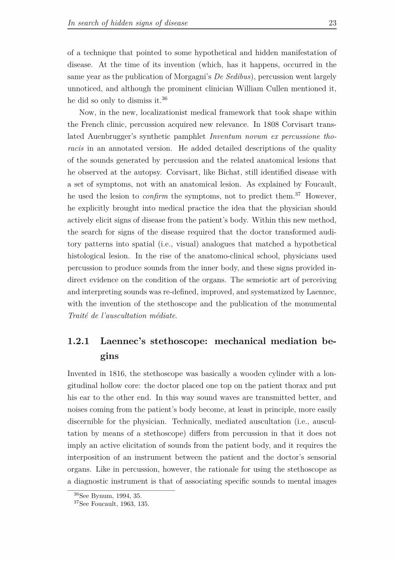
In search of hidden signs of disease 23
of a technique that pointed to some hypothetical and hidden manifestation ofdisease. At the time of its invention (which, has it happens, occurred in thesame year as the publication of Morgagni’s De Sedibus), percussion went largelyunnoticed, and although the prominent clinician William Cullen mentioned it,he did so only to dismiss it.36
Now, in the new, localizationist medical framework that took shape withinthe French clinic, percussion acquired new relevance. In 1808 Corvisart trans-lated Auenbrugger’s synthetic pamphlet Inventum novum ex percussione tho-racis in an annotated version. He added detailed descriptions of the qualityof the sounds generated by percussion and the related anatomical lesions thathe observed at the autopsy. Corvisart, like Bichat, still identified disease witha set of symptoms, not with an anatomical lesion. As explained by Foucault,he used the lesion to confirm the symptoms, not to predict them.37 However,he explicitly brought into medical practice the idea that the physician shouldactively elicit signs of disease from the patient’s body. Within this new method,the search for signs of the disease required that the doctor transformed audi-tory patterns into spatial (i.e., visual) analogues that matched a hypotheticalhistological lesion. In the rise of the anatomo-clinical school, physicians usedpercussion to produce sounds from the inner body, and these signs provided in-direct evidence on the condition of the organs. The semeiotic art of perceivingand interpreting sounds was re-defined, improved, and systematized by Laennec,with the invention of the stethoscope and the publication of the monumentalTraité de l’auscultation médiate.
1.2.1 Laennec’s stethoscope: mechanical mediation be-
gins
Invented in 1816, the stethoscope was basically a wooden cylinder with a lon-gitudinal hollow core: the doctor placed one top on the patient thorax and puthis ear to the other end. In this way sound waves are transmitted better, andnoises coming from the patient’s body become, at least in principle, more easilydiscernible for the physician. Technically, mediated auscultation (i.e., auscul-tation by means of a stethoscope) differs from percussion in that it does notimply an active elicitation of sounds from the patient body, and it requires theinterposition of an instrument between the patient and the doctor’s sensorialorgans. Like in percussion, however, the rationale for using the stethoscope asa diagnostic instrument is that of associating specific sounds to mental images
36See Bynum, 1994, 35.37See Foucault, 1963, 135.
24 Disease and diagnosis in the 19th century
that hypothetically point to actual organic lesions, which in turn correspond toa disease.
The idea of a sort of synaesthesis between hearing and vision is patent inthe instrument name (from the Greek stèthos, chest and skopèo, I see), and themetaphor of vision was recurrent in the medical literature between 1820 and1850, when the idea of “seeing” a disease by listening through the stethoscopewas largely endorsed by the promotors of mediated auscultation. “We anatomiseby auscultation (if I may say so), while the patient is yet alive,” wrote onephysician, and in the preface to the first English translation of Laennec’s treatise,John Forbes claimed: “[auscultation] is a window in the breast through which wecan see the precise state of things within.”38 This confidence in the scopic powersof the stethoscope was backed by the studies of Laennec, which had shown aclose correspondence between the sounds coming from the chest of hundreds ofdiseased individuals, and the lesions revealed by their autopsies (Figure 1.1).
As his teachers Bichat and Corvisart, Laennec studied and worked in anhôpital géneral, where he had access to the dissecting rooms and to many seri-ously ill patients, those paupers who “offered” their bodies to science in exchangefor a shelter and basic care. Dividing his time between the hospital lanes andthe morgue, he could systematically examine a vast number of cases. In 1819,he published the first edition of the Traité de l’auscultation mediate, where hepainstakingly described the characteristics of the sounds he perceived by auscul-tation and their relations to the damaged tissues he observed post mortem. Re-markably, his descriptions of clinical cases covered not only the different stagesof a disease, but also the variations observed in people with different physicalmakeups. In the introduction to the Traité, he claimed:
The diseases of the thoracic viscera are very numerous and diversified,and yet have almost all the same class of symptoms [cough, dyspnea,expectoration]. The consequence is, that the most skillful physician whotrusts to the pulse and general symptoms, is often deceived in regard tothe most common and best known complaints of this cavity.39
Laennec’s critique of the diagnostic method based on general symptoms was notcontingent on the idiosyncrasies of chest anatomy and ailments. It mirrored thatseminal concept of clinical anatomy, according to which diseases cannot be un-derstood and defined through transient symptoms that disappear as soon as thevital functions come to a stop. They should rather be understood and definedthrough the recurrent lesions observed in cadavers. This idea was expoundedby G.L. Bayle (colleague of Laennec and disciples of Bichat and Corvisart) in
38Both authors are quoted in Reiser, 1978, 30.39Laennec, 1826, 2.
In search of hidden signs of disease 25
Figure 1.1: Figure representing different forms of tubercular matter and some ofits effects. (a) Developed tubercle, completely yellow; (b) group of early tubercles,whose exterior is still grey and semi-transparent; (c) small cartilaginous cyst whichcontained tubercular matter and that discharged its content due to its own softening;(d) tubercular excavation completely empty and covered by two membranes [. . . ];(e) Small tubercular excavation completely empty and uncovered; (f) part of theexternal surface of the lung; (g) partially softened and empty tubercle; (h) tubercularinfiltration starting from the pulmonary tissue. T. Laennec, Traité de l’auscultationmediate, 2nd Edition, 1826, Paris, Plate II, Fig. 2. Original caption, my translation.Public domain.
26 Disease and diagnosis in the 19th century
his doctoral thesis in 1802. In a text published a few years laterBayle wrote:“individuals must be regarded as phthisic who are neither feverous, nor thin,nor suffering from purulent expectoration; it is enough that the lungs should beaffected by a lesion that tends to disorganize and ulcerate them; phthisis is sim-ply that lesion.”40 It was through the works of Laennec and Bayle that phthisisceased to be “phthisis,” that is, a condition defined by manifest symptoms ofconsumption, and became “tuberculosis,” which consists in the presence of “tu-bercles” (characteristic lesions) in the lungs or in other organs, for example, thebones or the kidneys.41 This transformation was not accomplished overnight.The words phthisis and consumption were still in use in medical literature atthe end of the nineteenth century, which is an evidence of how long it can takefor radical changes to be accepted by the medical community and integratedinto daily practice. That notwithstanding, a new direction for medicine was set.
An epistemological rupture With the invention of the stethoscope and withthe publication of the Traité de l’auscultation mediate (in particular, the sec-ond edition of 1826), the conceptualization of disease as organic lesion becameoperative, because it had a proper diagnostic instrument and its nosology. Thisis why medical historian Giorgio Cosmacini has called the stethoscope a “philo-sophic instrument, connected to an epistemological rupture.”42 Cosmacini alsostresses the fact that since the stethoscope was the first diagnostic instrument ofgeneral use “it transformed the practice of medicine, helped changing doctors’perception of illness, and inaugurated a tendency to widen the distance betweendoctor and patient, introducing between them the first, rudimental technologicalapparatus.”43 This transformation was by no means an anodyne process, and ittriggered a heated debate. The medical community did not consensually acceptit, neither in the academic circles nor in actual medical practice. On the onehand, there were Laennec’s supporters (mostly out of France), who consideredmediated auscultation not only an extremely valuable diagnostic tool, but alsoan indispensable instrument for transforming medicine into an empirical science,in which causal connections could be verified through laboratory evidences (inthis case the laboratory was the morgue), and whose precision was comparable
40Bayle, 1810, 8-9, quoted in Foucault, 1963, 138-139.41According to Grmek, the official “birth certificate” of the modern anatomo-clinical concept
of disease is to be found in G.L. Bayle’s doctoral thesis and in the second edition of Laennec’streatise, organized according to the anatomo-clinical definition of the chest ailments. Grmek,1999, 150.
42Cosmacini, 1997, 329, my translation.43Cosmacini, 1997, 329, my translation.
In search of hidden signs of disease 27
to that “attainable in some of the higher branches of physics.”44 These doctors,as their mentor Laennec, were generally highly dismissive of patient’s narrative,considered untrustworthy and of little clinical (hence, medical tout court) rele-vance. On the other hand, there were physicians who dismissed the stethoscope,considering it a quite useless, if not ridiculous, instrument. Many other prac-titioners, even if less interested in general medical theory, could simply makeno sense of auscultation. Transforming into a diagnosis ephemeral sounds –that Laennec and his successors had subdivided in endless tones – was no easytask, and to rely on usual techniques as patient’s story, observation and manualinspection of the chest seemed far more secure.45 Furthermore, even among doc-tors who appreciated the method of auscultation, there were many who foundmediated auscultation difficult to perform, and felt that they would be betteroff with the naked ear, rather than the stethoscope. Indeed, it was not clear ifthe stethoscope actually improved the reception of sounds from the chest.
Instrument-mediated perception In the second edition of his treatise Laen-nec himself recognized that for doctors who had little experience with the in-strument, direct auscultation might be easier and more fruitful. He neverthelessadvocated the superiority of the mediated technique, and his arguments were ofdifferent orders: moral, practical, and medical. For what concerns the moral as-pect, Laennec observed that: “[Immediate auscultation] is always inconvenientboth to the physician and patient; in the case of females it is not only indelicatebut often impracticable; and in that class of persons found in hospitals it is dis-gusting.”46 Concerning the practical reasons, he specified that there are pointsof the body where it is impossible to apply the ear directly (e.g., on the axilla,or under the female breast), and that the strong pressure on the chest requiredby the application of the naked ear for immediate auscultation is uncomfortablefor the patient and thus “gives rise to extraneous sounds from the contractionof the muscles.”47 Finally, he briefly expounded the medical argument:
Moreover, some of the most important of the stethoscopic signs havefor one of their causes the stethoscope itself . Thus, perfect pectorilo-quism, which consists in the transmission of the voice through the tubeof the instrument, is changed, in the trial of immediate auscultation, intoa simple resonance, stronger no doubt than in the natural condition ofthe parts, but such as to be with difficulty discriminated from oegophon-ism and bronchophonism. For these and other reasons I do not hesitate
44J. Taylor, Introductory lecture on the opening of the medical session of 1841-1842 inUniversity College, quoted in Reiser, 1978, 30.
45See Cosmacini, 1997, 327-331.46Laennec, 1826, 4.47Laennec, 1826, 26.
28 Disease and diagnosis in the 19th century
to affirm, that the physicians who shall confine themselves to immedi-ate auscultation, will never acquire great certainty in diagnosis, and willevery now and then fall into serious mistakes.48
Hence, Laennec claimed that the stethoscope itself was one of the causes of themost important signs of disease that could be perceived through it. Does thismean that he believed that the stethoscope created the signs of disease (and,eventually, the disease itself)? No. What Laennec was saying is that “perfectpectoriloquism” – a specific sign that the pulmonary texture around the mainexcavation had undergone considerable condensation accompanied by the for-mation of further cavities – was “the transmission of the voice through the tubeof the instrument,” characterized by a specific clarity and intensity. Immediateauscultation (without the interposition of the stethoscope) would still reveal athoracic resonance stronger than that one would hear if the organs were healthy.However, the quality of the perceived sound would not allow to discriminate per-fect pectoriloquism from other signs such as oegophonism or bronchophonism,indicators of other specific forms of necrobiosis. Hence, the instrument causedthe signs of disease to appear more clearly to the human senses, and in a morevaried range of tones, thus allowing for finer distinctions. This means that itcreated finer audible patterns and a range of possible gnoseological constructscompatible with them.
According to the French physician and epistemologist François Dagognet,Laennec’s invention was groundbreaking because it provided an instrumentthat, in spite of its simplicity, was able to convey valuable information notjust about heart and lungs’ anatomy but, more importantly, about their func-tionality. Through the stethoscope the trained doctor could “fix, on the basisof an uninterrupted natural language [of the chest], the place, the volume, thenature and the evolution of the disease.”49 However, Dagognet stresses that thestethoscope’s translation of the natural language of the body was trustworthyonly to the ear of those who were able to decode it.50 Well before the diffusionof photography and the invention of radiography, the stethoscope introduced inmedicine the question of instrument-mediated perception and mediated observa-tion. Inventing the stethoscope, using it and making sense of it, were activitiesthat implied different forms and different levels of mediation. In the first place,doctors had to agree that mediated experience was as valuable as (if not morevaluable than) direct experience. They should trust the instrument and believethat it gave access to some information relevant for the diagnosis. Moreover,since medicine is meant to be a relationship between a doctor and a patient,
48Laennec, 1826, 26-27.49Dagognet, 1986, 100, my translation.50See Dagognet, 1986, 100.
In search of hidden signs of disease 29
turning the stethoscope into a full-fledged medical tool also implied that pa-tients recognized the authority of the instrument in the hand of the doctor.This meant that the patient too, and not only the practitioner, had to believethat the signs collected by the physician through this new medium – signs whichthe patient himself could not perceive – were as truthful as the symptoms he orshe could actually feel and display.
The stethoscope as a medium
I will elaborate on the concept of mediation from different perspectives in thedevelopment of this thesis. Here I just want to begin to introduce the problemand make some preliminary remarks on the basis of what I consider the mostsalient features of mediated auscultation.
1. In auscultation (mediated or immediate) sounds from the inner body aretaken as valid proxies of the disease. Through a quite complex and artic-ulate cognitive process, the doctor converts thoracic sounds in mental im-ages that match the anatomical lesions revealed by pathological anatomy,which, in turn, coincide with a certain disease. Hence, the sound perceivedby the doctor becomes a medium of the condition of the patient’s internalorgans and tissues. It is a medium because it conveys indirect evidence ofan invisible condition (in the specific case of tuberculosis, the presence oftubercles).
2. The stethoscope in itself transmits a range of tones (e.g., pectoriloquism,oegophonism, bronchophonism) that could not be perceived just by placingthe ear right on the patient’s body (they are the specific sound of breathingor voice passing through a wooden cilinder). It mediates the sensorialexperience of the physician enhancing his or her hearing capabilities. Thatis, it is a medium between the auditive system of the doctor and the soundsemitted from within the body of the patient. Thus, the stethoscope is adifferential transmitter (a go-between) of sounds produced by the innerorgans.
3. Auscultation and the stethoscope are thinkable and epistemically soundwithin the localizationist paradigm of clinical anatomy, which entails thatwe accept the cadaver as a medium for the living, diseased body. In clinicalanatomy, in fact, the dead body is considered more reliable than the livingone in manifesting the signs of disease, since post mortem anatomical lesionare stable testimonies of the transient event of illness (with its related andovert symptoms) experienced by the patient. In other words, within the
30 Disease and diagnosis in the 19th century
localizationist paradigm of clinical anatomy the corpse becomes a mediumof the living being in that it constitutes the material support of the signs,traces and marks of the disease.51 In other words, it replaces the livingbody. It is a medium because it allows the pathologist to observe the innerbody in all its details (as I will show in Chapter 3, with the invention ofradiography X-ray images replaced the cadaver as media of the inner body,with the advantage of granting access to the interior of the living body).
4. Once the signs collected by the doctor become more relevant than thepatient’s transient and subjective symptoms in defining and diagnosingthe disease, the very experience of illness becomes potentially mediated.In a sense, we can say that the doctor becomes the medium (and theoracle) of the patient’s experience, that is, the doctor enters in contactwith and gives voice to what happens inside the patient’s body bypassingthe patient himself.
In relation to this latter point, it could be objected that it has always been thecase, since only the physician, and before him the priest and the magician, hasthe social authority to tell the patient what is going on within the sick (and thehealthy) body. Before the institution of modern clinical diagnosis, however, thedoctor had to rely exclusively on the patient’s narrative, in addition to manifestbodily symptoms and signs, in order to establish what was going on with thatspecific person. Modern clinic did without the patient’s story and symptoms,it diagnosed the disease before it could even manifest itself. Laennec praisedthe stethoscope of allowing “to detect or even to suspect, diseases in their verycommencement,”52 when the patient was not aware of them, yet.
Historians of medicine have reported that many patients were embarrassed,perplexed or even frightened by the stethoscopic examination. At the sametime, most of them were amazed by the ability of the physician to tell whatwas happening inside the body from sounds inaudible to the patient himself.53
This is why Dagognet locates in the stethoscope the beginning of a medicinethat, in its ideal limit, will be without patient, without doctors and, paradoxi-cally, without disease.54 Without patient, because technology enables medicineto discover the disease independently from, or even in spite of, the confusionof subjective symptoms (most symptoms are unspecific, and some diseases aresimply asymptomatic). This becomes possible because technology, even in the
51For a very interesting analysis of the body as material medium of inscription of differentsigns and images see Belting, 2001.
52Laennec, 1826, 3.53See, Reiser, 1978, 36; Cosmacini, 1997, 329-330.54See Dagognet, 1986, 100.
Endoscopic visions 31
very primitive form of the stethoscope, forces the disease to come to the surface,making abstraction of the patient’s lived experience. Without doctor, becausethe ideal diagnostic technology exposes so clearly the disease that no hypothet-ical inference is required. It speaks the “truth” of the disease and needs nopotentially faulty interpretation. Without disease, because an ideal medicinecatches the disease even before it appears, and diverts the natural course ofpathological events.
1.3 Endoscopic visions
With the French school of clinical anatomy, physical diagnosis became a tenetof medical practice. Semeiotics, the search of signs of disease through percus-sion and auscultation was just one form of physical examination. The otherswere, and still are, anatomical exploration, performed through palpation or withendoscopic tools, and functional diagnosis. Anatomical exploration shares thelocalizationist paradigm of semeiotics, but implies a different kind of sensorialcontact with the disease: the doctor can touch (through the skin) a swollenliver by palpating the abdomen, or see a polyp on the vocal cords by means ofa laryngoscope.55 Many endoscopic devices, which allowed looking literally intothe body exploiting its natural orifices, were developed by surgeons during thenineteenth century.
Although archaeological testimonies show that anal and vaginal specula werealready familiar to ancient Greek and Roman physicians, the first attempt toactually illuminate the inner organs was made by the German surgeon PhilippBozzini, between 1804 and 1805. He officially presented his invention, which hecalled Leichtleiter (light conductor), to the Austrian Faculty of Medicine in Vi-enna, in 1806, raising harsh criticism and a heated controversy.56 In his analysisof the origins of modern endoscopy, the historian of science Claudio Poglianoshows that the negative reaction to this new instrument was due to a numberof reasons,57 including different conceptions of the relative epistemic cogency ofvision and touch. While Bozzini claimed that vision renders more certain eventhe touch of the most trained expert, and that “only seldom are the other sensesprepared to dispense with its assistance,”58 the director of the Vienna Faculty
55See Peitzman and Maulitz, 1999, 180.56See Reiser, 1978, 51; Bynum, 2006, 169-170; Pogliano, 2011, 52.57The Leichtleiter provided a good visualization of the pharynx and nasal cavities, but when
employed in other orifices, the illuminated spot was too small to provide valuable information.In addition, it was very difficult to operate. Institutional factors also hampered Bozzin’sinvention: supported by the progressive Josephsakademie, it was harshly criticized by themore conservative Faculty of Medicine. See Pogliano, 2011, 57-58.
58Bozzini, quoted in Pogliano, 2011, 53.
32 Disease and diagnosis in the 19th century
of Medicine, who wrote a negative report on the Leichtleiter, maintained that,in order to receive the best treatment, “the patient should place his confidencein the judgement of a reasonable doctor and the hand of an experienced inves-tigator.”59 It seems also reasonable to suppose that a technique which requirednot only to touch, but literally penetrate into the living body generated somemoral discomfort both in doctors and patients (for the latter the discomfort wasalso physical, given that endoscopy is a very painful examination).
Touch vs. vision In spite of the institutional resistances, Bozzini’s workpaved the way for further research. First successes came in 1812, with An-thelme Récamier’s vaginal speculum, and in 1822, with Pierre Ségalas’ urethro-cystoscope. However, the main developments occurred between the 1830s andthe 1860s, when the otoscope, the urethroscope, the laryngoscope, and the oph-talmoscope appeared. And yet, even if endoscopy was making significant tech-nical progresses, still in 1855, on occasion of a demonstration of the use of theurethroscope by his inventor, Antonin Jean Desormeaux, a former teacher of hiscommented: “One can see very well with your instrument, but what is thereto see, really?”60 Indeed, endoscopy was not unanimously accepted by Frenchphysicians. Pogliano mentions the criticisms of Félix Guyon, a prominent pro-fessor of urology in Paris, who repeatedly expressed his conviction that in mostcases skilled touch coupled with clinical intuition would gather enough informa-tion without the aid of artificial devices. In his Leçons cliniques sur les maladiesdes voies urinaires, published in 1881, Guyon claimed that one should not con-sider a great progress in medicine the simple fact of being able to see somethingwhich until then had been usefully perceived by the sense of touch. Sight, he ar-gued, will tell us nothing about the consistence and sensitivity of a constrictionof the urinary tract, and it will say even less on its extension. Consequently,he warned his students that even if the techniques to illuminate the urethraand the bladder (endoscopy) would improve in the future, they could never dowithout the “endless resources of touch.”61 Guyon was suspicious of the media-tion between doctor and patient (and between doctor and the signs of disease)introduced by the endoscope, with its systems of lenses and mirrors. His mis-trust, however, was not so much about the instrument, but more substantiallyabout the epistemic virtues of vision. In a time in which the confidence in thecognitive accountability of sight was gaining momentum, thanks also to the pro-gressive diffusion of photography, he insisted on that sensorial experiences are
59Quoted in Pogliano, 2011, 57.60Desormeaux, 1865, 8, my translation.61Guyon, 1881, 625.
Endoscopic visions 33
interchangeable only to a limited extent, and that certain information providedby the sense of touch (e.g., consistence and sensitivity of a section of an organ)could not be replaced by eyesight. He was ready to admit that in the humanbody only a few organs, or sections of organ, are available to direct touch, buthe nevertheless stressed the fact that endoscopy could supersede haptic exami-nation only in organs like the eye and the larynx, which could not be touchedat will.62
Guyon’s position did not pass the test of history, as the current developmentsin video-endoscopy definitely demonstrate. Not only has vision largely replacedthe sense of touch in diagnosis, but also the development of telesurgery seems toprove that a whole reversibility of touch into sight is actually possible. However,we can take Guyon’s reluctance about vision as a source of medical knowledgeas an indicator of how complex was the process that led to the “dematerializa-tion” of the body characteristic of modern medicine. Vision, we know, is thesense of distancing and objectivation par excellence, while touch is the sense ofproximity, whereby the observer melts with the perceived object.63 For Guyon,the very texture of the organ and its sensitivity were essential components ofmedical perception. According to him, touch granted a form of direct knowledgethat vision, being physically removed from its object, could not entail. Yet, Des-ormeaux, who was also a surgeon, in his De l’endoscope, of 1865, praised visionunconditionally (he opened the book with a quotation from the fifth paradoxin Cicero’s Paradoxa Stoicorum: “Nosquoque oculos eruditus habemus”), andclaimed that compared with the advancement of general pathology and of theclinical study of different organs, urology scored far behind, mostly because untilthe invention of the endoscope the urinary tract had been inaccessible to vision.He placed the endoscope among the means – which also included percussion andauscultation – that allowed medicine to explore human organs directly, by ex-tending and perfecting the doctor’s senses.64 Clearly, two very different notionsabout what count as direct observation, and about the primacy of vision in theacquisition of medical knowledge were at stake.
1.3.1 The case of the ophtalmoscope
Within the domain of endoscopic devices, the case of the ophtalmoscope, de-signed in 1851 by Hermann Von Helmholtz, is particularly interesting, becauseunlike the other endoscopic tools, it did not need to be inserted into the humanbody. Almost fifty years before the discovery of X-rays, this invention enabled
62See Guyon, 1881, 621.63I develop the topic of the relation between touch and vision in Chapter 4, Section 4.1.64See Desormeaux, 1865, xi and 1-2.
34 Disease and diagnosis in the 19th century
physicians to visually explore a (small) part of the living inner body from theoutside. Thanks to the ophtalmoscope, anatomical causes of eye’s pathologies,which previously could be seen only in post mortem autopsy, became easily vis-ible in vivo. Moreover, it did not simply allowed the visualization of the innereye (retina, optic nerve head, and eye’s arteries), but also proved valuable inthe indirect diagnosis of diseases of the kidneys and arteries, as well as of someneurological conditions.65 With this instrument, the physician could see both inand through the eye: in, because it became possible to actually look at the innerstructures of the organ of vision; through, because on the basis of the alterationof some of these structures it was possible to infer the state of other parts andfunctions of the body.
As already noted in the case of Laennec and auscultation, an aspect of phys-ical diagnosis particularly praised by physicians was the fact that it freed thediagnostician from patient’s subjective testimony. In the case of the ophtalmo-scope, for example, it became easier to diagnose astigmatism in persons whocould not read.66 To stress this point, Reiser quotes a physician who, on theoccasion of a meeting of the American Medical Association, in 1894, wrote thatthe ophtalmoscope reduced the physician’s dependence “on the faltering judg-ment of the untrained patient, substituting therefor the skill of the expert who,reasoning from scientific data, is thus able to fit glasses in less time and more ac-curately with consequent satisfaction to himself and his patient.”67 This sentenceexemplifies quite well some fundamental ideas about the relationship betweenbody, first-person experience, and knowledge that informs scientific medicine.In fact, if medicine is a science, then it requires a specialized knowledge and aspecialized language developed within a community of peers, who collect, share,and elaborate scientific data (in this case the lesions or malformations revealedby the ophthalmoscope), and cannot rely on the imprecise words that a pa-tient normally uses to describe the experience of illness. All the patient can dois to describe symptoms, but scientific medicine can track disease even beforesymptoms appear, and a great deal of its efficacy comes from this possibility.With the ophtalmoscope, for example, the nosology of eye diseases changed,because the instrument revealed a range of different lesions that could not evenbe imagined before. At the same time, both the prevalence (the total numberof cases of disease in a given population at a specific time) and curability ofthese diseases were redefined.68 In fact, if a disease can be detected in an earlystage, before the patient can feel or display any clear symptom, its prevalence
65See Bynum, 2006, 169.66See Reiser, 1978, 47.67H.V. Wuerdemann, 1894, 341, quoted in Reiser, 1978, 49.68See Bynum, 2006, 167.
Looking into the microscopic body 35
will rise. Simultaneously, since many disorders incurable in their advanced stagecan still be treated when discovered in their pre-symptomatic state, curabilitywill improve. In turn, the prevalence of secondary diseases, that is disease asso-ciated with other pathological conditions, will decrease. Instrument creates newperceptions of the body that do not only reorganize medical knowledge creatingnew nosological entities, but also redefine the very meaning of being ill.
1.4 Looking into the microscopic body
A different way of accessing the inner human body, much less obvious thanendoscopy, came from the microscope. With the improvement of the compoundmicroscope69 and the development of microtomes, cellular staining, and fixationmethods, by the late nineteenth century the microscopic dimension entered therealm of diagnostic medicine in two very different areas: (1) cellular pathology,which entailed a refinement of clinical pathology; and (2) microbiology, whichbroke completely with clinical pathology and put forward a new conceptionof disease, rotted in the germ theory. While in the first case the microscopetransformed the body into a jigsaw puzzle of minute interwoven pieces, in thesecond it turned the body into a porous and fluid entity, open to the attacks ofexternal invisible pathogens.
1.4.1 Cellular pathology
Cellular pathology was first theorized in 1858 by the German pathologist RudolfVirchow. With the help of the microscope and of histochemical analysis, Vir-chow demonstrated that cells found in diseased tissues were modifications ofnormal cellular types.70 On these grounds he posited that it was the cell, andthe disruption of its functions, the primary locus of disease. Consequently thework of microscopists – carried out in the laboratory, far away from the patient’sbody – became fundamental for studying these invisible units of life.71 Virchowworked within the localizationist paradigm and considered his ideas the naturaldevelopment of Morgagni, Bichat and Laennec’s morphological views of the dis-ease. However, he did not restrain his analysis to the structural changes of theimpaired cell, but rather insisted on the importance of chemical and physicalalterations. Although he believed that the organism’s vital properties could not
69The first compound microscope was built in 1595, but the first scientific book on mi-croscopy was Robert Hook’s Micrographia, published in 1665. In the 1670s Anton VanLeeuwenhoek described sperm cells, bacteria and protozoa. Achromatic lenses (correctedfor chromatic aberrations) became available only in the early nineteenth century.
70See Grmek, 1999, 155.71See Reiser, 1978, 79.
36 Disease and diagnosis in the 19th century
be completely reduced to physics and chemistry, he nevertheless thought thatliving organisms function according to chemical and physical laws, and that onthe basis of these laws it is possible to understand the mechanisms of disease.Bynum remarks:
By urging doctors to think about disease in terms of cells, Virchowemphasized process rather than result. Looking at the cell behaviour indiseases placed the pathological centre-of-gravity towards the beginning,rather than at the end stages of a disease. [. . . ] The anatomo-pathologicaldefinitions of many diseases remained pretty secure, but notions of causeand mechanisms also came to the fore. [. . . ] The microscope as well asthe dissecting scalpel became constant tools of the pathologist. Stains,microtomes, and fixation methods were developed, enabling thin sectionsof tissues to be cut and different kinds of cells to be more clearly differ-entiated.72
One of the most relevant practical outcomes of this new conceptual approachwas that it became possible to discriminate malignant tumors from the benignones, with enormous consequences in terms of prognosis and therapeutics (thesurgeon would operate the tumor only if the pathologist declared it malignant).In turn, observing thin layers of tissue under the microscope became a specializedactivity, and for the first time emerged the figure of a clinician who spent histime in the laboratory, rather than in the hospital wards, and made his diagnosesaway from the bedside.
With Virchow, the cell became the ultimate morphological and functionalunit of life, an idea that he summarized in the precept omins cellula e cellula(all cells come from cells). However, Grmek points out that even though hestarted from a localizationist assumption, Virchow had a dynamic view of thedisease and the concept of Krankheitsprozess was a tenet of his thought. Heexplicitly maintained that the aim of pathology was to study vital activity underits deviations and hindrances. As his contemporary Claude Bernard, Virchowsaw no intrinsic difference between health and disease, physiology and pathology,and his thought played a seminal role in the debate about the antinomies whole-part, solid-fluid, process-state, structure-function.73 To him, the organism was acombination of cells, and disease was the sum and the result of cellular affections.It was inside the cell that the ens morbi resided, because the cell reacted indifferent ways to internal and external stimuli (physical and chemical) thatcould determine a pathological condition.74 The ens morbi, the disease, was apossible state of the cell (Figure 1.2).
72Bynum, 2006, 121-122.73See Grmek, 1999, 155.74See Grmek, 1999, 155-156.
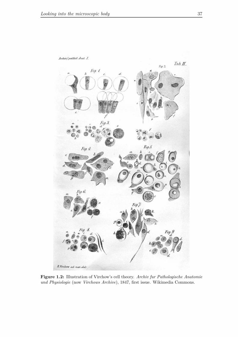
Looking into the microscopic body 37
Figure 1.2: Illustration of Virchow’s cell theory. Archiv fur Pathologische Anatomieund Physiologie (now Virchows Archive), 1847, first issue. Wikimedia Commons.
38 Disease and diagnosis in the 19th century
1.4.2 Microbiology
Microbiology and the related germ theory of disease gained momentum sometwenty years after cellular pathology. It presented a quite different vision ofdisease and, consequently, of the body. While cellular pathology focused on theinternal pathological process (pathogenesis), the germ theory was centered onthe external causes (etiology) of disease, which was seen as the result of theinteraction between two living beings, namely, a human being and a parasite.More precisely, at least according to Grmek, the germ theory of disease implieda biologic, non-anthropocentric vision of illness, in which “the infectious diseaseis nothing but a specific aspect of the food chain of organisms.”75
Ideas about the existence of “atoms” of biological material that could trans-mit disease were already discussed in the sixteenth century,76 but it was only inthe second half on the nineteenth century, with the discoveries of Louis Pasteurand Robert Koch, that the microbial theory established itself as a completemedical doctrine and the microscope became an indispensable diagnostic tool.In particular, Koch’s successes in isolating the bacteria responsible for cholera,anthrax and tuberculosis, in the 1880s, encouraged many clinicians to rede-fine the diagnostic process, identifying the micro-organism, with the diagnosis.For these doctors the disease acquired a specific ontological status, because thegerm was the disease. Within the germ theory of disease, elusive cognitive clueslike signs and symptoms were replaced by the certainty of the microbiologicaltest. Tuberculosis was no more an illness specified by its characteristic symp-toms (phthisis), nor a specific histological lesion revealed by auscultation andthe histological examination (the tubercle), it was rather the infection provokedby Koch’s bacillus (Mycobacterium tuberculosis). As a consequence of this re-definition, a number of apparently different ailments, like pulmonary phthisis,scrofula, and white tumor of the knee became particular forms of the same dis-ease.77 Diagnosis was made in the laboratory, growing colonies of bacteria onPetri dishes, staining them and observing them under the microscope. As incellular pathology, which required the specialization of pathologists who cut,stained and analyzed histological samples, with the germ theory a new figureof clinician emerged: the specialist in bacteriology who could deal with cell cul-tures and microscopic images. A medical specialist who did not spend time at
75Grmek, 1999, 160.76Girolamo Fracastoro published his De contagione et contagiosis morbis in 1546, and prior
to Luis Pasteur’s work on silkworm disease, in 1835-1836, Agostino Bassi had suggested thatit was caused by the microorganism muscardine. Favus (a skin disease) and anthrax wereknown to have, respectively, different microbic origins. In 1840 Jacob Henle defended thatmany diseases were probably caused by a contagium vivum (living contagion). See Cosmacini,2007, 232; Bynum, 1994, 128; Reiser, 1978, 78-79.
77See Grmek, 1999, 160.
Functional anatomy: disease in numbers and graphs 39
the bedside of the patient, but rather examined little fragments of his or herbody, or minuscule particles that infected it.
With the development of microscopy, the perception of the body mediated bymechanical instruments entered a new phase. The body could still be thoughtof as an ordered landscape of organs, but in order to reveal its constitutingelements and expose the structure of cells, one should walk away from it andenter the laboratory, where an array of instruments and experimental techniquesallowed the visualization of the invisible. In terms of perception of the humanbody, this implied recognizing that it was opaque not simply because its innerorgans were concealed by the skin, but also because its very components as wellas its parasites were extremely minute. An image of the body removed fromany possible natural perception came to the fore.78
1.5 Functional anatomy: disease in numbers
and graphs
In the same years in which Virchow developed cellular pathology, Claude Bernardwas revolutionizing functional anatomy while creating experimental medicine.To him, function not structure was the key to understand the living body anddisease was no more to be understood as a static entity (the anatomical lesion),but rather as the alteration of a process. Indeed, like Virchow, Claude Bernardthought that the difference between the physiological (normal) and the patho-logical state was of quantitative rather than qualitative order.79 This meantthat, as Cosmacini puts it: “function and disfunction, physiology and pathol-ogy constituted a graduated, measurable continuum.”80 Within this vision, therelation between tissue and disease was deeply transformed: the lesion of a tissuewas irrelevant as far as it did not affect its function, while an impaired functiondid not necessarily lead to a structural alteration, even though it produced astate of disease.81
78I discuss in depth microscopic images and microscopic vision in Chapter 3, Section 3.2.79See Grmek, 1999, 157. Grmek points out that before Bernard and Virchow, François
Magéndie, Bernard’s teacher, had already defended that pathology was physiology underspecial conditions.
80Cosmacini, 1997, 343, my translation. It should be noted that Bernard’s conceptualizationof disease as a mere quantitative modification of normal (healthy) states, has been criticizedby Georges Canguilhem (1966) on both conceptual and empirical grounds.
81Anatomo-pathologists were already aware of this problem, and they tried to solve it byreducing the impaired process to an alteration of the structure. According to Laennec, the factthat some diseases did not present any clear morphological alteration did not mean that theirorigin was not anatomical, it barely meant that pathological anatomy was still an imperfectdiscipline and thus was not able to reveal all the possible anatomical alterations that could vexthe human body. Nevertheless, for what concerns mental illnesses he was willing to accept
40 Disease and diagnosis in the 19th century
Now, while the description of structure implies the recourse to space only, thedescription of a process entails space and time, thus requiring different researchmethods and instruments. Moreover, if pathology differs from the healthy stateby degree, and not by some fixed property, then measuring quantities becomesimportant both for understanding disease and for making diagnoses. For medi-cal research, the second half of the nineteenth century was the time of Bernard’sexperimental pathology, with his laboratory experiments on living animals, andof Etienne-Jules Marey’s graphic method, which included studies on blood cir-culation (with the sphygmograph) as well as on animal locomotion (with theinvention of chronophotography and instant diagrams).82 Accordingly, in thedomain of medical practice, it was the time in which new diagnostic strategieswere developed. They were based on graphic and quantitative methods, andtheir aim was to record such elusive data as breathing, blood pressure, heart-beat or temperature. As noted by Reiser, in this renewed epistemic context, “thestatement of the body’s vivifying activities in numbers and graphs provided afactual transcription of pathology equivalent to the discovery of an anatomicallesion.”83 In clinical anatomy the corpse was intended to be the medium thatmade visible the disease located in the tissues and organs; in physiology a newform of visibility was put forward: graphs were perceived as faithful inscrip-tion of vital functions, and numbers were taken as exact quantifications of vitalparameters.
Although clinical anatomy and physiology were very different endeavors –they looked at different things using different methods, different languages anddifferent images – they shared the same goal, namely providing a good rep-resentation of the state of health of a human body. Bichat and his disciples,thanks to the concept of disease as death within life, were able to track thecontinuum life-disease-death fixing the transient history of the illness in the se-cure geography of the corpse. Bernard and the other physiologists, working onthe continuum between the normal and the pathological could bracket death,and re-conceptualize the disease as a range of possible vital states. While theanatomists had been forced to come to terms with the geography and history ofthe dying tissue, physiologists needed to come to terms with the quantities anddegrees of the functions of the body, in order to capture them in their patholog-ical state. The solution to this problem was to measure functions and fix them
that the lesion could directly concern the vital principle, and consequently could never beobserved. See Grmek, 1991, 151.
82Claude Bernard published the two volumes of his Léçons de physiologie expérimentaleappliquée à la medicine between 1855 and 1856; Etienne-Jules Marey published La méthodegraphique dans les sciences expérimentales, et principalement em physiologie et medicine in1878.
83Reiser, 1978, 91.
Functional anatomy: disease in numbers and graphs 41
in numbers and graphs. Instruments as the spirometer, the thermometer, thesphygmometer and the sphygmograph gradually entered the diagnostic toolbox.
1.5.1 The spirometer
One of the first attempts to make “a numerical portrait”84 of the patient wasmade in the 1840s by John Hutchinson, an English surgeon, with the inventionof the spirometer (Figure 1.3). This device measures lungs capacity on a grad-uate scale, thus transforming the process of breathing into a precise and visiblequantity.85
Hutchinson tested his instrument on more than 2,000 individuals (26 women),most of whom were healthy, and in 1846 published a monograph where he pre-sented his results organized in charts and tables, putting in relation lungs’ vitalcapacity,86 age, height, weight and profession (Figures 1.4 and 1.5). He reachedthe conclusion that his quantitative method not only detected lung diseases atan earlier stage than auscultation or percussion, but was also a reliable instru-ment for judging men’s overall physical fitness. Just like Laennec had praisedthe exactitude of the stethoscope, Hutchinson claimed: “When all these [quan-titative] observations are made, and noted in a book properly headed, the stateof the person examined is then expressed in the unerring measure of figures –which immediately presents to the eye a certain state of things.”87 Yet, in spiteof Hutchinson’s confidence, it took many years before the spirometer becamepart of respiratory medicine’s routine.88
As the other vital indexes, lungs’ vital capacity is basically a statistical indi-cator, that is, it acquires clinical meaning only by comparing the data concerningthe members of a population with specific characteristics. Now, for integratinga statistical index into medical practice, physicians should grant numbers a farhigher epistemic import than they did in the 1840s. At that time,the veryidea that knowledge based on the laws of chance was as valuable as knowl-edge based on the deterministic comprehension of phenomena was still underconstruction.89 However, even if probabilistic thinking took a long time to be
84Reiser, 1978, 91.85The spirometer consists of a tube – through which the patient has to breath – connected
to a balanced receiver, which is elevated by each increment of expired air inside a graduatedpipe. A slightly modified version of Hutchinson’s device is still in use today.
86Vital capacity is the quantity of air expelled by the greatest voluntary expiration followingthe deepest inspiration. It is still used as a first line test for evaluating pulmonary functionality.
87Hutchinson, 1846, 244.88As remarked by Bynum, this was true for the majority of diagnostic techniques, whose
passage from elite experimentation to general practice normally took quite a long time. SeeBynum, 2006, 169.
89For a study on how statistical and probabilistic thinking eroded determinism during thenineteenth century, see Hacking, 1990.
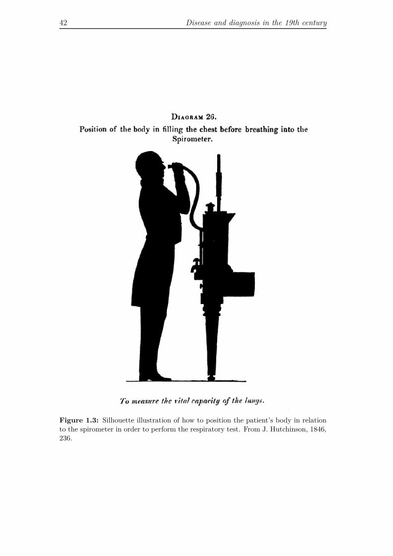
42 Disease and diagnosis in the 19th century
Figure 1.3: Silhouette illustration of how to position the patient’s body in relationto the spirometer in order to perform the respiratory test. From J. Hutchinson, 1846,236.
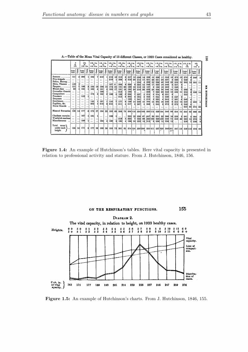
Functional anatomy: disease in numbers and graphs 43
Figure 1.4: An example of Hutchinson’s tables. Here vital capacity is presented inrelation to professional activity and stature. From J. Hutchinson, 1846, 156.
Figure 1.5: An example of Hutchinson’s charts. From J. Hutchinson, 1846, 155.
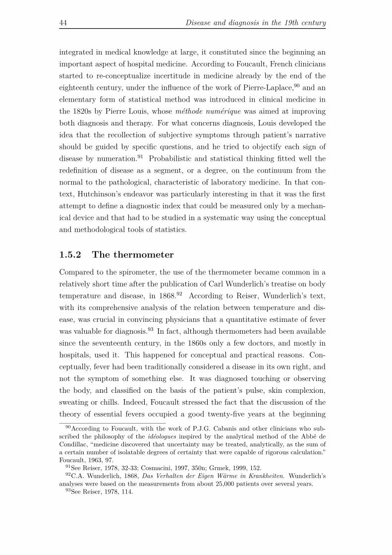
44 Disease and diagnosis in the 19th century
integrated in medical knowledge at large, it constituted since the beginning animportant aspect of hospital medicine. According to Foucault, French cliniciansstarted to re-conceptualize incertitude in medicine already by the end of theeighteenth century, under the influence of the work of Pierre-Laplace,90 and anelementary form of statistical method was introduced in clinical medicine inthe 1820s by Pierre Louis, whose méthode numérique was aimed at improvingboth diagnosis and therapy. For what concerns diagnosis, Louis developed theidea that the recollection of subjective symptoms through patient’s narrativeshould be guided by specific questions, and he tried to objectify each sign ofdisease by numeration.91 Probabilistic and statistical thinking fitted well theredefinition of disease as a segment, or a degree, on the continuum from thenormal to the pathological, characteristic of laboratory medicine. In that con-text, Hutchinson’s endeavor was particularly interesting in that it was the firstattempt to define a diagnostic index that could be measured only by a mechan-ical device and that had to be studied in a systematic way using the conceptualand methodological tools of statistics.
1.5.2 The thermometer
Compared to the spirometer, the use of the thermometer became common in arelatively short time after the publication of Carl Wunderlich’s treatise on bodytemperature and disease, in 1868.92 According to Reiser, Wunderlich’s text,with its comprehensive analysis of the relation between temperature and dis-ease, was crucial in convincing physicians that a quantitative estimate of feverwas valuable for diagnosis.93 In fact, although thermometers had been availablesince the seventeenth century, in the 1860s only a few doctors, and mostly inhospitals, used it. This happened for conceptual and practical reasons. Con-ceptually, fever had been traditionally considered a disease in its own right, andnot the symptom of something else. It was diagnosed touching or observingthe body, and classified on the basis of the patient’s pulse, skin complexion,sweating or chills. Indeed, Foucault stressed the fact that the discussion of thetheory of essential fevers occupied a good twenty-five years at the beginning
90According to Foucault, with the work of P.J.G. Cabanis and other clinicians who sub-scribed the philosophy of the idéologues inspired by the analytical method of the Abbé deCondillac, “medicine discovered that uncertainty may be treated, analytically, as the sum ofa certain number of isolatable degrees of certainty that were capable of rigorous calculation.”Foucault, 1963, 97.
91See Reiser, 1978, 32-33; Cosmacini, 1997, 350n; Grmek, 1999, 152.92C.A. Wunderlich, 1868, Das Verhalten der Eigen Wärme in Krankheiten. Wunderlich’s
analyses were based on the measurements from about 25,000 patients over several years.93See Reiser, 1978, 114.
Functional anatomy: disease in numbers and graphs 45
of the nineteenth century,94 and it was only when the metamorphosis of feverfrom disease into symptom was accomplished that temperature could be assimi-lated to other vital indexes and treated statistically. A confirmation of the fact,emphasized by many historians of medicine, that the sheer availability of a tech-nology does not entail its immediate use, and that a new diagnostic techniquecan be integrated into medical practice only when its results make sense withinthe scientific context of the moment.95 At the technical level, what restraineddoctors from taking up thermometry was the fact that the first thermometers96
were cumbersome instruments, difficult to use, and with low accuracy. Addi-tionally, they used different temperature scales, and this made the comparisonof data extremely difficult.
However, by the time Wunderlich published his treatise, these problems hadbeen overcome: the thermometer could be easily used and data from differentsubjects, or from the same subject at different times, could be easily compared.For the German clinician, thermometry had quite a few characteristics thatmade it a superior diagnostic approach in relation to other diagnostic methods.Compared to patient anamnesis it had the advantage that temperature could“neither be feigned nor falsified,”97 while compared to percussion and auscul-tation, it provided quantitative data, thereby offering “materials for diagnosiswhich are incontestable and indubitable, which are independent of the opinionor the amount of practice or the sagacity of the observer.”98 Moreover, in con-trast with spirometry and lungs vital capacity, it measured a phenomenon that,although ever changing, was more clearly related to the disease. Tracking vari-ations in body temperature, Wunderlich maintained, would allow defining thenatural laws of fever in diseases as diverse as typhus, neuroses, or injuries of thespinal cord.99 The temperature graph portrayed the disease, and in this portraitneither the patient nor the doctor could spoil the objectivity of the representa-tion by introducing their subjective feelings or lack of skills. In fact, since thethermometer was simple to use and provided an automatic output, a new figurecould enter the diagnostic arena: the unskilled measurer. While a qualitative
94See Foucault, 1963, Ch. 10. According to Foucault, the debate on the essential fevers wasthe final step in the definition of anatomo-clinical perception. Fever, in fact, questioned thevery being of the disease in its relation to the lesional phenomena.
95See, for instance, Faure, 2005, 22-23; Bynum, 2006, 169.96The first thermometer was probably build by Galileo, between 1593 and 1597, while San-
torio Santorio, who built his thermometer in1625, is credited for having extensively attemptedto measure the temperature of the human body. See Reiser, 1978, 110-11.
97Wunderlich, 1868, vi. It should be noted that distrust of patients’ subjective reports wasa tenet of clinical medicine, and it was quite common already in the eighteenth century. SeePeitzman and Maulitz, 1999, 176-177.
98Wunderlich, 1868, 48.99See Wunderlich, 1868, 51.
46 Disease and diagnosis in the 19th century
description of fever required a highly educated sense of touch and an adequatevocabulary, a precise quantitative measure was necessarily mechanical, and wasexpressed in numbers on a graduate scale. In this scenario, the unskilled mea-surer was valuable not only because he or she would save time to the doctor,but also because, as argued by the author of an article published in the BritishMedical Journal in 1868, not having any previous theory about temperatureand disease, he “would see things as they really occurred.”100 Portable and easyto use, the thermometer was finally adopted by a large number of doctors, andit encouraged the growing belief that the physiological conditions of health anddisease could be measured precisely.101
Confidence in the mechanical recording of phenomena, and in the virtues ofthe naïve eye of the unskilled observer, were part of the scientific ethos of thesecond half of the nineteenth century,102 so it is not surprising that it also perme-ated medicine, which, as already noted, was struggling to become a full-fledgedempirical science at least since Sydenham’s time. It should also be noted thatwith the thermometer, and with Wunderlich’s conceptualization of temperatureas a marker of the course of diseases, for the first time appeared the idea thatthe doctor could collect physical signs for diagnosis and follow-up without beingat the bedside. The stethoscope had introduced the first mechanical mediationbetween doctor and patients, but it still required a physical proximity and con-tinuity. With the thermometer, this continuity was potentially broken, since thephysician could touch, as it were, the patient’s body even being far away fromhim.
1.5.3 The sphygmometer and the sphygmograph
If we think about the relationship between vital indices and the instrumentsused to measure them, we observe different dynamics. The very idea of lungscapacity as a medical concept with a specific diagnostic meaning could not existwithout an instrument that measured it,103 while body temperature passed frombeing a disease (fever), to be a qualitative generic symptom, and with the ther-mometer, it eventually became a quantitative index. On the contrary, for whatconcerns the pulse, the proxy of blood circulation and heartbeat, the diagnostic
100Quoted in Reiser, 1978, 117.101See Reiser, 1978, 119.102A famous and much debated account of “mechanical objectivity” have been provided by
Lorraine Daston and Peter Galison in Daston and Galison, 1992 and 2007. On the same topicsee also Galison 1998 and 1999. I criticize Daston and Galison’s idea of mechanical objectivityin Chapter 3.
103More precisely, the idea that the lungs could contain a certain volume of oxygen alreadyexisted, but without a proper instrument that actually measured such volume it was impossibleto use lungs capacity as a diagnostic marker.
Functional anatomy: disease in numbers and graphs 47
index existed long before the invention of its measuring device. Indeed, pulsehas been the first index of bodily function, known both by Western and East-ern physicians.104 In the second century AD, Galen had developed a complexdoctrine of the pulse, but in the nineteenth century only a few doctors reliedon pulse taking for their diagnoses. It did not depend only on the fact that bythen Galen’s theories had lost currency, but also on the fact that a semiology ofthe pulse demanded great sensorial training and experience, in addition to anexquisite sense of touch, which most practitioners lacked.105
Pulse-feeling machines Attempts to build an instrument that would helprecording the pulse date back at least to 1450,106 but the first effective device wasdeveloped in 1834 by the physician Julius Hérisson, who called the instrumentsphygmometer. It allowed visualizing the intervals between pulse beats, as wellas the duration and force of contractions, by transmitting the impulse of theheart beat to a column of mercury. In the memoir in which he presented thesphygmometer to the Institut de France, Hérisson enumerated its advantages,praising with special emphasis the fact that – besides making possible a moreprecise monitoring of the pulse, recorded by a mechanical device rather than bysubjective touch feelings – it enabled “the physician to write down in his notebook, an exact description of the state of the pulse.”107 Pulse feeling, so difficultto describe in words, could be finally expressed in the clear language of numbers.This would allow comparing the state of heart and arteries in the same subjectunder normal and pathological conditions, as well as during the course of thedisease. Moreover, the numbers on the graduated scale of the sphygmometerprovided a description of the pulse that could be shared by different peoplesimultaneously, or even in different places at different moments. This was anadvantage not only in medical consultations, but also in medical education:students could judge pulse motions with their own eyes (as variations of themercury’s column), rather than rely on the verbal description of the sensationsof their teachers. Finally, the sphygmometer would create a common languagefor describing pulse beat, because, as Hérisson put it: “the instrument being thesame every where, the measure obtained at St. Petersburgh will be perfectlyunderstood at Paris.”108
104The Chinese Maijing (pulse manual) dating back to the third century AD, classifiedtwenty-four kinds of pulse. See Cosmacini, 1997, 159 and 160n.
105See Reiser, 1978, 97.106See Reiser, 1978, 96.107Hérisson, 1834, 11.108Hérisson, 1834, 14.
48 Disease and diagnosis in the 19th century
In a few pages Hérisson sketched some fundamental epistemological prob-lems: the question of mechanical objectivity versus human sensorial subjectivity;the possibility of accessing at different times and places the same information asa condition for inter-subjectivity; the creation of a common language among themembers of a scientific community, rooted in the use of the same instruments. Interms of medical practice these epistemic issues mirrored the problem of defin-ing a stable, reproducible semiology that could improve diagnosis. Moreover, asremarked by Dagognet, the sphygmometer allowed the clinician to measure theforce that propels the cardiac wave, without needing to penetrate the vessels,thus without trespassing the body’s boundaries. It allowed “to know quicklyand from without what was going on within.”109
Pulse-visualizing graphs Although Hérisson’s instrument, too cumbersomeand difficult to use, never entered into medical practice, it gained various sup-porters and many physiologists worked to its improvement.110 One of the mostnotable innovations in the domain of pulse examination was the invention of thesphygmograph. First designed by the German physiologist Karl Vierodt, it wasperfected by the French physiologist Etienne-Jules Marey, in 1860. This deviceprovided a graphic representation of the pulse: it measured the frequency ofthe blood flow and visualized its rhythm and form (duration and amplitude).Interestingly, although in the publication in which he presented his inventionMarey warned the readers that the sphygmograph was not ready yet for beingused as diagnostic tool,111 it nevertheless attained a relative diffusion within themedical community. This success probably depended on the fact that, as thesphygmometer, the sphygmograph provided a visible record of the pulse, butcontrary to Hérisson’s device, Marey’s was small, portable, and relatively easyto operate. Additionally, the sphygmograph – the first of Marey’s “inscribing
109Dagognet, 1986, 105, my translation.110Eventually, in 1896, the Italian Scipione Riva-Rocci designed the modern blood pressure
cuff (sphygmomanometer). This instrument was portable of easy application, reasonablyaccurate and produced automatic results. Widely used today (often in its computerizedversion), this instrument played a pivotal role in defining the semiology of one of the mostcommon diseases of our time: hypertension. As remarked by Faure, the sphygmometer is agood example of how a diagnostic technique can go beyond revealing a disease, it can createone. This phenomenon, he maintains, does not depend on the technique itself, but on socialfactors. The sphygmometer has been in use in Paris clinics since the 1860s as physical sign ofseveral illness, but its use spread widely and contributed to create the notion of hypertensiononly by the end of the nineteenth century. By then health insurances, born in England in theeighteenth century, had about one million clients in France. Health insurances were interestedin foreseeing diseases (predictive medicine), and thus developed different methods to estimatethe risk of disease. Measuring arterial tension was one of these methods: hypertension becamefirst an indicator of risk and in time a full-fledged disease. See Faure, 2005, 24.
111See Marey, 1860, 32.
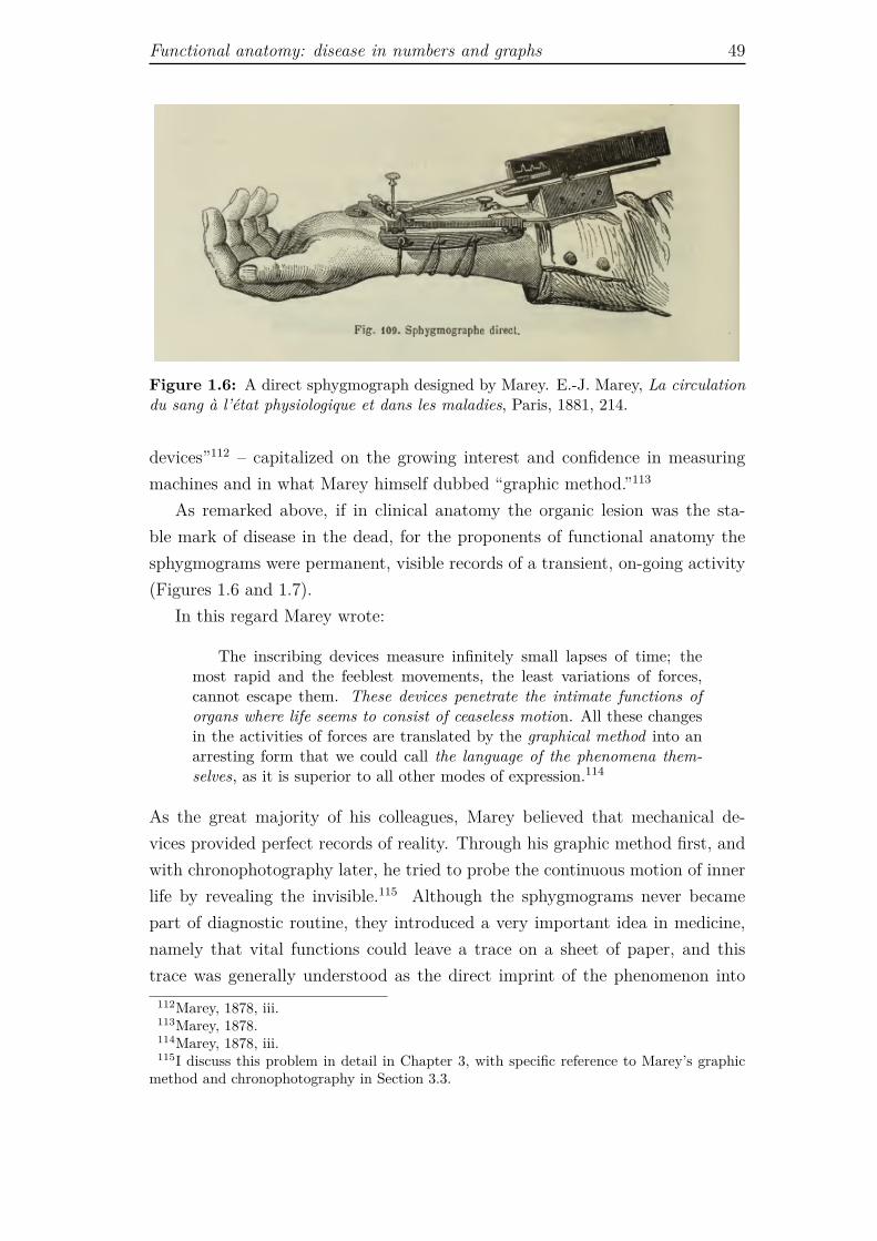
Functional anatomy: disease in numbers and graphs 49
Figure 1.6: A direct sphygmograph designed by Marey. E.-J. Marey, La circulationdu sang à l’état physiologique et dans les maladies, Paris, 1881, 214.
devices”112 – capitalized on the growing interest and confidence in measuringmachines and in what Marey himself dubbed “graphic method.”113
As remarked above, if in clinical anatomy the organic lesion was the sta-ble mark of disease in the dead, for the proponents of functional anatomy thesphygmograms were permanent, visible records of a transient, on-going activity(Figures 1.6 and 1.7).
In this regard Marey wrote:
The inscribing devices measure infinitely small lapses of time; themost rapid and the feeblest movements, the least variations of forces,cannot escape them. These devices penetrate the intimate functions oforgans where life seems to consist of ceaseless motion. All these changesin the activities of forces are translated by the graphical method into anarresting form that we could call the language of the phenomena them-selves, as it is superior to all other modes of expression.114
As the great majority of his colleagues, Marey believed that mechanical de-vices provided perfect records of reality. Through his graphic method first, andwith chronophotography later, he tried to probe the continuous motion of innerlife by revealing the invisible.115 Although the sphygmograms never becamepart of diagnostic routine, they introduced a very important idea in medicine,namely that vital functions could leave a trace on a sheet of paper, and thistrace was generally understood as the direct imprint of the phenomenon into
112Marey, 1878, iii.113Marey, 1878.114Marey, 1878, iii.115I discuss this problem in detail in Chapter 3, with specific reference to Marey’s graphic
method and chronophotography in Section 3.3.
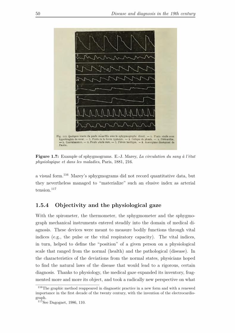
50 Disease and diagnosis in the 19th century
Figure 1.7: Example of sphygmograms. E.-J. Marey, La circulation du sang à l’étatphysiologique et dans les maladies, Paris, 1881, 216.
a visual form.116 Marey’s sphygmograms did not record quantitative data, butthey nevertheless managed to “materialize” such an elusive index as arterialtension.117
1.5.4 Objectivity and the physiological gaze
With the spirometer, the thermometer, the sphygmometer and the sphygmo-graph mechanical instruments entered steadily into the domain of medical di-agnosis. These devices were meant to measure bodily functions through vitalindices (e.g., the pulse or the vital respiratory capacity). The vital indices,in turn, helped to define the “position” of a given person on a physiologicalscale that ranged from the normal (health) and the pathological (disease). Inthe characteristics of the deviations from the normal states, physicians hopedto find the natural laws of the disease that would lead to a rigorous, certaindiagnosis. Thanks to physiology, the medical gaze expanded its inventory, frag-mented more and more its object, and took a radically new perspective on what
116The graphic method reappeared in diagnostic practice in a new form and with a renewedimportance in the first decade of the twenty century, with the invention of the electrocardio-graph.
117See Dagognet, 1986, 110.
Functional anatomy: disease in numbers and graphs 51
could be revealed by the inner, living body. Moreover, an articulated idea ofwhat an objective representation of the body is became operative. All the in-struments described above measure different bodily functions, but they sharea fundamental feature: they make visible through graphs or numbers dynamicprocesses of invisible nature, allowing their permanent record. In this way thevital indices become objective data.
Objectivity here works at different levels. On the one hand, it is understoodas the suppression of the subjectivity of the patient’s narrative describing hisor her symptoms and sensations (a process that began with Laennec’s medi-ated auscultation), as well as the suppression of the sensorial subjectivity ofthe doctor that potentially impaired the examination of physiological indicesor the search for symptoms. It is very difficult to describe and note down thequality of, say, the pulse measured by touch, but its intensity and duration be-come immediately accessible to different people when it is translated into anincrease of mercury on a graded scale or into a line on a sheet of paper. Inthis respect, the machine provides objective data in the sense that it producesdata that are open to collective, inter-subjective scrutiny. These records cantravel from place to place, they can be copied and compared. On the otherhand, objectivity is understood as the adequate correspondence between repre-sentations (graphical or numerical) and phenomena. In other words, objectivitybecomes a synonym for truth, and objective representation is a correct, truthfulrepresentation. Marey believed that his inscribing devices spoke the “languageof phenomena themselves”118 and most of his contemporaries agreed with him.This idea was certainly reinforced by the development of photography. However,as I will show in Chapters 2 and 3, the belief that mechanical devices spoke thevery language of natural phenomena was multifaceted and quite more complexthat it might seem at first sight.
118Marey, 1878, iii.

52 Disease and diagnosis in the 19th century
Chapter 2
The beginnings of medical
photography: visualizing illness
In the previous chapter it was argued that in the course of the nineteenth centuryWestern medicine conceptualized the human body and its diseases in several dif-ferent ways, and that these conceptual transformations were intertwined withthe development of a range of diagnostic instruments. All these instruments,from the scalpel of the pathologist to the measuring instruments of the physiol-ogist, had the aim to make the inner body and its functions accessible from theoutside, thus in a way or another, visible. Visibility was indeed the imperativeset by clinical anatomy at the outset of modern medicine. And visualization, inthe form of anatomical and microscopy images, endoscopic inspections, as wellas graphs and tables of numbers, became a mark of medical practice well beforethe invention of radiography. I went through a analysis of medical diagnostictechniques and technologies, in order to outline the epistemological backgroundagainst which to frame radiography as a diagnostic tool belonging to a avastand heterogeneous medical tradition. Yet, radiography belongs also to anotherline of filiation, namely, medical photography. Hence, in the present chapter Iexplore how photography was co-opted in medicine shortly after its invention,and how it affected the way physicians looked at the patient’s body.
Photography never was a diagnostic instrument proper, yet with its promiseto provide a perfect visual record of reality, it was perceived as the naturalallied of all morphological sciences, including medicine. By outlining the earlydevelopment of medical photography I try to understand how the photographictechnology was embedded in medical theories and practices. To this aim, Idraw on Charles Sanders Peirce’s conceptualization of photographs as indicesand icons, because it encapsulates quite well the nineteenth-century discourse onphotography, simultaneously offering conceptual categories that help examining
53
54 The beginnings of medical photography: visualizing illness
that very discourse. I integrate Peirce’s intuitions with Allan Sekula’s analysisof how early photographic portraits could work according to the indexical orsymbolic mode of signification. I also draw on Walter Benjamin’s considerationson the optical unconscious and the intrinsic analytic potential of photography,as well as on his idea (largely borrowed from other theorists such as LászlóMoholy-Nagy and Béla Balász) that photography works as a prosthetic sense,which enriches and transforms our sensory experience. I argue that, althoughvirtually all the scientists and medical doctors of the nineteenth century praisedthe mimetic realism of photographic image, they often used the new opticalmedium to produce pictures of such abstract entities as pathological conditions.More precisely, they used photographs to train their ability to extract general,abstract features of disease from the contingent pictures of singular individuals.In this sense, we can say that they used photographs to visualize the invisible.I also maintain that the body represented in these photographs is not the bodyas we see it. It is a body reconfigured as part of an experimental apparatus inwhich patient, camera and observer are components of a larger structure (whichincludes the archive and the medical journal) capable of producing scientific andmedical evidence.
2.1 Londe: medical photography in practice
In the preface of his book La photographie médicale, of 1893, Albert Londe,pioneer in scientific photography and director of the photographic service ofthe Hôpital de la Salpêtrière,1 claimed that photography was “one of the mostbeautiful discoveries of mankind.”2 He believed that the photographic labora-tory would soon become a necessary facility for any hospital, and envisaged thecreation of photography courses in medical schools. Photography, Londe main-tained, with its ability to keep track of transient states was an invaluable tool formedical research and practice,3 and he consistently backed this claim by making
1At the time Londe worked there, the Salpêtrière, a female insane asylum, hosted about5,000 patients, many of whom institutionalized for life (Jean-Martin Charcot, director of thehospital for many years said that it was a sort of living museum of pathological conditions).See Sicard, 1995, 19.
2Londe, 1893, ix, my translation. For Londe photography was both a discovery and ascience in its own right. To the contemporary reader, the idea that photography can beconsidered a science, rather than a technique (or technology), might sound strange. However,we should consider that for those who were involved in the development of photographicinstrumentation and practices, photography posed an open problem in applied science. Thevery activity of taking pictures was bounded with the problem of understanding how the wholeprocess functioned. This understanding, in turn, was related to the study of the physical andchemical phenomena that subtended the working principles of the apparatus.
3See Londe, 1893, 3.
Londe: medical photography in practice 55
a list of the actual and possible applications of photography to medicine andphysiology. Photography, he said, was the necessary iconographic complementof the patient’s file in all those cases in which the morphological presentation andevolution of the disease were relevant (e.g., deformities and wounds). It couldbe used to record endoscopic examinations, as well as surgical and obstetricalinterventions, in a much more precise way than any written description, and thevisual records could be shared and retrieved at any time for further study. Inthe same way, photography could improve the much honored tradition of patho-logical anatomy, by enabling the comparison of the lesions revealed by differentautopsies. For what concerns the study of histology and cytology, it would allowto preserve “authentic and enduring records”4 of tissues that were doomed toperish in a short time. Moreover, in the case of the study of nervous and men-tal pathologies, in which the observation of unexpected and quick movementswas paramount, photography could provide access to new fields of knowledge,while in the domain of physiology it had the great advantage of allowing therecording of body motions without directly interfering with the subject underexamination (Londe collaborated with Etienne-Jules Marey in the developmentof chronophotography).5 Furthermore, Londe contended that photography hadan eminently pedagogical quality because, by facilitating the production of largecollections of images, it fostered observational skills in students and taught bothstudents and qualified physicians how to recognize permanent and recurrent fea-tures within the intrinsic variability of the morphological alterations provokedby pathology. As he put it: “Photography provides different ways to multiplythe original picture, without loosing any of its tracts of sincerity and truth”.6
Londe concluded his introductory list of applications of medical photographymentioning its relevance in the medico-legal context. In this case, however,rather than praising the evidential role of the photographic image, he empha-sized its emotional power: put under the eyes of judges and jury, the shockingpictures of a face disfigured by a lethal wound would discourage any feeling ofpity for the author of the crime.7
Within the list of medical applications of photography expounded by Londe,I would like to focus on two points: (1) the use of photographic records in the in-dividual patient’s file, and (2) the pedagogical relevance of collections, or series,of images. For what concerns the second point, it is important to emphasize
4Londe, 1893, 6, my translation.5Chronophotographs are pictures taken in extremely rapid sequences, above the tempo-
ral resolution of the eye. The first successful attempts to produce this sort of images wereaccomplished by the photographer Eadweard Muybridge in the United States and by thephysiologist Etienne-Jules Marey in France. I analyze in detail Marey’s work in Chapter 3.
6Londe, 1893, 6, my translation.7See Londe, 1893, 6 and 215.
56 The beginnings of medical photography: visualizing illness
that it was through the series, the gallery of images, that photographs acquireda diagnostic meaning. By training the eye, the photographic series taught torecognize the facies of a disease at a glance.8 In fact, as Walter Benjamin willlater remark, the series is endowed with two antithetic properties: it gives visi-bility to the exceptional, and simultaneously fosters our “sense for sameness,”9
our ability to recognize, or extract, recurrent patterns within a variety of sin-gular phenomena. The series of photographs, collected in specialized journals,books of case studies or file archives, has the function to stimulate a sort ofinductive process whereby a general visual rule (the facies of the disease) isderived from a set of individual occurrences. Hence, there is a movement fromthe particular to the general.10 In medicine, however, general knowledge needsalways to be referred to an individual case. More precisely, there is always atension towards the construction of a science of the individual, because the fieldof action of the physician is a specific person. This is why Londe recommendedto keep photographic records of patients in their medical file. In explaining therules for the proper making of medical photographs, Londe insisted that it wasalways necessary to produce sets of images of the same patient, because thiswas the only way to keep track of the evolution of his or her condition over timeand under different stimuli.11 Only through sets of images it became possible“to rigorously establish [pathological] types that corresponded to specific affec-tions.”12 Photography promised to foster the advancement of medicine becauseit offered an opportunity to shift from the general rule to the particular casewith relative ease.
As we will see in this chapter, establishing a human type for specific diseaseswas one of the main purposes of early medical photography. We can considerthis endeavor as the visual analogue of the attempts to organize and inscribedisease within the body pursued by all the diagnostic technologies developedduring the nineteenth century. Similarly to the stethoscope, the thermometer,the sphygmograph and all the other diagnostic devices discussed in Chapter 1,medical photography was meant to objectify the body, to turn it into a scientificobject that could be described through the combination of simplified, measur-able, standard elements. It was thus necessary for medical photography to havea standard, seemingly neutral aesthetics, corresponding to a specific visual code.Such aesthetics was summarized by Londe in a few rules: he recommended to
8See Londe, 1893, 74-77.9Benjamin, 1936, 256.
10This means that the photographic series had not simply a pedagogical value (transmissionand sharing of knowledge), but also a scientific value proper, for it helped to construct newknowledge by induction.
11See Londe, 1893, 66.12Londe, 1893, 5.
Photographs as indices and icons 57
photograph the body according to an ordered sequence,13 against a uniform darkgrey background “against which the naked body stands out very clearly,”14 andwith the camera placed right in front of the subject, in order to avoid any de-formation. Not surprisingly, this was also the aesthetics of ethnography andcriminology, two social sciences that received great impetus between the 1870sand 1880s. In these disciplines, too, what was at stake was the creation ofobjective, scientific portraits of the average man (or woman).
Londe’s book, a reference text written more than fifty years after the intro-duction of photography in medicine, can be considered a compendium of theaccepted wisdom about scientific photography in the late nineteenth century.The high value attached to the specific ability of the camera to produce faith-ful visual records, its eminently documental character, is patent in Londe’s listof medical applications of photography. For him, as for his contemporaries,the camera was a “wonderful recording instrument,”15 and photographs were“truthful images” [images fidèles]16 of the world. Tellingly, even Charles Baude-laire’s condemnation of photography in the Salon of 1859 was chiefly groundedin the idea that the camera is a picture-making machine characterized by “anabsolute material exactitude.”17 For the poet, photography was a device thatmerely recorded trivial external reality, and therefore it could certainly be the“very humble servant”18 of the sciences and the arts, but it was divested of anycreative and artistic potential.19
2.2 Photographs as indices and icons
To better understand Londe and Baudelaire’s conception of photography, with-out limiting ourselves to certify their endorsement of photographic realism, itis useful to refer to the semiotic conceptualization of photography put forwardby Peirce. Indeed, it seems to me that Londe and Baudelaire, as virtually alltheir contemporaries, implicitly subscribed to the Peircean truth-value theory ofphotography. According to the American phylosopher, photographs, especiallyinstantaneous photographs, are simultaneously icons and indices. They are in-dices because they are physically connected to their referent, as a thumbnail tothe finger or the smoke to the fire. At the same time they are icons, because
13Namely, the whole body view from front, sides and behind, the head alone, the hands,the feet and the lower limbs, and, finally, detailed images of the skin.
14Londe, 1893, 66.15Londe, 1893, 6, my translation.16Londe, 1893, 68.17Baudelaire, 1859, 261, my translation.18Baudelaire, 1859, 261, my translation.19See Baudelaire, 1859, 254-263.
58 The beginnings of medical photography: visualizing illness
their meaning depends on their resemblance to the object they represent. Moreexactly, in the case of photography the iconic mode of signification of the signdirectly springs from its indexical mode. Peirce wrote: “Photographs [are] pro-duced under such circumstances that they [are] physically forced to correspondpoint by point to nature.”20 And also: “A photograph [...] owing to its opti-cal connection with its object, is evidence that that appearance corresponds toreality.”21 That is, the truth-value of photographs is predicated on their phys-ical mode of production. Indeed, it is interesting to consider Peirce’s idea ofphotographs as iconic indices in the light of a description of the photographicprocess provided by one of its inventors, Henry Fox Talbot. In 1844, in hisgroundbreaking photographic album The Pencil of Nature,22 Talbot wrote:
The idea occurred to me [...] how charming it would be if it werepossible to cause these natural images imprint themselves durably, andremain fixed upon the paper! [...] Now, light, where it exists, can ex-ert an action, and, in certain circumstances, does exert one sufficient tocause changes in material bodies. Suppose, then, such an action could beexerted on paper; and suppose the paper could be visibly changed by it.In that case surely some effect must result having a general resemblanceto the cause which produced it.23
As a matter of fact, photographs were the product of two distinct processes: anoptical process, that led to the production of an image in the camera, and achemical one, that allowed to fix and reproduce on a sensitive surface the imagecast upon it by the lens. This chain of physicochemical phenomena preserved,through a stream of causal relations, a resemblance between the object thathad produced an image within the camera, and the image that had been finallyfixed on the sensitive plate. The very materiality of the photographic processbounded the iconicity of the picture to its indexicality, and it was the source ofits perceived realism.
Peirce did not developed a theory of photography in its own right, but heused photographs as examples of how signs can signify. Although it is beyondthe scope of this dissertation to explore in detail Peirce’s semiotics, I will resortto his classification of signs into icons, indices and symbols as a conceptual andmethodological tool for part of my analysis of photography and medical imagingin the following chapters For Peirce, the sign is a unity of what is represented (the
20Peirce, CP 2.281.21Peirce, CP, 4.447.22Together with the Frenchman Louis Daguerre, Henry Fox Talbot is credited for being one
of the inventors of photography. The Pencil of Nature was the first book ever published inwhich illustrations were printed through a photographic process. The book was composed ofsix installments released between 1844 and 1846, and included 24 calotype prints.
23Talbot, 1844, n.p.
Photographs as indices and icons 59
Object), how it is represented (the Representamen) and how it is interpreted (theInterpetant). More exactly, the Representamen is the sensible form taken bythe sign (it is also called sign-vehicle); the Interpretant is the sense made of thesign (it can be understood as a translation of the primitive sign into a further,more developed sign in the mind of the interpreter); and the Object is thereferent, that is, something beyond the sign to which it refers (importantly, theobjects puts constraints on the sign’s possibility to signify). The classificationof signs in indices, icons, and symbols depends on the quality of the relationshipthat connects Object, Representamen, and Interpretant.24 This relationship, ormode of signification, is characterized by different degrees of conventionality,with indices at the bottom of the scale (where, ideally, no convention is involvedin the process of signification) and symbols at the top. Thus, a sign is a symbolif it is connected to its object by a convention, which, as such, has to be learnedand agreed upon.25 Instances of symbols are language in general, numbers, theMorse code, and nautical flags. An index, on the contrary, relates to its referentvia a physical or factual connection.26 For example, a footprint is an index of aperson walking on a trail, because it is the physical imprint let by the foot, andpain can be an index of disease. Finally, a sign is an icon when it has a relationof resemblance with its referent.27 Similarity is the defining feature of iconicity,and indeed, Peirce also terms such mode of signification likeness. Instances oficons are pictorial portraits, scale models, but also metaphors, synthetic aromas,and imitative gestures.
Peirce’s precise thoughts about the nature of icons, indices and symbolschanged and became more complex overtime, as far as he developed his signtheory. In particular, he came to realize that it was virtually impossible tofind any pure instance of icons and indices, and that these signs were alwayspartly symbolic or conventional.28 This alerts us about the fact that not onlywe cannot treat photographs as pure indices, but we should not even considerthem as simple combinations of index and icon. Indeed, as we will see in thischapter and in the following one, the symbolic or conventional component of themode of signification of photographs and radiographs plays a fundamental role in
24See Atkin, 2013, and Chandler, 2007, 39-44.25“A Symbol is a sign which refers to the Object that it denotes by virtue of a law, usually an
association of general ideas, which operates to cause the symbol to be interpreted as referringto that Object.” Peirce, CP 2.249.
26“An Index is a sign which refers to the Object that it denotes by virtue of being reallyaffected by that Object.” Peirce, CP 2.248.
27“An Icon is a sign which refers to the Object that it denotes merely by virtue of charactersof its own, and which it possesses, just the same, whether any such Object actually exists ornot.” Peirce, CP 2.247.
28See Atkin, 2013.
60 The beginnings of medical photography: visualizing illness
endowing these images with meaning.29 The interplay between the indexical andsymbolic modes of signification is crucial to an understanding of photographyand other forms of scientific imaging technology, especially when it comes to thevisualization of the invisible.
Indexicality and likeness Before exploring at length the problem of the vi-sualization of the invisible, I task I undertake in this chapter and in the next one,I want to insist on how powerfully the idea of resemblance was entrenched withthat of indexicality in the earliest conceptualizations of photography.30 ForLonde and his contemporaries, the epistemic authority of photography camefrom its indexical nature, from its being a direct imprint of nature. This phys-ical contiguity was the guarantee of iconic exactitude, of perfect resemblance:the photographic image was epistemically meaningful because it was identical toits referent and it was identical to its referent because it was its direct physicalimprint. In other words, the photograph was perceived as scientifically mean-ingful (that is, objective and endowed with cognitive value) because it was anicon, but it was an icon only in so far as it was an index (that is, its ontologicalstatus secured its truth-value). This conflation of the indexical and iconic na-ture of photography was quite productive, for it allowed scientists to use theseimages in flexible (sometimes conceptually conflicting) ways.
Embedded in Londe’s apparently naïve photographic realism we can in factdiscern a range of instrumental uses of photography. As highly contingent im-ages, taken directly from the body of the patient and referring to no one butthat patient, photographs are important complements in the medical file, forthey keep memory of what the doctor saw with his own eyes, with a fidelity anddetail that a written description could not convey. In this case, what was atstake was the ability of the photograph to be identical to its referent. And it wasstill the contingency of the photographic picture that conveyed its evidentiaryand emotional meaning in the murder trial.31 In this instance what matteredwas the indexical nature of the photograph not only in the strictest sense of be-
29In Peirce’s semiotics the meaning of a sign is not contained within it, but arises in itsinterpretation. Moreover, the object is crucial to the meaning of the sign (it puts constraintson the sign). Hence the meaning of a sign includes both its reference and its sense. SeeChandler, 2007, 33.
30Of course, the notion of “resemblance” is very problematic and controversial. It is rootedin a mimetic-realistic conception of “likeness,” as if this were an intrinsic property of the image,rather than an anthropological and cultural construct. That is, it is a conception which doesnot take into account that what counts as similar to something else is not only species-specific(what counts as similar for a human being is not recognized as such by, say, a dog or a fly),but also culture-specific. The need to resort to the symbolic mode of signification to interpreta photograph can be considered a symptom of the fact that the iconicity of a picture, itslikeness, is not as self-evident and self-contained as one might believe on first thought.
31See Londe, 1893, 6 and 125.
A new optical truth 61
ing an evidence of the crime, but also in what concerned the emotional cogencyof the index. It was because they knew, by virtue of common sense, that thatslaughtered person was really and necessarily before the camera, and becausethey felt that that body was in metonymical relation with the photographicplate, that the members of the jury would be revolted by the photograph muchmore than by any painting or drawing, no matter how realistic.
However, photographs are not only about what we can see with our owneyes. They are also about what we struggle to see (the elusive movements of theneurological patient), or what we cannot see at all (the all too rapid movementsstudied through chronophotography). In these cases the iconic aspect of thephotograph is taken to a level that goes beyond visual resemblance, and canacquires a diagrammatic function. Also, the contingent nature of the photographfades, or is tamed, once pictures are collected in organized series (in reviews oratlases). In this case the medical value of the camera resides not so much in itsability to provide exact records of specific occurrences, but rather in the factthat it allows to easily multiply images. Hence, it allows to create those visualgalleries, those pathological and teratological collections that ideally would allowthe recognition of general prototypes, the facies of each and any disease.
2.3 A new optical truth
Since its official invention, in 1839, photography has been simultaneously aninstrument and an object of research (scientific and artistic alike), but untilthe turn of the century photographs continued to be conceived of as the fix-ation of the image of the camera obscura, thus as an imprint of the visible.Although in their practice the scientists used photography in very creative waysand demanded of photographs to reveal much more than the surface of objects,their conceptual (and rhetorical) discourse on photography essentially revolvedaround the idea of “perfect and truthful record”32 of visible phenomena.33 Ben-jamin, in his Little History of Photography, of 1931, ascribed this lack of concep-tual variety to the tumultuous development of the new technique, “which longprecluded any backward glance.”34 More specifically, he considered the theo-rization of photography put forward before the 1920s “entirely rudimentary,”35
32Diamond, 1856, 24.33We can find some meaningful exceptions in the writings of the physicist and philosopher
Ernst Mach and of physiologist Etienne-Jules Marey. Both were interested in the use ofphotography for the study of dynamic phenomena. Mach expounded his ideas on photographyin the article “An account of scientific applications of photography,” of 1893.
34Benjamin, 1931, 507.35Benjamin, 1931, 508.
62 The beginnings of medical photography: visualizing illness
because based on two misconceptions: on the one hand, the conviction that aphotograph was nothing more than a mirror image of the visible world; on theother hand, the attempt to legitimize (or delegitimize) photography in the faceof traditional painting.36
This vision was challenged by theoreticians and practitioners of photogra-phy and cinema of the 1920s and 1930s, who developed a broad reflection on thespecific cognitive and analytic power of the new optical media.37 For instance,for the Hungarian artist and theorist Lázló Mohly-Nagy, Benjamin’s friend andmain exponent of the photographic movement New Vision [Neues Sehen], thetask of the camera was to provide a new, enriched way to perceive and under-stand the world. Moholy-Nagy emphasized the ability of photography to provide“an unbiased optical vision” [eine unvoreingenommene Optik], that is, a visionnot bound to the association laws that rule over natural perception.38 The cam-era allowed to enlarge and frame the field of view in ways the human eye couldnot. Indeed, for Moholy-Nagy, the distortions produced by the lens and by allsort of photographic experimentation, from oblique views to superimpositions,should not be considered photographic errors, but rather important epistemicdevices that, by severing all habitual connection between vision, imaginationand judgement, would reveal a new “optical truth” [das Optisch-wahre], whichwould precede any subjective stance.39 As pointed out by media scholar AntonioSomaini, Moholy-Nagy’s strong statements about the objectivity of the camera,which clearly did not take into account the subjectivity and intentionality ofthe photographer, are to be understood in the context of the reflection on theepistemic cogency of photography and cinema that animated the debate aboutthese media in the 1920s. For the Russian film maker and theorist Dziga Vertov,for instance, the camera had specific analytic properties that revealed what hecalled Kino-Pravda (literally, film-truth), i.e., the reality of life that escapedcommon perception. Similarly, the formalist Osip Brik, in the essay What the
36Benjamin maintained that photography could not be judged according to the categoriesof painting, for it actually overturned those categories. He particularly criticized what heconsidered to be a “fetishistic and fundamentally antitechnological concept of art,” that is,a concept of art grounded in the idea of divine inspiration, detached from any historical andmaterial context. Benjamin, 1931, 508.
37See Somaini, 2010, ix-lvi; Pinotti and Somaini, 2012, xxv-xxviii; Landeker, 2005 and 2006.38See Moholy-Nagy, 1925, 5. In the introduction to the Italian edition of Moholy-Nagy’s
Malerei Photographie Film, Antonio Somaini points out that already in 1844 Henry Fox Talbothad hinted at the idea of unbiased vision, by stressing that photography does not discriminatebetween a chimney sweep and the Apollo of the Belvedere. See Somaini, 2010, xxxix. InThe Pencil of Nature, commenting on the plate with the view of the boulevards at Paris,Talbot wrote: “The instrument [the camera] chronicles whatever it sees, and certainly woulddelineate a chimney-pot or a chimney-sweeper with the same impartiality as it would theApollo of Belvedere.” Talbot, 1844, 18.
39See Somaini, 2010, xxxix-xl.
A new optical truth 63
Eye Does Not See, of 1926, maintained that photography and cinema did nothave to imitate the human eye, but rather unveil what the human eye is unableto see. The same line of thought was hold by French avant-garde film directorssuch as Germaine Dulac, who considered cinema a sort of microscope focused onlife, Abel Gance, who challenged traditional vision with multiple projections andframe superimpositions, and Jean Epstein, for whom cinema, through montage,slow motion, time-lapse, and reversed projection, could radically reconfigure ourexperience of space and time. Within the German speaking context, to whichboth Moholy-Nagy and Benjamin belonged, Béla Balász thought of cinema as anew sense organ, which entailed new sensorial faculties and thus paved the wayto the advent of a new visual culture [visuelle Kultur ]. This new visual culture,Balász posited, entailed a distinctively modern way to see the world, and overtime would lead to a reconsideration of the epistemic import of human senso-rial faculties.40 For Balász, as for Moholy-Nagy, the new optical media couldbe considered prosthesis of the human sensorium, for they reorganize, integrateand perfect our senses.41
2.3.1 Pictures from the optical unconscious
Balász and Moholy-Nagy’s intuitions on the epistemic and analytic possibilitiesof the camera were condensed by Benjamin in the metaphor of the “opticalunconscious,” expounded in the essays Little History of Photography and TheWork of Art in the Age of Mechanical Reproduction. In the latter he wrote:
With the close-up, space expands; with slow motion, movement isextended. And just as enlargement not merely clarifies what we see in-distinctly “in any case,” but brings to light entirely new structures ofmatter, slow motion not only reveals familiar aspects of movements, butdiscloses quite unknown aspects within them [. . . ]. Clearly it is anothernature which speaks to the camera as compared to the eye. “Other” aboveall in the sense that a space informed by human consciousness gives way toa space informed by the unconscious. [. . . ] Whereas it is a commonplacethat, for example, we have some idea what is involved in the act of walk-ing (if only in general terms), we have no idea at all what happens duringthe split second when a person actually takes a step. [. . . ] This is wherethe camera comes into play, with all its resources for swooping and rising,disrupting and isolating, stretching or compressing a sequence, enlargingor reducing an object. It is through the camera that we first discoverthe optical unconscious, just as we discover the instinctual unconsciousthrough psychoanalysis.42
40See Somaini, 2010, xl-xliii.41See Somaini, 2012, 206.42Benjamin, 1939, 266.
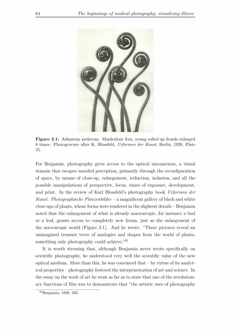
64 The beginnings of medical photography: visualizing illness
Figure 2.1: Adiantum pedatum. Maidenhair fern, young rolled-up fronds enlarged8 times. Photogravure after K. Blossfeld, Urformen der Kunst, Berlin, 1928, Plate55.
For Benjamin, photography gives access to the optical unconscious, a visualdomain that escapes unaided perception, primarily through the reconfigurationof space, by means of close-up, enlargement, reduction, isolation, and all thepossible manipulations of perspective, focus, times of exposure, development,and print. In the review of Karl Blossfeld’s photography book Urformen derKunst. Photographische Planzenbilder – a magnificent gallery of black and whiteclose-ups of plants, whose forms were rendered in the slightest details – Benjaminnoted that the enlargement of what is already macroscopic, for instance a budor a leaf, grants access to completely new forms, just as the enlargement ofthe microscopic world (Figure 2.1). And he wrote: “These pictures reveal anunimagined treasure trove of analogies and shapes from the world of plants,something only photography could achieve.”43
It is worth stressing that, although Benjamin never wrote specifically onscientific photography, he understood very well the scientific value of the newoptical medium. More than this, he was convinced that – by virtue of its analyt-ical properties – photography fostered the interpenetration of art and science. Inthe essay on the work of art he went as far as to state that one of the revolution-ary functions of film was to demonstrate that “the artistic uses of photography
43Benjamin, 1928, 350.
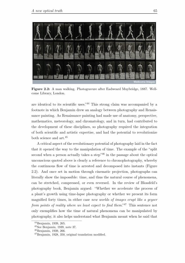
A new optical truth 65
Figure 2.2: A man walking. Photogravure after Eadweard Muybridge, 1887. Well-come Library, London.
are identical to its scientific uses.”44 This strong claim was accompanied by afootnote in which Benjamin drew an analogy between photography and Renais-sance painting. As Renaissance painting had made use of anatomy, perspective,mathematics, meteorology, and chromatology, and in turn, had contributed tothe development of these disciplines, so photography required the integrationof both scientific and artistic expertise, and had the potential to revolutionizeboth science and art.45
A critical aspect of the revolutionary potential of photography laid in the factthat it opened the way to the manipulation of time. The example of the “splitsecond when a person actually takes a step”46 in the passage about the opticalunconscious quoted above is clearly a reference to chronophotography, wherebythe continuous flow of time is arrested and decomposed into instants (Figure2.2). And once set in motion through cinematic projection, photographs canliterally show the impossible: time, and thus the natural course of phenomena,can be stretched, compressed, or even reversed. In the review of Blossfeld’sphotography book, Benjamin argued: “Whether we accelerate the process ofa plant’s growth using time-lapse photography or whether we present its formmagnified forty times, in either case new worlds of images erupt like a geyserfrom points of reality where we least expect to find them.”47 This sentence notonly exemplifies how the time of natural phenomena can be manipulated byphotography, it also helps understand what Benjamin meant when he said that
44Benjamin, 1939, 265.45See Benjamin, 1939, note 37.46Benjamin, 1939, 266.47Benjamin, 1928, 350, original translation modified.
66 The beginnings of medical photography: visualizing illness
the nature that speaks to the camera is “other” from the nature that speaks tothe eye, nevertheless it is no less real.48 The world becomes “other” because theway our perception structures our apprehension of the world and its phenomenais reinvented: we have new mechanical senses through which we can exploredimensions of reality that we could not even conceive of before. And throughthis new mechanical perception we discover “entirely new structures of matter.”49
Benjamin’s ideas on the optical unconscious and the reconfiguration of theperception of space and time were grounded in his analysis of photography andcinema (enlargement, slow motion, time-lapse, and so on).50 In this account,the ability of the camera to disclose the optical unconscious (the unknown, theunexpected) seems to depend chiefly on highly conscious processes of photo-graphic manipulation and experimentation, such as enlargement, time-lapse,slow motion, and so on. Still, in the early days of photography many werefascinated by another property of the photographic process, namely, its abilityto capture perfectly visible aspects of everyday life that had not been noticedby the photographer. Photography theorist Joel Snyder explains that: “the au-thors of the earliest descriptions of daguerreotypes dwelled on the profusion ofdetail carried on the surface of the mirrored plate, noting, for example, puddlesor stray reflections that had gone unnoticed at the time the plate was made.”51
Photography had the surprising ability to show aspects of the visible world, atthe visible scale, and within the range of the eye resolution that the eye did notcatch. In this case we can talk of an optical unconscious revealed by the camera,not by rearranging space and time, but by bypassing the neuro-psychologicalassociative laws of natural vision. It is the optical unconscious correspondingto Moholy-Nagy’s idea of unbiased vision. Such unbiased vision was made pos-sible by the (partial) independence of the camera from the photographer.52 ForMoholy-Nagy there was a fundamental divide between photography (mechan-ical images in general) and hand-made representations. In his view, paintingsand drawings always bear traces of the author’s will, or at least of his or herperceptual schemes. For him, the activity of painting and drawing was inher-ently limited by the laws of human perception. In representing the world, thepainter cannot show more than he or she has perceived. Even if the artist de-
48Benjamin, 1939, 266. See quote above.49Benjamin, 1939, 266.50For a very interesting discussion on how early scientific cinema influenced the space and
time perception of spectators’, and exerted a strong impact on the early theories of cinema,see Landecker, 2005 and 2006.
51Snyder, 1998, 391.52Moholy-Nagy considered the camera as completely independent from the photographer.
We cannot forget, however, that it is always a human agent who selects a specific lens, time ofexposure, perspective, and so forth. A photographic image, like any other mechanical image,can never be completely independent from the human operator.
A new optical truth 67
picted an imaginary world, this would still be in a way or another linked tothe functioning of the human psychology and neurophysiology. Photography,on the contrary, seemed to break the bond between the image and the humanproducer. The developed plates could show something that might well be infront of the camera, but that the photographer had not seen for a multiplicityof reasons (from simple lack of attention to any other form of natural limitationor impairment of human perception). In this sense we can say that photogra-phy has an inherent analytic, anatomizing quality, and an enormous potentialto transform perception. Of course, in contrast to the idea that painting anddrawing are heavily limited by the rules of natural perception, one could take apsychoanalytic stance and claim that every artwork is first and foremost a repre-sentation of the artist’s unconscious. Or, alternatively, one could maintain thatthe hand of the artist is guided by formal and ideal determinants that allow toreveal aspects of the world that escape ordinary perception. In this respect, artin itself can transform our way of seeing objects no less than photography andother optical media. In this dissertation, however, I am specifically interestedin mechanically produced images, thus I do not explore how image productionin general impinges on our perception of the world.
2.3.2 The photographer as anatomist
A suggestive description of how photography and cinema bring about a transfor-mation of sensory experience is provided by Benjamin in the essay on the workof art, in the famous metaphor of the cinematographic operator as surgeon:
How does the camera operator compare with the painter? In answerto this, it will be helpful to consider the concept of the operator as it isfamiliar to us from surgery. The surgeon represents the polar oppositeof the magician. The attitude of the magician, who heals a sick personby a laying-on of hands, differs from that of the surgeon, who makes anintervention in the patient. The magician maintains the natural distancebetween himself and the person treated; more precisely, it reduced itslightly by laying on his hands, but increases it greatly by his authority.The surgeon does exactly the reverse; he greatly diminishes the distancefrom the patient by penetrating the patient’s body, and increases it onlyslightly by the caution with which his hand moves among the organs.[...] Magician is to surgeon as painter is to cinematographer. The paintermaintains in his work a natural distance from reality, whereas the cine-matographer penetrates deeply into its tissues. The images obtained byeach differ enormously. The painter’s is a total image, whereas that ofthe cinematographer is piecemeal, its manyfold parts being assembled ac-cording to a new law.53
53Benjamin, 1939, 263-264.
68 The beginnings of medical photography: visualizing illness
In the two figures of the surgeon and the magician Benjamin embodied a numberof dialectical antinomies that characterized his reflection on perception, namely,first vs. second technique; proximity vs. distance; appearance vs. play (Scheinand Spiel); cult value of the auratic artwork (the artwork still invested of itsmagic-sacral properties derived from its being unique and inscribed in an au-thoritative tradition) vs. exhibition value of the non-auratic work, (the workreproduced in series, freed from any ritual meaning, easily transportable andaccessible to the masses).54 Here, however, I focus on the figure of the surgeonas exemplification of the analytic, enquiring gaze of the camera.
Unlike the magician-painter, who intervenes on the patient (correspondingto the object to be represented) from the distance of his sacral authority, thesurgeon-cameraman, from the proximity of his technical competence, penetrateswith his hands and his eyes straight into the body. The painter creates a “to-tal image,” an image that requires of its author to always keep the necessarydistance from its object in order to be able to see it. Ideally, once the work iscompleted, the beholder will see what the painter saw. On the contrary, the lensof the cameraman, for Benjamin, is like the gaze of the surgeon, a haptic gaze,a gaze in action. This is so because in the making of a film the cameraman hasthe option to zoom in and out, to chose different planes, to stop time or to makeit move faster. Moreover, the frames must be further edited (the film is liter-ally cut into sections that are subsequently reassembled or discarded), so thatthe final stream of images will appear to the eye of the spectator not as it hadbeen seen by the cameraman,55 but as it was manufactured by a team of peopleworking under a film director. The cinematographer creates an image which is“piecemeal” because it is necessarily the output of the composition of “many-fold parts” (the different shots) “assembled according to a new law,” namelythe laws of cinematographic narrative. Similarly, the surgeon, in the lineage ofthe anatomist and the clinician, does not contemplate the body from without,he literally cuts and stitches its parts according to the rules of anatomy andphysiology. Thus, the cinematic gaze is much more akin to the scientific way oflooking at the world than to artistic contemplation, or even to natural vision. Itis a structured, analytic way of seeing that dissects and recombines the elementsof reality according to its own rules, dictated by the purpose of investigation.In the Little History of Photography, Benjamin hinted again at the proximitybetween photography and medicine when he claimed: “Details of structure, cel-lular tissue, with which technology and medicine are normally concerned – all
54See Pinotti and Somaini, 2012, 11.55As a matter of fact, at the time of shooting the cameraman sees no image stream at all,
but rather a number of individual events.
A new optical truth 69
this is, in its origins, more native to the camera than the atmospheric landscapeor the soulful portrait.56 And he immediately added:
Yet at the same time, photography reveals in this material physiog-nomic aspects, image worlds, which dwell in the smallest things – mean-ingful yet covert enough to find a hiding place in waking dreams, butwhich, enlarged and capable of formulation, make the difference betweentechnology and magic visible as a thoroughly historical variable.57
That is, getting closer and closer to its object, in accordance with its specificanalytic habit, photography has stripped the world of its magic (which doesnot mean that it has stripped it of its beauty) and replaced “waking dreams,”by definition elusive and impalpable, with enlarged images, from which we canpick up the smallest details. The surgeon has finally overruled the magician,for technology has shortened the distance between object and observer, betweenthe invisible and the visible, the unknown and what is “capable of formulation.”In Benjamin’s terms, photography has “suck[ed] the aura out of reality.”58
2.3.3 Image series and the aura of the body
Benjamin’s concept of aura is a complex one, and he elaborated on it from dif-ferent perspectives. In The Work of Art in the Age of Mechanical Reproductionthe notion of aura encapsulates a node of intertwined properties of the artworkbefore the age of its mechanical reproducibility. It is the here and now of thework of art, that is, “its unique existence in a particular place,”59 which deter-mines its distinctiveness and authenticity, but it is also its permanence in time,which entails its transmissibility and the related value as historical testimony. Inthe same text Benjamin also defines the aura as an irreducible distance betweenthe artwork and the observer.60 An unbridgeable distance created by the factthat the work of art belongs to a tradition whereby it derives an authority anda cult value that set it beyond the reach of the beholder. Mechanical reproduc-tion, and the mass-production that goes with it, strips the artwork of its aura,because through the proliferation of cheap copies it obliterates its here and now,jeopardizing simultaneously its uniqueness, authenticity, historical meaning andcult value. The aura, however, is not just a property of the works of art. Nat-ural objects can be auratic too. Benjamin defined the aura of a natural objectas “the unique appearance of a distance, however near it may be.”61 But if the
56Benjamin, 1931, 512.57Benjamin, 1931, 512.58Benjamin, 1931, 518.59Benjamin, 1939, 253.60See Benjamin, 1939, 255 and 1931, 253-256.61Benjamin, 1939, 255, see also Benjamin, 1931, 518.
70 The beginnings of medical photography: visualizing illness
aura of a natural object is the irreducible distance between the object and thebeholder, then, just as the works of art, natural objects loose their aura whenwe get very close to them, and look at them with the analytic, anatomizing gazeof science.
Now, if we try to apply the concept of aura to the domain of medical repre-sentations, we find that the whole endeavor of Western medicine since the birthof the clinic can be seen as a laborious attempt to peel the aura away from thehuman body. The anatomist not only obliterates the distance of the observerand cuts directly into the body, but thanks to all the techniques put in placeto explore the inner organs from without, the human body has entered a spe-cific domain of mechanical visibility and reproducibility. With the stethoscopeLaennec put the body of the patient at a distance by interposing an instrument,but he did so in order to gain a deeper access into the heart and lungs (thesame happened with the endoscopic devices, meant to visualize other inner or-gans). With the microscope cellular pathologists pushed their senses down intothe cell. With the thermometer, the sphygmometer and all the other measuringinstruments, records from the inner body could be easily taken, copied, and sentfrom one doctor to the other, so that, ideally, there was no longer the need tobe in the presence of the original (the patient) to make a diagnosis. Of course,this whole process was progressively enhanced by the subsequent developmentof imaging technologies that replicate our body, or parts of it, in countless sam-ples. However, unlike the work of art, which loses its unique existence due tothe proliferation of its reproductions, the uniqueness of the human body is notundermined by its copies (a human body does not lose its uniqueness just be-cause its portrait can be reproduced indefinitely). The uniqueness of a humanbody fades instead in the repetition of the series, more precisely, when it be-comes just one picture among many other pictures of human beings (similarly,as human beings we lose our singular identity when we become part of a crowd,a multitude, and as such we can be studied in statistical terms).
We have seen in Section 2.1 that Londe recommended to take patients’ pic-tures in sets, because it was the best way to bring to the fore the facies of thedisease. Within the pathological series, in fact, each singular body becomes therough copy of an inexistent original. Interestingly, Benjamin wrote the following:
The destruction of the aura is the signature of a perception whose‘sense for sameness in the world’ has so increased that, by means ofreproduction, it extracts sameness even from what is unique. Thus ismanifested in the field of perception what in the theoretical sphere isnoticeable in the increasing significance of statistics.62
62Benjamin, 1939, 256, see also Benjamin, 1931, 519.
A new optical truth 71
The creation of series of images made possible by mechanical reproduction spursour quest for similarities, our tendency to make associations, to discover analo-gies and common patterns. Because of the proliferation of images of each andany object, we can exercise our “sense for sameness” on a virtually endless visualdomain, and therefore we are able to standardize even what is unique. Just asstatistics extracts averages and distribution trends by collecting and analyzingsingular events, so our perception, trained by the continuous flux of images,learns to extract resemblances between individual visual instances. And is itnot the systematic, methodic and organized search for “sameness” in what isunique, a mark of the scientific way of organizing the world?
In Benjamin’s reflexion on the perception of sameness it is possible to estab-lish an interesting parallel with the anatomo-clinical perception as described byFoucault. For Foucault, the clinical gaze became possible in the hospital, werethe eye of the doctor could easily encompass endless series of diseased bodies,which in some cases would be eventually anatomically dissected. In the wards ofthe clinic the unique, individual patient became a public and visible body thatwould be compared to other bodies in order to find out similarities and differ-ences, and thus organize disparate signs, symptoms and anatomical lesions intotaxonomical categories. In a way, the mechanical production and reproductionof images impinged on everyday life perception as the hospital had impinged onmedical perception. It normalized the world, and made it accessible in a waysimilar to the way in which clinical anatomy had normalized the human bodyand gained access to it.
Benjamin’s analysis of photography and cinema provides relevant conceptualtools for understanding the epistemic value of these optical media: the idea ofan analytical potential proper to the technology itself, the idea of optical uncon-scious, the notion of aura, and its connection with the issue of the recognitionof similarities within a series of images. In considering the photographic appa-ratus as inherently analytic and aimed at the invisible, Benjamin offers a firmground to understand how the technology works and how it impinges on hu-man perception. Yet, we still have to ask ourselves how photographs were mademeaningful. We have to understand how the potentiality of the technologicalmedium was actualized in the decades that followed the invention of photogra-phy. In particular, within the scope of this dissertation, we aim at understandinghow photographs acquired their epistemic relevance, and thus their usefulnessin the context of medicine. In other words, taking for granted the ontologicalproperties of photography put forward by Benjamin, we must now understandhow they were made operative and meaningful by early medical photographers.To this aim I turn to Sekula’s idea that the physiognomic and phrenological
72 The beginnings of medical photography: visualizing illness
paradigms played a fundamental role in defining the meaning of photographicportraits in the nineteenth century, and that the photographic realism professedby the beholders of the time was connected to specific archival practices thatoperated according to the indexical or to the symbolic modes of signification.
2.4 Bodies, archives, and the photograph as
symbol
In the work “The Body and the Archive,” of 1986, photographer and theoristAllan Sekula defended the view that if we are to understand how photogra-phy became epistemically relevant in the nineteenth-century scientific discourseabout the body, we need to take into account two factors. First, we have toconsider that by the mid of the century physiognomy and phrenology workedas a unified hermeneutic paradigm, which governed both scientific and artisticportrayal.63 Second, we have to understand the photographic realism professedby the scientists of the time in terms of “instrumental and technical realism,”64
embedded in specific scientific and technical practices.This means that we cannot really understand the emergence of photography
as a truth-apparatus, if we think of it only as a product of the optical empiri-cism provided by the camera. It is fundamental to take into account that theoutputs of the camera were integrated into a pre-existing theoretical framework(physiognomic and phrenological theories), as well as into an archive, that is,into a theoretical and material system that allowed to collect, organize, andretrieve each photograph whenever necessary.65 Sekula summarizes the spe-cific properties of the photographic archive as follows: “In structural terms, thearchive is both an abstract paradigmatic entity and a concrete institution. Inboth senses, the archive is a vast substitution set, providing for a relation ofgeneral equivalence between images.” And still: “The capacity of the archiveto reduce all possible sights to a single code of equivalence was grounded inthe metrical accuracy of the camera.”66 Hence the archive, as the specializedjournal or the case studies collection, was a material institution that organizedseries of photographs. At the same time, it was the embodiment of a theoreticalparadigm (phrenology and physiognomy), which allowed to select, organize and
63Sekula, 1986, 11.64Sekula, 1986, 16.65Sekula claims that: “Since physiognomy and phrenology were comparative, taxonomic
disciplines, they sought to encompass an entire range of human diversity. In this respect,these disciplines were instrumental in constructing the very archive they claimed to interpret.”Sekula, 1986, 12.
66Sekula, 1986, 17.
Bodies, archives, and the photograph as symbol 73
interpret individual pictures within the series. Working as a “substitution set,”it allowed to substitute images for real bodies. In this way it helped finding outvisual equivalences and relations among individuals. It did not simply fosteredwhat Benjamin called our “sense of sameness,”67 but it imposed a quite strictcode of vision and classification. This code bounded the viewer to interpretthe photograph in a univocal way, and at the same time it dictated how theactual bodies should be. To put it differently, physiognomy was the conceptualframework that justified and required the constitution of the archive, and thearchive, in turn, provided physiognomy with a standard gauge whereby it couldposition each single body within a larger ensemble.68
“Realist” and “nominalist” approaches to photographic meaning Yetthis project of photographic classification of human types was made difficult bythe “messy contingency”69 of photographs. Such contingency could be tamedfollowing a realist or a nominalist approach, each approach corresponding to aspecific way of endowing photographs with meaning.70 Within the realist ap-proach, which posited the real existence of species and types, the circumstantialand idiosyncratic features of each picture could be distilled, as it were, intoone typical or characteristic image (through a process of visual abstraction andsynthesis). On the contrary, within the nominalist approach, which denied thereality of generic categories (if not as mental constructs), it was necessary todevelop a filing system that allowed to retrieve a concrete individual picturewhenever needed. Sekula exemplifies the nominalist stance with the work ofthe French criminologist Alphonse Bertillon, and the realist position with thecomposite portraitures by the British statistician and pioneer of eugenics Fran-cis Galton. Through these examples he links the nominalist approach to theindexical mode of signification, and the realist approach to the symbolic mode.
In the 1880s, Bertillon created a practical signaletic system (known as Bertillon-age) for keeping a record of criminals (Figure 2.3). Each offender was identifiedby photographs showing frontal and profile views of the face and whole body,in addition to the details of individual markings, such as tattoos and scars.An attached written file specified the salient characteristics of the personalityof the individual. According to Sekula, Bertillon, with his accurate morpho-logical measurements and classifications, “reinvent[ed] physiognomy in precisenonmetaphysical ethnographic terms.”71 The body reproduced in the signaletic
67Benjamin, 1939, 256. See also Section 2.3 above.68See Sekula, 1986, 17.69Sekula, 1986, 17.70See Sekula, 1986, 18.71Sekula, 1986, 30-33.
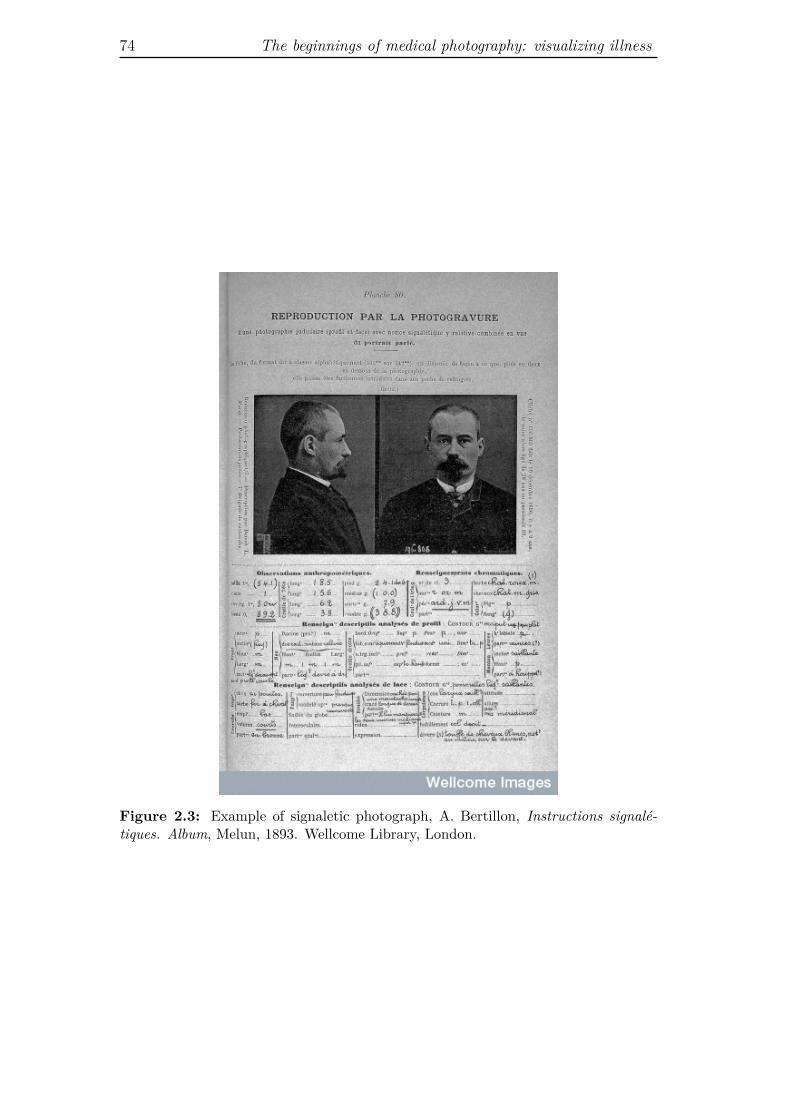
74 The beginnings of medical photography: visualizing illness
Figure 2.3: Example of signaletic photograph, A. Bertillon, Instructions signalé-tiques. Album, Melun, 1893. Wellcome Library, London.
Bodies, archives, and the photograph as symbol 75
photographs expressed nothing but its own appearance, and it was this ap-pearance that allowed to recognize each specific criminal. On the contrary,Galton firmly believed that the body revealed the true nature of the individual,and in fact he developed his composite portraits within the scope of a largereugenic project, aimed at defining typical classes of human beings.72 His pe-culiar photographic technique was an attempt to merge optical and statisticalprocedures, in order to create a mechanical and statistical method that wouldallow to identify the real physiognomy of different groups of individuals. Itconsisted of re-photographing portraits of subjects belonging to a given groupon a single photographic plate. Each photograph received a fractional exposurecorresponding to the inverse of the total number of images in the sample. Asa consequence, the individual idiosyncratic features faded away, due to under-exposure, and what remained was a blurred configuration of the features whichrecurred in all the subjects. This mechanical method was meant to replace hu-man judgement in the recognition of the fundamental features of a human typus(Figure 2.4).
Drawing on Peirce, Sekula describes Bertillon’s endeavor as grounded inthe indexical order of meaning of photography, whereby each photograph is thephysical trace of a contingent, material referent that actually exists in the world.Galton’s, on the contrary, was based on an attempt “to elevate the indexicalphotographic composite to the level of the symbolic, thus expressing a generallaw through accretion of contingent instances.”73 Yet, since Galton believed thatphysiognomy revealed the true metaphysical nature of the individual, he didnot understand his technique of composite portrayals as a mere mechanizationof visual pattern recognition; he took it as a mechanization of the inferentialreasoning that leads to the formulation of general laws. Thus, in Peircean terms,the composite portraits were meant to work as symbols, because they referred totheir object by virtue of a universal category.74 On the contrary, in Bertillonage,each photograph literally corresponded point by point to its referent (the bodyof the criminal), and was intended to always refer to that and only that referent.In the system of composite portraits, the final photograph embodied a synthesis
72As most social scientists of his time, Galton was obsessed with the idea of establishingthe physiognomy of the insane, the criminal and the Jew, but he also searched for the typusof the consumptive.
73Sekula, 1986, 55. In Peirce’s semiotics, a symbol is defined as “a sign which refers tothe Object that it denotes by virtue of a law, usually an association of general ideas, whichoperates to cause the Symbol to be interpreted as referring to that Object.” Peirce, CP, 2.249.
74Galton certainly did not conceive of his general physiognomic laws as conventional corre-spondence rules. He was firmly convinced of the reality and correctness of the laws he couldextract by operating the process of composite portraiture.
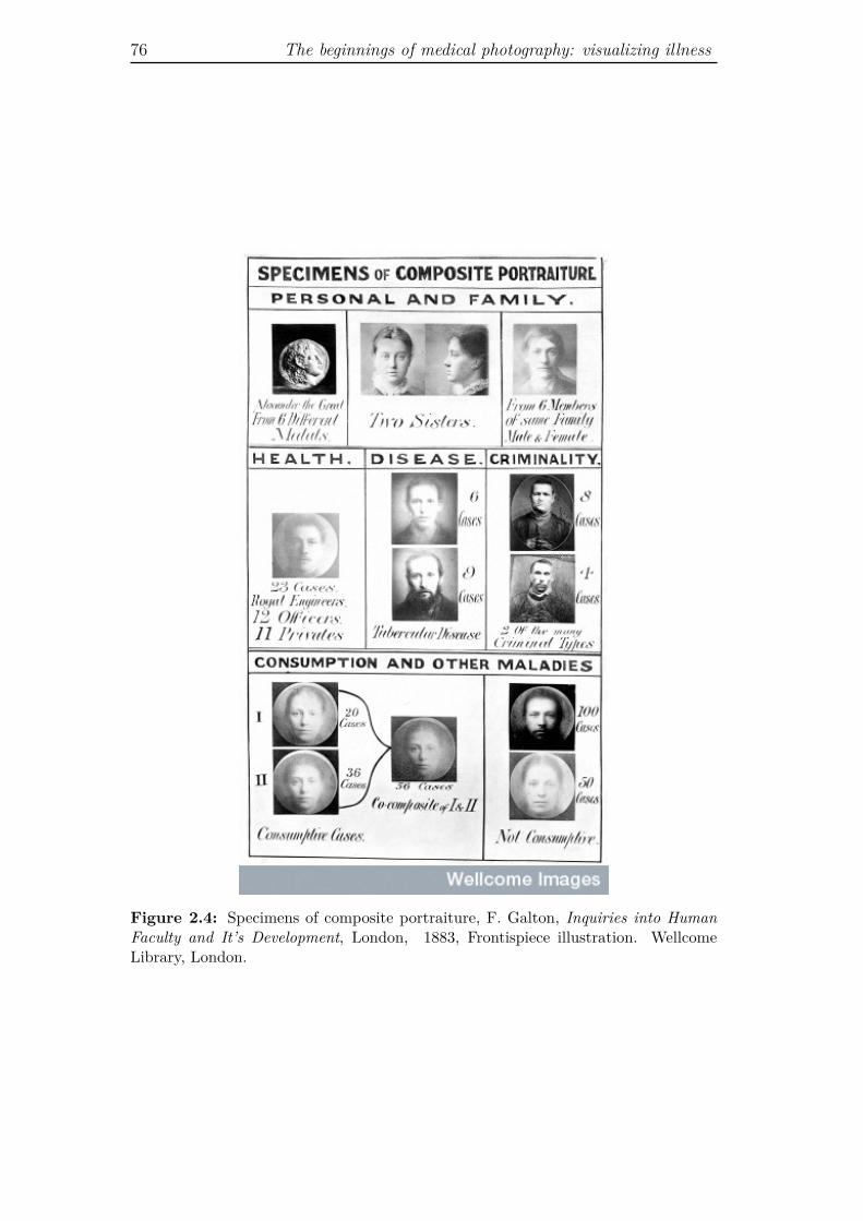
76 The beginnings of medical photography: visualizing illness
Figure 2.4: Specimens of composite portraiture, F. Galton, Inquiries into HumanFaculty and It’s Development, London, 1883, Frontispiece illustration. WellcomeLibrary, London.
Bodies, archives, and the photograph as symbol 77
of the images of many referents, and it was intended to refer to an indefinitenumber of individuals on the basis of their resemblance to the synthetic image.
Sekula’ analysis is particularly useful because Bertillon and Galton’s workare extreme examples of two opposite, yet complementary forms of makingsense of and using photographs not only in the social sciences (and pseudo-sciences), but also in medicine. Bertillonage can be taken as the epitome of thenominalist approach (which denies the reality of generic categories and focuseson singular, actual instances). Here the photograph works as an index andthe archive is used to properly assign an individual to his or her signaleticpicture, in order to check and certify his or her identity. In this case, the task ofphotography is to ratify a univocal correspondence: one criminal-one picture.75
There is no hidden truth beyond this correspondence, or more precisely, thephotograph cannot say anything that goes beyond such correspondence. Wecould say that in this case the contingency of the photographic image prevailedover the theoretical ambitions of the physiognomic paradigm. And in fact,Sekula argues that Bertillon’s reinvention of physiognomy in nonmetaphysicalterms had the ambition to constitute a real science of the individual. That is, ascience that established for each individual the idiosyncratic rules of his or herappearance and behavior. This science, however, could only be a descriptive,ethnographic endeavor, since it would not allow to (it was not meant to) inferuniversal laws about the countenance and actions of all human beings. Yet, usedin this way, the photograph and the archive would help the criminologist, as wellas the ethnographer or the physician, to carefully follow the transformationsoccurring on the body of a singular individual. Photography identified theindividual. When Londe recommended to keep photographic records in thepatient’s file, he had in mind this specific use of photography.76
Galton’s endeavor went in precisely the opposite direction to Bertillon’s. Iffor Bertillon the fundamental function of the archive was to associate one por-trait to one and only one individual, for Galton a collection of photographsbecame useful and scientifically meaningful as far as it allowed to make abstrac-tion of particular instances and generate the universal face of the mentally ill,the consumptive, the criminal, or any other possible group of human beings.Through the technique of the composite portrayals, Galton collapsed severalphotographs of the archive into one, so that the contingency of each singularportrait could literally fade away to give way to the typus. Composite por-trayals were not portraits of human beings, they were portraits of conceptual
75This function of photographs and of the photographic archive perfectly fits Peirce’s def-inition of photographs as signs that are “produced under such circumstances that they [are]physically forced to correspond point by point to nature.” Peirce, CP 2.247.
76See Section 2.1 above.
78 The beginnings of medical photography: visualizing illness
categories, general ideas. Galton, however, believed that such categories wereas real as the bodies that were classified. He worked in strict accordance withthe physiognomic framework, and the originality of his work resides in the factthat he tried to automate the abstract thinking that leads from the particular tothe universal. He did so by attempting to treat visual information in statisticalterms. In this sense, he literally conceived of his photographic technique as anomological machine, a mechanical device that reproduced the laws of humannature.
Although physicians did not necessarily subscribe to Galton’s purpose ofmechanizing clinical judgement, they nevertheless shared with him a fundamen-tal assumption, namely, that working on series of photographs one could performthe same kind of observations, apply the same patterns of reasoning and judge-ment, and reach the same conclusions, as if one was working with series of actualindividuals. In other words, it was possible to substitute the body of the actualperson for its photographic image. This implies that it was necessary to believein the indexical nature of photography in order to believe that the archive couldreveal a universal truth. For Galton the individual photograph per se could notpoint to a universal human typus, and this is why he had to develop a systemto produce a composite image. The individual photograph would just containthe truth of the individual instance it represented. For a universal truth toemerge, it was necessary to combine different pictures. Galton and Bertillonshared the same beliefs about the ontology of the photograph (it was a perfectimprint of the real object, burden with contingency), but they diverged on theepistemology of the archive. Galton worked according to a realist stance, whileBertillon followed a nominalist approach. Galton wanted to develop a scienceof the individual in the sense of a science of typical classes of human beings(identification and definition of human categories). Bertillon, on the contrary,aimed at a positivist science of the individual as ethnography of the particular,which withdrew from any generalization. Accordingly, Galton’s endeavor wasenvisaged as science (criminology), while Bertillon’s was deemed a technique(criminalistics). The former looked for the criminal (or the consumptive, theinsane, etc.), the latter looked for this or that offender.
Now, if in the enforcement of law criminalistics could do without criminol-ogy,77 in medicine the analysis of a single photographic portrait acquired diag-nostic cogency only when it could be referred to a general image of the disease.Hence, in medicine the two epistemological approaches concerning the archive
77In this case the aim was to verify the identity of the suspect and to check if he or she hadalready been charged, in the past, for other crimes. The photograph said who the person was,not how he or she was.
Physiognomic laws and therapeutic portraits 79
had to work in tandem, and if a physician was to use photographs as diagnosticdevices he had to continuously shift between the general (realist) and the par-ticular (nominalist) approach. In the following sections I analyze how these twoinstances were realized in actual medical practice, taking into examination thework of different pioneers of medical photography.
2.5 Physiognomic laws and therapeutic portraits
As shown above, the phrenological and physiognomic theories provided a fun-damental epistemic paradigm for making sense of photographic portraits, henceit does not come as a surprise that psychiatry was one of the first clinical disci-plines that tried to put photography in the service of diagnostics. In fact, if thehuman character and mental faculties expressed themselves through facial con-figuration and in the form and measures of the head (the tenets of physiognomyand phrenology, respectively), then photographs would be the ideal diagnosticand nosological instrument. The photographic portrait could be the empiricalevidence of the visual symptomatology of psychosis.
The first attempt to systematically use photography to record the physiog-nomy of the mentally ill for clinical use was carried out by the British psychi-atrist Hugh Welch Diamond, superintendent of the Female Department of theSurrey County Asylum. Importantly, he was also a founding member of theRoyal Photographic Society. In 1852, Diamond produced a photographic seriesof types of insanity, and four years later he gave a talk at the Royal Society inwhich he expounded what he considered to be the three functions of photogra-phy in the diagnosis and treatment of mental illness.78 Photography, Diamondmaintained, could record the appearance of the insane for study (physiognomicparadigm); it could be used in the treatment of patients, by presenting them astrikingly accurate self-image; and it could be valuable in case of re-admission(the photographic portrait would confirm the identity of the patient and recallto the doctor’s mind the case and its treatment).79 Hence, the photograph wassimultaneously embedded in a gnoseological structure (the physiognomic the-ory), a therapeutic protocol, and a controlling apparatus. For what regards thediagnostic function, it was conceivable because, by catching the expressions andcountenance of the patient with perfect accuracy, it allowed the psychiatrist toevaluate the corresponding mental state with the highest precision. However,
78Historian Jennifer Tucker remarks that Diamond used his photographic albums not onlyfor medical purposes, but also to promote scientific photography. His work circulated amongphotographers, beyond the medical community. See Tucker, 2005, 18-19.
79See Diamond, 1856.
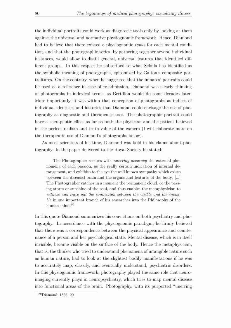
80 The beginnings of medical photography: visualizing illness
the individual portraits could work as diagnostic tools only by looking at themagainst the universal and normative physiognomic framework. Hence, Diamondhad to believe that there existed a physiognomic typus for each mental condi-tion, and that the photographic series, by gathering together several individualinstances, would allow to distill general, universal features that identified dif-ferent groups. In this respect he subscribed to what Sekula has identified asthe symbolic meaning of photographs, epitomized by Galton’s composite por-traitures. On the contrary, when he suggested that the inmates’ portraits couldbe used as a reference in case of re-admission, Diamond was clearly thinkingof photographs in indexical terms, as Bertillon would do some decades later.More importantly, it was within that conception of photographs as indices ofindividual identities and histories that Diamond could envisage the use of pho-tography as diagnostic and therapeutic tool. The photographic portrait couldhave a therapeutic effect as far as both the physician and the patient believedin the perfect realism and truth-value of the camera (I will elaborate more onthe therapeutic use of Diamond’s photographs below).
As most scientists of his time, Diamond was bold in his claims about pho-tography. In the paper delivered to the Royal Society he stated:
The Photographer secures with unerring accuracy the external phe-nomena of each passion, as the really certain indication of internal de-rangement, and exhibits to the eye the well known sympathy which existsbetween the diseased brain and the organs and features of the body. [...]The Photographer catches in a moment the permanent cloud, or the pass-ing storm or sunshine of the soul, and thus enables the metaphysician towitness and trace out the connection between the visible and the invisi-ble in one important branch of his researches into the Philosophy of thehuman mind.80
In this quote Diamond summarizes his convictions on both psychiatry and pho-tography. In accordance with the physiognomic paradigm, he firmly believedthat there was a correspondence between the physical appearance and counte-nance of a person and her psychological state. Mental disease, which is in itselfinvisible, became visible on the surface of the body. Hence the metaphysician,that is, the thinker who tried to understand phenomena of intangible nature suchas human nature, had to look at the slightest bodily manifestations if he wasto accurately map, classify, and eventually understand, psychiatric disorders.In this physiognomic framework, photography played the same role that neuro-imaging currently plays in neuropsychiatry, which tries to map mental diseaseinto functional areas of the brain. Photography, with its purported “unerring
80Diamond, 1856, 20.
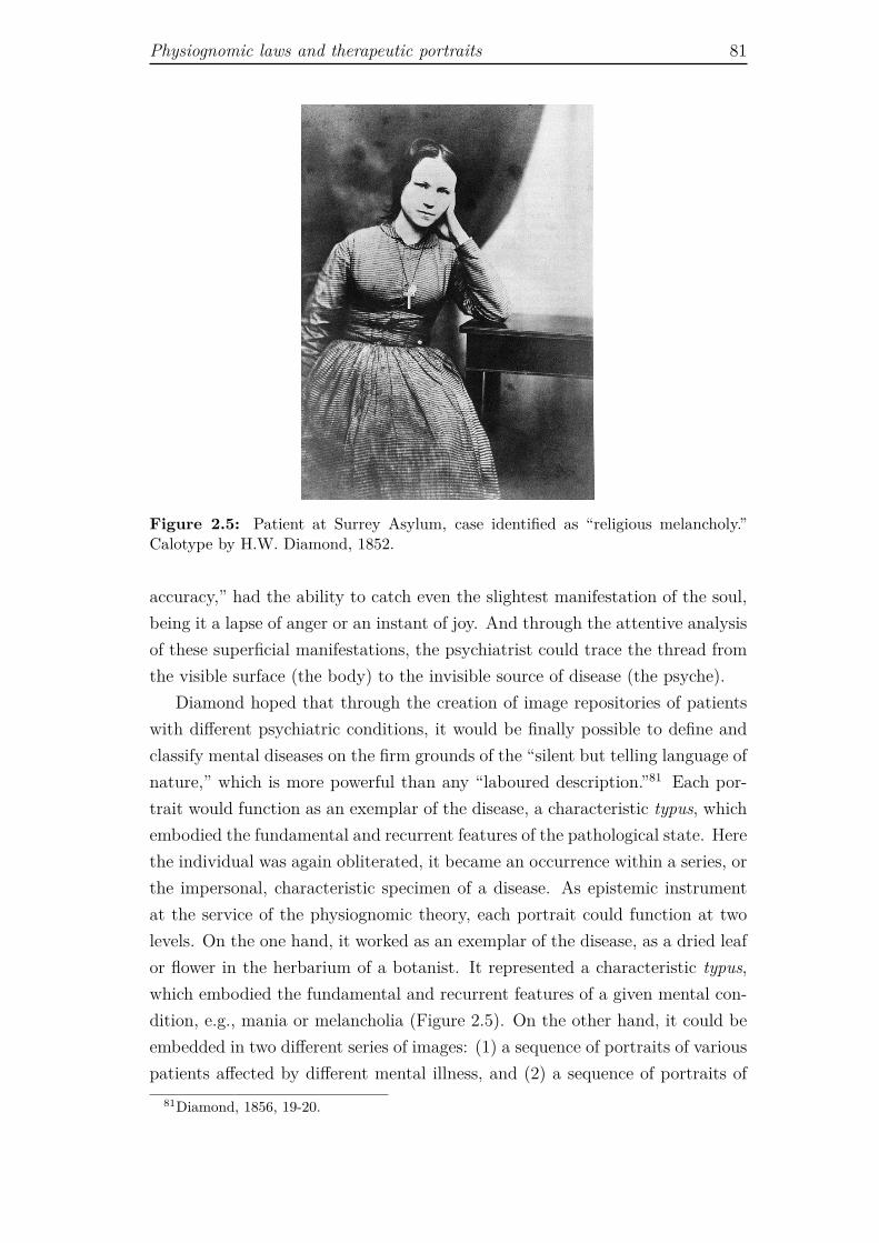
Physiognomic laws and therapeutic portraits 81
Figure 2.5: Patient at Surrey Asylum, case identified as “religious melancholy.”Calotype by H.W. Diamond, 1852.
accuracy,” had the ability to catch even the slightest manifestation of the soul,being it a lapse of anger or an instant of joy. And through the attentive analysisof these superficial manifestations, the psychiatrist could trace the thread fromthe visible surface (the body) to the invisible source of disease (the psyche).
Diamond hoped that through the creation of image repositories of patientswith different psychiatric conditions, it would be finally possible to define andclassify mental diseases on the firm grounds of the “silent but telling language ofnature,” which is more powerful than any “laboured description.”81 Each por-trait would function as an exemplar of the disease, a characteristic typus, whichembodied the fundamental and recurrent features of the pathological state. Herethe individual was again obliterated, it became an occurrence within a series, orthe impersonal, characteristic specimen of a disease. As epistemic instrumentat the service of the physiognomic theory, each portrait could function at twolevels. On the one hand, it worked as an exemplar of the disease, as a dried leafor flower in the herbarium of a botanist. It represented a characteristic typus,which embodied the fundamental and recurrent features of a given mental con-dition, e.g., mania or melancholia (Figure 2.5). On the other hand, it could beembedded in two different series of images: (1) a sequence of portraits of variouspatients affected by different mental illness, and (2) a sequence of portraits of
81Diamond, 1856, 19-20.
82 The beginnings of medical photography: visualizing illness
the same patient at different stages of the disease. In the first case, by puttingin contrast characteristic representatives of a variety of mental pathologies, thephotographic series was meant to make more visible the idiosyncratic featuresof each exemplar, thus making the individual picture more eloquent and rein-forcing its meaning. In the second case, the series showed the transformationsthat might occur within the same patient, thus adding a temporal, dynamicdimension to the study of the disease. From this multiplicity of uses of patient’sportraits emerges, once again, the unresolved tension between the will to rep-resent the universal features of a disease (to identify a typus) and the need todeal with singular, idiosyncratic individuals.
As seen above, in the discussion of Londe’s ideas on medical photography andSekula’s analysis of archival strategies, it seems that early medical photographers(and, more generally, scientists) tried to come to terms with the problem ofthe relation between the general and the particular by creating different formsof photographic series. On the one hand, they produced series of pictures ofdifferent patients affected by the same ailment, in order to visualize the general(universal) morphology of a disease; on the other hand, they gathered series ofpictures of the same patient, in order to reconstruct the history of the diseasein that specific individual. These two movements correspond to the realist andnominalist modes of signification, respectively. They show how, in medicine, twoirreconcilable philosophical positions are forced to come to terms. Medicine, asa practice, has to treat idiosyncratic unique individuals, hence, it must workaccording to a nominalist approach. However, medicine, as a science, has toextrapolate universal features from individual cases, and is thus forced to workby induction, according to an ultimately realist mode of signification.
2.5.1 Clinical cases: articulating pictures and words
The search of a typus for a group of patients, corresponding to the facies of thedisease, was certainly the main core of the use of photography in medicine in thenineteenth century, and the most common and widely available form of archivewas the collection of photographically illustrated clinical cases. The publicationof clinical cases in journals and atlases was meant to train the sort of reason-ing and intuitive judgement that Galton tried to automate with his compositeportraits. A fundamental aspect of clinical cases, however, is that images arewedded to a text, sometimes as mere examples, sometimes as non verbal argu-ments. In this case, in order to understand the functioning of photography asepistemic device, we must look also to its relation with the accompanying text.
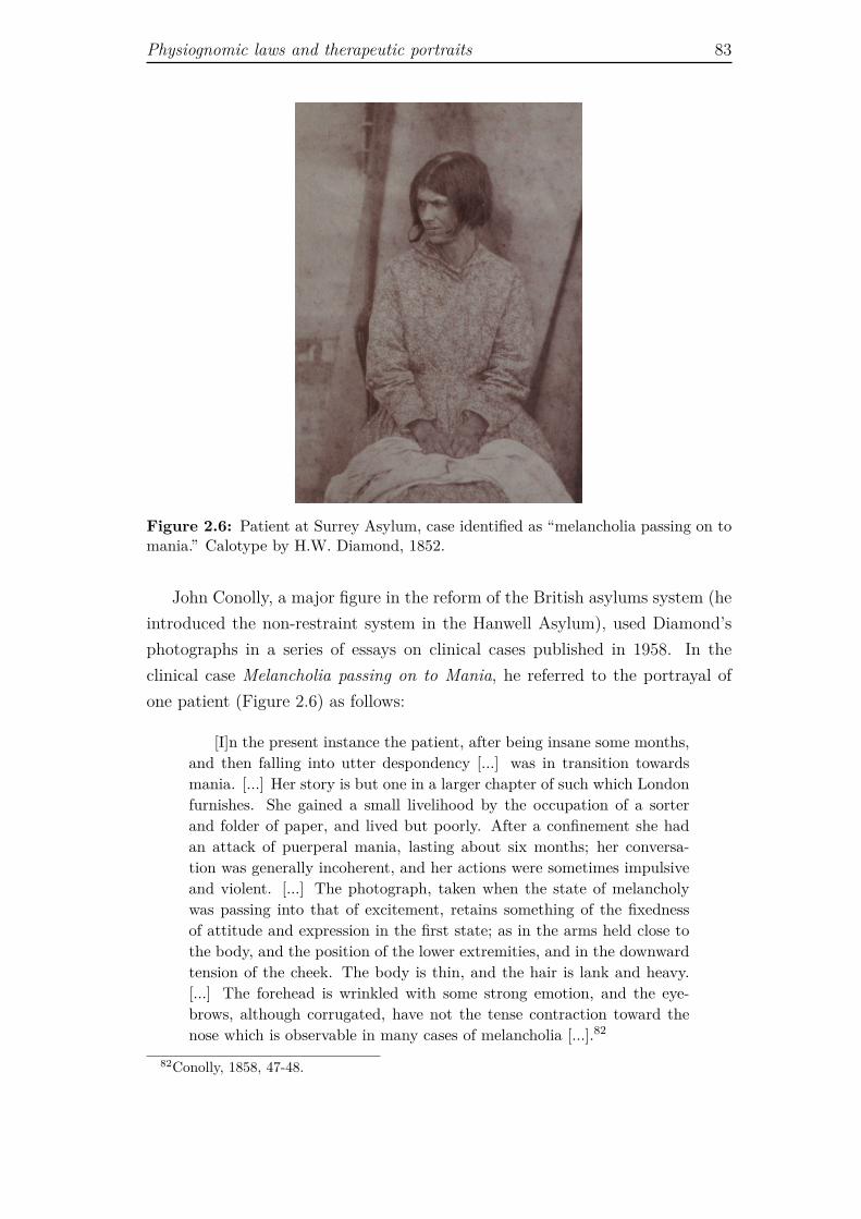
Physiognomic laws and therapeutic portraits 83
Figure 2.6: Patient at Surrey Asylum, case identified as “melancholia passing on tomania.” Calotype by H.W. Diamond, 1852.
John Conolly, a major figure in the reform of the British asylums system (heintroduced the non-restraint system in the Hanwell Asylum), used Diamond’sphotographs in a series of essays on clinical cases published in 1958. In theclinical case Melancholia passing on to Mania, he referred to the portrayal ofone patient (Figure 2.6) as follows:
[I]n the present instance the patient, after being insane some months,and then falling into utter despondency [...] was in transition towardsmania. [...] Her story is but one in a larger chapter of such which Londonfurnishes. She gained a small livelihood by the occupation of a sorterand folder of paper, and lived but poorly. After a confinement she hadan attack of puerperal mania, lasting about six months; her conversa-tion was generally incoherent, and her actions were sometimes impulsiveand violent. [...] The photograph, taken when the state of melancholywas passing into that of excitement, retains something of the fixednessof attitude and expression in the first state; as in the arms held close tothe body, and the position of the lower extremities, and in the downwardtension of the cheek. The body is thin, and the hair is lank and heavy.[...] The forehead is wrinkled with some strong emotion, and the eye-brows, although corrugated, have not the tense contraction toward thenose which is observable in many cases of melancholia [...].82
82Conolly, 1858, 47-48.
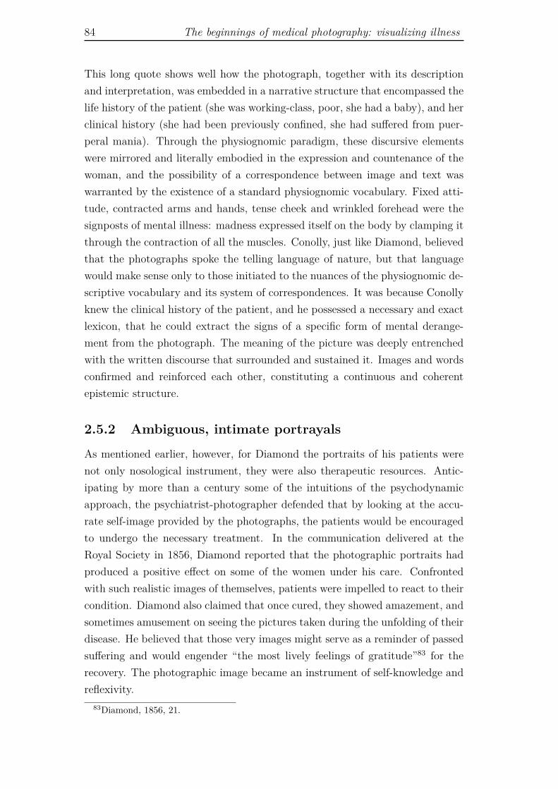
84 The beginnings of medical photography: visualizing illness
This long quote shows well how the photograph, together with its descriptionand interpretation, was embedded in a narrative structure that encompassed thelife history of the patient (she was working-class, poor, she had a baby), and herclinical history (she had been previously confined, she had suffered from puer-peral mania). Through the physiognomic paradigm, these discursive elementswere mirrored and literally embodied in the expression and countenance of thewoman, and the possibility of a correspondence between image and text waswarranted by the existence of a standard physiognomic vocabulary. Fixed atti-tude, contracted arms and hands, tense cheek and wrinkled forehead were thesignposts of mental illness: madness expressed itself on the body by clamping itthrough the contraction of all the muscles. Conolly, just like Diamond, believedthat the photographs spoke the telling language of nature, but that languagewould make sense only to those initiated to the nuances of the physiognomic de-scriptive vocabulary and its system of correspondences. It was because Conollyknew the clinical history of the patient, and he possessed a necessary and exactlexicon, that he could extract the signs of a specific form of mental derange-ment from the photograph. The meaning of the picture was deeply entrenchedwith the written discourse that surrounded and sustained it. Images and wordsconfirmed and reinforced each other, constituting a continuous and coherentepistemic structure.
2.5.2 Ambiguous, intimate portrayals
As mentioned earlier, however, for Diamond the portraits of his patients werenot only nosological instrument, they were also therapeutic resources. Antic-ipating by more than a century some of the intuitions of the psychodynamicapproach, the psychiatrist-photographer defended that by looking at the accu-rate self-image provided by the photographs, the patients would be encouragedto undergo the necessary treatment. In the communication delivered at theRoyal Society in 1856, Diamond reported that the photographic portraits hadproduced a positive effect on some of the women under his care. Confrontedwith such realistic images of themselves, patients were impelled to react to theircondition. Diamond also claimed that once cured, they showed amazement, andsometimes amusement on seeing the pictures taken during the unfolding of theirdisease. He believed that those very images might serve as a reminder of passedsuffering and would engender “the most lively feelings of gratitude”83 for therecovery. The photographic image became an instrument of self-knowledge andreflexivity.
83Diamond, 1856, 21.
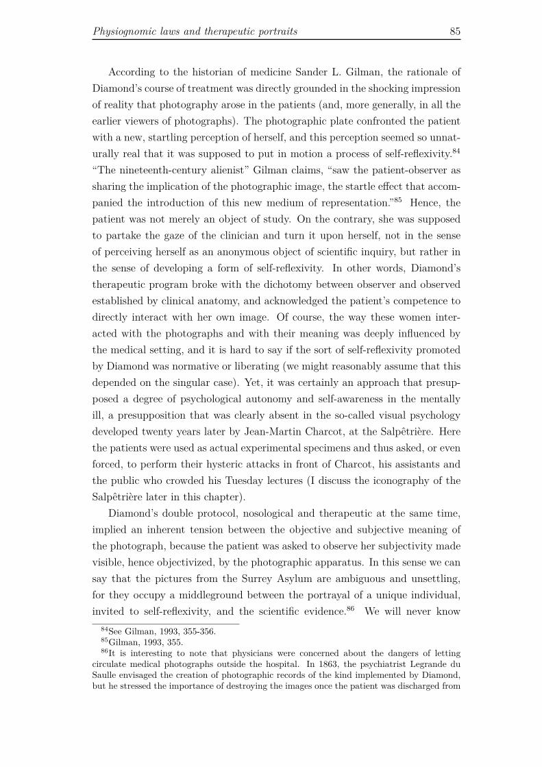
Physiognomic laws and therapeutic portraits 85
According to the historian of medicine Sander L. Gilman, the rationale ofDiamond’s course of treatment was directly grounded in the shocking impressionof reality that photography arose in the patients (and, more generally, in all theearlier viewers of photographs). The photographic plate confronted the patientwith a new, startling perception of herself, and this perception seemed so unnat-urally real that it was supposed to put in motion a process of self-reflexivity.84
“The nineteenth-century alienist” Gilman claims, “saw the patient-observer assharing the implication of the photographic image, the startle effect that accom-panied the introduction of this new medium of representation.”85 Hence, thepatient was not merely an object of study. On the contrary, she was supposedto partake the gaze of the clinician and turn it upon herself, not in the senseof perceiving herself as an anonymous object of scientific inquiry, but rather inthe sense of developing a form of self-reflexivity. In other words, Diamond’stherapeutic program broke with the dichotomy between observer and observedestablished by clinical anatomy, and acknowledged the patient’s competence todirectly interact with her own image. Of course, the way these women inter-acted with the photographs and with their meaning was deeply influenced bythe medical setting, and it is hard to say if the sort of self-reflexivity promotedby Diamond was normative or liberating (we might reasonably assume that thisdepended on the singular case). Yet, it was certainly an approach that presup-posed a degree of psychological autonomy and self-awareness in the mentallyill, a presupposition that was clearly absent in the so-called visual psychologydeveloped twenty years later by Jean-Martin Charcot, at the Salpêtrière. Herethe patients were used as actual experimental specimens and thus asked, or evenforced, to perform their hysteric attacks in front of Charcot, his assistants andthe public who crowded his Tuesday lectures (I discuss the iconography of theSalpêtrière later in this chapter).
Diamond’s double protocol, nosological and therapeutic at the same time,implied an inherent tension between the objective and subjective meaning ofthe photograph, because the patient was asked to observe her subjectivity madevisible, hence objectivized, by the photographic apparatus. In this sense we cansay that the pictures from the Surrey Asylum are ambiguous and unsettling,for they occupy a middleground between the portrayal of a unique individual,invited to self-reflexivity, and the scientific evidence.86 We will never know
84See Gilman, 1993, 355-356.85Gilman, 1993, 355.86It is interesting to note that physicians were concerned about the dangers of letting
circulate medical photographs outside the hospital. In 1863, the psychiatrist Legrande duSaulle envisaged the creation of photographic records of the kind implemented by Diamond,but he stressed the importance of destroying the images once the patient was discharged from
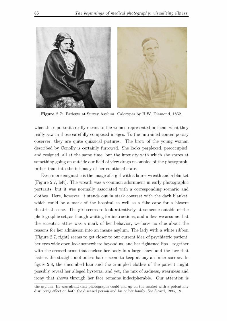
86 The beginnings of medical photography: visualizing illness
Figure 2.7: Patients at Surrey Asylum. Calotypes by H.W. Diamond, 1852.
what these portraits really meant to the women represented in them, what theyreally saw in those carefully composed images. To the untrained contemporaryobserver, they are quite quizzical pictures. The brow of the young womandescribed by Conolly is certainly furrowed. She looks perplexed, preoccupied,and resigned, all at the same time, but the intensity with which she stares atsomething going on outside our field of view drags us outside of the photograph,rather than into the intimacy of her emotional state.
Even more enigmatic is the image of a girl with a laurel wreath and a blanket(Figure 2.7, left). The wreath was a common adornment in early photographicportraits, but it was normally associated with a corresponding scenario andclothes. Here, however, it stands out in stark contrast with the dark blanket,which could be a mark of the hospital as well as a fake cape for a bizarretheatrical scene. The girl seems to look attentively at someone outside of thephotographic set, as though waiting for instructions, and unless we assume thatthe eccentric attire was a mark of her behavior, we have no clue about thereasons for her admission into an insane asylum. The lady with a white ribbon(Figure 2.7, right) seems to get closer to our current idea of psychiatric patient:her eyes wide open look somewhere beyond us, and her tightened lips – togetherwith the crossed arms that enclose her body in a large shawl and the lace thatfastens the straight motionless hair – seem to keep at bay an inner sorrow. Infigure 2.8, the uncombed hair and the crumpled clothes of the patient mightpossibly reveal her alleged hysteria, and yet, the mix of sadness, weariness andirony that shows through her face remains indecipherable. Our attention is
the asylum. He was afraid that photographs could end up on the market with a potentiallydisrupting effect on both the diseased person and his or her family. See Sicard, 1995, 18.
Physiognomic laws and therapeutic portraits 87
Figure 2.8: Patient at Surrey Asylum. Calotype by H.W. Diamond, 1852.
caught by the ring on her left hand (what Roland Barthes would identify as thepunctum of this photograph), which points to her life outside the asylum, herfamily, her private history.
There is a decorous calm in the bodies of these women, which contrastsstarkly with the spectacular bodies of the hysterics depicted by Charcot in col-laboration with his assistants and photographers. Unlike Charcot’s patients,captured in the uncontrolled agitation of their seizures, Diamond’s ladies standstill, posing for the photographer in profound concentration, whether they lookinto the camera or to some invisible event removed from our field of view.Art historian Martin Kemp has remarked that Diamond’s pictures were stagedwithin the aesthetics (framings, backgrounds, posing) of the photographic por-trayal of his time,87 and indeed, most of them could find their place in a familyalbum. Looking at these photographs we do not see clinical cases, we rather havea feeling similar to that described by Benjamin in relation to David OctaviusHill’s portrait of the Newhaven fishwife. Benjamin wrote:
In Hill’s Newhaven fishwife, her eyes cast down in such indolent, se-ductive modesty, there remains something that goes beyond testimony tothe photographer’s art, something that cannot be silenced, that fills youwith an unruly desire to know what her name was, the woman who was
87See Kemp, 2006, 292.
88 The beginnings of medical photography: visualizing illness
alive there, who even now is still real and will never consent to be whollyabsorbed in ‘art.’88
As the Newhaven fishwife, the women from the Surrey Asylum tell us that theywere “alive there.” They assert their presence, their life. It is a life that haspassed away for long now, and yet it left its sparkle there, indisputable. Thephotograph captured that instant of life that cannot be erased, because it hasalready happened, it is accomplished, and nothing can un-make it. As in the caseof the Newhaven fishwife, the presence of the women from the Surrey Asylumin front of the camera goes beyond the photographer’s art (and the scientificcategories of the psychiatrist), imposes the sheer reality of their existence, andmake us wonder about their names, their real lives.
2.6 Dermatology: the struggle for realism
Together with psychiatry, the other clinical specialty that immediately sizedupon photography was dermatology. As discussed in Chapter 1, clinical anatomyrevolutionized medicine by positing the internal, hidden lesion as the origin ofany disease. In the case of dermatology, however, there was no hidden lesion tolook for: the lesion was at the surface, consequently its very visibility was thesymptom, the sign and the diagnosis at the same time. Accordingly, photog-raphy was immediately perceived as an ideal recording and diagnostic tool fordermatologists. Dermatology is a discipline of total visibility, hence, for the der-matologist, the superficial gaze of the camera perfectly fitted with the eye of theclinician. The closeup, the selection of details on the very surface of the body,was the clinic. Not surprisingly then, it was a dermatologist, Albert Hardy, whocoined the expression “photographic clinic” [clinique photographique], in 1867.
With its ability to carefully record what the eye of the doctor saw, photog-raphy was an ideal means of communication and knowledge sharing: picturescirculate easily, and rare skin diseases started to travel in effigy across coun-tries. Moreover, thanks to its capacity to keep record of what is unnoticeable,it fostered the observation of new phenomena. Therefore it was thought thatit could help in both diagnosis and nosology. Yet, media theorist MoniqueSicard points out that with the introduction of photography in dermatology un-certainty tended to prevail over knowledge. In the absence of well establishedtaxonomical frames, in fact, the accumulation of scattered visual data lead tothe proliferation of painstakingly detailed descriptions, which had little use foractual medical practice.89 For some years discussions about the proper identi-
88Benjamin, 1931, 510.89See Sicard, 1995, 15, and 1998, 149.
Dermatology: the struggle for realism 89
fication of papules, pustules, spots and the like burgeoned, in the attempt toestablish an ever more specific terminology. The debate went as far as to raiseHardy’s preoccupation that the proliferation of images could paradoxically leadto an arid nominalism, with dermatologists more preoccupied in labeling themanifestations of the diseases rather than understanding and treating them.90
Hardy, in collaboration with his intern A. de Montméja, was the men-tor of the first photographically illustrated medical review, the Clinique pho-tographique de l’Hôpital Saint-Louis, which was published in fourteen issuesbetween 1867 and 1868, when all the installments were collected in a singlevolume atlas.91 This work had an explicit pedagogical intent. In the prefaceHardy explained that the objective of the publication was to provide medicalstudents and practitioners with a visual aid to train the eye in the recognitionof skin diseases. To this aim each pathology was sketched out in a short verbaldescription and exemplified by a photograph. Photographs, with their allegedrealism, were supposed to have a higher degree of exactitude, compared to otherforms of illustration, and leave a stronger mnemonic impression. In order to as-sure his readers about the accountability of the images, Hardy specified that allthe pictures had been skillfully created by his assistant, “who combine[d] an indepth knowledge of skin diseases to an indisputable talent as photographer andcolorist.” The plates, he insisted, “represented nature caught in the act.”92 Yet,in spite of the absolute visibility of skin diseases, to catch them in the act, thatis, to represent them faithfully, was a real challenge for photography. Actually,the attempt to truthfully depict the skin called into question the very axiom ofphotography’s fidelity to nature. The most obvious difficulty came from colors.How much realistic could be a representation that reduced the endless paletteof the real world to a fading scale of grey? Color shadows of the skin lesion area relevant diagnostic elements in dermatology, but good quality and affordablecolor photography became available only as late as 1950s.
Between fiction and reality Undoubtedly, in terms of realism, photographslagged far behind another form of dermatological representation, the so calledmoulages. Used as teaching tools and research documents, moulages were three-dimensional wax models of skin diseases, characterized by the astonishing, hap-tic naturalism distinctive of wax sculptures. After being casted directly from the
90See Sicard, 1995, 15, and 1998, 149.91Hardy became familiar with the dermatological applications of photography in 1866,
when he came to know the work of Alexander Belmanno Squire in London. He conse-quently asked his student de Montméja to work on this new imaging technique and ina very short time they created the Clinique Photographique. See http://www.bium.univ-paris5.fr/histmed/medica/dermato.htm (retrieved on Aug 16, 2013).
92Hardy, 1868, n.p.
90 The beginnings of medical photography: visualizing illness
patient’s body, these models were painted and, in many cases, real hair, nailsand clothes fabric were added to reinforce the appearance of real life.93 Com-pared to photographs, however, moulages were cumbersome, fragile, took muchspace, and were difficult to transport. Crucially, they were not mechanicallyreproducible and could not be produced in large series.
Still, in spite of all the obvious differences that separate wax models fromphotographs, the excessive, disturbing naturalism of the moulages finds an oddcounterpart in the iconography of the Clinique photographique. In fact, in orderto overcome the problem of color, de Montméja had to retouch each photo-graphic plate with watercolors, a common practice at the time, creating hybridimages that attempted to merge photographic and pictorial realism. These hy-brid pictures expose a contradiction that apparently eluded both scientists andphotographers in the nineteenth century. On the one hand, photographs weretrusted on the basis of their indexical nature, more specifically, on the basisof the belief that the realism of a photograph scored above the realism of apainting, because the photographic image was the result of a direct imprint ofnature on the sensible plate; the photograph was perceived as a natural object,the un-mediated appearing of reality on a photographic plate. On the otherhand, those who actually produced the photographs realized that in order toreally look like reality, the photographic image had to be retouched, using tradi-tional artistic techniques as watercolor. That is, far from being unproblematicimprints of nature, dermatological photographs required intense labor in orderto become similar to the real objects they were meant to represent.
That notwithstanding, the earliest practitioners of photography did not dis-cuss or problematize the contradictions arising from their ideas about photog-raphy (perceived as imprint of nature) and their actual practice (intensive laborto make photographs look similar to reality). We could say that they werenot disturbed by the ambiguity of the new optical medium. They trusted andpraised its fidelity to nature, even though they were quite aware of its actuallimitations. In this regard, photography theorist Geoffrey Batchen has arguedthat in the early days of photography the ontological status of photographicpictures was understood as complex and unstable, wavering between fictionand reality.94 Through the analysis of Hyppolite Bayard’s Self-Portrait as aDrowned Man (1940), an ostensibly staged (hence fake) photographic portrait
93Wax moulages were produced and used between the 1890s and the 1950s for clinicalcollection and medical training. They were completely let aside only in the second half of thetwentieth century, when the quality of color photograph substantially improved, allowing fora more powerful visual realism. See Schnalke, 1995 and 2004.
94Batchen, 1999, 202.
Dermatology: the struggle for realism 91
that depicted the photographer as if he had died by drowning,95 Batchen showsthat the photograph was understood as both perfomative and documentary, anatural object and a cultural artifact.96 In a similar vein, Sabine Kriebel haspointed out that during the nineteenth century the conception and reception ofthe photographic image was “bifurcated.”97 On the one hand, photographs wereperceived as the product of a technical process, and the camera was conceived ofas a picture-machine whose mechanical functioning was a warrant of objectivity.On the other hand, the language used to refer to photographs was often basedon nature, as if they were quasi-natural objects (common expressions were “sunpictures” and “impressed by nature’s hand”). They were “taken” from naturein quite the same way as naturalists took their specimens from the wild.98
The pictures of the Clinique photographique, with their superimposition oftwo media (photography and watercolor) produced an involuntary and unset-tling carnivalesque effect (Figure 2.9), and yet they provided medical evidence.Used to illustrate the discursive description of the different pathologies (in thepagination of the atlas each image follows its corresponding text), these pho-tographs were meant to transform living and suffering individuals into the rep-resentative of a disease. Embedded as they are in the structure of the atlas,the bodies and the faces of the patients became part of a scientific discourse, anargumentation grounded in an optical empiricism. Nevertheless, if we removethem from this context and look at them outside of the series of clinical cases,those images taken almost a century and a half ago, still convey private histo-ries of suffering and shame. They are two-sided. On one side they are scientificmodels (of the disease), on the other they are portraits (of the singular patient).In other words, these photographs embody the problem of the relationship be-tween a scientific representation which is, by definition, the representation of ageneral instance, and the representation of an individual, a particular case, withits unicity and idiosyncrasies.
95Hyppolite Bayard claimed to have invented photography before Daguerre, and that hehad been persuaded by a friend of Daguerre to postpone the announcement of his findings.Consequently he missed the opportunity to be recognized as the inventor of the medium. In1840 he reacted to this injustice by creating a portrait of himself as a drowned man. Heaccompanied the photograph with the following caption: “The corpse which you see here isthat of M. Bayard, inventor of the process that has just been shown to you. As far as I knowthis indefatigable experimenter has been occupied for about three years with his discovery.The Government, which has been only too generous to Monsieur Daguerre, has said it can donothing for Monsieur Bayard, and the poor wretch has drowned himself. Oh the vagaries ofhuman life...!”
96Batchen, 1999, 202.97Kriebel, 2007, 8.98See Kriebel, 2007, 8.
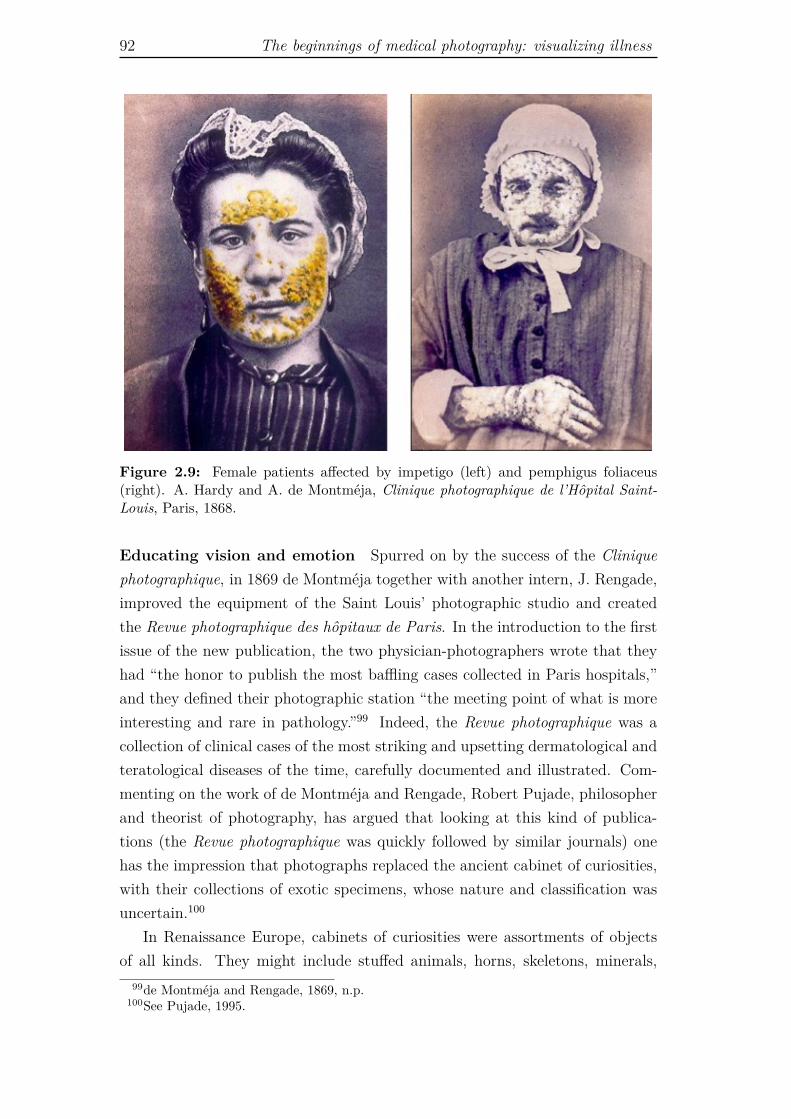
92 The beginnings of medical photography: visualizing illness
Figure 2.9: Female patients affected by impetigo (left) and pemphigus foliaceus(right). A. Hardy and A. de Montméja, Clinique photographique de l’Hôpital Saint-Louis, Paris, 1868.
Educating vision and emotion Spurred on by the success of the Cliniquephotographique, in 1869 de Montméja together with another intern, J. Rengade,improved the equipment of the Saint Louis’ photographic studio and createdthe Revue photographique des hôpitaux de Paris. In the introduction to the firstissue of the new publication, the two physician-photographers wrote that theyhad “the honor to publish the most baffling cases collected in Paris hospitals,”and they defined their photographic station “the meeting point of what is moreinteresting and rare in pathology.”99 Indeed, the Revue photographique was acollection of clinical cases of the most striking and upsetting dermatological andteratological diseases of the time, carefully documented and illustrated. Com-menting on the work of de Montméja and Rengade, Robert Pujade, philosopherand theorist of photography, has argued that looking at this kind of publica-tions (the Revue photographique was quickly followed by similar journals) onehas the impression that photographs replaced the ancient cabinet of curiosities,with their collections of exotic specimens, whose nature and classification wasuncertain.100
In Renaissance Europe, cabinets of curiosities were assortments of objectsof all kinds. They might include stuffed animals, horns, skeletons, minerals,
99de Montméja and Rengade, 1869, n.p.100See Pujade, 1995.
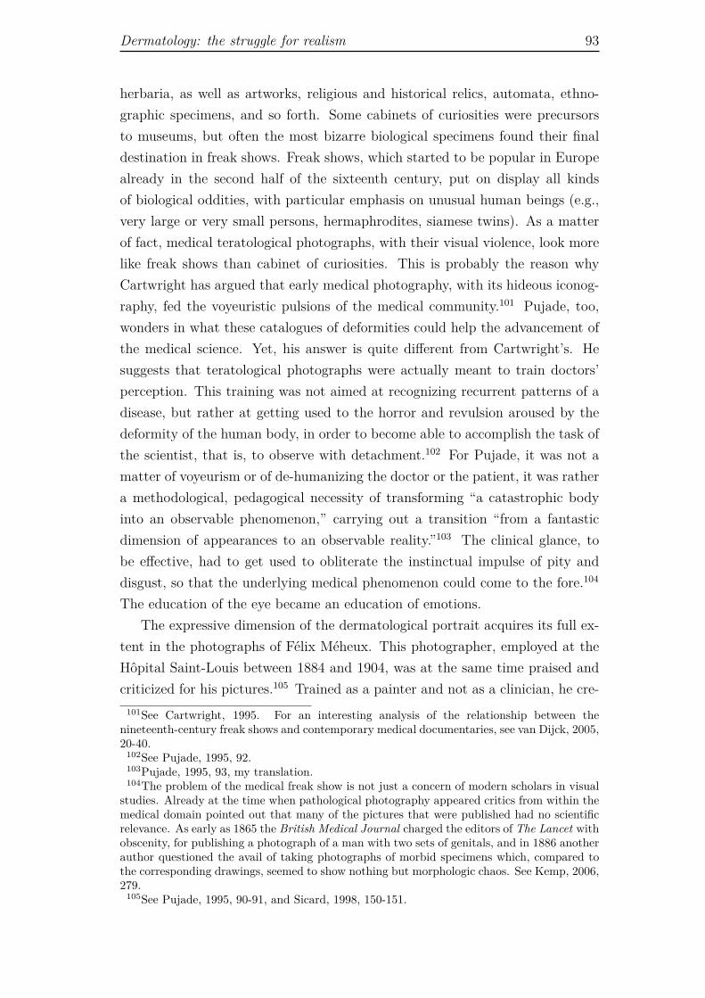
Dermatology: the struggle for realism 93
herbaria, as well as artworks, religious and historical relics, automata, ethno-graphic specimens, and so forth. Some cabinets of curiosities were precursorsto museums, but often the most bizarre biological specimens found their finaldestination in freak shows. Freak shows, which started to be popular in Europealready in the second half of the sixteenth century, put on display all kindsof biological oddities, with particular emphasis on unusual human beings (e.g.,very large or very small persons, hermaphrodites, siamese twins). As a matterof fact, medical teratological photographs, with their visual violence, look morelike freak shows than cabinet of curiosities. This is probably the reason whyCartwright has argued that early medical photography, with its hideous iconog-raphy, fed the voyeuristic pulsions of the medical community.101 Pujade, too,wonders in what these catalogues of deformities could help the advancement ofthe medical science. Yet, his answer is quite different from Cartwright’s. Hesuggests that teratological photographs were actually meant to train doctors’perception. This training was not aimed at recognizing recurrent patterns of adisease, but rather at getting used to the horror and revulsion aroused by thedeformity of the human body, in order to become able to accomplish the task ofthe scientist, that is, to observe with detachment.102 For Pujade, it was not amatter of voyeurism or of de-humanizing the doctor or the patient, it was rathera methodological, pedagogical necessity of transforming “a catastrophic bodyinto an observable phenomenon,” carrying out a transition “from a fantasticdimension of appearances to an observable reality.”103 The clinical glance, tobe effective, had to get used to obliterate the instinctual impulse of pity anddisgust, so that the underlying medical phenomenon could come to the fore.104
The education of the eye became an education of emotions.The expressive dimension of the dermatological portrait acquires its full ex-
tent in the photographs of Félix Méheux. This photographer, employed at theHôpital Saint-Louis between 1884 and 1904, was at the same time praised andcriticized for his pictures.105 Trained as a painter and not as a clinician, he cre-
101See Cartwright, 1995. For an interesting analysis of the relationship between thenineteenth-century freak shows and contemporary medical documentaries, see van Dijck, 2005,20-40.
102See Pujade, 1995, 92.103Pujade, 1995, 93, my translation.104The problem of the medical freak show is not just a concern of modern scholars in visual
studies. Already at the time when pathological photography appeared critics from within themedical domain pointed out that many of the pictures that were published had no scientificrelevance. As early as 1865 the British Medical Journal charged the editors of The Lancet withobscenity, for publishing a photograph of a man with two sets of genitals, and in 1886 anotherauthor questioned the avail of taking photographs of morbid specimens which, compared tothe corresponding drawings, seemed to show nothing but morphologic chaos. See Kemp, 2006,279.
105See Pujade, 1995, 90-91, and Sicard, 1998, 150-151.
94 The beginnings of medical photography: visualizing illness
Figure 2.10: Syphilitic alopecia (left). Pigmentation probably caused by photo-sensitization due to a cosmetic product (right), F. Mehéux, ca. 1884-1893.
ated his images from the perspective of the visual artist. Through a skillful workwith light, shadows, and framing, he deliberately composed photographs whichdid not just exemplify the pathology, but were first and foremost portraits ofthe diseased individual, conveying a personal history and an intimate suffering(Figure 2.10). As noted by Pujade: “[In the work of Méheux] the artistic gazeputs on stage the simple presence of subjects who pose for the ‘alopecia areatadecalvans’, the ‘herpetic diseases’, or the ‘fetal ichthyosis’, with the result of bur-dening with beauty the image of their catastrophic condition.”106 Like allegoricfigurines, the women and men of these portraits stand for something else thanthemselves, they pose as images of a disease, but the choice of focus and fram-ing, the darkness that surrounds them, the small details (a ring, a curl), directour look towards a human being, not a clinical case. It is not a matter of aes-theticizing the disease, there is neither morbid voyeurism in these photographs,nor moralizing intentions. What happens is that the narrative, personal dimen-sion of the portrayal overrules its informational content. Not surprisingly, then,physicians acknowledged and appreciated the uncanny beauty of Méheux’s por-traits, but some of them accused him of not being scientifically sound, becausehis pictures were not produced according to standardized protocols (the rulessummarized by Londe in his book of 1893), but rather according to artistic
106Pujade, 1995, 91, my translation.
Duchenne de Boulogne: electricity meets photography 95
canons.107 The shattered humanity of Méheux’s portraits was perceived as aptfor contemplation and emotion, not for scientific observation and examination.
Photography might well be endowed with an inherent analytic or dissectingquality, but in order to be scientifically meaningful and acceptable, photographshad to be inscribed within a proper aesthetic canon, a visual code which imposedstandardized protocols of framing, lighting, exposure, pose and distance betweencamera and sitter. This visual code had the aim to produce an effect of aestheticneutrality, that is, to convey the feeling that photographs were blank from thestylistic and interpretive point of view. This, however, was a paradoxical project,because at the very moment in which such visual rules were implemented, aspecific style of depiction was born. A style that possibly responded to specificneeds of clarity, standardization, and reproducibility, but that was also aimedat displaying its objectivity.
2.7 Duchenne de Boulogne: electricity meets
photography
In the same period in which de Montméja dealt with the difficulties of repro-ducing the surface of the body and Diamond developed his physiognomic pro-gram, the neurologist Guillaume Duchenne, known as Duchenne de Boulogne,was experimenting with what he called “electrophysiological photographs,”108
in search of the the laws that govern the muscular underpinning of both motionand emotion. A founding father of clinical medicine, Duchenne, who worked atthe Salpêtrière, specialized in muscular and neurological disorders. He was thefirst to describe pseudo-hypertrophic muscular paralysis (which today is knownunder the name of Duchenne muscular dystrophy) and he studied extensivelyprogressive muscle atrophy and atrophic paralysis in children. His study of emo-tions was in part a consequence of his research on muscular conditions, since themuscles of the face, the place where human emotions show through, are oftenaffected by amyotrophic diseases.
Scientific observation through photography It is important to note that,as pointed out by Sicard, Duchenne’s method was simultaneously electrophys-iological and photographic.109 The electrophysiological protocol required theapplication of a low intensity current to a circumscribed area of the body, in
107See Sicard, 1998, 150.108Duchenne, 1862a, n.p.109See Sicard, 1998, 133.
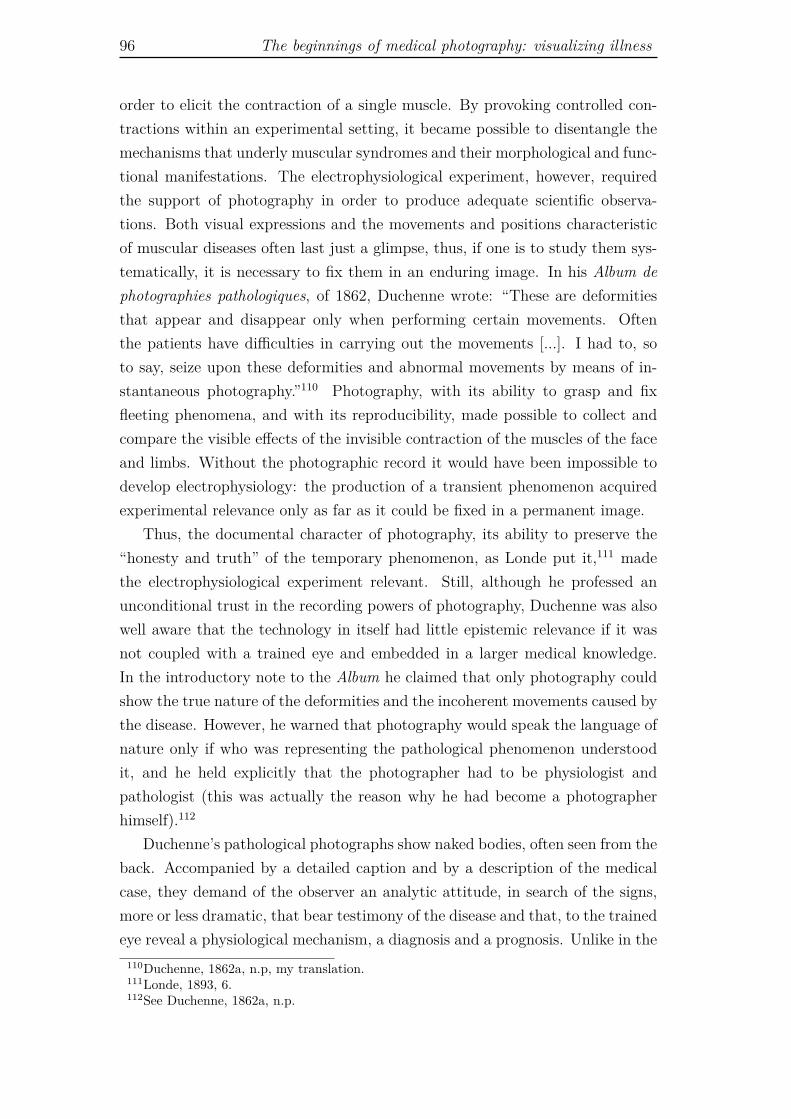
96 The beginnings of medical photography: visualizing illness
order to elicit the contraction of a single muscle. By provoking controlled con-tractions within an experimental setting, it became possible to disentangle themechanisms that underly muscular syndromes and their morphological and func-tional manifestations. The electrophysiological experiment, however, requiredthe support of photography in order to produce adequate scientific observa-tions. Both visual expressions and the movements and positions characteristicof muscular diseases often last just a glimpse, thus, if one is to study them sys-tematically, it is necessary to fix them in an enduring image. In his Album dephotographies pathologiques, of 1862, Duchenne wrote: “These are deformitiesthat appear and disappear only when performing certain movements. Oftenthe patients have difficulties in carrying out the movements [...]. I had to, soto say, seize upon these deformities and abnormal movements by means of in-stantaneous photography.”110 Photography, with its ability to grasp and fixfleeting phenomena, and with its reproducibility, made possible to collect andcompare the visible effects of the invisible contraction of the muscles of the faceand limbs. Without the photographic record it would have been impossible todevelop electrophysiology: the production of a transient phenomenon acquiredexperimental relevance only as far as it could be fixed in a permanent image.
Thus, the documental character of photography, its ability to preserve the“honesty and truth” of the temporary phenomenon, as Londe put it,111 madethe electrophysiological experiment relevant. Still, although he professed anunconditional trust in the recording powers of photography, Duchenne was alsowell aware that the technology in itself had little epistemic relevance if it wasnot coupled with a trained eye and embedded in a larger medical knowledge.In the introductory note to the Album he claimed that only photography couldshow the true nature of the deformities and the incoherent movements caused bythe disease. However, he warned that photography would speak the language ofnature only if who was representing the pathological phenomenon understoodit, and he held explicitly that the photographer had to be physiologist andpathologist (this was actually the reason why he had become a photographerhimself).112
Duchenne’s pathological photographs show naked bodies, often seen from theback. Accompanied by a detailed caption and by a description of the medicalcase, they demand of the observer an analytic attitude, in search of the signs,more or less dramatic, that bear testimony of the disease and that, to the trainedeye reveal a physiological mechanism, a diagnosis and a prognosis. Unlike in the
110Duchenne, 1862a, n.p, my translation.111Londe, 1893, 6.112See Duchenne, 1862a, n.p.
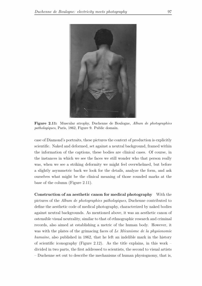
Duchenne de Boulogne: electricity meets photography 97
Figure 2.11: Muscular atrophy, Duchenne de Boulogne, Album de photographiespathologiques, Paris, 1862, Figure 9. Public domain.
case of Diamond’s portraits, these pictures the context of production is explicitlyscientific. Naked and deformed, set against a neutral background, framed withinthe information of the captions, these bodies are clinical cases. Of course, inthe instances in which we see the faces we still wonder who that person reallywas, when we see a striking deformity we might feel overwhelmed, but beforea slightly asymmetric back we look for the details, analyze the form, and askourselves what might be the clinical meaning of those rounded marks at thebase of the column (Figure 2.11).
Construction of an aesthetic canon for medical photography With thepictures of the Album de photographies pathologiques, Duchenne contributed todefine the aesthetic code of medical photography, characterized by naked bodiesagainst neutral backgrounds. As mentioned above, it was an aesthetic canon ofostensible visual neutrality, similar to that of ethnographic research and criminalrecords, also aimed at establishing a metric of the human body. However, itwas with the plates of the grimacing faces of Le Mécanisme de la physionomiehumaine, also published in 1862, that he left an indelible mark in the historyof scientific iconography (Figure 2.12). As the title explains, in this work –divided in two parts, the first addressed to scientists, the second to visual artists– Duchenne set out to describe the mechanisms of human physiognomy, that is,
98 The beginnings of medical photography: visualizing illness
Figure 2.12: The facial expression of profound attention on the human face being in-duced by electrical current. Duchenne de Boulogne, Le Mécanisme de la physionomiehumaine, Paris, 1862, Plate 1, figure 9. Wellcome Library, London.
to understand the physiological rules that govern the expression of emotions. Hismethod consisted of triggering muscular contractions with electrical probes andrecording the resulting grimaces by means of photography. He thus created anextensive visual gallery of what he considered to be the mechanical expressionsof inner emotions.
Art historian Martin Kemp has defined Le Mécanisme de la physionomiehumaine “the most remarkable of all the photographically illustrated books inmedical science before 1900,” and he has remarked that the portraits of the oldshoemaker of the Salpêtrière, who was Duchenne’s favorite model, “are visuallystriking as the often exaggerated portrayals in the drawn manuals of expres-sion.”113 Yet, although Duchenne mentions the painter Charles Le Brun amongthe predecessors of his visual research, he does so just to stress in what re-spect his endeavor was different. Namely, he insisted on the fact that while theartist merely represented the different physiognomical conformations producedby passions, he, as a neurologist, wanted to understand the physiological mech-anisms that ruled the movements of the face that made emotions visible.114 Hestated: “By tracing the pathway from the expressive muscle to the soul thatputs it into action, I was able to study and discover the mechanism, the laws
113Kemp, 2006, 289.114See Duchenne, 1862b, 2.
Duchenne de Boulogne: electricity meets photography 99
of human physiognomy,”115 and “[by means of electrophysiological experiments]we understand how each passion is always delineated on the face by the samemuscular contractions, and neither fashion nor whim can make them change.”116
For Duchenne, the laws of the expression of emotions coincided with theirmechanism of motion. In this respect I think that Kemp misses the point whenhe writes:
[Duchenne’s] instrumental analysis of the expression of emotions achievescompelling visual results, yet the definition of the subject’s emotionalstate remains that of the observer, because the ‘sitter’ was not actuallyexperiencing that particular emotion at the time of the making of theimage.117
Here Kemp implies that Duchenne failed to represent the true emotion of thesubjects of his experiments, because they were not feeling that emotion whenthe picture was taken. This was not, however, what Duchenne was attemptingto achieve. He was not concerned with exploring the real nature of emotions.He had no doubt that emotions reside in the soul, and that it is the soul whichunder natural circumstances triggers the muscular movements that lead to theexpression of emotions. Tellingly, he did not talk of “emotional states” butrather of “motions of the soul.” Working within the framework of positivist andmechanist philosophy, Duchenne understood the laws of nature, including theexpression of emotions, in terms of mechanical laws. He did not question theidea that emotions spring from the soul. His point was that through electrostim-ulation one can activate the muscles just as the soul does. And this is exactlythe reason why under electrostimulation a person can express an emotion eventhough he or she is not actually feeling that specific emotion. Duchenne’s workrevolved around the idea that, given that the soul needs the muscles to find avisible expression on the surface of the body, then by studying the muscles onecan understand the mechanical laws that underpin the expression of emotions.Kemp comments on Duchenne’s photographs as if they were artistic portrayals,but they were not.
A more pertinent criticism to Duchenne’s approach came from Charles Dar-win. In The Expression of Emotion in Man and Animals (1872), Darwin re-peatedly mentioned Duchenne and reproduced some of his electrophysiologicalphotographs. He acknowledged the importance of the work of the French neurol-ogist and praised his research, but he pointed out that Duchenne had overstatedthe relevance of the contraction of a single muscle in the expression of emotion.
115Duchenne, 1862b, xii.116Duchenne, 1862b, 51.117Kemp, 2006, 290.
100 The beginnings of medical photography: visualizing illness
More importantly, Duchenne had not understood that expressions had not beeninscribed on the human face by God at the moment of Creation, they wererather the result of an evolutionary process. For Darwin, Duchenne’s pictureswere relevant in that they allowed the comparison of human and animal expres-sions. The laws of these expressions, however, were to be found in their commonancestor and not in muscular activity.
Models not portraits As aptly observed by Sicard, Duchenne’s mechanicalfaces – like Marey’s human machines that appeared a couple of decades later– are models not portraits.118 They are “experimental creations of a series ofphotographs that mimic reality without necessarily being similar to it, but thatallows understanding its operating principles.”119 This is an extremely impor-tant point, an essential feature of all modern medical imaging. If in Diamond’sportrayals patients were still endowed with their here and now, their aura, theirunique humanity, Duchenne started a process of visual abstraction and analysisof the human body by means of photography. On the one hand, the patient’sbody became part of the experimental setting and was integrated into the imag-ing apparatus, in order to constitute a scientific object proper (the patient, theelectric devices, and the photographic studio are bound together to form theexperimental setting). On the other hand, the mimetic properties of mechanicalimages, although routinely praised, became secondary, or disappeared altogetherin the context of more and more complex experimental forms of photographicvisualization such as those discussed by Benjamin and the other theorists of hisgeneration (enlargement, slow-motion, and so on).
2.8 Charcot: the aesthetics of hysteria
Duchenne’s innovative approach to photography and photographic experimen-tation was taken on by his most famous student, Jean-Martin Charcot, whobecame director of the Salpêtrière in 1862. Charcot, considered the founderof modern neurology, was the first to describe multiple sclerosis and a numberof other neuro-degenerative pathologies. Convinced that hysteria (one of themost elusive mental diseases since the times of Hippocrates), was also a neu-rological disorder, he developed the most famous and debated iconography ofhystero-epileptic patients ever. Images were fundamental in Charcot’s researchand teaching. His anatomo-clinical method consisted of two steps: the firstfocused on the clinical description of the neurological condition, complemented
118See Sicard, 1998, 34.119Sicard, 1998, 134, my translation.
Charcot: the aesthetics of hysteria 101
by drawings or photographs that documented the static deformities of chronicdiseases, as well as the evolving signs of acute illnesses. The second focused onanatomical and microscopic analysis, and here, too, visual representation waswidely used to document his findings.120 Working from within the tradition ofclinical pathology, Charcot considered visual documentation a pivotal elementto his effort to describe and categorize neurological disorders. As Gilman putsit: “To describe was to understand, to describe in the most accurate mannermeant to avoid the ambiguity of words, and to rely on the immediate, real imageof the sufferer.”121
Under Charcot’s direction the Salpêtrière enrolled several artists, sculptorsand photographers (he appointed Londe as superintendent of the photographicstation of the hospital).122 At the Salpêtrière patients were regularly pho-tographed as part of their neurological evaluation, and as Charcot’s clinicalinterests moved increasingly towards the study of dynamic disorders, with hys-teria ranking in the first place, in the 1880s his photographers developed se-quential and time-lapse photographic methods. This was a major break in thehistory of photography. Photographs, whose main characteristic was that offreezing time, were now used to record the duration of movement, with theresult of transforming motion and time in a sequence of self-contained instants.
Several thousands of photographs of mentally deranged and geriatric pa-tients were taken between the 1870s and the 1910s, and they were collected intwo different publications, the Iconographie photographique de la Salpêtrière andthe Nouvelle iconographie de la Salpêtrière. The first, published between 1875and 1880 was a limited edition of hand-pasted photographs that documentedvarious disorders, but had a strong emphasis on epilepsy and hysteria. Thesecond, appeared between 1888 and 1918 was a large circulation journal thatreported neurological case studies based on pictorial documents (photographs ordrawings). In the introduction of the first volume of the Nouvelle iconographie,the editors (Paul Richer, Gilles de la Tourette, and Albert Londe) wrote:
With the aid of this immediate record [drawings and photographs],we are able to freeze the abnormality, to decompose the various abnormalmovements one by one, and thereby capture the disorder with precision.[...] We can also say that with the aid of instantaneous photography, weare able to capture and dissect on the sensible plate abnormal movementsthat were impossible to analyze with all the desirable precision using theclinical examination only.123
120See Coetz, 1991, 241.121Gilman, 1993, 352.122See Goetz, 1991; Pujade, 1995, 91-92.123Richer, de la Tourette, and Londe, 1888, i-ii, my translation.
102 The beginnings of medical photography: visualizing illness
This insistence on the ability of photography to freeze and dissect movementis symptomatic of the methodological problems that Charcot and his assistantsmet in their research on neurological diseases in general, and hysteria in partic-ular. They wanted to use photographs to see what the eye could not catch.
The malleability of the symptoms of hysteria made it a very difficult ob-ject of study, and already in Charcot’s time many physicians questioned itsvery status as real disease. In order to be recognized as such, hysteria had toshow observable symptoms, and since its symptoms were fleeting, they neededphotography to be stabilized. Frustrated in his search for the organic, neuro-logical substrates of hysteria, in fact, Charcot had been forced to fall back onan epiphenomenalist, clinical account of the regularities of this disease, focusingon its external manifestations. In this way he believed he could find the rela-tionships that would place hysteria within the taxonomy of an extended familyof related deficits, all governed by known physiological laws, such as hemilat-eral anesthesias, grandes paroxysmes, palpitations, Saint Vitus dance, tertiaryneurosyphilitic infections, and so on.124 In this project photography becamea taxonomic as well as diagnostic instrument. As noted in an early review ofthe Iconographie photographique, the camera was as necessary for the study ofhysteria as the microscope was for histology.125
While Diamond had tried to understand and classify insanity as a psycholog-ical disorder, on the basis of the patients’ facial expression, posture and attire,Charcot was committed to a comprehensive clinical scrutiny of hysteria, whichincluded motor and sensory symptoms, tics, epileptiform seizures, somnambu-lism, contractures, and a range of other pathologic behaviors.126 Accordingly,if at the Surrey Asylum patients were represented within the boundaries of thedecorum of the bourgeois portrayal, at the Salpêtrière they were captured intheir crises (often instigated by the medical staff) in hallucinated and halluci-nating instant pictures, at moments in which they looked out of control, bothmentally and physically. As a consequence, Charcot created his own image ofhysteria, made of twisted bodies, contracted limbs, ecstatic figures and cryingfaces (Figures 2.13 and 2.14).
This iconography inevitably shocks the viewer, and it is difficult not to per-ceive it as voyeuristic. The women of the Salpêtrière have been described bothas victims of relentless medical misogyny and violence,127 and as protagonists ofa mute resistance to institutional power, brought about by simultaneously se-
124See Porter, 1993, 258.125See Gilman, 1993, 352.126See Porter, 1993, 257.127See Didi-Huberman, 1982.
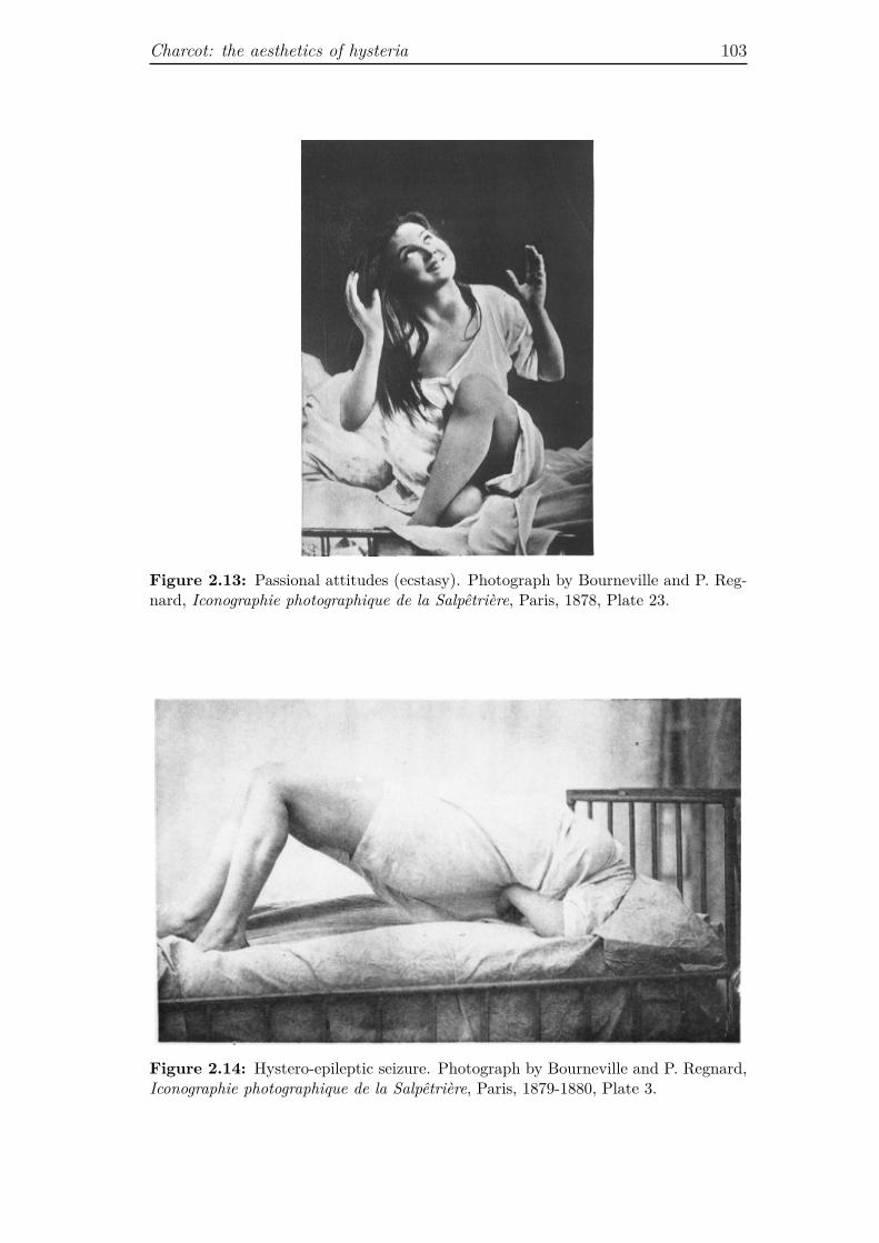
Charcot: the aesthetics of hysteria 103
Figure 2.13: Passional attitudes (ecstasy). Photograph by Bourneville and P. Reg-nard, Iconographie photographique de la Salpêtrière, Paris, 1878, Plate 23.
Figure 2.14: Hystero-epileptic seizure. Photograph by Bourneville and P. Regnard,Iconographie photographique de la Salpêtrière, Paris, 1879-1880, Plate 3.
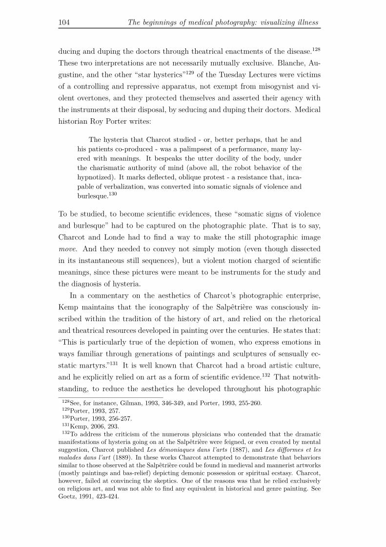
104 The beginnings of medical photography: visualizing illness
ducing and duping the doctors through theatrical enactments of the disease.128
These two interpretations are not necessarily mutually exclusive. Blanche, Au-gustine, and the other “star hysterics”129 of the Tuesday Lectures were victimsof a controlling and repressive apparatus, not exempt from misogynist and vi-olent overtones, and they protected themselves and asserted their agency withthe instruments at their disposal, by seducing and duping their doctors. Medicalhistorian Roy Porter writes:
The hysteria that Charcot studied - or, better perhaps, that he andhis patients co-produced - was a palimpsest of a performance, many lay-ered with meanings. It bespeaks the utter docility of the body, underthe charismatic authority of mind (above all, the robot behavior of thehypnotized). It marks deflected, oblique protest - a resistance that, inca-pable of verbalization, was converted into somatic signals of violence andburlesque.130
To be studied, to become scientific evidences, these “somatic signs of violenceand burlesque” had to be captured on the photographic plate. That is to say,Charcot and Londe had to find a way to make the still photographic imagemove. And they needed to convey not simply motion (even though dissectedin its instantaneous still sequences), but a violent motion charged of scientificmeanings, since these pictures were meant to be instruments for the study andthe diagnosis of hysteria.
In a commentary on the aesthetics of Charcot’s photographic enterprise,Kemp maintains that the iconography of the Salpêtrière was consciously in-scribed within the tradition of the history of art, and relied on the rhetoricaland theatrical resources developed in painting over the centuries. He states that:“This is particularly true of the depiction of women, who express emotions inways familiar through generations of paintings and sculptures of sensually ec-static martyrs.”131 It is well known that Charcot had a broad artistic culture,and he explicitly relied on art as a form of scientific evidence.132 That notwith-standing, to reduce the aesthetics he developed throughout his photographic
128See, for instance, Gilman, 1993, 346-349, and Porter, 1993, 255-260.129Porter, 1993, 257.130Porter, 1993, 256-257.131Kemp, 2006, 293.132To address the criticism of the numerous physicians who contended that the dramatic
manifestations of hysteria going on at the Salpêtrière were feigned, or even created by mentalsuggestion, Charcot published Les démoniaques dans l’arts (1887), and Les difformes et lesmalades dans l’art (1889). In these works Charcot attempted to demonstrate that behaviorssimilar to those observed at the Salpêtrière could be found in medieval and mannerist artworks(mostly paintings and bas-relief) depicting demonic possession or spiritual ecstasy. Charcot,however, failed at convincing the skeptics. One of the reasons was that he relied exclusivelyon religious art, and was not able to find any equivalent in historical and genre painting. SeeGoetz, 1991, 423-424.
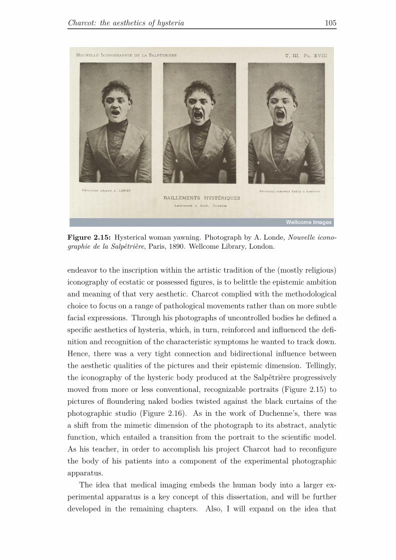
Charcot: the aesthetics of hysteria 105
Figure 2.15: Hysterical woman yawning. Photograph by A. Londe, Nouvelle icono-graphie de la Salpêtrière, Paris, 1890. Wellcome Library, London.
endeavor to the inscription within the artistic tradition of the (mostly religious)iconography of ecstatic or possessed figures, is to belittle the epistemic ambitionand meaning of that very aesthetic. Charcot complied with the methodologicalchoice to focus on a range of pathological movements rather than on more subtlefacial expressions. Through his photographs of uncontrolled bodies he defined aspecific aesthetics of hysteria, which, in turn, reinforced and influenced the defi-nition and recognition of the characteristic symptoms he wanted to track down.Hence, there was a very tight connection and bidirectional influence betweenthe aesthetic qualities of the pictures and their epistemic dimension. Tellingly,the iconography of the hysteric body produced at the Salpêtrière progressivelymoved from more or less conventional, recognizable portraits (Figure 2.15) topictures of floundering naked bodies twisted against the black curtains of thephotographic studio (Figure 2.16). As in the work of Duchenne’s, there wasa shift from the mimetic dimension of the photograph to its abstract, analyticfunction, which entailed a transition from the portrait to the scientific model.As his teacher, in order to accomplish his project Charcot had to reconfigurethe body of his patients into a component of the experimental photographicapparatus.
The idea that medical imaging embeds the human body into a larger ex-perimental apparatus is a key concept of this dissertation, and will be furtherdeveloped in the remaining chapters. Also, I will expand on the idea that
106 The beginnings of medical photography: visualizing illness
Figure 2.16: Episode of male hysteria. Photograph by A. Londe, Nouvelle icono-graphie de la Salpêtrière, Paris, 1888.
medicine, like biology and physiology, deals with the problem of creating im-ages of invisible objects and phenomena (Chapter 3); that the mimetic imagesprovided by photography are routinely transformed into diagrammatic repre-sentations (Chapter 4); and that photography has an analytic potential, whichexploded with the introduction of digital technologies (Chapter 5).
Chapter 3
Photography, radiography, and
the visualization of the invisible
As discussed in the previous chapter, since its origins medical photography hasdealt, one way or another, with the problem of producing pictures of somethingthat is not directly visible. In some cases, namely in psychiatry and neurol-ogy, the problem laid in the fact that the visual dimension of the disease understudy was elusive. Consequently, a form of visibility had to be actively producedthrough predefined theoretical constructs (for instance, phrenology and phys-iognomy in psychiatry), or by developing devices that could produce a specificexpressivity of the body (as in the case of electrophysiology). Even in derma-tology, the clinical discipline devoted to the surface of the body and thereforeto eminently visible diseases, the aim of photographic collections was less torecord the individual, idiosyncratic condition of a specific patient (what Sekulacalls the “nominalist” mode of signification),1 than to establish a human type,an abstract entity, corresponding to a disease (“realist” mode of significationin Sekula’s account). More precisely, photographic series were meant to re-veal the facies of a pathological condition, that is, its characteristic yet generalfeatures. In the photographic series the uniqueness of the patient’s body wasideally obliterated, and illness showed itself as an objective, visible fact.
In the present chapter I discuss how photography was used to deal withother forms of invisibility, namely, the invisibility of extremely small objects (mi-crophotography), very fast phenomena (chronophotography), and macroscopicobjects concealed inside the body (radiography). My main goal is to discussthe semiotic and epistemological problems encountered by the late-nineteenth-century physicians and scientists who used photographic processes to createimages of different invisible objects and phenomena. The questions I address
1See Sekula, 1986. See also Section 2.4.
107
108 Photography, radiography, and the visualization of the invisible
are: what kind of pictures were these? Through which modes of significationwere they made meaningful? Could they be considered iconic images, takinginto account that in many cases it was impossible to corroborate their “likeness”with an actual referent? What sort of photographic realism could be endorsedby the photographers of the invisible? This set of questions is coupled withanother one: could all these scientists genuinely rely on mechanical objectivity,as has been influentially defended by the historians of science Lorraine Dastonand Peter Galison?
Drawing on the theoretical analysis of photography developed in Chapter 2,I show how the iconic, indexical and symbolic modes of signification were com-bined in order to make sense of microscopic, chronographic and radiographicpictures. As in the previous chapter, this theoretical approach is intertwinedwith a historical one. That is, I sustain my conceptual analysis and epistemo-logical arguments with the examination of historical literature (both primaryand secondary sources) concerning representational and experimental practices.Starting from a critical appraisal of Daston and Galison’s claims about the late-nineteenth century discourses about mechanical images, I demonstrate that theidea of mechanical objectivity is far too poor to account for the richness of prac-tical and conceptual strategies put in place to produce and make sense of thephotographs of the invisible. In the light of this historical analysis, I suggestthat the notion of mechanical objectivity should be replaced, on the one hand,with that of intersubjectivity sustained by operational conventions; on the otherhand, with the idea of mechanical sensibility. Finally, closing the circle with theclaims I made in Chapter 1 about the relationship between X-ray diagnosticsand clinical anatomy, I show how the process that led to make sense of radio-graphs was indebted to Bichat’s conceptualization of the cadaver as proxy ofthe living body, and to the clinical gaze that was born in the hospital at thedawn of the nineteenth century.
3.1 Truth-to-nature, mechanical objectivity, and
trained judgment
A relevant part of the discussion developed in this chapter is built against theconcept of mechanical objectivity put forward by Daston and Galison in a seriesof works published between 1992 and 2007.2 Consequently, the first thing to dois to provide a brief outline of the ideas of these authors.
2See Daston and Galison, 1992; Galison, 1998; Galison, 1999; Daston and Galison, 2007.
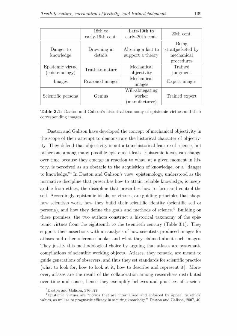
Truth-to-nature, mechanical objectivity, and trained judgment 109
18th toearly-19th cent.
Late-19th toearly-20th cent. 20th cent.
Danger toknowledge
Drowning indetails
Altering a fact tosupport a theory
Beingstraitjacketed by
mechanicalprocedures
Epistemic virtue(epistemology) Truth-to-nature Mechanical
objectivityTrained
judgment
Images Reasoned images Mechanicalimages Expert images
Scientific persona GeniusWill-abnegating
worker(manufacturer)
Trained expert
Table 3.1: Daston and Galison’s historical taxonomy of epistemic virtues and theircorresponding images.
Daston and Galison have developed the concept of mechanical objectivity inthe scope of their attempt to demonstrate the historical character of objectiv-ity. They defend that objectivity is not a transhistorical feature of science, butrather one among many possible epistemic ideals. Epistemic ideals can changeover time because they emerge in reaction to what, at a given moment in his-tory, is perceived as an obstacle to the acquisition of knowledge, or a “dangerto knowledge.”3 In Daston and Galison’s view, epistemology, understood as thenormative discipline that prescribes how to attain reliable knowledge, is insep-arable from ethics, the discipline that prescribes how to form and control theself. Accordingly, epistemic ideals, or virtues, are guiding principles that shapehow scientists work, how they build their scientific identity (scientific self orpersona), and how they define the goals and methods of science.4 Building onthese premises, the two authors construct a historical taxonomy of the epis-temic virtues from the eighteenth to the twentieth century (Table 3.1). Theysupport their assertions with an analysis of how scientists produced images foratlases and other reference books, and what they claimed about such images.They justify this methodological choice by arguing that atlases are systematiccompilations of scientific working objects. Atlases, they remark, are meant toguide generations of observers, and thus they set standards for scientific practice(what to look for, how to look at it, how to describe and represent it). More-over, atlases are the result of the collaboration among researchers distributedover time and space, hence they exemplify believes and practices of a scien-
3Daston and Galison, 376-377.4Epistemic virtues are “norms that are internalized and enforced by appeal to ethical
values, as well as to pragmatic efficacy in securing knowledge.” Daston and Galison, 2007, 40.
110 Photography, radiography, and the visualization of the invisible
tific community.5 Daston and Galison’s taxonomy of epistemic virtues can besummarized as follows:
1. The ideal of truth-to-nature emerged in the late-eighteenth century as aweapon against the danger to be overwhelmed by the details of the mon-strous variability of nature. In accordance with Enlightenment sensa-tionalist psychology, the self was perceived as fragmented and excessivelyreceptive. Hence, in order to secure knowledge, the savant – who workedin strict relationship with the artist-illustrator – had to actively controlsensations, by selecting and interpreting them. The scientific persona cor-responding to the virtue of truth-to-nature was the genius.
2. The ideal of mechanical objectivity, on the contrary, aimed at the sup-pression of subjectivity. It appeared around the 1830s and seeminglyguided scientists until the 1920s, acting against the hindrances of the post-Kantian self, perceived as too active and eager to impose its interpretationsand hypotheses on the world. The core demand of mechanical objectivitywas to “let nature speak for itself,”6 which implied that images had to beproduced according to strict mechanical protocols.7 Photography was nota precondition for the emergence of mechanical objectivity, but it never-theless played a seminal role as paradigmatic form of mechanical imagingtechnology. The scientific persona corresponding to the epistemic virtueof mechanical objectivity was an automaton, a will-abnegating worker.
3. Finally, by the 1930s the ideal of trained judgement emerged, partly in re-action to mechanical objectivity. Drawing support from the many theoriesof the unconscious that blossomed at the turn of the twentieth century,trained judgment downplayed the importance of the unbending applica-tion of procedural routine proper of mechanical objectivity, and put highvalues on educated intuition, the “physiognomic sight”8 that comes withtraining and a long acquaintance with an object. The scientific personashaped by trained judgement was the trained expert, a self-confident pro-fessional who trusted his or her own intuitions, which could be educated
5See Daston and Galison, 2007, 19-27. In my view, it is dubious that one can take therules which prescribe how to make, compose, and present images in atlases as proxies for theepistemic ideals that govern the making of science as a whole. I develop this criticism inSection 3.4.3.
6Daston and Galison, 1992, 81.7“Objectivity in its mechanical guise emerges as a ferociously austere, self-denying virtue,
a virtue present when all the special skills, intuitions, and inspirations of the scientist could bequieted and nature could be transferred to the page without intervention or interpretation.”Galison, 1999, 19.
8Daston and Galison, 2007, 314.
Microphotography: intersubjectivity vs. mechanical objectivity 111
and refined by studying and working in renowned schools and scientificinstitutions.
In opposition to both truth-to-nature and trained judgment, self-erasing was thecharacterizing feature of mechanical objectivity. In Daston and Galison’s words:“In the making of images, the taking of measurements, the tracing of curves, andmany other scientific practices of the latter half of the nineteenth century, self-elimination became an imperative.”9 They base this strong claim in the textualanalysis of the introductions of a considerable number of nineteenth-centuryatlases, and in the discussion of a few debates about scientific images whoseright to claim objectivity was contentious.10 Yet, historical and conceptualanalyses of the same period provided by other authors show that if mechanicalobjectivity as suppression of the observer ever existed, it was far from being anoverarching ideal, and that a much more nuanced approach to the mechanicalproduction of images was actually in place. In the following pages I criticizethe notion of mechanical objectivity and the alleged divide between mechanicalobjectivity and trained judgment through the analysis of three case studies: theuse of photography in microbiology, chronophotography, and radiography.
3.2 Microphotography: intersubjectivity vs. me-
chanical objectivity
Microphotography was one of the earliest applications of photography in science.Indeed, it was through the microscope that photography entered the domain ofmedicine.11 Both bacteriologists and cellular pathologists, who in the second halfof the nineteenth century promoted new conceptions of disease and new ways ofpracticing medicine, relied on the combination of an old and a new technology(the microscope and the photographic camera) to back their discoveries. AsSicard puts it: “At the edge of knowledge, microscopy met photography.”12
3.2.1 The myth of the faithful record
In February 1840, just a few months after François Arago’s official presentationof the invention of photography at the Académie des Sciences, Alfred Donné,a physician and bacteriologist who gave microscopy classes in Paris, showed to
9Daston and Galison, 2007, 196.10For example, the diatribe between Camillo Golgi and Santiago Ramón y Cajal about the
individuality of neural cells. See Daston and Galison, 2007, 115-120, and 183-184.11See Sicard, 1995, 11-12.12Sicard, 1998, 122, my translation.
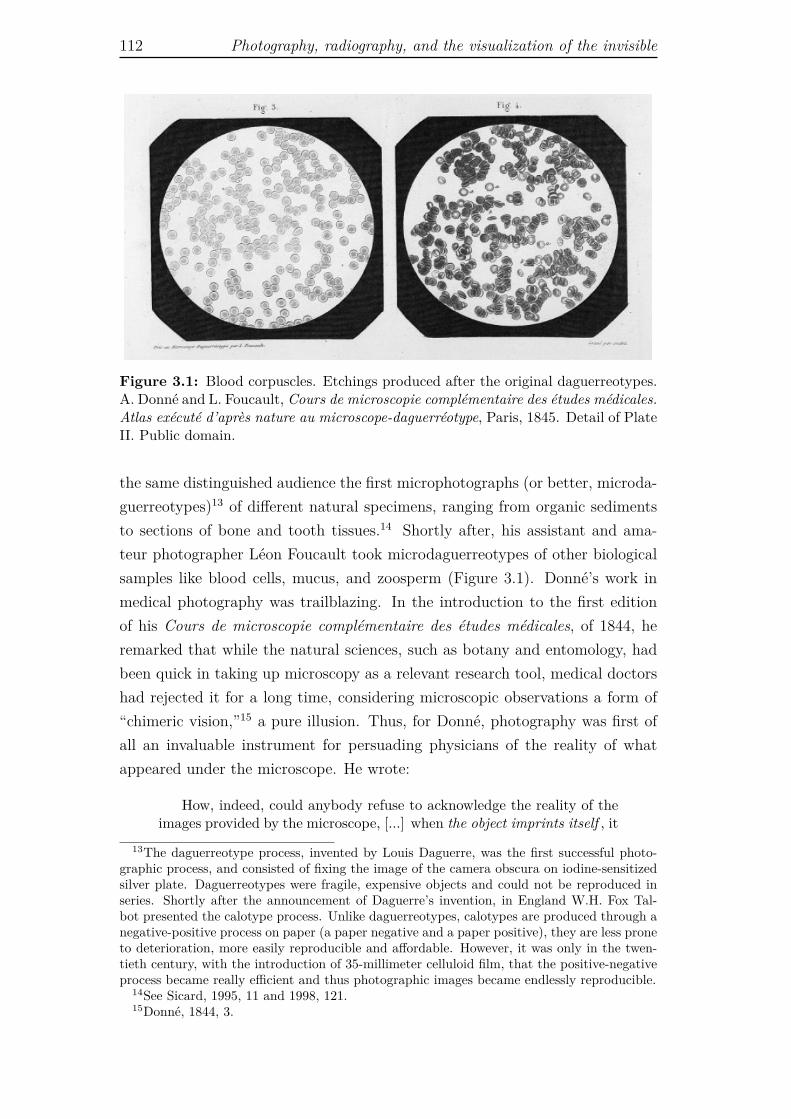
112 Photography, radiography, and the visualization of the invisible
Figure 3.1: Blood corpuscles. Etchings produced after the original daguerreotypes.A. Donné and L. Foucault, Cours de microscopie complémentaire des études médicales.Atlas exécuté d’après nature au microscope-daguerréotype, Paris, 1845. Detail of PlateII. Public domain.
the same distinguished audience the first microphotographs (or better, microda-guerreotypes)13 of different natural specimens, ranging from organic sedimentsto sections of bone and tooth tissues.14 Shortly after, his assistant and ama-teur photographer Léon Foucault took microdaguerreotypes of other biologicalsamples like blood cells, mucus, and zoosperm (Figure 3.1). Donné’s work inmedical photography was trailblazing. In the introduction to the first editionof his Cours de microscopie complémentaire des études médicales, of 1844, heremarked that while the natural sciences, such as botany and entomology, hadbeen quick in taking up microscopy as a relevant research tool, medical doctorshad rejected it for a long time, considering microscopic observations a form of“chimeric vision,”15 a pure illusion. Thus, for Donné, photography was first ofall an invaluable instrument for persuading physicians of the reality of whatappeared under the microscope. He wrote:
How, indeed, could anybody refuse to acknowledge the reality of theimages provided by the microscope, [...] when the object imprints itself , it
13The daguerreotype process, invented by Louis Daguerre, was the first successful photo-graphic process, and consisted of fixing the image of the camera obscura on iodine-sensitizedsilver plate. Daguerreotypes were fragile, expensive objects and could not be reproduced inseries. Shortly after the announcement of Daguerre’s invention, in England W.H. Fox Tal-bot presented the calotype process. Unlike daguerreotypes, calotypes are produced through anegative-positive process on paper (a paper negative and a paper positive), they are less proneto deterioration, more easily reproducible and affordable. However, it was only in the twen-tieth century, with the introduction of 35-millimeter celluloid film, that the positive-negativeprocess became really efficient and thus photographic images became endlessly reproducible.
14See Sicard, 1995, 11 and 1998, 121.15Donné, 1844, 3.
Microphotography: intersubjectivity vs. mechanical objectivity 113
fixes itself on the plate without the help of the art, without any interven-tion of the human hand, by the only effect of light, and always identicalup to the smallest details to the image perceived by the observer under themicroscope?16
Donné, as virtually all his contemporaries, considered photography a direct im-print of nature, “a material image printed by nature itself.”17 This does not meanthat he was not aware that both the microscope and the daguerreotype createartificial visual effects. Sicard reports that Donné and Foucault – both preoc-cupied with the pedagogical difficulties of training physicians in microscopy –knew very well the practical difficulties related to the use of microdaguerrotypes,and actually asked themselves how to sensibly use these images as tools for par-taking knowledge.18 As a matter of fact, daguerrotypes have a good definition,but they have a low sensitivity for red, and this, at a time in which the most im-portant histological dyes were cochineal and carmine, carried a major technicallimitation. Moreover, in order to look at the silver plate properly, one had toincline it at a specific angle, and therefore for the untrained eye proper observa-tion could be quite difficult.19 That notwithstanding Donné was confident thatwhat the daguerreotype showed was “identical” to what the microscopist saw,and this would allow sharing microscopic vision. In spite of all the actual dividebetween reality and its photographic representation – a divide that was partic-ularly deep in the early days of photography – photographs were immediatelyperceived as “the evidence of the present or past existence of a reality that re-sembles to them.”20 For Donné, photography produced representations that wereas accurate as the quantitative methods used in physiology and, accordingly, hebelieved that it could be extremely useful for medical diagnosis.
Still, regardless of Donné’s pioneering work, microphotographs became anaccepted tool within the scientific community only in the 1880s, thanks to thework of Robert Koch. As the historian of science Olaf Breidbach has shown,Koch conceptualized and used photographs as an instrument to cross-check mi-croscopists’ observations, rather than an unquestionable imprint of reality, andin so doing he fostered the creation of experimental standards and a commonway of seeing among bacteriologists. It was ultimately on these grounds thatmicrophotography became a fundamental instrument for bacteriology.21
The problem of sharing microscopic observations was one of Donné’s mainconcerns, and before turning to photography, he had developed a sun microscope
16Donné, 1844, 36-37, my translation.17Donné, 1845, 10.18See Sicard, 1995, 12.19See Sicard, 1998, 124.20Sicard, 1995, 12, my translation.21See Breidbach, 2002, 222
114 Photography, radiography, and the visualization of the invisible
which projected the image of the specimen under analysis on a screen. This inge-nious system, however, was hampered by its dependence on the availability andquality of natural light. As seen above, Donné thought that microphotographsgranted the solution to his search for a shared microscopic sight, but, accordingto Breidbach, until well into the 1880s, the majority of microscopists did not be-lieve that photography could provide reliable representations of their specimens.Paradoxically, such distrust was a consequence of the excessive enthusiasm pro-fessed by some of the early defendants of microphotography. Breidbach explainsthat those arguing in favor of microphotography saw photographs as a realityin their own right. The consequence was that pictures, and not the originalsamples, became the primary object of microscopic analysis. Instead of tryingto optimize the photographic processes, these practitioners set out to dissectthe hidden qualities of the images, because they assumed that what they sawon the photographic plate was a perfect reproduction of the world, or moreemphatically, the world itself captured by the camera.22 Many introductionsto scientific microphotography were published in the 1860s, and in general theywere not devoted to a specific subject. As Donné’s atlas, they were collectionsof photographs of different microscopic objects, and their aim was to demon-strate what microphotography could show, rather than to tackle a well definedscientific problem. Breidbach points out that in the manuals published in thisdecade, three main arguments were put forward in favor of the new imagingtechnology: (1) microphotographs allowed better measurements of the sample;(2) they showed details which were invisible when looking through the micro-scope; and (3) they were free from the bias of the observer.23
The rationale underlying the first claim was that microphotographs, morepractical to handle than actual microscopic preparations, granted precise mea-surements even in the case of complicated objects. Critics of photography, how-ever, stressed that this putative advantage could be very well attained by em-ploying a camera lucida, an optical device widely used by microscopists at thetime, which required much less technical effort than early photography.24 Thesecond argument, namely that photography showed more than the microscope,derived from one of the properties of photography largely praised by Benjamin,i.e., the possibility of enlargement. In this respect, Breidbach reports that ina publication of 1868, the naturalists Oscar Reichard and Carl Sturenburg ex-plained that they obtained high magnification by re-photographing a micropho-tograph put under the microscope. The process was repeated twice and this
22See Breidbach, 2002, 222.23See Breidbach, 2002, 230-231.24See, Breidbach, 2002, 231.
Microphotography: intersubjectivity vs. mechanical objectivity 115
allowed to allegedly attain magnifications of 8000x/30,000x, at a time in whichthe best microscope could provide a maximum enlargement of 2000x. Throughthis procedure granularity and other physical properties of the photographicnegatives became salient, but Reichard and Sturenburg – as many of their col-leagues fascinated by photography – did not take these elements for artifactsproduced by the enlargement. On the contrary, they assumed that they were ac-tual properties of the biological specimen made visible by microphotography.25
In their approach, it was the photographic camera, and not the microscope, thatprovided relevant information about nature. As Breidbach explains: “The pho-tograph gave the world its own quality, set apart from those things actually seenin the microscope.”26 Photography was the ultimate scientific instrument, theone which set the paragon of truth and reality. On this very ground was foundedthe third claim, according to which photographs were objective because inde-pendent from the photograph maker’s subjectivity. For the proponents of thisidea of objectivity, microphotographs were the proper representation of whatactually existed in nature, for the details of the image were not restricted tothose deemed relevant or particularly important by the observer.27
3.2.2 The problem of the invisible referent
This conception of photography, founded on the assumption that the photog-rapher is a passive agent in the production of pictures, fits the definition ofmechanical objectivity as suppression of subjectivity proposed by Daston andGalison. As seen above, according to Daston and Galison, the ideal of me-chanical objectivity emerged in the second half of the nineteenth century andwas characterized by the scientists’ strenuous attempt “to eliminate the mediat-ing presence of the observer.”28 In the account provided by the two historians,mechanical images, of which photographs represented the most accomplishedexample, granted objectivity for two reasons. On the one hand, they by-passedhuman judgement, which tended to select the details of objects or phenomenato be represented; on the other hand, they overcame the limitations of humansenses and drawing abilities, which undermined the accuracy of observationsand representations, respectively. Mechanical objectivity was clearly predicatedon the indexicality of the photographic image, because its independence fromthe photographer and its pictorial accuracy (iconic mode of signification) were
25See Breidbach, 2002, 233.26Breidbach, 2002, 234.27See Breidbach, 2002, 231. This is the conception of photography that was later theorized
and brought to the artistic arena by Moholy-Nagy, with his idea of unbiased vision and hisexperimentation with the photographic medium. See Section 2.3.
28Daston and Galison, 1992, 82.
116 Photography, radiography, and the visualization of the invisible
a consequence of the fact that the image was produced through an automatedprocess that physically forced, to use Peirce’s words, a correspondence betweensign and referent. And yet, this account of the objectivity of photography (andmechanical images in general), which Daston and Galison present as an overarch-ing scientific ideal and guiding principle for the scientists of the late-nineteenthand early-twentieth centuries,29 was far from being unanimously endorsed bybacteriologists. In fact, when it came to the study of the microscopic world, theepistemic authority of photographs could no longer rely on the combination ofindexical and iconic modes of signification, because, being the referent invisible,it was impossible to certify any resemblance. Indeed, Breidbach shows that theidea that microphotographs were objective simply by virtue of being a mechan-ical imprint of nature had little currency among the majority of microscopists.Evidence of this, he maintains, comes from the fact that microphotographswere almost completely absent from micro-morphological and histological pa-pers prior to the late 1880s, even though inexpensive methods for photographicproduction and printing became available in the 1850s and 1870s, respectively.30
Objectivity from operational conventions As a general rule, the scien-tists who worked with microscopes were very much aware of the many variablesinvolved in their observations. They knew that the selection, preparation, fixa-tion, and staining of the specimen, together with the optical properties of eachmicroscope, played a seminal role in determining the results of any observationof the invisible world. Accordingly, they would not subscribe easily to the ideathat a microphotograph showed the objective, true features of a microscopicentity (e.g., a bacterium or a plant cell) merely by virtue of being mechanicallyproduced. The act of looking at a specimen under the microscope was just thelast of a chain of actions that started with the very complex tasks of selecting,collecting and preparing a sample suitable for microscopic observation. Hence,for those who studied the microworld, the ideal of objectivity had to be by ne-cessity much more specific and elaborated than the ideal of suppression of theobserver described by Daston and Galison. For the majority of microscopists,as Breidbach shows, an objective representation was not simply a mechanicalimage, but rather “a reliable reproduction of a microscopic preparation,”31 andit could be attained “by the extent to which the image seen in the microscope[could] be accurately reproduced and its reproduction technically controlled.”32
This implied not only that the photographs had to be taken under controlled29See Daston and Galison, 2007, 174-190.30See Breidbach, 2002, 226 and 234.31Breidbach, 2002, 235.32Breidbach, 2002, 235.
Microphotography: intersubjectivity vs. mechanical objectivity 117
conditions, but also that the whole process of sample fixation, staining, han-dling, and conservation had to be in line with explicit rules and standards.In other words, objectivity was not an effect of the use of a mechanical tech-nology, but rather the result of operational procedures. The whole process ofmicrobiological investigation had to be carried out within a set of operationalconventions.
Now, signs that signify by virtue of learned conventions are symbols. Peirce’sdefinition, however, refers to the conventions entailed in the process of signs’interpretation, not production (although he stresses that the object puts con-straints on the sign’s ability to signify). This is probably the reason why theauthors who suggest that microscopic imaging has to be understood in termsof symbols rather than icons, emphasize the fact that one has to learn the con-ventions that allow to interpret the images (conventions related to the way ofseeing), and do not elaborate much on the topic of the relevance of conventionsin the very production of such images.33 I think, however, that if we pay atten-tion to the fact that in the processes of image making conventions are embodiedin standard procedures and methodologies, we are in a better position to under-stand how the symbolic and indexical orders of signification are entangled whenit comes to assign truth-value to an image of invisible entities. In other words,we have to keep in mind that the meaning of a microphotograph depends on thefact that we trust it as an index (mechanical imprint), only as far as it has beenproduced according to well defined conventions, that is, experimental standards.The application of operational conventions in the phase of image production isan essential component of the scientists’ trust in images, and creates the ba-sis, both material and epistemological, for the definition of a symbolic mode ofsignification in image interpretation.
It was this specific version of objectivity grounded in operational conventions,put forward by Koch in the 1880s, that led to the acceptance of microphotog-raphy as a meaningful scientific tool in bacteriology.34 Koch did not explicitlycontest the idea that photographs provided an accurate reproduction of reality.However, he understood the epistemic potential of photography in strict relationwith the specific characteristics, problems, and needs of his field of research. Ina paper published in 1886, he wrote:
It must be remembered that [in bacteriology] we have to do entirelywith microscopic objects, and that two observers with the microscopecannot see the same object simultaneously and form a joint opinion withregard to it [. . . ]; and as all microscopists know, even the slightest turnof the fine adjustment causes so small an object as a bacterium either to
33See, for example, Serpente, 2011.34See Breidbach, 2002, 243.
118 Photography, radiography, and the visualization of the invisible
disappear entirely from the field, or to appear with different markings andshadows. Agreement will always be facilitated when the observations aremade with the same instrument, that is to say, with the same illumination,the same objective, and the same magnifying power. But if the conditionsunder which the microscopic object is seen are very different, [. . . ] ifthe preparation and staining of the object is dissimilar , if moreover it ismounted in fluids of different degrees of refrangibility, how can anyonewonder when one microscopist asserts that the object as seen by him isquite different from that described by another? [. . . ] Photography, on thecontrary, gives the microscopical picture once and for all, and reproducesit without the slightest error, in exactly the same focus, magnification,and illumination as when it was taken.35
For Koch, photography was capital to investigation in microbiology, because itwarranted that two or more researchers, even if working with different micro-scopes in different places, could observe the same thing, under the same exper-imental and observational conditions. They would see the same cellular cultureunder the same focus, magnification, and illumination. In other words, theywould construct the same image and, consequently, the same scientific object.
Objectivity from intersubjectivity Photography provided a permanentrecord of a phenomenon that otherwise could be observed by only one personat the time. It allowed intersubjective evaluations, by assuring that all subjectswere looking at the same micro-organism or, more precisely, to the same pictureof a given microscopic sample, produced and handled under standard condi-tions. Such a picture could be retrieved at any moment for later comparisonsand could travel long distances, reaching new observers. In a field like microbi-ology, that in the second half of the nineteenth century was still immature butburgeoning, microphotographs were invaluable means for communication withinthe scientific community. Bynum has remarked that: “Koch’s pioneering pho-tomicroscopy [...] helped in the search for standards, which were so importantin the last, heady decades of the [nineteenth] century, when new pathogenicorganisms were being announced every few months.”36 Microphotographs wereparamount in creating a common practice and a common vision among micro-scopists. These photographs could not rely on the likeness with the referent(iconicity) to secure their epistemic authority, because there was no way to di-rectly assess any similarity. Neither could such authority come exclusively fromtheir indexicality. As shown by Breidbach, the indexical mode of significationwas not enough to make mechanical images meaningful, useful, and accepted inthe microbiologists’ community. To meet this goal, microphotographs had to be
35Koch, 1886, 20.36Bynum, 1994, 129.
Microphotography: intersubjectivity vs. mechanical objectivity 119
embedded in a specific set of conventional practices that created the conditionsfor a symbolic mode of signification grounded in indexical premises. This meansthat for microbiologists, the idea of objectivity of photography bore a profoundrelation with the notion of intersubjectivity. The sharing of conventions can bein fact considered a precondition for intersubjective evaluations. A photographwas objective in the sense that it was intersubjective, and it was intersubjectivebecause it was produced according to well defined standards. Importantly, itwas acknowledged that the photograph did not necessarily show the real object(e.g., a bacterium), since it could show artifacts as well. That is, the notionof photographic objectivity was severed from that of photographic realism as amark of truth. To better understand this point it is useful to resort to PatrickMaynard’s distinction between depiction and detection, and the further elabo-ration of such distinction put forward by Laura Perini.
3.2.3 Depictions and detections
Maynard defines photography as a technology that exploits light and other radi-ations to mark surfaces.37 A fundamental feature of his theory of photographyis that photographic images can be used as depictions or to make detections.We use photographic images as depictions when we rely on their mimetic prop-erties in order to imagine that we see the object through the image; we usephotographic images to make detections when we use them as sources of infor-mation. For Maynard, the depictive and detective functions can be separated,but in most cases we rely on the depictive character of photographs to makedetections: “Photography might be most simply characterized as the site of his-torically the most spectacular interaction of depictive and detective functions.”38
For instance, when I look at a photograph of my sister or of the Tour Eiffel, Idetect my sister or the Parisian monument via the depiction provided by thephotograph. I imagine that I am seeing that person or that object through thepicture. However, what is depicted does not always coincide with what canbe detected. Maynard uses the example of King Kong to clarify this point. Aphotograph of King Kong is a photographic depiction of a giant ape, but onecannot use it to detect an ape, because the colossal gorilla known under thename of King Kong never actually existed. In this case one can use the photo-graph to detect a disguised actor, a big puppet, or a virtual reality animation,but not the subject-matter depicted in the photograph or in a movie.39 This
37Maynard defines material images as physical states of a surface, and he calls them “mark-ings” or “marked surfaces.” See Maynard, 1997, Ch. 1
38Maynard, 1997, 120.39See Maynard, 1997, 114.
120 Photography, radiography, and the visualization of the invisible
means that what can be detected in a photograph, and the level of accuracy ofsuch detection, depends to a large extent on the background information thatthe observer possess.
This is why the philosopher of science Laura Perini argues that Maynard’sdistinction between photographic depiction and detection can help us under-standing scientific imaging only if we think of detection as independent from,or at least problematically related to, depiction. In fact, although scientistslargely rely on photographic evidence in their experimental work, they are oftenin the situation of possessing very little background information about the de-picted object.40 Perini remarks that when knowledge about an object is scant,“using photographs as depictions is often a threat to detection, because it fos-ters a way of working with the image that needs have no connection with thekind of information it actually presents.”41 Seeing (recognizing) something asdepicted incorporates a number of assumptions, and what appears as depicteddoes not always correspond to what should be detected. A typical instance inwhich depiction is detrimental to detection is that of microscopy artifacts. Perinidiscusses the concrete example of an electron microscope photograph showinga putative mesosome (dense structure) inside the bacterium Bacillus subtilis,published in the Journal of Bacteriology in 1964.
The problem of artifacts When the authors of this article on B. subtilisobserved a dense structure inside the bacterium, they thought that it was anorganelle with some biological function, whose presence was related to the pro-toplast state of the bacterial cells, and they named it mesosome. Subsequentanalyses, however, demonstrated that the alleged bacterial organelle was, infact, an artifact. The biologists had used the micrographs as a depiction of B.subtilis and this had led them to make a wrong detection: they took an artifactfor a real organelle. In this case, to use the photograph to imagine to see the realobject through this picture was not the right path to detection. In order not tobe mistaken it is necessary to keep in mind that when our knowledge about anobject is limited, extracting information about such objects from depictions canbe misleading. To avoid the pitfall, the researchers should have taken the micro-graph simply as a depiction of the structure of a sample specifically prepared forelectronic microscopy, rather than a direct depiction of the bacillus they werestudying. Under this caveat, they could just claim that the image allowed todetect small dense regions inside the prepared specimen, because this was in-deed the only information that one could extract from the micrograph. To know
40See Perini, 2012, 151-152.41Perini, 2012, 153.
Microphotography: intersubjectivity vs. mechanical objectivity 121
something about the origins and function of that dense region – whether it wasmaterial that clumped up during sample processing, or an actual organelle inthe cytoplasm of B. subtilis – required a number of additional observations andexperiments. Only in this way, and not simply relying on the perceived realismof the electron microscope image, it becomes possible to clarify the relationshipbetween what is depicted in the micrograph and which structures really belongto the natural specimen.42 The biologist can certainly imagine that he is lookingat a bacterium, but he must be aware that he is looking at it through the electronmicroscope and through the sample. It is a depiction that is twice removed fromits object. Accordingly, if one is to extract reliable information from microscopicimages, one has to approach the depiction in a quite restrained way, keeping inmind that it shows a processed specimen, and not the ultimate object of one’sresearch (the microscopic entity). If one looks at an object through the doublemediation of the microscope and of the treated sample, then both the condi-tions of operation of the optical device and the conditions of preparation of thespecimen must be standardized, otherwise we could never replicate and shareour observations. This leads us back to Koch’s insistence on the standardizationof both samples’ preparation and microphotography procedures, which allowedhim to develop an operational notion of photographic objectivity, independentfrom general claims of photographic realism.
The nineteenth-century bacteriologists who endorsed Koch’s vision of mi-crophotography did certainly believe in the reality of the microorganisms theystudied, yet their belief was not primarily grounded in photographic depiction,but rather in a complex set of laboratory practices concerning the isolation,growth, and preparation of the samples. Ultimately, it was this set of practicesthat allowed them to make detections (i.e., to extract information) from pho-tographic images. In this respect, it is worthy to remember that according toKoch’s famous “postulates” for the demonstration of the parasitic nature of adisease, a microorganism had to be: (1) “constantly present in characteristicform and arrangement in the diseases tissue;” (2) it should also be “isolated andgrown in pure culture;” and (3) it had to be “shown to induce the disease exper-imentally.”43 Microphotographs could play an important role in the first step,but they had little or no relevance in the second and in the third. I will comeback to the relationship between images and laboratory practices in Chapter4, where I discuss Ian Hacking’s idea that scientific representations are deeplyentrenched with a larger range of experimental interventions.
42See Perini, 2012, 155.43F. Loeffler, quoted in Bynum, 1994, 128. Loeffler was a pupil of Koch’s. Koch took his
postulates from his teacher Friedrich Henle, who had expressed them in an implicit form inhis essay De miasmata et contagia of 1840.
122 Photography, radiography, and the visualization of the invisible
3.3 Chronophotography: revealing the optical
unconscious
Chapter 2 ended with an image from the Nouvelle iconographie de la Salpêtrièrethat shows a naked body in the grip of convulsive movements against a blackbackground. Commenting on this picture I pointed out that in his attemptsto capture motion, Charcot followed in the steps of his teacher Duchenne deBoulogne, who used photography to produce models in order to study the me-chanical principles of movement rather than produce portraits of the humanbody. However, the definitive transformation of the human body into a di-agram by means of photography was accomplished by another French physi-cian, Etienne-Jules Marey, with his studies on animal locomotion. Importantly,Marey was a physiologist not a clinician. His preoccupation was to understandmovement in general, rather than as a specific symptom of some pathologicalcondition. He was not involved in diagnosing specific patients, and thus he couldgive up altogether the personal, individual dimension of medical photography,its dimension of portrait, so to say. He wanted to understand the general func-tioning of the human machine, and to this end he needed to set aside all itscontingent idiosyncrasies.
An important analysis of Marey’s chronophotographic method has been pro-vided by Snyder, in the article “Visualization and Visibility,” of 1998, written inresponse to Daston and Galison’s idea of mechanical objectivity. I showed abovethat the ideal of mechanical objectivity paid a very poor service to the credi-bility of microscopys during the 1860s. Microphotographs were widely acceptedas valuable scientific evidence only when Koch replaced the idea of mechanicalobjectivity (conceived as the suppression of the subjectivity of the observer andmimetic realism) with the idea of intersubjectivity supported by shared labora-tory practices that summed up to operational conventions (microphotographyprovided a common controlled vision of the microscopic world). Snyder showsthat also in the case of chronophotography the idea of mechanical objectivity iscompletely misleading. Chronophotographs, he notes, were not meant to sup-press any subjectivity, for the simple reason that that they showed somethingthat no human observer could ever see (this observation holds true also formicrophotography, because it shows an invisible referent).
3.3.1 Subjectivity superseded
At the outset of “The Image of Objectivity,” Daston and Galison quote a passagefrom Marey who, in the book La Méthode graphique dans les sciences expérimen-
Chronophotography: revealing the optical unconscious 123
tales, after surveying the countless visual instruments available to the scientistof the late nineteenth century, concluded:
There is no doubt that graphical expression will soon replace all oth-ers whenever one has at hand a movement or a change of state – in aword, any phenomenon. Born before science, language is often inappro-priate to express exact measures or definite relations. [Images recordedby mechanical instruments, on the contrary, speak] the language of thephenomena themselves.44
Daston and Galison comment on this quote as follow:
“Let nature speak for itself” became the watchword of a new brand ofscientific objectivity that emerged in the latter half of the nineteenth cen-tury. At issue was not only accuracy but morality as well [. . . ]. Marey andhis contemporaries turned to mechanically produced images to eliminatesuspect mediation.45
Here “suspect mediation” stands for the inevitable amount of idealization, inter-pretation, or lack of precision that a human observer introduces in any descrip-tion, verbal or visual of a phenomenon. For Daston and Galison, the scientistsin the late nineteenth and early twentieth centuries praised mechanical rep-resentation, because they saw in it a way to suppress the subjectivity of theobserver. In the case of Marey’s images of movement, however, the suppressionof subjectivity was not an issue. As Snyder puts it:
Most of Marey’s work does not fit the mold Daston and Galison makefor it. [. . . ] For the most part, Marey did not conceive of his precisioninstruments as impartial mediators substituting for and improving uponan observer’s eye or an illustrator’s hand. His mechanically originatedgraphs and photographically generated pictures are visualizations of dis-placements charted against precisely determined units of time. Thesemovements fall outside the scope of human detection and accordingly,their inscriptions cannot be characterized as especially accurate visual-izations of what might otherwise have been registered by an illustrator orscientist.46
When the machine records and displays something that falls squarely outsidethe realm of human sensibility (for example, vectors of motion as in the caseof many of the works of Marey’s), then the act of observing is fully displaced
44Marey, 1878, iii.45Daston and Galison, 1992, 81. It should be noted that the discussion of Marey’s texts,
which in the article of 1992 had a prominent role in the argumentation of Daston and Galison’s,disappeared from their book of 2007. Although in the book the idea of mechanical objectivityis presented in a much more articulated and nuanced form than in the article, the authors donot touch upon, hence do not provide any answer to, the objections raised by Snyder.
46Snyder, 1998, 379.
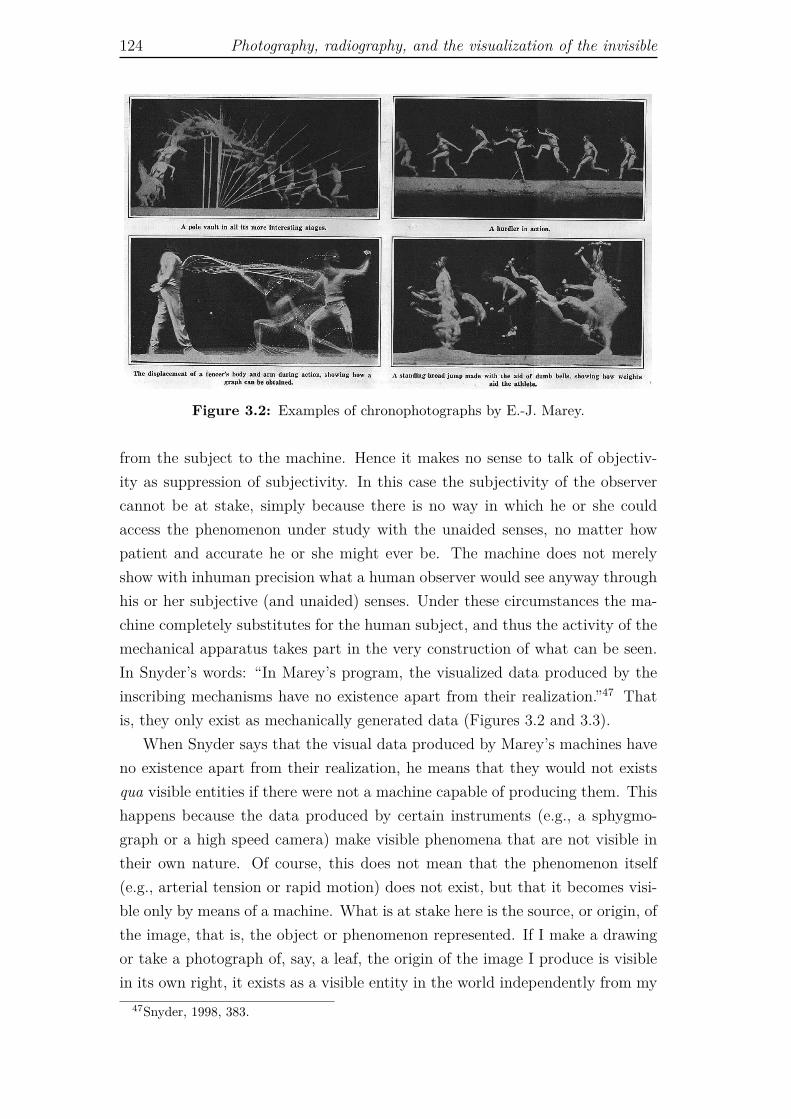
124 Photography, radiography, and the visualization of the invisible
Figure 3.2: Examples of chronophotographs by E.-J. Marey.
from the subject to the machine. Hence it makes no sense to talk of objectiv-ity as suppression of subjectivity. In this case the subjectivity of the observercannot be at stake, simply because there is no way in which he or she couldaccess the phenomenon under study with the unaided senses, no matter howpatient and accurate he or she might ever be. The machine does not merelyshow with inhuman precision what a human observer would see anyway throughhis or her subjective (and unaided) senses. Under these circumstances the ma-chine completely substitutes for the human subject, and thus the activity of themechanical apparatus takes part in the very construction of what can be seen.In Snyder’s words: “In Marey’s program, the visualized data produced by theinscribing mechanisms have no existence apart from their realization.”47 Thatis, they only exist as mechanically generated data (Figures 3.2 and 3.3).
When Snyder says that the visual data produced by Marey’s machines haveno existence apart from their realization, he means that they would not existsqua visible entities if there were not a machine capable of producing them. Thishappens because the data produced by certain instruments (e.g., a sphygmo-graph or a high speed camera) make visible phenomena that are not visible intheir own nature. Of course, this does not mean that the phenomenon itself(e.g., arterial tension or rapid motion) does not exist, but that it becomes visi-ble only by means of a machine. What is at stake here is the source, or origin, ofthe image, that is, the object or phenomenon represented. If I make a drawingor take a photograph of, say, a leaf, the origin of the image I produce is visiblein its own right, it exists as a visible entity in the world independently from my
47Snyder, 1998, 383.
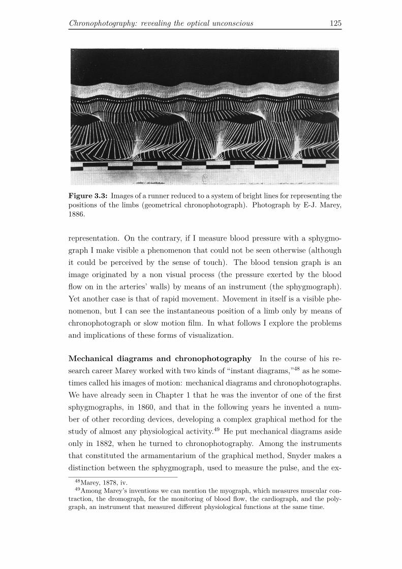
Chronophotography: revealing the optical unconscious 125
Figure 3.3: Images of a runner reduced to a system of bright lines for representing thepositions of the limbs (geometrical chronophotograph). Photograph by E-J. Marey,1886.
representation. On the contrary, if I measure blood pressure with a sphygmo-graph I make visible a phenomenon that could not be seen otherwise (althoughit could be perceived by the sense of touch). The blood tension graph is animage originated by a non visual process (the pressure exerted by the bloodflow on in the arteries’ walls) by means of an instrument (the sphygmograph).Yet another case is that of rapid movement. Movement in itself is a visible phe-nomenon, but I can see the instantaneous position of a limb only by means ofchronophotograph or slow motion film. In what follows I explore the problemsand implications of these forms of visualization.
Mechanical diagrams and chronophotography In the course of his re-search career Marey worked with two kinds of “instant diagrams,”48 as he some-times called his images of motion: mechanical diagrams and chronophotographs.We have already seen in Chapter 1 that he was the inventor of one of the firstsphygmographs, in 1860, and that in the following years he invented a num-ber of other recording devices, developing a complex graphical method for thestudy of almost any physiological activity.49 He put mechanical diagrams asideonly in 1882, when he turned to chronophotography. Among the instrumentsthat constituted the armamentarium of the graphical method, Snyder makes adistinction between the sphygmograph, used to measure the pulse, and the ex-
48Marey, 1878, iv.49Among Marey’s inventions we can mention the myograph, which measures muscular con-
traction, the dromograph, for the monitoring of blood flow, the cardiograph, and the poly-graph, an instrument that measured different physiological functions at the same time.
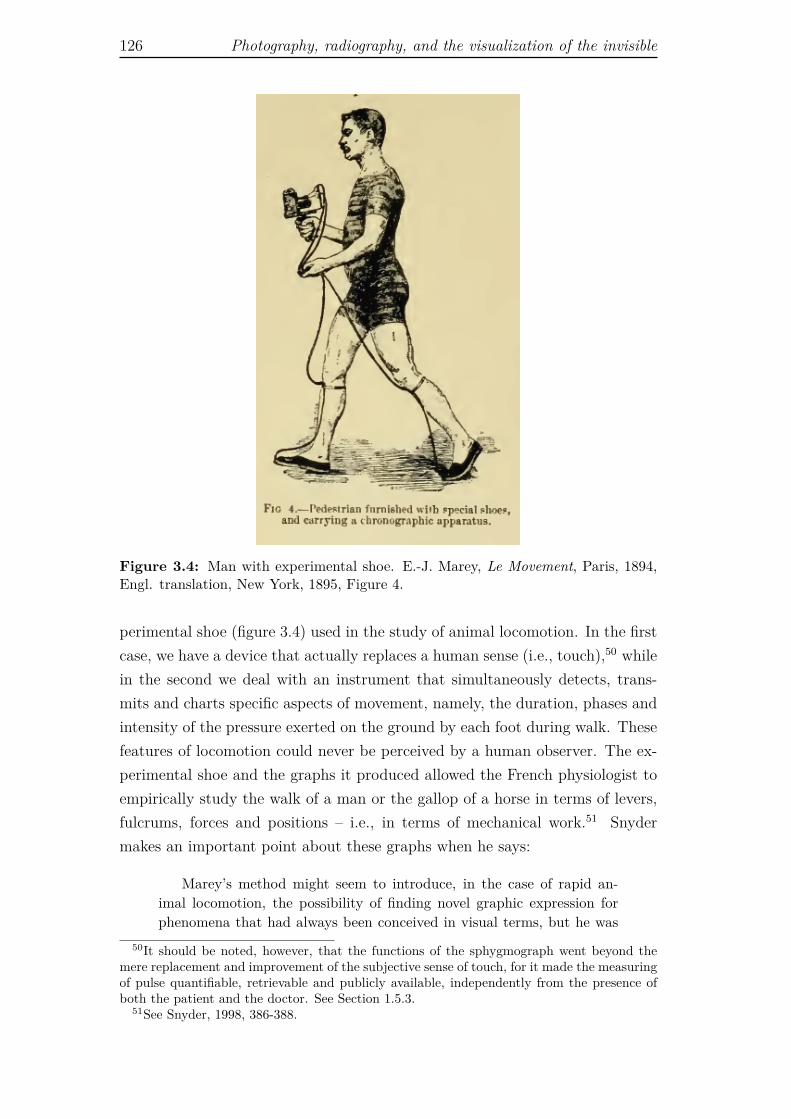
126 Photography, radiography, and the visualization of the invisible
Figure 3.4: Man with experimental shoe. E.-J. Marey, Le Movement, Paris, 1894,Engl. translation, New York, 1895, Figure 4.
perimental shoe (figure 3.4) used in the study of animal locomotion. In the firstcase, we have a device that actually replaces a human sense (i.e., touch),50 whilein the second we deal with an instrument that simultaneously detects, trans-mits and charts specific aspects of movement, namely, the duration, phases andintensity of the pressure exerted on the ground by each foot during walk. Thesefeatures of locomotion could never be perceived by a human observer. The ex-perimental shoe and the graphs it produced allowed the French physiologist toempirically study the walk of a man or the gallop of a horse in terms of levers,fulcrums, forces and positions – i.e., in terms of mechanical work.51 Snydermakes an important point about these graphs when he says:
Marey’s method might seem to introduce, in the case of rapid an-imal locomotion, the possibility of finding novel graphic expression forphenomena that had always been conceived in visual terms, but he was
50It should be noted, however, that the functions of the sphygmograph went beyond themere replacement and improvement of the subjective sense of touch, for it made the measuringof pulse quantifiable, retrievable and publicly available, independently from the presence ofboth the patient and the doctor. See Section 1.5.3.
51See Snyder, 1998, 386-388.
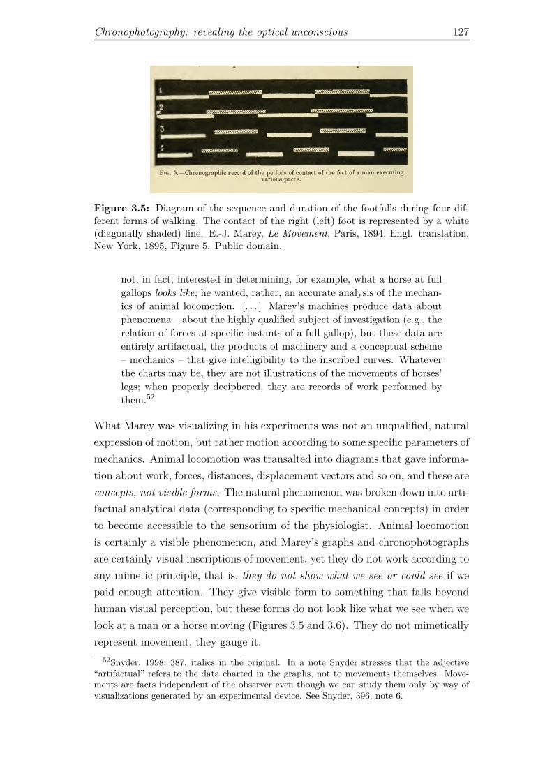
Chronophotography: revealing the optical unconscious 127
Figure 3.5: Diagram of the sequence and duration of the footfalls during four dif-ferent forms of walking. The contact of the right (left) foot is represented by a white(diagonally shaded) line. E.-J. Marey, Le Movement, Paris, 1894, Engl. translation,New York, 1895, Figure 5. Public domain.
not, in fact, interested in determining, for example, what a horse at fullgallops looks like; he wanted, rather, an accurate analysis of the mechan-ics of animal locomotion. [. . . ] Marey’s machines produce data aboutphenomena – about the highly qualified subject of investigation (e.g., therelation of forces at specific instants of a full gallop), but these data areentirely artifactual, the products of machinery and a conceptual scheme– mechanics – that give intelligibility to the inscribed curves. Whateverthe charts may be, they are not illustrations of the movements of horses’legs; when properly deciphered, they are records of work performed bythem.52
What Marey was visualizing in his experiments was not an unqualified, naturalexpression of motion, but rather motion according to some specific parameters ofmechanics. Animal locomotion was transalted into diagrams that gave informa-tion about work, forces, distances, displacement vectors and so on, and these areconcepts, not visible forms. The natural phenomenon was broken down into arti-factual analytical data (corresponding to specific mechanical concepts) in orderto become accessible to the sensorium of the physiologist. Animal locomotionis certainly a visible phenomenon, and Marey’s graphs and chronophotographsare certainly visual inscriptions of movement, yet they do not work according toany mimetic principle, that is, they do not show what we see or could see if wepaid enough attention. They give visible form to something that falls beyondhuman visual perception, but these forms do not look like what we see when welook at a man or a horse moving (Figures 3.5 and 3.6). They do not mimeticallyrepresent movement, they gauge it.
52Snyder, 1998, 387, italics in the original. In a note Snyder stresses that the adjective“artifactual” refers to the data charted in the graphs, not to movements themselves. Move-ments are facts independent of the observer even though we can study them only by way ofvisualizations generated by an experimental device. See Snyder, 396, note 6.
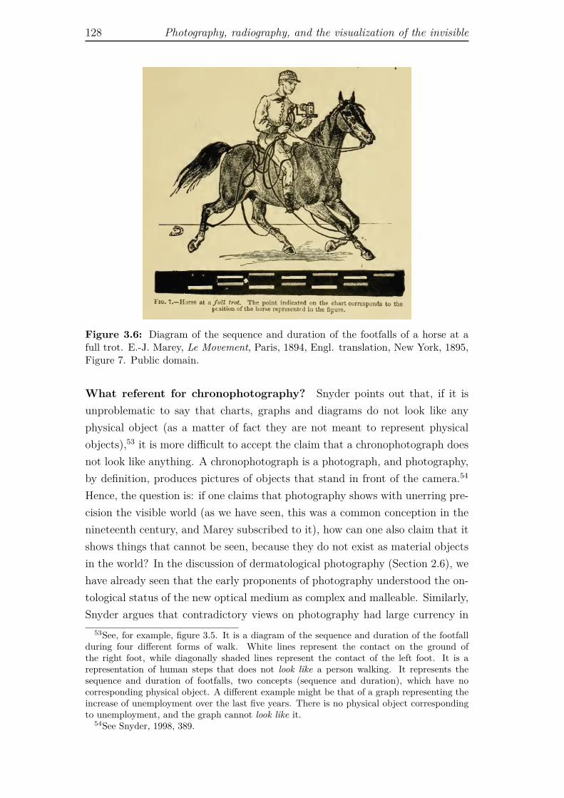
128 Photography, radiography, and the visualization of the invisible
Figure 3.6: Diagram of the sequence and duration of the footfalls of a horse at afull trot. E.-J. Marey, Le Movement, Paris, 1894, Engl. translation, New York, 1895,Figure 7. Public domain.
What referent for chronophotography? Snyder points out that, if it isunproblematic to say that charts, graphs and diagrams do not look like anyphysical object (as a matter of fact they are not meant to represent physicalobjects),53 it is more difficult to accept the claim that a chronophotograph doesnot look like anything. A chronophotograph is a photograph, and photography,by definition, produces pictures of objects that stand in front of the camera.54
Hence, the question is: if one claims that photography shows with unerring pre-cision the visible world (as we have seen, this was a common conception in thenineteenth century, and Marey subscribed to it), how can one also claim that itshows things that cannot be seen, because they do not exist as material objectsin the world? In the discussion of dermatological photography (Section 2.6), wehave already seen that the early proponents of photography understood the on-tological status of the new optical medium as complex and malleable. Similarly,Snyder argues that contradictory views on photography had large currency in
53See, for example, figure 3.5. It is a diagram of the sequence and duration of the footfallduring four different forms of walk. White lines represent the contact on the ground ofthe right foot, while diagonally shaded lines represent the contact of the left foot. It is arepresentation of human steps that does not look like a person walking. It represents thesequence and duration of footfalls, two concepts (sequence and duration), which have nocorresponding physical object. A different example might be that of a graph representing theincrease of unemployment over the last five years. There is no physical object correspondingto unemployment, and the graph cannot look like it.
54See Snyder, 1998, 389.
Chronophotography: revealing the optical unconscious 129
the nineteenth century, and he considers Marey an “ideal commentator”55 ex-actly because the French physiologist held nearly all the conflicting positionson photography maintained by his contemporaries.56 In one page of his bookMovement, of 1894, referring to the problem of perspective, Marey could claimthat photography reproduces “the appearance of natural objects, as seen bylooking with one eye only,” and a few pages later, discussing the chronopho-tographs of a rotating brass band (Figure 3.7) he could state: “In reality weare dealing with a hypothetical figure, which finds no counterpart in Nature.”57
How to reconcile these seemingly contradictory positions? Part of the answersuggested by Snyder impinges on the technical evolution of the photographicmedium. Early photography, he argues, with its long times of exposure helpedto corroborate the cognitive primacy of vision: the image on the photographicplate displayed what a bystander or the photographer would have seen frombehind the camera at the time of exposure, “if only he or she had carefullyattended to the scene.”58 Until the 1880s, when instantaneous photography be-came widely available, every photograph was a still life, and it was perceivedas a mirror reflecting a static superficial reality. The sensible plate would fixwhat the eye could see. With chronophotography the reciprocal corroborationof photography and human vision no longer applied. In Snyder’s words:
Such pictures work against the conception of photographs as repro-ducing the appearance of objects. And so here we enter another newdomain of mechanical sensibility, which permits us to see, though only inpictorial form, what happens in front of us – before our eyes. [...] [Withchronophotography] the observer is left wobbling between what is visibleto the naked eye and photographically depictable and what is unseeableby the eye but nonetheless reproducible by chronophotographic means.59
Chronophotographs confront the viewer with the paradox of a photograph thatshows something that is right in front of him, and yet remains invisible. Thisparadox belongs to the domain of the optical unconscious theorized by Benjaminand exemplified by the famous pictures of galloping horses. For ages equestrianpainters had pictured “flying gallop” with the horse completely detached fromthe ground when its four legs are extended. And this is indeed what we believe wesee when we look at a galloping horse. In 1878, however, Eadweard Muybridgepublished the photography sequence Silhouettes of Horses in Motion, whichrevealed that, during gallop, horses are detached from the ground when the
55Snyder, 1998, 390.56See Snyder, 1998, 390.57Marey, 1894, 19 and 31, quoted in Snyder, 1998, 391.58Snyder, 1998, 391.59Snyder, 1998, 393.
130 Photography, radiography, and the visualization of the invisible
four legs are bent. Moving horses are certainly visible objects, and yet thereare aspects of their motion that deceive our eyes. Imaging machines endow uswith “mechanical sensibility,”60 that is, they allow us to explore objects andphenomena that fall beyond the reach of unaided human senses.
3.3.2 Mechanical sensibility
By conceptualizing photography in terms of mechanical sensibility we adopta perspective that is diametrically opposed to Daston and Galison’s idea ofmechanical objectivity as suppression of subjectivity. Within the frameworkof mechanical objectivity, the image produced by the machine is a mechanicalcopy of the visible word, and the scientist is the passive recipient of such image.On the contrary, if we think in terms of mechanical sensibility, the imagingapparatus acquires creative properties. It produces images that can reveal theinvisible, rather than merely reproduce what is already visible, and it engagesthe scientists in a productive relation. In fact, it is the scientist (or a collectiveof scientists) that designs and operates the imaging apparatus and, in turn,through such apparatus his senses are improved. With chronophotography thecamera is no longer a picture-machine that produces likenesses, it becomes aninstrument wedded into an experimental setting and a theoretical structure,that gauges the world and its phenomena going beyond the experience providedby unaided senses.
Yet, I think that it is important to note that this paradox of a photographthat shows the invisible was already implicit in the endeavors of all the medicalphotographers discussed in the previous chapter. All of them, from differentperspectives and with different photographic strategies, used the superficial eyeof the camera to disentangle a range of non visual phenomena. In the contextof clinical medicine, Duchenne looked for the laws of emotions, Charcot triedto track down the neurological underpinning of hysteria, and Diamond claimedthat in the search for the causes of mental illnesses, photography would enablethe metaphysician “to witness and trace out the connection between the visibleand the invisible.”61 Even in dermatology, the domain of the surface of thebody and therefore of total visibility, photographs deeply influenced nosology,by taking permanent track of details that went usually unnoticed.
Microphotography, by coupling the camera to the microscope, entered therealm of the extremely small, but with the work of Marey’s, photography de-parted from the tradition of photographic realism in an unprecedented way. He
60Snyder, 1998, 393.61Diamond, 1856, 20.
Chronophotography: revealing the optical unconscious 131
claimed: “Although chronophotography represents the successive attitudes ofa moving object, it affords a very different picture from what is actually seenby the eye when looking at the object itself,” and added: “[these] images ap-peal rather to the imagination than to the senses.”62 As seen above, Marey wasnot bothered by the brisk divide between the idea of photography as producerof perfect visual correspondences with what we can see, and photography asa device that produces imaginary pictures. According to Snyder, this lack ofproblematization was a sign of the little theorization undergone by photogra-phy in the nineteenth century. This may well be the case, but I think thatthe contradiction disappears if we recall Peirce’s idea that the iconic status ofphotographs is predicated on their indexicality.
Symbolic indices and prosthetic senses If a photograph is an icon byvirtue of its being an index (in Peirce’s definition a photograph is physicallyforced to correspond point by point to nature),63 there is nothing that preventsus from accepting that it can correspond to nature in points that are not di-rectly visible to the human eye. To put it differently, if the epistemic authorityof photography lies primarily in its indexical relation with nature, then resem-blance with the represented object becomes an ancillary feature. Consequently,it is not contradictory to take photography as a medium that shows the appear-ance of things (icon), and that can also show phenomena that fall beyond thescope of natural vision. While in the former case the photograph is a sign thatworks as index and icon, in the latter it can work as index and symbol. In thecase of microphotography, I inscribed the symbolic mode of signification of theimage within the operational conventions of laboratory practice. In the case ofchronophotography, the symbolic dimension of the picture depends chiefly upona specific theoretical framework that conceives of continuous movement as thesum of instantaneous components, and thus allows to study motion through thevisual fragments of a continuum. In the geometrical chronophotograph shownin Figure 3.3 we see an index of the position of moving limbs, but the imagethat we see is not an icon, because it does not look like a man running, at leastnot according to a criterion of similarity with what we can see with the nakedeye. Yet, for Marey it was a valuable tool for studying human locomotion ac-cording to the principles of mechanics.64 Another example is provided by the
62Marey, 1894, 304, quoted in Snyder, 1998, 391.63See Peirce, CP 2.281. See also Section 2.2.64Marey subscribed to positivist philosophy and was a firm opponent of biological vitalism.
He nevertheless criticized traditional mechanistic accounts of life, maintaining that they hadfailed because they used as models machines such as pumps, levers, and ropes, that do notrely on an integrated engine as source of motive force. On the contrary, modern machines,
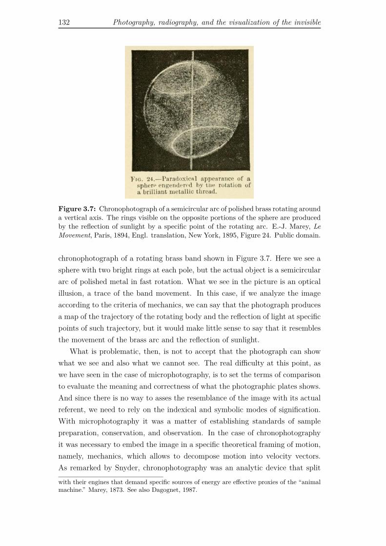
132 Photography, radiography, and the visualization of the invisible
Figure 3.7: Chronophotograph of a semicircular arc of polished brass rotating arounda vertical axis. The rings visible on the opposite portions of the sphere are producedby the reflection of sunlight by a specific point of the rotating arc. E.-J. Marey, LeMovement, Paris, 1894, Engl. translation, New York, 1895, Figure 24. Public domain.
chronophotograph of a rotating brass band shown in Figure 3.7. Here we see asphere with two bright rings at each pole, but the actual object is a semicirculararc of polished metal in fast rotation. What we see in the picture is an opticalillusion, a trace of the band movement. In this case, if we analyze the imageaccording to the criteria of mechanics, we can say that the photograph producesa map of the trajectory of the rotating body and the reflection of light at specificpoints of such trajectory, but it would make little sense to say that it resemblesthe movement of the brass arc and the reflection of sunlight.
What is problematic, then, is not to accept that the photograph can showwhat we see and also what we cannot see. The real difficulty at this point, aswe have seen in the case of microphotography, is to set the terms of comparisonto evaluate the meaning and correctness of what the photographic plates shows.And since there is no way to asses the resemblance of the image with its actualreferent, we need to rely on the indexical and symbolic modes of signification.With microphotography it was a matter of establishing standards of samplepreparation, conservation, and observation. In the case of chronophotographyit was necessary to embed the image in a specific theoretical framing of motion,namely, mechanics, which allows to decompose motion into velocity vectors.As remarked by Snyder, chronophotography was an analytic device that split
with their engines that demand specific sources of energy are effective proxies of the “animalmachine.” Marey, 1873. See also Dagognet, 1987.
X-rays and the transparent body 133
movement in its instantaneous components, and reduced the body to a systemof fulcrums and levers. In this sense, it required of the observer an effort ofimagination to recompose movement as a synthetic continuous process.65 Moregenerally, it assumed a way of looking that was different from natural vision,which relies on the iconic mode of signification, and required instead a way oflooking that had to be learned (symbolic mode).
To make his point Snyder brings to our attention a comment of Marey’son Muybridge’s equestrian chronophotographs. Marey wrote that although thepositions of gallop revealed by the American photographer at first appeared un-natural, they eventually “have taught us to discover attitudes in Nature which weare unable to see for ourselves, and we begin almost to resent a slight mistake inthe [traditional] delineation of a horse in motion.”66 As Benjamin, Moholy-Nagyand the avant-garde film directors of the 1920s, Marey envisaged chronophotog-raphy as a prosthesis of natural perception, an instrument that would reconfig-ure, improve and transform our vision and, accordingly, our understanding ofthe world. Photography, according to Marey, would teach us to see in a differentway. As Benjamin put it three decades later, it would grant us access to theoptical unconscious, by making visible those aspects of the world that escapethe bare senses. The education of the eye foresaw by Marey had nothing incommon with the suppression of the subjectivity of the observer. It implied, onthe contrary, an active training of imagination and, in the last instance, a deeptransformation of our sensory apprehension of the world.
3.4 X-rays and the transparent body
When radiography was invented, at the end of 1895, it was perceived as a furtherinnovation in an already rich domain of scientific images, optical technologiesand experimental practices. In his book on the medical technologies of thenineteenth century, Reiser wrote that radiography “catapulted medicine into avisual age.”67 Yet, it should be stressed that this transformation capitalized onan already existing and extremely rich visual culture. The century had startedwith a new way of looking at the inner body, promoted by clinical anatomy.By its end doctors could visualize the human body (from the outside and fromthe inside), its functions, and its impairments in completely new ways thatranged from endoscopy to physiological tables, passing through the lenses ofthe camera and the microscope. With the invention of radiography a new form
65See Snyder, 1998, 394-396.66Marey, 1894, 395, quoted in Snyder, 1998, 391.67Reiser, 2009, 14.
134 Photography, radiography, and the visualization of the invisible
of clinical anatomy became possible. X-rays dramatically improved medicine’sdiagnostic capability and, unlike what happened with many of the previousmedical innovations, the diagnostic potential of this discovery, which in factoccurred outside the realm of medicine, was instantly realized.
Wilhelm Roentgen discovered X-rays quite serendipitously, and invented ra-diography in the process of testing and documenting his unexpected findings.Like many other physicists of his time, he was engaged in exploring the proper-ties of electrical discharges and cathode rays using Crookes’ tubes. The acceptedhistory goes that one evening, while operating a tube covered by black card-board, he noticed that some rays escaped the apparatus, leaving a faint glowon a fluorescent screen that somebody had forgotten on the bench nearby theCrookes’ tube. Surprised by this phenomenon, he replicated it several times,placing a number of objects between the tube and the photographic plates.Some of these objects, as books, blocks of wood, and rubber bands, left therays pass through and sensitize the photographic screen; some others, as leadsand other metal objects, blocked them. These properties were definitely not theproperties of cathode rays: Roentgen had found a new kind of rays, which hedubbed X-rays with the X of ignoramus.68
In the days following his discovery Roentgen took radiographs of many otherobjects, including the left hand of his wife Bertha, which became a real icon ofthe time. It should be noted that Roentgen did not actually use the term“radiograph.” He dubbed his images skiagraphs or skiagrams, which literallymeans an image formed by the shadow of an object.69 Indeed, shadows, andnot well delineated and contrasted figures, were what one could see in the X-ray photographs. The terms radiographs and radiography quickly appeared inthe literature, together with many others, such as shadowgraphs, roentographs,katographs, electrographs, fluorographs and so forth. Such variety of namesremained in use for many years, and it is symptomatic of the novelty of X-rayimages. These images were so new that they did not even have a proper name,although many referred to them as new photography. The instability of theterminology went hand in hand with the provisional nature of the knowledge thatsurrounded radiographs.70 Within the scope of physics and technology it was notclear how radiographs were produced: on the theoretical hand, physicists whostudied the composition and behavior of matter did not know exactly what X-
68See Kevles, 1997, 17-21; Reiser, 2009, 21; Friedman and Friedland, 1998, 115-132.69Roentegen, 1896.70It has been noted that as doctors grew comfortable interpreting X-ray images, they were
less prone to refer to them as shadows. By 1900 the British journal Archives of ClinicalSkiagraphy had changed its name into Archives of the Roentgen Ray. The terms skiagram andskiagraph disappeared altogether after the 1920s. See Lerner, 1992, 387.
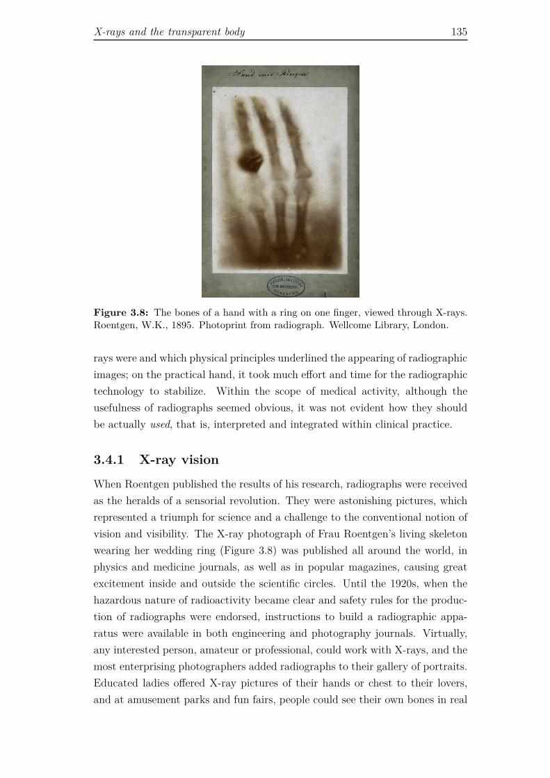
X-rays and the transparent body 135
Figure 3.8: The bones of a hand with a ring on one finger, viewed through X-rays.Roentgen, W.K., 1895. Photoprint from radiograph. Wellcome Library, London.
rays were and which physical principles underlined the appearing of radiographicimages; on the practical hand, it took much effort and time for the radiographictechnology to stabilize. Within the scope of medical activity, although theusefulness of radiographs seemed obvious, it was not evident how they shouldbe actually used, that is, interpreted and integrated within clinical practice.
3.4.1 X-ray vision
When Roentgen published the results of his research, radiographs were receivedas the heralds of a sensorial revolution. They were astonishing pictures, whichrepresented a triumph for science and a challenge to the conventional notion ofvision and visibility. The X-ray photograph of Frau Roentgen’s living skeletonwearing her wedding ring (Figure 3.8) was published all around the world, inphysics and medicine journals, as well as in popular magazines, causing greatexcitement inside and outside the scientific circles. Until the 1920s, when thehazardous nature of radioactivity became clear and safety rules for the produc-tion of radiographs were endorsed, instructions to build a radiographic appa-ratus were available in both engineering and photography journals. Virtually,any interested person, amateur or professional, could work with X-rays, and themost enterprising photographers added radiographs to their gallery of portraits.Educated ladies offered X-ray pictures of their hands or chest to their lovers,and at amusement parks and fun fairs, people could see their own bones in real
136 Photography, radiography, and the visualization of the invisible
time through a fluoroscope. As a whole, radiographs were perceived not just aspowerful medical devices, but as actual portraits.
Thomas Mann’s novel The Magic Mountain, of 1924, provides interestinginsights into the socio-cultural but also emotional impact of X-ray imaging.Dr. Hofrat Behrens, the director of the sanatorium in the Alps where thestory takes place, explained to his bewildered patients that radiography was“anatomy by the means of light,”71 but for Hans Castorp, the young hero of thenovel, radiographs were both omens of death and romantic keepsakes. Whenhe was invited to take a look at the radiograph of his cousin’s chest, Hanshad the uncomfortable feeling of being intruding into his intimacy and peeringinto his imminent death, but later on in the story, he kept a radiograph ofClavdia Chauchat as a fetish of his love for her.72 Far from being perceived onlyas objective medical records, X-ray images were endowed with several extra-scientific meanings. Indeed, as described by the historian of science Bettyann H.Kevles, radiography had a great impact on both popular and high culture in theearly twentieth century. Kevles points out that the X-rays’ ability to turn visiblewhat was hidden by the skin was quickly associated to the notion of X-ray vision,the power to peer not only through the skin, but also through clothes and walls.The idea of transparency associated to the human body triggered voyeuristicimagination, as if transparency meant nudity. Several humorous cartoons onthis theme appeared in the popular press, and some manufacturers advertisedtheir radiographic products with pictures of naked ladies standing in front ofan X-ray screen, as if they stood in front of a mirror.73 A more sophisticatedversion of the idea of X-ray vision can be found among modernist artists. Kevlesquotes from the Technical Manifesto of Futurist Painting, published in 1910:
Our growing need of truth is no longer satisfied with Form and Colors asthey have been understood hitherto. [. . . ] Who can still believe in theopacity of the bodies, since our sharpened and multiplied sensitivenesshas already penetrated the obscure manifestation of the medium? Whyshould we forget in our creations the power of our sight, capable of givingresults analogous to those of the X-rays?74
71Mann, 1924, 225.72Media scholar José van Dijck argues that The Magic Mountain can be considered an
ante litteram cultural analysis of the tremendous social impact of the new visual systeminaugurated by the X-rays. On the one hand, Mann reflects on the use of radiography in thediagnosis of tuberculosis, inducing the reader to reconsider the dichotomy between instrumentand observer, object and representation, science and art. On the other hand, he piercinglydescribes the erotic imaginary associated to X-ray imaging. See van Dijck, 2005, Ch. 5.
73See Kevles, 1998, 17-30; Cartwright, 1995, Ch. 5.74Quoted in Kevles, 1998, 130. The manifesto was signed by the Italian painters Umberto
Boccioni, Carlo Dalmazzo Carrà, Giacomo Balla, Gino Severini and Luigi Russolo.
X-rays and the transparent body 137
For avant-garde artists, radiography was a metaphor of the mathematical andscientific achievements that since the final quarter of the nineteenth centuryhad shattered the positivist view of knowledge and reality, as well as a formalmodel for expressing their novel conceptions of vision and space, matter andforce, visibility and invisibility.75 In the hand of these artists, X-rays became aformal device whereby the human body was dissolved into lines of force, trans-parent surfaces, and dissected volumes. They deployed the analytical functionsdescribed by Benjamin in relation to photography and cinema.
3.4.2 Oculis subjecta fidelibus
Roentgen himself was never interested in the medical potential of radiography.It seems that he discussed the issue only once, on occasion of a lecture given tothe Würzburg Physical-Medical Society, shortly after his invention. He reachedthe conclusion that the avail of X-rays for medicine was very limited, becausehe thought that since all the soft tissues of the body have the same density,they would leave a uniform, uninformative shade on the radiographic plate.76
On the contrary, the very first comment published in the lay press (Die Presse,Wien, January 5, 1896), showed considerable optimism about the diagnosticpotential of X-rays imaging. The journalist wrote: “we can foresee that oneday these rays will be so perfect that [...] they could be of immeasurable helpfor the diagnosis of countless diseases other than those of the bones.”77 Theenthusiasm of the Austrian reporter was shared by a handful of physicians whostarted to make radiographs as soon as the discovery went public. Roentgenpublished his preliminary report on X-rays on December 28, 1895, and withina few days the first medical radiographs appeared. At the technical level, thetransition from photography to radiography was smooth. As an imaging device,radiography was considered a special kind of photography, one that exploitedX-rays rather than visible light to create images on a sensible surface. Indeed,in the early years of radiology, photographers contributed to the developmentof the new technology as much as physicists, engineers, and physicians. Medicalphotographers were very quick at learning how to make radiographs. Londe,for example, set up a radiographic laboratory at the Salpêtrière as early asJanuary 1896, and shortly after he was appointed director of the Departmentof Radiology of the Parisian hospital.78
75See Lamata Manuel, 2010.76See Friedman and Friedland, 1998, 125-126.77Quoted in Posner, 1971, 233.78See Sicard, 1998, 211. Since so many different professionals were involved in the making of
radiographs, an important debate arose about who was entailed to make (and interpret) them.In France, for example, the issue was raised already in December 1896 by Antoine Béclère,
138 Photography, radiography, and the visualization of the invisible
In April 1896, the British Medical Journal began to publish the Archives ofClinical Skiagraphy and appointed a “special commissioner for investigation ofthe application of the new photography to medicine and surgery” (a Mr. SydneyRowland). In the first issue of the Archives Rowland wrote:
The progress of this new Art has been so rapid that, although Prof.Roengen’s discovery is only a thing of yesterday, it has already takenits place among the approved and accepted aids to diagnosis. At thefirst moment, the statement that it had been found possible to penetratethe fleshy coverings of the bones, and to photograph their substance andcontour, seemed the realization of almost an impossible scientific dream.[...] At the present time, we are in the position to obtain a visible imageof every bone and every joint in the body. [...]
Whatever may be the scientific explanation of the exact character ofthe X rays, the value of their discovery to surgeons and physicians is in-estimable. By means of the new radiation it is now possible to rendervisible certain of the interiors structures of the living body, and their pre-cise conditions of disease or of health can be objectively demonstrated, andfacts which heretofore could not be known or guessed at by a complicatedsystem of inference, are now oculis subjecta fidelibus.79
This quotation captures quite well the enthusiasm with which the medical world,or at least part of it, received the discovery of the X-rays. As Rowland put it, itwas a scientific dream that came true. It was finally possible to gain a definitivevisual access to the inner body, and thereby attain objective demonstrations ofconditions that the traditional inferential method of medicine could never re-veal. For Rowland, radiography was objective because it offered visual evidenceof something that, under natural conditions, was hidden to the human senses.His idea of objectivity was explicitly rooted in the sheer equivalence between ob-jective and visible (oculis subjecta fidelibus), therefore his trust in the objectivityof radiography depended implicitly on the fact that X-rays made available to hu-man perception something that otherwise would be strictly concealed, namely,the bones and joints of a living person.
For Rowland, objectivity was related with the very possibility of seeing some-thing and of sharing such vision, rather than with the sheer automatization ofthe process of image production. However, although his excitement was widelyshared within the medical community, the integration of radiography in routineclinical practice was neither uncontroversial nor instantaneous. The new tech-nology was almost immediately adopted in surgery, especially in the domains ofthe virologist-immunologist founder of French Radiology. Béclère, as many other physicians,argued that both radiography and radiotherapy should be controlled by medical specialists,but the conflict of competences went on until 1934, when a law established that a medicaldegree was needed to use X-rays for diagnostic and therapeutic aims. Similar measures wereadopted in all countries where hospitals had radiographic facilities.
79Rowland, 1896, 3 and 9.
X-rays and the transparent body 139
military medicine80 and dentistry, but it took a few years before it could makesome impact on internal medicine. This is not surprising if we consider that,as Roentgen had warned, soft tissues are scarcely opaque to X-rays and that,given the poor quality of the early radiographs, it was much easier to identifybullets, bones and teeth than soft organs, such as the lungs and the heart.
The difficulties in visualizing the lungs were particularly compelling, becauselungs were the most important seat of tuberculosis, one of the most commonand deadly diseases of the eighteenth and nineteenth centuries. However, med-ical historian E. Posner has shown that the attitude of the medical communityin respect to such hindrances was not uniform. In an early comment on theRoentgen rays, the British scientific journal The Electrician, reported: “withthe stethoscope it has become possible to listen to the working of the humanframe, with the new radiation it is to be hoped that the human frame willbecome – to skilled vision – something akin to transparent.”81 Thus, radio-graphy, with its ability to reveal the inner body, was immediately associatedto the stethoscope, which was the leading diagnostic technology of the time,at least for chest diseases. Commentators in the medical press, however, werequite cautious, and in the issue of the British Medical Journal that appearedon February 1, 1896 a doctor wrote that only “the uneducated imagination” oflay people could dream of immediate benefits from the X-rays in the domain oftuberculosis and thorax diseases.82 At the beginning of the 1920s some medicaltextbooks still expressed skepticism in relation to the ability of X-rays to re-veal pulmonary tuberculosis before the manifestation of symptoms and physicalsigns. As a telling example, Posner reminds that in the eight edition of ThePrinciples and Practice of Medicine, of 1920, the renowned clinician WilliamOsler (one of the founding professors of the Johns Hopkins Hospital School ofMedicine) maintained that a careful clinical examination would normally saymore than any radiograph, and disparagingly wrote: “radiographers need thesalutary lesson of the Dead House to correct their Visionary interpretation ofshadows.”83 To Osler, radiographs were mere shadows, and the only way to gaintrue knowledge of disease remained pathological anatomy.
French doctors, in general, seemed more optimistic. Already in 1897, theRevue de la Tubercolose et de Pneumologie reported on the work of L. Douchardand colleagues, at the Hôpital Charité in Paris, saying that thanks to their “belles
80Italy was the first country to use X-rays on the battlefield: in May 1896 field hospitalsin the east Africa war were equipped with X-ray units. In the following year the British sentfield-type X-ray apparatuses to the Balkans and Afghanistan. See Kevles, 1997, 39.
81The Electrician, 1896, 36:448, quoted in Posner, 1971, 233.82See Posner, 1971, 233.83Quoted in Posner, 1971, 234.
140 Photography, radiography, and the visualization of the invisible
radiographies” they had been able to detect tuberculous lesions that had beenmissed by percussion and auscultation. And although Bouchard himself thoughtthat the new method had limitations similar to those of physical examination,the author of the article was much more confident and expressed no doubt thatradioscopy would have allowed for earlier diagnosis of tuberculosis.84
3.4.3 Radiography and the challenges of internal medicine
Tuberculosis, a leading cause of death until the discovery of penicillin (1929),was since the beginning a major field of investigation for radiographers, but itwas only by the beginning of the First World War that X-ray technology wasstable enough to allow for a significant advance in chest radiology. Until then,the work of doctors and technicians had been hampered by relevant technicalshortcomings. Indeed, technical problems were a strong limiting factor. In 1949,a radiologist wrote: “For the first five years X-ray apparatus was more an in-teresting toy than a weapon of value in medicine [...] looking back, it seemsremarkable that any results could be obtained with such makeshift and unre-liable apparatus – still more remarkable the range of examinations attemptedand their comparative success.”85 As a matter of fact, especially when it cameto soft tissues, it is surprising that physicians could see something at all in earlyradiographs. Not only the image quality was very low, but most physicians,even among those actively engaged in the development of the new technology,did not always know what they were looking for when they looked at a radio-graph. A telling example of such difficulties is provided by the frontispiece ofthe book Practical Radiography, published in 1896. It proudly displayed “TheHuman Heart in situ” (so went the caption), but the heart it showed was upsidedown, and so it remained in consecutive reprints for twenty years.86
So, how do we explain the faith of the early proponents of radiography inthose blurred, distorted images? What could justify their stubbornness, theirwill to see something relevant in images that very often proved to be quite de-ceiving? To answer these questions we have to take into account two factors.One is that radiography was automatically granted the same epistemic author-ity as photography; the other is that radiography joined a field, instrumentaldiagnostics, that was in full development (as shown in Chapter 1), and col-lected the enthusiasm of the youngest and most dynamic part of the medicalcommunity. As remarked by Rowland, radiographs transformed the invisiblestructures and hypothetical impairments of the inner body into something that
84See Posner, 1971, 234.85Barclay, 1949.86See Posner, 1971, 234; Kevles, 1998, 92.
X-rays and the transparent body 141
was “oculis subjecta fidelibus.”87 For medicine, which had long been strugglingto become a full-fledged empirical science, the possibility of transforming indi-rect evidence into visible facts looked like a very reliable way to acquire secureand well founded knowledge. Additionally, we can speculate that for physicians,used to deal with ambiguous signs and symptoms, the lack of clarity in radio-graphic images did not represent a strong argument against the possibility ofgaining useful knowledge through them. This does not mean, of course, thatthere was unanimous agreement among doctors about the evidentiary power ofX-ray imaging. As mentioned above, although surgery and dentistry were veryquick at integrating radiography in daily practice, in internal medicine the pro-cess was longer and much more complex. Those clinicians who engaged in thediagnosis of diseases with undefined morphology, or characterized by functionalrather than morphological features were often skeptical about the diagnosticrelevance of radiography.
In internal medicine, similarly to what had previously happened with mi-crophotography and chronophotography, the corroboration of the content of theimage was a complex issue, because it was necessary to endow with meaningimages whose iconic dimension was problematic. It goes without saying thatit was relatively easy to verify the similarity between an X-ray image and abroken rib, but in which sense could one say that a shadow in the radiographicplate looked like a tubercular lesion or an aortic aneurysm? For radiographstoo, a number of cognitive strategies ranging from technology stabilization andstandardization to the development of interpretive ability had to be put in placeto produce clinical meaning.
3.4.4 On the interpretation of X-ray images
The visual appearance of radiographs gave an immediate impression of trans-parency, and yet one had to learn to see before one could see something mean-ingful in these images. In 1903, J.F. Halls Dally, one of the British pioneers ofclinical radioscopy, wrote in The Lancet:
A good radiograph in some respects may be said to resemble a paintingby Turner. Without intuition or previous study the one is almost asincomprehensible as the other, but as we gaze the wealth of detail risesbefore our vision, until finally we are able to interpret the meaning ofstreaks and shadows that to the untrained eyes are meaningless.88
In 1905, C.L. Leonard, an American surgeon who used radiography to improvethe differential diagnosis of ureteral calculus argued:
87Rowland 1896, 9. See quotation above.88Halls Dally, 1903, 1806.
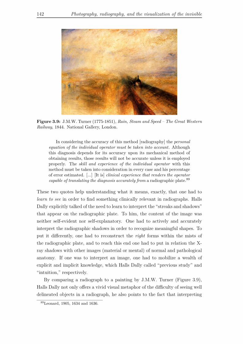
142 Photography, radiography, and the visualization of the invisible
Figure 3.9: J.M.W. Turner (1775-1851), Rain, Steam and Speed – The Great WesternRailway, 1844. National Gallery, London.
In considering the accuracy of this method [radiography] the personalequation of the individual operator must be taken into account. Althoughthis diagnosis depends for its accuracy upon its mechanical method ofobtaining results, those results will not be accurate unless it is employedproperly. The skill and experience of the individual operator with thismethod must be taken into consideration in every case and his percentageof error estimated. [...] [It is] clinical experience that renders the operatorcapable of translating the diagnosis accurately from a radiographic plate.89
These two quotes help understanding what it means, exactly, that one had tolearn to see in order to find something clinically relevant in radiographs. HallsDally explicitly talked of the need to learn to interpret the “streaks and shadows”that appear on the radiographic plate. To him, the content of the image wasneither self-evident nor self-explanatory. One had to actively and accuratelyinterpret the radiographic shadows in order to recognize meaningful shapes. Toput it differently, one had to reconstruct the right forms within the mists ofthe radiographic plate, and to reach this end one had to put in relation the X-ray shadows with other images (material or mental) of normal and pathologicalanatomy. If one was to interpret an image, one had to mobilize a wealth ofexplicit and implicit knowledge, which Halls Dally called “previous study” and“intuition,” respectively.
By comparing a radiograph to a painting by J.M.W. Turner (Figure 3.9),Halls Dally not only offers a vivid visual metaphor of the difficulty of seeing welldelineated objects in a radiograph, he also points to the fact that interpreting
89Leonard, 1905, 1634 and 1636.
X-rays and the transparent body 143
is by necessity a creative activity, open to qualitative nuances.90 This entailsthat as far as the observer becomes more experienced he or she can learn tosee more details. At the same time, it implies that different observers mightpay attention to different elements. In this case, their interpretations will differwithout this necessarily entailing one of them to be wrong. Another possibilityis that different beholders can understand the same visual elements in distinctways, while the same observer could revise his or her interpretation over time.Halls Dally had no doubts about the possibility to establish a correct, stableinterpretation of a radiographic image. In fact, he went as far as to claim that:“Although observers may differ widely as to the interpretation of evidence basedon tactile and auditory impressions, ocular evidence in most cases admits of nodifference of opinion.”91 In this, he partook in Rowland’s assurance about theobjective nature of vision. Yet, he was aware that vision was inextricably linkedto interpretation, and that the process that led to the proper understandingof the streaks and shadows on the radiographic plate was not an automatic,merely optical operation. He knew that without training pattern recognitionand clinical judgment one could see nothing but elusive shapes.
The skillful operator Leonard, too, was conscious of the fact that the mean-ing of radiographs was neither explicit nor transparent, and he resorted to themetaphor of translation to warn the reader that the making of a diagnosis froman X-ray plate was not a passive task. One does not read the diagnosis straightinto the image, one has to carry on an accurate translation (which is alwaysan interpretative process), in order to transform what is seen in the radiographinto knowledge of what is happening within the patient’s body. But, how doesone learn to perform such translations? For Leonard, as for Halls Dally, one
90J.M.W. Turner (1775-1851) was an English Romantic painter whose style is regardedas an anticipation of Impressionism. He was known by his contemporaries as “the painter oflight.” The artist and curator Lawrence Gowing has described Turner’s work as “the indefinitetransmission and dispersal of light by an infinite series of reflections from an endless varietyof surfaces and materials, each contributing to its own color that mingles with every other,penetrating ultimately to every recess, reflected everywhere.” Gowing, 1966, 21.
91Halls Dally, 1903, 1803. Halls Dally optimism about differences of opinion concerningocular evidences was somewhat unjustified. Already in the 1920s doctors started discussingthe problem of errors in the interpretation of radiographs, and a radiologist wrote that onesource of error was the “thoughtless belief in the infallibility of the roentgen ray by the medicalprofession as a whole.” Musser, 1923, 252, quoted in Reiser, 1978, 189. And in 1947, the firstsystematic study on the magnitude of variation in the interpretation of X-ray films showedthat in about one-third of cases different doctors differed with their interpretations, and eventhe same doctor watching the same radiographs at different times differed in his interpretationabout a fifth of the time. See Reiser, 1978, 189-190. The advancement of technology has notsolved the problem, as shown by the rich literature that deals with the problem of error inradiography. See, for example, Sabih, et al., 2011; Alexander, 2010; Kundel, 2006; Griscom,2002.
144 Photography, radiography, and the visualization of the invisible
needed clinical experience, with its combination of practical skills and theoreti-cal knowledge. Still, if one is to rely on radiographs to make a medical diagnosis,to formulate the right interpretation or to establish the right translation is notenough. In the first place one has to produce the right image. Halls Dally im-plicitly hints at this problem, because he talks of a “good radiograph,” but it isin the quote from Leonard that we find an explicit reference the process of imageproduction. In fact, he insists on that the accuracy of the radiographic methoddepends chiefly on the “skill and experience of the individual operator,”92 be-cause the mechanical system of image production can be accurate only as faras it is “employed properly.”93 As the image has to be interpreted in order toacquire meaning, so the mechanical apparatus has to be skillfully operated inorder to produce a trustworthy image. This was particularly true at a time inwhich the technology was still under development, and doctors and techniciansworked by trial and error, dealing with the unpredictable behavior of new in-struments and processes. As Posner put it, the early history of radiography was“a story of erratic electric supplies, nitrous fume-producing cell batteries, inef-fective electrolytic rectifiers, and temperamental gas tubes.”94 The technologyto produce radiographs was as novel as the images themselves, and as such itrequired the same ingenuity and critical judgment. The apparatus was mechan-ical, but it was not a self-operating agent. Images did not emerge passively frommachines, they had to be actively produced, and they had to be produced inthe right way.
3.4.5 Mechanical images and trained judgment
Halls Dally’s and Leonard’s clear awareness of the need to use clinical experienceand critical judgment both in the production and interpretation of radiographsis at odds not only with Daston and Galison’s idea of mechanical objectivity,but also with their claim that another epistemic virtue, namely trained judg-ment, emerged after mechanical objectivity and as a backlash against it.95 Asexplained in the introductory section of this chapter, according to Daston andGalison, mechanical objectivity was the overarching ideal that regulated themaking and use of scientific images between the second half of the nineteenthand the first decades of the twentieth centuries. It prescribed to suppress theintervention of the scientist in the process of image production and reproduc-
92Leonard, 1905, 1634. It should be noted that when Leonard talks of an “operator” hemeans the physician himself, who took the radiographs with the help of an assistant technician.
93Leonard, 1905, 1634.94Posner, 1971, 238.95See Daston and Galison, 2007, 18 and 377.
X-rays and the transparent body 145
tion, since the ultimate goal of the scientific endeavor was to represent the exactappearance of the object as recorded by the mechanical apparatus. Any hu-man interference with the photographic camera, the radiographic machine, orany other recording device had to be minimized, so that the subjectivity ofthe observer-operator would not affect the direct imprint of nature on a sheetof paper or any other sensible support. Mechanical objectivity also imposedrigorous self-restraint in the interpretation of images. Ideally, Daston and Gal-ison say, images had not to be interpreted at all, and in those cases in whichinterpretation was unavoidable, it should be performed according to rigid andstandardized procedures.96
According to Daston and Galison’s chronological taxonomy of epistemic ide-als, by the third decade of the twentieth century, partly in reaction to me-chanical objectivity, the principle that governed science became one of trainedjudgment. To use their own words: “[By 1920s] the edict of mechanical ob-jectivity to abstain from all interpretation turned out to be sterile.”97 Unlikemechanical objectivity, trained judgment prescribed to produce, reproduce, andinterpret images by resorting to the tacit knowledge acquired through practi-cal training. Interpreted images, the form of representation corresponding totrained judgement, were still produced by mechanical devices and respectinggood laboratory practices, but subjective interpretation, fed by accurate train-ing, was no longer seen as standing in stark opposition with scientific rigor andobjectivity.98 Comparing mechanical objectivity and trained judgment, Das-ton and Galison maintain that for the scientist who subscribed to the ideal oftrained judgment, judgment was “an act of cultivated perception and cognitionwas associated with a picture of reading that was both anti-algorithmic andantimechanistic.”99 And they specify:
Scientific image judgment had to be acquired through a sophisticatedapprenticeship, but it was a labor of a very different sort from the re-hearsed moves of the nineteenth-century mechanical objectivist. Inter-preted images got their force not from the labor behind automation, self-registration, or absolute self-restraint, but from the expert training of theeye, which drew on a historically specific way of seeing. Scientific sighthad become an ‘empirical art.’100
Now, the articles from Leonard and Halls Dally clearly show that even at thetime of alleged mechanical objectivity, clinicians who worked with radiographs
96See Daston and Galison, 2007, 315, 321, 328, 344.97Daston and Galison, 2007, 344.98See Daston and Galison, 2007, 314.99Daston and Galison, 2007, 331.
100Daston and Galison, 2007, 331.
146 Photography, radiography, and the visualization of the invisible
were clearly aware of the many variables the operators had to take into consid-eration when making a radiograph, of the ambiguity of the shadows they had tomake sense of, and of the complexity of the conditions they wanted to diagnose.This is to say that they did not believe that radiographs were imprints of realitysic et simpliciter. Even when they praised radiography as being more objectivethan other diagnostic instruments, Halls Dally and Leonard did not claim thatobjectivity was the suppression of the judgment either of the observing subject,or of the image maker. They did not recommend the blind application of proce-dures in the process of image production, quite the contrary, and they did nottake objectivity for self-evidence. They explicitly acknowledged that the skilland experience of the operator were paramount, and invited their colleagues tolearn to interpret radiographs, to translate images into diagnosis on the basisof study, educated intuition, and clinical experience. Importantly, they werenot innovators in this respect. Their predecessors, the pioneers of photography,had followed the same approach. As pointed out in Chapter 2, Duchenne deBoulogne became a photographer himself because he was convinced that pho-tography would let nature speak for herself only if the image maker was wellacquainted with the phenomenon that was to be recorded,101 and the wholepoint of all medical photographic publications of the nineteenth century was totrain the eye of the observer, in order to improve his physiognomic sight and hisclinical judgment.102 Contrary to Daston and Galison’s claims, scientific sighthas always been, by necessity, an empirical art, and possibly no one was moreconscious of this than physicians, whose commitment to intuitive medical judg-ment not only long predated the twentieth century, but was also constitutive ofthe very authority of medical profession.103
Legitimization of scientific photography in the nineteenth century Itis true, as Daston and Galison stress, that in the introductions to photographicatlases the nineteenth-century authors reiterated over and over again that theyhad let nature imprint itself, without intervening on the photographic plate.However, it should be remarked that they also praised the skill of the photog-rapher. Hardy, for example, eulogized de Montméja’s “indisputable talent asphotographer and colorist,”104 and Donné emphasized that the results attained
101See Section 2.7.102In this same line of criticism, in a review of Daston and Galison’s book Theodor Porter has
pointed out that: “If all the confusing details of a real photograph are preserved [complyingwith the edict of mechanical objectivity] – if seasoned judgment in its preparation is disbarred– the need for expert discernment among those who consult the images becomes all the morepressing.” Porter, 2008, 644.
103For a criticism to Daston and Galison along similar lines see Porter, 2008.104Hardy, 1868, n.p.
X-rays and the transparent body 147
by his photographer Léon Foucault were “remarkable.”105 In an essay on therole of photography in science, published in 1882 in the journal PhotographicNews, the anonymous author not only highlighted the relevance of technologyand of the photographer’s technical skill, he also stated that the making of ascientific photograph inevitably involved aesthetic choices, judgments and inter-ventions.106 Moreover, one should not forget that, as remarked by the historianof photography Jennifer Tucker, the expression “judging eye” was coined byVictorian photographers to refer to the great skill and different kinds of knowl-edge that one had to develop in order to make good photographs.107 All thesedocumental evidences contradict Daston and Galison’s claim that the work ofscientists in the late-nineteenth and early-twentieth centuries was governed bythe ambition to attain mechanical objectivity, and that only during the twenti-eth century “the espousal, celebration, and cultivation of trained judgment”108
imposed itself as a new regulative ideal that supplemented objectivity.Objectivity was never purely mechanical, neither in the practice nor in the
discourses of scientists. In her book Nature Exposed. Photography as Eyewit-ness in Victorian Science, of 2005, Tucker has convincingly disputed the strongassumption, common among historians, that in the nineteenth century the au-thority of photography was unchallenged. By examining scientific, technical andpopular nineteenth-century literature on scientific photography, she providesplenty of evidence that concepts of skill (both visual and manual), judgment,and human agency informed what counted as objective and subjective duringthe Victorian era. Referring explicitly to Daston and Galison’s article of 1992,Tucker argues that: “Although nineteenth-century faith in photography waspowerful, the idea that people over a hundred years ago accepted photographsat face value is exaggerated and misleading.”109 Gathering testimonies from dif-ferent sources (scientific journals and congress reports, but also popular lecturesand magazines), Tucker demonstrates that Victorians – both scientists and thegeneral public – were keenly aware of the manufactured nature of photographs.Most of them also knew that the production of mechanical images required in-tense skilled labor. Indeed, in the process of professionalization of scientificphotography, those who promoted this new medium as a scientific tool stressedthe importance of qualified knowledge that had to be coupled with artful craft.How to make proper data with a scientific instrument, Tucker remarks, was atopic of great concern at the time photography emerged.
105Donné, 1844, 36.106See Tucker, 2005, 259.107See Tucker, 2005, 60.108Daston and Galison, 2007, 321.109Tucker, 2005, 4.
148 Photography, radiography, and the visualization of the invisible
A paradigmatic case is provided by spirit photography, because it provedhow misleading the unconditional association of mechanical representation toobjective and scientific evidence could be. Defendants of spiritualism, in fact,tried to draw on the claims about the mechanical objectivity of photography toreinforce their contention that spirit photographs were direct proofs of an oth-erwise unseen reality. Thus, in order to reject the belief that it was possible tophotograph spirits and ghosts, scientists were forced to explicitly argue that me-chanical objectivity was not ipso facto scientific and trustworthy.110 “Through-out the nineteenth century,” Tucker notes “people debated truth claims based onphotographs and, in the process, established the criteria by which a photographcould be accepted as scientific evidence. These debates erupted in laboratories,observatories, and scientific meetings as well as at world’s fairs and in court-rooms, illustrated periodicals, and spiritualist séances.”111 Indeed, as shown inChapter 2 and in the discussion of Marey’s ideas about photography in Section3.3, people in the nineteenth century held quite contradictory views about me-chanically produced images. They maintained that photography was the pencilof nature and that one had to be a skilled photographer to produce picturesthat looked like the original object; they were ready to say that photographyfaithfully reproduced the appearance of objects and that it created hypotheticalimages. This ambiguity, however, is less a testimony of a lack of sophisticationin the understandinhg of photography, than a symptom of the complex natureof photography itself.
Recognizing the unknown Another important aspect overlooked by Das-ton and Galison is that early photographers and early radiographers, as well asthose who developed other technologies for visualization, such as cloud cham-bers or stellar spectrographs, were creating images that no one had seen before.They were literally creating new phenomena and, accordingly, they had to learnto see anew. Now, how could they produce anything new, and learn to seeanything at all, if they had restrained themselves from applying judgment andinterpretation? How could they have invented and perfected a panoply of imag-ing technologies, if they had been afraid to intervene? How could they haveventured in the wide range of examinations they performed with erratic appa-ratuses and blurred images, if they had been the shy, will-abnegating workersafraid to interfere with machines and images described by Daston and Gali-
110See Tucker, 2005, Ch. 2. In Ch. 3 and Ch. 4 Tucker analyses the case of photographyin meteorology and bacteriology, respectively, and in Ch. 5 she focuses on the questionof scientists’ disagreement in the interpretation of images, drawing on the specific case ofplanetary photography (Mars, in particular).
111Tucker, 2005, 6.
X-rays and the transparent body 149
son?112 They argue that the fact that under mechanical objectivity scientistsdid actually resort to intervention and judgment cannot be used as a counter-argument against the claim that mechanical objectivity was an epistemic ideal.Ideals, they rightly argue, are never fully accomplished, nevertheless they regu-late people’s behavior.113 This is quite obvious, since it directly proceeds fromthe definition of ideal. However, it seems to me that it cannot be used as anargument to refute strong historical and conceptual counter-evidences, as thoseput forward by Breidbach, Snyder and Tucker concerning photography, and bymy examples from radiography.
Some problems with Daston and Galison’s methodology In a reviewof Objectivity, the philosopher of science Martin Kusch has noted that Dastonand Galison exemplify mechanical objectivity on the basis of the analysis ofabout seventy non-geographical atlases114 (although they say that almost twothousand were published between 1830 and 1930), but they do not provide anyquantitative data about the alleged rise and fall of the various epistemic virtues,or their role in different disciplinary fields. They select and synthesize theirsources in ways that are not transparent, and “a critical reader might well feelthat she is asked to take a lot – perhaps too much – simply on trust.”115 The verychoice to use as primary sources scientific atlases and manuals is problematic.
As a matter of fact, atlases are supposed to fix knowledge and consolidatescientific practices. This is why they work as references or guides. Atlasesand manuals have, in general, an eminently pedagogical function. However,the ideas and practices involved in the production, selection and presentationof images aimed at stabilizing knowledge are not necessarily the same as theideas and practices involved in the production and handling of images aimed atgenerating new knowledge. Atlases, and particularly what scientists write in theintroductions to these atlases, are not necessarily the best route to understand-ing how images actually work in the construction of new knowledge.116 Tuckerremarks that Daston and Galison’s choice to focus on atlases, letting aside other
112Porter makes a similar criticism. He comments that it is quite surprising to see the periodbetween 1850 and 1920, the golden age of public science and of scientists’s public role, depictedas an era of mechanical objectivity. In Daston and Galison’s construal, mechanical objectivityprescribed absolute self-restrained from making assertions that could not be demonstratedimpersonally (mechanically). The regulatory ideal of mechanical objectivity, by Daston andGalison’s own admission, summed up to intellectual timidity and an ethos of self-abnegation.Porter ironically wonders: “Were Huxley, Helmholtz, and Bernard paralyzed by anxietiesregarding the validity of their own insights and interpretations?” Porter, 2008, 645.
113See, for example, Daston and Galison, 2007, 321.114Namely, atlases of anatomy, bacteriology, neurology, pathology, physics, and zoology.115Kusch, 2009, 129.116For similar criticisms see Porter, 2007 and 2008.
150 Photography, radiography, and the visualization of the invisible
scientific media, hampers from the outset the possibility to make general claimsabout what scientists really thought, and how they actually worked. She pointsout that, exactly because atlases serve as descriptive guides to idealized scien-tific identities, a sort of conduct books for scientists, they cannot help to shedlight on crucial questions about actual scientific practice, which is related lessto atlases’ production than to their reception and use. To make her point sheironically comments: “We do not really accept that nineteenth-century womenlived according to prescriptive literature.”117
The aim of this extensive critique of Daston and Galison’s notion of me-chanical objectivity was not to deny that in the second half of the nineteenth,and well into the twentieth century, scientists, artists and the lay people wereutterly fascinated by machines and automation. I do not dispute that in manycases mechanization of data recording was associated to the idea of objectivityas suppression of the subjective idiosyncrasies of a human observer or measurer.I rather wanted to stress that Daston and Galison make too strong a case formechanical objectivity, as if it were a uniform and universal dogma. We failto understand how automation and the development of measuring and imagingdevices transformed science and scientific practice during the nineteenth cen-tury, if we do not take into account that the struggle to adhere to standards ofmechanical objectivity was deeply entrenched with an equally important strug-gle for intersubjectivity, the development of a mechanical sensibility (prostheticsenses), and the acute awareness of the importance, in the empirical sciences,of skilled operators and educated interpreters.
This completes my critical remarks concerning the relationship between me-chanical objectivity and trained judgment. Now it is necessary to understandhow radiographers faced the problem of image corroboration, and through whatspecific strategies and practices they endowed radiographs with diagnostic mean-ing and relevance.
3.4.6 Making sense of shadows
As seen above, the integration of radiography in clinical medicine was a complexand relatively long process. Bernike Pasveer, a scholar in Science and Technol-ogy Studies, has convincingly argued that the fact that internal medicine wasmuch slower than surgery in making sense of radiography depends only in parton technical difficulties and image poor quality. This was just one aspect of theproblem. Equally important was the fact that X-rays did actually open a newvisual world, and traditional anatomical knowledge (normal or pathological)
117Tucker, 2008, 655.
X-rays and the transparent body 151
gained through autopsies could only partly help making sense of the shadowson an X-ray plate. Pasveer suggests that if surgery, dentistry and orthopedicswere quicker than internal medicine at taking on radiography, it was not onlybecause shrapnels, teeth and bones are more radio-opaque than the lungs. Itwas also because recognizing the anatomical look of the skeleton in a radiographrequires a less radical sensorial and cognitive transformation than connecting theanatomical image of dead body organs to the shadow of living ones.118 As sheputs it: “The X-ray images were trusted for their ability to represent reality, butin the pre-Roentgen era reality looked enormously different from the shadowsthat were now said to be mirroring the inner parts of patients.”119 One tellingexample of this transformation of what reality should (or could) look like, is thatof the stomach and intestine as represented in anatomy atlases or seen through askiagram. In 1910, in fact, the use of bismuth, an X-ray opaque substance thatcan be safely ingested, showed that the stomach, often horizontal and curved inthe cadaver, is generally vertical in the living, while the intestine, which in thedead occupies a relatively fixed position, in the living can occupy almost anyplace in the abdomen.120
As argued in Chapter 1, clinical anatomy and the localizationist paradigm itentailed was a necessary precondition for making sense of radiology, neverthe-less anatomical knowledge gained through cadavers’ dissection did not alwayssmoothly overlap with the anatomy revealed by the X-rays. As Laennec hadneeded a highly educated ear to be able to use the stethoscope, radiographersneeded a highly educated eye. One had to develop a skilled vision, as many com-mentators of the time asserted. This process of education of the eye was muchmore than sensorial training. Pasveer distinguishes four phases in the processthat turned the shadows of X-ray plates into meaningful and useful diagnosticimages: (1) Technology control and stabilization; (2) Validation of the visualcontent of radiographs by comparing them with real organs; (3) Comparisonof the information acquired through X-ray images with that provided by otherdiagnostic techniques; (4) Comparison of different X-ray images. These stepspartially overlap both in time and content (problems to be solved), but each ofthem corresponds to a specific method for shaping the content and meaning ofshadow images.
Technology stabilization The first step in the process of X-ray imagingsignification involved experimentation with technology. This phase occupied
118See Pasveer, 1989, 361.119Pasveer, 1989, 361.120See Reiser, 1978, 65.
152 Photography, radiography, and the visualization of the invisible
about twenty years since the discovery of Roentgen rays, and was meant tostabilize and standardize machines and practices. X-ray workers (physicians,physicists, engineers, and photographers) experimented with photographic ap-paratuses, materials, procedures, and X-rayed a wide range of objects, so thatthe whole system could be finally controlled, black-boxed (the radiologist wouldno longer need to know how the technology worked in order to use it) and rou-tinely operated without major problems. In other words, the aim of this phasewas to warrant the liability, reproducibility and comparability of X-ray images,distinguishing the shadows produced by the object under examination, fromthe artifacts introduced by the machine or the procedure.121 Pasveer points outthat the early articles on the use of Roentgen rays in medicine reflected theexploratory character of the work of physicians and technicians at that time.122
Before describing clinical cases and discussing the diagnostic meaning of the im-ages, they specified what tubes had been used, the amount of current, the lengthof the exposure and the respective positions of tube, photographic plate and pa-tient. At the beginning these descriptions did not have prescriptive functions,but in time they became sets of operational rules: how to use an apparatus withcertain characteristics; to position the patient in a specific way; to accustomthe eyes to darkness if one was working with fluorescent screens rather thanphotographic plates, and so on.
As already mentioned, one of the advantages of producing medical visualdata was that they provided information that could be easily retrieved, shared,and compared. The production and use of these data, however, demandedstandardization of objects and actions. It also demanded a proper language todescribe the new reality that had entered the medical domain. Patients hadto be positioned “screen to back”, “screen to chest”, “lateral oblique;” and anormal thorax would present a “well-defined median opacity” with a lateral“transradiant area.”123 Pasveer remarks that all these sentences could simplynot exist in the medical vocabulary before the discovery of X-rays. There wouldhave been no meaning attached to them.124 This was true not only for internalmedicine, but also for surgery and orthopedics. Radiographs enabled surgeonsto view, for the first time, preoperative bony fragments and the postoperativeopposition. In the long run these visual criteria replaced older methods of
121Of the more than 1,000 articles and fifty books published on X-rays in 1896, more thantwo thirds treated technical issues, while the remaining third was on clinical and surgicalsubjects. See Pasveer, 1989, 379.
122See Pasveer, 1989, 369.123Halls Dally, 1903, 1801.124See Pasveer, 1989, 365.
X-rays and the transparent body 153
physical examinations in the evaluation of fractures and of the success of theintervention.125
Image external validation The second moment described by Pasveer con-cerned the validation of the X-ray image by comparison with organs’ directobservation. During this phase, which overlapped chronologically with the tech-nological experimentation, the iconic dimension or radiographic imaging, thatis, the resemblance between shadows and organs, was secured. Radiologistschecked their interpretation of a radiographs through direct observation of theorgans during a surgical procedure or at the autopsy, in a process which clearlyfollowed the logic of Laennec’s validation of the stethoscope and, more gener-ally, of pathologic anatomy since Morgagni. Very similarly to how Laennec hadchecked the diagnosis he made by auscultation with the results of post mortemexaminations, radiologists assessed their vision by comparing what they saw inthe X-ray plate with what they saw in the actual body. It should be noted,however, that the task of radiologists was comparatively easier, because whileLaennec had to match auditive with visual perceptions, they had to connect dif-ferent visual objects, consequently it was relatively simple to find firm points ofreference to link the image to the cadaver. Additionally, while auscultation wasa solitary endeavor difficult to share at the intersubjective level (only one listenerat the time could use the stethoscope), radiographs allowed different observers towatch simultaneously the same objects, so that the process of matching organsand their X-ray shadows had an overtly collective dimension. Radiographersused the organs to cross-check the images, and the images to cross-check theirown vision. Pasveer remarks that, as anatomists and pathologists already knew,the cadaver was not always a faithful model of the living body. In the case ofthe lungs, for example, it was sometimes difficult to compare the shadow imagesof the living lungs with their anatomical appearance in the dead, because theseorgans tend to collapse when ventilation is interrupted. In this respect it shouldbe highlighted that, as in the case of the visualization of the digestive system,the comparison of X-ray images with anatomical specimens was not only valu-able in educating the eye and validating the content of radiographs, it also ledto a reassessment of previous anatomical knowledge.126
As a general rule, it was soon clear that there were systematic mismatchesbetween macroscopic anatomy and radiological images. In the X-ray imagesdenser structures would screen the soft tissues, some elements that appeared in
125See Lerner, 1992, 388.126Physiological facts were also questioned. In his article of 1903 Halls Dally contested, on
the grounds of fluoroscopic observations, the widespread belief that the diaphragm flattenedon inspiration.
154 Photography, radiography, and the visualization of the invisible
the photographic plate did not correspond to anything that could be directlyobserved in the real body and, obviously, there was the problem of collapsinga three-dimensional complex structure in a multilayered bidimensional image.Moreover, according to the relative positions of cathodic tube, photographicplate and patient’s body, some shadows came to the fore and others receded. Toovercome these problems of visualization different solutions were implemented.On the one hand, procedures for taking the radiographs were standardized,in order to ensure images’ reliability and comparability. On the other hand,to check the correspondence between organs and shadows, two methods wereadopted. One consisted of tagging organs with metal coils before taking theradiograph; the other consisted of removing in a sequence different anatomicalstructures (e.g., visceral pleura and pericardium) from the cadaver and takingsuccessive X-ray pictures, so that it became possible to recognize which shadowcorresponded to which structure.127 Furthermore, to teach visual skills andcreate a common way of seeing, radiographic atlases, manuals, and handbookwere published, so that it could be possible to systematically compare images(the first radiology atlases and handbooks were published as soon as 1896). As awhole, an articulated complex of activities was put in place in order to guide theeye of the physician in the passage from the actual body to its shadow-image,so that anatomy and radiography could check on each other.
Comparison of different diagnostic techniques In order to transform ra-diographs into diagnostic instruments relevant to clinical practice, it was alsonecessary to show that it was possible to translate the information acquired withother, well established, diagnostic tools into the shadows of the X-ray plate. Thiswas the third moment in the process of X-ray imaging signification. To illustratethis problem and the solutions that were put forward, Pasveer resorts to the ex-emplary case of the diagnosis of pulmonary tuberculosis. At the turn of thenineteenth century, this disease was diagnosed by the anamnesis of the patientand his family, by physical examination, and by bacteriological analysis. So, inorder to give meaning to the shadow images of the lungs, it was necessary tointegrate the new visual information into this pre-existing body of knowledge.But how to relate images with clinical data and levels of infiltration by bacte-ria? A common solution, Pasveer explains, was that of reporting in a sequencethe clinical case (patient’s anamnesis, symptoms, physical signs, bacteriology)and a description of the images, often accompanied by pictures. She specificallyrefers to the article of 1903 by Halls Dally mentioned above,128 in which the
127See Pasveer, 1989, 371.128See Section 3.4.3.
X-rays and the transparent body 155
clinician-radiologist made a direct comparison between the visual appearance ofa chest radiograph and the signs collected through traditional physical exams.For example, brightness in the X-ray image corresponded to hyper-resonance inpercussion, trans-radiancy to normal resonance, dense shadow to a dull sound.Physical signs already had a well established diagnostic meaning, which wastransferred to radiographs through these correspondences. That is, the diagnos-tic significance of X-ray images was built upon a symbolic mode of signification,by resorting to a conventional – although not arbitrary – system of correlationsamong different signs. I elaborate further on this point in Chapter 4, where Iput forward a semiotic analysis of Halls Dally’s article.
Image internal validation The final step of the epistemic authenticationof radiography defined by Pasveer was the comparison of different radiologi-cal images. In my view, this process should be considered an act of internalvalidation. It warranted the inherent coherence of the new visual system andsanctioned its relative autonomy from other diagnostic strategies (subjectivesymptoms and physical signs). This passage could be accomplished by capi-talizing on the amount of knowledge acquired in the earlier phases, which ata certain point of the process became implicit. That is, radiologists came toa point in which they were reasonably confident that the apparatus they usedwas reliable, that a regimen of correspondences between radiological images andnormal and pathological anatomy subsisted, and that radiological images hadproper clinical relevance. It became fundamental to learn to recognize healthyorgans, normal variations, and pathological states by simply looking at the ra-diograph. This process required the comparison of images. In this task, aswe have already seen in Chapter 2, collections of images gathered in journalsand atlases were extremely valuable tools. Pasveer points out that in medicaljournals of the first decade of the twentieth century the description of a normalchest “for the sake of comparison”129 was standard, but by 1910 the specializedreaders were expected to be already familiar with it.130 Once the boundaries be-tween normal morphological variability and overt pathological states had beensettled, normal and pathological variability did not need to be verified over andover again, it became part of the given knowledge of radiology.131
On these premises it can be said that X-ray images acquired an inherentcognitive content, reaching the status of autonomous epistemic objects. It wason the basis of this relative autonomy that radiographs became what Reiser
129Pasveer, 1989, 376.130Pasveer, 1989, 376.131See Pasveer, 1989, 374-377.
156 Photography, radiography, and the visualization of the invisible
has called “the transcendent instrument of visualization in medicine.”132 Tran-scendent because, at least virtually, a whole, self-contained system of visualknowledge about the body could be built comparing images: images of thesame person at different stages of a disease, as well as images of different pa-tients affected by the same disease (all radiographs were produced followingroughly the same protocols).
3.4.7 Radiography and the legacy of clinical anatomy
The lesson of Bichat on how to use a cadaver as a trustworthy representation ofthe living body was implicitly recovered in this crossed comparison of images.Tellingly, it was with the First World War that radiography made a major stepforward. As the hospitals for the clinicians at the beginning of the nineteenthcentury, the war provided an enormous field of experimentation and study onX-rays from both the technical and medical point of view. At the technicallevel, the need of operating the X-ray apparatus in the war field demanded thedevelopment of portable, reliable and easy to handle instruments, as well asthe training of a large number of technicians;133 at the clinical level, the wargave doctors the opportunity to submit to radiological scrutiny a very largeand relatively uniform population of young men. In doing so they did not onlytrained their eyes to recognize hidden bullets and shrapnel, they actually gainedcumulative experience in looking into the human body by means of radiologicalimages. Once again thorax examination and the diagnosis of tuberculosis was ofparticular importance. As noted by Kevles, war doctors submitted to radiolog-ical examination hundreds of thousands of recruits, in a sort of unplanned massscreening, and discovered that almost half of them suffered from some formof chest disease.134 The very idea of mass screening became possible, some-thing that would have been quite implausible with the stethoscope and othertechniques of physical examination. Radiography, in fact, allowed examiningand comparing images rather than real people, and this provided considerablepractical advantages. Pasveer argues:
The comparison of the shadow-images with each other was an activitythat violated the nineteenth-century romantic view of the uniqueness andwholeness of disease and patients. Patients had to become interchange-
132Reiser, 1978, 58.133In France, Marie Curie designed an apparatus for the battlefront and created a school
where she trained hundreds of X-ray operators (more than 150 were women). Similarly, in theUnited States the Roentgenological division of the Army Medical Corps was strengthened,Radiology schools were set up in several cities and American physicians were sent to attendspecial classes in Paris and Tours. See Kevles, 1997, 72-74.
134See Kevles, 1997, 72-79.
X-rays and the transparent body 157
able, reproducible, quantifiable, and the disease had to be “isolated” fromits bearer.135
Yet, as we have seen in Chapter 1, the “romantic view of the uniqueness andwholeness of disease and patients” had already been largely eroded all along thenineteenth century. Beginning with clinical anatomy, which equated disease andhistological lesion or cell degeneration (cellular pathology), and passing throughphysiology, with its proliferation of vital indexes, medicine had carried on anenormous effort to objectivize the human body and its illnesses.
From bodies to images and back At the very beginning of the nineteenthcentury, the medicine of the individual started to be translated into hospitalmedicine. It was then that the hospital became, as Foucault wrote: “a neutraldomain, one that is homogeneous in all its parts and in which comparison is pos-sible and open to any form of pathological event, with no principle of selectionor exclusion. In such a domain everything must be possible, and possible in thesame way.”136 The standardization of images had exactly the same function: tocircumscribe a neutral visual space against which the pathological could emergewithin the variations of the boundaries of the normal, and within the variationsof the pathological itself. It was at the intersection of these two sets of varia-tions that the trained expert could recognize, at a glance, an individual disease.If the gaze of the clinician travelled from body to body in the hospital wards,the gaze of the radiologist travelled from image to image. This became possiblebecause a vast system of references had been set. It was a composite system,made of methodological and technical standards, scientific societies, specializedjournals, handbooks, and atlases. In this way a bulk of new implicit and ex-plicit knowledge spread out within the scientific community of X-ray workers(physicians and technicians). With clinical radiography, the anatomizing powerof photography and its ability to foster our “sense of sameness”137 merged intotangible objects, which fixed on a celluloid plate the shadows of our living innerbody.
Radiography, the younger surprising daughter of photography transformedthe inner body into a seemingly transparent, portable and reproducible object.X-ray images stripped the aura of the human body for good. They did not getsimply closer and closer to it, they entered it. As a true analytic and dissectingdevice, they became a full-fledged medical medium of the body. To clarify this
135Pasveer, 1989, 374-375.136Foucault, 1963, 109.137Benjamin, 1936, 256. See Section 2.3.
158 Photography, radiography, and the visualization of the invisible
point it is useful to compare X-ray vision with the salient features of Laennec’smediated auscultation that were expounded in Chapter 1.
1. While in mediated auscultation the stethoscopic sounds convey indirectevidence of the histological lesion, with the X-rays the diagnostic instru-ment seems to directly show the lesion, that is, the disease (from this pointof view the conceptual dependence of radiography on clinical anatomy ispatent).
2. With radiography, the senses of the physicians are not just enhanced. Tosee a lung distended by air or a moving diaphragm is simply beyond thepossibilities of the human eye. These images do not exist in the realmof natural vision. The X-ray machine is a medium of human vision notin that it improves it, but in that it supersedes it. The stethoscope,too, helped hearing better and provided a finer spectrum of sounds thatwent beyond those perceivable by the naked ear. However, a well trainedclinician can do without the stethoscope and gather comparable sensoryinformation by unmediated auscultation. X-ray imaging, on the contrary,shows something that no naked eye could ever see. In a sense, the X-ray machinery has its own way of seeing, independent of the human one,and yet in a continuum with it. It brought photography and cinema to afurther degree of revelation of the optical unconscious.
3. Like mediated auscultation, traditional diagnostic imaging makes senseonly within a localizationist medical paradigm, in which the disease isstrictly associated to the anatomical lesion. However, while in clinicalanatomy the cadaver became the medium of the living body, because itwas the material support on which the disease presented itself as a mark, alesion, with the introduction of radiography and its further developments,the corpse could be finally set aside, since the disease presented itselfdirectly on the photographic plate. As a consequence, the image becamea medium of the body because it stood in the place of the body and, evenmore importantly, because it took to the surface what the body concealed.In some cases, it could also reveal a disease which had not yet expresseditself through symptoms and signs. The capacity of the image to act asan absolute medium of the living body has been taken to its full potentialby contemporary digital imaging. Digital technologies allow performingon the image manipulations that would be impossible in the real body,as well as in traditional analogical imaging. With MRI, for instance, it ispossible to manipulate image parameters, so that fat content is excludedand the biological structures of diagnostic interest become more visible.
X-rays and the transparent body 159
4. Finally, as was anticipated by the stethoscope, with the introduction of X-ray imaging the experience of illness in itself could be potentially mediated,anticipated, reconfigured. In fact, one of the main advantages attributedto radiography by practitioners was that it enabled the doctor to makeearlier diagnosis, if compared to physical examination and microbiologicaltests.
Proponents of mediated auscultation had made very similar claims about thestethoscope in relation to the diagnosis based on general symptoms. In the caseof mediated auscultation, the doctor could become the medium of the patient’sexperience because, thanks to his trained senses and clinical knowledge, he couldperceive signs of the disease of which the patient was still unaware. Nevertheless,the patient had to suspect that something was wrong, otherwise he or she wouldnot even call for the doctor. However, since the invention of preventive medicinein the 1960s, people who undergo mass screening are all apparently healthy, thatis, potentially ill. The diagnostic image will establish reasonable evidence forsuspecting that one is sick rather than healthy, bypassing and anticipating anysuspicion of disease that might be raised by the patient or the doctor.138
In the medicine of symptoms, the perception of disease was necessarily thesubjective experience of the patient. With clinical anatomy we passed to amedicine of physical examination, with signs experienced by the doctor. Withlaboratory tests and diagnostic imaging, medicine tries more and more success-fully to identify the disease at its very beginning or, better, before it even begins.This takes us back to Dagognet’s observation that diagnostic technologies areaimed at pushing medicine to its ideal limit, that of a medicine without patient,without doctor and, in its ultimate development, without disease.139 An idealstate in which medicine deals with perfectly transparent objects, in which bodyand disease are no longer individual, fluctuating experiences, but objective, ob-servable, quantifiable, and foreseeable facts. In other words, medicine tries tobridge the gap between the real, lived body and the body constructed by scienceas an object of scientific research.
138The passage from being a healthy person to being a sick one is always frightening, some-times disrupting. Susan Sontag captured very well the dramatic implications of this transition,which could occur at any moment of our life, when she wrote: “Illness is the night-side of life,a more onerous citizenship. Everyone who is born holds dual citizenship, in the kingdom ofthe well and in the kingdom of the sick. Although we all prefer to use only the good passport,sooner or later each of us is obliged, at least for a spell, to identify ourselves as citizens ofthat other place.” Sontag, 1978, 3.
139See Chapter 1, Section 1.2.

160 Photography, radiography, and the visualization of the invisible
Chapter 4
Scientific images are tools
In the previous chapter I examined the problem of the visualization of the invis-ible in nineteenth-century science by analyzing the cases of microphotography,chronophotography, and early radiographic imaging. I showed that the threePeircean modes of signification – indexical, iconic, and symbolic – were all co-opted to endow those images with meaning, and that the problem of corrobo-rating the truth value of signs that represent invisible referents was tackled bystandardizing experiments, imaging practices, and the rules for image interpre-tation. Part of my argument was built against Daston and Galison’s notion ofmechanical objectivity, which, as I demonstrated, is too poor to provide a sat-isfactory account of the theoretical frameworks and actual practices developedby nineteenth-century scientists and physicians in order to visualize invisibleobjects and processes. My arguments were mostly, although not exclusively,historical. In the present chapter, which is divided in two parts, I move to amore general, theory-oriented discussion of what it is to work with scientificimages. In the first part I engage with the longstanding problem of visual rep-resentation, arguing that to represent is a poietic activity and not an act ofmimesis or passive repetition. As in Chapter 3, I define my position in contrastwith Daston and Galison’s. By the end of their book Objectivity, in fact, it be-comes clear that their theory of scientific images is built on a passive conceptionof representation, conceived of as reproduction and copy of reality. Hence, I usethe discussion of such position as a heuristic path to sustain an opposite view,one which takes representation as a creative endeavor that engages image mak-ers and observers in a range of sensorial and cognitive activities. In the secondpart of the chapter I further develop this idea along two axes of argumentation.Firstly, I discuss David Gooding’s account of scientific visual thinking, because itprovides a sound description of the trans-historical cognitive strategies adoptedby scientists when working with images. Secondly, I argue that scientific andmedical images must be understood as signs within semiotic networks, as well as
161
162 Scientific images are tools
components of empirical practices. Here, again, my approach is twofold. On theone hand, I resort to textual and visual semiotics to analyze how images acquiremeaning by referring to other pictures and written signs. On the other hand,I draw on works from laboratory ethnography and philosophy of experiment toshow that the signification of images is a process entrenched with experientialknowledge, that is, a form of cognition which demands an interplay of people,objects, instruments and experiments.
4.1 Representation as presentation
In the last chapter of Objectivity, Daston and Galison introduce the epistemicideal of the twenty-first century, which they call “presentation.”1 Comparedto the previous epistemic virtues (truth-to-nature, mechanical objectivity, andtrained judgment), presentation entails a very deep transformation in scien-tific imaging, namely, the shift from a practice of mirror-like copy of objects(old-fashioned representation) to a practice of objects and images manipulation(presentation). Daston and Galison bring up their concept of presentation, asopposed to representation, by comparing traditional atlases, made of differentsorts of printed images, with contemporary image repositories, based on digitalarchives of manipulable interactive pictures. According to the authors, two greatdivides separate the images collected in traditional atlases from those containedin digital archives: the former are to be looked at, and re-present somethingwhich already exists (they are copies, as accurate as possible, of reality); thelatter, on the contrary, are to be used, and they present something that wasnot already there.2 This account of how digital technologies impinge on thevery nature of atlases entails an implicit theory of (scientific) images, accord-ing to which the development of digital technologies produced a fundamentaltransformation in the domain of images from both the phenomenological andepistemological perspective. Before computer graphics, images were somethingto be looked at, and they provided more or less accurate copies of somethingthat already existed (representation). After computer graphics, images becamesomething to be used, devices that can show to our eyes something that cannotexist outside of the image (presentation).
The conceptual pairs representation-presentation and looking-using are deeplyentangled. However, for the sake of clarity I try to discuss them separately: firstI explore and criticize the difference between images that re-present and images
1Daston and Galison, 2007, 383.2See Daston and Galison, 2007, 382-385.
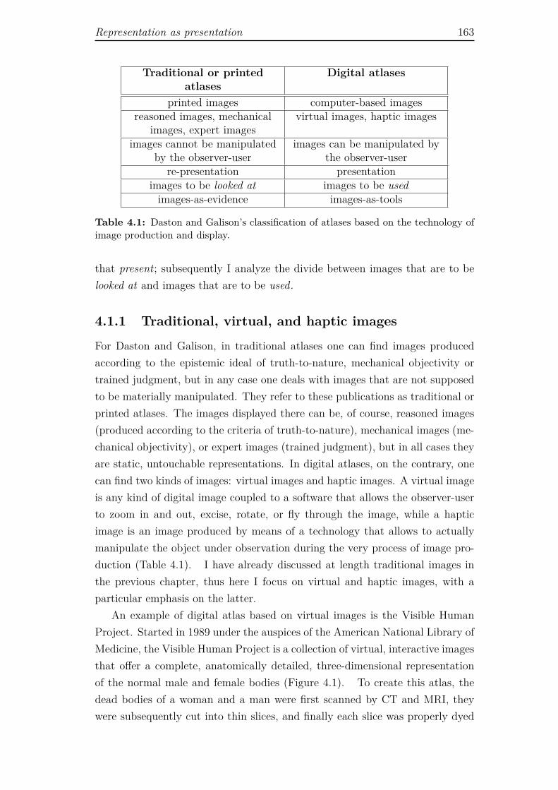
Representation as presentation 163
Traditional or printed
atlases
Digital atlases
printed images computer-based imagesreasoned images, mechanical
images, expert imagesvirtual images, haptic images
images cannot be manipulatedby the observer-user
images can be manipulated bythe observer-user
re-presentation presentationimages to be looked at images to be used
images-as-evidence images-as-tools
Table 4.1: Daston and Galison’s classification of atlases based on the technology ofimage production and display.
that present; subsequently I analyze the divide between images that are to belooked at and images that are to be used.
4.1.1 Traditional, virtual, and haptic images
For Daston and Galison, in traditional atlases one can find images producedaccording to the epistemic ideal of truth-to-nature, mechanical objectivity ortrained judgment, but in any case one deals with images that are not supposedto be materially manipulated. They refer to these publications as traditional orprinted atlases. The images displayed there can be, of course, reasoned images(produced according to the criteria of truth-to-nature), mechanical images (me-chanical objectivity), or expert images (trained judgment), but in all cases theyare static, untouchable representations. In digital atlases, on the contrary, onecan find two kinds of images: virtual images and haptic images. A virtual imageis any kind of digital image coupled to a software that allows the observer-userto zoom in and out, excise, rotate, or fly through the image, while a hapticimage is an image produced by means of a technology that allows to actuallymanipulate the object under observation during the very process of image pro-duction (Table 4.1). I have already discussed at length traditional images inthe previous chapter, thus here I focus on virtual and haptic images, with aparticular emphasis on the latter.
An example of digital atlas based on virtual images is the Visible HumanProject. Started in 1989 under the auspices of the American National Library ofMedicine, the Visible Human Project is a collection of virtual, interactive imagesthat offer a complete, anatomically detailed, three-dimensional representationof the normal male and female bodies (Figure 4.1). To create this atlas, thedead bodies of a woman and a man were first scanned by CT and MRI, theywere subsequently cut into thin slices, and finally each slice was properly dyed
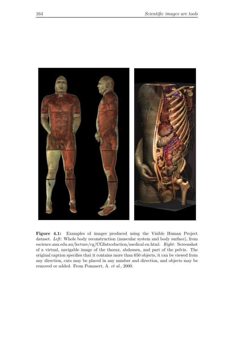
164 Scientific images are tools
Figure 4.1: Examples of images produced using the Visible Human Projectdataset. Left: Whole body reconstruction (muscular system and body surface), fromescience.anu.edu.au/lecture/cg/CGIntroduction/medical.en.html. Right: Screenshotof a virtual, navigable image of the thorax, abdomen, and part of the pelvis. Theoriginal caption specifies that it contains more than 650 objects, it can be viewed fromany direction, cuts may be placed in any number and direction, and objects may beremoved or added. From Pommert, A. et al., 2000.
Representation as presentation 165
and photographed. These different sets of images (photographs, TC scans, MRIscans), associated to image reconstruction and simulation algorithms, becamethe constitutive items of a digital database. They can be virtually manipulatedfor didactical purposes, e.g., in the study of anatomy, or for professional simula-tion of surgical interventions. Haptic images are not collected in proper atlases,but rather in image galleries available in universities, research centers, and in-dustries’ websites. A haptic image is, literally, an image that elicits the sensationof touch, or that can be actually perceived through touch. Daston and Galisonuse this term to refer to images produced resorting to nanotechnological imagingtechniques. The peculiarity of these images is that in order to produce them, itis necessary to interfere with the nanostructure of the portion of matter that isvisualized. In other words, in order to produce haptic images (or nanoimages),one has to manipulate the structure of matter at the nanoscale. Nanoimagesare the only example of haptic images provided by Daston and Galison, andthey use the terms haptic images and nanoimages interchangeably. So, what isso special about these images? How are they produced?
Scanning Tunneling Microscope (STM) and Atom Force Microscope (AFM)were among the first nanotechnologies to be developed. In STM the tip of aconductive tiny probe is used to scan a surface very closely. During this processthe voltage difference applied between the probe and the surface produces atunneling current (a flow of electrons) which is a function of the probe position,the applied voltage, and the local density of states of the sample. Thus, bymeasuring the current, we acquire information that can be displayed in visualform and that provides an image of the surface of the sample.3 Daston andGalison put great value on the fact that the tip of the scanning probe can beused to manipulate nanostructures by literally interacting with one atom at atime. While looking at the image, one can move the atoms of the sample byusing the very same instrument that is used to produce the image. Hence, onedoes really touch the object (although by means of a very particular probe)and manipulate the image simultaneously, for the manipulating and imagingprocesses coincide.4
For Daston and Galison, unlike other scientific images, whose function isto reproduce nature as faithfully as possible, haptic images create somethingthat was not already there, hence they present something rather than merelyre-present it. In their own words:
3The AFM works according to the same principles of the STM, but intead of monitoring acurrent, it measures the force caused by the interaction between the probe tip and the sample.
4See Daston and Galison, 2007, 383.
166 Scientific images are tools
In the context of the more engineering-inspired, device-oriented workthat surrounds much of nanotechnology, images function less for repre-sentation than for presentation. [...] [B]ecause nanomanipulation is nolonger necessarily focused on copying what already exists – and insteadbecomes part of a coming-into-existence – we find it makes more sense todrop the prefix re-, with its meaning of repetition.5
This is to say that what makes haptic images presentations rather than rep-resentations is the fact that with nanotechnology the interest of scientists andengineers has shifted from observation to manipulation. The activities of ob-serving and manipulating a given object have merged into a single action, theyoccur at the same moment and using the same apparatus, and we assist to amove from the reproduction of reality to the production of a new reality. Hapticimages, instantiated by nanoimages, are presentations rather than representa-tions because they are images in which observation and manipulation of theimaged object coincide.
Daston and Galison, however, provide also another definition of presentation,which applies to virtual images. They say:
Epistemically, these virtual atlases [created by the Visible HumanProject] differed from the old medical atlases such as those of Jean Cru-veilhier, Albinus, and William Hunter: using the navigation and image-modifying capability of the program, they could produce in a moment animage that no one had ever seen – this was not, sensu stricto, a matterof re-presentation.6
This means that for Daston and Galison an image can be considered a presenta-tion not only if it allows to manipulate the specimen during the imaging process,but also if no one has never seen that specific image before (because it has beenreconfigured by the viewer-user), or it presents objects or phenomena that noone has ever seen before.
However, once the definition of image-as-presentation is formulated in thisway, it becomes difficult to understand, by Daston and Galison’s own stan-dards, why printed images such as drawings and analogical photographs do notcount as presentations. On the one hand, if an image is a presentation if noone has never seen it before, then all original images could count as presenta-tions independently from the technique of production and the material supportof the image. In this case, speaking in rigorous terms, one should apply theterm re-presentation, as synonymous of repetition, only to identical copies andreproductions of already existing images (I will develop the idea that each repre-sentation is a presentation in the following pages). On the other hand, if images
5Daston and Galison, 2007, 383.6Daston and Galison, 2007, 390.
Representation as presentation 167
are presentations when they make visible obejcts and phenomena that could notbe seen without the mediation of the image, then it is not clear why Donné’s da-guerreotypes, Koch’s microphotographs, Marey’s chronophotographs, or HallsDally’s radiographs cannot count as presentations, provided that all of themshowed something that no one could have seen before the development of anadequate imaging instrument. This is what scientific images are supposed todo: to present us a world that we could not see otherwise.
4.1.2 Image manipulation before the digital era
What about the point, rightly stressed by Daston and Galison, that virtualimages can be manipulated by the observer-user in very specific ways? Todiscuss this issue I will resort to a brief analysis of the same example used bythe authors, the Visible Human Project. As stated in the website of the NationalLibrary of Medicine, the aim of this project is to provide an extremely faithfulrepresentation of the human body, so that medicine students can learn anatomyreducing as much as possible the need to dissect actual human corpses.7 Thishas been, in general, the aim of all traditional anatomical atlases and of theirthree-dimensional elder cousins: anatomical wax or wooden models.8 Being avirtual atlas, the Visible Human Project has transferred to the computer screenthe third dimension which, in the past, could be afforded only by resorting toexpensive, cumbersome, and fragile sculptures. Still, it is interesting to notethat in Juan Valverde de Amusco’s anatomical atlas Vivae imagines partiumcorporis humani aereis formis expressae, printed in 1566, there was an attemptto make printed images manipulable. Bound at the end of the atlas we finda pair of male and female figures composed of paper flaps that, when lifted,revealed the internal organs (Figure 4.2). What I want to emphasize here isthat draughtsmen and painters have always dealt with the problem of renderingthe multiple layers of three-dimensional reality in bidimensional representations,and the anatomical fugitive sheets were an ingenuous means developed to allowthe atlas user to enter the body, by providing a rudimentary form of imagenavigation and manipulation. The Flemish engravers who produced the atlasillustrations were really pushing to the limit the possibilities of printed imagesto function as three-dimensional haptic objects. Valverde’s example is by nomeans an isolated case, and it testifies to a conception of images – well before
7See www.nlm.nih.gov/research/visible/, retrieved on Jun 13, 2012.8For a study of the role of wax anatomical models in medical education see Ballestriero,
2010; Hopwood, 2004; Schnalke, 1995 and 2004. Riva et al., 2010, explore the relationshipbetween anatomical illustration and wax modeling in Italy between the sixteenth and theearly-nineteenth centuries.
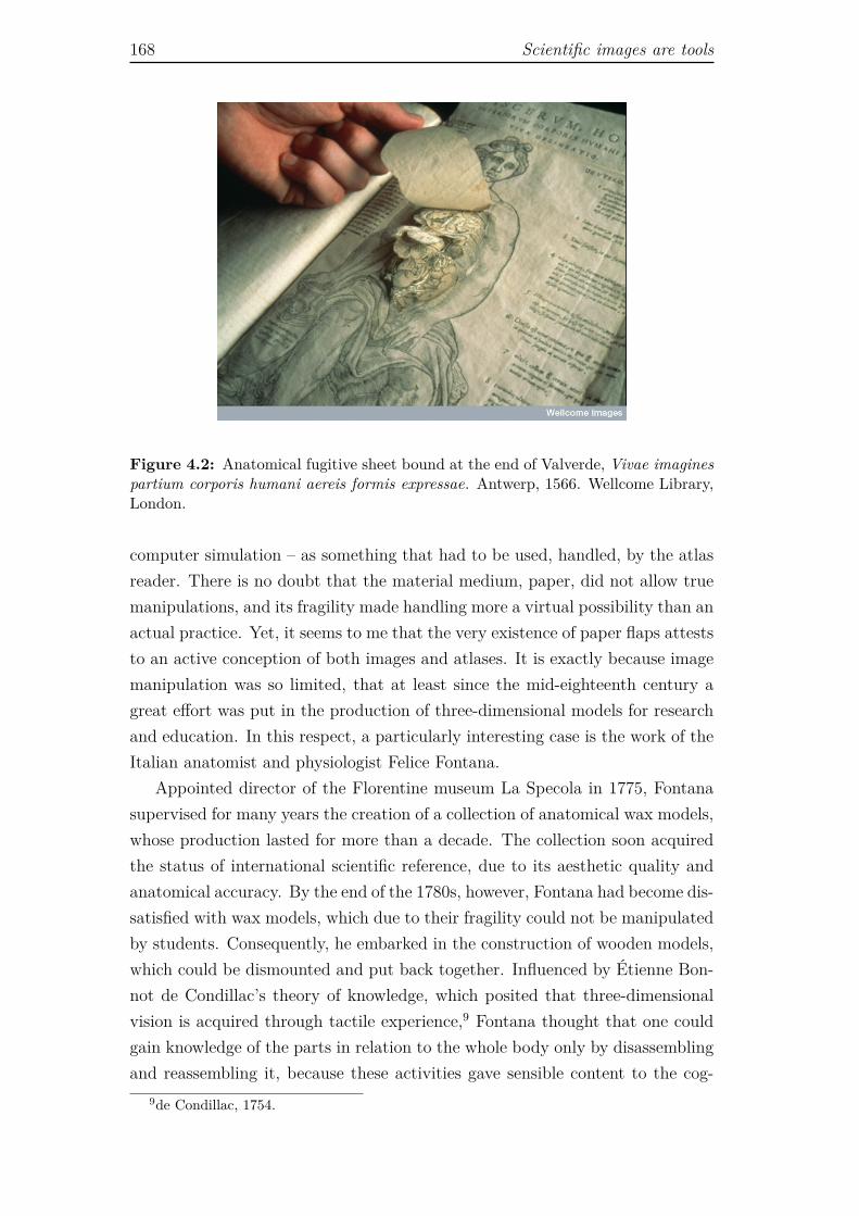
168 Scientific images are tools
Figure 4.2: Anatomical fugitive sheet bound at the end of Valverde, Vivae imaginespartium corporis humani aereis formis expressae. Antwerp, 1566. Wellcome Library,London.
computer simulation – as something that had to be used, handled, by the atlasreader. There is no doubt that the material medium, paper, did not allow truemanipulations, and its fragility made handling more a virtual possibility than anactual practice. Yet, it seems to me that the very existence of paper flaps atteststo an active conception of both images and atlases. It is exactly because imagemanipulation was so limited, that at least since the mid-eighteenth century agreat effort was put in the production of three-dimensional models for researchand education. In this respect, a particularly interesting case is the work of theItalian anatomist and physiologist Felice Fontana.
Appointed director of the Florentine museum La Specola in 1775, Fontanasupervised for many years the creation of a collection of anatomical wax models,whose production lasted for more than a decade. The collection soon acquiredthe status of international scientific reference, due to its aesthetic quality andanatomical accuracy. By the end of the 1780s, however, Fontana had become dis-satisfied with wax models, which due to their fragility could not be manipulatedby students. Consequently, he embarked in the construction of wooden models,which could be dismounted and put back together. Influenced by Étienne Bon-not de Condillac’s theory of knowledge, which posited that three-dimensionalvision is acquired through tactile experience,9 Fontana thought that one couldgain knowledge of the parts in relation to the whole body only by disassemblingand reassembling it, because these activities gave sensible content to the cog-
9de Condillac, 1754.
Representation as presentation 169
nitive activities of analysis and synthesis.10 He claimed: “[wooden body parts]can be handled, rotated into any position, and inspected at leisure. Plates,wax [models], or a cadaver lend themselves to these operations only with diffi-culty and unsatisfactorily.”11 Fontana wanted to build a fictional human bodywhich, as the virtual body of the Visible Human Project, would allow studentsto navigate inside artificial viscera and manipulate them at leisure, overtakingthe limits of both bidimensional representations and real, corpse-based anatomy.The project proved unattainable. The prototype life-size statue made of 3,000independent wooden parts turned out to be extremely complex to build. More-over, most of Fontana’s colleagues, as well as his patron and employer, PeterLeopold, Grand Duke of Tuscany, did not concur with him about the necessity ofreplacing wax models, characterized by astounding realism, with wooden mod-els, apparently less artistic or sophisticated. Fontana’s project was visionary.Nevertheless, it provides a clear example of a form of anatomical representationthat attempted to provide the viewer the illusory, yet tactile, experience of en-tering the body and observing it from the inside. Even more importantly, it wasmeant to give the opportunity to take the model’s pieces apart and carry outan artificial dissection. This historical example corroborates my claim that ma-nipulable representations are not a product of computer technology. The needto handle objects, or their reproductions, in order to understand their structureand functioning appears to be a fundamental, trans-historical feature of thehuman cognitive system. What is historical is the way in which such need formanipulable representations can be satisfied.
4.1.3 Aesthesiological considerations
The contrast between images that re-present and images that present is con-nected to the distinction between images one looks at, and images one uses.Daston and Galison call the former “images-as-evidence” and the latter “images-as-tools.”12 In their taxonomy, haptic images are tools par excellence, becausethey are produced using a technology that allows to literally touch atoms withthe tip of a probe. Now, it is interesting to note that the polarity haptic-optic,translated into its spatial constituents proximity-distance, had already been usedby Benjamin to discuss the tendency of photography and cinema to bring thebeholder closer and closer to objects and people, stimulating a scientific, anato-mizing gaze.13 As we have seen in Section 2.3.2, for Benjamin, the photographic
10See Mazzolini, 2004, 56.11Fontana, quoted in Mazzolini, 2004, 61.12Daston and Galison, 2007, 385.13See Benjamin, 1932, 519; 1939, 255.
170 Scientific images are tools
gaze metaphorically touches and dissects objects and phenomena. It does so byenlarging what is small, sectioning what is continuous, accelerating what is slow,and slowing down what is too fast for human perception. Benjamin borrowedthe concept of haptic image from the Vienna School of Art History, in partic-ular from Alois Riegl. For Riegl, a history of artistic styles entailed a historyof the way each style stimulates and organizes human perception. Accordingly,to write a history of styles was to analyze the distinct ways in which differentimages affect the observer and his corporeal relation with the work of art. Somestyles, e.g., Impressionism, put the beholder at a distance, because only fromthe distance one can properly perceive the painting. In this case Riegl talks ofa style that produces optical images. Other styles, e.g., Egyptian hieroglyphics,force the observer to get closer to the images and invite to close-up view. Theseare haptic images: the eye goes along the contours of the figures as if it was afinger.14
On the poietic function of representation Indeed, the topic of the en-trenchment of optical and tactile sensations in aesthetic fruition is a classicaltheme in both aesthetics and art criticism. Art history is rich in anecdotesthat illustrate how the phenomenological body, with its ability to grasp objects,to get close to them and to manipulate them, is mobilized by visual images.15
Pliny the Elder in his Naturalis Historia described a contest between the Greekpainters Zeuxis and Parrhasius: Zeuxis painted some grapes so perfectly thatbirds flew down to peck them, but Parrhasius painted such a realistic curtainthat his adversary tried to draw it aside.16 It goes without saying that in Pliny’shistory Parrhasius was honored as the best artist, because his work was so realis-tic that it had baffled not just animals, but also a fellow painter. In more recenttimes, the art historian and critic Michael Fried, describing Adolph Menzel’sCemetery Among Trees, with an Open Grave (1846-1847), wrote: “It leads usto project ourselves as if corporeally onto the nearest portion of the scaffold [...],or at least loose firm hold of the distinction between our implied situation [...]and the depicted scene.”17 And another art critic, Bernhard Berenson, ascribedGiotto’s greatness to his ability to stimulate “the tactile consciousness”18 of thebeholder. Berenson maintained that: “Every time our eyes recognize reality, we
14See Pinotti and Somaini, 2012, xvi-xix.15In recent art history and aesthetics the relationship between images and the phenomeno-
logical body is studied mostly in connection with the question of emotion and empathy. See,for example, Elkins, 1999 and 2001; Freedberg, 2007; Freedberg and Gallese, 2007.
16Naturalis Historia, XXXV, 15. I took this example and the followings from Cappelletto,2010.
17Fried, 2002, 34.18Berenson, 1896, 5.
Representation as presentation 171
are, as a matter of fact, giving tactile value to retinal impressions,” and sug-gested that the artist’s main duty “is to rouse the tactile sense, for I must havethe illusion of being able to touch a figure, I must have the illusion of varyingmuscular sensations inside my palm and fingers corresponding to the variousprojections of this figure, before I shall take it for granted as real, and let itaffect me lastingly.”19 For all these authors the realism of the image appeals toan embodied reaction and not to just a cognitive one: the beholder does notrecognize the object represented by the image, he corporeally experiences it. InPliny’s story Zeuxis is moved to touch and draw aside Parrhasius’ curtain, Friedfeels like he is entering Menzel’s drawing moved by empathetic impulsion, whileBerenson makes a normative statement, claiming that art has to elicit tactilesensations if it is to produce lasting impressions.
These examples show how the sense of reality conveyed by images drawsmore on the poietic value of mimesis than on its mimetic function.20 In thisperspective, the image is productive because it engages the observer in theexperience of a sensible object, not merely in the vicarious experience of thesubject-matter of the representation. The experience of looking at an imageis the experience of a human body that encounters the material body of theimage and enters in a relation with it. Indeed, we can say that images are toolsinvolved in the production of knowledge because they lay in between materialreality and abstract thought. An image, notes the philosopher Jean-JacquesWunenburger, is an ambivalent object, which occupies a middle ground betweenreality and thinking, the pure datum of sensible experience and its concept. Animage is a real object in its own right, and at the same time it is the fictionof something that is there only in effigies, or that has simply left a trace on asensible surface. An image, Wunenburger maintains, allows to reproduce andinternalize the world in a plastic way: it can attempt to mirror it, but at thesame time it modifies and transforms reality, creating fictional worlds. An imageis a place of mediation and a point of convergence between a thing and a thought,it is an intermediate construction that partakes in the production of knowledgeabout the world and in its dissolution into imagination.21
Image perception and mirror neurons The aesthesiological intuitions presentin art literature, from Pliny to Fried, passing through the Vienna School of ArtHistory and Benjamin, seem to find empirical corroboration in recent neurolog-ical theories about the so called mirror neuron system. In apes and in humans
19Berenson, 1896, 5.20See Cappelletto, 2010, 11.21See Wunenburger, 1997, i.
172 Scientific images are tools
the mirror neuron system occupies distinct cortical regions, which are activatedwhen an action is observed, as well as when it is actually executed. This meansthat our brain reacts in the same way when we perform an action and whenwe observe it, and this simulation occurs even when we look at static images ofactions. In fact, the same neurons are involved in the recognition of facial ex-pressions, and they seem to underpin not only the understanding of actions, butalso the comprehension of the intentions that underlie an action.22 Moreover,brain imaging studies have shown that the observation of images of manipula-ble objects leads to the activation of canonical neurons in a cortical region thatis assumed to be responsible for action control, rather than for visual objectsrecognition.23 This neural mechanism has quite an intuitive explanation: to gethold of an object, one needs to be informed about its shape (one grabs a balldifferently than a pen), thus the function of the sensory input is to enable us toperform the appropriate grabbing gestures. As a whole, these experimental dataon mirror and canonical neurons corroborate an embodied approach to visionand spatial perception, and they suggest that there is nothing passive in thefruition of images, because images involve the observer in an active process ofembodied imagination, a process of corporeal simulation. Within this perspec-tive, vision becomes a function of the kinetic body, not an act of disembodiedcognition. This does not mean, of course, that cognition and culture are notinvolved in the understanding and interpretation of images. To be aware ofthe biological, embodied dimension of image perception just adds another layerof complexity to our understanding of how we relate to, and interact with vi-sual representations. In other words, the biological, cognitive, and cultural axesof perception should not be seen as mutually exclusive, but as interlocked indynamic interaction.
Even without endorsing a neuro-reductionist epistemological stance, I thinkthat we can find in experimental data insights relevant to the elucidation, enrich-ment, and possibly reformulation of some longstanding philosophical questions.The idea that spatial awareness is linked to motion and touch has a long historyin philosophy of knowledge. For example, in his New Theory of Vision of 1710,George Berkeley argued that humans develop tridimensional vision only afterthey learn what it is to move around the world. According to Berkeley, in fact,our retinal vision is originally bidimensional, and it becomes three-dimensionalonly through a cognitive process whereby we learn to correlate tactile sensations
22See Freedberg and Gallese, 2007; Urgesi et al., 2006; Johnson-Frey et al., 2003.23See Freedberg and Gallese, 2007; Ponseti et al., 2006; Boronat et al., 2005; Martin et al.,
1996.
Representation as presentation 173
with vision.24 Similarly, in the Treatise of Sensations of 1754, Condillac main-tained that the awareness of external objects, as well as the ideas of extension,distance, magnitude, and solidity depended on the sense of touch, and that theattribution of form and color hinged upon the motion of the eyes and the hand:to learn to see was to learn to coordinate sight and touch25 (as seen above,Fontana translated Condillac’s ideas on perception into a pedagogical theory).
Vittorio Gallese, one of the proponents of the mirror neurons theory, remarksthat there are many points of contact between recent neurological claims con-cerning the motor nature of spatial awareness, and the theories developed bydifferent philosophers of knowledge. He mentions Hermann von Helmholtz, whotried to substitute the Kantian notion of space as an a priori with the notionthat this a priori consists of the manifold of possible orientations in space, whichmeans that it is generated by exploratory behavior.26 Gallese also highlights theaffinities between the mirror neuron theory and the views on space perceptionelaborated in phenomenology by Edmund Husserl and Maurice Merleau-Ponty.Merleau-Ponty clearly linked space perception to motion and intentionality whenhe wrote that space is “[. . . ] not a sort of ether in which all things float [. . . ].The points in space mark, in our vicinity, the varying range of our aims andour gestures.”27 In a similar fashion, for Husserl, we see things as tactile ob-jects directly related to the lived body (Leib). It is the tactile lived body thatprovides the constitutive foundations of our cognitive self-referentiality, of ourdetermination of reality. Yet, while Husserl considered the physiological bodya material object, not directly involved in the phenomenological awareness ofthe lived body’s relation with the world, contemporary neurophysiology sug-gests that this is not the case.28 It seems, in fact, that at the neurological levelthere is no clearcut distinction between motion and cognition, because the samephysiological processes that underpin the functioning of our body in the world(the sensory-motor system), also contribute to our awareness of the objects thatthe world contains, and of our own lived body as part of it. The world is notout there, we are intertwined with it. According to Gallese’s theory of embodiedcognition, which goes under the name of embodied simulation, action simula-tion integrates visual and auditive stimuli within a specific neural circuit. He
24Ian Hacking refers to Berkeley’s theory of vision in the discussion of his idea of scientificobservation as acquired skill. I present Hacking’s position in Section 4.2.2.
25In his previous work, the Essay on the Origin of Human Knowledge of 1746, Condillacdefended a less radical position. Though he already maintained that the cognitive facultiesare developed as a consequence of sensation, he took sensation itself largely for granted andnot as a learned skill. See Falkenstein, 2010.
26See Patton, 2012; Gallese, 2005, 26.27Merleau-Ponty, 1945, 243.28See Gallese, 2005, 27.
174 Scientific images are tools
explains: “The sight of an object at a given location, or the sound it produces,automatically triggers a ‘plan’ for a specific action directed toward that location.What is a ‘plan’ to act? It is a simulated potential action.”29 This means thatwhen a visual stimulus is presented to our perception, it evokes the inner simu-lation of the corresponding motor schema, which maps position in motor terms,independently from the existence of an actual action or movement. Galleseclarifies his conception of embodied simulation as follows:
Simulation is not conceived of as being confined to the domain ofmotor control, but rather as a more general and basic endowment ofour brain. It is mental because it has content, but it is sensory-motorbecause its function is realized by the sensory-motor system. I call it‘embodied’ – not only because it is neurally realized, but also becauseit uses a pre-existing body-model in the brain, and therefore involves anon-propositional form of self-representation.30
Through embodied simulation we literally, although not materially, enter incontact with the world around us by moving within it and touching it. Accord-ingly, an image is not merely seen by the eye, it is experienced by the lived body.The lived experience, with its intertwining of visual and motor reactions is apre-condition for any cognitive apprehension of the image content and meaning.
Some problems with the mirror neurons theory of image perception
This neurological account of embodied cognition is quite compelling especiallybecause, by positing that lived experience is a precondition for the cognitiveelaboration of images, it is congruous with our basic intuition that some sort ofsensorial and physiological apprehension of the world must necessarily precedeany cognitive-cultural operation. Still, intuitive as it might be, this assumptionis problematic. The mirror neurons theory seems to fit nicely to our empa-thetic response to artworks, where we recognize bodies that could be our body,movements that could be our movements, and emotions that could be our emo-tions. However, it is much more difficult to understand how the neuro-motorsystem could be involved in the apprehension of scientific images which, at leastin principle, are emotionally neutral. Even more importantly, we should re-member that in the majority of cases scientific images show something that wecannot recognize as part of our ordinary world and of our lived experience. Itcould be claimed that in this case, as in the case of conceptual art, the neuro-motor system is involved at the level of canonical neurons that, as seen above,
29Gallese, 2005, 27.30See Gallese, 2005, 41-42.
Representation as presentation 175
underpin shape recognition and our capacity to manipulate objects.31 Indeed,in medical images we might see objects that we recognize as something thatcan be actually touched (a bone, a vein, the liver). However, most scientificimages show objects that cannot be grabbed as we grab a pen or a ball. Do werecognize a cell under the microscope as a seizable object? Or do we perceiveit as undistinguished patches of colors, which acquire the shape of a cell onlywithin a well-defined cognitive framework? In front of the image of an objectthat can be recognized only by someone provided with specific knowledge, theneuro-motor reaction of the competent observer will not be different from thatof the naïve viewer? To put it differently, could not the neuro-motor reaction be,in this case, post-cognitive rather than pre-cognitive, through a sort of ricocheteffect of the cognitive dimension on the neural dimension?
To the inexperienced patient, an arterial angiogram shows something akin tofilaments, to the radiologist, it shows a pervious or occluded pipe (the artery), tothe surgeon, an artery that, if occluded, can be stripped. Will the neuro-motorreaction of these three subjects be the same? Gallese and colleagues wouldprobably answer that these different conscious perceptions come after a primor-dial embodied reaction, whereby both expert and inexperienced observers seesomething that can be grabbed. Yet, we do not grab in the same way a spread-ing filament, a pipe-artery, or an artery that we know to be firm or slipperyunder the lancet. So, it is difficult to organize an order of temporal prioritiesbetween cultural and embodied perceptions, and this might not even be themost relevant or instructive question. It might turn out that a more interestingphilosophical and scientific challenge is to understand the entanglements of bio-logical and cultural domains, rather than splitting them in sequential operations(theories about neuroplasticity and epigenetics seem to move in this direction).Thus, problematic as they might be, neuroscientific theories of embodied pre-cognition offer interesting arguments for thinking visual representation, becausethey blur the boundaries between cognitive-visual experience and bodily-hapticexperience.
4.1.4 Manipulating images and objects
Although I rise objections to Daston and Galison’s views on the divide betweenprint and digital images, it is important to stress that, by emphasizing the em-bodied dimension of image fruition as a universal and fundamental aspect ofour interaction with all kind of pictures, I do not intend to downplay the po-
31For the putative role of canonical neurons in the fruition of conceptual art see Freedbergand Gallese, 2007.
176 Scientific images are tools
tential transformative effect of digital images on our sensorial perception; onthe contrary. I think that digital technologies produce extremely powerful ef-fects on our perception, and this happens for two reasons. On the one hand,digital images can stimulate to a very high degree the haptic dimension of im-age’s perception (but this dimension concerns the perception of all images, notonly of computer-generated images). On the other hand, they strengthen ourability to relate to virtual reality, by taking advantage of the fact that images,independently from the technology used to produce and display them, occupya middle-ground between reality and imagination. It seems thus reasonable toposit that virtual images and the interactive devices that support them workby reproducing and amplifying our innate processes of embodied simulation. Itwould be unreasonable to deny that virtual images can be manipulated by boththeir producers and final users in unprecedented ways, and that virtual imagesmanipulation provides us with novel sensorial and cognitive experiences. Yet,this does not mean that images could not be, and were not, manipulated be-fore the digital era. Indeed, as Benjamin expressed so well with his metaphorof the camera operator as surgeon, photography and cinema brought to life avery specific practice of image manipulation. Zooming in and out, strong mag-nification, slow and fast motion were exactly the features of photography andcinema that compelled Benjamin to argue that they brought to light “entirelynew structures of matter,” and made us “discover the optical unconscious.”32
As mentioned in Section 3.2, a common practice in the early days of photomi-crography was to re-photograph a microphotograph put under the microscope,in order to attain magnifications beyond the technical possibilities of the mi-croscope optics.33 This procedure proved scientifically unappropriate, becauseit magnified artifacts rather than the real specimen, nevertheless it attests tothe intrinsic manipulability of images. Indeed, one of the reasons why scientistsproduce images is that one can perform operations on an image that could notbe carried out on the real object.
Time-lapse cinema and image malleability If photography allowed tomanipulate images in the spatial dimension, cinema allowed temporal manipu-lation. The historian and philosopher of media Hannah Landecker has shownthat time-lapse microcinematography played a capital role in making possiblethe study of life phenomena and it contributed to the emergence of the ideathat, at the microscopic level, life is characterized by relentless, frantic activ-
32Benjamin, 1939, 266.33See Section 2.3.
Representation as presentation 177
ity.34 This happened because many life phenomena, such as the movement ofbacteria, the division of a cell or the blossoming of a flower have a temporalresolution well below the human sensory threshold or possibility of continuousobservation. That is, they are too slow to be directly observed in their becoming.With time-lapse cinema this problem is solved by projecting an image sequenceat a much faster frequency than that used to capture the individual frames.In other words, while with chronophotography and slow-motion cinema we canslow down very fast phenomena, with the time-lapse technique we can acceleratevery slow processes in order to see their sequential unfolding, rather than thediscrete stages of the process. We can visualize the motion of the stretchingpetals, rather than a bloom and, a few days later, an open flower. Through thistechnique one can manipulate the time of experiment, observation, and demon-stration, condensing the long hours, or even days, of real-time observation intoa very short film (acceleration through projection). Unlike chronophotography,which was meant to dissect movement into its instantaneous components, time-lapse cinema was meant to reconstruct a continuous phenomenon on the basisof instantaneous images. One of the first time-lapse microfilms was made bythe Swiss biologist Julius Ries in 1907, to study the fertilization and develop-ment of a sea urchin. Ries made these two events visible by condensing thefourteen hours of the natural process (documented through photographs takenevery seven minutes) into a two minutes film. The time of the living process wasreadjusted to the time of observation: through the careful manipulation of thetime of static and dynamical modes of imaging, a morphological transformationthat was too slow to be observed as continuous process, was brought within therange of human senses.35
These examples of photomicrography and microcinema show that image mal-leability largely predates digital technology. Indeed, we could say that malleabil-ity, or plasticity, is probably one of the reasons why images are so ubiquitous inscience.
Nanoimages and objects malleability In making their case about images-as-tools, Daston and Galison state the following:
In [...] haptic images, seeing and making entered together – unlike themore familiar image making that marked so many generations of science,holding fast to a two-step sequence. The older method meant first smash-ing a proton against an antiproton in an accelerator, then imaging thedetritus for analysis in a bubble-chamber photograph or digital display.Or, in a very different domain of science, first preparing a tissue sample,
34See Landecker, 2005, 918-920.35See Landecker, 2005 and 2006.
178 Scientific images are tools
then imaging it in the electron microscope. For early twenty-first-centurynanoscientists, such after-the-fact representations were often entirely be-sides the point.
Frequently, the nanographers want images to engineer things. In thefirst instance, these were images-as-tools, entirely enmeshed in making,much more than images-as-evidence to be marshaled for a later demon-stration.36
Although these paragraphs can be easily subscribed for what concerns the defi-nition of what makes nanoimages different from other microscopic images (theycan be used to engineer things), it is quite problematic in its implications aboutimages that do not belong to nanoscience. What does it mean that non-nanoimages are after-the-fact representations? Is it a technical statement or an epis-temological one? If it is a technical statement, it simply means that when weuse an imaging apparatus which does not involve nanotechnology, we have toprepare the sample first, and then we can make a picture of it. But is it true?To microinject a sample during observation is a common practice in microscopy(even with simple optical microscopes). Also, moving to a completely differ-ent scale, in Duchenne’s photographies éléctrophysiologiques the patient’s bodywas embedded in the experimental machinery, and literally wired to the elec-trostimulator. Was this not a form of physical manipulation, although not atthe nanoscale, that occurred during the process of visualization of a specificphenomenon? (In this case, human expression of emotions).37 So, in techni-cal terms, the claim that only nanotechnology allows to manipulate the objectduring observation is not strictly correct.
Things become even more problematic if we take Daston and Galison’s sen-tence about after-the-fact representations as an epistemological statement. Theysay that such representations are “images-as-evidence to be marshaled for laterdemonstration.” I interpret their use of the term “demonstration” as referringto the fact that – unlike in the case of images-as-tools, where images are use toproduce new phenomena – in the case of images-as-evidence, the image is usedas a demonstration, an evidence, of the existence of a given phenomenon. Forexample, the existence of a bacterium with some morphological characteristic.But why should such demonstration occur later? Later in relation to what? Theuse of this temporal adverb conveys the idea that images-as-evidence (which,in this context, correspond to all images that are not nanoimages) are used asillustrations of something that has already been discovered or understood byother means. It is true that physicians suspected the existence of microorgan-isms already before they were able to see them with a microscope, and physicists
36Daston and Galison, 2007, 384-385, italics in the original.37See Section 2.6.
Representation as presentation 179
had already made many observations and developed many theories about sub-atomic particles before they came out with the idea of smashing protons againstantiprotons and visualize the result. Yet, in both cases visualization was partand parcel of the experimental process, not a subsidiary activity. Particle ex-periments would be incomplete, or better, useless without visualization, becausethey would be experiments whose results are concealed and inaccessible. Kochcould not have developed his germ theory of disease without the images he sawunder the microscope, which he fixed in drawings and photographs. Roentgenwould have not discovered the X-rays if they had not left a trace on a sen-sible plate. He used those marks as evidences of his discovery, and he alsoused them as an instrument to study the behavior of the rays when passingthrough different objects and materials. Physicians and surgeons used radio-graphs as evidences of disease, and as means for entering with their eyes intothe patient’s body. And outside the domain of technology-mediated images, wellbefore the development of computerized molecular modeling, the immunologistPaul Ehrlich used pictorial diagrams as real heuristic tools in the developmentand defense of his controversial side-chain theory of antibody formation.38
Daston and Galison put great emphasis on the fact that in nanoimagingobject manipulation and vision occur simultaneously, and that the technologyused for manipulation coincides with the technology for visualization. However,they do not provide compelling reasons to justify their claims about the allegedlyunique epistemic status of nanoimages in relation to other scientific pictures.Most objects, independently from their scale and from the imaging technology,have to be manipulated in order to produce a scientific image. In my view, in atheory of scientific images this is more relevant than whether the visualizationand manipulation occur synchronically or diachronically. Cells, microorganisms,and all sorts of living tissues have to be properly prepared to become visibleunder the microscope and in microphotographs;39 an object has to be bombardedwith X-rays to produce a radiograph; and a patient has to ingest bismuth forthe radiologist to see the shape of the digestive system, in real time, on thefluoroscopic screen.
How are we to understand these practices if not as manipulations, inter-ventions on natural objects aimed at turning them into objects for scientificvisualization and analysis? Even images that do not reach the technological so-
38Ehrlich made his drawings public in the 1900 Croonian Lecture “On immunity with specialreference to cell life,” later published in the Proceedings of the Royal Society and widely cited.For a study of Ehrlich’s pictures and their role in the development of immunological theory,see Cambrosio et al., 1993 and 2005. Among studies of scientific images and the ways theyare used by scientists, see Dagognet, 1986; Lynch and Woolgar, 1990; Baigre, 1996; Pomboand Di Marco, 2010.
39See Section 2.3.
180 Scientific images are tools
phistication of nanomanipulation command some sort of actual handling of theobject of study, and this happens because images are part of an experimentalpraxis, a complex set of activities whose aim is not merely to copy objects in themost accurate way, but to learn something about them. We can thus concludethat, even if we accept Daston and Galison’s definition of images-as-tools as im-ages that allow to manipulate the object of study during the imaging process, wecannot concur with them in applying this definition only to nanoimages. Manyscientific images produced by means of some technological apparatus allow orrequire the manipulation of the sample during the making of the image. Moreexactly, the preparation of the sample should be considered part of the processof visualization. Still, I have to clarify first both the semiotic and experien-tial processes entailed in scientific images signification, in order to reinforce andmake more precise my claims on the poietic nature of scientific representation.To say that representing is an active endeavor is not enough. An account of howthis activity is brought about is necessary. To put it differently, it is necessaryto explore how representation and meaning are interlocked.
4.2 From vision to meaning
As seen above, Daston and Galison’s technology-driven dichotomy between im-ages that present and images that re-present entails a passive understanding ofrepresentation. This way of conceiving representation not only overlooks thequestions posed by disciplines as diverse as art theory, philosophy, and neu-roscience; it also makes difficult to understand how the images that merelyrepresent – allegedly burdened with passivity and repetition – can produce newknowledge as they actually do. In contrast with this view, I have defendedthe idea that scientific representations always entail some form of presentation.On the one hand, they command specific manipulations of the object of study;on the other hand, images themselves can be manipulated in order to betterunderstand the represented object; finally, the viewer, too, is activated, so tospeak, by the image both at the sensory and cognitive level. In the remainder ofthis chapter, I back my claims through the analysis of how images are used andendowed with meaning in actual scientific practice. My account is organizedalong three lines: first, I briefly summarize David Gooding’s theory of scientificvisual thinking, which has the merit of highlighting the trans-historical aspectsof visual cognition without neglecting the cultural and local character of actualpractices of visualization; second, I propose a semiotic analysis of the processesof image signification; finally, I introduce the idea that scientific images mustbe understood as essential components of experimental practices.
From vision to meaning 181
4.2.1 Six general features of scientific representations
Gooding, an historian and philosopher of science who has worked on the cog-nitive theory of scientific images and models, describes scientific representationas a practice where cognition and culture meet.40 He remarks that, althoughmany philosophers and scientists (physicists and mathematicians in particular)are convinced that science proceeds by eliminating sense experience in orderto produce abstractions that can be formally manipulated, the growing impor-tance of data visualization attests to our cognitive need to translate abstractor invisible data into objects or forms that can be manipulated and interpretedresorting to non-formalized visual skills.41 For Gooding, it is possible to identifya distinct scientific “visual method” of thinking, whereby “images, narratives,meanings, arguments and objects” converge.42 Such visual method, he argues,is trans-historical, in spite of the diversity of the local contexts of practices(different scientific disciplines and different imaging technologies). He states:“The existence of common features behind the diversity of visual representa-tions suggests a common dynamical structure for visual thinking, showing howvisual representations facilitate cognitive processes such as pattern-matchingand visual inference through the use of tools, technologies and other culturalresources.”43 Gooding identifies six general persistent features of scientific visualrepresentation:
1. Representations are hybrid, that is, in scientific visualization differentmodes of representation (visual, verbal, numerical, symbolic) are broughttogether to display an interpretation. The simplest and most straightfor-ward example is the use of captions that describe explicitly what is to beseen in the image, but we can also think of the pointers and letters that areoften superimposed on microscopy images to guide the gaze of the viewer,or the lines and points traced on bubble chamber photographs to clarifythe trajectory of the tracks.
2. Representations are multimodal, that is, they combine information thatcomes from different sources and appeal to different sensory modalities.For instance, in maps color codes can be used to visualize temperatureor altitude, while the functional differentiation of an anatomical structurecould be displayed using different symbols.
40See Gooding, 2006.41See Gooding, 2003, 263-268.42Gooding, 2004a, 280.43Gooding, 2004b, 551.
182 Scientific images are tools
3. Representations are plastic, because they can be subjected to a wide rangeof variations that externalize, but also guide mental operations, and ac-commodate interpersonal communicative needs. For instance, a represen-tation can be manipulated materially or mentally through operations suchas rotation or color change, a photograph can be cut and worked in severalways, or it can be translated into a diagram. Although digital technologiesenable specific forms of image plasticity, the plasticity of representation isprimarily related to our cognitive needs, and not to the technique of imageproduction.
4. Representational variation takes often the form of recursive transforma-tions between bidimensional forms (patterns and diagrams), three- dimen-sional forms (structures) and four-dimensional representations (temporaltransformations). For example, to reconstruct an extinct animal from fos-sil imprints, paleontologists typically take several imprints’ pictures. Fromthe photographs they will extract bidimensional diagrammatic represen-tations that serve as a basis for three-dimensional modeling. However,the information provided by the bidimensional diagrams is not enough toreconstruct the morphology of the original specimen. It must be com-plemented with ecological, physiological, and biological information fromdifferent sources (e.g., knowledge about the physiology and behavior ofsimilar organisms, or about the processes of mineralization of specificstructures). Importantly, the process is not linear, but dialectical, be-cause the knowledge generated at each step of visualization can lead toa reassessment of the previous passages and thus open new possibilitiesof representing, i.e., understanding, the object of study. Transformationscan be informal or formalized.
5. Representational plasticity is constrained, because it has to respect trans-formation rules which might be explicit and articulated verbally, or em-bodied in representational techniques and technologies. Verbal rules arepredicated on the background knowledge of the specific scientific disciplineand on information derived from different sources. Representational ruleencompass, for example, geometrical methods of image transformation, orcomputational methods for image reconstruction.
6. Representations are distributed in three different ways: (1) between minds(individuals) and machines, because researchers use instruments to pro-duce and work with different kinds of images; (2) between the mental andthe material (sensory) domain, because of the very double nature of im-
From vision to meaning 183
ages; and (3) among individuals, machines, and organizations, because ofthe collective dimension of scientific knowledge.44
For Gooding, it is through the distributed character of visualization that thetrans-historical cognitive domain intersect the socio-cultural dimension of ev-ery human endeavor. He argues that studies on practices of representationand visual thinking help to elucidate our comprehension of science as a dis-tributed system under different perspectives. By providing information on howresearchers produce, manipulate, and communicate with and about visual im-ages, they undermine the traditional distinction between internal (mental) andexternal (public) representations. In doing so they foster the idea that sci-ence constitutes a “truly hybrid cognitive system,”45 wherein the boundariesbetween collective and individual knowledge are blurred. Mental and materialimages work through a continuous interplay: material images can be generatedby machines (their production does not involve a human agent), but they can-not be manipulated without engaging in mental processes. Mental processes, inturn, are proper of individuals, and nevertheless they are influenced by collectiveknowledge and communication. Additionally, studies in scientific visual think-ing show that “representations are neither cognitively nor socially neutral.”46 Atthe social level, representations have to respect the conventions of a disciplineand are necessarily inscribed in a wider cultural tradition (e.g., artistic styles),at the cognitive level, they have to accommodate different cognitive capacitiesand goals.
In the following sections I discuss how these general features are instanti-ated in specific cases of scientific representations. First, I analyze images inthe context of the semiotic network of a scientific paper, examining the 1903radiology article by Halls Dally already mentioned in Chapter 3.47 I chose thisarticle because it was published at a time in which the very meaning of X-rayimaging was still in the making, thus it offers a privileged vantage point to un-derstand the processes of image signification. Subsequently, I look at images ascomponents of experimental practices, extending my considerations from med-ical imaging to visualization technologies in molecular biology and microscopy,in order to highlight the instrumental, embodied, and collective dimension ofscientific representation and visual thinking.
44See Gooding, 2006, 689-692; Gooding, 2004b.45Gooding, 2006, 694.46Gooding, 2006, 695.47See Section 3.4.3.
184 Scientific images are tools
4.2.2 Images as part of semiotic networks
According to the semiotician Françoise Bastide, a scientific article is, as a whole,“a technique of visualization for the public, for the scientific community con-cerned [with the problems of a field of knowledge].”48 Through an article, thefirst-hand visual experience of one scientist, or of a limited group of scientists,becomes a larger, shared experience that can be discussed and related to a sys-tem of collective knowledge. This idea applies quite well to Halls Dally’s paper,because the author, in his attempt to validate X-ray imaging as an effective diag-nostic technology for pulmonary diseases, described quite accurately the actionsand cognitive processes involved in chest radiographs signification, and madeexplicit a number assumptions that were subsequently internalized in medicalknowledge through formal education and routine medical practice. Hence, byanalyzing in detail this document we can have an idea of the processes that ledto the definition of a wide range of conventions and cognitive strategies thatmade radiographic imaging meaningful for the doctors of the early twentiethcentury. In Halls Dally’s article, the radiographic and fluoroscopic images aresystematically translated into words, diagrammatic pictures, and tables, thuscreating an elaborated semiotic network that accomplishes two functions. Onthe one hand, it transforms the streaks and shadows that appear on the radio-graphic plate or the fluorescent screen into anatomical images (the lungs and theheart) and physiological activities (respiration in the form of diaphragm move-ment). On the other hand, by putting in relation X-ray images with physicalexamination and the description of clinical cases (patient histories), images areinvested with a concrete medical meaning. Thus, throughout the article we seeat work the hybrid, multimodal, plastic character of representation, as well asthe definition of the constraints for image interpretation, and the distributednature of the whole endeavor.
Conditions of visibility Halls Dally’s paper can be divided in two parts:one is aimed at establishing the primary conditions of visibility of the X-rayimages of the thorax, while the other deals with the construction of their diag-nostic meaning. Halls Dally’s first concern was to clearly define the position ofradiographic imaging within the context of respiratory medicine. He wrote: “Inaddition to the established methods of medical investigation of disease of thechest comprised under the headings of inspection, palpation, percussion, aus-cultation, and mensuration we now have at our disposal the Roentgen rays, anewer yet no less potent factor in diagnosis.”49 In a single sentence he stressed
48Bastide, 1990, 189.49Halls Dally, 1903, 1800.
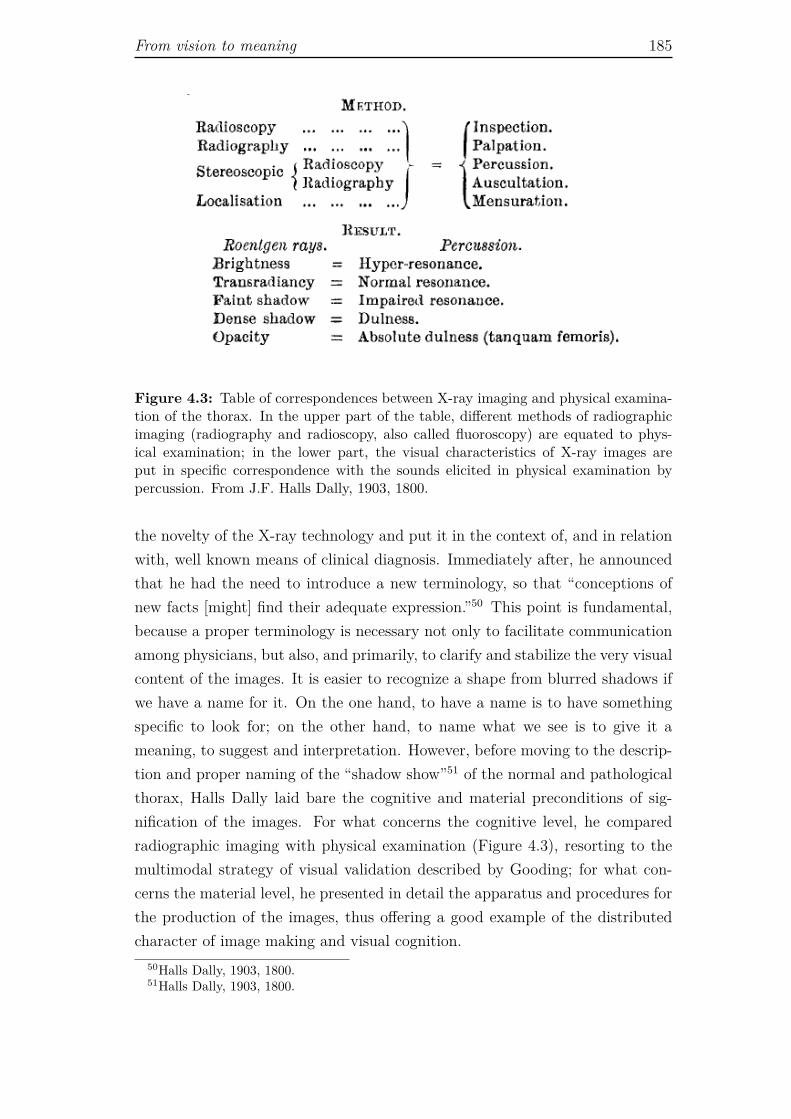
From vision to meaning 185
Figure 4.3: Table of correspondences between X-ray imaging and physical examina-tion of the thorax. In the upper part of the table, different methods of radiographicimaging (radiography and radioscopy, also called fluoroscopy) are equated to phys-ical examination; in the lower part, the visual characteristics of X-ray images areput in specific correspondence with the sounds elicited in physical examination bypercussion. From J.F. Halls Dally, 1903, 1800.
the novelty of the X-ray technology and put it in the context of, and in relationwith, well known means of clinical diagnosis. Immediately after, he announcedthat he had the need to introduce a new terminology, so that “conceptions ofnew facts [might] find their adequate expression.”50 This point is fundamental,because a proper terminology is necessary not only to facilitate communicationamong physicians, but also, and primarily, to clarify and stabilize the very visualcontent of the images. It is easier to recognize a shape from blurred shadows ifwe have a name for it. On the one hand, to have a name is to have somethingspecific to look for; on the other hand, to name what we see is to give it ameaning, to suggest and interpretation. However, before moving to the descrip-tion and proper naming of the “shadow show”51 of the normal and pathologicalthorax, Halls Dally laid bare the cognitive and material preconditions of sig-nification of the images. For what concerns the cognitive level, he comparedradiographic imaging with physical examination (Figure 4.3), resorting to themultimodal strategy of visual validation described by Gooding; for what con-cerns the material level, he presented in detail the apparatus and procedures forthe production of the images, thus offering a good example of the distributedcharacter of image making and visual cognition.
50Halls Dally, 1903, 1800.51Halls Dally, 1903, 1800.
186 Scientific images are tools
The comparison between radiographic imaging and physical examination al-ready points to the diagnostic meaning of the images and locates them in aspecific domain of knowledge. On the contrary, the technicalities about imageproduction concern the very possibility to use mechanical images as reliablerepresentations of the object of study (this section in Halls Dally’s article cor-responds to the the phase of technology control and stabilization described byPasveer).52 For instance, Halls Dally warned that if the patient’s exposure wastoo long, or if the current intensity in the vacuum tube too high, then theanatomical structures that should appear dense would look transradiant (trans-parent). If the tube was placed too close to the patient’s body, the shadows ofthe inner structures near to the tube would appear larger and more indefinitethan those which were far.53 The importance of these technical details lay inthe fact that to use different technical parameters means to produce differentimages. That is, the image is not an unqualified copy of nature: it shows whatit shows because it has been produced under specific conditions, and if thoseconditions vary, the image also changes. Mechanical images are highly con-tingent objects, whose variability can be tamed by setting technical standards(operational conventions).54 Consequently, the definition, discussion and imple-mentation of such standards for image production plays a fundamental role inthe process of mechanical images signification.
Having settled the general preconditions for X-ray images signification, HallsDally moved to the description of the radiograph of the normal, healthy thoraxaccording to the new terminology he had previously mentioned. He suggestedthe adoption of the term “pleuro-pericardial lines” to refer to ill-defined streaksthat appear alongside the mediastinal area and follow the outline of the heart.He remarked that in earlier publications those lines had been taken for lowerbronchi and bronchioles, mediastinal glands, or visceral pleura, and referringdirectly to both the descriptions and images published by his colleagues, he ex-posed the shortcomings of their interpretations. The streaks, he argued, cannotbe bronchi and bronchioles because of their position, and they cannot be causedby either mediastinal glands or the visceral pleura because they are still visibleafter removing part of the organ for post mortem radiography. The tentativenature of his own interpretation, however, was explicitly acknowledged by theradiologist. He wrote: “I venture to suggest (under correction) the term ‘pleuro-pericardial lines’ as being more expressive of what I take to be the origin of these
52See Section 3.4.4.53Halls Dally, 1903, 1800.54I discussed this topic at length in Chapter 3. In particular, in Section 3.2 I showed how
the setting of experimental standard played an essential role in the process of signification ofmicroscopy images.
From vision to meaning 187
shadows.”55 At the dawn of diagnostic imaging, to learn to see something intoradiographs was envisaged as a collective process of iterative corrections, andthe definition of what could be seen went hand in hand with the developmentof the terminology that allowed to discuss the new knowledge. By discussingalternative interpretations, radiologists checked on each other’s ability to see,developed a specialized language, and built a collective way of seeing. Thisstrategy, in more or less formalized forms is enacted each time a new imagingtechnique is introduced in medicine or in other fields of science.56
Building clinical meaning In the second part of the article, Halls Dallydealt with the problem of how to use radiographs as diagnostic tools. Howto recognize pathological states by means of images? For the clinicians of theearly-twentieth century the pressing question was: how to diagnose tubercu-losis promptly and correctly using X-rays? Halls Dally suggested that earlysigns of pulmonary disease could be detected only by fluoroscopy, because thisreal-time visualization technique allowed to see the movements of the internalorgans. A reduction in the mobility of the diaphragm, he maintained, was theearliest indication of tubercular lesions in the lungs. He explained: “Diminutionin the extent of the excursion of the diaphragm will be noted in the great ma-jority on the side most affected, the general rule being that in quiet and normalbreathing the excursions are equal on the two sides, but on maximum respi-ration they are slightly greater in extent on the right than on the left side.”57
This description was accompanied by a table (Figure 4.4) that displayed thereference number of the clinical cases described in the article, the sex and age ofthe patient, the measurements of diaphragmatic mobility, and some qualitative
55Halls Dally, 1903, 1801.56The problem of defining a shared language to describe images and their diagnostic meaning
is still paramount in medicine, and various thesauruses and taxonomies have been developedto tackle this issue. RadLex (Radiology Lexicon), for example, is a general controlled vocab-ulary developed by the Radiology Society of North America, DICOM (Digital Imaging andCommunications in Medicine) is an industry-driven standard for the production of medicalimaging files, their storage and their transmission over networks, while BI-RADS (BreastImaging-Reporting and Data System) is a standard way to describe breast imaging findingsand results developed by the American College of Radiology (it exists in specific versions formammography, ultrasound imaging, and MRI). More recently there have been attempts to re-place thesauruses and taxonomies with ontologies, which are more dynamic and are meant toretain multiple layers of relationships between different terms (for an overview of the problemsencountered in the integration of different lexicons and the development of an ontology seeOverton et al., 2011). The main objective of all these standards is to improve communicationamong health professionals an researchers, and to allow data retrieval for both clinical andresearch purposes. I think, however, that it would be interesting to analyze if and how thesestandards, by putting limits to the language, also influence the possible interpretations of animage. Does the definition of a standardized lexicon prescribe a specific form of seeing? Doesit push medical vision from the qualitative to a semi-quantitative domain?
57Halls Dally, 1903, 1802.
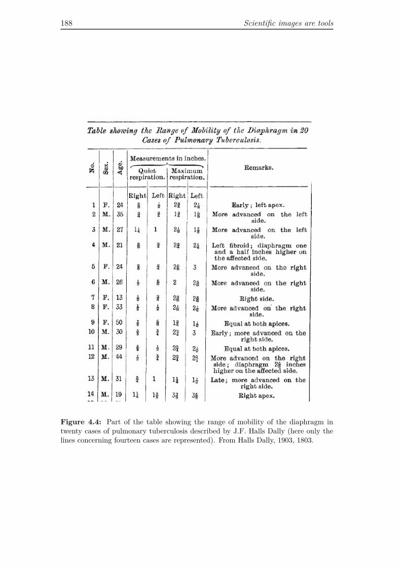
188 Scientific images are tools
Figure 4.4: Part of the table showing the range of mobility of the diaphragm intwenty cases of pulmonary tuberculosis described by J.F. Halls Dally (here only thelines concerning fourteen cases are represented). From Halls Dally, 1903, 1803.
From vision to meaning 189
annotations concerning diaphragmatic movements. In this case, the imagingdevice (the fluoroscope) was used as a measuring tool (because it allows to mea-sure the movement of the diaphragm during respiration), and the table becamea surface on which to visualize and organize different cognitive elements. Moreprecisely: (1) quantitative information, corresponding to the measurements ofdiaphragmatic excursion (diagnostic indicator); (2) qualitative description ofthe diaphragm’s movement, which re-connects the contents of the table to thevisual nature of the original information provided by the fluoroscope; (3) qual-itative information, indirectly provided through the numbers corresponding tothe patient histories described in the text of the article. In turn, that verbaldescription referred to the corporeal dimension of physical examination, whichengaged the bodies of the patient and of the physician, since the latter had totouch, tap, and literally listen to the body of the former. As a whole, throughthe table of diaphragmatic mobility, the visual dimension of diagnosis was ex-plicitly and systematically put in relation with its narrative and quantitativedimensions.
A similar semiotic operation was performed with radiographs. The authordid not compare X-ray imaging and physical examination only verbally as shownin Figure 4.3. He also provided a diagrammatic representation of the clinicalcases, in which the results of physical examination were displayed visually along-side drawings of the radiographic images (Figure 4.5). As in the table analyzedabove, where Halls Dally organized and endowed with meaning the measure-ments of diaphragmatic movement measured through fluoroscopy, in the di-agrammatic drawings of Figure 4.5 different cognitive elements were broughttogether, namely: (1) the narrative, temporal dimension of the clinical case, towhich each image explicitly refers through the indication of the case referencenumber; (2) the pictorial rendering of auscultation, based on a visual codifica-tion of the sounds perceived during auscultation (“impaired note”, “dull note”,“dry crepitation”, etc.); (3) the pictorial simplification of the radiograph, wherethe diagrammatic representation of the shadows is accompanied by a verbaldescription (“faint shadow”, “dense shadow”, “opacity”). In this way the ra-diographic image was fully embedded in, and articulated with, the pre-existingknowledge about pulmonary diagnosis. At this point the complex relation be-tween representation and meaning seems quite clear. However, we need to saymore about these images.
From photographs to diagrams The choice of using drawings rather thanprints of the X-ray images possibly depended on editorial and typographicalreasons, but it also answered specific needs in terms of elaboration and trans-
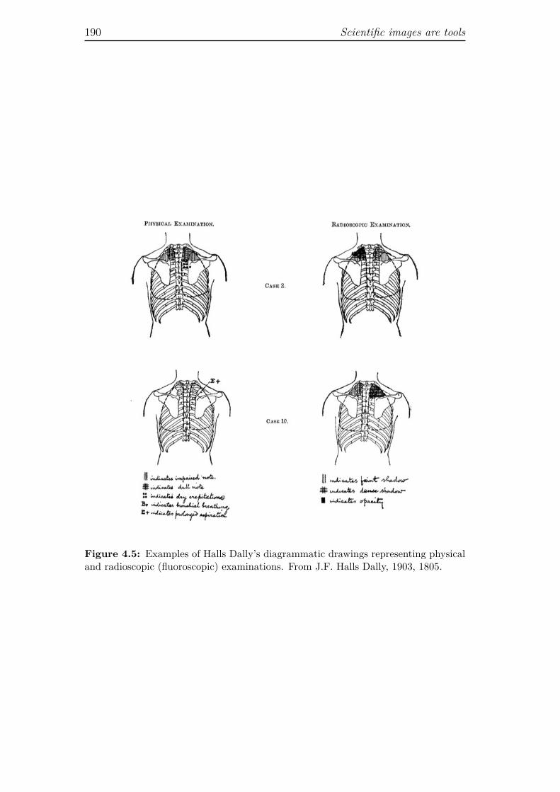
190 Scientific images are tools
Figure 4.5: Examples of Halls Dally’s diagrammatic drawings representing physicaland radioscopic (fluoroscopic) examinations. From J.F. Halls Dally, 1903, 1805.
From vision to meaning 191
mission of information. At the outset of the article the author insisted on thatradiographs are shadows, not maps.58 Accordingly, they could not be read em-ploying a univocal, fully formalized procedure, they had to be interpreted, andthe proper interpretation could be brought about “only by careful and repeatedobservations.”59 Careful and repeated observations allow the radiologist to learnto recognize patterns which, coupled with other knowledge about the disease,can be translated into a diagnosis. To recognize patterns in the shadows is a skillthat has to be learned. Using drawings with captions rather than radiographicimages, Halls Dally provided a quasi-map, a clarification of the shadow-image,which in this way could be more easily integrated in, and articulated with thesemiotic network of the article. This operation attest to the hybrid and con-strained plasticity of scientific images (a very rich visual source is resolved intopatterns that correspond to an interpretation). By selecting, reorganizing, andintegrating with other sources of knowledge the visual content of the originalimage, the diagrammatic representation of the radiograph reduces its complex-ity and at the same time improves its explanatory power, because it displaysa possible interpretation. In this regard, Bastide remarks that published scien-tific images tend to avoid the proliferation of meanings cherished by artists, andchannel the reader into a codified (although not necessarily explicit) significa-tion.60 This codified signification, that we can call guided interpretation, canbe made explicit through the association of a diagram to the original image. Inthis case, the diagrammatic depiction prevents the proliferation of meanings. Itrestricts the number of possible interpretations of the original image, because itcontains and reveals the interpretation of the author of the image. Such interpre-tation can subsequently work as reference for other observers in the significationof other images in the same domain.61 Compared to indexical images, such asphotographs or radiographs, a diagram is a simplified representation. However,Bastide points out that, unlike indices, diagrams can integrate information froman elevated number of sources, adding a different kind of complexity to the orig-inal visual sign without reducing its cognitive accessibility. As she puts it: “[A]graph is probably more ‘convincing’ because it economizes the time and atten-tion of the reader. If we compare it to a photograph, we see that a graph can
58See Halls Dally, 1903, 1800.59Halls Dally, 1903, 1800.60“The reading of the signification of a [scientific] photograph (with scarcely any relation
to its aesthetic character) relies on the use of systems that semiotics calls ‘semi-symbolic.’They couple a difference at the level of the signified to a marked difference at the level of thesignifier.” Bastide, 1990, 201.
61Bastide provides a detailed semantic analysis of the passage from photograph to diagramin Bastide, 1990, 213-220.
192 Scientific images are tools
support many more dimensions than a photograph while remaining readable.”62
Halls Dally’s composite drawings of physical and radioscopic examination offeran example of this semiotic process, whereby the confusing shadows of the origi-nal X-ray image are turned into a diagrammatic representation which is simplerthan a radiograph and at the same time richer, because it gathers additionalinformation from the physical examination.63
Paired representation The question of scientific images interpretation as aprocess whereby simplification and addition of complexity are not mutually ex-clusive has been further explored by the sociologist of science Michael Lynch.According to Lynch, the idea that diagrammatic renderings are simplificationsof more dense images is useful, but it fails to account for the actual intricaciesof the processes of transformation that lead from one representation to another.In the article “The externalized retina: Selection and mathematization in thevisual documentation of objects in the life sciences” he states: “Instead of re-ducing what is visibly available in the original, a sequence of reproductionsprogressively modifies the object’s visibility in the direction of generic pedagogyand abstract theorizing.”64 That is, diagrammatic drawings and other pictorialrenderings of complex images such as radiographs or microphotographs do notsimply select and show in a simplified way the visual content of the original im-age, they transform the very visibility of the original object to accomplish somepedagogical or theoretical aim. Lynch elaborates on his argument by examin-ing how biologists make sense of, and teach novices to make sense of, picturesproduced by electron microscopy. He remarks that they systematically resort topaired representation, which consists of placing diagrammatic renderings along-side an electron microphotograph, in order to orientate the viewer towards aparticular interpretation of the raw image.65 He separates such diagrammaticrenderings in two categories: tracings (that he also calls diagrams) and models.
Tracings are pictorial renderings of specific photographs, while models rep-resent general entities, by synthesizing what can be seen in various photographs,and by assigning visual codes to features, such as biochemical properties, thatcannot be seen under any microscope. In other words, the model summarizes
62Bastide, 1990, 213-214.63An additional level of complexity emerges if we consider the number of the clinical cases
as part of the diagrammatic representation (see Figure 4.5). This would be consistent withthe idea that it is the scientific article as a whole that works as visualizing device, and notjust its individual components. We have to acknowledge, however, that the drawings remainintelligible even if we eliminate the case study reference. Indeed, the textual part of Figure4.5 that is inherent to the intelligibility of the image is the caption, which provides the key tothe symbolic mode of signification.
64Lynch, 1990, 181.65See Lynch, 1990. On paired representation in electron microscopy see also Serpente, 2011.
From vision to meaning 193
visual, experimental, and theoretical information collected not only through dif-ferent techniques, but also in different research projects.66 Hence, while in thecase of tracings we are in the domain of selection and simplification (Lynch usesthe metaphor of the filter), in the case of diagrammatic models things are muchmore complex. Lynch emphasizes that such pictorial renderings are essentialto how “scientific objects and orderly relationships are revealed and made an-alyzable.”67 Diagrammatic models do not simply entail the selection of someelements of the raw image, they also require the synthesis of new forms and thedisplay of different sorts of information, because the model “strives to identifyin the particular specimen under study the ‘universal’ properties which ‘solid-ify’ the object in reference to the current state of the discipline.”68 This goalis reached, according to Lynch’s analysis, putting in place two representationaloperations: selection and mathematization.
The notion of selection (simplification and schematization of the originalimage) gathers four transformative practices that lead from one representationto another: filtering, uniforming, upgrading, and defining.
1. Filtering is the operation whereby the diagrammatic picture reduces theamount of visual elements present in the raw image. As Lynch puts it:“Unused visibility is simply discarded out of the picture.”69 Hence, thisis a process of simplification which contains an implicit interpretation,because it is based on the selection of elements that are deemed importantaccording to the aims of the research.
2. Uniforming entails the application of visual conventions in order to groupelements that share some characteristic (morphological or functional), evenif in the original image such elements might have variable dimensions, het-erogeneous shapes, or occupy distant positions. Uniforming is an explicitform of interpretation, because it requires the decision to aggregate - byusing the same pictorial code - different parts of the image.
3. Upgrading consists of highlighting certain shapes visible in the originalimage, in order to assign them a specific identity (i.e., the borders betweendistinct areas are made artificially clear and distinct), therefore it worksin complementary opposition with uniforming.
66See Lynch, 1990, 168.67Lynch, 1990, 154.68Lynch, 1990, 157, italics in the original.69Lynch, 1990, 161.
194 Scientific images are tools
4. Defining consists of assigning linguistic labels and pointers to the dia-grammatic image, so that the identity of each element is unambiguouslyestablished.70
All these operations are functional to mathematization, that is, they prepare theobject (the original image, and through it, the original specimen), for mathe-matical operations. In Lynch the notion of mathematization has two interlockedmeanings. On the one hand, it refers to the generalization from the specific fea-tures of an individual image (or set of images) to the general morphological andfunctional properties of a natural object. On the other hand, it refers to the setof geometrical and quantitative analyses that can be performed on images oncethey undergo the different processes of selection. In fact, once visual objectsare properly coded, they can be aggregated, examined and compared in relationto a variety of features, such as line slopes, points distribution, recurrence ofspecific patterns, and so forth. Throughout this process “specimen materialsare ‘shaped’ in terms of the geometric parameters of the graph, so that mathe-matical analysis and natural phenomena do not so much correspond as do theymerge indistinguishably on the ground of the literary representation [providedby a textbook or a scientific article].”71 In other words, the diagrammatic rep-resentation is not a pale mirror of the original image, or even of the naturalobject, it is not merely constituted by progressively reducing chaos. It is ratherthe product of an articulated set of operations that produce multiple forms ofvisibility.
Lynch encapsulates this idea in the expression “eidetic image.”72 The termis clearly adapted from Husserl’s philosophy, but in Lynch’s use it is stripped ofits transcendental dimension and refers “concretely to the generalized or ideal-ized version of an object portrayed in a visual document.”73 Hence, by eideticimage Lynch does not refer to a mental picture, but to material visual docu-ments that synthesize and publicly display the knowledge and assumptions ofa scientific domain. Importantly, he stresses that an image is always eidetic inrelation to another image. That is, while the diagram is an eidetic image inrelation to the microphotographs, a microphotograph can be considered eideticin relation to the image one sees when peering through the microscope, becausethe photograph fixes a choice of magnification, framing, and so on. In turn,the microscopic slide (the prepared sample) is eidetic in relation to the originalspecimen, because it is prepared according to criteria for fixation and staining
70“Entities are not only made more like one another, they are more clearly distinguishedfrom unlike entities.” Lynch, 1990, 161.
71Lynch, 1990, 181, italics in the original.72Lynch, 1990, 162.73Lynch, 1990, 183.
From vision to meaning 195
that depend on the aims of the research. This leads us back to the idea of semi-otic network, in which each isolated figure and form of representation acquiresmeaning within a system of internal and external comparisons that bound dif-ferent images and, through them, vision and thinking. It also leads us back tothe issue of the material preparation of the scientific image, which entails thetransformation of the natural object into a scientific object.
4.2.3 Images as part of experiments
I used the idea of semiotic network to describe how the process of significationof scientific images (including diagnostic images) is related to the productionand connection of different images and other print signs, such as words andtables of numbers. However, in the analysis of the first part of Halls Dally’sarticle, I pointed to two moments in which the possible meaning of the image isconnected not to other images or signs, but to material objects and activities:(1) when Halls Dally describes the machinery and the procedures for imageproduction; (2) when in discussing the meaning of the alleged “pleuro-pericardiallines,” he makes reference to other radiographs produced after removing from thecorpse part of the organs under study. In both cases experiential knowledge issummoned to sustain visual and theoretical knowledge, and the two operationspoint to the distributed nature of scientific representation. In the first case,representation is distributed between instruments and operators; in the secondcase, between the radiology cabinet and the morgue, the living body and thecorpse.
In order to interpret an X-ray image, the doctor has to know how it hasbeen produced, and in those cases in which a mark on the radiographic platecannot be unequivocally associated to an anatomical structure or a pathologicalstate, he has to go back to the real body and manipulate it to make sense of theimage.74 This necessity to get out of the domain of images and enter the domainof material objects is particularly strong in the stages of development of a novelimaging technology. It tends to fade away once instruments and procedureshave been black-boxed, and once the points of correspondence between imagesand bodies have been secured. However, the need to resort to the domain ofmaterial objects re-emerges each time an image obtained with a well knowntechnology shows something unexpected. In the case of medical diagnostics thephysician possesses a reasonable amount of knowledge about the imaged objects(human anatomy and physiology as well as pathological states). Thus, after
74The manipulation of the patient’s body might consists in a biopsy, if the uncertainty con-cerns the diagnosis, or even in post mortem autopsy, if the uncertainty is about morphologicaland anatomical elements, as in the case described by Halls Dally.
196 Scientific images are tools
excluding possible artifacts, he will try to make sense of the unexpected marksby searching some clue in the patient history. For example, the lung radiographof a patient with mild symptoms of pneumonia might show lesions that lookmore serious than the symptoms would make one think. This could mean thatthe pneumonia is actually worse than it seems from the symptoms, that there isanother pulmonary disease besides the pneumonia, or that the patient alreadysuffered from pneumonia or another lung disease in the past and this left scarson the lungs after remission. In any case, the physician has at his disposal anumber of additional tests to solve the doubts posed by the image. Things areeven more complicated in basic research, where background knowledge about theobject of study is scant and the range of possible interpretations much widerthan in clinical medicine. Here the experiential and experimental dimensionof image interpretation appears more clearly than in the case of the imagesproduced in contexts in which the correspondence between the image and itsreferent is well defined.
Visual and experiential knowledge In a seminal ethnographic study con-ducted in a German laboratory of molecular biology in the late 1980s, KarinKnorr-Cetina and Klaus Amann have shown that the interpretive process carriedout by biologists to make sense of DNA and RNA autoradiographs involves awhole “repertoire of chunks of visual and experiential knowledge.”75 Autoradio-graphy, associated to gel electrophoresis, is used in molecular biology to analyzenucleic acids macromolecules (DNA and RNA) on the basis of their size andelectrical charge. The electrophoresis gel is a thin layer of agarose with holes(called wells) at one end. To produce an autoradiograph, samples of macro-molecules digested and marked with radioactive labels are loaded in the wells,and an electric current is turned on, creating an electric field that makes the neg-atively charged molecules move through the gel. Since smaller molecules movemore easily through the pores of the agarose matrix, they are faster and thusmigrate farther than the larger ones. As a consequence, after a given lapse oftime molecules of different size will form distinct bands on the gel. Because themolecules have radioactive labels, if a sensible film is exposed to the gel, it willbe marked producing the autoradiograph of the fragments of RNA and DNAarrayed on the matrix (Figure 4.6). It is important to note that the size of themolecule is not related to the dimension of the bands, but to their position onthe gel, which is a direct function of the distance travelled during electrophore-sis. This is why in order to calculate their dimension, it is necessary to analyzethe location of each fragment in relation to a marker, a built-in visual reference
75Knorr-Cetina and Amann, 1990, 263.
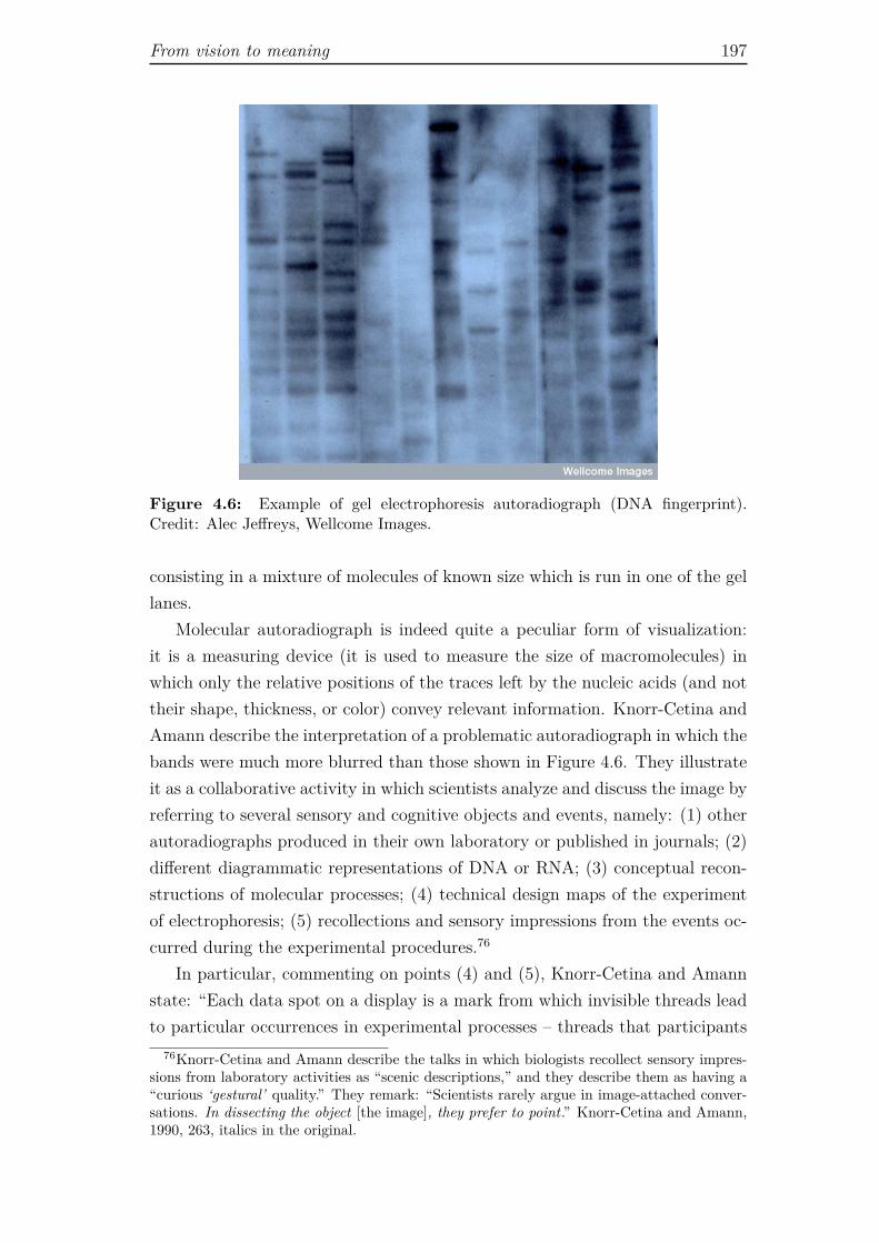
From vision to meaning 197
Figure 4.6: Example of gel electrophoresis autoradiograph (DNA fingerprint).Credit: Alec Jeffreys, Wellcome Images.
consisting in a mixture of molecules of known size which is run in one of the gellanes.
Molecular autoradiograph is indeed quite a peculiar form of visualization:it is a measuring device (it is used to measure the size of macromolecules) inwhich only the relative positions of the traces left by the nucleic acids (and nottheir shape, thickness, or color) convey relevant information. Knorr-Cetina andAmann describe the interpretation of a problematic autoradiograph in which thebands were much more blurred than those shown in Figure 4.6. They illustrateit as a collaborative activity in which scientists analyze and discuss the image byreferring to several sensory and cognitive objects and events, namely: (1) otherautoradiographs produced in their own laboratory or published in journals; (2)different diagrammatic representations of DNA or RNA; (3) conceptual recon-structions of molecular processes; (4) technical design maps of the experimentof electrophoresis; (5) recollections and sensory impressions from the events oc-curred during the experimental procedures.76
In particular, commenting on points (4) and (5), Knorr-Cetina and Amannstate: “Each data spot on a display is a mark from which invisible threads leadto particular occurrences in experimental processes – threads that participants
76Knorr-Cetina and Amann describe the talks in which biologists recollect sensory impres-sions from laboratory activities as “scenic descriptions,” and they describe them as having a“curious ‘gestural’ quality.” They remark: “Scientists rarely argue in image-attached conver-sations. In dissecting the object [the image], they prefer to point.” Knorr-Cetina and Amann,1990, 263, italics in the original.
198 Scientific images are tools
[scientists] pursue in image dissection.”77 That is, when an image is particularlyresistant to interpretation, when its very visual content is difficult to decipher,scientists tend to retrace the whole process of image production. They look out-side of the image, into a whole range of objects and activities that are part of theexperimental setting. Indeed, the autoradiograph does not simply visualize theresults of the experiment (which entails samples preparations and electrophore-sis), it contains a range of information about various steps of the laboratoryprocess. It says, for example, if the electrophoresis worked properly, or if thesamples were treated adequately. It literally bears a trace, or a “signature,” asKnorr-Cetina and Amann call it, of all those steps. An autoradiograph, theysay, “is the result of an imaging technology that creates visible traces of invisiblereactions.”78 Accordingly, although the goal of the electrophoresis is to measurethe length of nucleic acids fragments, what the autoradiograph shows is a com-pound of information that points back to all the invisible reactions that precededthe impression of the X-ray film. As a consequence, in order to interpret theimage one has to be able to reconstruct at least in part the wider experimentaldomain in which those reactions occur (for instance, to know if the amount ofnuclease used to process RNA and DNA before the electrophoresis was enough,if the time of exposure of the X-ray film was adequate, and so forth).
As Knorr-cetina and Amann aptly put it: “[Biologists] treat images as self-contained subjects that carry the analysis of an event within them.”79 Theautoradiography of gel electrophoresis is an example of a sophisticated imagingtechnique in which the problem of image signification is twofold. On the onehand, it is impossible to directly compare the information provided by the imagewith the object of study. On the other hand, due to the complexity of the wholeexperimental process that leads to the production of the autoradiograph, thecausal relationship between object and image has not warranted stability andreproducibility.
Representing and intervening The analysis provided by Knorr-Cetina andAmann supports Ian Hacking’s well known claim that, in science, represen-tation and intervention are deeply enmeshed activities. In order to representsomething, one has to manipulate instruments and the object of study.80 Hack-ing’s argument, rooted in the debate on scientific realism vs. anti-realism, isaimed at contrasting what he calls “the spectator theory of knowledge,”81 that
77Knorr-Cetina and Amann, 1990, 274.78Knorr-Cetina and Amann, 1990, 263.79Knorr-Cetina and Amann, 1990, 262, italics in the original.80See Hacking, 1983.81Hacking, 1983, 130.
From vision to meaning 199
is, the idea that scientific knowledge is an abstract theoretical representation ofthe world. He criticizes this stance, claiming that any doctrine of knowledgebased on the “contemplation between theory and the world” is doomed to leadto “an idealist cul-de-sac.”82 In order to avoid this dead end, he puts forwardan instrumentalist theory of knowledge, according to which we can have a firmgrip on reality, and get to know something about it, only when we intervene innature, when we create new phenomena,83 for example by spraying positronsand electrons on niobium droplets to increase or decrease their charge.84
Hacking’s instrumental theory of knowledge informs his theory of observationand of scientific images. Images, he maintains, become intelligible not becausewe look at them very carefully and with a well defined preconceived theoryabout what we are going to see, but because we perform on and around thema number of interlocked different activities. He uses the example of microscopyimages, and makes two fundamental, interrelated claims: (1) “You learn tosee through a microscope by doing, not just by looking,”85 and (2) “We donot in general see through a microscope; we see with one.”86 In both sentencesHacking points to the fact that seeing something under a microscope, that is,recognizing something meaningful and scientifically relevant, is not simply amatter of watching, it entails a range of bodily and instrumental activities.When we look for the first time through a microscope, all we see are patchesof colors. Students in high school and university learn to recognize what theyare seeing by exercising with reference microphotographs and drawings dulycaptioned. This is a good basic training for routine work, but it is not how wediscover something new. If we are to learn something new about a specimen weare observing under a microscope, we will have to use several microscopes andseveral ways to prepare the specimens. We will need different images of the samesample, or of different samples of the same object, in order to compare them. Theresearcher interested in finding something new or in sorting out structures fromartifacts has to learn to produce different images with a suitable instrument,under suitable conditions. Hacking draws a parallel between his idea that wesee with and not through a microscope, and Berkely’s theory of vision, accordingto which three-dimensional vision is an acquired skill correlated with the sense
82Hacking, 1983, 130.83Curiously enough, Hacking never mentions the work of Gaston Bachelard who, in the
1930s, coined the term “phénoménotechnique” to refer exactly to the fact that modern sciencecreates its own phenomena. I discuss the concept of phenomenotechnique in Chapter 5.
84“Now, how does one alter the charge on the niobium ball? ‘Well, at that stage,’ said myfriend, ‘we spray it with positrons to increase the charge or with electrons to decrease thecharge.’ From that day forth I’ve been a scientific realist. So far as I’m concerned, if you canspray them then they are real.” Hacking, 1983, 23, italics in the original.
85Hacking, 1983, 189.86Hacking, 1983, 207.
200 Scientific images are tools
of touch and with actual tampering with the world. He writes: “Whether ornot Berkeley was right about primary vision, new ways of seeing, acquired afterinfancy, involve learning by doing, not just passive looking.”87
Hacking works this idea through the example of the discovery of dense bod-ies in red blood cells. Dense bodies are small spots visualized in erythrocytesobserved using low power electron microscopy. They are called “dense” simplybecause they are electron dense, which means that they appear at the elec-tron microscope without any staining or preparation. When they were noticedfor the first time, nobody knew if they were actual biological structure or justartifacts. To discriminate artifacts from real objects is a common problem inmicroscopy, and in visualization technologies in general. So, how do scientistsknow that what they are seeing is real and not just an erratic creation of theimaging apparatus? We have already encountered this problem in Chapter 3,discussing the question of depiction and detection in photography. We saw thatPerini considers microscopy artifacts as paradigmatic stances in which depictionhampers detection, because they provide an example of how we can be mistakenby the apparent mimetic properties of an image. In Perini’s analysis, scientistscan overcome this pitfall by granting microscopy images a well circumscribedmeaning, that is, by seeing them not as more or less indirect depictions of thereal microscopic entity, but as depictions of the visual features of a sample pre-pared and visualized under specific conditions.88 On the basis of this restraintand cautious vision, as it were, the biologist is motivated and entitled to carryon a range of different activities aimed at establishing if the forms observedunder the microscope correspond or not to some actual quality of the naturalobject. In this respect, Perini’s position overlaps with Hacking’s. Still, in heraccount the epistemic warrant of mechanically-produced images relies primarilyon the causal interaction between the object under study and the detector.89
Hacking, on the contrary, tends to downplay the relevance of the microscope asmapping device.90 He rather insists on the fact that when it comes to distin-guish artifacts from reality, scientists implement different practical strategies:(1) they check if the same structures appear under different microscopes thatwork on the basis of unrelated – and well understood – physical processes;91 (2)they construct objects with specific known features and they observe them with
87Hacking, 1983, 189.88See Section 3.2.89See Perini, 2006, 868.90The microscopy image can be conceived of as “a map of interactions between the specimen
and the imaging radiation.” Hacking, 1983, 207.91In the case of dense bodies the results of the electron microscope were checked with a
fluorescence microscope.
From vision to meaning 201
a microscope;92 (3) they manipulate the objects they want to observe, for ex-ample, by microinjecting them, and look at how these manipulations affect theimage; (4) they cross the observations done with the microscope with the resultsfrom other sciences, namely biochemistry and crystallography, which can con-firm if the structures that are visualized might correspond to specific chemicaland physical properties.93 This means, again, that scientists endow instrument-mediated observations with meaning not merely by looking at the mechanicalrecord provided by the machine, but through a range of activities that combinetheoretical and experimental work. Scientists are not remote spectators, theyare hand-on sense-makers.
When doctors look at what might be a lesion revealed by a diagnostic scan,they judge their findings and weigh them against an idea they have formedabout the lesion, a mental image which is the product of previous experience.The experiences on which this mental process is based relate to medical educa-tion (anatomy, physiology, pathology, and so forth), experiential knowledge ofthe inner body (direct observation of intraoperative findings or corpse dissec-tion), comparison between images produced with the same imaging modality,and comparison between images and the results of other tests. In other words,the diagnostic meaning of a particular lesion in a particular image emerges fromprevious experience, which is constructed at both the historical-cultural (knowl-edge developed within the scientific community) and individual level (knowledgeacquired and developed by the single practitioner). Books, articles, dissectingroom and surgical table, images form the same patient or from different subjects,laboratory tests, discussions at ward meetings or conferences, all these objectsand events enter the process of image interpretation. In the case of well knownpathologies, black-boxed technologies, and experienced radiologists, the imagebecomes a self-contained epistemic object, and the doctor can see at a glancethe disease in the radiographic plate (or, more likely, on the computer screen).Still, each time doubts arise, the practitioner can get outside of the image andconsult other objects, machines, events, and people involved in the distributedprocess of signification and interpretation. Doctors work with images in order
92Hacking calls this argument “the argument of the grid,” because he uses as example themetal grid employed by biologists to compare a specimen under an electron and a fluorescencemicroscope. Back in the 1980s, the procedure consisted of drawing on a paper a grid witha letter in each square, and then to photographically reduce it. The biologist, who knewexactly the characteristics of the grid, fixed the specimen on it and mounted the slice on themicroscope. Through the microscope he would see exactly the grid he had constructed, withits lines and letters. Hacking concludes that, in this case, we have good reasons to believe thatwhat we see under the microscope is a feature of the specimen and not an artifact because:we made the grid, we know that the manufacturing process is reliable, and we can check theresult through different microscopes. See Hacking, 1983, 202-204.
93Hacking, 1983, 209.
202 Scientific images are tools
to produce a specific visibility of the body and of the disease. This visibility isa presentation of the body and the disease under experimental conditions. Itturns such complex entities into objects amenable to medical comprehension andpractice. Images are tools pertaining to the larger toolbox of medical practiceand they allow the physician to work on the human body and treat its diseases.Resorting to Lynch’s notion of eidetic image, we could say that doctors producean eidetic image of the human body not only because the radiograph fixes spe-cific choices in terms of image production (radiation intensity, time of exposure,reciprocal position of body and machine, etc.), but also because it fixes a specificway of understanding (theorizing) both the body and disease.
In this chapter I approached the notion of images as tools by covering a long,somewhat heterogeneous route. I went through the discussion of the haptic, em-bodied dimension of image perception, engaged in the semantic analysis of howscientists make sense of photograph-like images, and finally dealt with the expe-riential and collective dimension of image production and signification. In thebackground always lingered the tension between material and mental represen-tation, representations and objects, pictures and language, vision and gesture,images and laboratory practices. This unresolved tension is due, I think, to theambiguous nature of images themselves. It depends on the fact that they area go-between that links material and mental dimensions, presence and absence,sensible experience and concept. In the following chapter I engage more deeplywith the idea that scientific images are experimental tools. More precisely, Iexpound the thesis that diagnostic images are to be understood as experimentaldevices because they present the body under experimental conditions, througha process that entails a specific reconfiguration of both body and illness. I doso by drawing on Gaston Bachelard’s idea of phenomenotechnique. I apply theconcept of phenomenotechnique to both medical images and bodies, in orderto show how the lived experience of illness is effectively turned into a visualscientific object.
Chapter 5
Medical imaging as
“phenomenotechnique”
Bachelard coined the concept of phenomenotechnique in the article “Noumèneet microphysique,” of 1931, to describe the activity of mature sciences. In thistext the philosopher claimed that the science of his time, characterized by thetheoretical revolutions and empirical successes of quantum mechanics and space-time relativity, was no longer a phenomenography [phénoménographie].1 Thatis, it was not a representation of the world based on the observation and analysisof natural phenomena. It was a “phenomenotechnique, whereby new phenom-ena are not simply found, but invented, created anew.”2 This concept, furtherdeveloped in Le nouvel ésprit scientifique, of 1934, became a fundamental cat-egory of Bachelard’s epistemology. To him, non-Euclidean geometry, relativitytheory, quantum mechanics, and quantum chemistry are the outcomes of a sci-ence that has broken any relation with the data of our everyday experience.Referring to modern chemistry, Bachelard remarked that it had completely bro-ken with ancient natural history, which described all sorts of natural matter(mineral, vegetal, animal) on the basis of collections of facts rooted in sensibledata and connected through ambiguous relations of similarity. To study matter,Bachelard claimed, one has “to make profession of facticity,”3 that is, one hasto produce substances that do not exist in nature, and to re-make those thatexist, in order to analyze them under the proper degree of purity.4 He wrote:
[In science] phenomena must be selected, filtered, purified, shaped byinstruments; indeed, it may well be the instruments that produce thephenomenon in the first place. And instruments are nothing but theories
1Bachelard, 1931, 18.2Bachelard, 1931, 18, my translation.3Bachelard, 1953, 22, my translation, italics in the original.4See Bachelard, 1953, 22.
203
204 Medical imaging as “phenomenotechnique”
materialized. The phenomena they produce bear the stamp of theorythroughout.5
Modern science does not find its objects in nature, it realizes them in the formof new physical effects (e.g., Zeeman effect),6 purified chemical substances, syn-thetic molecules. It has broken with the primacy of primitive sense data properof naïve realism and praised by positivist philosophers, and has turned into a“realization of the rational,”7 characterized by the integration of theory andexperiment, bounded in a dialectical relation.8 For Bachelard, “science realizesits objects without ever just finding them ready-made. Phenomenotechniqueextends phenomenology. A concept becomes scientific in so far as it becomesa technique, in so far as it is accompanied by a technique that realizes.”9 Inother words, science must replace a phenomenology that describes natural phe-nomena10 with a phenomenotechnique that inscribes the natural phenomenonwithin scientific rationality. To quote Bachelard again:
A truly scientific phenomenology is [...] essentially a phenomeno-technology. Its purpose is to amplify what is revealed beyond appear-ance. It takes its instructions from construction [Elle s’instruit par cequ’elle construit]. Wonderworking reason designs its own miracles. Sci-ence conjures up a world, by means not of magic immanent in realitybut of rational impulse immanent in mind. [. . . ] Scientific work makesrational entities real, in the full sense of the word.11
In his effort to elaborate a philosophy of science situated beyond the receivedtraditions (positivism vs. formalism, empiricism vs. conventionalism, realismvs. idealism), which he considered the product of unjustifiable abstractions andgeneralizations,12 Bachelard developed an original brand of rationalism thatpointed towards an absolute mathematization of the world, but at the sametime commanded the reification of reason into artificial material entities. In atext written in 1936 for the first and only issue of the Surrealist review Inquisi-tions, he wrote that human reason has to be turbulent and aggressive, in order
5Bachelard, 1934, 13.6The Zeeman effect, named after the Dutch physicist Pieter Zeeman, is the effect of splitting
a spectral line into several components in the presence of a static magnetic field. It was firstproduced by Zeeman in 1896, by holding a flame between strong magnetic poles.
7Bachelard, 1934, 5.8Rheinberger aptly defines this relationship between theory and experiment as a dialecti-
cally constituted material-discursive circle. See Rheinberger, 2005, 316.9Bachelard, 1938, 70, italics in the original.
10It is not clear if when Bachelard uses the term phenomenology in opposition to thatof phenomenotechnique he is referring to the Husserlian tradition, or if he simply means“phenomenography,” that is, the observation and description of natural phenomena based onprimary sense data. I incline to the latter interpretation, but this is a problem open to debate.
11Bachelard, 1934, 13.12See Bachelard, 1949, 5.
205
to found a surrationalism. It has to establish “an experimental reason capable ofsurrationally [surrationellement] organize the real, just as Tristan Tzara’s exper-imental dream organizes poetic freedom in a surrealist way.”13 Rationalism mustbe untamed and experimental at the same time, it has to break with the real,in order to create new material objects and phenomena that are the only pos-sible scientific reality. Based on a dialectical relation between rationalism andmaterialism, Bachelard’s epistemology is quite complex, and it would deservea detailed discussion that I cannot put forward here (it would require anotherdissertation). What is important to stress in the scope of my work is that thetechnical realization of the rational was so relevant to Bachelard epistemology,that he was not afraid to call his rationalism realism. Of course, Bachelard’srealism, just as his rationalism, is of a peculiar sort. And indeed he uses the ex-pressions “educated realism,”14 “realism at one remove,”15 or “technical realism”[réalisme téchnique]16 in opposition to common scientific realism. Bachelard’srealism concerns a reality that can be grasped only through the technical real-ization of the rational.17 In Le Nouvel esprit scientifique, he explained:
[Technical realism] has nothing to do with traditional philosophicalrealism. It is rather realism at one remove, conceived in reaction to theusual notion of reality, as a polemic against the immediate; it consistsof realized reason, reason subject to experimentation [un réalisme faitthe raison réalisée, de raison expérimentée]. The ‘reality’ to which thisrealism corresponds is not transferred into the realm of the unknowablething-in-itself . It has a noumenal richness of quite another order. Thething-in-itself is a noumenon by exclusion of phenomena, whereas scien-tific reality, I would argue, consists in a noumenal context suitable fordefining axes of experimentation.18
This idea of a non-Kantian noumenon appeared also in the 1931 article onmicrophysics where the notion of phenomenotechnique was first introduced:
We could therefore say that mathematical physics corresponds to anoumenology quite different from the phenomenography to which sci-entific empiricism condemns itself. This noumenology implies a phe-
13Bachelard, 1936, 8, italics in the original, my translation.14Bachelard, 1937, 3.15Bachelard, 1934, 5.16Bachelard, 1934, 5.17Jean Gayon has called Bachelard’s scientific realism an “unprecedented realism” [réalisme
inédit]. He remarks that during the development of his epistemology, Bachelard moved com-pletely away from idealism, while submitting realism to a profound reformulation, throughthe separation of the concept of reality from that of thing. In Bachelard’s epistemology realitybelongs to the realm of the noumenon technically materialized, not of nature. See Gayon,1994, 22-23.
18Bachelard, 1934, 5-6, italics in the original.
206 Medical imaging as “phenomenotechnique”
nomenotechnique whereby new phenomena are not simply found, butinvented, that is, created anew.19
For Bachelard, the noumenon is not the unknowable thing-in-itself that liesbeyond the phenomenon. It is the deep mathematical structure that shapesphenomena, the set of relations that inform reality and that are described bytheoretical physics.20 When we study nature at the scale of the infinitely small,Bachelard says, as in the case of microphysics and quantum chemistry, we can-not think anymore in terms of reality of things, we must think in terms of realityof organization, of relations.21 These relations are described by the equations ofadvanced physics and, in turn, such equations allow designing the experimentsthat materialize the very world they describe.22 It is not simple to provide anexact definition of the term noumenon as used by Bachelard. However, evenwithout entering into the details of an exegesis of this concept, we can highlightthe fact that Bachelard conceived the noumenon in a strict relation with phe-nomenotechnique. Regardless of how one exactly defines the noumenon, it hasto be considered as a rational substratum that defines the lines of thought alongwhich experiments can be envisaged and brought about. As seen in the quoteabove, to Bachelard any “noumenology implies a phenomenotechnique.”23 Ra-tionality must be applied, in the sense that it has to be technically implemented.The objects of science are real, yet they are not natural: they are technicallyproduced and theoretically invested entities.24 Now, if scientific objects are ra-tional constructs technically reified, then scientific instruments and technologiesare not secondary products of science, but rather part and parcel of scientificactivity.
By integrating technology (technical production of phenomena) into science,Bachelard drew a line between the reality of science and the reality of the naturalworld. In this vision, scientific reality belongs to the realm of the noumenontechnically materialized, not of nature, and the role of phenomenotechniqueis to realize mathematical rationality at the phenomenal level. Reason andtechnique enter a dialectical circle of reciprocal shaping. Bachelard gave severalexamples of materialization of mathematical rationality. For instance, if one isto study atomic isotopes, one has to produce them in the mass spectroscope,25
and to study piezo-electric phenomena (i.e., the production of electricity bycompression or dilatation of a crystal) one needs to work with an ideal, i.e. pure,
19Bachelard, 1931, 18-19, my translation.20See Bachelard, 1931, 18-19.21See Bachelard, 1931, 13.22See Bachelard, 1931, 17.23Bachelard, 1931, 19.24See Rheinberger, 2005, 316.25See Bachelard, 1949, 103.
207
quartz, which cannot be found in nature and must be produced in the laboratory.The crystal thus obtained, he explained “is not simply matter provided withgeometrical properties. It is materialized geometry. The crystal created inthe laboratory is not an object, really, it is an instrument.”26 For Bachelard,this synthetic crystal is a rational device that produces scientific phenomena, itprovides the conditions for proper scientific observation and analysis.
The emphasis put by Bachelard on the creation of phenomena implies anemphasis on the role of instruments and technology in the production of scien-tific knowledge. Bachelard did not develop a philosophy of technology, yet, asalready explained, the concept of phenomenotechnique entails a view of scienceapplication not as a mere byproduct of fundamental research, but as a consti-tutive element of the scientific process. Consequently, technology, in the formof scientific instruments and artificially produced phenomena, is embedded intoscience since the outset, because our scientific understanding of the world is nec-essarily mediated by instruments and scientific instruments are an embodimentof knowledge.27 Discussing the processes whereby scientists select the relevantfeatures of a phenomenon before they set out to study it, Bachelard noted thatthe role of technological apparatuses in scientific research is that of reducingand controlling the richness and interrelation of natural phenomena:
The will to neglect is especially active in contemporary technology.A piece of apparatus can indeed be described negatively, if we may beallowed the expression, as well as positively. It is defined in terms of theperturbations it guards against, the technique isolating it, the assuranceit gives that clearly defined influences can be neglected, in short in termsof the fact that it comprises a closed system. There is a whole complex ofshields, casing, and immobilisers that fences in the phenomenon. All thisassembled negativism that a piece of apparatus is in modern physics runscounter to the sloppy affirmations of the possibility of some undeterminedphenomenological interaction.28
The technological apparatus required by advanced physics is a piece of “as-sembled negativism” because its function is that of remaking the natural phe-nomenon into a scientific one. Scientific instruments tame natural phenomenaby reducing their variability, their interconnection with multiple related phe-nomena, and all the contingencies that influence their actual manifestations.
26Bachelard, 1949, 202, italics in the original, my translation.27It is worth noting that for Bachelard mathematical models and calculus techniques are
scientific instruments just in the same manner as the material instruments used in the labo-ratory. “Psychologically, tensor calculus is the matrix of relativistic thinking. Contemporaryphysical science has been created by this mathematical instrument, much as microbiology wascreated by the microscope. None of the new knowledge is accessible to anyone who has notmastered the use of this new instrument.” Bachelard, 1934, 56.
28Bachelard, 1938, 221.
208 Medical imaging as “phenomenotechnique”
The task of scientific instruments and scientific rationality is that of isolat-ing phenomena, dissecting them into fundamental components, and assuringtheir stability and reproducibility. In the laboratory the natural phenomenonis fenced. It becomes a “closed system” that can be manipulated and examinedat will.
By constructing its own objects, modern science has transformed the veryconcept of phenomenon. A scientific phenomenon is no longer a datum, some-thing exceptional and surprising that one finds in nature, it is rather an effect,something that is produced in the laboratory. In this way science creates thecircumstances for producing objective knowledge, because a phenomenotech-nique is a method of objectification of the real, a process whereby well definedobjects and regular phenomena are created.29 The reality of Bachelard’s epis-temology is a reality in which the world has been dissolved into rationality andre-materialized into experimental phenomena produced by technological appa-ratuses.
5.1 Diagnostic images and phenomenotechnique
Having discussed Bachelard’s concept of phenomenotechnique, it is now neces-sary to explain how it can be applied to medical imaging. How does his visionof science as an activity that creates its own objects apply to the productionof diagnostic images? According to Bachelard, phenomenotechnique is the pro-cess whereby science creates the technical entities it can study. In the case ofmedicine, the new objects created by the technological apparatus are imagesof the human body. Hence, one might claim that if these objects (the images)are new and artificial, because phenomenotechnically created, they have no cor-respondence with any real object (the body). In such case the application ofthe concept of phenomenotechnique to medical imaging would imply the absurdproposition that the imaging technology produces human bodies and diseases.More generally, it would imply that the whole activity of medicine consists ofproducing bodies and diseases as technical phenomena. The human body andits illnesses would thus become inventions of medicine technically implemented.This would be, indeed, the position of radical constructivists.30 Since I do not en-dorse this perspective, I have to clarify how the concept of phenomenotechnique
29“In early instrumental knowledge, the same obstacle can be seen to arise as in ordinaryobjective knowledge: the phenomenon does not necessarily make its most regular variableavailable for measurement. On the other hand, as instruments are improved, their scientificproduct will be better defined. Knowledge becomes objective in proportion to it becominginstrumental.” Bachelard, 1938, 217.
30See, for instance, Latour, 1984 and 1999, Ch. 5.
Diagnostic images and phenomenotechnique 209
can help thinking medical imaging without falling into radical-constructivism.To this end, it is important to make some distinctions. For what concernsthe application of the concept of phenomenotechnique to medicine, one mustfirst distinguish between medical research and clinical practice. If we take intoaccount medical research, in particular that branch of contemporary medicalresearch based on molecular analysis, we can say that it works quite preciselyaccording to the idea of phenomenotechnique. In the molecular laboratory,mechanisms of disease, immunological response, and therapeutical action areroutinely produced as artificial phenomena in a variety of forms. This requiresthe manufacturing of a variety of entities, that range from cellular cultures togenetically engineered viruses, from model organisms, wherein human diseasesare induced,31 to computational simulations. Genes and biological moleculesare the mathematical (or rather biochemical) noumenon of molecular medicine:genetic and molecular models provide the rational structures along whose linesnew experiments can be conceived and artificial objects and phenomena pro-duced.
The relationship between the concept of phenomenotechnique and medicineis less straightforward when it comes to clinical practice and, more precisely,to the part of clinical practice that deals with diagnosis. Unlike the medicalresearcher, the clinician is not engaged in explaining the fundamental mecha-nisms of health and disease. The daily work of the medical practitioner is notto produce experimental models of disease, but rather to recognize what diseaseaffects a specific person. The practitioner does not creates artificial objects, hehas to deal with that natural object which is the patient’s body. Still, his accessto such body and to his illness is by necessity mediated. He has to reconfigurethe lived experience of illness in medical terms and within a medical praxis. Hiswork is not to create a diseased body as a technical entity, yet he must reframethe natural phenomenon (the patient’s body and his being ill) within medicalrationality. In this endeavor the physician resorts to a vast array of instru-ments, including imaging technologies. In clinical medicine, the methodologicalimperative of phenomenotechnique to create the scientific object is transformedinto the imperative to reconfigure the object of study under artificial and con-trolled conditions. This was indeed the founding principle of clinical anatomy,as articulated by Bichat at the turn of the eighteenth century.
31Model organisms are also called experimental models of disease and belong to a wide rangeof species. The most common are Saccharomyces cerevisiae (yeast), Drosophila melanogaster(fruit fly), Caenorhabditis elegans (roundworm), Danio rerio (zebra fish), Mus musculus(house mouse), and Rattus norvegicus (brown rat). Animal models employed to study hu-man disease are usually selected because of their similarity to humans in terms of genetics,anatomy, and physiology, as well as for their unlimited supply and ease of manipulation. SeeSimmons, 2008.
210 Medical imaging as “phenomenotechnique”
Bichat set the foundations for the birth of clinical anatomy on the assumptionthat the confusion of symptoms presented by the living patient and recorded bythe doctor at the bedside presented nothing but “a train of incoherent phenom-ena.”32 To impose a coherence on such disorder it was necessary to observe thecadaver, a proxy of the living body where the confusion of the disease is fixed inthe form of an organic lesion. With the birth of the clinic, the lived experienceof the patient was ideally let aside, and physicians had to learn make sense ofillness on the basis of a newly defined geography of the corpse. It was still aphenomenography (as opposed to phenomenotechnique), but one that forced thepathologist to separate the disease from the ambiguity of its symptomatic man-ifestations. Anatomical dissection itself can be conceived as a technique thatallowed to materialize a specific conception of disease, namely, the idea thata disease is an organic lesion rather than a train of symptoms. Accordingly, itcommanded the development of countless technologies to explore the inner bodyand its functions. Every diagnostic technology introduced in clinical medicineenhanced the visibility of the body, and our knowledge about it, by introducingdifferent layers of instrumental mediation between the observer (the physician)and the object of study and intervention (the patient).
It is exactly because the concept of phenomenotechnique emphasizes themediated and instrumental character of scientific knowledge, that it can beuseful for thinking medical imaging and diagnostic images. The concept ofphenomenotechnique can provide important insights into the epistemology ofmedical imaging because, by highlighting the relevance of instruments in theproduction of scientific knowledge, and the indirect, mediated nature of sci-entific experience, it helps clarifying different issues concerning the relationshipbetween technology and science. Consequently, it helps thinking the relationshipbetween diagnostic images (the product of a technology), and medical knowl-edge (a specific form of scientific knowledge). To say that medical imaging canbe thought resorting to the concept of phenomenotechnique does not mean,of course, that medical imaging is a straight example of phenomenotechnique.Thus, my aim is not that of fitting radiology into phenomenotechnique, but ofcombining this concept with the other concepts I have explored in the differentchapter of this dissertation in order to reflect upon the epistemology of medicalimaging. This means that I use Bachelard’s phenomenotechnique as a point ofdeparture, a stimulus for thinking. An intellectual inspiration rather than a setof stiff rules that must be applied point by point.
To Bachelard, scientific perception is the opposite of natural perception be-cause it transcends the immediate and is always polemical in relation to common
32Bichat, 1801, vol. 1, 60. See Section 1.1.
Diagnostic images and phenomenotechnique 211
sense knowledge.33 This is why the scientific spirit has to be formed againstnatural mental habits.34 In my appropriation, as it were, of the Bachelardeanconcept, I bracket the polemical aspect of scientific perception and highlight itsartificial nature. I stress the fact that scientific experience transcends the im-mediate because, thanks to mechanical and electronic instruments, the scientistgains access to a sensorial domain, which is also an intellectual domain, that isprecluded to natural perception. Understood in this way, the concept of phe-nomenotechnique can be applied to medical imaging and put in relation withBenjamin’s optical unconscious. Imaging technologies create phenomena thatreveal “entirely new structures of matter.”35 They extend natural perceptionand allow analyzing and dissecting reality from novel, unexpected angles.
As a large set of imaging technologies, medical imaging can be conceptualizedin terms of phenomenotechnique because it produces artificial, controlled phe-nomena – diagnostic images – that help gaining knowledge about what happensinside the human body. The function of these images is to replace the actualpatient’s body when we need to answer specific diagnostic questions. Also, theytransform the physician’s perception, by providing instrumentally mediated ac-cess to his object of study, and they simultaneously turn a natural object (thehuman body affected by a disease) into a scientific object. Moreover, both theimaging apparatus and the images it produces can be considered reified theoriesfor two reasons: on the one hand, especially in the case of digital imaging, theyreally materialize mathematical equations; on the other hand, they materializea vast array of knowledge from different domains and, in turn, influence thedevelopment of this very knowledge. In what follows I argue in favor of theseideas along four lines of analysis:
1. Diagnostic images act as a technical medium of the human body, thusmediate the access of the doctor to the patient’s body. By introducingan experiential and intellectual separation (the doctor looks at the bodyby means of the image) they make visible that which is concealed. Bymediating perception they grant cognitive access to objects and processesthat are otherwise hidden within the body.
2. Diagnostic images are a technical reconfigurations of the patient’s body,in the sense that they selectively display information about the inner bodyin a way that is useful for diagnostic purposes. Although they often looklike mimetic representations of the inner body, they are better understood
33See Bachelard, 1934, 12-13.34See Bachelard, 1938, 33.35Benjamin, 1939, 266. See Section 2.3.1.
212 Medical imaging as “phenomenotechnique”
as visual reconfigurations of non visual data that carry information aboutthe internal organs and their functioning.
3. Diagnostic images are embodied knowledge, in the sense that they are theoutcome of a technology that relies on theories, hypotheses, concepts, andmaterial practices produced in different scientific fields, such as physics,chemistry, biology and medicine.
4. Diagnostic images are diagnostically meaningful only if the observer (thephysician) has a proper training and is in command of adequate theoreticalknowledge and practical skills, whereby he is placed at the core of anepistemic network.
5.1.1 Diagnostic images as a medium of the inner body
The idea that diagnostic instruments in general, and medical images in partic-ular, are a medium of the patient’s body has been defended repeatedly in thisdissertation. Discussing the introduction of the stethoscope in clinical medicine,I argued that it marked the beginning of mediated perception in medical di-agnosis. I showed that such mediation invested different levels of perceptionand relied on well-defined theoretical assumptions. In particular, the use ofthe stethoscope required the creation of a precise correspondence between theacoustic signals coming from the chest of the living patient and the anatomicallesions studied on the cadaver. These correspondences had diagnostic importbecause the organic lesions had been previously associated to specific patho-logical conditions. Through the network of correspondences between acousticand visual signs, living body and cadaver, the lived experience of disease, whichthe patient could express through the narration of his or her symptoms, wasturned into a collection of evidences that could be gathered and interpreted bythe educated ear and eye of the physician. The lived experience of the patientwas, at least in part, put aside in the diagnostic process. Concurrently, theimmediate experience of the doctor was reshaped into a mediated experiencethat relied on the conceptualization of disease put forward by clinical anatomy,and that required a specific training of the ear. On these grounds, I maintainedthat the stethoscope was at the centre of an epistemological rupture.36 That is,it was part of the complex and still ongoing process whereby Western medicinehas attempted to mutate from ars curandi into an empirical science, on themodel of physics, chemistry, and biology. In this respect we can say that clini-cal anatomy and the stethoscope set the premises for medicine to create its own
36See Section 1.2.1.
Diagnostic images and phenomenotechnique 213
brand of phenomenotechnique. Namely, a phenomenotechnique that reformu-lates and mediates the way illness is perceived by the patient and the physician,in an attempt to turn both the body and disease into objective and measurable– although not mathematical – entities.
With the development of instruments such as the spirometer and the sphyg-mograph, the technical character of diagnostic mediation was refined throughthe definition of quantifiable indices of health and disease. With photographyand radiography it acquired new peculiar features. For what concerns medicalphotography, I highlighted that it required the embedding of the patient’s bodyinto an experimental and theoretical apparatus. This was particularly clear inthe electrophysiological portraits by Duchenne, whereby the human subject wasliterally wired to the electrical stimulator, and photographs turned the fleet-ing motions of the muscles into physiological evidence.37 Similarly, in Marey’sgeometrical chronophotographs, the body of a running or jumping man was re-duced to a system of fulcrums and levers (see Figure 3.3). This metamorphosiswas achieved by dressing the runner in a black velvet costume, with silver linesmarking the limbs and the key pivots, and by taking pictures with a camera thatshot at a speed of 1/720 of a second. Through a technical expedient, the con-tinuous movements of a running man were dissected into analytical componentsthat could be imagined and endowed with meaning only within the theoreticalframework of mechanics.38
Compared to medical photography and to the vast array of diagnostic instru-ments that preceded its invention,39 radiography has a specific phenomenotech-nical character, because it produced the first artificial phenomenon that madethe interior of the living body visible. Of course, here the concept of phe-nomenotechnique is not used in a proper Bachelardean sense. I use it to em-phasize the artificial nature of X-ray images (a point I develop in detail in thefollowing Section), and its effects on our sensory perception. With radiographsthe body became partially transparent, reproducible, and transportable. Asa medium of the patient’s body, radiography accomplished the work that hadbeen started with the stethoscope. If the stethoscope mediated the access to thesounds from the chest, and through them to the organic lesions, X-ray images,going beyond the sensorial constraints of human vision, provided visual accessto the signs inscribed by disease on the organs. By fixing such signs on a pho-tographic plate, radiography turned them into public visual objects that couldbe collectively scrutinized and compared. Through an elaborated process of
37See Section 2.7.38See Section 3.3.39See Chapter 1.
214 Medical imaging as “phenomenotechnique”
signification,40 radiographs became a self-contained system of visual knowledgeabout the inner body: that which was concealed by the body, could be revealedby the radiographic image. In this sense, radiography is a phenomenotechniquebecause it diverts the gaze of the physician from the natural object (the pa-tient) toward an artificial phenomenon (the radiograph) that reconstructs theinner body on a photo-sensible plate, and this passage from the natural to theartificial is essential to the construction of new knowledge.
Importantly, the radiological image performs its cognitive function not be-cause it is a copy of the body, but because it selectively shows some features ofthe inner tissues and organs. In the words of a radiologist:
You might suppose that the value of a [radiological] picture increaseswith its visual similarity to the part of the body that it examines. Butmuch of that information content will invariably be medically irrelevantat best, or detract from or even hide the diagnostically critical features.[...] A diagnostic imaging system must [...] be able to display the specific,distinctive aspects of a patient’s anatomy or physiology that are causinga problem, and be sensitive enough to pick up even very faint signs ofit.41
This means that the diagnostic value of a radiological image does not dependprimarily on its similarity with the natural object (although such similarityplays an important cognitive role). Indeed, the more details pile up on theradiographic plate, the less readable the picture is, because much of the infor-mation it displays is not related to the pathological condition. The diagnosticmeaning of the image depends to a large extent on the fact that it shows onlycertain anatomical or physiological features (e.g., soft rather than dense tissuesor clusters of cells with a particularly enhanced metabolic activity) and that itreveals even the slightest alterations (e.g., a very small lesion). We could saythat the imaging apparatus works as a sort of sieve that makes some informationvisible, while neglecting some other. By transforming the visibility of the body,it enhances its intelligibility.
5.1.2 Diagnostic images as visual reconfigurations of non-
visual signals
Natural vision is based on the ability of our visual system to be stimulated byvisible light42 reflected by objects around us (the retina works as a transducer
40See Sections 3.4.5 and 4.2.2.41Wolbarst, 1999, 6-7.42Visible light corresponds to electromagnetic waves in the wavelength between 750 and 400
nm (low energy photons).
Diagnostic images and phenomenotechnique 215
that converts patterns of light into neural stimuli). Similarly, photography relieson the fact that photo-sensible plates or films are impressed by light bouncingoff the surface of things. Medical imaging works in an altogether different way,because it exploits non-optical properties of the inner tissues. Radiographyand TC are based on the degree to which biological tissues attenuate X-rays;43
ultrasound imaging relies on the tissues’ echogenicity, that is, their ability toreflect ultrasound;44 MRI is based on the magnetic properties of the protonsof water molecules distributed within and around cells; and PET relies on thefact that certain metabolic activities induce the absorption of specific moleculesthat can be labelled with radioactive markers. Although each imaging modalityworks according to a variety of physical processes, they all share a fundamentalcommonality: they create images by measuring non-visual properties of thebiological tissues. As the radiologist Anthony Wolbarst explains:
[Imaging technologies] create medical images by following and recording, bysome means, the progress of suitable probes that are attempting to pass through apatient’s body. The body must be partially, but only partially, transparent to theprobes. If the probes all slip right through bones and organs without interactingwith them, like light through a pane of clear glass, no difference among the tis-sues can be visualized. Similarly, if their passage is completely blocked, nothingmuch shows up. But if we choose probes that are only somewhat absorbed, scat-tered, reflected, delayed, or otherwise affected, we may be able to detect smalldifferences in how they interact with different biological materials. And thesedifferences can then serve as the raw material for the creation of diagnosticallyuseful pictures.45
Hence, in order to produce a medical image, a selective probe (X-rays, ultra-sounds, radiopharmaceuticals, and so on) has to be sent into the body. Differentprobes measure different properties of the biological tissues (we can call suchproperties biological indicators),46 the interaction between the probe and the bi-ological indicator produces a signal (e.g., an attenuated X-ray beam or a delayedultrasound), and the signal is recorded by a detector. Subsequently a convertertranslates this non-visual signal (the “raw material” for the creation of the im-age)47 into a visual output. More precisely, an increased/decreased value of thesignal produced by the interaction between the biological tissue and the probe ismapped out as increased/decreased luminosity on a radiographic plate or com-puter screen. Diagnostic images can be thus understood as maps of luminosity
43X-rays are electromagnetic waves with wavelengths of 10 nm or less (high energy photons).44Ultrasounds are sound waves with a frequency higher than the upper limit of the human
hearing range (20 KHz).45Wolbarst, 1999, 8.46See Bogen, 2002.47See quote above.
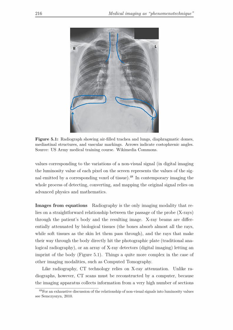
216 Medical imaging as “phenomenotechnique”
Figure 5.1: Radiograph showing air-filled trachea and lungs, diaphragmatic domes,mediastinal structures, and vascular markings. Arrows indicate costophrenic angles.Source: US Army medical training course. Wikimedia Commons.
values corresponding to the variations of a non-visual signal (in digital imagingthe luminosity value of each pixel on the screen represents the values of the sig-nal emitted by a corresponding voxel of tissue).48 In contemporary imaging thewhole process of detecting, converting, and mapping the original signal relies onadvanced physics and mathematics.
Images from equations Radiography is the only imaging modality that re-lies on a straightforward relationship between the passage of the probe (X-rays)through the patient’s body and the resulting image. X-ray beams are differ-entially attenuated by biological tissues (the bones absorb almost all the rays,while soft tissues as the skin let them pass through), and the rays that maketheir way through the body directly hit the photographic plate (traditional ana-logical radiography), or an array of X-ray detectors (digital imaging) letting animprint of the body (Figure 5.1). Things a quite more complex in the case ofother imaging modalities, such as Computed Tomography.
Like radiography, CT technology relies on X-ray attenuation. Unlike ra-diographs, however, CT scans must be reconstructed by a computer, becausethe imaging apparatus collects information from a very high number of sections
48For an exhaustive discussion of the relationship of non-visual signals into luminosity valuessee Semczyszyn, 2010.
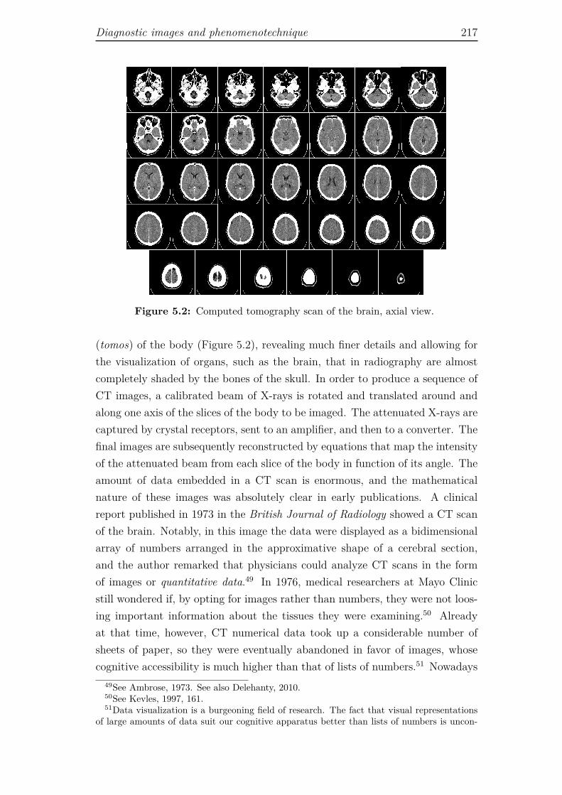
Diagnostic images and phenomenotechnique 217
Figure 5.2: Computed tomography scan of the brain, axial view.
(tomos) of the body (Figure 5.2), revealing much finer details and allowing forthe visualization of organs, such as the brain, that in radiography are almostcompletely shaded by the bones of the skull. In order to produce a sequence ofCT images, a calibrated beam of X-rays is rotated and translated around andalong one axis of the slices of the body to be imaged. The attenuated X-rays arecaptured by crystal receptors, sent to an amplifier, and then to a converter. Thefinal images are subsequently reconstructed by equations that map the intensityof the attenuated beam from each slice of the body in function of its angle. Theamount of data embedded in a CT scan is enormous, and the mathematicalnature of these images was absolutely clear in early publications. A clinicalreport published in 1973 in the British Journal of Radiology showed a CT scanof the brain. Notably, in this image the data were displayed as a bidimensionalarray of numbers arranged in the approximative shape of a cerebral section,and the author remarked that physicians could analyze CT scans in the formof images or quantitative data.49 In 1976, medical researchers at Mayo Clinicstill wondered if, by opting for images rather than numbers, they were not loos-ing important information about the tissues they were examining.50 Alreadyat that time, however, CT numerical data took up a considerable number ofsheets of paper, so they were eventually abandoned in favor of images, whosecognitive accessibility is much higher than that of lists of numbers.51 Nowadays
49See Ambrose, 1973. See also Delehanty, 2010.50See Kevles, 1997, 161.51Data visualization is a burgeoning field of research. The fact that visual representations
of large amounts of data suit our cognitive apparatus better than lists of numbers is uncon-
218 Medical imaging as “phenomenotechnique”
the volume of data generated by CT equipments is enormous. These data allowcreating very detailed three-dimensional images, and they pose new challengesto radiologists, namely for what concerns their manipulation (data processing),presentation, storage, and interpretation.52
Similar considerations can be made for all contemporary medical imagingmodalities, because algorithms for image reconstruction play a fundamentalrole in all of them. In ultrasound imaging, it is necessary to turn into im-ages several pulses of ultrasound beams that are reflected by bodily tissues.In MRI, the images are produced resorting to the spatial coordinates obtainedfrom the magnetic relaxation times of the hydrogen nuclei (protons) of the watermolecules which compose our body. In PET, an image of metabolic activity isreconstructed on the basis of the spatial coordinates of the gamma-rays emit-ted by radioactive molecules previously injected in the patient’s blood.53 Thismeans that when the radiologist looks at a medical image, what he really seesare non-visual signals translated into areas of different luminosity. Of course,he perceives this information in the form of a visual object, and thus uses thedepictions to make detections. That is, he relies on the mimetic features ofthe image to extract non-visual information, namely, information relevant for amedical diagnosis.54 Being an expert observer, however, the radiologist is awareof the problematic relation between depiction and detection. He knows, for in-stance, that certain features of the image might refer to artifacts produced bythe imaging apparatus, rather than to actual properties of the biological tissue.He also knows that there is no univocal correspondence between what appearsin the image and the actual health conditions of the patient.55 It is also worthnoting that the more sophisticated the imaging technology, the higher the pos-sibility of producing artifacts during the imaging process, and thus the greaterthe expertise required to correctly acquire and interpret the images.
This concise and simplified description of how medical images are producedprovides the background for some reflections about their phenomenotechnicalnature. First of all, if we focus on the idea that these images are produced “byfollowing and recording, by some means, the progress of suitable probes that are
troversial. What is not always clear is if semi-naturalistic, photograph-like images hold anyspecial advantage compared to other visual formats such as graphs (see Delehanty, 2010). Inmedical imaging the advantage of highly naturalistic visual displays is quite obvious when onewants to visualize morphological and structural elements, or locate an event (e.g., blood flowrate) in a specific area of the body. The advantage of naturalistic renderings is less clear whenimaging is used to quantify metabolic processes, as in the case of PET and functional MRI.
52See Pisco, 2009, 33.53See Wolbarst, 1999; Pope, 1999.54See Maynard, 1997, 119-120. I discuss the distinction between depiction and detection in
Section 3.2.3.55See Perini, 2012. See also Section 3.2.3.
Diagnostic images and phenomenotechnique 219
attempting to pass through a patient’s body”56 we can start to understand themin terms of experimental practice. To produce a diagnostic image is to create anartificial, controlled phenomenon whereby specific signals are elicited from thebiological tissues, and subsequently converted into visual outputs. A diagnosticimage is really a representation of the body and disease under experimentalconditions, for three reasons:
1. An appropriate probe has to be sent into the patient’s body in order toemit the desired signal;
2. The patient has to adhere to specific protocols before and during theimaging process to optimize the quality of the output signal (the body ispart of the imaging apparatus, not just an object of representation);57
3. It is possible to properly compare different images only if they are pro-duced with the same imaging modality and following the same operationalstandards.58
Another observation that we can make in relation to the phenomenotechnicalnature of radiological images is that they are not conventional images: theyare mathematical reconstructions of quantitative data derived from non-visualsignals. This unconventional, counterintuitive feature of diagnostic images wasalready present, although to a lesser degree, in analogical imaging. In fact,although traditional radiography does not depend on a mathematical recon-struction of the original signal (X-ray attenuation), it nevertheless creates animage by exploiting non-visual properties of the biological tissues. It producesa phenomenon (the X-ray image) that can only exist as far as it is technicallyproduced. This character of novelty is one reason why the process of significa-tion of radiographs was complex and involved a combination of technical andcognitive operations that helped create and consolidate new visual skills. Com-pared to analogical imaging, in digital medical imaging the phenomenotechnical
56Wolbarst, 1999, 8.57Depending on the technology employed, a patient might be asked not to eat previously to
the examination, to drink a contrast liquid, or to undergo other forms of bodily preparations.During the imaging process, one will be typically asked to stand still, to hold one’s breath, tobreath deeply, and so forth.
58In order to elaborate a diagnosis it can be useful to combine different imaging modalities(e.g., ultrasound and MRI) because they allow to collect different information. However, tostudy the evolution of a disease in a single patient, or to compare the radiological presentationof the same disease in different patients, one has to compare images of the same modalityproduced under the same conditions (e.g., instruments with identical, or very similar, technicalcharacteristics, same time of exposure, same physical preparation of the patient, and so on).For the relevance of operational standards to the creation of scientific objectivity see Section3.2.2.
220 Medical imaging as “phenomenotechnique”
nature of images is taken to a more sophisticated and accomplished level.59 Thereconfiguration of quantitative data into images is performed by mathematicalalgorithms that embody a specific epistemic context: the pre-existing knowledgeabout the object under study; the questions that need to be answered (the diag-nostic problem); the working hypothesis; the expected outcomes; and so forth.We could say that contemporary medical images entail the materialization ofmathematical noumena. This does not mean that medical images are math-ematical inventions, because there is a counterfactual causal chain that linksbiological indicators, probes, and images (if the tissue were different, the signalemitted would be different, and therefore the image would be different). Yet,diagnostic images are not copies of the human body. They are rather simula-tions of the real object, because they can be manipulated in order to increasethe visibility of specific aspects of the body or of the disease, and to erase thoseelements that are irrelevant to the diagnosis and would provide only visual noise.
For instance, in MRI, by selecting specific sequences of radio frequency pulsein the phase of image acquisition, it is possible to selectively enhance the bright-ness of soft tissues (e.g., fat or brain matter), and neglect watery tissues (diseasedareas) or liquids (e.g., urine or cerebrospinal fluid). Similarly, in the phase ofimage reconstruction, the selection of specific parameters allows to better visu-alize certain areas rather than others. Moreover, the final user (the radiologistwho interprets the scan) can manipulate several parameters, such as color, whilelooking at the image.60 The mathematical substructure of these images allowsfor an increased representational plasticity, and thus enhances their cognitiveeffectiveness. In this respect it is important to stress that the selection of pa-rameters in the phase of image acquisition determines the kind of informationthat can be extracted from the final output. For example, if an MRI image wasacquired according to parameters that neglect watery tissues, it will not be pos-sible to retrieve information about such tissues from the final image, no matterhow many software-based manipulations one performs on it. These manipula-tions only affect the way the information gathered during image acquisition ispresented to the observer.
Blurred boundaries As the great majority of contemporary scientific images,todays’ diagnostic images are image-data, that is, they are lists of numbers cor-
59This is not, of course, an instance of teleological progress, but rather the result of scientificand technological development. I use the adjective “accomplished” to point out that newdiagnostic images are better suited to carry out the task of medical imaging, which is toextract information from the patient’s body by reconfiguring its visibility.
60For an informative study on software-based interaction between radiologists and imagessee Friedrich, 2010.
Diagnostic images and phenomenotechnique 221
responding to physical quantities which are displayed in the form of a bidimen-sional visual object.61 They are hybrid objects that blur the boundaries betweenquantitative data and qualitative representation: we use quantitative data toproduce the image, and we resort to the qualitative features of the image to in-terpret the data. As all mechanical images of invisible referents, contemporarydiagnostic images also blur the difference between the indexical and symbolicmodes of signification. We are justified in trusting the indexical nature of theseimages as far as they have been produced according to well defined operationalconventions, because these protocols assure the stability of both the causal andcounterfactual relationship between the referent and its image. In turn, theapplication of the operational standards lays the foundations for the symbolicmode of signification of the images (it creates the material preconditions to de-fine the rules according to which the image must be interpreted).62 In digitalimaging, however, an important role in the process of image making is playedby the iconic mode of signification, too. Icons are signs that signify by virtue ofsimilarity, by sharing some quality with the referent.63 In morphological medicalimaging, the mathematical algorithms for image reconstruction have preciselythe aim to create images that resemble the original object by preserving its ge-ometry. In the same way, in functional imaging the original signal is processedso as that the intensity of the physiological phenomenon under study is visuallyrendered in terms of intensity of the luminosity of a specific area of the image.The preservation of a resemblance between the image and its referent is thusa principle built into the mathematical equations that determine the aspect ofthe image. This means that the interpretation of the original data (the nonvisual signal) begins during the very production of the image, on the basis ofthe knowledge used to design the algorithms for image reconstruction.
5.1.3 Diagnostic images as reified knowledge
Bachelard claimed that instruments, in modern science, are “reified theorems”64
and that the phenomena produced by scientific instruments “bear the stamp oftheory throughout.”65 For him, Millikan’s oil drop experiment or Stern andGarlach’s space quantization experiment were “conceived directly as a functionof the electron or the atom.”66 That is, they were specifically designed to testa well defined theory, they materialized the theory. A common interpretation
61See Prasad, 2005; Israel-Jost, 2011.62See Section 3.2.2.63See Peirce, CP 2.247. See also Section 2.2.64Bachelard, 1971, 137; 1949, 103; 1934, 13.65Bachelard, 1934, 13.66Bachelard, 1971, 137.
222 Medical imaging as “phenomenotechnique”
of these statements is that they point to the self-referentiality of theory, thus,to the theory-ladenness of observation and the underdetermination of scientifictheories.67 However, considering the importance attributed by Bachelard toexperiment, and taking into account that he saw science application as a con-stitutive element in the creation of scientific concepts, we can regard his claimson instruments as reified theorems as a general statement concerning the factthat scientific instruments materialize a large body of pre-existing knowledge,and that they help produce the object under study as a technophenomenon.68
If instruments materialize different bodies of knowledge (not only well definedtheories about the object of study), and if they help recreate the object ofinterest as a technophenomenon, then we can really think of technology not asa mere application of science, but as an active constituent of it. Accordingly,we can understand medical imaging not simply as the application to medicineof technologies based on physical theories, but as the reification of knowledge(both theoretical and practical) developed in different domains, such as, physics,chemistry, pharmacology, biology, physiology, pathology, and so forth. Thisimplies that medical imaging technologies embody and blend together theories,concepts, hypotheses and practices concerning the machinery that produces theimage as well as the human body that through those images is examined. Byreconfiguring natural objects and by creating totally new phenomena, imagingtechnologies extend the scope of our sensory experience, and at the same timethey extend the domain of what can be thought and how it can be thought.They extend the scientific mind.69 Conversely, for a technology to become partof a given scientific domain it must accommodate the pre-existing knowledge ofthat domain, or at least part of it.
In the specific case of medical imaging, I have shown in Chapter 1 thatphysicians could be receptive to the possibilities open by radiography becausethey conceptualized disease in terms of organic lesions. However, the creationand stabilization of proper clinical knowledge based on radiographs was a longand complex process. It entailed:
67See Galison, 1997, 18.68See Rheinberger, 2005, 320. Bachelard wrote: “The instrument is the necessary interme-
diary in the study of a definitely instrumented phenomenon [un phénomène vraiment instru-menté] that has been designed as an object of a phenomenotechnique.” Bachelard, 1949, 2-3,my translation.
69Castelao-Lawless makes a similar point when she says that Bachelard’s claim that in-struments are reified theories refers to the fact that, for him, both mathematical models andscientific instruments were extensions of the scientific mind. See Castelao-Lawless, 1995, 50.In La formation de l’esprit scientifique Bachelard wrote: “A measuring instrument alwaysends up as a theory: the microscope has to be understood as extending the mind rather thanthe eye.” Bachelard, 1938, 240.
Diagnostic images and phenomenotechnique 223
1. Technology control and stabilization (an operation that brought togetherphysicians, physicists, engineers, photographers, and other professionals).
2. The matching of what was shown by radiographic images and what couldbe directly observed in the cadaver (an operation that led to the revision ofsome well entrenched assumption about human anatomy based on autopsicobservations).
3. The comparison of the information provided by radiographs and infor-mation collected through other diagnostic techniques (an operation thatrequired the development not only of new visual skills, but also of a newlanguage).
4. The comparison of different images among them (an operation wherebyimages were turned into a medium of the patient’s body).70
Indeed, the history of the discovery of X-rays and their subsequent introduc-tion in medicine shows that instruments are less a direct reification of a single,well-defined theory, than the embodiment of complex dynamics of practices, the-ories, hypotheses and concepts. Roentgen discovered X-rays by chance. Whileexploring the properties of cathode rays, he inadvertently set the conditions forthe appearance of a new phenomenon that seemed anomalous in the context ofwhat he knew about the electromagnetic waves. It took several years to developa proper physical theory of X-rays, but this did not prevent the development ofa more efficient radiographic technology for medical purposes. Such improve-ment was brought about on empirical bases, not as the direct application ofbetter theories. In turn, the refinement of the technology for X-rays produc-tion and manipulation contributed to the growth of theoretical knowledge aboutradiation, as well as about human anatomy and disease. At each step of theirdevelopment X-ray machines and the related practices of image making were thematerialization of a heterogeneous body of knowledge distributed among differ-ent professionals. This included, for example, theoretical and empirical knowl-edge about X-rays production, image fixation, use of contrast agents, tissues’densities in healthy and diseased tissues, and so forth. The imaging apparatusemerged at the intersection of different domains of knowledge.
An exemplary case of knowledge crossing The intersection of knowledgecoming from different disciplinary fields is particularly marked in positron emis-sion tomography. While X-ray, ultrasound, and magnetic resonance imaging
70See Sections 3.4.5, 3.4.6, and 4.2.2.
224 Medical imaging as “phenomenotechnique”
provide morphological information, PET allows visualizing the physiological ac-tivity of the inner tissues, by showing the variation of concentration of certainmolecules in specific areas of the body over a given amount of time. It has sev-eral applications: it helps diagnosing cancer and monitoring possible metastasesby detecting the elevation of glucose uptake by cells in rapid division; it allowstracking the levels of oxygenation of different areas of the heart or the brain;and it can be used to measure the activity of specific neural receptors. This kindof imaging is possible thanks to so called radiotracers or radiopharmaceuticals.These are molecules in which an atom of carbon, oxygen, nitrogen or fluorinehas been replaced by a correspondent radioactive isotope. To produce a PETimage, the radiotracer, selected according to the physiological process that onewants to monitor, has to be injected into the patient’s blood. At this point itflows through the body as a normal molecule, but after a precise span of time(the isotope half life)71 it emits radioactivity in the form of a positron that, aftercolliding with an electron, produces two γ-rays that shoot off the body almost180º apart. These two γ-rays are the signal detected by the imaging apparatus,and they are used to map the flow rate of the labelled molecule. As in thecase of CT scans, the machinery delivers a set of images corresponding to theslices in which the original volume (the human body) has been divided. And asin the case of CT, the production of the images entails complex mathematicaloperations which require considerable computing power.
This means that for PET technology to be possible, many branches of knowl-edge have to converge: physiology, medicine, biochemistry, nuclear chemistry,physics, electronic engineering, and mathematics. PET as a technology, andPET images as the material output of such technology materialize knowledgeabout normal and pathological physiology, physiological modeling, tracers ki-netics, radioactivity production and detection, image processing, image recon-struction, and so forth. Indeed, PET images are particularly complex for whatconcerns both their production and interpretation. The information they presentis both quantitative (they are meant to literally measure biological activity) andqualitative (they show the spatial distribution of the phenomenon under study).Consequently they demand complex physiological modeling. Moreover, to bemedically relevant, they have to be processed in terms of relationships betweenmolecular circulation in the organism, radioactive decay, and specific propertiesof the physiological process under examination (e.g., the relationship betweenglucose concentration in the cells, cells’ proliferation, and tumor malignity).72
71The half life is the length of time required for half of any given amount of an element todecay into another element. It is specific for each isotope.
72See Dumit, 2004, 30.
Diagnostic images and phenomenotechnique 225
In his study on the role of PET imaging in neuropsychiatry, the anthro-pologist of science Joseph Dumit has shown that the different researchers thatcontributed to the invention of PET conceptualized this technology in differentterms. For one of them, the physicist Michel M. Ter-Pogossian, PET was notexactly an invention, it was rather a “recognition.”73 That is, it was the result ofnoting that certain positron emitting radionuclides have physiological propertiesand thus can be used to image biological processes. Once the imaging potentialof these molecules was realized, it was just a matter of finding a way to takeadvantage of them. This was possible thanks to the insights and instrumentsproduced by the researchers in the area of CT scanning. For Ter-Pogossian,designing the first PET machine was essentially a matter of putting together“different building blocks”74 from different scientific areas. The building blocksto which Ter-Pogossian refers are both ideas (theories, hypothesis, kinetic mod-els, and so on) and material objects (such as the cyclotron for the productionof radioisotopes, the imaging apparatus, and the radiopharmaceuticals). Allthese elements converge in the technology that goes under the name of positronemission tomography. Ter-Pogossian’s account of the invention of PET pointsin the direction of a conceptualization of technology as materialization of dif-ferent strands of knowledge, yet it provides a picture of medical imaging as anapplication of technology to medicine. In his account, first came the realizationthat specific radiotracers could be used to monitor physiological activity, then itwas developed into an apparatus that could take advantage of these molecules,and finally physicians embraced the new technology for diagnostic and researchpurposes.
The physician Henry N. Wagner, another contributor to the invention ofPET,75 offers a different perspective. For him, the development of PET “was achain of events”76 that started in the nineteenth century with Claude Bernard’sconceptualization of disease in physiological terms.77 The other fundamentalevents in the chain were Georg von Hevesy’s success in using radioactive isotopesto study metabolic processes, in 1923, and the medical measurements of thyroidactivity by scintigraphy, the first technology for functional imaging, developedsome twenty years before PET. For Wagner, “thyroid scanning started because
73Dumit, 2004, 43.74Ter-Pogossian in an interview with Dumit. Dumit, 2004, 45.75The attribution of an invention is a classical problem for history of science. Given the
multidisciplinary nature of PET, it is consensual that many people contributed to its invention.Ter-Pogossian himself claims that “there are masses of fathers of PET.” Dumit, 2004, 40.
76Wagner, in an interview with Dumit. Dumit, 2004, 47.77Note that the idea that the preconditions for functional imaging were set by Bernard’s
conceptualization of body and disease in terms of physiological functions is specular to theidea that clinical anatomy set the preconditions for morphological imaging.
226 Medical imaging as “phenomenotechnique”
of nodules in the thyroid,”78 not because there was a technology that madefunctional scanning possible. This shift of perspective is important, because ithighlights the fact that medical technologies embody the need to solve medicalproblems, medical questions. Physicians needed to know whether the nodulesin the thyroid were functional or not. They needed to measure blood flow andto associate it to a variety of problems, from the effects of anesthesia to thediagnosis of schizophrenia. To solve these problems they did what they couldwith the tracers and detectors they had at their disposal at any given time.The diagnostic questions and problems were not simply an external force thatprovided the practical goal which demanded the development of a technology.Rather, they were the core around which successive technologies acquired theirspecific form, within a number of constraints posed by all the other factors atstake (tracers, detectors, image reconstruction models, and so on). But there ismore than this.
By integrating the medical problem, the technology can reshape the waythe problem itself is posed. Wagner gives the example of the development ofsingle photon emission radiopharmaceuticals that simulate somatostatin, a pep-tide with a wide range of inhibiting effects on various organs. Single photonemission tracers are akin to those used in PET, and they started to be used inthe 1980s to study and diagnose neuroendocrine tumors through single photonemission computed tomography (SPECT).79 Such studies showed that in certaintumors there are specific metabolic alterations, a discovery that played an im-portant role in directing cancer research towards a molecular approach. SPECTand single photon emission tracers were part of the solution to the problem ofdiagnosing cancer, in turn they helped rethinking cancer in terms of cellulargrowth and communication, and this led to the development of new molecularapproaches and technologies in oncological research.80 A medical imaging tech-nology materializes the techniques, practices, problems, hypotheses, conceptsand theories of different domains, and that by changing the kind of informationavailable, it can redirect the approach to the medical questions it addresses.
Transparent and visible theories The interweaving between medical the-ories, instruments, and images can be more or less complex. It depends onwhat must be visualized (it is easier to visualize a structure than a process);
78Wagner, in an interview with Dumit. Dumit, 2004, 47.79SPECT is an imaging technology akin to PET, but it uses a different family of tracers
(single photon emission rather than positron emission radionuclides), and a different scanner.Its spatial resolution is significantly lower than that of PET’s, but it is considerably lessexpensive.
80See Dumit, 2004, 48; Pepe et al., 2012.
Diagnostic images and phenomenotechnique 227
how it is visualized (some technologies are more theory-loaded than others);and for what specific reason (some diseases are better understood than oth-ers). As discussed in Chapter 3, the visualization of teeth and broken bonesby X-ray imaging was uncontroversial and relatively straightforward, while thesame technology demanded a much more complex process of image significationin order to acquire meaning in clinical medicine.81 In the case of tuberculo-sis, such process implied, among other things, the construction of a semioticnetwork that bounded radiological images with already established diagnostictechniques.82 Contemporary imaging offers even more compelling examples ofhow theories and images influence each other. It also shows that the theoryembedded in an imaging apparatus and in the interpretation of the images itproduces becomes transparent when the approach to the problem at stake isconsensual. On the contrary, in contested domains, where the conceptualiza-tion of a medical problem is not settled, the theory beyond images productionand interpretation becomes visible because controversial. PET images providea good example of both situations.
The use of PET scanning to monitor cancer, to verify cardiac perfusion, orto evaluate brain disorders such as Alzheimer’s disease and epileptic seizures,is uncontroversial. Our medical understanding and conceptualization of theseconditions, although not static, is sufficiently settled to render unproblematicthe idea that we can visualize information about them by means of PET. Thissignifies that, although there might be doubts whether in specific cases PET isthe best option compared to other diagnostic modalities, there is no disagree-ment upon the fact that PET images provide information actually related tothe disease under study. In this case the theory (or theories) that binds patient,apparatus and images is transparent in virtue of being well corroborated andgenerally accepted, it becomes implicit and thus almost invisible.
This is not the case when PET is used to study and diagnose psychiatricdisorders.83 At present, in spite of the great amount of studies carried out inthis field, there are no validated brain imaging techniques to diagnose psychiatricdiseases. This is so not only because of technical reasons,84 but also becausethere is no agreement upon whether what we can visualize through cerebral PET(the relative oxygenation or the metabolic rate of glucose in different areas of thebrain) does correlate to specific psychiatric conditions.85 Hence, if a psychiatrist
81See Section 3.4.82See Section 4.2.2.83Together with PET, the other brain imaging technologies used to study and diagnose
psychiatric conditions are SPECT and functional MRI (fMRI).84See Farah and Gillihan, 2012.85The discussion over the neural correlates of psychiatric disorders occurs at two levels.
On the one hand, the very existence of such correlates is contested, especially by scholars
228 Medical imaging as “phenomenotechnique”
resorts to brain imaging to study such conditions, he is explicitly endorsing aspecific theory of the mind, of psychiatric illness, and of psychiatric illnesses’classification. These theories motivate and justify the choice of the imagingapparatus, which is supposed to allow to see a putative diagnostic indicator inthe image in question. Since the theory is contested, it becomes visible throughthe technology and through the image.
5.1.4 Diagnostic images and the “phenomenotechnique
of the observer”
Independently from the fact that the theory about the phenomenon under studyis transparent or visible, diagnostic images are always “belief-opaque.”86 In thedefinition of the philosopher and cognitive scientist Adina Roskies, an imagingtechnique is belief-transparent “if it is possible to appropriately interpret theimage it produces with information contained in the image,” and it is belief-opaque “when the information needed for the interpretation is not present inthe resultant image.”87 Typically, a photograph of a visible object is belief-transparent, because we have a previous knowledge of how the thing representedin the picture looks like. It has a visible referent and thus we can rely onour intuition, that is, on the visual habits produced by everyday experience tointerpret the image (of course, this does not mean that our interpretation isalways correct). When the referent is invisible, however, not only we lack suchintuitive knowledge, but relying on commonsense visual habits might lead togross mistakes in the interpretation of the image. For instance, a non-expertviewer who looks at the PET scan of an oncological patient will probably thinkthat all the “spots” on the image are an indication of active disease, and thatone can easily see the disease in the image. The expert viewer, on the contrary,knows that the image must be interpreted in the light of a wider set of knowledge,because noncancerous conditions such as chronic or acute inflammations havethe same appearance of cancer on PET images, while many types of tumorsdo not show up. The gaze of the experienced observer incorporates a widerset of knowledge that is not directly available in the image and that allows
from philosophy and from the human and social sciences. On the other hand, within theneuroscientific community who endorses this approach to mental illness, the debate revolvesaround the use of brain imaging in the actual identification of neurocorrelates. For a reviewof the two debates see Vidal and Ortega, 2012. The debate over the neural correlates ofpsychiatric diseases can be seen as part of a larger debate over brain modularity and the use ofneuroimaging in cognitive studies. For an epistemological analysis of the use of neuroimagingin cognitive science see Roskies, 2007. For an epistemological critique of the localizationistand modularity approach in neuroscience see Hardcastle and Stewart, 2002.
86Roskies, 2007, 868.87Roskies, 2007, 868.
Diagnostic images and phenomenotechnique 229
making such distinctions. The expert viewer knows the rules, both implicitand explicit, necessary to make sense of the extended sensory domain producedby the phenomenotechnique. This is to say that, although visual experienceis fundamental for the radiologist to deal with the data generated by imagingtechnologies, his visual experience is of a different order compared to primary,everyday life visual experience. It is a peculiar form of perception that requiresa phenomenotechnique of the observer.88
A fundamental element of the divide between naïve perception and educatedperception is the comprehension of the complex nature of the causal and coun-terfactual relation between an image and the object of study. The untrainedviewer is not aware of the complexity of the causal chain of events that linksthe original phenomenon to its image, and consequently has no awareness ofthe number of variables at stake (for instance, the production of artifacts). Hehas an equally simplified understanding of the counterfactual relation, that is,he does not know that different phenomena (e.g., benign rather than malignanttumor) could result in the same image. The expert viewer, on the contrary,not only is aware of such problems, but is also in command of the theoreticalknowledge and practical skills necessary to deal with them. In other words, thenaïve viewer engages in an intuitive process of signification that is misleading,he takes a complex sign for a simple index, or an icon.
In Chapter 3 I argued that we are warranted in inferring properties of invis-ible objects and phenomena from mechanical images because such images areindices. I also argued that the indexical mode of signification is not enough,because it does not allow to endow an image with meaning and to directly dis-tinguish the properties of the object from the artifacts created by the imagingprocess. To tackle this problem, scientists resort to three strategies: (1) they putin place a symbolic mode of signification based on the creation of operationalstandards for image production;89 (2) they inscribe the image into a specific
88I use the term “phenomenotechnique” rather than simply “technique” of the observer fortwo reasons. First, because by maintaining a homogeneous terminology along the descriptionof technologies, images and observers I stress the entanglement of the processes of imageproduction and image signification, the continuum that binds the production of an image to itsinterpretation. Second, I want to mark a conceptual distance from the expression “techniqueof the observer” as used by Jonathan Crary. In the book Techniques of the Observer (1990),Crary explores the dynamics of embodiment and disembodiment of the act of vision broughtabout by different optical devices, and he analyzes such devices as part of a larger assemblageof historical events and powers. By talking of phenomenotechnique of the observer, I framemy analysis in a different domain. I am not interested in the embodied act of seen, nor inthe relations of power in which optical devices are embedded. I rather focus on how scientificvision is constructed as an explicitly and necessarily artificial form of perception, and as adistributed process.
89See Section 3.2.
230 Medical imaging as “phenomenotechnique”
theoretical framework that defines what can be seen;90 (3) they manipulate theobject under study performing different sorts of experiments in order to gatheradditional information to combine with that obtained through the image.91 Iconsider all these activities, both practical and intellectual, a phenomenotech-nique of the observer, because they put the viewer at the centre of an epistemicnetwork that makes possible the emergence of a perception removed from thatwhich is granted by everyday experience.
This way of understanding the process whereby scientific perception is pro-duced is quite different from Bachelard’s idea of psychoanalysis of the scientificspirit.92 To Bachelard, science is continuously getting rid of conventions andmental habits so that new forms of rationality can emerge. It aims to a surra-tionality that constantly breaks with the past and will necessarily change in thefuture. The phenomenotechnique of the observer I have just described works ata different level. It has a pragmatic value, in the sense that its aim is to makepossible actual scientific observations in the present, rather than to break withthe past. It produces and stabilizes perceptual habits that are different fromthose of commonsense experience, but they are habits nevertheless. It leadsto the creation of conventions for image production and interpretation. Theseconventions are responsive to conceptual change, because they rely on the prac-tical and theoretical knowledge of a given scientific domain, but they do notnecessarily emerge in polemic with past knowledge. Finally, what is demandedof the observer is not the ability to dissolve reality into pure rationality. Rather,it is demanded that he learns to deal with the materiality of the instrumentsand of the objects that through the instruments are made visible and examined(the human bodies).
The phenomenotechnique of the observer entails the training of the scientistsin relation to the strategies of image signification mentioned above: operationalconventions, theoretical frameworks, experimental practices. This means thatmedical images acquire meaning within a distributed process that goes wellbeyond the act of watching. It is an educated act of perception that is possiblebecause there are standards for image making; semiotic networks created anddistributed on the pages of journals, books and atlases; practices of samplespreparation and manipulation. Since early medical photography, the meaningof diagnostic images has emerged from a combination of all these factors, and theprocess has been further enriched and complicated by the introduction of digitalimaging, with its mathematical models for image reconstruction. Through this
90Section 3.3. See also Section 2.491See Sections 3.2.3 and 4.2.3.92See Bachelard, 1938.

Diagnostic images and phenomenotechnique 231
multilayered process the act of seeing is deeply transformed. It moves awayfrom intuitive natural perception and acquires a scientific character.

232 Medical imaging as “phenomenotechnique”
Conclusions
In this dissertation I attempted to develop an epistemology of medical imagingthat took into account the fact that diagnostic images are simultaneously di-agnostic instruments and scientific images. It was thus necessary to highlightdistinct epistemological problems concerning, respectively, medical diagnosticsand the cognitive statute of instrument-mediated images.
Although, as a whole, my study is mainly focused on the problems posedby diagnostic images qua images, in the first chapter I proposed an account ofthe historical and conceptual conditions of possibility of diagnostic images quadiagnostic instruments. I did so by going back to the birth of clinical anatomy,when diagnosis became a matter of scrutinizing the inner body, and medicinestarted to establish itself as an empirical science capable of producing a clearand precise visibility of illness. My basic intuition was that one can diagnosea disease by means of an image of the inner body only if illness is understoodin visual and spatial terms. Drawing on Foucault’s The Birth of the Clinic andon historical medical literature, I argued that the conceptual roots of medicalimaging as a diagnostic technique go back to the understanding of body anddisease defined by clinical anatomy at the end of the eighteenth century, andthat diagnostic images are to be understood first and foremost as instrumentsthat help turning diseases into an objective, visual entity. The clinical paradigmthat emerged from clinical anatomy commanded specific forms of objectivationof the human body: to make a diagnosis, the physician could no longer relyexclusively on the symptoms related by the patient, he had to actively elicit oreven fabricate the signs that pointed to the disease, which was conceptualized interms of organic lesion. In this context it became necessary to develop diagnosticinstruments that could produce a specific expressivity of the human body.1 Byturning the inside out, such instruments transformed invisible structures andprocesses into visible indices of health or disease.
By analyzing the wide range of diagnostic modalities developed in the nine-teenth century, I tracked down the clinical tradition wherein medical imagingcan be inscribed, and highlighted the entanglement between the development of
1See Section 1.1.
233
234 Conclusions
the new instruments and the transformations in the understanding of anatomy,physiology and of the nature of disease.2 From this part of the research it waspossible to conclude that although the invention of radiography has been oftendescribed as a revolution in medicine and in medical visual culture,3 it shouldbe understood less as an epistemological than as a technological and culturaltransformation. On the one hand, the conceptual bases for medical imaging arerooted in the conceptualization of disease put forward by clinical anatomy; onthe other hand, the vast array of diagnostic instruments developed during thenineteenth century posed some epistemological questions that were subsequentlyinherited by radiology. In particular, the replacement of subjective sensationsof both patients and physicians with objective (or at least inter-subjective) me-chanical indices of health and disease; the production of visual records of theinterior of the body that could be filed, retrieved and shared by several ob-servers; the need to develop a specialized language that allowed to describe anddiscuss such visual data; the introduction of a mediation between the observer(the physician) and the object (the patient), which generated different forms ofinstrument-mediated perception.
Subsequently I turned my attention to the image-related problems posed bymedical imaging. Through the analysis of early photography, I explored therole played by this groundbreaking optic media in reshaping the visibility ofboth the human body and disease.4 More precisely, I tried to understand howmedical photography contributed to the reconfiguration of a natural object (thehuman body and its ailments) into a scientific object. Following Peirce’s semi-otics, more precisely his organization of signs in indices, icons and symbols, Iargued that although nineteenth-century theorization of photography revolvedessentially around the idea of photographs as perfect records of reality (iconicmode of signification), predicated on the mechanical nature of the photographicprocess (indexical mode of signification),5 the symbolic mode of significationplayed a fundamental role in establishing specific forms of visibility of diseasein such diverse fields such as psychiatry, dermatology and neurology. In fact,what the physician-photographers struggled to see in their pictures were less thevisible signs of illness in a specific patient, than the facies of the disease, that is,its general yet characteristic appearance. On the one hand, photographs wereused to keep records of individual patients and monitor the evolution of theircondition. In this case each picture was an index, in the sense that it identi-
2Sections 1.2 to 1.5.3See, for instance, Reiser, 1978; Cartwright, 1995.4See Chapter 2.5It should be noted that our commonsensical intuition about photography and its epistemic
reliability has not changed since the nineteenth century.
Conclusions 235
fied in a strict one-to-one relation the individual patient affected by a specificmanifestation of the disease. On the other hand, photographic collections wereused to make abstraction of the specific circumstances of a patient and generatea universal image of the disease. These collections were nomological machines,they worked according to the symbolic mode of signification and were meant toreveal the laws of nature that govern the appearing of a disease.
The analysis of medical photography was further enriched by taking intoconsideration Benjamin’s reflections on the photographic series (mechanical pro-duction and reproduction of an image and of the body it represents), and on theintrinsic analytic and dissecting potential of photography (the photographer asa surgeon). Looking at the works of the early physician-photographers in thelight of Benjamin’s ideas, I reached the conclusion that the photographic seriescollected in medical journals, manuals, and hospital archives produced a clini-cal gaze in the Foucauldian sense. That is, they created a neutral visual spacesimilar to that of the hospital wards, where each body was open to medical ob-servation and could be put in relation with other bodies, so that the pathologicalcould emerge as a stable pattern from the heterogeneous variability of the indi-vidual instances. The photographic collection recreated on paper the sensorialand cognitive domain of the clinic. Moreover, in the new regime of mechani-cal visibility and reproducibility created by the photographic camera, the bodybecame not only an object of scientific analysis, it was also directly embeddedinto a complex apparatus for visualization and experimentation (an emblematiccase is that of the electrophysiologic portraits by Duchenne de Boulogne), whichenhanced and transformed the visibility of the body.
From the scrutiny of early medical photography and of its role as a prede-cessor of radiology, I zoomed out, so to speak, to a problem that concerns notonly medical imaging, but scientific imaging in general. Namely, the problem ofthe visualization of the invisible and of the validation of instrument-mediatedimages that are meant to show invisible referents. Taking into account specificexamples from microphotography, chronophotography and early radiography, Idefended the idea that the process of signification of the images of invisible ref-erents entails a complex interplay of indexical, iconic and symbolic semiosis. Inthese cases, the creation of operational conventions for image production and thedefinition of theoretical frameworks for image interpretation play a fundamentalrole in securing the epistemic authority of the image. My argumentation, builtin opposition to Daston and Galison’s idea of mechanical objectivity as suppres-sion of the subjectivity of the image maker and of the observer, was organizedin three moments. Firstly, I stressed that in the case of microphotography theacceptance of the truthfulness of an image and of its scientific value largely
236 Conclusions
depended on the definition of experimental standards for samples’ preparation,observation and imaging. These standards provided operational conventionsthat grounded a symbolic mode of signification. That is, the microscopists be-lieved that two microphotographs showed the same thing, and that what theyshowed was the actual specimen and not an artifact produced by a specificapparatus, when the pictures had been produced under the same conditions.The microphotograph was considered the result of a complex experimental pro-cess, not the direct and transparent imprint of nature. Accordingly, their ideaof objectivity was grounded in inter-subjectivity, not in the suppression of thesubjectivity of the individual researcher.6
In a second step, through the analysis of the chronophotographic methoddeveloped in the 1880s by the French physiologist Etienne-Jules Marey to studyanimal locomotion, I argued that Daston and Galison’s notion of mechanicalobjectivity should be replaced by that of “mechanical sensibility,”7 a conceptstrictly related to Benjamin’s idea of optical unconscious. The aim of chronopho-tography, in fact, was not that of suppressing the subjectivity of the observer,but rather to extend his sensorial domain. As theorized by Benjamin, imagingdevices can be considered sensorial prosthesis, because they grant us access tothe optical unconscious, that is, to a perceptual domain that lies beyond oursenses. This sensorial enhancement, however, cannot be passive. The fragmen-tation of motion produced by high-speed cameras does not pertain to naturalperception. As a consequence, in order to make sense of this new world of im-ages, vision has to be complemented by imagination, and the indexical and iconicmodes of signification must leave space to symbolic semiosis. This means thatwhen we deal with pictures of invisible objects and phenomena, the notion ofimage objectivity and truthfulness must be separated from that of photographicrealism and visual likeness.8
Finally, I delved into the different phases of the process of signification ofradiographs. This process, that spanned over several years, entailed the stan-dardization of instruments and procedures (as in the case of microscopy); thevalidation of the visual content of radiographs by comparing them with real or-gans; the comparison of X-ray images with other diagnostic techniques, in orderto validate the diagnostic value of the images; and the comparison of imagesof different patients or of the same patient in different moments, in order tocheck the internal coherence of a system of images and turn radiographs intorelatively autonomous instruments for diagnosis. As a whole, the questions I
6See Section 3.2.7Snyder, 1998, 393.8See Section 3.3.
Conclusions 237
dealt with in this discussion suggested that scientific images show their objectsthrough several layers of mediation, which are related to the preparation of theobject to be imaged (e.g., the treatment of a sample for microscopy analysis);to the imaging process proper (the functioning and the conditions of operationof the imaging device); and to the theoretical framework wherein the image hasto acquire a meaning.9
From these considerations emerged the need to further explore the statuteof scientific images as epistemic tools, and to shed more light on the processesof signification of diagnostic images, considered as a specific case of scientificimages. For what concerns the first point, I argued in favor of an epistemologyof images grounded in a poietic rather than mimetic conception of visual repre-sentation. Drawing on both historical and theoretical arguments, I emphasizedthe embodied dimension of image production and fruition, and suggested thatscientific images should be considered cognitive tools in a strong, non metaphor-ical sense.10 To clarify this idea I examined three cases of clinicians and scien-tists working with images.11 First, I developed the concept of semiotic networkthrough the analysis of a radiology article published in 1903. I observed that insuch article text, diagrams, and tables of numbers were combined into a nexusof interrelated signs, in order to prove that X-ray images can be effectivelyused to diagnose different pathologies of the heart and the lung. Subsequently,drawing on a classical study in laboratory ethnography carried out by KarinKnorr-Cetina and Klaus Amann,12 I stressed the relevance of the experientialand collective dimension of image interpretation in a discipline, such as molecu-lar biology, that deals with forms of visualization and invisibility quite differentfrom those of radiography. Finally, following Ian Hacking’s theory of scientificobservation and his discussion of microscopy images,13 I pointed out that, inorder to learn something about invisible entities from instrument-mediated im-ages, watching is not enough.14 One has to perform a range of activities, such asusing imaging equipments that work according to different physical principles,and preparing the sample in different ways. Also, one has to complement vi-sual observations with the results from other sciences (in the case examined byHacking, microscopy observations are complemented by biochemical and crys-tallographic data, in the case of medical imaging, additional information can becollected using a variety of diagnostic methods).
9See Section 3.4.10See Section 4.1.11See Section 4.2.12See Knorr-Cetina and Amann, 1990.13See Hacking, 1983, Ch. 11.14Hacking refers to this methodological imperative with the catchphrase “Don’t just peer:
interfere.” Hacking, 1983, 189.
238 Conclusions
What became apparent from this analysis is that the process of significationof scientific images has a distributed character. It is not simply a matter ofinterpreting a ready-made image released by a mechanical or electronic appara-tus. It rather requires a complex set of operations, both material and cognitive,that might involve different people at different moments and the use of a varietyof instruments. Scientific images can be considered experimental tools, in thesense that scientists and physicians handle them in several forms in order to ex-plore different aspects of their object of study. More importantly, these imagesare to be understood as controlled, artificial phenomena produced with the aimof redefining the visibility of natural objects.
The need to elaborate more on this latter idea drew me to Bachelard’s con-cept of phenomenotechnique. Although I could not directly apply this concept tomedical imaging, for the reasons explained in Chapter 5,15 it proved useful andinspiring for summarizing and reorganizing the ideas and arguments developedin the dissertation, as well as to analyze some specific features of contemporarymedical imaging, characterized by a high level of mathematization. A centralpoint I retained from Bachelard’s idea of phenomenotechnique is that science hasto create its own objects.16 Scientific instruments – that for Bachelard are reifiedtheories – produce scientific objects by isolating and dissecting natural objectsinto partial components that can be reassembled and observed under stable andreproducible conditions. Although we cannot say that medical imaging createsthe human body or its diseases, we can reasonably say that it creates artificialobjects (images) that in turn produce a specific visibility, and thus intelligibil-ity, of both the body and disease. This visibility sums up to the reconfigurationof the natural phenomenon under controlled, experimental conditions. By an-alyzing medical imaging through the lens of phenomenotechnique, I reachedfour conclusions that summarize and refine the arguments and considerationsdeveloped in the course of the dissertation: (1) diagnostic images should be con-sidered simulations of the patient’s body, rather than mimetic representations;(2) diagnostic images are an example of materialized knowledge; (3) diagnosticimages are a medium of the human body and of the individual experience ofillness; (4) a phenomenotechnique of the observer is needed in order to makesense of diagnostic images.
For what concerns the first consideration (diagnostic images as simulations),it emerged from the observation that diagnostic images can be conceived of asartificial, technical entities generated by an apparatus that extracts informationfrom the patient’s body and displays it in a visual form. If we actually examine
15See Section 5.1.16See Bachelard, 1938, 221.
Conclusions 239
the technologies for medical images production, we understand that the imag-ing apparatus elicits the emission of biochemical and biophysical signals fromthe inner organs (quantitative data), and transforms such signals into images.It is thus possible to say that medical imaging creates images by probing non-visual properties of the biological tissues (e.g., X-ray attenuation in the caseof radiography, ultrasounds reflection in the case of ultrasound imaging). Incontemporary digital imaging the detection and elaboration of the original sig-nal relies on advanced physics and complex mathematical algorithms for imageacquisition and reconstruction. The image that results from these processes hasan enhanced plasticity for two reasons. On the one hand, during the phase ofdata acquisition it is possible to fine tune the kind of biochemical and biophys-ical signals we want to collect (e.g., signals corresponding to high metabolicalactivity in specific areas of the body); on the other hand, in the phase of imagereconstruction the selected algorithms will further refine the information to beretained (e.g., fat tissue or liquids could be phased out). Finally, during thephase of analysis and interpretation the observer can manipulate the image inseveral ways, for instance rotating it or changing the color scale. This is whydiagnostic images are akin to computer simulations: they are virtual objects onwhich one can perform a number of operations that would be impossible or verydifficult on the natural object. These virtual objects, however, are more thanthe assembling of millions of bits of mathematical data, they are the technical(or electronic) reconfiguration of real bodies. That is to say that the image,as an artificial object created by making visible non-visual data, gives a newsensible form to the human body. It brings into presence an artificial figurewhereby the observer can see the inner body. The diagnostic image is not amirror, it does not work by mimesis, it is rather a dissecting device that makesvisible that which the body by transforming disease into a visual entity.
The formulation of the second conclusion (diagnostic images as material-ized knowledge) owes much to Bachelard’s claims about instruments as reifiedtheories and to the fact that the concept of phenomenotechnique implies thatscientific instruments and technologies are not a secondary product of scientificdevelopment, but an intrinsic facet of the scientific process. By analyzing in thelight of these ideas the history of the invention of positron emission tomography,it became clear to me that a medical imaging as a technology, and diagnosticimages as the product of such technology, can be seen as the materialization ofa wide set of knowledge. They embody theories, concepts, problems and prac-tices pertaining to different scientific areas, not only medicine, but also physics,chemistry, biology, engineering, and so on. Medical imaging reconfigures the vis-ibility of the natural objects (the human body and its ailments), and in doing so
240 Conclusions
it extends the domain not only of what can be perceived (mechanical sensibilityand the optical unconscious), but also of what can be thought and how it canbe thought. Indeed, to think of technology as the reification of various strandsof knowledge and, at the same time, as an active constituent of scientific con-cepts (thus also medical concepts) helps to better understand why radiology –which according to my analysis was grounded in the conceptualization of dis-ease put forward by clinical anatomy at the end of the eighteenth century – notonly accompanied the transformation of medical theories during the twentiethcentury, but also exerted a very strong impact on the theoretical and practicaldevelopment of different areas of medicine (from oncology to obstetrics).
The concept of phenomenotechnique also helped clarify the idea that diag-nostic images are a medium of the patient’s body. Discussing the introduction ofradiography in clinical medicine and its relation with clinical anatomy, in Chap-ter 3 I argued that X-ray images can be considered a medium of the patient’sbody for different reasons: because they physically mediate the sensorial accessof the physician to the interior of the body of the diseased person; because theyreplace (or displace) human vision creating an extra-sensory form of perception;because they are a material support that shows outside and away from the realbody that which the actual body normally conceals; because they mediate thefirst-person experience of illness, by virtually allowing the physicians to recog-nize signs of disease well before they manifest themselves through symptomsthat can be perceived by the patient.17 Now, if we consider the radiographicimage as the product of a phenomenotechnique, we can say that it works as amedium because it is an artificial, technical entity that replaces a natural ob-ject, namely the human body, and in so doing it grants sensorial and cognitiveaccess to anatomical structures and physiological processes that under normalconditions are screened by the flesh and the skin. Medical imaging is thus oneof the instruments developed by medicine to transform disease into a scientific,although not mathematical, object. Through medical imaging disease acquiresa novel visual dimension, but the meaning of such visibility is discernible onlyin the context of a wider set of knowledge.
This latter point is related with the idea that medical imaging does not justentail the creation of artificial phenomena that objectify the human body. It nec-essarily implies a transformation of the perceptual habits of the observer, and ofhis modes of image signification. I coined the expression “phenomenotechniqueof the observer” to emphasize the fact that scientific instruments do not onlyproduce specific objects, they also mold specific observers. I used this phrasing,instead of simply talking of techniques of interpretation, because I wanted to
17See Section 3.4.6.
Conclusions 241
maintain a linguistic unity that underlined the continuity and interrelatednessof the processes of image production and signification. In the case of medicalimaging technologies, the observer can make sense of the new entities (a varietyof complex images of the inner body) only if he has gone through a cycle ofinstruction and education that allows to resort to the sophisticated modes ofimage signification examined above (combination of indexical, iconic and sym-bolic semiosis related to operational conventions, theoretical frameworks andexperimental practices). The proposed notion of phenomenotechnique of theobserver is akin to Bachelard’s idea of formation of the scientific spirit in that itstresses the importance of education and scientific training,18 and the ongoingcharacter of the formative process19 (potentially, each new instrument opensnew possibilities for experimenting and thinking). Unlike Bachelard’s formationof the scientific spirit, however, the idea of phenomenotechnique of the observerdoes not focus on the systematic abandonment of pre-existing mental habitsand forms of rationality. It rather tries to account for the process whereby onelearns to make sense of a continuously enhanced sensorial domain. In this itis akin to the idea defended by Benjamin and other theorists of photography,such as Moholy-Nagy and Balász, according to whom the new optical fieldsopen by photography forced a training of the eye and led to a reorganizationand evolution of our senses.20 However, while the training of the eye envisagedby these authors was related to the idea of optical unconscious and “unbiasedoptical vision,”21 thus emphasized the autonomy of the imaging apparatus inrelation to the sensorial capabilities and cognitive intentions of the viewer, theidea of phenomenotechnique of the observer stresses not only the intentionalnature of the training of visual perception, but also its constructed and socialdimension (the education of the eye occurs within the scientific community andthrough the composite, distributed process of image signification described inChapters 3 and 4). In other words, the notion of phenomenotechnique of theobserver refers to the fact that the creation of a new perceptual domain mustbe accompanied by a specific education of the observer if it is to have scientific(or medical) import. It is a platitude to say that an image has to be interpretedin order to acquire a meaning. Still, it is interesting to remark how wide the
18In French, as in Italian and Portuguese, but also German, the word formation (formazione,formação, Bildung) also means teaching and training. Indeed, the discussion of school teach-ing is pursued by Bachelard throughout La formation de l’esprit scientifique, alongside theconsideration of epistemic obstacles, and in the last page of the book he wrote: “The principleof continued culture is [...] at the root of modem scientific culture. [...] There is science onlyif schooling is permanent.” Bachelard, 1938, 249, italics in the original.
19“The mind must be formed by being reformed.” Bachelard, 1938, 33.20See Benjamin, 1936; Moholy-Nagy, 1925. See Section 2.3.21Moholy-Nagy, 1925, 5.
242 Conclusions
range of significations attributed to diagnostic images can be, and how equallywide can be the divide between naïve and expert perception. In this regard atelling example is provided by fetal ultrasound. The hyper-real images of the un-born are nowadays part of popular visual culture. They enter the family albumand the social media as the baby’s first picture. In the eye of the lay observerthey are portraits and are treated as such, both at the emotional and rhetoricallevel. In the eye of the expert viewer, the medical operator who went throughthe necessary process of training and education, they are diagnostic tools andcan be used to take therapeutical decisions, often resorting to complex proba-bilistic reasoning, rather than to some indisputable evidence displayed by theimage.22 Medical imaging technologies do not only redefine the visibility of thehuman body and of disease, they also impose a reshaping of the perception ofthe radiographer.
By producing new optical domains, medical imaging has deeply transformeddoctor’s perception of the human body. Through medical imaging the dark,dense and amorphous visceral body has been turned into a seemingly transpar-ent volume, wherein overlapping shadows or neat sectional images reveal thegeography of an orderly anatomy and the marks of disease. Together with theother diagnostic techniques developed by Western medicine over the last twocenturies, medical imaging has contributed to transform both the lived bodyand its ailments into scientific objects. Reconfigured into a vast array of images,the natural object has been cleared of the redundant features of its materialityand of the sensations it produces, and thus made amenable to scientific analy-sis. The body reconstructed through the imaging apparatus can be observed atwill, phasing out irrelevant aspects and enhancing others, it can be measured,compared, filed, retrieved, manipulated in unprecedented ways.
Radiology, and the perceptual domain it produces, acquired diagnostic signif-icance within the conceptualization of body and disease put forward by clinicalmedicine. It subsequently evolved through technological innovations and, moreimportantly, through the integration of physiological and molecular theories.Functional imaging has been in use since the 1950s to monitor physiological andmetabolic processes, and molecular imaging (also called quantitative imaging)is emerging as a diagnostic and research instrument in the context of so-called
22Fetal ultrasound scans render a clinical and emotional message at the same time. Thisambiguity is unproblematic when the fetus appears to be healthy. However, when the imageshows indications of potential malformations or syndromes, prospective parents are asked tosuspend the iconic mode of signification (the idea that the sonographic image is a photographicportrait of their baby), and to focus on the clinical meaning of the ultrasound examination.This passage can be extremely difficult not only for psychological reasons, but also becausemost parents do not posses the knowledge required to assess an image in diagnostic terms.
Conclusions 243
personalized medicine.23 By producing new phenomena, each innovation inmedical imaging brings with it a reconfiguration of the human body and diseaseinto different scientific objects organized around morphology, physiology, cellu-lar metabolism or genetic expression. At each reorganization of the scientificobject corresponds a reorganization of the perception of medical researchers andpractitioners. Old and new objects and modes of perception are not mutuallyexclusive, they rather coexist in quite complex dynamics, and in some cases tendtowards integration.24 For instance, in oncology the physical examination of thepatient is still part of the assessment of the disease. The physician inspects bytouch and auscultation the body of the patient and listens to his or her descrip-tion of symptoms. On occasions he might also pay attention to the patient’semotional distress or practical daily difficulties. In the clinician’s office, cancer isstill perceived and conceptualized in clinical terms. But the construction of thescientific objects corresponding to “cancer X” and “patient with cancer X” relieson a variety of additional elements gathered through radiological, pathological,and molecular examinations. These are the phenomenotechniques whereby thescientific objects are brought into being, that is, these are the prisms throughwhich the physician operatively perceives the patient and the disease.
One of the main struggles of this study was to keep the focus on image epis-temology, resisting the temptation to follow the endless threads of research thatappeared along the way. For instance, I could not fully engage in a discussionof the ontology of disease, a fundamental topic in philosophy of medicine, andI could only couch the complexity of the process of medical diagnosis. Anotherimportant and fascinating topic that was merely alluded to is the relationshipbetween scientific images and specialized language. A question that has relevantpractical implications when it comes to define standardized lexicons to improvecommunication among health practitioners and researchers, and to allow dataretrieval for both clinical and research purposes. Also, much remains to be saidabout the paradoxical statute of diagnostic images as images of a specific indi-vidual. Unlike other scientific images, in fact, the function of diagnostic imagesis not that of leading from the particular (the contingent phenomenon capturedin the image) to the general (the scientific model or law that accounts for all thecontingent manifestations of that phenomenon), but rather to provide informa-
23There is no univocal definition of what personalized medicine (PM) is, or might be. Theconcept of PM was born in 2003 with in the sequence of the completion of the Human GenomeProject. Put in a nutshell, PM devises a different treatment for each patient, tailored on his orher genetic makeup. In recent years, however, there have been many attempts to redefine PMin more realistic and operative terms, in order to encompass factors that go beyond genetics.
24For an insightful description of how the same disease can be conceptualized and made theobject of actual medical practice by different specialists, see Annemarie Moll’s ethnographicstudy on atherosclerosis. Moll, 2002.
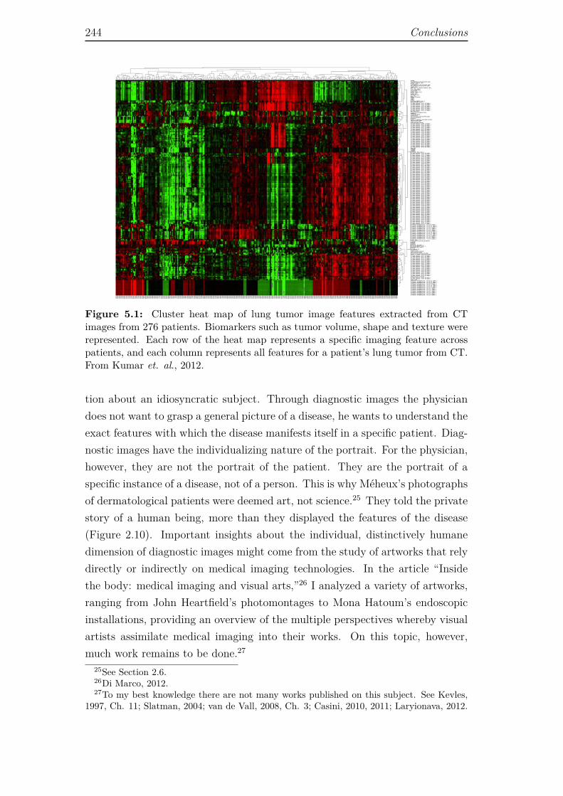
244 Conclusions
Figure 5.1: Cluster heat map of lung tumor image features extracted from CTimages from 276 patients. Biomarkers such as tumor volume, shape and texture wererepresented. Each row of the heat map represents a specific imaging feature acrosspatients, and each column represents all features for a patient’s lung tumor from CT.From Kumar et. al., 2012.
tion about an idiosyncratic subject. Through diagnostic images the physiciandoes not want to grasp a general picture of a disease, he wants to understand theexact features with which the disease manifests itself in a specific patient. Diag-nostic images have the individualizing nature of the portrait. For the physician,however, they are not the portrait of the patient. They are the portrait of aspecific instance of a disease, not of a person. This is why Méheux’s photographsof dermatological patients were deemed art, not science.25 They told the privatestory of a human being, more than they displayed the features of the disease(Figure 2.10). Important insights about the individual, distinctively humanedimension of diagnostic images might come from the study of artworks that relydirectly or indirectly on medical imaging technologies. In the article “Insidethe body: medical imaging and visual arts,”26 I analyzed a variety of artworks,ranging from John Heartfield’s photomontages to Mona Hatoum’s endoscopicinstallations, providing an overview of the multiple perspectives whereby visualartists assimilate medical imaging into their works. On this topic, however,much work remains to be done.27
25See Section 2.6.26Di Marco, 2012.27To my best knowledge there are not many works published on this subject. See Kevles,
1997, Ch. 11; Slatman, 2004; van de Vall, 2008, Ch. 3; Casini, 2010, 2011; Laryionava, 2012.
Conclusions 245
Diagnostic images are by their own nature multidisciplinary objects that callinto question problems of philosophy of science and technology, philosophy ofmedicine, image theory, aesthetics, media theory, and anthropology, to namejust a few. Consequently, they provide a virtually endless field of research,because one can always approach them from a different perspective. Moreover,the emerging field of radiomics, which relies on advanced image analysis toinfer genomic and proteomic patterns from image data,28 promises to blur moreand more the boundaries between quantitative and qualitative information, toreshape the visual landscape of clinical medical imaging (Figure 5.1), and toredefine the theory and practice of diagnostic images’ interpretation. Hence,future research in the philosophy of medical imaging will necessarily come acrossthe methodological and philosophical problems posed by so-called Big Data,29
as well as with the conceptual and ethical issues posed by personalized medicine.Hopefully, the epistemological work developed in this dissertation will serve asa point of departure and conceptual basis for further investigations.
28See Gillies et al., 2010; Lambin et al., 2012; Kumar et al., 2012.29This term is used to refer to any collection of data sets too large and complex to be
properly processed using traditional computational methods. Among the problems posedby Big Data we can count data collection, analysis, visualization, mining, sharing, storage,transfer, and privacy protection.

246 Conclusions
Bibliography
[1] Alexander, K. (2010), Reducing error in radiographic interpretation, CVJ,51:533-335
[2] Ambrose, J. (1973), Computerized transverse axial tomography scanning(tomography): Part 2. Clinical application, British Journal of Radiology,46:1023–1047
[3] Atkin, A. (2013), Peirce’s theory of signs, in The Stanford En-cyclopedia of Philosophy, (Summer 2013 edition), E.N. Zalta(Ed.), <http://plato.stanford.edu/archives/sum2013/entries/peirce-semiotics/>, retrieved on Apr 9, 2014
[4] Azevedo Tavares, C. (Ed.) (2012), Imagens do Corpo na Ciência a naArte, Fim de Século, Lisbon
[5] Bachelard, G. (1931) [2002], Noumène et microphysique, in G. Canguilhem(Ed.) Études, Vrin, Paris, 1970, pp. 11-22
[6] Bachelard, G. (1934) [1984], Le Nouvel esprit scientifique, [English transla-tion by A. Goldhammer, The New Scientific Spirit, Beacon Press, Boston]
[7] Bachelard, G. (1936), Le surrationalisme, Inquisitions, 1:1-10
[8] Bachelard, G. (1937), L’Expérience de l’espace dans la physique contem-poraine, Félix Alcan, Paris
[9] Bachelard, G. (1938) [2002], La Formation de l’esprit scientifique, [Englishtranslation by M. McAllester Jones, The Formation of the Scientific Spirit.A Contribution to a Psychoanalysis of Objective Knowledge, ClinamenPress, Manchester]
[10] Bachelard, G. (1949) [1975], Le Rationalisme appliqué, Presses Universi-taires de France, Paris
[11] Bachelard, G. (1953) [1963], Le Matérialisme rationnel, Presses Universi-taires de France, Paris
247
248 Bibliography
[12] Bachelard, G. (1971), Epistémologie: textes choisis par DominiqueLecourt, Presses Universitaires de France, Paris
[13] Baigrie, B.S. (Ed.) (1996), Picturing Knowledge: Historical and Philo-sophical Problems Concerning the Use of Art in Science, University ofToronto Press, Toronto
[14] Ballestriero, R. (2010), Anatomic models and wax Venuses: Art master-pieces or scientific craft works?, Journal of Anatomy, 216:223-234
[15] Barclay, A.E. (1949), The old order changes, British Journal of Radiology,22:300-308
[16] Barthes, R. (1980), La chambre claire: note sur la photographie, Galli-mard, Paris
[17] Bastide, F. (1990), The iconography of scientific texts: principles of anal-ysis, in M. Lynch and S. Woolgar (Eds.), Representation in ScientificPractice, MIT Press, Cambridge, pp. 187-230
[18] Batchen, G. (1999), Burning with Desire: The Conception of Photography,MIT Press, Cambridge
[19] Baudelaire, C. (1859) [1868], Salon de 1859, Curiosités esthétiques, MichelLévy Frères, Paris, pp. 245-358
[20] Bayle, G.L. (1810), Recherches sur la phthisic pulmonaire, Gabon, Paris
[21] Belting, H. (2001) [2004], Bild-Anthropologie: Entwürfe für eine Bildwis-senschaft [French translation by J. Torrent, Pour une anthropologie desimages, Gallimard, Paris]
[22] Benjamin, W. (1928)[1999], Neues von Blumen, in H.C. Adam (Ed.),Karl Blossfeld, [English translation by C. Goodden, News from flowers,Taschen, Koln, pp. 349-351]
[23] Benjamin, W. (1931)[1999], Kleine Geschichte der Photographie, [Englishtranslation by R. Livingstone, Little history of photography, The BelknapPress of Harvard University Press, Cambridge & London]
[24] Benjamin, W. (1936-1939)[2003], Das Kunstwerk im Zeitalter seiner tech-nischen Reproduzierbarkeit, [English translation by H. Zohn and E. Jeph-cott, The Work of Art in the Age of Its Technological Reproducibility,Third Version, The Belknap Press of Harvard University Press, Cambridge& London]
Bibliography 249
[25] Berenson, B. (1896), The Florentine Painters of the Renaissance, G.P.Putnam’s Sons, New York & London
[26] Bernard, C. (1855-1856), Léçons de physiologie expérimentale appliquée àla medicine, J.B. Baillie et fils, Paris
[27] Bichat, X., (1801)[1822], Anatomie générale appliquée à la physiologie età la medicine, [English translation by G. Hayward, General Anatomy Ap-plied to Physiology and Medicine, Richardson & Lord, Boston]
[28] Bogen, J. (2002), Epistemological custard pies from functional brain imag-ing, Philosophy of Science, 69:S59-S71
[29] Boronat, C.B. et al. (2005), Distinctions between manipulation and func-tion knowledge of objects: evidence from functional magnetic resonanceimaging, Cognitive Brain Research, 23:361–373
[30] Breidbach, O. (2002), Representation of the microcosm - The claim forobjectivity in 19th century scientific microphotography, Journal of theHistory of Biology, 35:221–250
[31] Bunzl, M. and Hanson, S. (Eds.) (2010), Foundational Issues of HumanBrain Mapping, MIT Press, Cambridge
[32] Bynum, W.F. (1994), Science and the Practice of Medicine in the Nine-teenth Century, Cambridge University Press, Cambridge & New York
[33] Bynum, W.F. et al. (Eds.) (2006), The Western Medical Tradition, 1800to 2000, Cambridge University Press, Cambridge & New York
[34] Bynum, W.F. (2006), The rise of science in medicine, 1850-1913, in W.F.Bynum et al. (Eds.), The Western Medical Tradition, 1800 to 2000, Cam-bridge University Press, Cambridge & New York
[35] Canguilhem, G. (1966), Le normal et le pathologique, Presses Universi-taires de France, Paris
[36] Cambrosio, A. et al., (1993), Ehrlich “beautiful pictures” and the contro-versial beginnings of immunological imagery, Isis, 84(4):662-699
[37] Cambrosio, A. et al. (2005), Arguing with images: Pauling’s theory ofantibody formation, Representations, 89:94-130
[38] Cappelletto, C. (2010), La natura finzionale dell’immagine nel confrontocon le neuroscienze, Psicoart – Rivista Online di Arte e Psicologia, doi:10.6092/issn.2038-6184/2133, retrieved on Feb 2, 2015
250 Bibliography
[39] Cartwright, L. (1995), Screening the Body. Tracing Medicine’s Visual Cul-ture, University of Minnesota Press, Minneapolis
[40] Carusi, A. (2011), Computational biology and the limits of shared vision,Perspectives on Science, 19(3):300-336
[41] Carusi, A. (2012), Making the visual visible in philosophy of science, Spon-taneous Generations, 6(1):106-114
[42] Casini, S. (2011), Magnetic resonance imaging (MRI) as mirror and por-trait: MRI configurations between science and the arts, Configurations,19(1):73-99
[43] Casini, S. (2010), The aesthetics of magnetic resonance imaging (MRI):from the scientific laboratory to an artwork, Contemporary Aesthetics,<http://www.contempaesthetics.org/newvolume/pages/article.php?articleID=569>,retrieved on Jan 27, 2015
[44] Castelao-Lawless, T. (1995), Phenomenotechnique in historical perspec-tive: its origins and implications for philosophy of science, Philosophy ofScience, 62(1):44-59
[45] Cazort, M. et al. (1996), The Ingenious Machine of Nature: Four Cen-turies of Art and Anatomy, University of Chicago Press, Chicago
[46] Chandler, D. (2007), Semiotics: the Basics, Routledge, London
[47] Conolly, J. (1858) [1976], Case studies from the physiognomy of insanity,in S.L. Gilman (Ed.), The Face of Madness. Hugh W. Diamond and theOrigin of Psychiatric Photography, Brunner Mazel, New York, pp. 25-72
[48] Cosmacini, G. (1997), L’arte lunga. Storia della medicina dall’antichità aoggi, Laterza, Rome & Bari
[49] Coulombe, M. (2009), Imaginer le posthumain. Sociologie de l’art etarchéologie d’une vertige, Les Presses de l’Université Laval, Québec
[50] Crary, J. (1990) [1992], Techniques of the Observer. On Vision and Moder-nity in the Nineteenth Century, MIT Press, Cambridge & London
[51] Chudleigh, T. (1999), Scanning for pleasure, Ultrasound Obstet Gynecol,14:369-371
[52] Dagognet, F. (1986), Philosophie de l’image, J. Vrin, Paris
Bibliography 251
[53] Dagognet, F. (1987), E.-J. Marey. La passion de la trace, Hazan, Paris
[54] Darwin, C. (1872), The Expression of Emotion in Man and Animals, JohnMurray, London
[55] Daston, L. and Galison, P. (1992), The image of objectivity, Representa-tions, 40:81-128
[56] Daston, L. and Galison, P. (2007), Objectivity, Zone Books, New York
[57] de Condillac, E.B. (1754), Traité des sensations, de Beure, Paris & London
[58] de Montméja, A. and Rengade, J. (1869), Revue photographique des hopi-taux de Paris, Delahaye, Paris
[59] Delehanty, M.C. (2010), Why Images? Medicine Studies, 2:161-173
[60] Desormeaux, J.A. (1865), De l’endoscope et de ses applications au diag-nostic et au traitement des affections de l’urèthre..., Baillière, Paris
[61] Diamond, H.W. (1856) [1976], On the application of photography to thephysiognomic and mental phenomena of insanity, in S.L. Gilman (Ed.),The Face of Madness. Hugh W. Diamond and the Origin of PsychiatricPhotography, Brunner Mazel, New York, pp. 17-23
[62] Didi-Huberman, G. (1982), Invention de l’hystérie. Charcot etl’Iconographie photographique de la Salpêtrière, Macula, Paris
[63] Di Marco, S. (2012a), Inside the body: Medical imaging and the visualarts, PsicoArt – Rivista Online di Arte e Psicologia, doi:10.6092/issn.2038-6184/2530
[64] Di Marco, S. (2012b), Naked to the flesh. Some notes on medical imaging,Modernism and Body Art, in C. Azevedo Tavares (Ed.), Imagens do Corpona Ciência a na Arte, Fim de Século, Lisbon, pp. 35-48
[65] Donné, A. and Foucault, L. (1844), Cours de microscopie complémentairedes études médicales, Baillière, Paris
[66] Duchenne de Boulogne, G. (1862a), Album de photographies pathologiquescomplémentaire du livre intitulé de l’électrisation localisée, Baillière, Paris
[67] Duchenne de Boulogne, G. (1862b), Le Mécanisme de la physionomie hu-maine, ou analyse électro-physiologique de l’expression des passions appli-cable à la pratique des arts plastiques, Baillière, Paris
252 Bibliography
[68] Duden, B. (1993), Disembodying Women: Perspectives on Pregnancy andthe Unborn, Harvard University Press, Cambridge
[69] Dumit, J. (2004), Picturing Personhood. Brain Scans and BiomedicalIdentity, Princeton University Press, Princeton & Oxford
[70] Elkins, J. (1995), Art history and images that are not art, Art Bulletin,77(4):553-571
[71] Elkins, J. (1999), Pictures of the Body. Pain and Metamorphosis, StanfordUniversity Press, Stanford
[72] Elkins, J. (2001), Pictures and Tears: A History of People Who HaveCried in Front of Paintings, Routledge, New York & London
[73] Elkins, J. (Ed.) (2007), Photography Theory, Routledge, New York &London
[74] Estival, C. (2009), La visualisation des clichés d’imagerie médicale par lespatients atteints d’un cancer. Analyse en début de trajectoire, Sociétés,doi:10.3917/soc.105.0009, retrieved on Jan 27, 2015
[75] Estival, C. (2010), Imagerie médicale et rapport au corps dans un centrede cancérologie, Corps, 1(8):105-110
[76] Falkenstein, L. (2010), Étienne Bonnot de Condillac, The Stan-ford Encyclopedia of Philosophy, (Fall 2010 Edition), E.N. Zalta(Ed.), <http://plato.stanford.edu/archives/fall2010/entries/condillac/>,retrieved on Apr 9, 2014
[77] Fangerau, H. et al. (Eds.) (2012), Medical Imaging and Philosophy. Chal-lenges, Reflections and Actions, Steiner Verlag, Stuttgart
[78] Farah, M. and Gillihan, S. (2012), Diagnostic brain imaging in psychiatry:current uses and future prospects, Virtual Mentor, 14(6): 464-471
[79] Faure, O. (2005), Le regard des médecins, in A. Corbin, et al. (Eds.),Histoire du corps, Seuil, Paris, vol. 2, pp. 15-50
[80] Foucault, M. (1963)[1973], Naissance de la clinique. Une archéologie duregard médical, Presses Universitaires de France, Paris, [English transla-tion by A.M.Sheridan, The Birth of the Clinic. An archaeology of MedicalPerception, Routledge, New York & London]
Bibliography 253
[81] Freedberg, D. (2007), Empatia, movimento ed emozione, in G. Lucignaniand A. Pinotti (Eds.), Immagini della mente. Neuroscienze, arte, filosofia,Raffaello Cortina Editore, Milan, pp. 13-67
[82] Freedberg, D. and Gallese, V. (2007), Motion, emotion and empathy inesthetic experience, Trends in Cognitive Sciences, 11(5):197-203
[83] French, S. (2003), A model-theoretic account of representation (Or I don’tknow much about art. . . but I know it involves isomorphism), Philosophyof Science, 70(5):1472–1483
[84] Fried, M. (2002), Menzel’s Realism: Art and Embodiment in Nineteenth-Century Berlin, Yale University Press, New Haven & London
[85] Friedman, M. and Friedland, G.W. (1998), Medicine’s 10 Greatest Dis-coveries, Yale Nota Bene, Yale University Press, New Haven & London
[86] Friedrich, K. (2010), “Sehkollektiv”: Sight styles in diagnostic ComputedTomography, Medicine Studies, 2:185-195
[87] Galison, P. (1998), Judgment against objectivity, in Picturing ScienceProducing Art, C. Jones and P. Galison (Eds.), Routledge, New York &London, pp. 327-359
[88] Galison, P. (1999), Objectivity is romantic, The Humanities and the Sci-ences, ACLS Occasional Paper, 47:15-43
[89] Gallese, V. (2005), Embodied simulation: From neurons to phenomenalexperience, Phenomenology and the Cognitive Sciences, 4(4):23-48
[90] Gayon, J. (1994), Bachelard: Le rationalisme appliqué, CNED-Quadrige,Paris
[91] Gillies, R.J. et al., (2010), The biology underlying molecular imaging inoncology: from genome to anatome and back again, Clinical Radiology,65:517-521
[92] Gilman, S.L. (Ed.) (1976), The Face of Madness. Hugh W. Diamond andthe Origin of Psychiatric Photography, Brunner Mazel, New York
[93] Gilman, S.L. (1976), Hugh W. Diamond and Psychiatric Photography,in S.L. Gilman (Ed.), The Face of Madness. Hugh W. Diamond and theOrigin of Psychiatric Photography, Brunner Mazel, New York, pp. 3-15
254 Bibliography
[94] Gilman, S.L. et al. (1993), Hysteria Beyond Freud, University of CaliforniaPress, Berkeley
[95] Gilman, S.L. (1993), The Image of the hysteric, in S.L. Gilman et al.,Hysteria Beyond Freud, University of California Press, Berkeley, pp. 345-436
[96] Goetz, C.G. (1991), Visual art in the neurologic career of Jean-MartinCharcot, Archives of Neurology, 48:421-425
[97] Gooding, D. (2003), Varying the cognitive span: Experimentation, visual-ization, and computation, The Philosophy of Scientific Experimentation,H. Radder (Ed.), University of Pittsburgh Press, Pittsburgh, pp. 255-283
[98] Gooding, D. (2004a), Envisioning explanations. The art in science, Inter-disciplinary Science Reviews, 29(3):279-294
[99] Gooding, D. (2004b), Cognition, construction and culture: visual theoriesin the sciences, Journal of Cognition and Culture, 4(3):551-593
[100] Gooding, D. (2006), Visual cognition: where cognition and culture meet,Philosophy of Science, 73:688-698
[101] Gowing, L. (1966), Turner: Imagination and Reality, MOMA, New York
[102] Griscom, N.T. (2002), A suggestion: look at the images first, before youread the history, Radiology, 223:9-10
[103] Grmek, M. (1999), Le concept de maladie, in M. Grmek (Ed.), Histoirede la pensée médicale en Occident, vol. 3, Seuil, Paris, pp. 147-167
[104] Guyon, F. (1881), Leçons cliniques sur lea maladies des voies urinaires:professées à l’Hôpital Necker, J.-B. Baillière et fils, Paris
[105] Hacking, I. (1983), Representing and Intervening. Introductory Topics inthe Philosophy of Natural Science, Cambridge University Press, Cam-bridge
[106] Hacking, I. (1990), The Taming of Chance, Cambridge University Press,Cambridge
[107] Hacking, I. (2002), Historical Ontology, Harvard University Press, Cam-bridge & London
[108] Halls Dally, J.F. (1903), On the use of the Roentgen rays in the diagnosisof pulmonary tuberculosis, The Lancet, 27 June:1800-1806
Bibliography 255
[109] Hardcastle, V.G. and Steward, C.M. (2002), What do brain data reallyshow?, Philosophy of Science, 69:S72–S82
[110] Hardy, A. and de Montméja, A. (1868), Clinique photographique del’Hôpital Saint-Louis, Chamerot et Lauwereyns, Paris
[111] Hérisson, J. (1834) [1835], Le sphygmomètre, instrument qui traduit à l’oeiltoute l’action des artères, [English translation by E.S. Blundell, Longman,Rees & Orme, London]
[112] Hopwood, N. (2004), Plastic publishing in embryology, in S. de Chadare-vian and N. Hopwood (Eds.), Models. The Third Dimension of Science,Stanford University Press, Stanford, pp. 170-206
[113] Huber, C.G. (2009), Interdependence of theoretical concepts and neu-roimaging data, Poiesis & Praxis, 6(3-4):203-217
[114] Huber, C.G. and Huber, J. (2009), Epistemological considerations on neu-roimaging - a crucial prerequisite for neuroethics, Bioethics, 23(6):340-348
[115] Hutchinson, J. (1846), On the capacity of the lungs. . . , Medico-ChirurgicalTransactions, 29:137-252
[116] Israel-Jost, V. (2011), L’observation scientifique: aspects philosophiques etpratiques, Doctoral dissertation, Université de Paris 1 Panthéon-Sorbonne,Paris
[117] Johnson-Frey, S.H. et al. (2003), Actions or hand–object interactions? Hu-man inferior frontal cortex and action observation, Neuron, 39:1053–1058
[118] Kemp, M. (2006), Seen | Unseen. Art, Science, and Intuition fromLeonardo to the Hubble Telescope, Oxford University Press, Oxford & NewYork
[119] Kevles, B.H. (1997), Naked to the Bone. Medical Imaging in the TwentiethCentury, Rutgers University Press, New Brunswick
[120] Klein, C. (2010), Images Are Not the Evidence in Neuroimaging, BritishJournal of Philosophy of Science, 61:265–278
[121] Knorr-Cetina K. and Amann, K. (1990), Image dissection in natural sci-entific inquiry, Science, Technology, & Human Values, 15(3):259-283
[122] Koch, R. (1886), On the investigation of pathogenic organisms, in W.W.Cheyne (Ed.), Recent Essays by Various Authors on Bacteria in Relationto Disease, New Sydenham Society, London, pp. 1-66
256 Bibliography
[123] Kosslyn, S.M. (1999), If neuroimaging is the answer, what is the question?Phil. Trans. R. Soc. Lond. B, 354:1283-1294
[124] Kriebel, S. (2007), Theories of photography. A short history, in J. Elkins(Ed.), Photography Theory, Routledge, New York & London, pp. 3-49
[125] Kumar, V. et al. (2012), Radiomics: the process and the challenges, Mag-netic Resonance Imaging, 30:1234-1248
[126] Kundel, H.L. (2006), History of research in medical image perception,Journal of the American College of Radiology, 3:402-408
[127] Kusch, M. (2009), Objectivity and historiography, Isis, 100:127–131
[128] Kusch, M. (2011), Reflexivity, relativism, microhistory: Three desideratafor historical epistemologies, Erkenntnis, 75:483–494
[129] Laennec, R.T.H. (1826), Traité de l’auscultation médiate et des maladiesdes poumons et du coeur, J.-S. Chaudé, Paris
[130] Lamata Manuel, A. (2010), Superrealistas: De la Contribución de losRayos-X a la Visión y Presentación de la Realidad en el Arte a Comienzosdel Siglo XX, Doctoral dissertation, Universidad Complutense de Madrid,Madrid
[131] Lambin, P. et al. (2012), Radiomics: Extracting more information frommedical images using advanced feature analysis, European Journal of Can-cer, 48:441–446
[132] Landecker, H. (2005), Cellular features: microcinematography and filmtheory, Critical Inquiry, 31(4):903-937
[133] Landecker, H. (2006), Microcinematography and the history of science andfilm, Isis, 97(1):121-132
[134] Laryionava, K. (2012), Medical imaging and contemporary art: redefi-nition of the human body, in H. Fangerau et al. (Eds.), Medical Imag-ing and Philosophy. Challenges, Reflections and Actions, Steiner Verlag,Stuttgart, pp. 145-156
[135] Latour, B. (1984), Les Microbes. Guerre et paix, suivi de Irréductions,Métailié, Paris
[136] Latour, B. (1999), Pandora’s Hope. Essays on the Reality of Science Stud-ies, Harvard University Press, Cambridge
Bibliography 257
[137] Leonard, C.L. (1905), Forty cases of ureteral calculus in which the Roent-gen diagnosis was confirmed by recovery of the calculus, The Lancet, June17: 1632-1636
[138] Lerner, B.H. (1992), The perils of “X-ray vision”: How radiographic im-ages have historically influenced perception, Perspectives in Biology andMedicine, 35(3):382-397
[139] Londe, A. (1893), La photographie médicale. Application aux sciencesmédicales et physiologiques, Gauthier-Villars et fils, Paris
[140] Lucignani, G. and Pinotti, A. (Eds.), Immagini della mente. Neuroscienze,arte, filosofia, Raffaello Cortina Editore, Milan
[141] Lynch, M. and Woolgar, S. (Eds.) (1990), Representation in ScientificPractice, MIT Press, Cambridge
[142] Lynch, M. (1990). The externalised retina: Selection and mathematisationin the visual documentation of objects in the life sciences, in M. Lynchand S. Woolgar (Eds.), Representation in Scientific Practice, MIT Press,Cambridge, pp. 153–186
[143] Mach, E. (1893), An account of scientific applications of photography, TheJournal of the Camera Club, 6:110–112
[144] Mann, T. (1924) [1970], Der Zauberberg, Fischer Verlag, Berlin [Por-tuguese translation by Herbert Caro, Livros do Brasil, Lisbon]
[145] Marey, E.-J. (1860), Recherches sur le pouls au moyen d’un nouvel appareilenregistreur, le sphygmographe, E. Thunot et Co., Paris
[146] Marey, E.-J. (1873), La machine animale, Librairie Germer Baillière, Paris
[147] Marey, E.-J. (1878/1885), La méthode graphique dans les sciences expéri-mentales, Masson, Paris
[148] Marey, E.-J. (1881), La circulation du sang à l’état physiologique et dansles maladies, Masson, Paris
[149] Marey, E.-J. (1894)[1895], Le Movement, Masson, Paris [English transla-tion by E. Pritchard, D. Appleton & C., New York]
[150] Martin, A. et al. (1996), Neural correlates of category-specific knowledge,Nature, 379:649–652
258 Bibliography
[151] Maynard, P. (1997), The Engine of Visualization. Thinking through Pho-tography, Cornell University Press, Ithaca & London
[152] Mazzolini, R.G. (2004), Plastic anatomies and artificial dissections, in S.de Chadarevian and N. Hopwood (Eds.), Models. The Third Dimensionof Science, Stanford University Press, Stanford, pp. 43-70
[153] Merleau-Ponty, M. (1945) [1962], Phénoménologie de la Perception, Galli-mard, Paris [English translation by Colin Smith, Humanities Press, NewYork]
[154] Mitchell, L.M. (2001), Baby’s First Picture: Ultrasound and the Politicsof Fetal Subjects, University of Toronto Press, Toronto
[155] Moholy-Nagy, L. (1925), Malerei, Fotografie, Film, Langen, Munich [Ital-ian translation by B. Reichlin, Pittura Fotografia Film, Einaudi, Turin]
[156] Mole, C. and Klein, C. (2010), Confirmation, refutation and the evidenceof fMRI, in M. Bunzl and S. Hanson (Eds.), Foundational Issues of HumanBrain Mapping, MIT Press, Cambridge
[157] Moll, A. (2002), The Body Multiple: Ontology in Medical Practice, DukeUniversity Press, Durham & London
[158] Morgan, M.L. and Michaels, M.W. (Eds.) (1999), Fetal Subjects, FeministPositions, University of Pennsylvania Press, Philadelphia
[159] Morgan, L.M. (2009), Icons of Life: A Cultural History of Human Em-bryos, University of California Press, Berkeley, Los Angeles & London
[160] Müller-Rostock, B. (2009), Imagin(in)g pregnancy in Northwest Tanzania:Networks, experiences and translations, in R. Zwijnenberger and R. vande Vall (Eds.), The Body Within. Art, Medicine and Visualization, Brill,Leiden & Boston, pp. 139-156
[161] Musser, J.H (1923), The internist and radiology, Radiology, 11:252
[162] Natale, S. (2008), Quella sensibilità esagerata della lastra: Raggi X erevival del mesmerismo nella fotografia di fine Ottocento, AFT – Rivistadi Storia e Fotografia, 48:53-61
[163] Natale, S. (2011), The invisible made visible, Media History, 17(4):345-358
[164] Natale, S. (2012), A short history of superimposition: From spirit photog-raphy to early cinema, Early Popular Visual Culture, 10(2):125-145
Bibliography 259
[165] Osler, W. (1820), The Principles and Practice of Medicine, 8th Edition,Young and Pentland, Edinburgh & London
[166] Overton, J. et al. (2011), Open biomedical ontologies applied to prostatecancer, Applied Ontology, 6: 35–51
[167] Pasveer, B. (1989), Knowledge of shadows: the introduction of X-rayimages in medicine, Sociology of Health and Illness, 4:360-381
[168] Patton, L. (2012), Hermann von Helmholtz, The Stanford Ency-clopedia of Philosophy, (Winter 2012 Edition), E.N. Zalta (Ed.)<http://plato.stanford.edu/archives/win2012/entries/hermann-helmholtz/>, retrieved on Apr 9, 2014
[169] Peirce, C.S., Collected Papers of Charles Sanders Peirce, C. Hartshorneand P. Weiss, The Belknap Press of Harvard University Press, Cambridge,1974
[170] Peitzman, S.J. and Maulitz, R.C. (1999), L’élaboration du diagnostique,in M. Grmek (Ed.), Histoire de la pensée médicale en Occident, vol. 3,Seuil, Paris, pp. 169-185
[171] Pepe, G. et al., (2012), Somatostatin receptor SPECT, European Journalof Nuclear Medicine and Molecular Imaging, 39(S1):S41–S51
[172] Perini, L. (2005a), The truth in pictures, Philosophy of Science72(1):262–285
[173] Perini, L. (2005b), Visual representations and confirmation, Philosophy ofScience, 72(5):913-926
[174] Perini, L. (2006), Visual representation, in J. Pfeifer and S. Sarkar (Eds.),The Philosophy of Science: An Encyclopedia, Routledge, New York &London, pp. 863-870
[175] Perini, L. (2012), Depiction, detection. And the epistemic value of pho-tography, The Journal of Aesthetics and Art Criticism, 70(1):151-160
[176] Petchesky, R. (1987), The power of visual culture in the politics of repro-duction, Feminist Studies, 3(2):263-292
[177] Pfeifer, J. and Sarkar, S. (Eds.) (2006), The Philosophy of Science: AnEncyclopedia, Routledge, New York & London
260 Bibliography
[178] Pinotti, A. and Somaini, A. (Eds.) (2012), Aura e choc. Saggi sulla teoriadei media di Walter Benjamin, Einaudi, Turin
[179] Pisco, J.M. (Ed.) (2009), Imagiologia básica. Texto e Atlas, Lidel, Lisbon
[180] Pogliano, C. (2011), Lumen in obscuris. The winding road of modernendoscopy, Nuncius, 26:50-82
[181] Pombo, O. and Di Marco, S. (Eds.) (2010), As imagens com que a ciênciase faz, Fim de Século, Lisbon
[182] Pombo, O. and Gerner, A. (Eds.) (2010), Studies in Diagrammatology andDiagram Praxis, College Publications, London
[183] Pommert, A. et al. (2000), A highly realistic volume model de-rived from the Visible Human Male, VHP Conference 2000,www.nlm.nih.gov/archive/20120702/research/visible/vhpconf2000,retrieved on Jan 13, 2014
[184] Ponseti, J. et al. (2006), A functional endophenotype for sexual orientationin humans, Neuroimage, 33:825–833
[185] Pope, J.A. (1999), Medical Physics: Imaging, Heinmann Educational Pub-lishers, Oxford
[186] Porter, R. (1993), The body and the mind, the doctor and the patient:negotiating hysteria, in S.L. Gilman et al. (Eds.), Hysteria Beyond Freud,University of California Press, Berkeley, pp. 225-266
[187] Porter, T.M. (2007), Eras of judgement, Nature, 449:985-986
[188] Porter, T.M. (2008), The objective self, Victorian Studies, 50(4):641-647
[189] Posner, E. (1971), The early years of chest radiology in Britain, Thorax,26:233-239
[190] Prasad A. (2005), Making images/making bodies: visibilizing and dis-ciplining through magnetic resonance imaging, Science, Technology, &Human Values, 30(2):291-316
[191] Pujade, R. (1995), La catastrophe et le phénomène, in M. Sicard, R. Pu-jade and D. Wallach, À corps et à raison: photographies médicales, 1840-1920, Marval, Paris, pp. 89-93 [Exhibition’s catalogue, Hotel de Sully,Paris, de April 14 to June 4, 1995]
Bibliography 261
[192] Radstake, M. (2009), Looking for a sponge: How a body learns to beaffected by ultrasound, in R. Zwijnenberger and R. van de Vall (Eds.), TheBody Within. Art, Medicine and Visualization, Brill, Leiden & Boston, pp.123-138
[193] Reiser, S.J. (1978) [1999], Medicine and the Reign of Technology, Cam-bridge University Press, Cambridge& New York
[194] Reiser, S.J. (2009), Technological Medicine. The Changing World of Doc-tors and Patients, Cambridge University Press, Cambridge & New York
[195] Rheinberger, H.-J. (2005), Gaston Bachelard and the notion of “phe-nomenotechnique”, Perspectives on Science, 13(3):313-328
[196] Richer, P., de la Tourette, G., Londe, A. (1888), Nouvelle iconographie dela Salpêtrière, Lecrosnier et Babé, Paris, Vol. 1
[197] Riva, A. et al. (2010), The evolution of anatomical illustration and waxmodelling in Italy from the 16th to early 19th centuries, Journal ofAnatomy, 216:209-222
[198] Roskies, A. (2007), Are neuroimages like photographs of the brain?, Phi-losophy of Science, 74:860-872
[199] Rowland, S. (1896), A series of collotype illustrations with descriptive text,illustrating applications of the new photograph to medicine and surgery,Archives of Clinical Skiagraphy, 1:3-31, reprint in British Journal of Ra-diography, 1995; 68:H2-H20
[200] Roberts, J. (2012), The Visualised Foetus: A Ccultural and Political Anal-ysis of Ultrasound Imagery, Ashgate Publishing, Surrey & Burlington
[201] Roentgen, W. (1896), On a new kind of rays, Nature, 53:274–276
[202] Sabih, D. et al. (2011), Image perception and interpretation of abnormal-ities; can we believe our eyes? Can we do something about it?, Insightsinto Imaging, 2:47-55
[203] Samuel, N. (2013), Images as tools. On visual epistemic practices in thebiological sciences, Studies in History and Philosophy of Biological andBiomedical Sciences, 44:225-236
[204] Schnalke, T. (1995), Diseases in Wax. The History of the Medical Moulage,Quintessence, Berlin
262 Bibliography
[205] Schnalke, T. (2004), Casting skin: meanings for doctors, artists, and pa-tients, in S. de Chadarevian and N. Hopwood (Eds.), Models. The ThirdDimension of Science, Stanford University Press, Stanford, pp. 207-241
[206] Sekula, A. (1986), The body and the archive, October, 39:3-64
[207] Semczyszyn, N. (2010), Signal into Vision: Medical Imaging as Instru-mentally Aided Perception, Doctoral dissertation, University of BritishColumbia, Vancouver
[208] Serpente, N. (2011), Cells from icons to symbols: Molecularizing cell bi-ology in the 1980s, Studies in History and Philosophy of Biological andBiomedical Sciences, 42:403–411
[209] Sicard, M., Pujade, R. and Wallach, D. (1995) À corps et à raison: pho-tographies médicales, 1840-1920, Marval, Paris [Exhibition’s catalogue,Hotel de Sully, Paris, de April 14 to June 4, 1995]
[210] Sicard, M. (1998) [2006], La fabrique du regard, Odile Jacob, Paris [Por-tuguese translation by P. Elói Duarte, Edições 70, Lisbon]
[211] Simmons, D. (2008), The use of animal models in studying genetic disease:transgenesis and induced mutation, Nature Education, 1(1):70
[212] Slatman, J. (2004), L’imagerie du corps interne, Methodos, 4, doi:10.4000/methodos.133
[213] Snyder, J. (1998), Visualization and visibility, in C. Jones and P. Galison(Eds.), Picturing Science Producing Art, Routledge, New York & London,pp. 379-397
[214] Somaini, A. (2010), Fotografia, cinema, montaggio. La “nuova visione” diLászló Moholy-Nagy, in L. Moholy-Nagy, Pittura Fotografia Film, Einaudi,Turin, pp. ix-lxxx
[215] Sontag, S. (1978), Illness as Metaphor, Penguin Books, London & NewYork
[216] Stephens, E. (2012), Anatomical imag(inari)es: The cultural impact ofmedical imaging technologies, Somatechnics, 2(2):159-170
[217] Sturm, T. (2011), Historical epistemology or history of epistemology?The case of the relation between perception and judgment, Erkenntnis,75:303–324
Bibliography 263
[218] Talbot, H.F. (1844-1846), The Pencil of Nature, Longman, Brown, Green& Longmans, London
[219] Taraborelli D. (2003), Verso una nuova frenologia? Considerazioni sull’usodei metodi di brain imaging e di strategie sottrattive per lo studio dellacognizione e delle sue basi neurali, L&PS – Logic and Philosophy of Sci-ence, 1(1):1-26
[220] Taylor, J.S. (2000), Of sonograms and baby prams: Prenatal diagnosis,pregnancy, and consumption, Feminist Studies, 26(2):391-418
[221] Taylor, J.S. (2008), The Public Life of the Fetal Sonogram: Technology,Consumption, and the Politics of Reproduction, Rutgers University Press,New Brunswick
[222] Tucker, J. (2005), Nature Exposed. Photography as Eyewitness in Victo-rian Science, The John Hopkins University Press, Baltimore
[223] Tucker, J. (2008), Objectivity, collective sight, and scientific personae,Victorian Studies, 50(4):648-657
[224] Urgesi, C. et al. (2006), Mapping implied body actions in the human,motor system, Journal of Neurosciences, 26:7942–7949
[225] van Dijck, J. (2005), The Transparent Body. A Cultural Analysis of Med-ical Imaging, University of Washington Press, Seattle
[226] van de Vall, R. (2008), At the Edges of Vision. A Phenomenological Aes-thetics of Contemporary Spectatorship, Ashgate, London
[227] Vidal, F. and Ortega, F. (2012), Are there neural correlates of depression?,in S. Choudhury and J. Slaby (Eds.), Critical Neuroscience: A Handbookof the Social and Cultural Contexts of Neuroscience, Blackwell Publishing,Oxford
[228] Wolbarst, A.B. (1999), Looking Within. How X-ray, CT, MRI, Ultra-sound, and Other Medical Images Are Created, and How They Help Physi-cians Save Lives, University of California Press, Berkeley and Los Angeles
[229] Wunderlich, C.A. (1868), Das Verhalten der Eigen Wärme in Krankheiten,[English translation from the second German edition by W. BalthurstWoodman, The New Sydenham Society, London]

264 Bibliography
[230] Wunenburger, J.J. (1997) [1999], Philosophie des images, Presses Univer-sitaires de France, Paris, [Italian translation by Sergio Arecco, Einaudi,Turin]
[231] Wurdemann, H.V. (1894), The status of skiascopy, JAMA, 23:341
[232] Zwijnenberger, R. and van de Vall, R. (Eds.) (2009), The Body Within.Art, Medicine and Visualization, Brill, Leiden & Boston


