Embed Size (px)
Citation preview
Accep
ted
Man
uscr
ipt
© Endocrine Society 2019. All rights reserved. For permissions, please e-mail: [email protected]
The role of sex and sex Hormones in Neurodegenerative Diseases
Elisabetta Vegeto, Alessandro Villa, Sara Della Torre, Valeria Crippa, Paola Rusmini,
Riccardo Cristofani, Mariarita Galbiati, Adriana Maggi*, Angelo Poletti*
Department of Excellence of Pharmacological and Biomolecular Sciences
and Center of Excellence on Neurodegenerative Diseases
Università degli Studi di Milano, Italy
Key terms: Sex Hormones, Alzheimer’s disease, Parkinson’s Disease, Amyotrophic Lateral
Sclerosis, Spinal and Bulbar Muscular Atrophy
*These Authors equally contributed to this manuscript
Corresponding author's contact information:
Angelo Poletti, PhD - Dipartimento di Scienze Farmacologiche e Biomolecolari, Center of
Excellence on Neurodegenerative Diseases. Università degli Studi di Milano, via Balzaretti 9,
20133, Milan, Italy. Ph. +390250318215; Fax +390250318204; e-mail [email protected]
Disclosure statement. The Authors have no item to disclose
Grants: National Institute of Health Grant RO1AG027713; European Union's Seventh Framework
Programme (FP7/2007-2013) under grant agreement no. 278850 (INMiND); Fondazione Cariplo,
Italy (2011-0591, 2014-0686 and 2017_0747); Fondazione Telethon, Italy (n. GGP14039,
GGP19218); Fondazione AriSLA, Italy (ALS_HSPB8, ALS_Granulopathy, MLOpathy,
Target_RAN), Italian Ministry of Health (n. GR-2011-02347198), Agenzia Italiana del Farmaco
(AIFA) (Co_ALS), Italian Ministry of University and Research (MIUR), PRIN - Progetti di ricerca
di interesse nazionale (n. 2015LFPNMN and 2017F2A2C5), MIUR progetto di eccellenza, Fondo
per il Finanziamento delle Attività Base di Ricerca (FFABR-MIUR), Fondazione Regionale per la
Ricerca Biomedica (FRRB) (Regione Lombardia, TRANS_ALS, project nr. 2015-0023),
Università degli Studi di Milano e piano di sviluppo UNIMI - linea B, European Molecular
Biology Organization (EMBO) short term fellowship (n. 537 – 2015); EU Joint Programme -
Neurodegenerative Disease Research (JPND) project European Union’s Horizon 2020 research
and innovation programme under grant agreement No 643417 (Grant ID: 01ED1601A, CureALS).
Dow
nloaded from https://academ
ic.oup.com/edrv/advance-article-abstract/doi/10.1210/endrev/bnz005/5572525 by guest on 22 O
ctober 2019
Accep
ted
Man
uscr
ipt
2
Abstract
Neurodegenerative diseases (NDs) are a wide class of disorders of the central nervous system
(CNS) with still unknown etiology. Several factors were hypothesized to be involved in the
pathogenesis of these diseases including genetic and environmental factors. Many of these diseases
show a sex prevalence and sex steroids where shown to have a role in the progression of specific
forms of neurodegeneration. Estrogens were reported to be neuroprotective though their action on
cognate nuclear and membrane receptors, while adverse effects of male hormones have been
described on neuronal cells, although some data also suggest neuroprotective activities. The
response of the CNS to sex steroids is a complex and integrated process that depends on: i.) the
type and amount of the cognate steroid receptor; ii.) the target cell-type, either neurons, glia or
microglia. Moreover, the levels of sex steroids in the CNS fluctuate due to gonadal activities and
to local metabolism and synthesis. Importantly, biochemical processes involved in the
pathogenesis of NDs are increasingly being recognized different among the two sexes and to be
influenced by sex steroids.
The aim of this review is to present current state-of-the-art understanding on the potential role of
sex steroids and their receptors on the onset and progression of major neurodegenerative disorders,
namely Alzheimer’s disease, Parkinson’s diseases, Amyotrophic Lateral Sclerosis and the peculiar
motoneuron disease Spinal and Bulbar Muscular Atrophy, in which hormonal therapy is
potentially useful as disease modifier.
Dow
nloaded from https://academ
ic.oup.com/edrv/advance-article-abstract/doi/10.1210/endrev/bnz005/5572525 by guest on 22 O
ctober 2019
Accep
ted
Man
uscr
ipt
3
Essential Points
Human neurodegenerative diseases are characterized by sex differences in term of
onset, progression of disease, but current knowledge does not allow to precisely
define the sex-related factors intervening in these diseases.
Epidemiological and clinical studies linked the sex-specific synthesis of sex steroids
to disease risk prevalence and incidence, but considering the hormonal pervasive
effects on sexual differentiation and on brain development or functions, their sex
specific influence in neurodegeneration remains obscure.
The role played by sex steroids into functionally priming male and female brains
also remains elusive thus impairing our ability to understand the extent to which
brain embryonal sex differentiation may be associated with the development of a
sex-specific vulnerability to neuronal death in adulthood.
Present state-of-the-art knowledge does not allow to definitely point to sex steroids
as a direct or indirect components for protective or detrimental activities in these
diseases.
The complexity of sex steroid physiological functions, the number of neural cells
potentially involved, epigenetic as well as environmental factors have impaired the
understanding of the role of sex on neurodegeneration so far.
Here we provide a wide and in depth analysis of the role of sex in in four common
neurodegenerative diseases: Alzheimer’s, Parkinson’s diseases, Amyotrophic
lateral Sclerosis and Spinobulbar Muscular Atrophy because all showing a sex-
specific incidence and progression.
Our effort is aimed at facilitating the identification of all aspects that in these
disorders associate sex and disease manifestation at both pre-clinical and clinical
level, hoping to enable a progress in this field and underline potential ways where
appropriate regulation of circulating hormones may provide benefits in these
disorders where we suffer a unique lack of positive therapeutic intervention.
Dow
nloaded from https://academ
ic.oup.com/edrv/advance-article-abstract/doi/10.1210/endrev/bnz005/5572525 by guest on 22 O
ctober 2019
Accep
ted
Man
uscr
ipt
4
INDEX
1. Introduction
2. The influence of sex on Neurodegenerative Diseases
a. Alzheimer’s Disease (AD): the pathogenesis
i. Pathogenic mechanisms in familiar and sporadic AD
ii. Non-neuronal cells in AD
iii. Sex difference in AD
b. Parkinson’s disease (PD): the pathogenesis
i. Common pathogenic mechanisms in genetic and idiopathic PD.
ii. Non-neuronal cells involved in PD
iii. Sex differences in PD
c. Amyotrophic lateral sclerosis (ALS): the pathogenesis
i. Sporadic versus familial forms of ALS
ii. Non-neuronal cells involved in ALS
iii. Sex differences in ALS
iv. The neuroinflammatory response in ALS affected regions and its possible
correlation with gender differences
d. The peculiar case of Spinal and Bulbar Muscular Atrophy (SBMA)
i. A mutation of Androgen Receptor, the molecular basis of the disease
ii. Non-neuronal cells in SBMA
3. Sex steroids in the mammalian nervous system
a. Morpho-functional differences between male and female brains
b. The essential role of sex steroids in brain sexual differentiation
c. Brain expression of steroid receptors as mediators of sex hormone activities
i. Sex hormone receptors
ii. Sex hormone receptors distribution in the brain.
d. Neurosteroids and neuroactive steroids
i. Local activation and de novo synthesis of steroids in the brain.
e. Sex steroid effects on neural cells
f. Predicting the activity of male sex hormones in the nervous systems
4. The influence of Sex hormones in Neurodegenerative diseases
a. Sex hormones and AD
i. Estrogen and AD
ii. Estrogen decreases A accumulation
iii. Estrogen effects on lipid metabolism
iv. Progesterone and AD
v. Female hormone replacement therapy and AD
vi. Role of male sex steroids in AD
b. Sex hormones and PD
i. Estrogens, NSDA and PD
ii. Estrogens in animal models of PD
iii. Progesterone and PD.
iv. Androgens, NSDA and PD
v. Sex steroid hormones and pathogenic mechanisms of PD
c. Sex hormones and ALS
Dow
nloaded from https://academ
ic.oup.com/edrv/advance-article-abstract/doi/10.1210/endrev/bnz005/5572525 by guest on 22 O
ctober 2019
Accep
ted
Man
uscr
ipt
5
i. Sex-related molecular and biochemical alterations occurring in ALS: the
role of sex steroids
ii. Male sex steroids
iii. Female sex steroids
iv. Neuroprotective activity of progesterone and estrogens
v. Estrogens and neuroinflammation in ALS
vi. Metabolic disorders and ALS
vii. Sex hormones, autophagy and proteasomal regulation
d. The case of SBMA
i. The role of testosterone.
5. Conclusions and future perspectives
Dow
nloaded from https://academ
ic.oup.com/edrv/advance-article-abstract/doi/10.1210/endrev/bnz005/5572525 by guest on 22 O
ctober 2019
Accep
ted
Man
uscr
ipt
6
1. Introduction
Neurodegenerative diseases (NDs) are devastating and largely fatal conditions that affect several
million people in the world. NDs onset may occur during the entire life in human, with juvenile
forms (that appear around birth, e.g.: some forms of spinal and muscular atrophy) or adult forms
that may also appear very late in life. Most NDs appear after the third decade of life possibly
correlating with the aging process. The causes leading to the massive neuronal death characteristic
of NDs remain to be fully understood, thus developing strategies for their prevention, treatment or
even delayed progression is a major challenge for modern medicine. Although it is indisputable
that the main symptoms of NDs reflect neuron-specific deficits, it is also conceivable that
dysfunction in other brain cells could precede and facilitate neuronal loss. Non-neuronal cells in
the central nervous system (CNS) include neuroglia (oligodendrocytes and astrocytes) and
microglia, a class of cells maintaining brain homeostasis through an appropriate exchange of
nutrients and protection against noxious stimuli (1). Oligodendrocytes enwrap axons with myelin
sheaths providing a structural and local metabolic and homeostatic protection (2); microglia ensure
immune protection, the control of synaptic connections and tissue repair (3,4); astrocytes regulate
the homeostasis of neurotransmission, metabolites and reactive oxygen species (5). Compelling
evidence shows that these cells are impaired in NDs (1,6), as discussed in the appropriate sections
of this review. Among the three major forms of ND, Alzheimer’s Disease (AD) is more frequent
in women than in men (ratio 2:1) (7), while Parkinson’s Disease (PD) and Amyotrophic Lateral
Sclerosis (ALS) affect primarily men (1.5:1, and 2:1 respectively) (8,9); peculiar is the case of
Spinal and Bulbar Muscular Atrophy (SBMA), which affects men only (10). The molecular bases
for this sex-related prevalence remain to be understood, although sex steroid hormones may be
involved in disease pathogenesis. However, the decline of sex steroid synthesis occurring during
Dow
nloaded from https://academ
ic.oup.com/edrv/advance-article-abstract/doi/10.1210/endrev/bnz005/5572525 by guest on 22 O
ctober 2019
Accep
ted
Man
uscr
ipt
7
aging or the changes due to pharmacological supplementation may be either a risk or protective
factors as a function of the neurodegenerative disease considered (11-13). Perhaps, the reason for
these opposite results is that the connections between sex steroids and the manifestation of NDs
are multifaceted as these hormones target all neuronal cells (neurons, glia and/or the immune cells)
and control brain sexual differentiation, a process that may be relevant for the degenerative
processes in adults. Moreover, another element of complexity further impairing our ability to
interpret the results of investigations is that, in addition to the gonads, sex steroids may be
synthesized in the brain. Epidemiological evidence indicates a sexual prevalence and incidence in
most forms of ND (table 1).
Considering the reported beneficial vs. deleterious effects of sex steroids, the aim of the present
review is to recapitulate current knowledge on estrogen, progesterone and androgen activities in
neural cells and their potential influence on manifestation of NDs. The review will also analyze
the impact of the relative contribution between circulating hormones, their local synthesis and
activation (Figure 1) including their complex interplay in target cells (Figure 2). Our anticipation
is that a better understanding of this field of study may lead to novel therapeutic strategies very
much in need for ND.
2. The influence of sex on Neurodegenerative Diseases
a. Alzheimer’s Disease: the pathogenesis
AD is the most common cause of dementia (47-49), and is a slowly progressive disease that begins
well before the onset of clinical symptoms (50). With disease progression, memory loss and
confusion become more severe (49), until impairments in basic physiological functions appear in
the terminal stage of the disease. Hallmarks of AD include the accumulation of aggregates and
Dow
nloaded from https://academ
ic.oup.com/edrv/advance-article-abstract/doi/10.1210/endrev/bnz005/5572525 by guest on 22 O
ctober 2019
Accep
ted
Man
uscr
ipt
8
deposits of misfolded proteins, like amyloid beta (Aβ) peptides, outside neurons, and of
neurofibrillary tangles (NFT), which contains abnormally phosphorylated microtubule-associated
tau protein, inside neurons (50,51). Increasing evidence points to a causal role of inflammation in
disease onset (52).
a.i. Pathogenic mechanisms in familiar and sporadic AD
Some AD cases (<1%) result from fully penetrant mutations of specific genes (Table 2) which
associated with an onset risk before 65 years (69). Sporadic, or late onset-form of AD (LOAD),
manifests later in life and is linked to genetic factors (70) acting together with environmental risk
factors (71) (Table 2). The most established genetic risk factor for LOAD is the ε4 allele of the
apolipoprotein E gene (APOE), a major cholesterol carrier responsible for lipid transport in the
brain (72,73); APOE4 accounts for differential risk to develop AD based on the gene dose (8.07
or 2.84 hazard ratio in homo- or heterozygosis, respectively) (56). Several other common genetic
variants associated with LOAD highlighting the multifactorial origins of sporadic AD (Table 2).
The blend of genetic, environmental and epigenetic factors (74-76) leads to a progressive
decrease in synaptic density and loss of integrity in neuronal networks, resulting into neuritic
atrophy and neuronal death. Several causes are involved, such as amyloidoses and tauopathy,
and impairment of lipid metabolism (77-80). In the case of APOE4 carriers
hypercholesterolemia is observed (~8% higher than baseline population) together with an
increased susceptibility to develop amyloid deposition (81,82). Other well-recognized
etiopathogenetic elements are neuroinflammation (3,52,83,84), impairment of autophagy (85),
or lysosomal degradation (86), loss of Ca2+ homeostasis (87,88), neuronal cycle control (89,90)
and metabolic dysregulation (91-93).
a.ii Non-neuronal cells in AD.
Dow
nloaded from https://academ
ic.oup.com/edrv/advance-article-abstract/doi/10.1210/endrev/bnz005/5572525 by guest on 22 O
ctober 2019
Accep
ted
Man
uscr
ipt
9
Neuroinflammatory in AD is due to a persistent activation of microglia associated with massive
release of molecules toxic to neurons and oligodendrocytes (e.g.: glutamate, free radicals, and
TNF-α (52)). Indeed, in early AD stages, microglia is neuroprotective by clearing Aβ deposits and
by internalize protofibrillar and fibrillar forms of Aβ peptide via endocytosis or macropinocytosis
(94-97). This activity decreases during disease progression, when Aβ receptor expression (such as
SRA, CD36, RAGE) is reduced and the degradation of the engulfed amyloid fibrils is impaired.
In turn, the formation of amyloid deposits within microglia sustains the production of pro-
inflammatory cytokines (IL-1β, IL-6, and TNFα) detrimental to neuron (83,98). Moreover, with
aging microglia becomes dystrophic, showing morphological characteristics distinct from young,
healthy cells (3,99); this process is further aggravated in pathologies involving systemic
inflammation (e.g. obesity and diabetes mellitus) (83,100,101).
Brain aging is often associated with a loss of astrocyte functions (6), prior to AD plaque or tangle
formation (102). Myelin breakdown is induced by neuroinflammation and oxidative stress and it
also occurs at early stages of AD, before AD plaque or tangle formation (2), but its relevance for
AD pathogenesis and the reasons why oligodendrocytes are unable to repair the initial myelin
breakdown remain to be elucidated.
a.iii Sex differences in AD.
Besides age and the genetic risk factors, also sex strongly influences the risk of AD. In fact, it is
estimated that almost two-thirds of Americans with AD are women (48), while among people aged
71 and older, 16% of women have AD compared with 11% of men (103,104). Also the cognitive
decline is sexually dimorphic and is faster in females (22,23) with AD, but not in mild cognitive
impairment (MCI) or other forms of dementia (105,106). Multimodality brain imaging indicates
that in females biomarkers of the preclinical phase of AD, including failures in cerebral glucose
metabolism, and a decrease in neuron mitochondrial function, appear early and overlaps the
Dow
nloaded from https://academ
ic.oup.com/edrv/advance-article-abstract/doi/10.1210/endrev/bnz005/5572525 by guest on 22 O
ctober 2019
Accep
ted
Man
uscr
ipt
10
endocrine transition of perimenopause (107). Neuropathological studies evidenced a more severe
β-amyloid accumulation in women (20,21), and increased levels of tau pathology in men (24).
These pathological hallmarks are reflected in most AD animal models (Tg2576,
APPswe/PSEN1E9, APP23, APPswexPS1, and 3xTg-AD). Notably, a poorer behavioral
performance and a greater increase of Aβ accumulation and hyperphosphorylated tau levels are
observed in females than age-matched males (14-19). Several biological and social reasons were
proposed to explain the AD prevalence in women (7), including the fact that women live longer
than men, and older age is the greatest risk factor for AD (108). It has been suggested that men
surviving to older ages may have a healthier cardiovascular risk profile, thus a lower risk for
dementia than women of the same age (109); however the fact that the higher AD incidence in
female is present in all age-matched groups (from 60–64 till 95 years of age) does not support this
hypothesis. The role of sex hormones is still to be clarified, since mixed results were reported on
the use of hormone replacement therapy (HRT) used to counteract the development and
progression of AD. A strong association between APO4 genotype with sporadic AD in women,
has been reported (110,111). Indeed, women carrying both the homo- and heterozygous Apo4
isoform have a higher rate of conversion from healthy aging to MCI and from MCI to AD.
Conversely, Apo4 variant in men has marginal effects when in homozygosity and null in
heterozygosity (110,112). Memory tests done in individuals with ε3/ε3, ε3/ε4 and ε4/ε4 APOE
genotypes showed a significant gene-dosage dependent effect of the ε4 allele on performance and
male ε4 carriers at midlife showed a significant behavioral advantage in short-term memory task
as compared with women (113).
b. Parkinson's Disease: the pathogenesis
Dow
nloaded from https://academ
ic.oup.com/edrv/advance-article-abstract/doi/10.1210/endrev/bnz005/5572525 by guest on 22 O
ctober 2019
Accep
ted
Man
uscr
ipt
11
PD is a progressive ND affecting 1-2% individuals over 65 years of age. It is characterized by
motor symptoms, that include tremor at rest, rigidity and bradykinesia, as well as non-motor
symptoms (cognitive impairment and mood, olfaction and autonomic dysfunctions). Motor
symptoms are due to the selective degeneration of dopaminergic neurons located in the substantia
nigra (SN) and innervating the striatum, forming the nigrostriatal dopaminergic (NSDA) system.
Dopaminergic neurons have a high oxidative status at baseline, possibly because of the natural
propensity of dopamine to oxidation. Antioxidant and detoxification systems are thus mainly
devoted to control dopamine metabolites in dopaminergic neurons; this makes the NSDA system
more susceptible and vulnerable to even mild forms of oxidative stress and energy dysmetabolism,
which instead spare other neuronal cell types (114,115). PD is characterized by Lewy’s bodies,
which are intraneuronal aggregates mainly consisting of α-synuclein (SNCA) protein (116), in the
central and enteric nervous systems. This suggested a gut-to-brain spreading of aggregated SNCA
possibly due to exposure to environmental toxins (117,118).
b.i Common pathogenic mechanisms in genetic and idiopathic PD.
Familial PD (fPD) is rare and generally monogenic, while most PD are sporadic forms (85-90%)
originating from both genetic causes and environmental factors (e.g.: pesticides and metals)
(119,120). Some causative genes and genetic predisposition factors have been identified (Table 3)
and suggested key molecular pathways as major elements in PD pathogenesis: 1) protein
aggregation, since SNCA gene mutations render the protein (and its dopamine-induced oxidized
forms) prone to oligomerize in neurons impairing protein trafficking and degradation (129-131);
2) mitochondrial defects and oxidative stress, since mutations in genes encoding leucine-rich
repeat serine/threonine kinase 2 (LRRK2), DJ-1, PTEN-induced putative kinase 1 (PINK1) and
Parkin produce mutated proteins causing respiratory chain dysfunctions and oxidative stress
(115,132); 3) protein and mitochondria quality systems, like autophagy and the lysosomal
Dow
nloaded from https://academ
ic.oup.com/edrv/advance-article-abstract/doi/10.1210/endrev/bnz005/5572525 by guest on 22 O
ctober 2019
Accep
ted
Man
uscr
ipt
12
pathways, since mutations have been found in the glucocerebrosidase gene (GBA) (133-138); 4)
neuroinflammation, since polymorphisms in pro-inflammatory genes potentiate the oxidative
microenvironment induced by dopamine, its metabolites and oligomeric SNCA (139-145).
Animal models of PD are widely used to study disease pathogenesis, particularly those obtained
by pharmacological treatments. Injection of the dopamine analogues, 6-hydroxydopamine (6-
OHDA) and metamphetamine (ME), or the dopamine transporter substrates, 1-methyl-4-phenyl-
1,2,3,6-tetrahydropyridine (MPTP) or the pesticide rotenone, fully reflect the biological defects of
fPD (146) by inducing dopamine accumulation and production of reactive and oxidative species
or by inhibiting mitochondrial activity and inducing oxidative stress and energy failure. This leads
to envision that oxidative stress is a major driver of dopaminergic neuron degeneration, sustained
by an interconnected network of genetic and biochemical alterations and inflammation. Oxidative
stress induces chemical modification of DNA, proteins and lipids in PD brains, and promote
protein aggregation engulfing the detoxification systems in dopaminergic neurons (147-151).
b.ii Non-neuronal cells involved in PD.
Besides neuron-specific defects, non-neuronal cells may be involved in PD pathogenesis.
Microglia activation is present in PD patients and animal models, even before neuronal loss or the
activation of other glial cells. Microglia may be detrimental to PD by secreting neurotoxic
inflammatory mediators and by a limited efficiency in removing misfolded proteins by
phagocytosis. Indeed, pharmacological inhibition of microglia is neuroprotective (152-163).
Interestingly, microglia activation in the whole brain leads to dopaminergic neuron death only in
the NSDA system, suggesting a key role for microglia and inflammation in PD motor defects (164-
169). Astrogliosis is detected in specific brain regions of PD patients and animal models of the
disease (170), and astrocytes contribution to PD pathogenesis is dual. On one side, activated
astrocytes release inflammatory and oxidative stress mediators and sustain iron-induced
Dow
nloaded from https://academ
ic.oup.com/edrv/advance-article-abstract/doi/10.1210/endrev/bnz005/5572525 by guest on 22 O
ctober 2019
Accep
ted
Man
uscr
ipt
13
neurotoxicity in; on the other side, astrocytes display induction of autophagy and produce neuron
survival factors (171-173). Importantly, many genes involved in PD are also expressed by
astrocytes, suggesting that pathogenic signals may also originate from these cells (174). A sexually
dimorphic reactivity of astrocytes has been reported in response to MPTP or LPS, giving rise to
specific patterns of ATP/ROS and inflammatory cytokines production in astrocytes generated from
male or female brains (170,175), supporting the hypothesis that they may contribute to sex
differences in neuroinflammatory diseases (176).
b.iii Sex differences in PD.
Together with aging, male sex is the strongest risk factor for PD. The risk to develop PD is 1.5–
fold greater in men than in women at all ages (31,177) (table 1). The phenotypic characteristics of
the disease are also sexually dimorphic, since women tend to be older than men at symptom onset
and more often present a tremorigenic form and a slower disease progression (31). Genomic
profiling of the SN pars compacta (SNpc) neurons from healthy donors and PD patients further
substantiated the different biological traits of men and women may have a role in disease etiology,
symptoms and response to therapy (178). The difference in PD incidence among the two sexes
may arise from substantial distinctions in healthy adult men and women in the composition and
reactivity of the NSDA system, and this is a peculiar feature of PD among other NDs. As compared
to women, the SNpc in men shows: a) a higher number of dopaminergic neurons with increased
expression of SNCA and PINK1; b) more robust dopamine release induced by stimuli, such as
psychomotor stimulants; and c) increased vulnerability to selective drugs in terms of addiction and
toxicity. Sexual differences are also observed in regulatory systems controlling NSDA plasticity,
a complex network made of interneurons, glia, microglia and input circuitries from other brain
areas that is sexually distinct in terms of cell composition, function and adaptation to signals. Male-
specific genetic determinants, and not organizational effects of sex steroid hormones, appear to
Dow
nloaded from https://academ
ic.oup.com/edrv/advance-article-abstract/doi/10.1210/endrev/bnz005/5572525 by guest on 22 O
ctober 2019
Accep
ted
Man
uscr
ipt
14
define this sexual dimorphism in NSDA circuitry. Importantly, the Y chromosome-specific gene
SRY (sex determining region Y) is expressed in NSDA neurons of males and its activity is
associated with a positive regulation of neuron number, dopamine synthesis and metabolism
(179,180). It is thus proposed that intrinsic biological properties related with SRY gene expression
predispose men to disorders associated with dopaminergic abnormalities in PD or schizophrenia.
Sex-related differences are also observed in the SNpc of rodents (26,27) and in the susceptibility
and progression of PD models. The loss of dopaminergic neurons in the SNpc and the reduction
in dopamine levels in the striatum are more robust in males, whereas the female sex is
neuroprotective when low doses of neurotoxins are used to evidence early stages of PD (27,28,30).
Genetic models are only recently being studied in terms of sexual differences; neurodegeneration
is increased in PARK2-null female mice, as compared with males, suggesting a higher sensibility
of females to ubiquitin-proteasomal defects (26,146). Neuronal primary cultures from females
survive longer and adapt to starvation through distinct metabolic programs, while autophagy is
associated with cell death in male cells (181).
Oxidative stress and mitochondrial functions are also sexually dimorphic in specific brain areas;
females have increased antioxidant defenses and respiratory chain activity compared to males, thus
with lower mitochondrial ROS production and oxidative damage as a consequence of the higher
expression of mitochondrial proteins and antioxidant enzymes (e.g.: paraoxynase-2 or thioredoxin)
(182-185). Male mesencephalic neurons exposed to 6-OHDA have reduced expression of selected
mitochondrial proteins, lower ATP levels and higher ROS levels compared to female neurons
(186). Moreover, females show a higher stress adaptation than males mediated by the expression
of stress response factors and the differential mitochondrial usage of energy sources (181,187).
Dow
nloaded from https://academ
ic.oup.com/edrv/advance-article-abstract/doi/10.1210/endrev/bnz005/5572525 by guest on 22 O
ctober 2019
Accep
ted
Man
uscr
ipt
15
Sexual differences in neuroinflammation have been scarcely analyzed in PD patients and in animal
models of the disease. Using MPTP, the increase in TNFα, IL-1β and IFNγ is faster and correlates
with earlier and greater decrease of TH-neurons in male than female mice striatum, (188).
c. Amyotrophic lateral sclerosis: the pathogenesis
Amyotrophic lateral sclerosis (ALS) has an incidence of 1-2:100,000 and is primarily associated
with functional alterations of the upper and lower motoneurons (placed in cerebral motor cortex
or brainstem, and in the ventral horns of the spinal cord or in the cranial nerves, respectively) and
their target muscle cells. Neurons in the fronto-temporal regions of the brain are seldomly involved
(189), and ALS may manifest as pure motor form or associated with fronto-temporal dementia
(ALS-FTD). Age of onset and progression rate are highly variable (onset generally occurs around
50-60 years of age, and juvenile forms are rare (189).
c.i Sporadic versus familial forms of ALS.
ALS occurs in two, clinically indistinguishable sporadic (sALS) and familial (fALS) forms. fALS
occurs only in 10-15% of patients (see table 4 for details). Several genes are associated with fALS
(189); the first identified is present in 20% of fALS and encodes the antioxidant enzyme
superoxide dismutase 1 (SOD1), a free radical scavenger enzyme ubiquitously expressed (232).
More recently described gene mutations include the genes encoding TAR DNA-binding protein
43 (TDP-43), ALS-linked fused in sarcoma/translocated in liposarcoma (FUS/TLS), optineurin,
and others (a list of gene mutations linked to ALS is reported in Table 4). Generally, the genetic
alteration is associated to a gain of a toxic function due to altered protein conformation (233)
(misfolding) and accumulation as protein aggregates poorly cleared by motoneurons.
Alternatively, the mutation affects essential biological functions by interfering with RNA
functions, axonal transport, mitochondrial and/or proteasome activities (234). Notably, the
Dow
nloaded from https://academ
ic.oup.com/edrv/advance-article-abstract/doi/10.1210/endrev/bnz005/5572525 by guest on 22 O
ctober 2019
Accep
ted
Man
uscr
ipt
16
proteins found mutated in fALS may show aberrant behavior also in their wild type (wt) form in
sALS: (e.g.: oxydized wtSOD1, cleaved C-terminus of wtTDP-43, etc.) (235-238), suggesting the
existence of common pathological mechanisms in fALS and sALS. These proteins may thus have
a natural propensity to misfold, and aggregate forming insoluble inclusions.
Recent studies showed that about 50% of fALS cases are associated to an expansion of a
hexanucleotide (G4C2) repeat located in its 5'-untranslated region of the C9ORF72 (chromosome
9 open reading frame 72) gene. This expansion generates five different highly hydrophobic
dipeptides (DPRs) via a novel mechanism named repeat-associated non-ATG (RAN) translation
(239-241). The DPRs, like misfolded proteins, aggregate as insoluble inclusions, and perturb
intracellular processes causing neurotoxicity (194,195,232,239,242-245).
c.ii Non-neural cells involved in ALS.
Although ALS is a motoneuron disease, it is now considered a complex multifactorial disease that
may involve other cell types (i.e. skeletal muscle cells (246-248), astrocytes (249-251),
oligodendrocytes (252), and Schwann cells (253,254)). Reactive microglia is generally present in
the areas where the disease is manifest (255), underlining the engagement of neuroinflammation,
and oxidative stress in the pathogenesis of the disease (256).
c.iii Sex differences in ALS.
ALS is characterized by high variability in the age of onset, and disease progression (table 1). Even
if the overall survival is similar in the two sexes, the disease appears earlier in males than females,
and with different symptomatology. In male, ALS initiates in motoneurons of the lumbar tract of
the spinal cord, while in females ALS tends to begins in bulbar regions (see (38) for extensive
review). The male/female ratio is between 1 and 3, depending upon the geographic area, the
population considered, and the age of disease onset (254,257,258). A potential biological marker
for the sex difference is mutant SOD1, whose concentration is dysregulated in the cerebrospinal
Dow
nloaded from https://academ
ic.oup.com/edrv/advance-article-abstract/doi/10.1210/endrev/bnz005/5572525 by guest on 22 O
ctober 2019
Accep
ted
Man
uscr
ipt
17
fluid (CSF) and it is higher in male than female ALS patients (41). This difference was not found
in other patients, suggesting a specific alteration of SOD1 metabolism in the two sexes (41). In
addition, a retrospective study, based on executive memory and language functions in ALS
patients, indicated the presence of a greater executive impairment in female than in male patients.
ALS females show a 2.6-relative risk for impaired executive functions (lower scores in ALS
females in Phonemic Fluency, Trial Making, and Wisconsin Card Sorting test) compared with
male patients, indicating an increased vulnerability in cognitive functions in female ALS patients
(39). These epidemiological studies suggest that circulating estrogens may protect from some ALS
alterations, while androgens might facilitate the manifestation of the pathology (259).
The molecular bases of sex-dimorphism in ALS are unknown at present. Sex steroids might
directly influence specific protective or detrimental mechanisms involved in disease or determine
the sex prevalence, also in relation to brain asymmetry between sexes, as further discussed. A
recent study, performed on a large number of ALS women with a natural menopause and well
defined oral contraceptive usage, has shown a positive association between a longer reproductive
condition, and the susceptibility to ALS and the survival of ALS patients, demonstrating that the
longer exposure to female hormones has neuroprotective effects on motoneurons in ALS (40).
Estrogens in both sexes might directly affect spinal cord motoneurons. Indeed, in the ventral horn
of the lumbar spinal cord of adult mice, cytoplasmic aromatase and nuclear estrogen receptors
(ERs: ERα and ERβ) both colocalize with the motoneuron specific marker SMI-32, and with
GPR30 (see below) (260); thus estradiol and phytoestrogens (which are neuroprotective in ALS
mouse models (261)) may directly acts as protective agent in spinal cord motoneurons.
Apart from hormonal milieu, other genetic factors may determine gender difference in ALS onset
and progression. C9ORF72 or CAG/polyglutamine (polyQ) tract expansions (e.g.: in the ataxin-2
or the androgen receptor (AR)) may play a role in ALS. Of note, AR is highly expressed in spinal
Dow
nloaded from https://academ
ic.oup.com/edrv/advance-article-abstract/doi/10.1210/endrev/bnz005/5572525 by guest on 22 O
ctober 2019
Accep
ted
Man
uscr
ipt
18
cord motoneurons and, when mutated with an expanded polyQ, causes an ALS-like form of
motoneuron disease (see below) (193,262). In addition, sex is a crucial factor in the C9ORF72-
linked ALS, since C9ORF72 expansion negatively impacts on survival time in men, but not in
women: fALS male patients carrying the C9ORF72 expansion characterized by spinal onset have
a reduced survival rate if compared to female with the same type of onset (263). Thus, this patient
cluster of may be more sensitive to adverse AR action as disease modifier in males (264).
Animal studies also support the existence of a sex-dependent susceptibility to ALS. Studies in the
classical animal models of ALS, tgG93A-SOD1 mice or rats (265), showed that the disease is
significantly more aggressive in males than in females (32,37). In fact, male ALS mice lose weight
and show motor symptoms earlier with faster symptom progression than females (32,33).
Surprisingly, sexual differences are exacerbated by the strain utilized (266) and allelic variants of
chromogranin B (CHGB) gene might act as ALS disease modifiers in a sex-dependent manner
(34,267). Chromogranin is an important component of the secretory vesicles and binds to mutant
SOD1 proteins, acting as a chaperone and promoting their secretions from neurons. Ohta and
colleagues showed that, the co-expression of CHGBL413 allelic variant in SOD1G37R mice results
in pathological changes and earlier disease onset specifically in female mice (34). These
differences may be due to a sex-determining region Y (SRY) silencer element of the CHGB
promoter, which allows higher neuronal expression of CHGB in females compared to males. In
patients, the sex-related effects of CHGB variants on ALS onset is still controversial: in fact, while
CHGB variants are linked to an earlier disease onset in females of cohorts of Japanese and French-
Canadian origins, no effect is observed in French, Swedish (34) or Italian ALS cohorts (268).
c.iv The neuroinflammatory response in ALS affected regions and in and its possible correlation
with the gender differences.
Dow
nloaded from https://academ
ic.oup.com/edrv/advance-article-abstract/doi/10.1210/endrev/bnz005/5572525 by guest on 22 O
ctober 2019
Accep
ted
Man
uscr
ipt
19
Neuroinflammation, together with oxidative stress, are among the main pathogenetic mechanisms
for ALS (256). Studies in autoptic tissues from ALS patients or in spinal cord of tg ALS mice
(255,269-271) showed local activation of microglia (positive for the markers CD11b, Iba1, and
CD68), astrocytes (GFAP and ALDH1L1 positive cells), and lymphocytes. In addition, spinal cord
astrocytosis spread from the ventral horn (the site primarily affected) to the dorsal horn, and to the
sites in which the cortico-spinal tract fibers enter the grey matter (272), while microgliosis was
present in the corticospinal tract, and in the spinal cord ventral horn where microglia interacts with
T-cell infiltrates (270). In the brain, astrocytosis was detectable in the motor cortex and in other
brain regions in the cortical grey matter (273) and subcortical white matter (274), while
microgliosis was present in the motor cortex, and in the motor nuclei of the brainstem (see (255)
for extensive review). ALS mouse models analyzed pre or post-symptomatically showed that the
presence of activated microglial cells anticipated the disease clinical manifestation and
motoneuron loss (271,275), while astrocytic activation paralleled motoneuronal death (276).
During disease progression, microglia and astrocytes activation increased (275,277) in parallel
with an up-regulation of the expression of cell-surface markers chemokines (such as CCL2) or the
colony stimulating factor 1 (CSF1) (255), that further contributed to microglia proliferation and
activation. The current opinion is that microglia play a dual role in NDs exerting both
neuroprotective and neurotoxic functions. In ALS, microgliosis is accompanied by the activation
of myeloid cells outside of the CNS (sciatic nerve) (278); T-cells (both CD4+ and CD8+) increase
and microglial activation occur approximately at the same time in the areas involved in ALS
(279,280). So far, no studies addressed microglia and neuroinflammation as a potential component
of the sex dimorphic risk of developing ALS.
d. The peculiar case of Spinal and Bulbar Muscular Atrophy.
Dow
nloaded from https://academ
ic.oup.com/edrv/advance-article-abstract/doi/10.1210/endrev/bnz005/5572525 by guest on 22 O
ctober 2019
Accep
ted
Man
uscr
ipt
20
Spinal and bulbar muscular atrophy (SBMA) is an inherited X-linked motoneuron disease
characterized by early onset (30-50 years) and a very slow progression (20 to 40 years), and it
occurs only in males. (44) and caused by mutations in the AR gene, and strictly depends upon the
presence of testosterone (table 1). In fact, in all animal models of SBMA developed so far, physical
or chemical castration counteracts disease onset and progression. SBMA is characterized by the
loss of motoneurons in the anterior horn of the spinal cord and in the brain stem (motoneurons of
the lower cranial nerves) (231,281-284) and of dorsal root ganglia (DRG) sensory neurons (285).
SBMA symptomatology includes muscle weakness and atrophy, fasciculations, dysphagia and
dysarthria with atrophy of the bulbar, facial and limb muscles, alterations in sensory function of
distal extremities (283,286). The skeletal muscle cells can also be directly affected in SBMA
(285,287-290). Endocrine dysfunctions, such as mild androgen insensitivity, and alterations in the
gonadal-hypothalamic axis, are part of the clinical manifestation of SBMA (291-293).
d.i A mutation of AR, the molecular basis of the disease.
SBMA is due to an expanded CAG (cytosine, adenine, guanine) triplet repeat sequence in the AR
gene (231). The CAG sequence is translated into elongated polyQ tract in the AR N-terminus
(ARpolyQ). In the healthy population, the AR CAG repeat is highly polymorphic (15-35
repetitions) (294), with variations within human races (295); in SBMA patients the AR CAG repeat
becomes expanded from 37 to a maximum of 72 repetition (282,294,296). An inverse correlation
exists between polyQ size and SBMA age-of-onset, progression rate and disease severity
(231,297), although exceptions to this rule (evidenced in siblings) suggest that some factors may
act as disease modifiers (298). The common etiopathological denominator which associates the
nine CAG/polyQ diseases is the presence of neurotoxic intracellular aggregates of the mutant
proteins (299). SBMA is not an exception to this rule, since ARpolyQ aggregates are present in
Dow
nloaded from https://academ
ic.oup.com/edrv/advance-article-abstract/doi/10.1210/endrev/bnz005/5572525 by guest on 22 O
ctober 2019
Accep
ted
Man
uscr
ipt
21
the nucleus of anterior horn spinal cord motoneurons, and in the cytoplasm of DRG sensory
neurons of SBMA patients (300,301).
d.ii Non-neuronal cells in SBMA.
Differently from ALS, no microglia activation has been reported in SBMA. This led to hypothesize
that the very slow progression of SBMA compared to ALS is due to a lack of the inflammatory
process. Notably, SBMA is now classified not only as motoneuron disease, but also as
neuromuscular disease. In fact, muscle biopsies from animal models and patients reveal that
myopathic symptoms anticipate motoneurons degeneration and SBMA patients present myopathic
symptoms not related to motoneuron degeneration (302). In a knock-in mouse model, muscle
degeneration precedes the loss of motoneurons and the selective overexpression of wtAR in
skeletal muscle recapitulates the disease (303). Moreover, the suppression of ARpolyQ expression
exclusively in muscle ameliorates and rescues from disease (287,288). Skeletal muscle
degeneration may also be responsible for alteration of retrograde axonal transport in motoneurons
(304) and also defects of the neuromuscular junctions are present before the motoneuron loss
(305). All these observations suggest that SBMA is not a cell-autonomous disease, and both
motoneuron and skeletal muscle represent primary targets of SBMA pathogenesis, and this new
perspective open new potential therapeutic approaches focused not only on the CNS, but also on
the skeletal muscle.
3. Sex steroids in the mammalian nervous system
a. Morpho-functional differences between male and female brains.
Significant morphometric differences exist in the mammalian brain of the two sexes. Even after
normalization for the body size, the total brain volume is significantly larger in males than females
Dow
nloaded from https://academ
ic.oup.com/edrv/advance-article-abstract/doi/10.1210/endrev/bnz005/5572525 by guest on 22 O
ctober 2019
Accep
ted
Man
uscr
ipt
22
(306-308); the global structure is different, since males, compared to females, have larger
hemispheres, frontal and temporal lobes, left parietal lobe, insula, cerebellum (309), amount of
CSF, volume of lateral ventricles and sulci (310). Compared to males, female brain has a higher
proportion of gray matter (310), and higher gray/white matter ratio in the frontal, temporal,
parietal, occipital lobes, cingulate gyrus and insula (reviewed in (310)). Such sexual dimorphism
present at the higher organizational levels (whole brain region, or selected brain regions) reflects
dimorphisms at cellular level. Indeed, neurons, neurite length and branching of specific neuronal
populations differ among sexes contributing to the dimorphism of specific brain regions. Astroglia
and microglia activities differ in the two sexes impacting on neuronal response to specific stimuli
or insults, including those leading to neurodegeneration. Depending of the brain region analyzed,
sex-specific activities may involve dopaminergic, serotonergic, and gamma-aminobutyric acid
(GABA)ergic neurons, explaining some of the sex-dependent responses to physiological or
pharmacological stimuli (310-313). The existence of functional differences in cognitive abilities
(e.g.verbal skills or spatial abilities, reported to be better in females and males, respectively) are
still object of debate (314). Studies in transsexual subjects (see (315)) showed that several sexually
dimorphic brain processes change in relation to the new sexual identity. Elements of feminization
can be observed in male transsexuals or masculinization in female transsexuals thus demonstrating
the validity of previous observations and suggesting that specific hormonal treatments may be able
to revert some of these parameters towards the characteristics of the desired sex.
b. The essential role of sex steroids in brain sexual differentiation.
During embryogenesis and early postnatal life, brain differentiation undergoes a sexually
dimorphic “organization” of the brain regions controlling gender identity (316,317), sexual
behavioral and endocrine functions (318,319). At this developmental stage, testosterone synthesis
Dow
nloaded from https://academ
ic.oup.com/edrv/advance-article-abstract/doi/10.1210/endrev/bnz005/5572525 by guest on 22 O
ctober 2019
Accep
ted
Man
uscr
ipt
23
by male gonads, and its estrogenic conversion by brain aromatase are responsible for brain
masculinization in males that “organizes” neural circuitries and neurons persisting for the entire
life to react to circulating or locally produced steroids in a male-specific manner. This process is
associated to a sexually-specific expression of sex hormone receptors in numerous brain areas
(3,320). The sexual differentiation involves all neural cells: in adult brains, astrocytes show a clear
sexual dimorphism (321-323) in their morphology (primary process length and number
distribution) (324-326), differentiation and function (327-330). Major microglia sex differences
occur at the neonatal stage and in the adult brain in terms of abundance, distribution within CNS
regions, response to exercise or stress, and expression of specific proteins (331,332). Male
microglia has a higher density and phagocytic capacity, while female microglia is more supportive
of neuronal functionality. Using transcriptomics and engrafting experiments to compare the
phenotype of microglia isolated from adult male and female mice, a strict association of male
microglia with inflammatory processes was observed, while female microglia was associated with
inhibition of inflammatory response and promotion of repair mechanisms (333).
c. Brain expression of steroid receptors as mediators of sex hormone activities.
c.i Sex hormone receptors.
Estrogen (ER), progesterone (PR) and androgen (AR) receptors belong to the superfamily of
hormone modulated-transcription factors. These receptors share structural homology in specific
domains, e.g.: the central DNA-binding domain (DBD)) or the ligand binding domain (LBD),
while the N-terminal domain and the very C-terminal tail greatly differ among nuclear receptors.
After their synthesis, steroid receptors are bound to heat shock proteins (e.g.: Hsp90, Hsp70, etc.)
that maintain the C-terminus folded in a way to expose the LBD. HSPs prevent activation and
nuclear translocation of AR and PR by masking the nuclear localization signal (NLS) and the DBD
Dow
nloaded from https://academ
ic.oup.com/edrv/advance-article-abstract/doi/10.1210/endrev/bnz005/5572525 by guest on 22 O
ctober 2019
Accep
ted
Man
uscr
ipt
24
(334,335). Ligand binding induces the HSPs release and receptor conformational changes which
enables post-translational modifications (PTM) (336,337), dimerization, nuclear translocation, and
binding to specific HRE located in the promoter region of their target genes. This allows the
recruitments of co-regulators and transcription factors to control gene transcription.
c.ii Sex hormone receptors distribution in the brain.
Brain distribution of sex steroid receptors is highly variable in function of the animal species, their
developmental stage, sex, and hormonal milieu. Moreover, data have been collected both with in
situ hybridization and immunocytochemistry and several differences have been found between
mRNA expression and protein production. However, it must be noted that not always mRNA
levels mirror protein levels and/or activity of any specific receptors because, in addition to
transcriptional control, a complex regulation influences translation and posttranslational
modifications. A brief overview of the major findings of the function of steroid receptor in the
animal and human brain will be provided below.
The brain estrogen receptors. Estrogens modulate brain functions by binding the two intracellular
steroid receptor subtypes, ERα and ERβ (338), and their several alternative splicing identified in
human brain (339). Membrane receptors, like the G-protein-coupled receptor, GPR30, which binds
17β-estradiol with very high affinity inducing a fast Ca++ mobilization is also present in the brain
(340,341). Estrogens modulate several neural functions, like mood, anxiety, fear and higher order
cognitive functions by enhancing learning and memory (342). In mammalian brain, ERs
distribution is generally similar throughout species (343). In humans, ERs are mainly expressed in
limbic-related areas, but the two isoforms localization may differ. Indeed, ERα mRNA is highly
expressed in the hypothalamus and amygdala, while the ERβ mRNA is highly expressed in the
hippocampal formation, entorhinal cortex, and thalamus (344). In mice and rats, the ERα isoform
is predominant in the preoptic area, in most of the hypothalamic nuclei and in the hippocampus,
Dow
nloaded from https://academ
ic.oup.com/edrv/advance-article-abstract/doi/10.1210/endrev/bnz005/5572525 by guest on 22 O
ctober 2019
Accep
ted
Man
uscr
ipt
25
while ERβ is predominant in the olfactory bulb, cerebral cortex, septum and preoptic area, bed
nucleus of the stria terminalis (BST), amygdala, paraventricular hypothalamic nucleus, thalamus,
ventral tegmental area, SN, raphe, locus coeruleus and cerebellum. Both ERs are expressed by all
neural cells (345), including microglia (346). ERs are differentially expressed in neural cells of
male and female rodents: ERα is higher in females than males in the hypothalamic ventromedial
nucleus, the periventricular and medial preoptic area (347,348), the periaqueductal grey neurons
(349) and the BST (350). Both isoforms are higher in females rat hippocampus than of male (351).
In humans, ER is higher in women in the diagonal band of Broca and in the medial mammillary
nucleus, in the suprachiasmatic nucleus and ventromedial nucleus. Conversely, ERα levels are
higher in men in the sexually dimorphic nucleus of the medial preoptic area, paraventricular
nucleus, and lateral hypothalamic area. ERα is present in neurons, astrocytes, plexus choroideus,
and other non-neuronal cells with some areas characterized by dimorphic distribution (348).
In the spinal cord, ERα and ERβ are present mainly in neurons in dorsal horn and in the area X,
and at lower levels in lumbar and sacral spinal cord (see (260)). In mouse, ERα and ERβ found in
SMI-32 positive anterior horn spinal cord motoneurons colocalize with aromatase, and the GPR30
(260). This localization correlates with the estrogenic induced improvement of locomotor function
after spinal cord injury or in in ALS (260). Estrogens neuroprotection is also recapitulated in
motoneuron cell lines and in cultured facial or spinal cord motoneurons (260).
By the availability of a transgenic model designed to induce the expression of the reporter gene
luciferase under the control of specific EREs inserted into a basal promoter (the ERE-Luc reporter
mice) (352), it was possible to quantify in which brain region of living animals estrogens
transcriptionally activated ERs. This system enabled to demonstrate the presence of the sexually
dimorphic ER activity in adult brains. In females, ER transcriptional activity is particularly
elevated in the arcuate, hypothalamus septum and amygdala; very little activity is observed in the
Dow
nloaded from https://academ
ic.oup.com/edrv/advance-article-abstract/doi/10.1210/endrev/bnz005/5572525 by guest on 22 O
ctober 2019
Accep
ted
Man
uscr
ipt
26
motor areas (e.g. striatum and substantia nigra). When female at proestrus were compared to
males, significant differences in ER activity were found. In females, ER activity was significantly
higher than in males in the arcuate, hypothalamus, thalamus and septum. Interestingly the brain
area with the relatively highest ER activity was the amygdala. (353). Notably, brain ER activation
associates both to circulating estrogens and, particularly in males, to locally produced estrogens
from circulating androgens via aromatase and 5α-reductases/3β-HSDs mediated conversion,
which produces 17β-estradiol (E2) and 3β-diol, a selective ERβ ligand (354) (see below for details).
The ERs differential localization and activity explain several sex-dimorphic brain functions, e.g.:
the hypothalamic GnRH release (355), the hippocampal synaptic plasticity (356), the
neuroprotection against neurotoxic insults (357), the pain modulation (358), the energy
metabolism regulation, the sensitivity to oxidative stress (27), the neuroinflammatory response (3),
and the neuroprotection exerted in several NDs, or in brain and spinal cord injuries (260).
With regards to GPR30, in rat brain in situ hybridization showed its presence in the cortex
(endopiriform nucleus, motor and somatosensory), hippocampus (CA1-CA3, dentate and
subiculum), habenula, hypothalamus (arcuate, paraventricular, suprachiasmatic, ventromedial,
central, dorsomedial and ventromedial hypothalamic nuclei), and in the SNpc (359). In these
regions, GPR30 is expressed in neurons (360,361), and in astrocytes (362), while in mouse spinal
cord GPR30 is found mainly in anterior horn motoneurons (260).
The brain progesterone receptors. Two forms of PR, a full-length (PR-B, 110 kDa), and a N-
terminally truncated form (PR-A, 86 kDa) derived by alternative transcription of the same gene
(363,364); splice PR variants have also been described (365,366). PR-A/PR-B expression ratio
varies in the different CNS regions (367) and it is influenced by hormonal variations, age and
estrous cycle after sexual maturity (364,368). In rodent brain, no PRs sex-dimorphism is observed.
The PR is highly expressed in the BST, in the centromedial amygdala, in the preoptic area, in the
Dow
nloaded from https://academ
ic.oup.com/edrv/advance-article-abstract/doi/10.1210/endrev/bnz005/5572525 by guest on 22 O
ctober 2019
Accep
ted
Man
uscr
ipt
27
ventromedial and dorsomedial nucleus of the hypothalamus, and the arcuate nucleus of female rat
(369), as well as in the norepinephrine neurons of the nucleus tractus solitarius of the brainstem
(369). In ovariectomized (OVX) female, 17-estradiol increases the expression of PR-B in the
preoptic area, of PR-A in hippocampus and olfactory bulb, and of both PRs in the hypothalamus;
no changes are present in the cortex and cerebellum (369,370). In male rats after gonadectomy PR-
A mRNA highly accumulates in the cerebellum only (369,370). The lack of estrogen regulation in
male rats is unexplained. However, since PR expression is regulated by estrogens, the brain area-
selective regulation of PR expression could be due to different ERs and co-regulators expression
in the various brain nuclei. The sexually dimorphic regulation of PR isoforms in the hypothalamus
and preoptic area is functionally relevant for the control sexual behavior (371,372), anxiety (373),
as well as for the production of somatostatin (374) and oxytocin receptors (375). At cellular levels,
PR mRNA is present in neurons (376), in new-born rat primary cultures of CNS-derived
oligodendrocytes and astrocytes (377,378), and in PNS-derived Schwann cells. No PRs expression
has been found in microglia (378,379). Notably, progesterone binding activities as also been
associated with the cell membrane (380-383). In female brain PRs mainly control to reproductive
behavior (384-386), and is involved in the regulation of myelination and its repair after traumatic
injury, neurogenesis and regeneration, inflammation, cognitive functions and mood (387-389).
The brain androgen receptor. The AR gene is located on the X chromosome (390); thus a single
AR allele exists in males; also females utilize only one AR allele because of the X chromosome
inactivation occurring randomly in each cell. The AR N-terminal region is encoded by exon 1 and
contains the polyQ, and proline or glycine stretches (231); the DNA-binding domain and the C-
terminal domain are homologous to those of ERs and PRs (391). The polyQ length is variable
among individuals also in relation to ethnic backgrounds (281) and if becomes longer than 37Q
Dow
nloaded from https://academ
ic.oup.com/edrv/advance-article-abstract/doi/10.1210/endrev/bnz005/5572525 by guest on 22 O
ctober 2019
Accep
ted
Man
uscr
ipt
28
causes SBMA. AR is activated by testosterone (T), and its more potent derivative
dihydrotestosterone (DHT) (392-396); both steroids positively regulate AR expression (397).
In the CNS, AR is expressed both in the CNS and PNS. In humans, AR is concentrated in specific
hypothalamic nuclei, in the horizontal diagonal band of Broca, in neurons of the latero-mammillary
nucleus, the medial mammillary nucleus, the sexually dimorphic nucleus of the preoptic area
(SDN-POA) and the infundibular nucleus. AR content is relatively lower in the BST, medial
preoptic area, dorsal and ventral zones of the periventricular nucleus, supraoptic nucleus, and
nucleus basalis of Meynert. AR is generally expressed at higher levels in males than in females,
particularly in several hypothalamic regions, where androgens organize the male hypothalamic-
pituitary-gonadal (HPG) axis (see (398) for details) (399), to regulate sexual dimorphic functions
(400), and might be responsible for the control of sex-dependent behaviors, or for the appearance
of selected psychiatric and neurological diseases, whose prevalence is sex-related (398). No major
sex differences in AR expression are present in the hippocampus, and in the temporal cortex (401),
which are rich of AR in both sexes. In the spinal cord, AR localizes in somatic motoneurons of the
anterior horn and of the bulbar regions which directly connect to striatal skeletal muscle (284). AR
is also present in the DRG sensory neurons which connect peripheral sensitive regions to posterior
areas of spinal cord. Upper motoneurons in the brain motor cortex do not express AR (334). In
rodents, AR localizes in somatic motoneurons of the trigeminal, facial, ambiguous and hypoglossal
nuclei (391,402,403), which are androgen-target cells (10,283,284,301,334,404-407). Here, AR
regulates the maturation of male motor functions, inducing neuromuscular junctions, regeneration
after resection, adult dendrites and axons growth and plasticity (334,391,403,408,409).
In conclusion, the dimorphic expression of brain sex steroid receptors may explain several sex-
specific behaviors. At present time, we are unable to discriminate the extent to which gonadal and
brain-derived steroids play a role in the sex specific activities controlled by the brain.
Dow
nloaded from https://academ
ic.oup.com/edrv/advance-article-abstract/doi/10.1210/endrev/bnz005/5572525 by guest on 22 O
ctober 2019
Accep
ted
Man
uscr
ipt
29
d. Neurosteroids and neuroactive steroids.
In the brain, sex steroid receptors are mainly activated by the circulating sex steroids produced by
the gonads, and to a less extent, by the adrenal gland, which freely enter the blood-brain barrier
(BBB). In adult males, the circulating androgen levels are relatively constant with limited
circadian, seasonal and annual fluctuations. In humans an androgenic peak occurs around birth and
lasts for the first month of life; then androgen levels become very low until puberty when raise to
high levels that gradually decline with age. However, in aged males, androgen levels are still
significantly higher than in aged females. In males, circulating estrogens and progestins are low,
however both steroids could be synthetized in cells expressing aromatase and even secreted into
the blood in endocrine dysfunctions (e.g.: estrogens in some feminized individuals). In females,
circulating levels of estrogens and progestins are very low prior puberty. After puberty, estrogens
and progesterone synthesis is strictly controlled during the menstrual cycle with a great increase
in case of pregnancy and lactation. In post-menopause, female sex steroid levels are very low.
Androgen levels are generally very low, except for specific pathological conditions (polycystic
ovary, hirsutism, etc.). In both sexes, all circulating sex steroids may reach the brain to participate
in the regulation of their cognate receptors. Alternatively, sex steroids can be locally converted to
more active metabolites as well as to compounds showing different biological activities. In
addition, both CNS and PNS de novo synthetize steroid hormones locally from cholesterol (410);
these steroids are indistinguishable from circulating steroids (Figure 1). CNS/PNS synthesized
steroids are named “neurosteroids” to be distinguished from the gonadal sex steroids that enter the
brain via the blood stream and called “neuroactive steroids”. Several steroidogenic enzymes are
present in all cell types of the CNS/PNS and neurosteroid synthesis is the result of a coordinated
interaction between neurons, macroglia and microglia (see Figure 1 for a detailed view of the
Dow
nloaded from https://academ
ic.oup.com/edrv/advance-article-abstract/doi/10.1210/endrev/bnz005/5572525 by guest on 22 O
ctober 2019
Accep
ted
Man
uscr
ipt
30
different processes). CNS/PNS steroid synthesis might be an adaptive mechanism following brain
damage (411) and neurodegenerative conditions which usually inversely correlate with CNS/PNS
steroid levels (412). The synthesis and regulation of neurosteroids and neuroactive steroids is
summarized in Figure 1. An overview of the complex distribution and activity of the various
enzymes involved in sex steroids synthesis and metabolism in the brain is reported in provided
below.
d.i Local activation and de novo synthesis of steroids in the brain.
Cholesterol cannot cross the BBB, thus it has to be locally produced (413). The enzyme essential
for cholesterol synthesis, 3-hydroxy-3-methylglutaryl-Coenzyme A reductase (HMG-CoA red)
which converts HMG-CoA into mevalonate, is expressed by all neural cells, with highest levels in
astrocytes (Figure 1). Neurosteroids biosynthesis starts with the internalization of cholesterol into
mitochondria mediated by the transduceosome (414), a protein complex composed of the
translocase TSPO (or “peripheral benzodiazepine receptor”) and the steroid acute regulatory
protein (STAR). TSPO is present both in neurons and activated glial cells (415,416) and also
microglia which is activated by TSPO ligands (417-419). TSPO up-regulation in glia is a major
hallmark of brain injury, inflammation and neurodegeneration (420). Because of this, TSPO is a
marker widely exploited by PET imaging to investigate the dynamics of microglia activation in
NDs (421).
Cholesterol conversion to pregnenolone is a rate-limiting step. Pregnenolone is substrate either of
the mitochondrial enzyme P450scc (and its human counterpart Cyp11a1), that generates
progesterone, or Cyp17 that produces dehydroepiandrosterone (DHEA) (Figure 1). In rodents
P450scc localizes mainly in the white matter and is more expressed in females than in males (422-
425). P450scc is also elevated in the cerebral cortex, hippocampus, midbrain, and amygdala (426-
Dow
nloaded from https://academ
ic.oup.com/edrv/advance-article-abstract/doi/10.1210/endrev/bnz005/5572525 by guest on 22 O
ctober 2019
Accep
ted
Man
uscr
ipt
31
428). The hortologue, CYP11A1, appears to have a similar distribution in the human brain
(422,429).
3-Hydroxysteroid Dehydrogenase (3-HDS) is responsible for progesterone synthesis. Several
3β-HSD isoforms were identified in rodents (four in rats and six in mice) (430), while in humans
only two are present: type I (mainly expressed in the placenta) (431) and type II (expressed in the
gonads, adrenal gland and brain) (432,433). In humans 3β-HSD mRNA is detectable in the
striatum, cortex, amygdala, midbrain, thalamus, hypothalamus, cerebellum (429). Astrocytes
mainly produce the 3β-HSD derivatives. In the rodent brain, the isoform 3β-HSD-1 is the most
expressed and with high concentrations in olfactory bulb, striatum, cortex, thalamus,
hypothalamus, habenula, septum, hippocampus and cerebellum (434,435). 3β-HSD mRNA, is
present in neurons, astrocytes and oligodendrocytes in primary culture (436,437) and Schwann
cells (438).
Androgen and estrogen synthesis require P450c17 (CYP17) (439-441), generally via DHEA (442).
P450c17 is highly expressed during brain development (426,443); in adult P450c17 is present in
hippocampus astrocytes and neurons (437,444), but not in microglia (418).
The 17β-Hydroxysteroid dehydrogenase (17β-HSD) converts androstenedione and estrone to
testosterone and 17β-estradiol, respectively (Figure 1). Different 17β-HSD isoforms exist (445),
that differ in tissue and subcellular localization (446-448). Type I 17β-HSD, which mainly
catalyzes the activation of estrone to estradiol (439), is present in glia of mammals and amphibians
(448-450); its expression is induced by inflammatory stimuli (such as lipopolysaccharide, LPS) in
primary murine microglia cultures (418). In rats, type I 17β-HSD is widely distributed in the CNS,
being localized particularly in the hippocampus, cerebral cortex, thalamus, and hypothalamus. In
the humans, Type I 17β-HSD is detectable in the hippocampus (451) and temporal lobe (452).
Type III and Type V are the androgenic forms of 17β-HSD (439), and their expression have been
Dow
nloaded from https://academ
ic.oup.com/edrv/advance-article-abstract/doi/10.1210/endrev/bnz005/5572525 by guest on 22 O
ctober 2019
Accep
ted
Man
uscr
ipt
32
detected in the hippocampus and cerebellum of human brains. Interestingly, a Type X 17β-HSD
is present in human cerebral cortex (453) and is potentially involved in AD pathogenesis as it binds
to Aβ (454) and is up-regulated in AD (455).
The enzymes mentioned above are mainly responsible for the local neurosteroids production. A
second group of enzymes has a catabolic role being capable to convert circulating sex steroids into
products with higher (activatory enzymes) or different biological activities. An example is the
enzyme 5α-Reductase (5α-R) (Figure 1), which converts testosterone to its more potent derivative
5α-dihydrotestosterone (5α-DHT) and progesterone to 5α-dihydroprogesterone (5α-DHP). DHT
is the precursor of the 3β-diol which has no androgenic activity, but acquires a potent estrogenic
activity (mainly mediated by ER binding). Instead, the 5α-DHP serves to produce
allopregnenolone or 3α-hydroxy-5α-pregnan-20-one or 3α,5α-tetrahydroprogesterone (3α,5α-
THP). 5α-DHP, but not allopregnenolone, binds PR; both 5α-DHP and allopregnanolone bind the
GABAa receptor (456). Two isoforms of 5-R (named type 1 and type 2) have been identified
(see (457-459), with different biochemical properties, but superimposable activity. 5α-Rs reduce
several androgens, progestagens and corticosteroids 3-keto-4-steroids. 5-R type 1 is expressed
both in neuronal and glial cells (459), while 5-R type 2 is confined in neuronal cells (459,460),
in specific brain regions, mainly in the neuroendocrine structures (461), including in GnRH
secreting neurons (293) and, at lower levels in the hippocampus. 5-R type 2 mRNA is present in
anterior horn spinal cord motoneurons (407), which also express considerably levels of AR.
Instead, very low levels of type 2 mRNA are present in the amygdala, olfactory bulb and in the
cerebral cortex (461). The temporal expression of the two 5-Rs isoforms considerably differs
indicating changes of their functional role from ontogeny to adulthood; 5-R type 2 mRNA is
undetectable during rat embryonic brain development at day 14, it appears at day 18 and increases
Dow
nloaded from https://academ
ic.oup.com/edrv/advance-article-abstract/doi/10.1210/endrev/bnz005/5572525 by guest on 22 O
ctober 2019
Accep
ted
Man
uscr
ipt
33
to reach a maximal level at postnatal day 2 (459). This expression pattern parallels the rate of
testosterone production from fetal and neonatal testis (461). Similar changes occur for aromatase
mRNA expression, but not for 5-R type 1. Studies in cultured neurons confirmed that both 5-R
type 2 and aromatase, but not 5-R type 1, are regulated by androgens.
Aromatase converts androgens to estrogens (Figure 1). A single gene (CYP19) encodes aromatase,
under the control of multiple cell/tissue specific promoters which codes different RNAs spliced at
the first exon, or 5’-untranslated region (5’-UTR) to a common splice junction immediately
upstream of the AUG translation initiation site giving an identical aromatase protein. Thus, the
promoter confers tissue-specific regulation to the aromatase gene (462) and those specific for the
brain are 33 kb upstream of the common splicing junction.
In women, brain aromatase activity is modulated by circulating estrogens, and fluctuates with the
menstrual cycle (463). In rat male brain, testosterone upregulates aromatase expression. Recent
studies by PET-based imaging in the baboon brain showed that aromatase is not evenly distributed
in mammalian brain as the highest uptake of the aromatase binding [11C]vorozole in the amygdala,
lower level in the preoptic area and hypothalamus, basal ganglia, and cortical areas (463).
Aromatase is expressed in human cerebrovascular endothelium, and has a sex dimorphic role in
the protection against stroke (464,465), since aromatase expression correlate with neuroprotection.
Indeed, mild neurodegenerative stimuli induce severe neurodegeneration when the enzyme is
genetically or pharmacologically inhibited (466), possibly because aromatase serves to locally
produce neuroprotective estrogens (463,466). In adult mice, aromatase has also been found in
motoneurons of lumbar ventral horn of the spinal cord, and it is in association to nuclear ERα and
ERβ expression, and with GPR30 cytoplasmic and neurite localization (260). Since ALS is more
frequent and appears earlier in men than women (see below) and gender difference exists also in
tg ALS mice (9) with OVX mice showing an exacerbation while estrogen treatment delays
Dow
nloaded from https://academ
ic.oup.com/edrv/advance-article-abstract/doi/10.1210/endrev/bnz005/5572525 by guest on 22 O
ctober 2019
Accep
ted
Man
uscr
ipt
34
symptoms progression in the same mice (467-469). This evidence suggests that estrogen may play
a protective role in motoneuron NDs and particularly in ALS.
e. Sex steroid effects on neural cells.
Estrogens are extremely pleiotropic in their activities. ERs are present in all neural cells and
estrogens regulate several molecular pathways, some of which involved in ND. In neurons,
estrogens prevent neuronal death by increasing the endogenous synthesis of anti-apoptotic
molecules (470) and the neuronal expression of growth factor receptors (471). Estrogens also
improve bioenergetic activity of neurons enhancing the ATP levels, the mitochondrial membrane
potential and the basal mitochondrial respiration (472), maintain the number of multisynaptic
boutons (473), and enhance synapse formation (474). In astrocytes, estradiol has neurotrophic
activity as facilitates the secretion of growth factors (475,476), represses the expression of glial
fibrillary acidic protein (GFAP) and reduces astrogliosis (476,477), stimulates the release of the
glial apolipoprotein E (apoE) necessary for neurite growth (478), and increases glutamate uptake
(479). Estrogens act on microglia by attenuating the response to inflammatory stimuli, maintaining
mitochondria viable, promoting the turnover of damaged proteins through the proteasome, and
regulating microglial proliferation (346,480-483). These effects were extensively reviewed
elsewhere (3).
f. Predicting the activity of male sex hormones in the nervous systems.
Circulating sex steroids have a major influence on brain functions, but their effects can be largely
changed by selective enzymatic modification in their structure. As an example, testosterone may
lead to a variety of androgen metabolites each endowed of biological specificity (Figure 2).
Dow
nloaded from https://academ
ic.oup.com/edrv/advance-article-abstract/doi/10.1210/endrev/bnz005/5572525 by guest on 22 O
ctober 2019
Accep
ted
Man
uscr
ipt
35
Aromatase changes radically the biological properties of the substrate by converting testosterone
into estradiol (484); 5-R transforms testosterone into the more potent AR ligand, DHT (293,407),
but 3-HSD and 3-HSD convert DHT into metabolites (3-diol and 3-diol) which signal
through membrane receptors (e.g. the 3-diol with GABAa) (485) or act as estrogens activating
ERs (e.g. 3-diol binds ER- specifically) (354). Therefore, the final biological function of
circulating testosterone in target cells is unpredictable. This complex action becomes even more
complicated when receptor dimerization is considered, since homo- and hetero-dimers may be
formed providing further levels of diversification to testosterone actions. To exemplify, each
monomer may associate with testosterone or DHT giving rise to three molecular species:
[AR:T/AR:T]; [AR:DHT/AR:DHT]; or [AR:T/AR:DHT] and, in the case of testosterone being
metabolized by CYP19 or the complex 5α-R/3β-HSDs, the active species generated may be:
[ER:E2/ER:E2]; [ER:E2/ER:E2]; [ER:E2/ER:3-diol], etc. Considering that each dimer
might be endowed of promoter selectivity, the response to the same hormone may be significantly
different in each given cell. An example of the complexity of the response to the various steroids
and their locally formed metabolites is reported in Figure 2.
4. The influence of Sex hormones in Neurodegenerative diseases
a. Sex hormones and AD.
The molecular bases of female prevalence of AD remain to be elucidated. The higher AD
susceptibility of women may result from a combination of organizational and activational effects
of sex steroids (486). In AD models, neonatal masculinization of female 3xTg-AD mice yielded
to selective, region-specific decrease in Aβ accumulation, while neonatal blockage of testosterone
Dow
nloaded from https://academ
ic.oup.com/edrv/advance-article-abstract/doi/10.1210/endrev/bnz005/5572525 by guest on 22 O
ctober 2019
Accep
ted
Man
uscr
ipt
36
in male 3xTg-AD mice led to an increase in Aβ (19). Thus, the brain masculinization induced
perinatally by estradiol, may certainly be a factor for AD predisposition. Conversely,
epidemiological studies in aging women undergoing or not HRT showed that estradiol may be
neuroprotective (487), particularly when abrupt hormonal decrease occurs as in post-menopause.
a.i Estrogen and AD.
Genetic studies suggested that an impaired ER signaling may predispose to AD, but by itself is
insufficient to determine an increased risk to develop AD. Indeed, a clear association between
impaired ERs signaling and neurodegenerative processes has not been found yet. Despite reports
showed an association between aromatase polymorphisms and AD (488), and a significantly lower
expression of ER mRNA splice variants in AD patients - particularly in women - than in healthy
individuals (489,490), the human genetic studies failed in providing evidence of a clear association
between AD and ER polymorphisms (491-495). Certainly, the Apo4 genotype, the most
established genetic AD risk factor, has a stronger association in women than in men in case of
homozygosity (110,111). This is possibly due to an interaction between the APO4 variant and
single nucleotide polymorphisms (SNP) of the ESR1 (rs9340799, rs2234693; rs2228480) and
ESR2 (rs4986938) genes (496,497). The pleiotropic effects of estrogens could also intervene at
different steps of AD pathogenesis, the first of which is certainly A accumulation (see Figure 3).
a.ii Estrogen decreases A accumulation.
Estrogens decrease the generation and secretion of -amyloid peptides (498). In rodents, 17β-
estradiol reduced Aβ accumulation in AD mouse models (482,499-502), in aromatase knockout
(KO) mice (502), and after OVX (503). Several mechanisms may be involved. In neurons, the
estrogen-ER complex was shown: a) to inhibit the gene expression of the -secretase1, the enzyme
required for the production of the neurotoxic Aβ peptide (504,505); b) to regulate APP processing
Dow
nloaded from https://academ
ic.oup.com/edrv/advance-article-abstract/doi/10.1210/endrev/bnz005/5572525 by guest on 22 O
ctober 2019
Accep
ted
Man
uscr
ipt
37
on cell membranes by affecting lipid composition and the lipid rafts; c) by modulating the -
secretase and Notch signaling (506), d) by increasing the synthesis of seladin-1 (selective
Alzheimer disease indicator-1, or 3-β-hydroxysteroid-Δ-24 reductase) that is involved in the
generation of detergent-resistant membrane domains that influence APP cleavage by β- and γ-
secretases (507,508). Finally, estrogens were shown to reduce TAU hyper-phosphorylation (509).
By acting on microglia, 17β-estradiol enhances Aβ peptide removal by modulating the phagocytic
activity of these cells. The proposed mechanism includes the modulation of the complement
pathway, which is part of the innate immune system and includes proteins associated with the rapid
recognition and internalization of pathogens, apoptotic cells, cellular debris, and misfolded
proteins (510). Activated ERs were shown to bind directly to the estrogen‐ responsive sequences
within the promoter of a key member of the complement pathway, the complement protein C3,
(511) which promotes Aβ phagocytosis and clearance (510,512). In addition, estrogens may
facilitate the turnover of oxidized or otherwise damaged proteins through upregulation of
microglia proteasome activity via p42/44 MAPK pathway (513,514) and by preventing the
induction of nitric oxide (515). It is conceivable that all these activities contribute to facilitate
estrogen-dependent transit from the inflammatory to the anti-inflammatory state of microglia,
perhaps contributing to slow down microglia aging. Following menopause, in the presence of Aβ
deposits, the unrestrained activation of microglia may accelerate the aging process, thus resulting
in the inability of these cells to acquire the anti-inflammatory phenotype involved in cell repair.
This impairment in microglial function may have detrimental consequences both for microglia and
surrounding cells (483). In AD models, such as the APP23 mice which overexpress the human
amyloid precursor protein with the Swedish mutation, ovariectomy was shown to be associated to
increased activation of the microglia at Aβ deposits; ovariectomy in these mice also facilitated the
progression of microglia to their reactive state. Long-term 17β-estradiol treatment counteracted
Dow
nloaded from https://academ
ic.oup.com/edrv/advance-article-abstract/doi/10.1210/endrev/bnz005/5572525 by guest on 22 O
ctober 2019
Accep
ted
Man
uscr
ipt
38
these effects reducing microglia reactivity to that found in control animals (482), suggesting that
estrogen anti-inflammatory effects on microglia protected the brain by limiting Aβ peptide
overburden. In addition, microglia isolated from adult mice have distinct identities and functions
determined by sex (333). In young healthy mice, male microglia expressed more inflammatory
biomarkers, while female microglia showed a more neuroprotective phenotype (333). The
landscape changes with aging, as aged brain females experience a higher level of microglial
activation compared to age-matched males, with implications for AD development (516).
a.iii Estrogen effects on lipid metabolism.
Epidemiological studies pointed to an association between metabolic syndrome and AD in women,
but not in man (517). The control of cholesterol and lipid metabolism is sexually dimorphic
(319,518) and the postmenopausal reduction of circulating estrogens alters liver lipid metabolism
with severe systemic consequences (319,519,520). Indeed, excessive circulating lipids causes
neuroinflammation (521,522) associated cognitive impairments (523,524); thus a protective
estrogenic effect could be exerted via reduced lipid biosynthesis (519). Moreover, in the
hippocampus and cortex, estrogens stimulate apoE release from astrocytes (478) and affect the
expression of the low-density lipoprotein receptor-related protein LRP, which triggers the
formation of Aβ (525).
a.iv Progesterone and AD.
Although the positive actions of estrogens on neurodegeneration are well characterized, the role
played by progesterone, also combined with estrogens, has not been fully explained yet.
Progesterone exerts beneficial effects on hippocampal-dependent spatial memory (526) and on
neuronal protection (526-529). These observations have not been confirmed in specific models of
neuronal damage and the role of progesterone is still controversial (530-532). In general,
progesterone-induced anti-oxidative (533) and anti-inflammatory actions (534) have been
Dow
nloaded from https://academ
ic.oup.com/edrv/advance-article-abstract/doi/10.1210/endrev/bnz005/5572525 by guest on 22 O
ctober 2019
Accep
ted
Man
uscr
ipt
39
described. In mice, after acute episode of global ischemia, progesterone prevented the caudate
nucleus neuronal death by reducing lipid peroxidation (535). For the specific case of AD,
progesterone, alone and combined with 17β-estradiol, has positive effects in murine AD models,
in particular on glutamate-induced oxidative injury (536,537), glucose deprivation, and FeSO4 and
Aβ toxicity in primary hippocampal cultures (538). This protection mediated by progesterone
could be associated both to classical genomic mechanisms (536) and to activation of membrane
receptors (539) leading to the neurotrophin expression. However, it is still unclear whether
progesterone or its metabolite allopregnanolone mediate these neuroprotective actions.
a.v Female hormone replacement therapy and AD.
In animal models of AD (3xTg-AD), estrogen beneficial effects on parameters like A
accumulation, TAU hyperphosphorylation, activity of cholinergic neurons in the hippocampus,
working memory and visual attention were not modified by progesterone in acute treatments, but
were decreased if the hormone was administered continuously. Most relevant, clear beneficial
effects of the HRT were observed when the hormone administration mimicked the ovarian cycle
(500,540) and this suggested a limited effectiveness in current HRT way of administration.
Indeed, despite these large number of data indicating that estrogens reduce AD risk, the studies on
the effects of HRT in the post-menopause did not reach conclusive results, as some HRT after
menopause did not provide a clear vision of the effects of the treatment. A beneficial effect of
estrogen therapy with regard to the onset and prevalence of AD was reported (541-543), but these
results were not reproduced in others (544-546). Studies in humans are complicated by differences
among HRT, the incongruity in the time at which HRT was started (547) and the use of estrogens
alone or in association with progesterone. All these elements may have a differential effect on the
final AD outcome. In fact, it is known that HRT initiated few years after menopause fails to provide
the beneficial effects, particularly in the nervous system. Moreover, continuous progesterone
Dow
nloaded from https://academ
ic.oup.com/edrv/advance-article-abstract/doi/10.1210/endrev/bnz005/5572525 by guest on 22 O
ctober 2019
Accep
ted
Man
uscr
ipt
40
treatment inhibits the E2-mediated induction of neurotrophin expression and spatial memory
performance (548). The inconsistency in the published results makes it impossible to produce a
definitive meta-analysis able to summarize the overall effects of HRT on cognition (549). This is
in contrast with pre-clinical data that are quite promising and solicit for the search of novel HRTs
aimed at slowing down the progression of dementia.
a.vi Role of male sex steroids in AD.
In males, circulating testosterone decreases slowly with aging (at an estimated rate of about 2%
per year) (550,551). However, with aging the Androgen Deficiency in Aging Males (ADAM)
syndrome may occur and cause dysfunctions in the androgen-responsive tissues, including brain
(551). In the brain of AD male rodents (552) and in AD patients (553,554) testosterone and DHT
were reported to be lower than in control subjects matched for age and indeed AD is associated
with a decrease in mood and libido (550,553,555,556) (see Figure 3). In orchiectomized (ORX)
3xTg-AD mice the Aβ accumulation in CSF and plasma is faster than in intact controls (557), and
this correlates with an impairment of behavioral performance, while a protective effect was
observed after replacement with testosterone or DHT (509). Besides the regulation of Aβ
deposition, androgens naturally exert neuroprotective actions in brain that include their support to
neuronal growth (558,559) and synaptic function (560-562), axonal regeneration (563,564), and
protection against neuronal death (565-567). In male brain, androgens are more potent than
estrogens on regulating spine density in specific regions (562,568), action that involves the AR
activation (562,569). The androgen-mediated protective effects on neurons can be exerted also
through the attenuation of the damages induced by serum deprivation (565,566), Aβ toxicity
(570,571), and oxidative stress (567), as demonstrated by in vitro experiments. While in vitro
studies were partially in vivo paralleled (572), a recent systematic meta-analysis of the available
Dow
nloaded from https://academ
ic.oup.com/edrv/advance-article-abstract/doi/10.1210/endrev/bnz005/5572525 by guest on 22 O
ctober 2019
Accep
ted
Man
uscr
ipt
41
randomized controlled trials did not support a beneficial effect of testosterone treatment on
cognitive functioning in men (573). The inconsistency of the trials may be due to still limited
knowledges of the mechanisms by which androgens regulate AD-related processes, including Aβ
production, degradation and clearance. Anyway, being low testosterone level a risk factor for AD,
androgen replacement is still an attractive therapeutic strategy worthy of more extensive studies.
b. Sex hormones and PD.
The NSDA neuronal circuit is highly responsive to sex steroid hormones. These signals induce
specific, sometime opposite, responses in female and male brains, possibly because of the
distinctive nature of the NSDA system and regulatory network in the two sexes (574) (Figure 4).
b.i Estrogens, NSDA and PD.
Under physiological conditions, the NSDA system is widely controlled by estrogens which trigger
the commitment of neuroblasts towards the dopaminergic phenotype and stimulate growth and
branching of neurites, metabolism and activity of DA through ERα-mediated mechanisms
(575,576). Although in adulthood estrogens retain the ability to potentiate DA neurotransmission
and provide neurotrophic support, differentiated TH-positive neurons do not express ERs and are
thus unable to directly respond to estrogens (577,578). Yet, ovarian ablation selectively depletes
DA neurons in the NSDA pathway of primates and rodents, while estradiol replacement prevents
this loss when added shortly (10 days) and not long (30 days) after ovarian surgery (579,580).
Estrogens action has been reconciled with the responsiveness of the microenvironment that
surrounds the NSDA tract that is a complex cellular network made of interneurons, glia, input
circuitries from other brain areas and microglia. Only ERα is expressed in the SN, while ERα, ERβ
and GPR30 are present in the striatum (574,581-584). This immune-neural network mediates
estrogens regulatory action on NSDA plasticity and, plausibly, in PD. Indeed, higher estrogens
Dow
nloaded from https://academ
ic.oup.com/edrv/advance-article-abstract/doi/10.1210/endrev/bnz005/5572525 by guest on 22 O
ctober 2019
Accep
ted
Man
uscr
ipt
42
levels inversely correlate with the severity of PD symptoms in women, while women who
experienced early natural or surgical menopause show a higher risk of PD (585-588). Thus,
estrogens are considered as neuroprotective agents, delaying the onset and mitigating the
symptoms of PD, although confounding factors such as timing, dosage, composition and route of
administration of HRT in women may limit the appreciation of hormonal effects (589,590).
b.ii Estrogens in animal models of PD.
As expected from clinical and preclinical evidence, estrogens were shown to reduce DA depletion
induced by neurotoxins in the female striatum of rats and mice (29,30). This neurotrophic effect is
achieved predominantly by ERα and should be activated before the toxic insult, suggesting that
estrogen action does not rescue neurotransmission in damaged neurons (357,591). Estrogen
responsiveness of the NSDA neural and immune network has been reconciled with organizational
effects of estrogens, since early life exposure to these hormones reduces their neuroprotective
effects in adult female mice following 6-OHDA injection (592). Some studies consistently showed
that estrogens are ineffective in preventing DA neuronal death in the SN (27,28), although other
reports recently pointed to neuroprotective effects using the 6-OHDA model of PD (593-595) and
in the MPTP-induced gut neurons degeneration (596). These contrasting results could be ascribed
to the specific neurotoxic mechanisms, the duration of hormone deprivation and the timing,
posology and drug identity used in hormone replacement regimens. It is thus still difficult to
discern whether estrogens prevent neurodegeneration or rather potentiate neurotransmission of
resilient neurons. Future studies will help to understand the role of estrogens in the sexual
dimorphism of neurodegeneration in PD and in addressing the role of pathogenic mechanisms,
such as protein aggregation, neuroinflammation or extra-nigral pathology (26).
Contrary to its action in females, estrogens administration worsens striatal DA depletion in the
male NSDA system, although the local production of estrogens in male brain may have protective
Dow
nloaded from https://academ
ic.oup.com/edrv/advance-article-abstract/doi/10.1210/endrev/bnz005/5572525 by guest on 22 O
ctober 2019
Accep
ted
Man
uscr
ipt
43
functions (27,30,597). Beyond effects related with the estrogen dose, the male regulatory network
that controls the NSDA system has sex-specific properties and estrogen responsiveness, as further
suggested by the observation that ERα and ERβ provide opposite effects in males in response to
estradiol (591,598). Estrogen responses in males are different from those observed in females and
are thus expected to provide specific effects in PD-like conditions.
b.iv Progesterone and PD.
Exiguous are the studies on progesterone and PD. There is no evidence for the presence of PR in
striatum and SN (367,369). Clinical data related with the risk of PD and progesterone levels are
not available. Only few reports have analyzed the effect of progesterone using animal models of
PD, showing that low doses of progesterone are able to exert neuroprotective activities in male
mice treated with MA or MPTP (599,600), while a higher dose is necessary to obtain
neuroprotective effects in OVX female mice treated with MA (577). In PR-KO mice, progesterone
treatment increases DA signaling in the striatum but decreases the number of TH-positive cells in
the SNpc when compared to wt mice (601,602).
b.v Androgens, NSDA and PD.
AR is expressed in the SN and within TH-positive neurons, making the NSDA a direct target of
androgens. Castration in adult mice causes an increase in the number of TH-positive cells in the
SNpc, whereas testosterone reduced this cell population (603,604). Interestingly, an opposite effect
has been reported in young mice, in which castration strongly reduces the density of TH-positive
neurons in the SNpc and impairs locomotor activity, effects reverted by DHT administration (605).
This suggests that organizational effects in mature, adult individuals may influence PD onset and
symptomatology. However, the role of androgens in the NSDA system and PD patients is unclear,
yet. Testosterone deficiency is associated with PD patients, and testosterone therapy may have
beneficial effects on motor and non-motor symptoms in selected PD patients (606-609).
Dow
nloaded from https://academ
ic.oup.com/edrv/advance-article-abstract/doi/10.1210/endrev/bnz005/5572525 by guest on 22 O
ctober 2019
Accep
ted
Man
uscr
ipt
44
Testosterone AR activation potentiates NSDA toxicity in adult PD animals through the increase in
intracellular DA and the production of oxidized forms and radical species; these effects are not
mediated by estrogens, since dutasteride, a 5α-R inhibitor that allow testosterone conversion to
estrogens, protects dopaminergic neurons in MPTP-lesioned male mice (610,611).
b.vi Sex steroid hormones and pathogenic mechanisms of PD.
Estrogens increase antioxidant responses and potentiating respiratory chain activity in the female
brain, by inducing the expression of mitochondrial proteins and enzymes.; also androgens may
play a role (182,183,612). Sex hormone-dependent effects on protein clearance and autophagy in
the brain have been scarcely investigated. Following brain iron overload in females, estrogens
were shown to reduce autophagy and severity of neuronal injury, while dopaminergic neurons
from male mice are more prone to autophagy activation and more vulnerable to toxicity (182).
Although the neuroprotective effects of estrogens in brain are well established and were shown to
involve inflammatory cells, little information is available on the effects of sex hormones on
inflammation in PD models (483,613). Morale and colleagues showed that the amounts of toxic
nitrites produced following MPTP injury in mice are inversely proportional to circulating
estrogens (614). A recent study suggests that estrogens modify the reactivity of microglia to 6-
OHDA in females, inducing a protective anti-inflammatory phenotype (595). Interestingly, enteric
macrophages express ERα, while enteric nerve cells express both ERα and ERβ (615) and
immunomodulatory and neuroprotective effects were observed for estrogens in the myenteric
plexus of MPTP-treated mice (596). Although the ENS system is receiving more attention in the
understanding of PD pathogenesis, the role of estrogens in this district still needs to be defined.
c. Sex hormones and ALS
c.i Sex-related molecular and biochemical alterations occurring in ALS: the role of sex steroids.
Dow
nloaded from https://academ
ic.oup.com/edrv/advance-article-abstract/doi/10.1210/endrev/bnz005/5572525 by guest on 22 O
ctober 2019
Accep
ted
Man
uscr
ipt
45
Up to date, very few data are available on the involvement of hormonal sex steroids in ALS.
However, it must be recalled that sex steroids may exert organizational effects both on neuronal
networks, as mentioned above, also on muscle cells (see Figure 5); these effects may be responsible
for gender differences. The skeletal muscle cells differentially respond to the anabolic activity of
androgens, which control muscle development in male, but also to estrogens and progestogens
(616-618). In female, not only the levels of female sex hormones change during the menstrual
cycle, but also the expression of their receptors (ERs and PR) is known to fluctuate in skeletal
muscles, suggesting a direct role of these steroids in muscle physiology (619,620).
c.ii Male sex steroids.
An increased ratio in the risk to develop ALS is observed in correlation with age of onset. This
observation apparently reinforces the hypothesis that circulating sex steroid hormones in adulthood
are more relevant than their possible priming action on the brain during development. So far the
contribution of the perinatal effect of sex steroids on masculinization of the nervous system is still
controversial. Notably, Vivekananda and colleagues (621) pointed out the influence of prenatal
factors in ALS development with the analysis of the ratio between the length of index and ring
fingers (2D:4D ratio), which is known to be modulated by high prenatal testosterone levels in both
sexes. In ALS patients, irrespective of sex, the 2D:4D ratio is lower than in controls, suggesting
that higher prenatal circulating levels of testosterone may be an independent risk factor for ALS
(621,622). Androgens might affect motoneuron development and axonal regeneration after injury,
in both sexes (334,409), suggesting that prenatal exposure to testosterone or its derivatives may
influence motoneurons organization, thus altering their vulnerability in later stages of life.
Moreover, it has been shown that motoneuron development may be affected by the exposure of its
progenitor cells to androgens (334,403). Interestingly, SOD1 mutations seem to have a significant,
sex-specific effect on the proliferation and differentiation rate of these cells. As an example, studies
Dow
nloaded from https://academ
ic.oup.com/edrv/advance-article-abstract/doi/10.1210/endrev/bnz005/5572525 by guest on 22 O
ctober 2019
Accep
ted
Man
uscr
ipt
46
performed in rat neural progenitor cells (rNPCs) showed that in male, but not in female rNPCs,
mutant SOD1 decreased significantly their proliferative and differentiating potential (35). This is
in line with studies where the exposure to prenatal testosterone was shown to directly influence
motoneuron organization (334). However, glial cells together with interneurons also play a role in
the androgen priming effects on the locomotor system. Herron and colleagues (36) suggested that
a specific class of synaptic input originating from a small cluster of spinal interneurons, the C-
boutons, may control the altered motoneuron excitability present in ALS. The C-bouton size is
increased during disease progression in tg ALS mice. Notably, C-boutons only are enlarged in
male ALS mice, further suggesting a sex-specific regulation of the interplay among motoneurons
and their surrounding cells (36). The role played by circulating androgens in ALS is mostly
associated with the expression of AR in the motoneurons, which has an organizational role in these
cells (407,408), and it is also suggested by the fact that neurons that do not express AR (cranial
nerves III, IV, and VI) are spared by ALS (623). As it will be discussed later, wt AR has the
propensity to form aggregates (exacerbated by the expanded polyQ tract responsible for SBMA,
(624)), suggesting that wt AR might contribute to the motoneuron distress, potentiating the
negative effects of other proteins capable to acquire aberrant biochemical behavior in motoneurons
and involved in sALS (in their wt form) and fALS (when mutated, e.g. TDP-43, SOD1, etc.).
However, so far, all efforts to correlate the length of the polyQ tract in wt AR with the
manifestation of ALS have failed (262,625). More convincing evidence of the involvement of AR
in ALS is provided by epidemiological studies on the incidence of the disease carried out in
veterans of the Gulf War (626), particularly in males (627), and in soccer players (628-630). These
segments of the population are thought to be inclined to the use of synthetic androgenic/anabolic
sex steroids (AAS) to increase muscle strength and resistance. Synthetic selective AR modulators
(SARMs) have been produced and proposed as AAS, able to circumscribe AR transcriptional
Dow
nloaded from https://academ
ic.oup.com/edrv/advance-article-abstract/doi/10.1210/endrev/bnz005/5572525 by guest on 22 O
ctober 2019
Accep
ted
Man
uscr
ipt
47
competence to the modulation of muscle anabolic activities. The effects of these AAS differs
significantly from the endogenous androgens and, considering the very high levels of AR in
motoneurons and muscle, it is conceivable that these AAS may have neurotoxic effects by inducing
an aberrant ligand-AR conformation and/or transcriptional activity (631,632) (see Figure 5).
By analyzing whether a correlation exists between serum sex steroids and respiratory parameters
in sALS, an increased level of circulating testosterone was found in female ALS patients compared
to control females. In addition, while in normal individuals (female controls) the circulating levels
of testosterone, dehydroepiandrosterone (and its sulfate) and progesterone, significantly declined
with age, testosterone levels remained elevated with increasing age in ALS patients. Notably, in
ALS patients who show higher testosterone levels and lower progesterone/free testosterone ratio
a faster worsening of respiratory parameters was observed (633). Recent data from the tg SOD1
ALS mouse model have shown that castration in male prolonged survival and disease duration,
while, nandrolone decanoate (an AAS) supplementation worsened motoneuron loss and reduced
survival (634-636). It is of note that castration decreased AR levels in the spinal cord and muscle;
nandrolone decanoate had the opposite effect and induced the formation of AR SDS-insoluble
inclusions in muscle and a more pronounced astrocytic activation, supporting the negative role of
androgens in ALS pathogenesis (634-636). On the contrary, in the same animal model AR
blockage, obtained with flutamide, resulted in an accelerated disease onset that may contribute to
muscle atrophy (637).{McLeod, 2019 #670}. These studies indicate that an impaired AR
signaling, both increased or diminished, is detrimental for ALS patients. Finally, gender
differences are evident in the autophagic process in muscle of tg ALS mice (248,638,639).
c.iii Female sex steroids.
The protective effects of female sex steroids may be owed to a dual action of these hormones: on
one side, they may prevent cell death by acting directly on the cognate receptors expressed by
Dow
nloaded from https://academ
ic.oup.com/edrv/advance-article-abstract/doi/10.1210/endrev/bnz005/5572525 by guest on 22 O
ctober 2019
Accep
ted
Man
uscr
ipt
48
motoneuron and muscle cells; on the other side, they may quench the inflammatory component of
the disease. A very recent report of the Euro-MOTOR Consortium in which over than 650 female
ALS patients and 1200 controls were analyzed by correlating hormonal exposures (reproductive
history, breastfeeding, contraceptive use, HRT, and gynecologic surgical history) with ALS
appearance have suggested that HRT correlates with a reduced risk of ALS (263).
c.iv Neuroprotective activity of progesterone and estrogens.
The potential direct cell protection is supported by a recent study in the tg SOD1 ALS mice where
progesterone slowed down the progression of the disease and extended the life span of the affected
male mice, without delaying the symptom onset. IHC analysis demonstrated that progesterone
reduces motoneuron death by activating the autophagy degradation of mutant SOD1, thereby
decreasing the load of the toxic protein in target cells (640). The positive effects of progesterone
may also be due to its action in the astrocytes that surround the diseased neurons. In fact, treatment
with the autophagy inhibitor 3-MA abolished the protective effects of progesterone in cultured
astrocytes derived from tg SOD1 ALS mice (640). Other data supporting the involvement of
progesterone have been obtained in the Wobbler mice. These animals display a spontaneous
recessive point mutation in vesicular/vacuolar protein sorting 54 (Vps54wr) that leads to a
progressive degeneration of both upper and lower motoneurons with striking similarities to ALS
(641). In this model, progesterone: i) decreased oxidative stress and mitochondrial membrane
disruption in motoneurons inducing the expression of BDNF; ii) restored choline-acetyltransferase
(ChAT) activity and immunoreactivity in the spinal cord; iii) prevented motoneuron mitochondria
vacuolization. In the same mice, progesterone treatment increased survival and muscle strength
(642-644). Of note, Wobbler mice are characterized by low levels of testosterone in the testis,
brain and spinal cord (645), but high levels of corticosterone and progesterone in several tissues
including the brain and all regions (cervical, thoracic, lumbar) of the spinal cord. The reduced
Dow
nloaded from https://academ
ic.oup.com/edrv/advance-article-abstract/doi/10.1210/endrev/bnz005/5572525 by guest on 22 O
ctober 2019
Accep
ted
Man
uscr
ipt
49
progesterone derivatives (5α-dihydroprogesterone, allopregnanolone, and 20α-
dihydroprogesterone) are also particularly elevated in the brain and in the spinal cord. It is
unknown whether these changes in steroids levels correlate with motoneuron degeneration (645).
Notably, a PR agonist (Nestorone) was shown to counteract several of the typical abnormalities
detectable in Wobbler mice, including the presence of vacuolated motoneurons, the reduced levels
of ChAT and of glutamine synthase. In the spinal cord, the Wobbler mice are characterized by
increased GFAP levels and astrogliosis (646), increased levels of Iba1+ microgliosis, of the
microglial marker CD11b mRNA and of the NFκB (while IκBα is reduced), TNFα and iNOS
mRNAs. All these parameters are reduced by the treatment with Nestorone, except for IκBα which
is increased by the treatment (646), supporting the notion that progestogens may have beneficial
effects in motoneuron diseases. These protective effects of progestagens are evident also in other
animal models of neurological disorders (647). With regards to PR expression, by analyzing the
spinal cord collected post-mortem from ALS patients, it has been shown that lumbar PR-A and
PR-B and cervical PR-B mRNA expression are higher in ALS than controls. Even if PR-A and
PR-B mainly localized in axonal processes and vessels (particularly in nerve roots and large
arteries) in ALS compared with controls, they occasionally were found in motoneurons (648).
Also, estrogens may exert a protective role in ALS. The treatment with 17β-estradiol performed
on pre-symptomatic or symptomatic male SOD1(G93A) mice resulted in enhanced motor
performance and an increased survival of lumbar spinal cord motoneurons. This correlated with a
reduced expression of the inflammasome proteins NLRP3, of the levels of activated caspase 1 and
of the mature IL1beta, normally increased in the tg SOD1 ALS mice (469). Indeed, estrogens may
directly target neurons and exert an antiapoptotic effect (357,470); this is also true for the
motoneurons which express the classical ERs in female and it was demonstrated in tg ALS mice
where treatments with 17β-estradiol delayed the disease progression and OVX had the opposite
Dow
nloaded from https://academ
ic.oup.com/edrv/advance-article-abstract/doi/10.1210/endrev/bnz005/5572525 by guest on 22 O
ctober 2019
Accep
ted
Man
uscr
ipt
50
effects (467,468,649). Further studies performed in cultured motoneurons showed that 17β-
estradiol, via the classical ERs, increased Akt phosphorylation and, in turn, the Akt anti-apoptotic
signaling pathway GSK-3β and Bcl-2 (650), effects blocked by the anti-estrogen ICI 182,780.
Aromatase expression was found to be altered in the spinal cords of tg SOD1 ALS mice. Prior to
ALS onset aromatase is mainly expressed in motoneurons of the anterior horn spinal cord, but
when ALS is manifested, aromatase expression is mainly present in astrocytes, and the overall
levels of aromatase were found to be reduced during disease progression (651).
c.v Estrogens and neuroinflammation in ALS.
Growing evidence suggests that estrogens may counteract neuroinflammation (see above, and
(652)) (Figure 5). TNFα and IFNγ were indicated as essential mediators in the neuroinflammatory
processes occurring in the ALS tissues (653) and both are increased in the spinal cord of tg SOD1
ALS mouse models (654) and in the blood of ALS patients (655). It has been shown that in male
tg SOD1 ALS mice the treatment with 17β-Estradiol increases the survival of motoneurons and
this is correlated with the downregulation of several component of the inflammatory response (e.g.:
NLRP3, IL1beta, etc.), abnormally elevated in the spinal cord of these mice (469).
In cultured motoneuronal cells derived from rat embryos explants, 17β-estradiol reduced the pro-
inflammatory activity of TNFα and IFNγ, an action fully reverted by the ER antagonist ICI 182,780
(650). Similar effects were also described with selective ER ligands (e.g.: PPT and DPN), which
prevented TNFα-induced apoptosis of the cultured motoneurons (656). Notably, estrogens induced
overexpression of ERα, ERβ, or both, activating a neuroprotective feedback circuit involved in the
upregulation of anti-apoptotic proteins (p-AKT, p-CREB, Bcl-2, and p-Src) (656).
c.vi Metabolic disorders and ALS.
Several recent studies underlined the major involvement of estrogens, in the regulation of energy
metabolism in specific cells and in most organs relevant for the metabolic control, including the
Dow
nloaded from https://academ
ic.oup.com/edrv/advance-article-abstract/doi/10.1210/endrev/bnz005/5572525 by guest on 22 O
ctober 2019
Accep
ted
Man
uscr
ipt
51
brain (319), an aspect potentially relevant also for ALS. Indeed, studies in humans and in the tg
SOD1 ALS model showed a direct correlation between spinal cord and motor cortex levels of both
mRNA and protein of PGC-1α [a Peroxisome Proliferator-activated Receptor γ (PPARγ)
coactivator 1 , the master regulator of cell metabolic demands] and reduced symptoms and
increased survival (657); the opposite was true when the PGC-1α levels were low (658).
Interestingly, this effect of PGC-1α was found in male, but not in female animals (659). In line
with this observation, it was shown that the anti-type II diabetes drug metformin, which induces
PGC-1α production (660), exerts negative effects on onset of neurological symptoms and on
disease progression in tg SOD1 ALS female mice, but not in male (661). The extent to which sex
steroid hormones are involved in these sex-dimorphic mechanisms is unknown. However, it was
shown (662) that, at least in the cerebral vascular endothelium, OVX increased the expression of
PGC-1α (663), thus it is likely that female mice have low PGC-1α levels in these districts.
Another interesting association was found in ALS patients with regards to cholesterol and lipid
metabolism: total cholesterol, LDL, triglycerides, protein levels, and LDL/HDL ratios were
significantly higher in men with ALS than in women or in the corresponding healthy controls. No
alterations were found in glucose levels (664). More interesting is a study in ALS patients taking
statins, drugs widely used to lower serum cholesterol. Women with ALS taking statins had a faster
functional decline when compared to untreated women and men. Thus, a reduction of cholesterol
levels, mediated by statins, had negative effects on ALS progression among females, but not males
(665). Still unknown are the cell populations affected by the changes in cholesterol levels. It is
possible that the effect of estrogens might be at the level of glia, microglia or motoneurons.
A recent study on PPARγ, which regulates genes encoding mitochondrial fatty acid β-oxidation
(FAO) (666), demonstrated that the PPARγ transcriptional activity is increased in the spinal cord
of symptomatic tg SOD1 ALS mice. This activity is correlated with the enhanced expression of
Dow
nloaded from https://academ
ic.oup.com/edrv/advance-article-abstract/doi/10.1210/endrev/bnz005/5572525 by guest on 22 O
ctober 2019
Accep
ted
Man
uscr
ipt
52
the PPARγ target genes (e.g.: lipoprotein lipase, glutathione S-transferase α-2) involved in
scavenging lipid peroxidation by-products. In parallel, the concentration of PPARγ increases in
the motoneuron indicating a role of the receptor located in motoneuron in neutralizing deleterious
lipoperoxidation derivatives within these cells (667). A potential involvement of sex steroid
hormones in PGC-1α and PPARγ activities may occur directly in the motoneuron through a
crosstalk between ERα and PPARs known to take place in other model systems (668). As lipid
metabolism is most relevant also for the state of activation of microglia cells, a contribution to the
final effect of estrogens cannot be ruled out. Alternatively, the effects of estrogens may be more
relevant at systemic level where estrogens regulate lipid metabolism and transport at hepatic level
(319,519). Thus, estrogens may regulate neural cell metabolism by affecting the nature of the
transport of lipoproteins facilitating or impairing the release of lipids to cells of the nervous system.
Lipid peroxidation has also been implicated in ALS because of the high abundance of
peroxidation-prone polyunsaturated fatty acids in the CNS accompanied by low antioxidant
content. Diet modification designed to modify composition in favor of unsaturated fatty acid was
shown to modify progression and survival of tg SOD1 ALS mice in a gender dependent manner,
with high unsaturated diet correlating with an accelerating effect in female. This was related to
increased levels of protein carbonyl and glycoxidative modifications and of cytoplasmic 8-oxo-
dG, which correlates with oxidation of mitochondrial DNA, likely linked to an increased
production of mitochondrial free radical species induced by the highly unsaturated diet (669). The
reasons why these effects are more pronounced in females are still unknown. However, an
evaluation of the oxygen consumption data (routine respiration is 41.7 % higher in females than
in males at end stage of disease) in lumbar spinal cord slices of tg SOD1 ALS mice demonstrated
the presence of early mitochondrial impairment in males due to complex I (but not in complexes
II and III) dysfunction. Thus, mitochondrial function seems to be better preserved in female than
Dow
nloaded from https://academ
ic.oup.com/edrv/advance-article-abstract/doi/10.1210/endrev/bnz005/5572525 by guest on 22 O
ctober 2019
Accep
ted
Man
uscr
ipt
53
in male tg SOD1 ALS mice, in line with clinical parameters. In these animals, also the fatty acid
profile evaluated using docosahexaenoic acid was significantly increased in female compared to
male mice. In cultured neuronal cells (N2A9 cells) overexpressing mutant SOD1, a reduced
activity of complex I function was identified, a function restored by 17β-estradiol. Thus, estradiol
may delay mitochondrial dysfunction in female mice in comparison with males (670).
It is of interest that the ERα was found to regulate some mitochondrial matrix proteases and heat
shock proteins involved in the classical mitochondrial unfolded protein response (UPRmt), a
process which maintains the normal mitochondrial proteostasis (671). In particular, ERα
upregulates the proteasome and the OMI, a mitochondrial intermembrane space (IMS) protease
(671). Importantly, sex differences are detectable in the IMS-UPRmt of the spinal cords of tg
SOD1 ALS mice; in fact, OMI and proteasome activity was found increased in female tg SOD1
ALS mice, while in male these parameters remained unchanged compared to age and sex matched
control mice (671). This increased IMS-UPRmt response in spinal cord was not present in tg SOD1
ALS mice crossed with the male or female ERαKO, suggesting that ERα is required to mediate
the activation of the OMI and proteasome activity of the IMS-UPRmt of the spinal cord (671).
c.vii Sex hormones, autophagy and proteasomal regulation.
Alternative biochemical pathways sensitive to sex-related functional alterations are those
involving the intracellular degradative systems. In a recent study carried out in the mutSOD1
model of ALS, it has been demonstrated that the genes encoding proteins involved in autophagic
or proteasomal regulation are differentially expressed in skeletal muscle and in spinal cord; in fact,
no mutSOD1 aggregates were present in the muscle despite that the functionality of this tissue is
affected by SOD1 mutation. Furthermore, autophagy or proteasomal regulatory proteins are
controlled in different manners in male and female mice. Our studies showed that, at the pre-
symptomatic stages, no sex related changes of heat shock protein B8 (HSPB8), BCL2-associated
Dow
nloaded from https://academ
ic.oup.com/edrv/advance-article-abstract/doi/10.1210/endrev/bnz005/5572525 by guest on 22 O
ctober 2019
Accep
ted
Man
uscr
ipt
54
athanogene (BAG) 3 and BAG1 expression, were observed in spinal cord and muscle of both tg
SOD1 ALS and control mice. At symptomatic stage, HSPB8 expression was robustly increased in
skeletal muscle of both female and male tg SOD1 ALS mice, and in spinal cord of male, but not
female tg SOD1 ALS mice (638). HSPB8 can be induced by estrogens, and enhances the
autophagic degradation of misfolded proteins (672-674), including those formed during prolonged
physical exercise in muscle. Since estrogens may modulate autophagy (675) and HSPB8, this may
help to explain the potential sex difference of cells to catabolize misfolded proteins (638,639,676).
Indeed, HSPB8 overexpression enhances the clearance of several misfolded proteins involved in
ALS (including mutant SOD1, C9ORF72 related DPRs and the disease associated TDP-43
fragments), both in flies and motoneuron cellular ALS models (673,674,676-678). HSPB8 acts in
conjunction with BAG3, HSP70 and CHIP to serve as a potent facilitator of the autophagy
clearance of aggregate-prone proteins, thus protecting from their neurotoxicity associated to their
reduced clearance. In addition, drugs like colchicine and doxorubicin, capable to induce the de
novo transcription of HSPB8 have been found active to reduce the accumulation of the ALS related
protein TDP-43 and their disease associated fragments in a HSPB8 and autophagy dependent
manner (677). In conclusion, since HSPB8 expression is strongly induced by estrogens (679),
estrogens in female may exert their beneficial effects also via a stimulation of the overall
autophagy processes.
d. The case of SBMA
d.i The role of testosterone.
The peculiarity of SBMA is that only male patients are affected; even women heterozygous for
the CAG expansion in the AR gene are asymptomatic (680); the hypothesis of the involvement of
the random inactivation X-chromosome which would have spared at least 50% of motoneurons
Dow
nloaded from https://academ
ic.oup.com/edrv/advance-article-abstract/doi/10.1210/endrev/bnz005/5572525 by guest on 22 O
ctober 2019
Accep
ted
Man
uscr
ipt
55
from AR toxicity initially put forward was disproven by the identification of two women
homozygous for SBMA. Both women did not show clinical symptom of ND (46). In addition,
female carriers of the mutant AR allele have a skewed inactivation by methylation of the wild-type
X-chromosome, a condition that favors the expression of mutant ARpolyQ in most cells. However,
no clinical symptoms or electrophysiological signs are present in carrier females (45). Thus, the
male incidence of SBMA is due to circulating testosterone. In line with this, also in tg SBMA
mouse models, where the mutant AR is randomly integrated into the genome (no link with X-
inactivation), only males develop motor alteration, these evidences support the involvement of
testosterone in disease (42,43). It is now clear that testosterone confers to the ARpolyQ the
capability to form aggregates that, at least under some circumstances, may be related to neurotoxic
events (Figure 6) (334). Indeed, these aggregates may form initially in an attempt to protect
neurons from mutant misfolded ARpolyQ that may alter cell homeostasis (including
transcriptional dysregulation), but later may directly exert cytotoxicity (334). In male SBMA mice,
castration prevents ARpolyQ accumulation and ameliorates the disease phenotype, while
testosterone administration to female SBMA mice induces ARpolyQ accumulation and causes
disease manifestation (see Figure 7). These observations are at the basis of the current hypothesis
that testosterone is the switch transforming the ARpolyQ from a “non-toxic” to “neurotoxic”
protein and for the proposal of the use of endocrine therapies in SBMA. So far, two endocrine
therapies have been tested in humans: Leuprorelin, a drug that reduces testosterone production
from testis (an agonist of the GnRH receptor which interrupts the gonadotropin pulsatile
production from the pituitary) and Dutasteride (681), an inhibitor of the isoenzymes 5α-reductase
1 and 2 (highly expressed in spinal cord motoneurons (407)), which activates testosterone to a
more potent androgen, DHT. Other studies have been based on the use of selective AR modulators
(SARMs), regulators of the AF-2 region of AR, inhibitors of HSP90s, as well as with synthetic
Dow
nloaded from https://academ
ic.oup.com/edrv/advance-article-abstract/doi/10.1210/endrev/bnz005/5572525 by guest on 22 O
ctober 2019

Accep
ted
Man
uscr
ipt
56
antisense oligonucleotides (ASO) directed against AR, and provided promising results
ameliorating the aberrant phenotype observed in male SBMA mice (see Figure 7 for details).
However, these preclinical studies were not replicated in clinical studies performed on SBMA
patients (43). A possible explanation is that SBMA patients are characterized by a large variability
of the symptoms and a very slow disease progression, thus the measurement of the efficacy of
these treatments is very challenging and perhaps only with very long time of treatment one will be
able to provide more convincing results (10,682,683). Recently, a male-to female transgender,
presented the typical SBMA symptoms, although she had been treated for 15 years with the anti-
androgen spironolactone to reduce the levels of testosterone. Spironolactone is a synthetic
aldosterone receptor antagonist with antiandrogen properties. It is widely used as antiandrogen for
feminizing hormone therapy for its ability to compete with testosterone for AR binding, it reduces
gonadal steroidogenesis and 5-R activity. In this case, spironolactone was able to induce
ARpolyQ nuclear localization and toxicity leading to SBMA pathogenesis (684). Based on our
better comprehension of the mechanisms governing intracellular receptor activities, two main
theories have been put forward to explain the mechanism of testosterone-induced toxicity of the
mutated AR form: nuclear toxicity and conformational alterations affecting protein folding
abilities. Nuclear toxicity was suggested by the finding that the survival of SBMA motoneurons in
culture and of SBMA mouse models was increased with mutations preventing ARpolyQ nuclear
translocation or inducing its cytoplasmic retention (685). The second is that a conformational
rearrangement is required for AR activation, since the long polyQ could alter proper folding. AR
folding requires post-translational modifications, including phosphorylation, sumoylation, and
acetylation, that are known to contribute to ligand-dependent AR proteotoxicity (See (685,686)).
Misfolding during activation results in ARpolyQ nuclear and cytoplasmic aggregation, which
sequesters chaperones, UPS components, transcription factors, etc. (624). However, ARpolyQ
Dow
nloaded from https://academ
ic.oup.com/edrv/advance-article-abstract/doi/10.1210/endrev/bnz005/5572525 by guest on 22 O
ctober 2019
Accep
ted
Man
uscr
ipt
57
neurotoxicity is not directly due to aggregate formations (624,687), since an inverse correlation
exists between ARpolyQ aggregation and cell viability (624). Thus, at least in the earlier stages of
their formation, cytoplasmic aggregates protect from cell death and ARpolyQ aggregation could
be due to the cell attempt to segregate potentially neurotoxic protein species into a physically
defined intracellular compartment. At late stages, aggregates may induce cell death by generating
different neuronal dysfunctions. This process occurs when the neurotoxic protein is poorly cleared
from neurons by specific degradative systems (see below) (632).
Interestingly, the ARpolyQ misfolding and aggregation triggered by testosterone is prevented
and/or counteracted by some anti-androgens: the steroidal Cyproterone acetate, a compound less
potent than testosterone in furthering AR nuclear translocation, induces the formation of few and
irregular ARpolyQ aggregates (Figure 6) (strongly sequestered into p62 bodies) (687,688). The
non-steroidal Bicalutamide (Casodex®) prevents ARpolyQ nuclear translocation, permitting a
more efficient cytoplasmic degradation of ARpolyQ via autophagy (Figure 6) (687). Interestingly,
both Cyproterone acetate and Bicalutamide reduced the formation of ARpolyQ insoluble species
induced by testosterone (290,631). Flutamide, another non-steroidal AR antagonist, did not induce
aggregate formation in in vitro models and exerts protective effects in SBMA mice models
(689,690). Similar data were found using selective modulators of the activation function-2 (AF2)
domain of the AR, which has been proposed to be essential for disease appearance. Recently, two
new molecules: the tolfenamic acid (TA) and the 1-[2-(4-methylphenoxy)ethyl]-2-[(2-
phenoxyethyl)sulfanyl]-1H-benzimidazole (MEPB) rescued from the loss of body weight, and the
altered rotarod activity or grip strength in SBMA mice models (691). In addition, protein
misfolding and their deleterious effects triggered in cells are counteracted by different HSPs
(Hsp40, Hsp70 and its C-terminus interacting protein, CHIP) (692). These observations may lead
to the generation of novel treatments, e.g. HSPs can be pharmacologically modulated with drugs
Dow
nloaded from https://academ
ic.oup.com/edrv/advance-article-abstract/doi/10.1210/endrev/bnz005/5572525 by guest on 22 O
ctober 2019
Accep
ted
Man
uscr
ipt
58
that enhance ARpolyQ degradation via autophagy activation (e.g.: 17-AAG, trehalose, etc.)
(688,693,694), preventing ARpolyQ mediated toxicity in motoneurons. Together, the data
collected using the SBMA as model for NDs are a promising proof-of-principle demonstrating that
by fully understanding the molecular mechanism(s) of activation of a given neurotoxic protein,
there might be a possibility for pharmacological interventions aimed to modulate misfolded protein
aberrant effects on neuronal survival. This clearly implies that each single mutant protein
responsible for NDs must be specifically targeted with selective drugs modulating one of its
functions associated to neurotoxicity (e.g., Bicalutamide, leuprorelin, dutasteride in the case of
SBMA). However, more selective drugs can be combined with compounds that modulate more
general pathways (e.g.: trehalose) controlling protective/aberrant functions in neurons (e.g.:
autophagy stimulators, chaperones inducers, etc.) clearly suggesting that NDs might be more
efficiently counteracted by an accurate formulation of drug cocktails that may greatly enhance the
activity of compounds poorly active when used as single treatment (631).
5. Conclusions and future perspectives
We know since decades that the activity of sex steroid hormones in the CNS is not only restricted
to the control of reproductive functions and sexual behavior, but it ranges from affective behavior
to learning, cognition and motor functions. We also learned that sex steroids have a large potential
for neuroprotection being able to prevent neuronal apoptosis, increase the synaptic interactions
and reducing neuroinflammatory processes. Yet, our ability to fully appreciate the complexity of
the action of sex steroids in the brain has been impaired by the multiplicity of their cellular and
intracellular targets and by the combined ability of the CNS to metabolize peripheral steroids or to
synthesize its own steroids locally. As a consequence, state-of-the-art understanding of the
Dow
nloaded from https://academ
ic.oup.com/edrv/advance-article-abstract/doi/10.1210/endrev/bnz005/5572525 by guest on 22 O
ctober 2019
Accep
ted
Man
uscr
ipt
59
physiological role of sex steroid hormones is still very basic and a number of questions are waiting
for an answer: i) Which are the effects of aging and decreased gonadal function on the synthesis
of sex hormones in the CNS? ii) How is receptor distribution controlled in the CNS? iii) May a
change in peripheral synthesis of hormones (e.g. due to menopause) result in up-regulation of brain
sex hormone receptors? iv) Which are the consequences of aging on the central activity of sex
hormones in the two sexes? v) Which is the influence of central sex steroid hormones on peripheral
functions not strictly associated with reproductive actions, such as energy control or behavior? vi)
Are these effects sexually dimorphic? vii) How do different receptor isoforms interact with each
other? viii) Which is the physio-pathological relevance of the sexual dimorphism of microglia?
The recent and major technological progress achieved in molecular imaging, genetic analysis and
stem cell biology enabled to localize and measure the specific action of each steroid hormone in
neural tissues along with the progression of a given neurological disease and provides the tools
that may enable us to answer to all the questions above. It is our hope that this review clearly
demonstrates the potential that these molecules might have in delaying and circumscribing the
progression of NDs and acts as a stimulus for the progression of studies in the field. Our aim was
to provide a deep perspective of our current understanding of sex steroid mode of action and by
collecting current understanding on the large variety of protective functions covered by sex
steroids in the brain of healthy and diseased males and females. Indeed, the identification of a
correlation between estrogens deficiencies and NDs may provide a mean for the study of the
efficacy of replacement therapies particularly in women after the menopause. Future studies should
also include the use of modulators of androgen functions as these male hormones appear to
potentiate the progress of neurodegenerative processes through different means. Even in this case,
the endocrine therapy could act as a relevant complement of therapies aimed at modifying
Dow
nloaded from https://academ
ic.oup.com/edrv/advance-article-abstract/doi/10.1210/endrev/bnz005/5572525 by guest on 22 O
ctober 2019
Accep
ted
Man
uscr
ipt
60
neurodegeneration course and investigations in ALS or SBMA models should be directed at
increasing our insights on endocrine therapies which decrease the toxicity of the activated AR.
To find answers, future studies should be aimed at better defining: i) the key physio-biochemical
processes by which sex steroids modulate neuroinflammatory processes and promote neuronal
synaptic functions and survival ii) the role played by sex steroids in neural cell interactions; iii)
the role of sex-specific genetics, epigenetics and hormones in the onset and progression of NDs;
iv) the relevance of brain sexual differentiation in all the previous processes.
Only a better understanding of these factors will enable the design of personalized preventive
strategies and therapies aimed to reduce the continuous NDs expansion in industrialized countries.
Dow
nloaded from https://academ
ic.oup.com/edrv/advance-article-abstract/doi/10.1210/endrev/bnz005/5572525 by guest on 22 O
ctober 2019
Accep
ted
Man
uscr
ipt
61
References
1. Kettenmann H, Kirchhoff F, Verkhratsky A. Microglia: new roles for the synaptic stripper.
Neuron. 2013;77(1):10-18.
2. Bartzokis G. Alzheimer's disease as homeostatic responses to age-related myelin
breakdown. Neurobiol Aging. 2011;32(8):1341-1371.
3. Villa A, Vegeto E, Poletti A, Maggi A. Estrogens, Neuroinflammation, and
Neurodegeneration. Endocr Rev. 2016;37(4):372-402.
4. Paolicelli RC, Bolasco G, Pagani F, Maggi L, Scianni M, Panzanelli P, Giustetto M,
Ferreira TA, Guiducci E, Dumas L, Ragozzino D, Gross CT. Synaptic pruning by microglia
is necessary for normal brain development. Science. 2011;333(6048):1456-1458.
5. Rodriguez-Arellano JJ, Parpura V, Zorec R, Verkhratsky A. Astrocytes in physiological
aging and Alzheimer's disease. Neurosci. 2016;323:170-182.
6. Heneka MT, Rodriguez JJ, Verkhratsky A. Neuroglia in neurodegeneration. Brain Res Rev.
2010;63(1-2):189-211.
7. Mielke MM, Vemuri P, Rocca WA. Clinical epidemiology of Alzheimer's disease:
assessing sex and gender differences. Clin Epidemiol. 2014;6:37-48.
8. Lo RY, Tanner CM. Epidemiology. Handbook of Parkinson's Disease 2013:24.
9. McCombe PA, Henderson RD. Effects of gender in amyotrophic lateral sclerosis. Gend
Med. 2010;7(6):557-570.
10. Katsuno M, Banno H, Suzuki K, Adachi H, Tanaka F, Sobue G. Clinical features and
molecular mechanisms of spinal and bulbar muscular atrophy (SBMA). Adv Exp Med Biol.
2010;685:64-74.
11. Takahashi S, Matsuura M, Tanabe E, Yara K, Nonaka K, Fukura Y, Kikuchi M, Kojima
T. Age at onset of schizophrenia: gender differences and influence of temporal
socioeconomic change. Psychiatry Clin Neurosci. 2000;54(2):153-156.
12. Nolen-Hoeksema S. Gender differences in depression. Curr Dir Psychol Sci.
2001;10(5):173-176.
13. Schwarz JM, Bilbo SD. Sex, glia, and development: interactions in health and disease.
Horm Behav. 2012;62(3):243-253.
14. Yue M, Hanna A, Wilson J, Roder H, Janus C. Sex difference in pathology and memory
decline in rTg4510 mouse model of tauopathy. Neurobiol Aging. 2011;32(4):590-603.
15. Lee JY, Cole TB, Palmiter RD, Suh SW, Koh JY. Contribution by synaptic zinc to the
gender-disparate plaque formation in human Swedish mutant APP transgenic mice. Proc
Natl Acad Sci U S A. 2002;99(11):7705-7710.
16. Halford RW, Russell DW. Reduction of cholesterol synthesis in the mouse brain does not
affect amyloid formation in Alzheimer's disease, but does extend lifespan. Proc Natl Acad
Sci U S A. 2009;106(9):3502-3506.
17. Sturchler-Pierrat C, Staufenbiel M. Pathogenic mechanisms of Alzheimer's disease
analyzed in the APP23 transgenic mouse model. Ann N Y Acad Sci. 2000;920:134-139.
18. Anderson GL, Manson J, Wallace R, Lund B, Hall D, Davis S, Shumaker S, Wang CY,
Stein E, Prentice RL. Implementation of the Women's Health Initiative study design. Ann
Epidemiol. 2003;13(9 Suppl):S5-17.
Dow
nloaded from https://academ
ic.oup.com/edrv/advance-article-abstract/doi/10.1210/endrev/bnz005/5572525 by guest on 22 O
ctober 2019
Accep
ted
Man
uscr
ipt
62
19. Carroll JC, Rosario ER, Kreimer S, Villamagna A, Gentzschein E, Stanczyk FZ, Pike CJ.
Sex differences in beta-amyloid accumulation in 3xTg-AD mice: role of neonatal sex
steroid hormone exposure. Brain Res. 2010;1366:233-245.
20. Barnes LL, Wilson RS, Bienias JL, Schneider JA, Evans DA, Bennett DA. Sex differences
in the clinical manifestations of Alzheimer disease pathology. Arch Gen Psychiatry.
2005;62(6):685-691.
21. Corder EH, Ghebremedhin E, Taylor MG, Thal DR, Ohm TG, Braak H. The biphasic
relationship between regional brain senile plaque and neurofibrillary tangle distributions:
modification by age, sex, and APOE polymorphism. Ann N Y Acad Sci. 2004;1019:24-28.
22. Proust-Lima C, Amieva H, Letenneur L, Orgogozo JM, Jacqmin-Gadda H, Dartigues JF.
Gender and education impact on brain aging: a general cognitive factor approach. Psychol
Aging. 2008;23(3):608-620.
23. Henderson VW, Buckwalter JG. Cognitive deficits of men and women with Alzheimer's
disease. Neurology. 1994;44(1):90-96.
24. Schultz C, Ghebremedhin E, Braak E, Braak H. Sex-dependent cytoskeletal changes of the
human hypothalamus develop independently of Alzheimer's disease. Exp Neurol.
1999;160(1):186-193.
25. Schwarting RK, Huston JP. Behavioral and neurochemical dynamics of neurotoxic meso-
striatal dopamine lesions. Neurotoxicol. 1997;18(3):689-708.
26. Rodriguez-Navarro JA, Solano RM, Casarejos MJ, Gomez A, Perucho J, de Yebenes JG,
Mena MA. Gender differences and estrogen effects in parkin null mice. J Neurochem.
2008;106(5):2143-2157.
27. Murray HE, Pillai AV, McArthur SR, Razvi N, Datla KP, Dexter DT, Gillies GE. Dose-
and sex-dependent effects of the neurotoxin 6-hydroxydopamine on the nigrostriatal
dopaminergic pathway of adult rats: differential actions of estrogen in males and females.
Neurosci. 2003;116(1):213-222.
28. Miller DB, Ali SF, O'Callaghan JP, Laws SC. The impact of gender and estrogen on striatal
dopaminergic neurotoxicity. Ann N Y Acad Sci. 1998;844:153-165.
29. Gillies GE, Murray HE, Dexter D, McArthur S. Sex dimorphisms in the neuroprotective
effects of estrogen in an animal model of Parkinson's disease. Pharmacol Biochem Behav.
2004;78(3):513-522.
30. McArthur S, Murray HE, Dhankot A, Dexter DT, Gillies GE. Striatal susceptibility to a
dopaminergic neurotoxin is independent of sex hormone effects on cell survival and DAT
expression but is exacerbated by central aromatase inhibition. J Neurochem.
2007;100(3):678-692.
31. Haaxma CA, Bloem BR, Borm GF, Oyen WJ, Leenders KL, Eshuis S, Booij J, Dluzen DE,
Horstink MW. Gender differences in Parkinson's disease. J Neurol Neurosurg Psychiatry.
2007;78(8):819-824.
32. Cervetto C, Frattaroli D, Maura G, Marcoli M. Motor neuron dysfunction in a mouse model
of ALS: gender-dependent effect of P2X7 antagonism. Toxicology. 2013;311(1-2):69-77.
33. Olivan S, Calvo AC, Rando A, Munoz MJ, Zaragoza P, Osta R. Comparative study of
behavioural tests in the SOD1G93A mouse model of amyotrophic lateral sclerosis. Exp
Anim. 2015;64(2):147-153.
34. Ohta Y, Soucy G, Phaneuf D, Audet JN, Gros-Louis F, Rouleau GA, Blasco H, Corcia P,
Andersen PM, Nordin F, Yamashita T, Abe K, Julien JP. Sex-dependent effects of
chromogranin B P413L allelic variant as disease modifier in amyotrophic lateral sclerosis.
Hum Mol Genet. 2016;25(21):4771-4786.
Dow
nloaded from https://academ
ic.oup.com/edrv/advance-article-abstract/doi/10.1210/endrev/bnz005/5572525 by guest on 22 O
ctober 2019
Accep
ted
Man
uscr
ipt
63
35. Li R, Strykowski R, Meyer M, Mulcrone P, Krakora D, Suzuki M. Male-specific
differences in proliferation, neurogenesis, and sensitivity to oxidative stress in neural
progenitor cells derived from a rat model of ALS. PloS one. 2012;7(11):e48581.
36. Herron LR, Miles GB. Gender-specific perturbations in modulatory inputs to motoneurons
in a mouse model of amyotrophic lateral sclerosis. Neurosci. 2012;226:313-323.
37. Stam NC, Nithianantharajah J, Howard ML, Atkin JD, Cheema SS, Hannan AJ. Sex-
specific behavioural effects of environmental enrichment in a transgenic mouse model of
amyotrophic lateral sclerosis. Eur J Neurosci. 2008;28(4):717-723.
38. Blasco H, Guennoc AM, Veyrat-Durebex C, Gordon PH, Andres CR, Camu W, Corcia P.
Amyotrophic lateral sclerosis: a hormonal condition? Amyotrophic Lateral Scl.
2012;13(6):585-588.
39. Palmieri A, Mento G, Calvo V, Querin G, D'Ascenzo C, Volpato C, Kleinbub JR, Bisiacchi
PS, Soraru G. Female gender doubles executive dysfunction risk in ALS: a case-control
study in 165 patients. J Neurol Neurosurg Psychiatry. 2015;86(5):574-579.
40. de Jong S, Huisman M, Sutedja N, van der Kooi A, de Visser M, Schelhaas J, van der
Schouw Y, Veldink J, van den Berg L. Endogenous female reproductive hormones and the
risk of amyotrophic lateral sclerosis. J Neurol. 2013;260(2):507-512.
41. Frutiger K, Lukas TJ, Gorrie G, Ajroud-Driss S, Siddique T. Gender difference in levels
of Cu/Zn superoxide dismutase (SOD1) in cerebrospinal fluid of patients with amyotrophic
lateral sclerosis. Amyotrophic Lateral Scl. 2008;9(3):184-187.
42. Katsuno M, Adachi H, Kume A, Li M, Nakagomi Y, Niwa H, Sang C, Kobayashi Y, Doyu
M, Sobue G. Testosterone reduction prevents phenotypic expression in a transgenic mouse
model of spinal and bulbar muscular atrophy. Neuron. 2002;35(5):843-854.
43. Katsuno M, Adachi H, Doyu M, Minamiyama M, Sang C, Kobayashi Y, Inukai A, Sobue
G. Leuprorelin rescues polyglutamine-dependent phenotypes in a transgenic mouse model
of spinal and bulbar muscular atrophy. Nat Med. 2003;9(6):768-773.
44. Ishihara H, Kanda F, Nishio H, Sumino K, Chihara K. Clinical features and skewed X-
chromosome inactivation in female carriers of X-linked recessive spinal and bulbar
muscular atrophy. J Neurol. 2001;248(10):856-860.
45. Paradas C, Solano F, Carrillo F, Fernandez C, Bautista J, Pintado E, Lucas M. Highly
skewed inactivation of the wild-type X-chromosome in asymptomatic female carriers of
spinal and bulbar muscular atrophy (Kennedy's disease). J Neurol. 2008;255(6):853-857.
46. Schmidt BJ, Greenberg CR, Allingham-Hawkins DJ, Spriggs EL. Expression of X-linked
bulbospinal muscular atrophy (Kennedy disease) in two homozygous women. Neurology.
2002;59(5):770-772.
47. Qiu C, Fratiglioni L. Epidemiology of Alzheimer's. Alzh Dis. 2017.
48. Hebert LE, Weuve J, Scherr PA, Evans DA. Alzheimer disease in the United States (2010-
2050) estimated using the 2010 census. Neurology. 2013;80(19):1778-1783.
49. Alzheimer's Association. 2018 Alzheimer's disease facts and figures. Alzheimers Dement.
2018;14(3):367-429.
50. Villemagne VL, Burnham S, Bourgeat P, Brown B, Ellis KA, Salvado O, Szoeke C,
Macaulay SL, Martins R, Maruff P, Ames D, Rowe CC, Masters CL, Australian Imaging
B, Lifestyle Research G. Amyloid beta deposition, neurodegeneration, and cognitive
decline in sporadic Alzheimer's disease: a prospective cohort study. Lancet Neurol.
2013;12(4):357-367.
Dow
nloaded from https://academ
ic.oup.com/edrv/advance-article-abstract/doi/10.1210/endrev/bnz005/5572525 by guest on 22 O
ctober 2019
Accep
ted
Man
uscr
ipt
64
51. Arendt T, Bigl V, Tennstedt A, Arendt A. Neuronal loss in different parts of the nucleus
basalis is related to neuritic plaque formation in cortical target areas in Alzheimer's disease.
Neurosci. 1985;14(1):1-14.
52. Heneka MT, Carson MJ, El Khoury J, Landreth GE, Brosseron F, Feinstein DL, Jacobs
AH, Wyss-Coray T, Vitorica J, Ransohoff RM, Herrup K, Frautschy SA, Finsen B, Brown
GC, Verkhratsky A, Yamanaka K, Koistinaho J, Latz E, Halle A, Petzold GC, Town T,
Morgan D, Shinohara ML, Perry VH, Holmes C, Bazan NG, Brooks DJ, Hunot S, Joseph
B, Deigendesch N, Garaschuk O, Boddeke E, Dinarello CA, Breitner JC, Cole GM,
Golenbock DT, Kummer MP. Neuroinflammation in Alzheimer's disease. Lancet Neurol.
2015;14(4):388-405.
53. Chartier-Harlin MC, Crawford F, Houlden H, Warren A, Hughes D, Fidani L, Goate A,
Rossor M, Roques P, Hardy J, et al. Early-onset Alzheimer's disease caused by mutations
at codon 717 of the beta-amyloid precursor protein gene. Nature. 1991;353(6347):844-846.
54. Hollingworth P, Harold D, Sims R, Gerrish A, Lambert JC, Carrasquillo MM, Abraham R,
Hamshere ML, Pahwa JS, Moskvina V, Dowzell K, Jones N, Stretton A, Thomas C,
Richards A, Ivanov D, Widdowson C, Chapman J, Lovestone S, Powell J, Proitsi P, Lupton
MK, Brayne C, Rubinsztein DC, Gill M, Lawlor B, Lynch A, Brown KS, Passmore PA,
Craig D, McGuinness B, Todd S, Holmes C, Mann D, Smith AD, Beaumont H, Warden
D, Wilcock G, Love S, Kehoe PG, Hooper NM, Vardy ER, Hardy J, Mead S, Fox NC,
Rossor M, Collinge J, Maier W, Jessen F, Ruther E, Schurmann B, Heun R, Kolsch H, van
den Bussche H, Heuser I, Kornhuber J, Wiltfang J, Dichgans M, Frolich L, Hampel H,
Gallacher J, Hull M, Rujescu D, Giegling I, Goate AM, Kauwe JS, Cruchaga C, Nowotny
P, Morris JC, Mayo K, Sleegers K, Bettens K, Engelborghs S, De Deyn PP, Van
Broeckhoven C, Livingston G, Bass NJ, Gurling H, McQuillin A, Gwilliam R, Deloukas
P, Al-Chalabi A, Shaw CE, Tsolaki M, Singleton AB, Guerreiro R, Muhleisen TW, Nothen
MM, Moebus S, Jockel KH, Klopp N, Wichmann HE, Pankratz VS, Sando SB, Aasly JO,
Barcikowska M, Wszolek ZK, Dickson DW, Graff-Radford NR, Petersen RC, Alzheimer's
Disease Neuroimaging I, van Duijn CM, Breteler MM, Ikram MA, DeStefano AL,
Fitzpatrick AL, Lopez O, Launer LJ, Seshadri S, consortium C, Berr C, Campion D,
Epelbaum J, Dartigues JF, Tzourio C, Alperovitch A, Lathrop M, consortium E, Feulner
TM, Friedrich P, Riehle C, Krawczak M, Schreiber S, Mayhaus M, Nicolhaus S,
Wagenpfeil S, Steinberg S, Stefansson H, Stefansson K, Snaedal J, Bjornsson S, Jonsson
PV, Chouraki V, Genier-Boley B, Hiltunen M, Soininen H, Combarros O, Zelenika D,
Delepine M, Bullido MJ, Pasquier F, Mateo I, Frank-Garcia A, Porcellini E, Hanon O,
Coto E, Alvarez V, Bosco P, Siciliano G, Mancuso M, Panza F, Solfrizzi V, Nacmias B,
Sorbi S, Bossu P, Piccardi P, Arosio B, Annoni G, Seripa D, Pilotto A, Scarpini E,
Galimberti D, Brice A, Hannequin D, Licastro F, Jones L, Holmans PA, Jonsson T,
Riemenschneider M, Morgan K, Younkin SG, Owen MJ, O'Donovan M, Amouyel P,
Williams J. Common variants at ABCA7, MS4A6A/MS4A4E, EPHA1, CD33 and CD2AP
are associated with Alzheimer's disease. Nat Genet. 2011;43(5):429-435.
55. Jansen IE, Savage JE, Watanabe K, Bryois J, Williams DM, Steinberg S, Sealock J,
Karlsson IK, Hagg S, Athanasiu L, Voyle N, Proitsi P, Witoelar A, Stringer S, Aarsland D,
Almdahl IS, Andersen F, Bergh S, Bettella F, Bjornsson S, Braekhus A, Brathen G, de
Leeuw C, Desikan RS, Djurovic S, Dumitrescu L, Fladby T, Hohman TJ, Jonsson PV,
Kiddle SJ, Rongve A, Saltvedt I, Sando SB, Selbaek G, Shoai M, Skene NG, Snaedal J,
Stordal E, Ulstein ID, Wang Y, White LR, Hardy J, Hjerling-Leffler J, Sullivan PF, van
der Flier WM, Dobson R, Davis LK, Stefansson H, Stefansson K, Pedersen NL, Ripke S,
Dow
nloaded from https://academ
ic.oup.com/edrv/advance-article-abstract/doi/10.1210/endrev/bnz005/5572525 by guest on 22 O
ctober 2019
Accep
ted
Man
uscr
ipt
65
Andreassen OA, Posthuma D. Genome-wide meta-analysis identifies new loci and
functional pathways influencing Alzheimer's disease risk. Nat Genet. 2019;51(3):404-413.
56. Corder EH, Saunders AM, Strittmatter WJ, Schmechel DE, Gaskell PC, Small GW, Roses
AD, Haines JL, Pericak-Vance MA. Gene dose of apolipoprotein E type 4 allele and the
risk of Alzheimer's disease in late onset families. Science. 1993;261(5123):921-923.
57. Boada M, Antunez C, Ramirez-Lorca R, DeStefano AL, Gonzalez-Perez A, Gayan J,
Lopez-Arrieta J, Ikram MA, Hernandez I, Marin J, Galan JJ, Bis JC, Mauleon A, Rosende-
Roca M, Moreno-Rey C, Gudnasson V, Moron FJ, Velasco J, Carrasco JM, Alegret M,
Espinosa A, Vinyes G, Lafuente A, Vargas L, Fitzpatrick AL, Alzheimer's Disease
Neuroimaging I, Launer LJ, Saez ME, Vazquez E, Becker JT, Lopez OL, Serrano-Rios M,
Tarraga L, van Duijn CM, Real LM, Seshadri S, Ruiz A. ATP5H/KCTD2 locus is
associated with Alzheimer's disease risk. Mol Psychiatr. 2014;19(6):682-687.
58. Seshadri S, Fitzpatrick AL, Ikram MA, DeStefano AL, Gudnason V, Boada M, Bis JC,
Smith AV, Carassquillo MM, Lambert JC, Harold D, Schrijvers EM, Ramirez-Lorca R,
Debette S, Longstreth WT, Jr., Janssens AC, Pankratz VS, Dartigues JF, Hollingworth P,
Aspelund T, Hernandez I, Beiser A, Kuller LH, Koudstaal PJ, Dickson DW, Tzourio C,
Abraham R, Antunez C, Du Y, Rotter JI, Aulchenko YS, Harris TB, Petersen RC, Berr C,
Owen MJ, Lopez-Arrieta J, Varadarajan BN, Becker JT, Rivadeneira F, Nalls MA, Graff-
Radford NR, Campion D, Auerbach S, Rice K, Hofman A, Jonsson PV, Schmidt H,
Lathrop M, Mosley TH, Au R, Psaty BM, Uitterlinden AG, Farrer LA, Lumley T, Ruiz A,
Williams J, Amouyel P, Younkin SG, Wolf PA, Launer LJ, Lopez OL, van Duijn CM,
Breteler MM, Consortium C, Consortium G, Consortium E. Genome-wide analysis of
genetic loci associated with Alzheimer disease. JAMA. 2010;303(18):1832-1840.
59. Lambert JC, Ibrahim-Verbaas CA, Harold D, Naj AC, Sims R, Bellenguez C, DeStafano
AL, Bis JC, Beecham GW, Grenier-Boley B, Russo G, Thorton-Wells TA, Jones N, Smith
AV, Chouraki V, Thomas C, Ikram MA, Zelenika D, Vardarajan BN, Kamatani Y, Lin CF,
Gerrish A, Schmidt H, Kunkle B, Dunstan ML, Ruiz A, Bihoreau MT, Choi SH, Reitz C,
Pasquier F, Cruchaga C, Craig D, Amin N, Berr C, Lopez OL, De Jager PL, Deramecourt
V, Johnston JA, Evans D, Lovestone S, Letenneur L, Moron FJ, Rubinsztein DC,
Eiriksdottir G, Sleegers K, Goate AM, Fievet N, Huentelman MW, Gill M, Brown K,
Kamboh MI, Keller L, Barberger-Gateau P, McGuiness B, Larson EB, Green R, Myers AJ,
Dufouil C, Todd S, Wallon D, Love S, Rogaeva E, Gallacher J, St George-Hyslop P,
Clarimon J, Lleo A, Bayer A, Tsuang DW, Yu L, Tsolaki M, Bossu P, Spalletta G, Proitsi
P, Collinge J, Sorbi S, Sanchez-Garcia F, Fox NC, Hardy J, Deniz Naranjo MC, Bosco P,
Clarke R, Brayne C, Galimberti D, Mancuso M, Matthews F, European Alzheimer's
Disease I, Genetic, Environmental Risk in Alzheimer's D, Alzheimer's Disease Genetic C,
Cohorts for H, Aging Research in Genomic E, Moebus S, Mecocci P, Del Zompo M, Maier
W, Hampel H, Pilotto A, Bullido M, Panza F, Caffarra P, Nacmias B, Gilbert JR, Mayhaus
M, Lannefelt L, Hakonarson H, Pichler S, Carrasquillo MM, Ingelsson M, Beekly D,
Alvarez V, Zou F, Valladares O, Younkin SG, Coto E, Hamilton-Nelson KL, Gu W,
Razquin C, Pastor P, Mateo I, Owen MJ, Faber KM, Jonsson PV, Combarros O,
O'Donovan MC, Cantwell LB, Soininen H, Blacker D, Mead S, Mosley TH, Jr., Bennett
DA, Harris TB, Fratiglioni L, Holmes C, de Bruijn RF, Passmore P, Montine TJ, Bettens
K, Rotter JI, Brice A, Morgan K, Foroud TM, Kukull WA, Hannequin D, Powell JF, Nalls
MA, Ritchie K, Lunetta KL, Kauwe JS, Boerwinkle E, Riemenschneider M, Boada M,
Hiltuenen M, Martin ER, Schmidt R, Rujescu D, Wang LS, Dartigues JF, Mayeux R,
Tzourio C, Hofman A, Nothen MM, Graff C, Psaty BM, Jones L, Haines JL, Holmans PA,
Dow
nloaded from https://academ
ic.oup.com/edrv/advance-article-abstract/doi/10.1210/endrev/bnz005/5572525 by guest on 22 O
ctober 2019
Accep
ted
Man
uscr
ipt
66
Lathrop M, Pericak-Vance MA, Launer LJ, Farrer LA, van Duijn CM, Van Broeckhoven
C, Moskvina V, Seshadri S, Williams J, Schellenberg GD, Amouyel P. Meta-analysis of
74,046 individuals identifies 11 new susceptibility loci for Alzheimer's disease. Nat Genet.
2013;45(12):1452-1458.
60. Harold D, Abraham R, Hollingworth P, Sims R, Gerrish A, Hamshere ML, Pahwa JS,
Moskvina V, Dowzell K, Williams A, Jones N, Thomas C, Stretton A, Morgan AR,
Lovestone S, Powell J, Proitsi P, Lupton MK, Brayne C, Rubinsztein DC, Gill M, Lawlor
B, Lynch A, Morgan K, Brown KS, Passmore PA, Craig D, McGuinness B, Todd S,
Holmes C, Mann D, Smith AD, Love S, Kehoe PG, Hardy J, Mead S, Fox N, Rossor M,
Collinge J, Maier W, Jessen F, Schurmann B, Heun R, van den Bussche H, Heuser I,
Kornhuber J, Wiltfang J, Dichgans M, Frolich L, Hampel H, Hull M, Rujescu D, Goate
AM, Kauwe JS, Cruchaga C, Nowotny P, Morris JC, Mayo K, Sleegers K, Bettens K,
Engelborghs S, De Deyn PP, Van Broeckhoven C, Livingston G, Bass NJ, Gurling H,
McQuillin A, Gwilliam R, Deloukas P, Al-Chalabi A, Shaw CE, Tsolaki M, Singleton AB,
Guerreiro R, Muhleisen TW, Nothen MM, Moebus S, Jockel KH, Klopp N, Wichmann
HE, Carrasquillo MM, Pankratz VS, Younkin SG, Holmans PA, O'Donovan M, Owen MJ,
Williams J. Genome-wide association study identifies variants at CLU and PICALM
associated with Alzheimer's disease. Nat Genet. 2009;41(10):1088-1093.
61. Lambert JC, Heath S, Even G, Campion D, Sleegers K, Hiltunen M, Combarros O,
Zelenika D, Bullido MJ, Tavernier B, Letenneur L, Bettens K, Berr C, Pasquier F, Fievet
N, Barberger-Gateau P, Engelborghs S, De Deyn P, Mateo I, Franck A, Helisalmi S,
Porcellini E, Hanon O, European Alzheimer's Disease Initiative I, de Pancorbo MM,
Lendon C, Dufouil C, Jaillard C, Leveillard T, Alvarez V, Bosco P, Mancuso M, Panza F,
Nacmias B, Bossu P, Piccardi P, Annoni G, Seripa D, Galimberti D, Hannequin D, Licastro
F, Soininen H, Ritchie K, Blanche H, Dartigues JF, Tzourio C, Gut I, Van Broeckhoven C,
Alperovitch A, Lathrop M, Amouyel P. Genome-wide association study identifies variants
at CLU and CR1 associated with Alzheimer's disease. Nat Genet. 2009;41(10):1094-1099.
62. Doran M, du Plessis DG, Ghadiali EJ, Mann DM, Pickering-Brown S, Larner AJ. Familial
early-onset dementia with tau intron 10 + 16 mutation with clinical features similar to those
of Alzheimer disease. Arch Neurol. 2007;64(10):1535-1539.
63. Cruchaga C, Kauwe JS, Mayo K, Spiegel N, Bertelsen S, Nowotny P, Shah AR, Abraham
R, Hollingworth P, Harold D, Owen MM, Williams J, Lovestone S, Peskind ER, Li G,
Leverenz JB, Galasko D, Alzheimer's Disease Neuroimaging I, Morris JC, Fagan AM,
Holtzman DM, Goate AM. SNPs associated with cerebrospinal fluid phospho-tau levels
influence rate of decline in Alzheimer's disease. PLoS genetics. 2010;6(9):e1001101.
64. Sherrington R, Rogaev EI, Liang Y, Rogaeva EA, Levesque G, Ikeda M, Chi H, Lin C, Li
G, Holman K, Tsuda T, Mar L, Foncin JF, Bruni AC, Montesi MP, Sorbi S, Rainero I,
Pinessi L, Nee L, Chumakov I, Pollen D, Brookes A, Sanseau P, Polinsky RJ, Wasco W,
Da Silva HA, Haines JL, Perkicak-Vance MA, Tanzi RE, Roses AD, Fraser PE, Rommens
JM, St George-Hyslop PH. Cloning of a gene bearing missense mutations in early-onset
familial Alzheimer's disease. Nature. 1995;375(6534):754-760.
65. Levy-Lahad E, Wasco W, Poorkaj P, Romano DM, Oshima J, Pettingell WH, Yu CE,
Jondro PD, Schmidt SD, Wang K, et al. Candidate gene for the chromosome 1 familial
Alzheimer's disease locus. Science. 1995;269(5226):973-977.
66. Guerreiro R, Wojtas A, Bras J, Carrasquillo M, Rogaeva E, Majounie E, Cruchaga C, Sassi
C, Kauwe JS, Younkin S, Hazrati L, Collinge J, Pocock J, Lashley T, Williams J, Lambert
JC, Amouyel P, Goate A, Rademakers R, Morgan K, Powell J, St George-Hyslop P,
Dow
nloaded from https://academ
ic.oup.com/edrv/advance-article-abstract/doi/10.1210/endrev/bnz005/5572525 by guest on 22 O
ctober 2019
Accep
ted
Man
uscr
ipt
67
Singleton A, Hardy J, Alzheimer Genetic Analysis G. TREM2 variants in Alzheimer's
disease. New Engl J Med. 2013;368(2):117-127.
67. Ruiz A, Heilmann S, Becker T, Hernandez I, Wagner H, Thelen M, Mauleon A, Rosende-
Roca M, Bellenguez C, Bis JC, Harold D, Gerrish A, Sims R, Sotolongo-Grau O, Espinosa
A, Alegret M, Arrieta JL, Lacour A, Leber M, Becker J, Lafuente A, Ruiz S, Vargas L,
Rodriguez O, Ortega G, Dominguez MA, Igap, Mayeux R, Haines JL, Pericak-Vance MA,
Farrer LA, Schellenberg GD, Chouraki V, Launer LJ, van Duijn C, Seshadri S, Antunez
C, Breteler MM, Serrano-Rios M, Jessen F, Tarraga L, Nothen MM, Maier W, Boada M,
Ramirez A. Follow-up of loci from the International Genomics of Alzheimer's Disease
Project identifies TRIP4 as a novel susceptibility gene. Transl psychiatr. 2014;4:e358.
68. Wetzel-Smith MK, Hunkapiller J, Bhangale TR, Srinivasan K, Maloney JA, Atwal JK, Sa
SM, Yaylaoglu MB, Foreman O, Ortmann W, Rathore N, Hansen DV, Tessier-Lavigne M,
Alzheimer's Disease Genetics C, Mayeux R, Pericak-Vance M, Haines J, Farrer LA,
Schellenberg GD, Goate A, Behrens TW, Cruchaga C, Watts RJ, Graham RR. A rare
mutation in UNC5C predisposes to late-onset Alzheimer's disease and increases neuronal
cell death. Nat Med. 2014;20(12):1452-1457.
69. Goldman JS, Hahn SE, Catania JW, LaRusse-Eckert S, Butson MB, Rumbaugh M,
Strecker MN, Roberts JS, Burke W, Mayeux R, Bird T, American College of Medical G,
the National Society of Genetic C. Genetic counseling and testing for Alzheimer disease:
joint practice guidelines of the American College of Medical Genetics and the National
Society of Genetic Counselors. Genet Med. 2011;13(6):597-605.
70. Lacour A, Espinosa A, Louwersheimer E, Heilmann S, Hernandez I, Wolfsgruber S,
Fernandez V, Wagner H, Rosende-Roca M, Mauleon A, Moreno-Grau S, Vargas L,
Pijnenburg YA, Koene T, Rodriguez-Gomez O, Ortega G, Ruiz S, Holstege H, Sotolongo-
Grau O, Kornhuber J, Peters O, Frolich L, Hull M, Ruther E, Wiltfang J, Scherer M, Riedel-
Heller S, Alegret M, Nothen MM, Scheltens P, Wagner M, Tarraga L, Jessen F, Boada M,
Maier W, van der Flier WM, Becker T, Ramirez A, Ruiz A. Genome-wide significant risk
factors for Alzheimer's disease: role in progression to dementia due to Alzheimer's disease
among subjects with mild cognitive impairment. Mol Psychiatr. 2017;22(1):153-160.
71. Hersi M, Irvine B, Gupta P, Gomes J, Birkett N, Krewski D. Risk factors associated with
the onset and progression of Alzheimer's disease: A systematic review of the evidence.
Neurotoxicol. 2017;61:143-187.
72. Strittmatter WJ, Saunders AM, Schmechel D, Pericak-Vance M, Enghild J, Salvesen GS,
Roses AD. Apolipoprotein E: high-avidity binding to beta-amyloid and increased
frequency of type 4 allele in late-onset familial Alzheimer disease. Proc Natl Acad Sci U
S A. 1993;90(5):1977-1981.
73. Rasmussen KL, Tybjaerg-Hansen A, Nordestgaard BG, Frikke-Schmidt R. Plasma
apolipoprotein E levels and risk of dementia: A Mendelian randomization study of 106,562
individuals. Alzheimers Dement. 2018;14(1):71-80.
74. Gong CX, Liu F, Iqbal K. Multifactorial Hypothesis and Multi-Targets for Alzheimer's
Disease. J Alzheimers Dis. 2018;64(s1):S107-S117.
75. Behl C. Amyloid in Alzheimer's Disease: Guilty Beyond Reasonable Doubt? Trends
Pharmacol Sci. 2017;38(10):849-851.
76. Moir RD, Lathe R, Tanzi RE. The antimicrobial protection hypothesis of Alzheimer's
disease. Alzheimers Dement. 2018;14(12):1602-1614.
77. Morris JC, Aisen PS, Bateman RJ, Benzinger TL, Cairns NJ, Fagan AM, Ghetti B, Goate
AM, Holtzman DM, Klunk WE, McDade E, Marcus DS, Martins RN, Masters CL, Mayeux
Dow
nloaded from https://academ
ic.oup.com/edrv/advance-article-abstract/doi/10.1210/endrev/bnz005/5572525 by guest on 22 O
ctober 2019
Accep
ted
Man
uscr
ipt
68
R, Oliver A, Quaid K, Ringman JM, Rossor MN, Salloway S, Schofield PR, Selsor NJ,
Sperling RA, Weiner MW, Xiong C, Moulder KL, Buckles VD. Developing an
international network for Alzheimer research: The Dominantly Inherited Alzheimer
Network. Clin Investig (Lond). 2012;2(10):975-984.
78. Foley P. Lipids in Alzheimer's disease: A century-old story. Biochim Biophys Acta.
2010;1801(8):750-753.
79. Chang TY, Yamauchi Y, Hasan MT, Chang C. Cellular cholesterol homeostasis and
Alzheimer's disease. J Lipid Res. 2017;58(12):2239-2254.
80. Di Paolo G, Kim TW. Linking lipids to Alzheimer's disease: cholesterol and beyond. Nat
Rev Neurosci. 2011;12(5):284-296.
81. Jofre-Monseny L, Minihane AM, Rimbach G. Impact of apoE genotype on oxidative stress,
inflammation and disease risk. Mol Nutr Food Research. 2008;52(1):131-145.
82. Mishra A, Brinton RD. Inflammation: Bridging Age, Menopause and APOEepsilon4
Genotype to Alzheimer's Disease. Front Aging Neurosci. 2018;10:312.
83. Mosher KI, Wyss-Coray T. Microglial dysfunction in brain aging and Alzheimer's disease.
Biochem Pharmacol. 2014;88(4):594-604.
84. McGeer PL, McGeer EG. The inflammatory response system of brain: implications for
therapy of Alzheimer and other neurodegenerative diseases. Brain Res Brain Res Rev.
1995;21(2):195-218.
85. Nixon RA, Yang DS. Autophagy failure in Alzheimer's disease--locating the primary
defect. Neurobiol Dis. 2011;43(1):38-45.
86. Nixon RA, Cataldo AM. Lysosomal system pathways: genes to neurodegeneration in
Alzheimer's disease. J Alzheimers Dis. 2006;9(3 Suppl):277-289.
87. Demuro A, Parker I, Stutzmann GE. Calcium signaling and amyloid toxicity in Alzheimer
disease. J Biol Chem. 2010;285(17):12463-12468.
88. Szydlowska K, Tymianski M. Calcium, ischemia and excitotoxicity. Cell Calcium.
2010;47(2):122-129.
89. Yang Y, Mufson EJ, Herrup K. Neuronal cell death is preceded by cell cycle events at all
stages of Alzheimer's disease. J Neurosci. 2003;23(7):2557-2563.
90. Kruman, II, Wersto RP, Cardozo-Pelaez F, Smilenov L, Chan SL, Chrest FJ, Emokpae R,
Jr., Gorospe M, Mattson MP. Cell cycle activation linked to neuronal cell death initiated
by DNA damage. Neuron. 2004;41(4):549-561.
91. Ferreira ST, Clarke JR, Bomfim TR, De Felice FG. Inflammation, defective insulin
signaling, and neuronal dysfunction in Alzheimer's disease. Alzheimers Dement. 2014;10(1
Suppl):S76-83.
92. Cholerton B, Baker LD, Craft S. Insulin, cognition, and dementia. Europ J Pharmacol.
2013;719(1-3):170-179.
93. Wang R, Li JJ, Diao S, Kwak YD, Liu L, Zhi L, Bueler H, Bhat NR, Williams RW, Park
EA, Liao FF. Metabolic stress modulates Alzheimer's beta-secretase gene transcription via
SIRT1-PPARgamma-PGC-1 in neurons. Cell Metab. 2013;17(5):685-694.
94. El Khoury J, Hickman SE, Thomas CA, Cao L, Silverstein SC, Loike JD. Scavenger
receptor-mediated adhesion of microglia to beta-amyloid fibrils. Nature.
1996;382(6593):716-719.
95. Bolmont T, Haiss F, Eicke D, Radde R, Mathis CA, Klunk WE, Kohsaka S, Jucker M,
Calhoun ME. Dynamics of the microglial/amyloid interaction indicate a role in plaque
maintenance. J Neurosci. 2008;28(16):4283-4292.
Dow
nloaded from https://academ
ic.oup.com/edrv/advance-article-abstract/doi/10.1210/endrev/bnz005/5572525 by guest on 22 O
ctober 2019
Accep
ted
Man
uscr
ipt
69
96. Mandrekar S, Jiang Q, Lee CY, Koenigsknecht-Talboo J, Holtzman DM, Landreth GE.
Microglia mediate the clearance of soluble Abeta through fluid phase macropinocytosis. J
Neurosci. 2009;29(13):4252-4262.
97. Liu Z, Condello C, Schain A, Harb R, Grutzendler J. CX3CR1 in microglia regulates brain
amyloid deposition through selective protofibrillar amyloid-beta phagocytosis. J Neurosci.
2010;30(50):17091-17101.
98. Hickman SE, Allison EK, El Khoury J. Microglial dysfunction and defective beta-amyloid
clearance pathways in aging Alzheimer's disease mice. J Neurosci. 2008;28(33):8354-
8360.
99. Streit WJ, Sammons NW, Kuhns AJ, Sparks DL. Dystrophic microglia in the aging human
brain. Glia. 2004;45(2):208-212.
100. Giunta B, Fernandez F, Nikolic WV, Obregon D, Rrapo E, Town T, Tan J. Inflammaging
as a prodrome to Alzheimer's disease. J Neuroinfl. 2008;5:51.
101. Peters R. Ageing and the brain. Postgrad Med J. 2006;82(964):84-88.
102. Nagele RG, Wegiel J, Venkataraman V, Imaki H, Wang KC, Wegiel J. Contribution of
glial cells to the development of amyloid plaques in Alzheimer's disease. Neurobiol Aging.
2004;25(5):663-674.
103. Mazure CM, Swendsen J. Sex differences in Alzheimer's disease and other dementias.
Lancet Neurol. 2016;15(5):451-452.
104. Plassman BL, Langa KM, McCammon RJ, Fisher GG, Potter GG, Burke JR, Steffens DC,
Foster NL, Giordani B, Unverzagt FW, Welsh-Bohmer KA, Heeringa SG, Weir DR,
Wallace RB. Incidence of dementia and cognitive impairment, not dementia in the United
States. Ann Neurol. 2011;70(3):418-426.
105. Farrer LA, Cupples LA, Haines JL, Hyman B, Kukull WA, Mayeux R, Myers RH, Pericak-
Vance MA, Risch N, van Duijn CM. Effects of age, sex, and ethnicity on the association
between apolipoprotein E genotype and Alzheimer disease. A meta-analysis. APOE and
Alzheimer Disease Meta Analysis Consortium. JAMA. 1997;278(16):1349-1356.
106. Giovagnoli AR, Erbetta A, Reati F, Bugiani O. Differential neuropsychological patterns of
frontal variant frontotemporal dementia and Alzheimer's disease in a study of diagnostic
concordance. Neuropsychologia. 2008;46(5):1495-1504.
107. Mosconi L, Berti V, Quinn C, McHugh P, Petrongolo G, Varsavsky I, Osorio RS, Pupi A,
Vallabhajosula S, Isaacson RS, de Leon MJ, Brinton RD. Sex differences in Alzheimer
risk: Brain imaging of endocrine vs chronologic aging. Neurology. 2017;89(13):1382-
1390.
108. Seshadri S, Wolf PA, Beiser A, Au R, McNulty K, White R, D'Agostino RB. Lifetime risk
of dementia and Alzheimer's disease. The impact of mortality on risk estimates in the
Framingham Study. Neurology. 1997;49(6):1498-1504.
109. Chene G, Beiser A, Au R, Preis SR, Wolf PA, Dufouil C, Seshadri S. Gender and incidence
of dementia in the Framingham Heart Study from mid-adult life. Alzheimers Dement.
2015;11(3):310-320.
110. Altmann A, Tian L, Henderson VW, Greicius MD, Alzheimer's Disease Neuroimaging
Initiative I. Sex modifies the APOE-related risk of developing Alzheimer disease. Ann
Neurol. 2014;75(4):563-573.
111. Ungar L, Altmann A, Greicius MD. Apolipoprotein E, gender, and Alzheimer's disease: an
overlooked, but potent and promising interaction. Brain Imag Behav. 2014;8(2):262-273.
112. Neu SC, Pa J, Kukull W, Beekly D, Kuzma A, Gangadharan P, Wang LS, Romero K,
Arneric SP, Redolfi A, Orlandi D, Frisoni GB, Au R, Devine S, Auerbach S, Espinosa A,
Dow
nloaded from https://academ
ic.oup.com/edrv/advance-article-abstract/doi/10.1210/endrev/bnz005/5572525 by guest on 22 O
ctober 2019
Accep
ted
Man
uscr
ipt
70
Boada M, Ruiz A, Johnson SC, Koscik R, Wang JJ, Hsu WC, Chen YL, Toga AW.
Apolipoprotein E Genotype and Sex Risk Factors for Alzheimer Disease: A Meta-analysis.
JAMA Neurol. 2017;74(10):1178-1189.
113. Zokaei N, Giehl K, Sillence A, Neville MJ, Karpe F, Nobre AC, Husain M. Sex and APOE:
A memory advantage in male APOE epsilon4 carriers in midlife. Cortex. 2017;88:98-105.
114. Zhou ZD, Lim TM. Roles of glutathione (GSH) in dopamine (DA) oxidation studied by
improved tandem HPLC plus ESI-MS. Neurochem Res. 2009;34(2):316-326.
115. Hwang O. Role of oxidative stress in Parkinson's disease. Exp Neurobiol. 2013;22(1):11-
17.
116. Spillantini MG, Crowther RA, Jakes R, Hasegawa M, Goedert M. alpha-Synuclein in
filamentous inclusions of Lewy bodies from Parkinson's disease and dementia with lewy
bodies. Proc Natl Acad Sci U S A. 1998;95(11):6469-6473.
117. Braak H, Del Tredici K, Rub U, de Vos RA, Jansen Steur EN, Braak E. Staging of brain
pathology related to sporadic Parkinson's disease. Neurobiol Aging. 2003;24(2):197-211.
118. Holmqvist S, Chutna O, Bousset L, Aldrin-Kirk P, Li W, Bjorklund T, Wang ZY, Roybon
L, Melki R, Li JY. Direct evidence of Parkinson pathology spread from the gastrointestinal
tract to the brain in rats. Acta Neuropathol. 2014;128(6):805-820.
119. Warner TT, Schapira AH. Genetic and environmental factors in the cause of Parkinson's
disease. Ann Neurol. 2003;53 Suppl 3:S16-23; discussion S23-15.
120. Wirdefeldt K, Adami HO, Cole P, Trichopoulos D, Mandel J. Epidemiology and etiology
of Parkinson's disease: a review of the evidence. Eur J Epidemiol. 2011;26 Suppl 1:S1-58.
121. Ramirez A, Heimbach A, Grundemann J, Stiller B, Hampshire D, Cid LP, Goebel I,
Mubaidin AF, Wriekat AL, Roeper J, Al-Din A, Hillmer AM, Karsak M, Liss B, Woods
CG, Behrens MI, Kubisch C. Hereditary parkinsonism with dementia is caused by
mutations in ATP13A2, encoding a lysosomal type 5 P-type ATPase. Nat Genet.
2006;38(10):1184-1191.
122. Bonifati V, Rizzu P, van Baren MJ, Schaap O, Breedveld GJ, Krieger E, Dekker MC,
Squitieri F, Ibanez P, Joosse M, van Dongen JW, Vanacore N, van Swieten JC, Brice A,
Meco G, van Duijn CM, Oostra BA, Heutink P. Mutations in the DJ-1 gene associated with
autosomal recessive early-onset parkinsonism. Science. 2003;299(5604):256-259.
123. Sidransky E, Nalls MA, Aasly JO, Aharon-Peretz J, Annesi G, Barbosa ER, Bar-Shira A,
Berg D, Bras J, Brice A, Chen CM, Clark LN, Condroyer C, De Marco EV, Durr A, Eblan
MJ, Fahn S, Farrer MJ, Fung HC, Gan-Or Z, Gasser T, Gershoni-Baruch R, Giladi N,
Griffith A, Gurevich T, Januario C, Kropp P, Lang AE, Lee-Chen GJ, Lesage S, Marder
K, Mata IF, Mirelman A, Mitsui J, Mizuta I, Nicoletti G, Oliveira C, Ottman R, Orr-
Urtreger A, Pereira LV, Quattrone A, Rogaeva E, Rolfs A, Rosenbaum H, Rozenberg R,
Samii A, Samaddar T, Schulte C, Sharma M, Singleton A, Spitz M, Tan EK, Tayebi N,
Toda T, Troiano AR, Tsuji S, Wittstock M, Wolfsberg TG, Wu YR, Zabetian CP, Zhao Y,
Ziegler SG. Multicenter analysis of glucocerebrosidase mutations in Parkinson's disease.
New Engl J Med. 2009;361(17):1651-1661.
124. Neumann J, Bras J, Deas E, O'Sullivan SS, Parkkinen L, Lachmann RH, Li A, Holton J,
Guerreiro R, Paudel R, Segarane B, Singleton A, Lees A, Hardy J, Houlden H, Revesz T,
Wood NW. Glucocerebrosidase mutations in clinical and pathologically proven
Parkinson's disease. Brain. 2009;132(Pt 7):1783-1794.
125. Funayama M, Hasegawa K, Kowa H, Saito M, Tsuji S, Obata F. A new locus for
Parkinson's disease (PARK8) maps to chromosome 12p11.2-q13.1. Ann Neurol.
2002;51(3):296-301.
Dow
nloaded from https://academ
ic.oup.com/edrv/advance-article-abstract/doi/10.1210/endrev/bnz005/5572525 by guest on 22 O
ctober 2019
Accep
ted
Man
uscr
ipt
71
126. Valente EM, Abou-Sleiman PM, Caputo V, Muqit MM, Harvey K, Gispert S, Ali Z, Del
Turco D, Bentivoglio AR, Healy DG, Albanese A, Nussbaum R, Gonzalez-Maldonado R,
Deller T, Salvi S, Cortelli P, Gilks WP, Latchman DS, Harvey RJ, Dallapiccola B,
Auburger G, Wood NW. Hereditary early-onset Parkinson's disease caused by mutations
in PINK1. Science. 2004;304(5674):1158-1160.
127. Deng H, Le WD, Hunter CB, Ondo WG, Guo Y, Xie WJ, Jankovic J. Heterogeneous
phenotype in a family with compound heterozygous parkin gene mutations. Arch Neurol.
2006;63(2):273-277.
128. Polymeropoulos MH, Lavedan C, Leroy E, Ide SE, Dehejia A, Dutra A, Pike B, Root H,
Rubenstein J, Boyer R, Stenroos ES, Chandrasekharappa S, Athanassiadou A,
Papapetropoulos T, Johnson WG, Lazzarini AM, Duvoisin RC, Di Iorio G, Golbe LI,
Nussbaum RL. Mutation in the alpha-synuclein gene identified in families with Parkinson's
disease. Science. 1997;276(5321):2045-2047.
129. Spillantini MG, Schmidt ML, Lee VM, Trojanowski JQ, Jakes R, Goedert M. Alpha-
synuclein in Lewy bodies. Nature. 1997;388(6645):839-840.
130. Venda LL, Cragg SJ, Buchman VL, Wade-Martins R. alpha-Synuclein and dopamine at
the crossroads of Parkinson's disease. Trends Neurosci. 2010;33(12):559-568.
131. Nakaso K, Tajima N, Ito S, Teraoka M, Yamashita A, Horikoshi Y, Kikuchi D, Mochida
S, Nakashima K, Matsura T. Dopamine-mediated oxidation of methionine 127 in alpha-
synuclein causes cytotoxicity and oligomerization of alpha-synuclein. PloS One.
2013;8(2):e55068.
132. Williams-Gray CH, Goris A, Saiki M, Foltynie T, Compston DA, Sawcer SJ, Barker RA.
Apolipoprotein E genotype as a risk factor for susceptibility to and dementia in Parkinson's
disease. J Neurol. 2009;256(3):493-498.
133. Yang Q, She H, Gearing M, Colla E, Lee M, Shacka JJ, Mao Z. Regulation of neuronal
survival factor MEF2D by chaperone-mediated autophagy. Science. 2009;323(5910):124-
127.
134. Gan-Or Z, Giladi N, Rozovski U, Shifrin C, Rosner S, Gurevich T, Bar-Shira A, Orr-
Urtreger A. Genotype-phenotype correlations between GBA mutations and Parkinson
disease risk and onset. Neurology. 2008;70(24):2277-2283.
135. Winslow AR, Chen CW, Corrochano S, Acevedo-Arozena A, Gordon DE, Peden AA,
Lichtenberg M, Menzies FM, Ravikumar B, Imarisio S, Brown S, O'Kane CJ, Rubinsztein
DC. alpha-Synuclein impairs macroautophagy: implications for Parkinson's disease. J Cell
Biol. 2010;190(6):1023-1037.
136. Sala G, Marinig D, Arosio A, Ferrarese C. Role of Chaperone-Mediated Autophagy
Dysfunctions in the Pathogenesis of Parkinson's Disease. Front Mol Neurosci. 2016;9:157.
137. Ebrahimi-Fakhari D, Cantuti-Castelvetri I, Fan Z, Rockenstein E, Masliah E, Hyman BT,
McLean PJ, Unni VK. Distinct roles in vivo for the ubiquitin-proteasome system and the
autophagy-lysosomal pathway in the degradation of alpha-synuclein. J Neurosci.
2011;31(41):14508-14520.
138. Martinez-Vicente M, Talloczy Z, Kaushik S, Massey AC, Mazzulli J, Mosharov EV,
Hodara R, Fredenburg R, Wu DC, Follenzi A, Dauer W, Przedborski S, Ischiropoulos H,
Lansbury PT, Sulzer D, Cuervo AM. Dopamine-modified alpha-synuclein blocks
chaperone-mediated autophagy. J Clin Invest. 2008;118(2):777-788.
139. McGeer PL, Itagaki S, Boyes BE, McGeer EG. Reactive microglia are positive for HLA-
DR in the substantia nigra of Parkinson's and Alzheimer's disease brains. Neurology.
1988;38(8):1285-1291.
Dow
nloaded from https://academ
ic.oup.com/edrv/advance-article-abstract/doi/10.1210/endrev/bnz005/5572525 by guest on 22 O
ctober 2019
Accep
ted
Man
uscr
ipt
72
140. Blandini F. Neural and immune mechanisms in the pathogenesis of Parkinson's disease. J
Neuroimmune Pharmacol. 2013;8(1):189-201.
141. Choi JG, Kim N, Ju IG, Eo H, Lim SM, Jang SE, Kim DH, Oh MS. Oral administration of
Proteus mirabilis damages dopaminergic neurons and motor functions in mice. Sci Rep.
2018;8(1):1275.
142. Hirsch EC, Hunot S. Neuroinflammation in Parkinson's disease: a target for
neuroprotection? Lancet Neurol. 2009;8(4):382-397.
143. Kruger R, Hardt C, Tschentscher F, Jackel S, Kuhn W, Muller T, Werner J, Woitalla D,
Berg D, Kuhnl N, Fuchs GA, Santos EJ, Przuntek H, Epplen JT, Schols L, Riess O. Genetic
analysis of immunomodulating factors in sporadic Parkinson's disease. J Neural Transm
(Vienna). 2000;107(5):553-562.
144. Lopez Gonzalez I, Garcia-Esparcia P, Llorens F, Ferrer I. Genetic and Transcriptomic
Profiles of Inflammation in Neurodegenerative Diseases: Alzheimer, Parkinson,
Creutzfeldt-Jakob and Tauopathies. Int J Mol Sci. 2016;17(2):206.
145. Stolzenberg E, Berry D, Yang, Lee EY, Kroemer A, Kaufman S, Wong GCL, Oppenheim
JJ, Sen S, Fishbein T, Bax A, Harris B, Barbut D, Zasloff MA. A Role for Neuronal Alpha-
Synuclein in Gastrointestinal Immunity. J Inn Imm. 2017;9(5):456-463.
146. Jagmag SA, Tripathi N, Shukla SD, Maiti S, Khurana S. Evaluation of Models of
Parkinson's Disease. Front Neurosci. 2015;9:503.
147. Bayir H, Kapralov AA, Jiang J, Huang Z, Tyurina YY, Tyurin VA, Zhao Q, Belikova NA,
Vlasova, II, Maeda A, Zhu J, Na HM, Mastroberardino PG, Sparvero LJ, Amoscato AA,
Chu CT, Greenamyre JT, Kagan VE. Peroxidase mechanism of lipid-dependent cross-
linking of synuclein with cytochrome C: protection against apoptosis versus delayed
oxidative stress in Parkinson disease. J Biol Chem. 2009;284(23):15951-15969.
148. Choi J, Sullards MC, Olzmann JA, Rees HD, Weintraub ST, Bostwick DE, Gearing M,
Levey AI, Chin LS, Li L. Oxidative damage of DJ-1 is linked to sporadic Parkinson and
Alzheimer diseases. J Biol Chem. 2006;281(16):10816-10824.
149. Bosco DA, Fowler DM, Zhang Q, Nieva J, Powers ET, Wentworth P, Jr., Lerner RA, Kelly
JW. Elevated levels of oxidized cholesterol metabolites in Lewy body disease brains
accelerate alpha-synuclein fibrilization. Nat Chem Biol. 2006;2(5):249-253.
150. Shimura-Miura H, Hattori N, Kang D, Miyako K, Nakabeppu Y, Mizuno Y. Increased 8-
oxo-dGTPase in the mitochondria of substantia nigral neurons in Parkinson's disease. Ann
Neurol. 1999;46(6):920-924.
151. Koprich JB, Reske-Nielsen C, Mithal P, Isacson O. Neuroinflammation mediated by IL-
1beta increases susceptibility of dopamine neurons to degeneration in an animal model of
Parkinson's disease. J Neuroinfl. 2008;5:8.
152. Gerhard A, Pavese N, Hotton G, Turkheimer F, Es M, Hammers A, Eggert K, Oertel W,
Banati RB, Brooks DJ. In vivo imaging of microglial activation with [11C](R)-PK11195
PET in idiopathic Parkinson's disease. Neurobiol Dis. 2006;21(2):404-412.
153. Gao HM, Jiang J, Wilson B, Zhang W, Hong JS, Liu B. Microglial activation-mediated
delayed and progressive degeneration of rat nigral dopaminergic neurons: relevance to
Parkinson's disease. J Neurochem. 2002;81(6):1285-1297.
154. Armentero MT, Levandis G, Nappi G, Bazzini E, Blandini F. Peripheral inflammation and
neuroprotection: systemic pretreatment with complete Freund's adjuvant reduces 6-
hydroxydopamine toxicity in a rodent model of Parkinson's disease. Neurobiol Dis.
2006;24(3):492-505.
Dow
nloaded from https://academ
ic.oup.com/edrv/advance-article-abstract/doi/10.1210/endrev/bnz005/5572525 by guest on 22 O
ctober 2019
Accep
ted
Man
uscr
ipt
73
155. Liberatore GT, Jackson-Lewis V, Vukosavic S, Mandir AS, Vila M, McAuliffe WG,
Dawson VL, Dawson TM, Przedborski S. Inducible nitric oxide synthase stimulates
dopaminergic neurodegeneration in the MPTP model of Parkinson disease. Nat Med.
1999;5(12):1403-1409.
156. Main BS, Zhang M, Brody KM, Ayton S, Frugier T, Steer D, Finkelstein D, Crack PJ,
Taylor JM. Type-1 interferons contribute to the neuroinflammatory response and disease
progression of the MPTP mouse model of Parkinson's disease. Glia. 2016;64(9):1590-
1604.
157. Wu XF, Block ML, Zhang W, Qin L, Wilson B, Zhang WQ, Veronesi B, Hong JS. The
role of microglia in paraquat-induced dopaminergic neurotoxicity. Antioxid Redox Signal.
2005;7(5-6):654-661.
158. Block ML, Zecca L, Hong JS. Microglia-mediated neurotoxicity: uncovering the molecular
mechanisms. Nat Rev Neurosci. 2007;8(1):57-69.
159. Wu DC, Jackson-Lewis V, Vila M, Tieu K, Teismann P, Vadseth C, Choi DK,
Ischiropoulos H, Przedborski S. Blockade of microglial activation is neuroprotective in the
1-methyl-4-phenyl-1,2,3,6-tetrahydropyridine mouse model of Parkinson disease. J
Neurosci. 2002;22(5):1763-1771.
160. Gao HM, Kotzbauer PT, Uryu K, Leight S, Trojanowski JQ, Lee VM. Neuroinflammation
and oxidation/nitration of alpha-synuclein linked to dopaminergic neurodegeneration. J
Neurosci. 2008;28(30):7687-7698.
161. Beraud D, Hathaway HA, Trecki J, Chasovskikh S, Johnson DA, Johnson JA, Federoff HJ,
Shimoji M, Mhyre TR, Maguire-Zeiss KA. Microglial activation and antioxidant responses
induced by the Parkinson's disease protein alpha-synuclein. J Neuroimmune Pharmacol.
2013;8(1):94-117.
162. Zhang W, Dallas S, Zhang D, Guo JP, Pang H, Wilson B, Miller DS, Chen B, Zhang W,
McGeer PL, Hong JS, Zhang J. Microglial PHOX and Mac-1 are essential to the enhanced
dopaminergic neurodegeneration elicited by A30P and A53T mutant alpha-synuclein.
Glia. 2007;55(11):1178-1188.
163. Qin L, Liu Y, Hong JS, Crews FT. NADPH oxidase and aging drive microglial activation,
oxidative stress, and dopaminergic neurodegeneration following systemic LPS
administration. Glia. 2013;61(6):855-868.
164. Vroon A, Drukarch B, Bol JG, Cras P, Breve JJ, Allan SM, Relton JK, Hoogland PV, Van
Dam AM. Neuroinflammation in Parkinson's patients and MPTP-treated mice is not
restricted to the nigrostriatal system: microgliosis and differential expression of
interleukin-1 receptors in the olfactory bulb. Exp Gerontol. 2007;42(8):762-771.
165. Qin L, Wu X, Block ML, Liu Y, Breese GR, Hong JS, Knapp DJ, Crews FT. Systemic
LPS causes chronic neuroinflammation and progressive neurodegeneration. Glia.
2007;55(5):453-462.
166. Liu Y, Qin L, Wilson B, Wu X, Qian L, Granholm AC, Crews FT, Hong JS. Endotoxin
induces a delayed loss of TH-IR neurons in substantia nigra and motor behavioral deficits.
Neurotoxicol. 2008;29(5):864-870.
167. Ling ZD, Chang Q, Lipton JW, Tong CW, Landers TM, Carvey PM. Combined toxicity
of prenatal bacterial endotoxin exposure and postnatal 6-hydroxydopamine in the adult rat
midbrain. Neurotoxicol. 2004;124(3):619-628.
168. McGeer EG, McGeer PL. The role of anti-inflammatory agents in Parkinson's disease. CNS
Drugs. 2007;21(10):789-797.
Dow
nloaded from https://academ
ic.oup.com/edrv/advance-article-abstract/doi/10.1210/endrev/bnz005/5572525 by guest on 22 O
ctober 2019
Accep
ted
Man
uscr
ipt
74
169. Qin L, Liu Y, Wang T, Wei SJ, Block ML, Wilson B, Liu B, Hong JS. NADPH oxidase
mediates lipopolysaccharide-induced neurotoxicity and proinflammatory gene expression
in activated microglia. J Biol Chem. 2004;279(2):1415-1421.
170. Sundar Boyalla S, Barbara Victor M, Roemgens A, Beyer C, Arnold S. Sex- and brain
region-specific role of cytochrome c oxidase in 1-methyl-4-phenylpyridinium-mediated
astrocyte vulnerability. J Neurosci Res. 2011;89(12):2068-2082.
171. Kim BW, Jeong KH, Kim JH, Jin M, Kim JH, Lee MG, Choi DK, Won SY, McLean C,
Jeon MT, Lee HW, Kim SR, Suk K. Pathogenic Upregulation of Glial Lipocalin-2 in the
Parkinsonian Dopaminergic System. J Neurosci. 2016;36(20):5608-5622.
172. Janda E, Lascala A, Carresi C, Parafati M, Aprigliano S, Russo V, Savoia C, Ziviani E,
Musolino V, Morani F, Isidoro C, Mollace V. Parkinsonian toxin-induced oxidative stress
inhibits basal autophagy in astrocytes via NQO2/quinone oxidoreductase 2: Implications
for neuroprotection. Autophagy. 2015;11(7):1063-1080.
173. Gan L, Vargas MR, Johnson DA, Johnson JA. Astrocyte-specific overexpression of Nrf2
delays motor pathology and synuclein aggregation throughout the CNS in the alpha-
synuclein mutant (A53T) mouse model. J Neurosci. 2012;32(49):17775-17787.
174. Booth HDE, Hirst WD, Wade-Martins R. The Role of Astrocyte Dysfunction in
Parkinson's Disease Pathogenesis. Trends Neurosci. 2017;40(6):358-370.
175. Santos-Galindo M, Acaz-Fonseca E, Bellini MJ, Garcia-Segura LM. Sex differences in the
inflammatory response of primary astrocytes to lipopolysaccharide. Biol Sex Differ.
2011;2:7.
176. Acaz-Fonseca E, Duran JC, Carrero P, Garcia-Segura LM, Arevalo MA. Sex differences
in glia reactivity after cortical brain injury. Glia. 2015;63(11):1966-1981.
177. Picillo M, Nicoletti A, Fetoni V, Garavaglia B, Barone P, Pellecchia MT. The relevance of
gender in Parkinson's disease: a review. J Neurol. 2017;264(8):1583-1607.
178. Cantuti-Castelvetri I, Keller-McGandy C, Bouzou B, Asteris G, Clark TW, Frosch MP,
Standaert DG. Effects of gender on nigral gene expression and parkinson disease.
Neurobiol Dis. 2007;26(3):606-614.
179. Czech DP, Lee J, Sim H, Parish CL, Vilain E, Harley VR. The human testis-determining
factor SRY localizes in midbrain dopamine neurons and regulates multiple components of
catecholamine synthesis and metabolism. J Neurochem. 2012;122(2):260-271.
180. Munro CA, McCaul ME, Wong DF, Oswald LM, Zhou Y, Brasic J, Kuwabara H, Kumar
A, Alexander M, Ye W, Wand GS. Sex differences in striatal dopamine release in healthy
adults. Biol Psychiatry. 2006;59(10):966-974.
181. Du L, Hickey RW, Bayir H, Watkins SC, Tyurin VA, Guo F, Kochanek PM, Jenkins LW,
Ren J, Gibson G, Chu CT, Kagan VE, Clark RS. Starving neurons show sex difference in
autophagy. J Biol Chem. 2009;284(4):2383-2396.
182. Wang LF, Yokoyama KK, Chen TY, Hsiao HW, Chiang PC, Hsieh YC, Lo S, Hsu C.
Male-Specific Alleviation of Iron-Induced Striatal Injury by Inhibition of Autophagy. PloS
One. 2015;10(7):e0131224.
183. Wang LF, Yokoyama KK, Lin CL, Chen TY, Hsiao HW, Chiang PC, Hsu C. Knockout of
ho-1 protects the striatum from ferrous iron-induced injury in a male-specific manner in
mice. Sci Rep. 2016;6:26358.
184. Cole TB, Coburn J, Dao K, Roque P, Chang YC, Kalia V, Guilarte TR, Dziedzic J, Costa
LG. Sex and genetic differences in the effects of acute diesel exhaust exposure on
inflammation and oxidative stress in mouse brain. Toxicology. 2016;374:1-9.
Dow
nloaded from https://academ
ic.oup.com/edrv/advance-article-abstract/doi/10.1210/endrev/bnz005/5572525 by guest on 22 O
ctober 2019
Accep
ted
Man
uscr
ipt
75
185. Demarest TG, McCarthy MM. Sex differences in mitochondrial (dys)function:
Implications for neuroprotection. J Bioenerg Biomembr. 2015;47(1-2):173-188.
186. Misiak M, Beyer C, Arnold S. Gender-specific role of mitochondria in the vulnerability of
6-hydroxydopamine-treated mesencephalic neurons. Biochim Biophys Acta. 2010;1797(6-
7):1178-1188.
187. Pomatto LCD, Carney C, Shen B, Wong S, Halaszynski K, Salomon MP, Davies KJA,
Tower J. The Mitochondrial Lon Protease Is Required for Age-Specific and Sex-Specific
Adaptation to Oxidative Stress. Curr Biol. 2017;27(1):1-15.
188. Joniec I, Ciesielska A, Kurkowska-Jastrzebska I, Przybylkowski A, Czlonkowska A,
Czlonkowski A. Age- and sex-differences in the nitric oxide synthase expression and
dopamine concentration in the murine model of Parkinson's disease induced by 1-methyl-
4-phenyl-1,2,3,6-tetrahydropyridine. Brain Res. 2009;1261:7-19.
189. Robberecht W, Philips T. The changing scene of amyotrophic lateral sclerosis. Nat Rev
Neurosci. 2013;14(4):248-264.
190. Yang Y, Hentati A, Deng HX, Dabbagh O, Sasaki T, Hirano M, Hung WY, Ouahchi K,
Yan J, Azim AC, Cole N, Gascon G, Yagmour A, Ben-Hamida M, Pericak-Vance M,
Hentati F, Siddique T. The gene encoding alsin, a protein with three guanine-nucleotide
exchange factor domains, is mutated in a form of recessive amyotrophic lateral sclerosis.
Nat Genet. 2001;29(2):160-165.
191. Greenway MJ, Andersen PM, Russ C, Ennis S, Cashman S, Donaghy C, Patterson V,
Swingler R, Kieran D, Prehn J, Morrison KE, Green A, Acharya KR, Brown RH, Jr.,
Hardiman O. ANG mutations segregate with familial and 'sporadic' amyotrophic lateral
sclerosis. Nat Genet. 2006;38(4):411-413.
192. Smith BN, Topp SD, Fallini C, Shibata H, Chen HJ, Troakes C, King A, Ticozzi N, Kenna
KP, Soragia-Gkazi A, Miller JW, Sato A, Dias DM, Jeon M, Vance C, Wong CH, de Majo
M, Kattuah W, Mitchell JC, Scotter EL, Parkin NW, Sapp PC, Nolan M, Nestor PJ,
Simpson M, Weale M, Lek M, Baas F, Vianney de Jong JM, Ten Asbroek A, Redondo
AG, Esteban-Perez J, Tiloca C, Verde F, Duga S, Leigh N, Pall H, Morrison KE, Al-
Chalabi A, Shaw PJ, Kirby J, Turner MR, Talbot K, Hardiman O, Glass JD, De Belleroche
J, Maki M, Moss SE, Miller C, Gellera C, Ratti A, Al-Sarraj S, Brown RH, Jr., Silani V,
Landers JE, Shaw CE. Mutations in the vesicular trafficking protein annexin A11 are
associated with amyotrophic lateral sclerosis. Sci Transl Med. 2017;9(388).
193. Elden AC, Kim HJ, Hart MP, Chen-Plotkin AS, Johnson BS, Fang X, Armakola M, Geser
F, Greene R, Lu MM, Padmanabhan A, Clay-Falcone D, McCluskey L, Elman L, Juhr D,
Gruber PJ, Rub U, Auburger G, Trojanowski JQ, Lee VM, Van Deerlin VM, Bonini NM,
Gitler AD. Ataxin-2 intermediate-length polyglutamine expansions are associated with
increased risk for ALS. Nature. 2010;466(7310):1069-1075.
194. Renton AE, Majounie E, Waite A, Simon-Sanchez J, Rollinson S, Gibbs JR, Schymick JC,
Laaksovirta H, van Swieten JC, Myllykangas L, Kalimo H, Paetau A, Abramzon Y, Remes
AM, Kaganovich A, Scholz SW, Duckworth J, Ding J, Harmer DW, Hernandez DG,
Johnson JO, Mok K, Ryten M, Trabzuni D, Guerreiro RJ, Orrell RW, Neal J, Murray A,
Pearson J, Jansen IE, Sondervan D, Seelaar H, Blake D, Young K, Halliwell N, Callister
JB, Toulson G, Richardson A, Gerhard A, Snowden J, Mann D, Neary D, Nalls MA,
Peuralinna T, Jansson L, Isoviita VM, Kaivorinne AL, Holtta-Vuori M, Ikonen E, Sulkava
R, Benatar M, Wuu J, Chio A, Restagno G, Borghero G, Sabatelli M, Consortium I,
Heckerman D, Rogaeva E, Zinman L, Rothstein JD, Sendtner M, Drepper C, Eichler EE,
Alkan C, Abdullaev Z, Pack SD, Dutra A, Pak E, Hardy J, Singleton A, Williams NM,
Dow
nloaded from https://academ
ic.oup.com/edrv/advance-article-abstract/doi/10.1210/endrev/bnz005/5572525 by guest on 22 O
ctober 2019
Accep
ted
Man
uscr
ipt
76
Heutink P, Pickering-Brown S, Morris HR, Tienari PJ, Traynor BJ. A hexanucleotide
repeat expansion in C9ORF72 is the cause of chromosome 9p21-linked ALS-FTD.
Neuron. 2011;72(2):257-268.
195. DeJesus-Hernandez M, Mackenzie IR, Boeve BF, Boxer AL, Baker M, Rutherford NJ,
Nicholson AM, Finch NA, Flynn H, Adamson J, Kouri N, Wojtas A, Sengdy P, Hsiung
GY, Karydas A, Seeley WW, Josephs KA, Coppola G, Geschwind DH, Wszolek ZK,
Feldman H, Knopman DS, Petersen RC, Miller BL, Dickson DW, Boylan KB, Graff-
Radford NR, Rademakers R. Expanded GGGGCC hexanucleotide repeat in noncoding
region of C9ORF72 causes chromosome 9p-linked FTD and ALS. Neuron.
2011;72(2):245-256.
196. Williams KL, Topp S, Yang S, Smith B, Fifita JA, Warraich ST, Zhang KY, Farrawell N,
Vance C, Hu X, Chesi A, Leblond CS, Lee A, Rayner SL, Sundaramoorthy V, Dobson-
Stone C, Molloy MP, van Blitterswijk M, Dickson DW, Petersen RC, Graff-Radford NR,
Boeve BF, Murray ME, Pottier C, Don E, Winnick C, McCann EP, Hogan A, Daoud H,
Levert A, Dion PA, Mitsui J, Ishiura H, Takahashi Y, Goto J, Kost J, Gellera C, Gkazi AS,
Miller J, Stockton J, Brooks WS, Boundy K, Polak M, Munoz-Blanco JL, Esteban-Perez
J, Rabano A, Hardiman O, Morrison KE, Ticozzi N, Silani V, de Belleroche J, Glass JD,
Kwok JB, Guillemin GJ, Chung RS, Tsuji S, Brown RH, Jr., Garcia-Redondo A,
Rademakers R, Landers JE, Gitler AD, Rouleau GA, Cole NJ, Yerbury JJ, Atkin JD, Shaw
CE, Nicholson GA, Blair IP. CCNF mutations in amyotrophic lateral sclerosis and
frontotemporal dementia. Nat Commun. 2016;7:11253.
197. van Rheenen W, Shatunov A, Dekker AM, McLaughlin RL, Diekstra FP, Pulit SL, van der
Spek RA, Vosa U, de Jong S, Robinson MR, Yang J, Fogh I, van Doormaal PT, Tazelaar
GH, Koppers M, Blokhuis AM, Sproviero W, Jones AR, Kenna KP, van Eijk KR,
Harschnitz O, Schellevis RD, Brands WJ, Medic J, Menelaou A, Vajda A, Ticozzi N, Lin
K, Rogelj B, Vrabec K, Ravnik-Glavac M, Koritnik B, Zidar J, Leonardis L, Groselj LD,
Millecamps S, Salachas F, Meininger V, de Carvalho M, Pinto S, Mora JS, Rojas-Garcia
R, Polak M, Chandran S, Colville S, Swingler R, Morrison KE, Shaw PJ, Hardy J, Orrell
RW, Pittman A, Sidle K, Fratta P, Malaspina A, Topp S, Petri S, Abdulla S, Drepper C,
Sendtner M, Meyer T, Ophoff RA, Staats KA, Wiedau-Pazos M, Lomen-Hoerth C, Van
Deerlin VM, Trojanowski JQ, Elman L, McCluskey L, Basak AN, Tunca C, Hamzeiy H,
Parman Y, Meitinger T, Lichtner P, Radivojkov-Blagojevic M, Andres CR, Maurel C,
Bensimon G, Landwehrmeyer B, Brice A, Payan CA, Saker-Delye S, Durr A, Wood NW,
Tittmann L, Lieb W, Franke A, Rietschel M, Cichon S, Nothen MM, Amouyel P, Tzourio
C, Dartigues JF, Uitterlinden AG, Rivadeneira F, Estrada K, Hofman A, Curtis C, Blauw
HM, van der Kooi AJ, de Visser M, Goris A, Weber M, Shaw CE, Smith BN, Pansarasa
O, Cereda C, Del Bo R, Comi GP, D'Alfonso S, Bertolin C, Soraru G, Mazzini L, Pensato
V, Gellera C, Tiloca C, Ratti A, Calvo A, Moglia C, Brunetti M, Arcuti S, Capozzo R,
Zecca C, Lunetta C, Penco S, Riva N, Padovani A, Filosto M, Muller B, Stuit RJ, Registry
P, Group S, Registry S, Consortium FS, Consortium S, Group NS, Blair I, Zhang K,
McCann EP, Fifita JA, Nicholson GA, Rowe DB, Pamphlett R, Kiernan MC, Grosskreutz
J, Witte OW, Ringer T, Prell T, Stubendorff B, Kurth I, Hubner CA, Leigh PN, Casale F,
Chio A, Beghi E, Pupillo E, Tortelli R, Logroscino G, Powell J, Ludolph AC, Weishaupt
JH, Robberecht W, Van Damme P, Franke L, Pers TH, Brown RH, Glass JD, Landers JE,
Hardiman O, Andersen PM, Corcia P, Vourc'h P, Silani V, Wray NR, Visscher PM, de
Bakker PI, van Es MA, Pasterkamp RJ, Lewis CM, Breen G, Al-Chalabi A, van den Berg
Dow
nloaded from https://academ
ic.oup.com/edrv/advance-article-abstract/doi/10.1210/endrev/bnz005/5572525 by guest on 22 O
ctober 2019
Accep
ted
Man
uscr
ipt
77
LH, Veldink JH. Genome-wide association analyses identify new risk variants and the
genetic architecture of amyotrophic lateral sclerosis. Nat Genet. 2016;48(9):1043-1048.
198. Bannwarth S, Ait-El-Mkadem S, Chaussenot A, Genin EC, Lacas-Gervais S, Fragaki K,
Berg-Alonso L, Kageyama Y, Serre V, Moore DG, Verschueren A, Rouzier C, Le Ber I,
Auge G, Cochaud C, Lespinasse F, N'Guyen K, de Septenville A, Brice A, Yu-Wai-Man
P, Sesaki H, Pouget J, Paquis-Flucklinger V. A mitochondrial origin for frontotemporal
dementia and amyotrophic lateral sclerosis through CHCHD10 involvement. Brain.
2014;137(Pt 8):2329-2345.
199. Parkinson N, Ince PG, Smith MO, Highley R, Skibinski G, Andersen PM, Morrison KE,
Pall HS, Hardiman O, Collinge J, Shaw PJ, Fisher EM, Study MRCPiA, Consortium FR.
ALS phenotypes with mutations in CHMP2B (charged multivesicular body protein 2B).
Neurology. 2006;67(6):1074-1077.
200. Mitchell J, Paul P, Chen HJ, Morris A, Payling M, Falchi M, Habgood J, Panoutsou S,
Winkler S, Tisato V, Hajitou A, Smith B, Vance C, Shaw C, Mazarakis ND, de Belleroche
J. Familial amyotrophic lateral sclerosis is associated with a mutation in D-amino acid
oxidase. Proc Natl Acad Sci U S A. 2010;107(16):7556-7561.
201. Munch C, Sedlmeier R, Meyer T, Homberg V, Sperfeld AD, Kurt A, Prudlo J, Peraus G,
Hanemann CO, Stumm G, Ludolph AC. Point mutations of the p150 subunit of dynactin
(DCTN1) gene in ALS. Neurology. 2004;63(4):724-726.
202. Simpson CL, Lemmens R, Miskiewicz K, Broom WJ, Hansen VK, van Vught PW, Landers
JE, Sapp P, Van Den Bosch L, Knight J, Neale BM, Turner MR, Veldink JH, Ophoff RA,
Tripathi VB, Beleza A, Shah MN, Proitsi P, Van Hoecke A, Carmeliet P, Horvitz HR,
Leigh PN, Shaw CE, van den Berg LH, Sham PC, Powell JF, Verstreken P, Brown RH,
Jr., Robberecht W, Al-Chalabi A. Variants of the elongator protein 3 (ELP3) gene are
associated with motor neuron degeneration. Hum Mol Genet. 2009;18(3):472-481.
203. Takahashi Y, Fukuda Y, Yoshimura J, Toyoda A, Kurppa K, Moritoyo H, Belzil VV, Dion
PA, Higasa K, Doi K, Ishiura H, Mitsui J, Date H, Ahsan B, Matsukawa T, Ichikawa Y,
Moritoyo T, Ikoma M, Hashimoto T, Kimura F, Murayama S, Onodera O, Nishizawa M,
Yoshida M, Atsuta N, Sobue G, JaCals, Fifita JA, Williams KL, Blair IP, Nicholson GA,
Gonzalez-Perez P, Brown RH, Jr., Nomoto M, Elenius K, Rouleau GA, Fujiyama A,
Morishita S, Goto J, Tsuji S. ERBB4 mutations that disrupt the neuregulin-ErbB4 pathway
cause amyotrophic lateral sclerosis type 19. Am J Hum Genet. 2013;93(5):900-905.
204. Couthouis J, Hart MP, Erion R, King OD, Diaz Z, Nakaya T, Ibrahim F, Kim HJ,
Mojsilovic-Petrovic J, Panossian S, Kim CE, Frackelton EC, Solski JA, Williams KL,
Clay-Falcone D, Elman L, McCluskey L, Greene R, Hakonarson H, Kalb RG, Lee VM,
Trojanowski JQ, Nicholson GA, Blair IP, Bonini NM, Van Deerlin VM, Mourelatos Z,
Shorter J, Gitler AD. Evaluating the role of the FUS/TLS-related gene EWSR1 in
amyotrophic lateral sclerosis. Hum Mol Genet. 2012;21(13):2899-2911.
205. Chow CY, Landers JE, Bergren SK, Sapp PC, Grant AE, Jones JM, Everett L, Lenk GM,
McKenna-Yasek DM, Weisman LS, Figlewicz D, Brown RH, Meisler MH. Deleterious
variants of FIG4, a phosphoinositide phosphatase, in patients with ALS. Am J Hum Genet.
2009;84(1):85-88.
206. Kwiatkowski TJ, Jr., Bosco DA, Leclerc AL, Tamrazian E, Vanderburg CR, Russ C, Davis
A, Gilchrist J, Kasarskis EJ, Munsat T, Valdmanis P, Rouleau GA, Hosler BA, Cortelli P,
de Jong PJ, Yoshinaga Y, Haines JL, Pericak-Vance MA, Yan J, Ticozzi N, Siddique T,
McKenna-Yasek D, Sapp PC, Horvitz HR, Landers JE, Brown RH, Jr. Mutations in the
Dow
nloaded from https://academ
ic.oup.com/edrv/advance-article-abstract/doi/10.1210/endrev/bnz005/5572525 by guest on 22 O
ctober 2019
Accep
ted
Man
uscr
ipt
78
FUS/TLS gene on chromosome 16 cause familial amyotrophic lateral sclerosis. Science.
2009;323(5918):1205-1208.
207. Vance C, Rogelj B, Hortobagyi T, De Vos KJ, Nishimura AL, Sreedharan J, Hu X, Smith
B, Ruddy D, Wright P, Ganesalingam J, Williams KL, Tripathi V, Al-Saraj S, Al-Chalabi
A, Leigh PN, Blair IP, Nicholson G, de Belleroche J, Gallo JM, Miller CC, Shaw CE.
Mutations in FUS, an RNA processing protein, cause familial amyotrophic lateral sclerosis
type 6. Science. 2009;323(5918):1208-1211.
208. Kaneb HM, Folkmann AW, Belzil VV, Jao LE, Leblond CS, Girard SL, Daoud H, Noreau
A, Rochefort D, Hince P, Szuto A, Levert A, Vidal S, Andre-Guimont C, Camu W,
Bouchard JP, Dupre N, Rouleau GA, Wente SR, Dion PA. Deleterious mutations in the
essential mRNA metabolism factor, hGle1, in amyotrophic lateral sclerosis. Hum Mol
Genet. 2015;24(5):1363-1373.
209. Kim HJ, Kim NC, Wang YD, Scarborough EA, Moore J, Diaz Z, MacLea KS, Freibaum
B, Li S, Molliex A, Kanagaraj AP, Carter R, Boylan KB, Wojtas AM, Rademakers R,
Pinkus JL, Greenberg SA, Trojanowski JQ, Traynor BJ, Smith BN, Topp S, Gkazi AS,
Miller J, Shaw CE, Kottlors M, Kirschner J, Pestronk A, Li YR, Ford AF, Gitler AD,
Benatar M, King OD, Kimonis VE, Ross ED, Weihl CC, Shorter J, Taylor JP. Mutations
in prion-like domains in hnRNPA2B1 and hnRNPA1 cause multisystem proteinopathy and
ALS. Nature. 2013;495(7442):467-473.
210. Brenner D, Yilmaz R, Muller K, Grehl T, Petri S, Meyer T, Grosskreutz J, Weydt P, Ruf
W, Neuwirth C, Weber M, Pinto S, Claeys KG, Schrank B, Jordan B, Knehr A, Gunther
K, Hubers A, Zeller D, Kubisch C, Jablonka S, Sendtner M, Klopstock T, de Carvalho M,
Sperfeld A, Borck G, Volk AE, Dorst J, Weis J, Otto M, Schuster J, Del Tredici K, Braak
H, Danzer KM, Freischmidt A, Meitinger T, Strom TM, Ludolph AC, Andersen PM,
Weishaupt JH, German ALSnMNDNET. Hot-spot KIF5A mutations cause familial ALS.
Brain. 2018;141(3):688-697.
211. Johnson JO, Pioro EP, Boehringer A, Chia R, Feit H, Renton AE, Pliner HA, Abramzon
Y, Marangi G, Winborn BJ, Gibbs JR, Nalls MA, Morgan S, Shoai M, Hardy J, Pittman
A, Orrell RW, Malaspina A, Sidle KC, Fratta P, Harms MB, Baloh RH, Pestronk A, Weihl
CC, Rogaeva E, Zinman L, Drory VE, Borghero G, Mora G, Calvo A, Rothstein JD,
Italsgen, Drepper C, Sendtner M, Singleton AB, Taylor JP, Cookson MR, Restagno G,
Sabatelli M, Bowser R, Chio A, Traynor BJ. Mutations in the Matrin 3 gene cause familial
amyotrophic lateral sclerosis. Nat Neurosci. 2014;17(5):664-666.
212. Figlewicz DA, Krizus A, Martinoli MG, Meininger V, Dib M, Rouleau GA, Julien JP.
Variants of the heavy neurofilament subunit are associated with the development of
amyotrophic lateral sclerosis. Hum Mol Genet. 1994;3(10):1757-1761.
213. Kenna KP, van Doormaal PT, Dekker AM, Ticozzi N, Kenna BJ, Diekstra FP, van
Rheenen W, van Eijk KR, Jones AR, Keagle P, Shatunov A, Sproviero W, Smith BN, van
Es MA, Topp SD, Kenna A, Miller JW, Fallini C, Tiloca C, McLaughlin RL, Vance C,
Troakes C, Colombrita C, Mora G, Calvo A, Verde F, Al-Sarraj S, King A, Calini D, de
Belleroche J, Baas F, van der Kooi AJ, de Visser M, Ten Asbroek AL, Sapp PC, McKenna-
Yasek D, Polak M, Asress S, Munoz-Blanco JL, Strom TM, Meitinger T, Morrison KE,
Consortium S, Lauria G, Williams KL, Leigh PN, Nicholson GA, Blair IP, Leblond CS,
Dion PA, Rouleau GA, Pall H, Shaw PJ, Turner MR, Talbot K, Taroni F, Boylan KB, Van
Blitterswijk M, Rademakers R, Esteban-Perez J, Garcia-Redondo A, Van Damme P,
Robberecht W, Chio A, Gellera C, Drepper C, Sendtner M, Ratti A, Glass JD, Mora JS,
Basak NA, Hardiman O, Ludolph AC, Andersen PM, Weishaupt JH, Brown RH, Jr., Al-
Dow
nloaded from https://academ
ic.oup.com/edrv/advance-article-abstract/doi/10.1210/endrev/bnz005/5572525 by guest on 22 O
ctober 2019
Accep
ted
Man
uscr
ipt
79
Chalabi A, Silani V, Shaw CE, van den Berg LH, Veldink JH, Landers JE. NEK1 variants
confer susceptibility to amyotrophic lateral sclerosis. Nat Genet. 2016;48(9):1037-1042.
214. Maruyama H, Morino H, Ito H, Izumi Y, Kato H, Watanabe Y, Kinoshita Y, Kamada M,
Nodera H, Suzuki H, Komure O, Matsuura S, Kobatake K, Morimoto N, Abe K, Suzuki
N, Aoki M, Kawata A, Hirai T, Kato T, Ogasawara K, Hirano A, Takumi T, Kusaka H,
Hagiwara K, Kaji R, Kawakami H. Mutations of optineurin in amyotrophic lateral
sclerosis. Nature. 2010;465(7295):223-226.
215. Wu CH, Fallini C, Ticozzi N, Keagle PJ, Sapp PC, Piotrowska K, Lowe P, Koppers M,
McKenna-Yasek D, Baron DM, Kost JE, Gonzalez-Perez P, Fox AD, Adams J, Taroni F,
Tiloca C, Leclerc AL, Chafe SC, Mangroo D, Moore MJ, Zitzewitz JA, Xu ZS, van den
Berg LH, Glass JD, Siciliano G, Cirulli ET, Goldstein DB, Salachas F, Meininger V,
Rossoll W, Ratti A, Gellera C, Bosco DA, Bassell GJ, Silani V, Drory VE, Brown RH, Jr.,
Landers JE. Mutations in the profilin 1 gene cause familial amyotrophic lateral sclerosis.
Nature. 2012;488(7412):499-503.
216. Chen YZ, Bennett CL, Huynh HM, Blair IP, Puls I, Irobi J, Dierick I, Abel A, Kennerson
ML, Rabin BA, Nicholson GA, Auer-Grumbach M, Wagner K, De Jonghe P, Griffin JW,
Fischbeck KH, Timmerman V, Cornblath DR, Chance PF. DNA/RNA helicase gene
mutations in a form of juvenile amyotrophic lateral sclerosis (ALS4). Am J Hum Genet.
2004;74(6):1128-1135.
217. Al-Saif A, Al-Mohanna F, Bohlega S. A mutation in sigma-1 receptor causes juvenile
amyotrophic lateral sclerosis. Ann Neurol. 2011;70(6):913-919.
218. Rosen DR, Siddique T, Patterson D, Figlewicz DA, Sapp P, Hentati A, Donaldson D, Goto
J, O'Regan JP, Deng HX, et al. Mutations in Cu/Zn superoxide dismutase gene are
associated with familial amyotrophic lateral sclerosis. Nature. 1993;362(6415):59-62.
219. Orlacchio A, Babalini C, Borreca A, Patrono C, Massa R, Basaran S, Munhoz RP, Rogaeva
EA, St George-Hyslop PH, Bernardi G, Kawarai T. SPATACSIN mutations cause
autosomal recessive juvenile amyotrophic lateral sclerosis. Brain. 2010;133(Pt 2):591-598.
220. Fecto F, Yan J, Vemula SP, Liu E, Yang Y, Chen W, Zheng JG, Shi Y, Siddique N, Arrat
H, Donkervoort S, Ajroud-Driss S, Sufit RL, Heller SL, Deng HX, Siddique T. SQSTM1
mutations in familial and sporadic amyotrophic lateral sclerosis. Arch Neurol.
2011;68(11):1440-1446.
221. Couthouis J, Hart MP, Shorter J, DeJesus-Hernandez M, Erion R, Oristano R, Liu AX,
Ramos D, Jethava N, Hosangadi D, Epstein J, Chiang A, Diaz Z, Nakaya T, Ibrahim F,
Kim HJ, Solski JA, Williams KL, Mojsilovic-Petrovic J, Ingre C, Boylan K, Graff-Radford
NR, Dickson DW, Clay-Falcone D, Elman L, McCluskey L, Greene R, Kalb RG, Lee VM,
Trojanowski JQ, Ludolph A, Robberecht W, Andersen PM, Nicholson GA, Blair IP, King
OD, Bonini NM, Van Deerlin V, Rademakers R, Mourelatos Z, Gitler AD. A yeast
functional screen predicts new candidate ALS disease genes. Proc Natl Acad Sci U S A.
2011;108(52):20881-20890.
222. Sreedharan J, Blair IP, Tripathi VB, Hu X, Vance C, Rogelj B, Ackerley S, Durnall JC,
Williams KL, Buratti E, Baralle F, de Belleroche J, Mitchell JD, Leigh PN, Al-Chalabi A,
Miller CC, Nicholson G, Shaw CE. TDP-43 mutations in familial and sporadic
amyotrophic lateral sclerosis. Science. 2008;319(5870):1668-1672.
223. Kabashi E, Valdmanis PN, Dion P, Spiegelman D, McConkey BJ, Vande Velde C,
Bouchard JP, Lacomblez L, Pochigaeva K, Salachas F, Pradat PF, Camu W, Meininger V,
Dupre N, Rouleau GA. TARDBP mutations in individuals with sporadic and familial
amyotrophic lateral sclerosis. Nat Genet. 2008;40(5):572-574.
Dow
nloaded from https://academ
ic.oup.com/edrv/advance-article-abstract/doi/10.1210/endrev/bnz005/5572525 by guest on 22 O
ctober 2019
Accep
ted
Man
uscr
ipt
80
224. Cirulli ET, Lasseigne BN, Petrovski S, Sapp PC, Dion PA, Leblond CS, Couthouis J, Lu
YF, Wang Q, Krueger BJ, Ren Z, Keebler J, Han Y, Levy SE, Boone BE, Wimbish JR,
Waite LL, Jones AL, Carulli JP, Day-Williams AG, Staropoli JF, Xin WW, Chesi A,
Raphael AR, McKenna-Yasek D, Cady J, Vianney de Jong JM, Kenna KP, Smith BN,
Topp S, Miller J, Gkazi A, Consortium FS, Al-Chalabi A, van den Berg LH, Veldink J,
Silani V, Ticozzi N, Shaw CE, Baloh RH, Appel S, Simpson E, Lagier-Tourenne C, Pulst
SM, Gibson S, Trojanowski JQ, Elman L, McCluskey L, Grossman M, Shneider NA,
Chung WK, Ravits JM, Glass JD, Sims KB, Van Deerlin VM, Maniatis T, Hayes SD,
Ordureau A, Swarup S, Landers J, Baas F, Allen AS, Bedlack RS, Harper JW, Gitler AD,
Rouleau GA, Brown R, Harms MB, Cooper GM, Harris T, Myers RM, Goldstein DB.
Exome sequencing in amyotrophic lateral sclerosis identifies risk genes and pathways.
Science. 2015;347(6229):1436-1441.
225. Mackenzie IR, Nicholson AM, Sarkar M, Messing J, Purice MD, Pottier C, Annu K, Baker
M, Perkerson RB, Kurti A, Matchett BJ, Mittag T, Temirov J, Hsiung GR, Krieger C,
Murray ME, Kato M, Fryer JD, Petrucelli L, Zinman L, Weintraub S, Mesulam M, Keith
J, Zivkovic SA, Hirsch-Reinshagen V, Roos RP, Zuchner S, Graff-Radford NR, Petersen
RC, Caselli RJ, Wszolek ZK, Finger E, Lippa C, Lacomis D, Stewart H, Dickson DW, Kim
HJ, Rogaeva E, Bigio E, Boylan KB, Taylor JP, Rademakers R. TIA1 Mutations in
Amyotrophic Lateral Sclerosis and Frontotemporal Dementia Promote Phase Separation
and Alter Stress Granule Dynamics. Neuron. 2017;95(4):808-816 e809.
226. Rademakers R, van Blitterswijk M. Excess of rare damaging TUBA4A variants suggests
cytoskeletal defects in ALS. Neuron. 2014;84(2):241-243.
227. Smith BN, Ticozzi N, Fallini C, Gkazi AS, Topp S, Kenna KP, Scotter EL, Kost J, Keagle
P, Miller JW, Calini D, Vance C, Danielson EW, Troakes C, Tiloca C, Al-Sarraj S, Lewis
EA, King A, Colombrita C, Pensato V, Castellotti B, de Belleroche J, Baas F, ten Asbroek
AL, Sapp PC, McKenna-Yasek D, McLaughlin RL, Polak M, Asress S, Esteban-Perez J,
Munoz-Blanco JL, Simpson M, Consortium S, van Rheenen W, Diekstra FP, Lauria G,
Duga S, Corti S, Cereda C, Corrado L, Soraru G, Morrison KE, Williams KL, Nicholson
GA, Blair IP, Dion PA, Leblond CS, Rouleau GA, Hardiman O, Veldink JH, van den Berg
LH, Al-Chalabi A, Pall H, Shaw PJ, Turner MR, Talbot K, Taroni F, Garcia-Redondo A,
Wu Z, Glass JD, Gellera C, Ratti A, Brown RH, Jr., Silani V, Shaw CE, Landers JE.
Exome-wide rare variant analysis identifies TUBA4A mutations associated with familial
ALS. Neuron. 2014;84(2):324-331.
228. Deng HX, Chen W, Hong ST, Boycott KM, Gorrie GH, Siddique N, Yang Y, Fecto F, Shi
Y, Zhai H, Jiang H, Hirano M, Rampersaud E, Jansen GH, Donkervoort S, Bigio EH,
Brooks BR, Ajroud K, Sufit RL, Haines JL, Mugnaini E, Pericak-Vance MA, Siddique T.
Mutations in UBQLN2 cause dominant X-linked juvenile and adult-onset ALS and
ALS/dementia. Nature. 2011;477(7363):211-215.
229. Nishimura AL, Mitne-Neto M, Silva HC, Richieri-Costa A, Middleton S, Cascio D, Kok
F, Oliveira JR, Gillingwater T, Webb J, Skehel P, Zatz M. A mutation in the vesicle-
trafficking protein VAPB causes late-onset spinal muscular atrophy and amyotrophic
lateral sclerosis. Am J Hum Genet. 2004;75(5):822-831.
230. Johnson JO, Mandrioli J, Benatar M, Abramzon Y, Van Deerlin VM, Trojanowski JQ,
Gibbs JR, Brunetti M, Gronka S, Wuu J, Ding J, McCluskey L, Martinez-Lage M, Falcone
D, Hernandez DG, Arepalli S, Chong S, Schymick JC, Rothstein J, Landi F, Wang YD,
Calvo A, Mora G, Sabatelli M, Monsurro MR, Battistini S, Salvi F, Spataro R, Sola P,
Borghero G, Consortium I, Galassi G, Scholz SW, Taylor JP, Restagno G, Chio A, Traynor
Dow
nloaded from https://academ
ic.oup.com/edrv/advance-article-abstract/doi/10.1210/endrev/bnz005/5572525 by guest on 22 O
ctober 2019
Accep
ted
Man
uscr
ipt
81
BJ. Exome sequencing reveals VCP mutations as a cause of familial ALS. Neuron.
2010;68(5):857-864.
231. La Spada AR, Wilson EM, Lubahn DB, Harding AE, Fischbeck KH. Androgen receptor
gene mutations in X-linked spinal and bulbar muscular atrophy. Nature.
1991;352(6330):77-79.
232. Bendotti C, Marino M, Cheroni C, Fontana E, Crippa V, Poletti A, De Biasi S. Dysfunction
of constitutive and inducible ubiquitin-proteasome system in amyotrophic lateral sclerosis:
implication for protein aggregation and immune response. Progr Neurobiol.
2012;97(2):101-126.
233. Seetharaman SV, Prudencio M, Karch C, Holloway SP, Borchelt DR, Hart PJ. Immature
copper-zinc superoxide dismutase and familial amyotrophic lateral sclerosis. Exp Biol Med
(Maywood). 2009;234(10):1140-1154.
234. Pasinelli P, Brown RH. Molecular biology of amyotrophic lateral sclerosis: insights from
genetics. Nat Rev Neurosci. 2006;7(9):710-723.
235. Bosco DA, Morfini G, Karabacak NM, Song Y, Gros-Louis F, Pasinelli P, Goolsby H,
Fontaine BA, Lemay N, McKenna-Yasek D, Frosch MP, Agar JN, Julien JP, Brady ST,
Brown RH, Jr. Wild-type and mutant SOD1 share an aberrant conformation and a common
pathogenic pathway in ALS. Nat Neurosci. 2010;13(11):1396-1403.
236. Bosco DA, Landers JE. Genetic determinants of amyotrophic lateral sclerosis as
therapeutic targets. CNS Neurol Disord Drug Targets. 2010;9(6):779-790.
237. Neumann M, Sampathu DM, Kwong LK, Truax AC, Micsenyi MC, Chou TT, Bruce J,
Schuck T, Grossman M, Clark CM, McCluskey LF, Miller BL, Masliah E, Mackenzie IR,
Feldman H, Feiden W, Kretzschmar HA, Trojanowski JQ, Lee VM. Ubiquitinated TDP-
43 in frontotemporal lobar degeneration and amyotrophic lateral sclerosis. Science.
2006;314(5796):130-133.
238. Daoud H, Valdmanis PN, Kabashi E, Dion P, Dupre N, Camu W, Meininger V, Rouleau
GA. Contribution of TARDBP mutations to sporadic amyotrophic lateral sclerosis. J Med
Genet. 2009;46(2):112-114.
239. Ash PE, Bieniek KF, Gendron TF, Caulfield T, Lin WL, Dejesus-Hernandez M, van
Blitterswijk MM, Jansen-West K, Paul JW, 3rd, Rademakers R, Boylan KB, Dickson DW,
Petrucelli L. Unconventional translation of C9ORF72 GGGGCC expansion generates
insoluble polypeptides specific to c9FTD/ALS. Neuron. 2013;77(4):639-646.
240. Lashley T, Hardy J, Isaacs AM. RANTing about C9orf72. Neuron. 2013;77(4):597-598.
241. Mori K, Lammich S, Mackenzie IR, Forne I, Zilow S, Kretzschmar H, Edbauer D, Janssens
J, Kleinberger G, Cruts M, Herms J, Neumann M, Van Broeckhoven C, Arzberger T, Haass
C. hnRNP A3 binds to GGGGCC repeats and is a constituent of p62-positive/TDP43-
negative inclusions in the hippocampus of patients with C9orf72 mutations. Acta
Neuropathol. 2013;125(3):413-423.
242. Al-Sarraj S, King A, Troakes C, Smith B, Maekawa S, Bodi I, Rogelj B, Al-Chalabi A,
Hortobagyi T, Shaw CE. p62 positive, TDP-43 negative, neuronal cytoplasmic and
intranuclear inclusions in the cerebellum and hippocampus define the pathology of
C9orf72-linked FTLD and MND/ALS. Acta Neuropathol. 2011;122(6):691-702.
243. Boeynaems S, Bogaert E, Kovacs D, Konijnenberg A, Timmerman E, Volkov A, Guharoy
M, De Decker M, Jaspers T, Ryan VH, Janke AM, Baatsen P, Vercruysse T, Kolaitis RM,
Daelemans D, Taylor JP, Kedersha N, Anderson P, Impens F, Sobott F, Schymkowitz J,
Rousseau F, Fawzi NL, Robberecht W, Van Damme P, Tompa P, Van Den Bosch L. Phase
Dow
nloaded from https://academ
ic.oup.com/edrv/advance-article-abstract/doi/10.1210/endrev/bnz005/5572525 by guest on 22 O
ctober 2019
Accep
ted
Man
uscr
ipt
82
Separation of C9orf72 Dipeptide Repeats Perturbs Stress Granule Dynamics. Mol Cell.
2017;65(6):1044-1055 e1045.
244. Freibaum BD, Taylor JP. The Role of Dipeptide Repeats in C9ORF72-Related ALS-FTD.
Front Mol Neurosci. 2017;10:35.
245. Carra S, Crippa V, Rusmini P, Boncoraglio A, Minoia M, Giorgetti E, Kampinga HH,
Poletti A. Alteration of protein folding and degradation in motor neuron diseases:
Implications and protective functions of small heat shock proteins. Progr Neurobiol.
2012;97(2):83-100.
246. Musaro A. State of the art and the dark side of amyotrophic lateral sclerosis. World J Biol
Chem. 2010;1(5):62-68.
247. Dobrowolny G, Aucello M, Rizzuto E, Beccafico S, Mammucari C, Boncompagni S, Belia
S, Wannenes F, Nicoletti C, Del Prete Z, Rosenthal N, Molinaro M, Protasi F, Fano G,
Sandri M, Musaro A. Skeletal muscle is a primary target of SOD1G93A-mediated toxicity.
Cell Metab. 2008;8(5):425-436.
248. Onesto E, Rusmini P, Crippa V, Ferri N, Zito A, Galbiati M, Poletti A. Muscle cells and
motoneurons differentially remove mutant SOD1 causing familial amyotrophic lateral
sclerosis. J Neurochem. 2011;118(2):266-280.
249. Nagai M, Re DB, Nagata T, Chalazonitis A, Jessell TM, Wichterle H, Przedborski S.
Astrocytes expressing ALS-linked mutated SOD1 release factors selectively toxic to motor
neurons. Nat Neurosci. 2007;10(5):615-622.
250. Boillée S, Vande Velde C, Cleveland DW. ALS: a disease of motor neurons and their
nonneuronal neighbors. Neuron. 2006;52:39-59.
251. Trotti D, Rolfs A, Danbolt NC, Brown RH, Jr., Hediger MA. SOD1 mutants linked to
amyotrophic lateral sclerosis selectively inactivate a glial glutamate transporter. Nat
Neurosci. 1999;2(9):848.
252. Philips T, Bento-Abreu A, Nonneman A, Haeck W, Staats K, Geelen V, Hersmus N,
Kusters B, Van Den Bosch L, Van Damme P, Richardson WD, Robberecht W.
Oligodendrocyte dysfunction in the pathogenesis of amyotrophic lateral sclerosis. Brain.
2013;136(Pt 2):471-482.
253. Lobsiger CS, Boillee S, McAlonis-Downes M, Khan AM, Feltri ML, Yamanaka K,
Cleveland DW. Schwann cells expressing dismutase active mutant SOD1 unexpectedly
slow disease progression in ALS mice. Proc Natl Acad Sci U S A. 2009;106(11):4465-
4470.
254. Manjaly ZR, Scott KM, Abhinav K, Wijesekera L, Ganesalingam J, Goldstein LH, Janssen
A, Dougherty A, Willey E, Stanton BR, Turner MR, Ampong MA, Sakel M, Orrell RW,
Howard R, Shaw CE, Leigh PN, Al-Chalabi A. The sex ratio in amyotrophic lateral
sclerosis: A population based study. Amyotrophic Lateral Scl. 2010;11(5):439-442.
255. Philips T, Robberecht W. Neuroinflammation in amyotrophic lateral sclerosis: role of glial
activation in motor neuron disease. Lancet Neurol. 2011;10(3):253-263.
256. Ferraiuolo L, Kirby J, Grierson AJ, Sendtner M, Shaw PJ. Molecular pathways of motor
neuron injury in amyotrophic lateral sclerosis. Nat Rev Neurol. 2011;7(11):616-630.
257. Kurtzke JF. Epidemiology of amyotrophic lateral sclerosis. Adv Neurol. 1982;36:281-302.
258. Haverkamp LJ, Appel V, Appel SH. Natural history of amyotrophic lateral sclerosis in a
database population. Validation of a scoring system and a model for survival prediction.
Brain. 1995;118 ( Pt 3):707-719.
259. Miller SC, Warnick JE. Protirelin (thyrotropin-releasing hormone) in amyotrophic lateral
sclerosis. The role of androgens. Arch Neurol. 1989;46(3):330-335.
Dow
nloaded from https://academ
ic.oup.com/edrv/advance-article-abstract/doi/10.1210/endrev/bnz005/5572525 by guest on 22 O
ctober 2019
Accep
ted
Man
uscr
ipt
83
260. Ji YX, Zhao M, Liu YL, Chen LS, Hao PL, Sun C. Expression of aromatase and estrogen
receptors in lumbar motoneurons of mice. Neurosci Lett. 2017;653:7-11.
261. Trieu VN, Uckun FM. Genistein is neuroprotective in murine models of familial
amyotrophic lateral sclerosis and stroke. Biochem Biophys Res Commun. 1999;258(3):685-
688.
262. Bruson A, Sambataro F, Querin G, D'Ascenzo C, Palmieri A, Agostini J, Gaiani A,
Angelini C, Galbiati M, Poletti A, Pennuto M, Pegoraro E, Clementi M, Soraru G. CAG
repeat length in androgen receptor gene is not associated with amyotrophic lateral sclerosis.
Eur J Neurol. 2012;19(10):1373-1375.
263. Rooney JPK, Visser AE, D'Ovidio F, Vermeulen R, Beghi E, Chio A, Veldink JH,
Logroscino G, van den Berg LH, Hardiman O, Euro MC. A case-control study of hormonal
exposures as etiologic factors for ALS in women: Euro-MOTOR. Neurology.
2017;89(12):1283-1290.
264. Trojsi F, Siciliano M, Femiano C, Santangelo G, Lunetta C, Calvo A, Moglia C, Marinou
K, Ticozzi N, Ferro C, Scialo C, Soraru G, Conte A, Falzone YM, Tortelli R, Russo M,
Sansone VA, Chio A, Mora G, Silani V, Volanti P, Caponnetto C, Querin G, Sabatelli M,
Riva N, Logroscino G, Messina S, Fasano A, Monsurro MR, Tedeschi G, Mandrioli J.
Comparative Analysis of C9orf72 and Sporadic Disease in a Large Multicenter ALS
Population: The Effect of Male Sex on Survival of C9orf72 Positive Patients. Front
Neurosci. 2019;13:485.
265. Suzuki M, Tork C, Shelley B, McHugh J, Wallace K, Klein SM, Lindstrom MJ, Svendsen
CN. Sexual dimorphism in disease onset and progression of a rat model of ALS.
Amyotrophic Lateral Scl. 2007;8(1):20-25.
266. Pfohl SR, Halicek MT, Mitchell CS. Characterization of the Contribution of Genetic
Background and Gender to Disease Progression in the SOD1 G93A Mouse Model of
Amyotrophic Lateral Sclerosis: A Meta-Analysis. J Neuromuscul Dis. 2015;2(2):137-150.
267. Gros-Louis F, Andersen PM, Dupre N, Urushitani M, Dion P, Souchon F, D'Amour M,
Camu W, Meininger V, Bouchard JP, Rouleau GA, Julien JP. Chromogranin B P413L
variant as risk factor and modifier of disease onset for amyotrophic lateral sclerosis. Proc
Natl Acad Sci U S A. 2009;106(51):21777-21782.
268. Ricci C, Battistini S, Avemaria F, Benigni M, Tarlarini C, Giannini F, Corbo M, Lunetta
C, Penco S. Lack of relationship between the P413L chromogranin B variant and a SALS
Italian cohort. Gene. 2015;568(2):186-189.
269. Engelhardt JI, Appel SH. IgG reactivity in the spinal cord and motor cortex in amyotrophic
lateral sclerosis. Arch Neurol. 1990;47(11):1210-1216.
270. Engelhardt JI, Tajti J, Appel SH. Lymphocytic infiltrates in the spinal cord in amyotrophic
lateral sclerosis. Arch Neurol. 1993;50(1):30-36.
271. Hall ED, Oostveen JA, Gurney ME. Relationship of microglial and astrocytic activation to
disease onset and progression in a transgenic model of familial ALS. Glia. 1998;23(3):249-
256.
272. Schiffer D, Cordera S, Cavalla P, Migheli A. Reactive astrogliosis of the spinal cord in
amyotrophic lateral sclerosis. J Neurol Sci. 1996;139 Suppl:27-33.
273. Nagy D, Kato T, Kushner PD. Reactive astrocytes are widespread in the cortical gray
matter of amyotrophic lateral sclerosis. J Neurosci Res. 1994;38(3):336-347.
274. Kushner PD, Stephenson DT, Wright S. Reactive astrogliosis is widespread in the
subcortical white matter of amyotrophic lateral sclerosis brain. J Neuropathol Exp Neurol.
1991;50(3):263-277.
Dow
nloaded from https://academ
ic.oup.com/edrv/advance-article-abstract/doi/10.1210/endrev/bnz005/5572525 by guest on 22 O
ctober 2019
Accep
ted
Man
uscr
ipt
84
275. Alexianu ME, Kozovska M, Appel SH. Immune reactivity in a mouse model of familial
ALS correlates with disease progression. Neurology. 2001;57(7):1282-1289.
276. Levine JB, Kong J, Nadler M, Xu Z. Astrocytes interact intimately with degenerating motor
neurons in mouse amyotrophic lateral sclerosis (ALS). Glia. 1999;28(3):215-224.
277. Gowing G, Philips T, Van Wijmeersch B, Audet JN, Dewil M, Van Den Bosch L, Billiau
AD, Robberecht W, Julien JP. Ablation of proliferating microglia does not affect motor
neuron degeneration in amyotrophic lateral sclerosis caused by mutant superoxide
dismutase. J Neurosci. 2008;28(41):10234-10244.
278. Chiu IM, Phatnani H, Kuligowski M, Tapia JC, Carrasco MA, Zhang M, Maniatis T,
Carroll MC. Activation of innate and humoral immunity in the peripheral nervous system
of ALS transgenic mice. Proc Natl Acad Sci U S A. 2009;106(49):20960-20965.
279. Chiu IM, Chen A, Zheng Y, Kosaras B, Tsiftsoglou SA, Vartanian TK, Brown RH, Jr.,
Carroll MC. T lymphocytes potentiate endogenous neuroprotective inflammation in a
mouse model of ALS. Proc Natl Acad Sci U S A. 2008;105(46):17913-17918.
280. Beers DR, Henkel JS, Zhao W, Wang J, Appel SH. CD4+ T cells support glial
neuroprotection, slow disease progression, and modify glial morphology in an animal
model of inherited ALS. Proc Natl Acad Sci U S A. 2008;105(40):15558-15563.
281. Brooks BP, Fischbeck KH. Spinal and bulbar muscular atrophy: a trinucleotide-repeat
expansion neurodegenerative disease. Trends Neurosci. 1995;18(10):459-461.
282. Fischbeck KH. Kennedy disease. J Inherit Metab Dis. 1997;20(2):152-158.
283. Sobue G, Hashizume Y, Mukai E, Hirayama M, Mitsuma T, Takahashi A. X-linked
recessive bulbospinal neuronopathy. A clinicopathological study. Brain. 1989;112 ( Pt
1):209-232.
284. Li M, Sobue G, Doyu M, Mukai E, Hashizume Y, Mitsuma T. Primary sensory neurons in
X-linked recessive bulbospinal neuropathy: histopathology and androgen receptor gene
expression. Muscle Nerve. 1995;18(3):301-308.
285. Chua JP, Lieberman AP. Pathogenic mechanisms and therapeutic strategies in spinobulbar
muscular atrophy. CNS Neurol Disord Drug Targets. 2013;12(8):1146-1156.
286. Polo A, Teatini F, D'Anna S, Manganotti P, Salviati A, Dallapiccola B, Zanette G, Rizzuto
N. Sensory involvement in X-linked spino-bulbar muscular atrophy (Kennedy's
syndrome): an electrophysiological study. J Neurol. 1996;243(5):388-392.
287. Cortes CJ, Ling SC, Guo LT, Hung G, Tsunemi T, Ly L, Tokunaga S, Lopez E, Sopher
BL, Bennett CF, Shelton GD, Cleveland DW, La Spada AR. Muscle expression of mutant
androgen receptor accounts for systemic and motor neuron disease phenotypes in spinal
and bulbar muscular atrophy. Neuron. 2014;82(2):295-307.
288. Lieberman AP, Yu Z, Murray S, Peralta R, Low A, Guo S, Yu XX, Cortes CJ, Bennett CF,
Monia BP, La Spada AR, Hung G. Peripheral androgen receptor gene suppression rescues
disease in mouse models of spinal and bulbar muscular atrophy. Cell Rep. 2014;7(3):774-
784.
289. Rinaldi C, Bott LC, Fischbeck KH. Muscle matters in Kennedy's disease. Neuron.
2014;82(2):251-253.
290. Rusmini P, Polanco MJ, Cristofani R, Cicardi ME, Meroni M, Galbiati M, Piccolella M,
Messi E, Giorgetti E, Lieberman AP, Milioto C, Rocchi A, Aggarwal T, Pennuto M, Crippa
V, Poletti A. Aberrant Autophagic Response in The Muscle of A Knock-in Mouse Model
of Spinal and Bulbar Muscular Atrophy. Sci Rep. 2015;5:15174.
Dow
nloaded from https://academ
ic.oup.com/edrv/advance-article-abstract/doi/10.1210/endrev/bnz005/5572525 by guest on 22 O
ctober 2019
Accep
ted
Man
uscr
ipt
85
291. Kazemi-Esfarjani P, Trifiro MA, Pinsky L. Evidence for a repressive function of the long
polyglutamine tract in the human androgen receptor: possible pathogenetic relevance for
the (CAG)n-expanded neuronopathies. Hum Mol Genet. 1995;4(4):523-527.
292. Belsham DD, Evangelou A, Roy D, Duc VL, Brown TJ. Regulation of gonadotropin-
releasing hormone (GnRH) gene expression by 5alpha-dihydrotestosterone in GnRH-
secreting GT1-7 hypothalamic neurons. Endocrinology. 1998;139(3):1108-1114.
293. Poletti A, Rampoldi A, Piccioni F, Volpi S, Simeoni S, Zanisi M, Martini L. 5Alpha-
reductase type 2 and androgen receptor expression in gonadotropin releasing hormone
GT1-1 cells. J Neuroendocrinol. 2001;13(4):353-357.
294. Kuhlenbaumer G, Kress W, Ringelstein EB, Stogbauer F. Thirty-seven CAG repeats in the
androgen receptor gene in two healthy individuals. J Neurol. 2001;248(1):23-26.
295. Edwards A, Hammond HA, Jin L, Caskey CT, Chakraborty R. Genetic variation at five
trimeric and tetrameric tandem repeat loci in four human population groups. Genomics.
1992;12(2):241-253.
296. Madeira JLO, Souza ABC, Cunha FS, Batista RL, Gomes NL, Rodrigues AS, Mennucci
de Haidar Jorge F, Chadi G, Callegaro D, Mendonca BB, Costa EMF, Domenice S. A
severe phenotype of Kennedy disease associated with a very large CAG repeat expansion.
Muscle Nerve. 2018;57(1):E95-E97.
297. La Spada AR, Roling DB, Harding AE, Warner CL, Spiegel R, Hausmanowa-Petrusewicz
I, Yee WC, Fischbeck KH. Meiotic stability and genotype-phenotype correlation of the
trinucleotide repeat in X-linked spinal and bulbar muscular atrophy. Nat Genet.
1992;2(4):301-304.
298. Morrison PJ, Mirakhur M, Patterson VH. Discordant repeat size and phenotype in Kennedy
syndrome. Clin Genet. 1998;53(4):276-277.
299. Ross CA. Polyglutamine pathogenesis: emergence of unifying mechanisms for
Huntington's disease and related disorders. Neuron. 2002;35(5):819-822.
300. Suzuki K, Katsuno M, Banno H, Takeuchi Y, Atsuta N, Ito M, Watanabe H, Yamashita F,
Hori N, Nakamura T, Hirayama M, Tanaka F, Sobue G. CAG repeat size correlates to
electrophysiological motor and sensory phenotypes in SBMA. Brain. 2008;131(Pt 1):229-
239.
301. Adachi H, Katsuno M, Minamiyama M, Waza M, Sang C, Nakagomi Y, Kobayashi Y,
Tanaka F, Doyu M, Inukai A, Yoshida M, Hashizume Y, Sobue G. Widespread nuclear
and cytoplasmic accumulation of mutant androgen receptor in SBMA patients. Brain.
2005;128(Pt 3):659-670.
302. Rhodes LE, Freeman BK, Auh S, Kokkinis AD, La Pean A, Chen C, Lehky TJ, Shrader
JA, Levy EW, Harris-Love M, Di Prospero NA, Fischbeck KH. Clinical features of spinal
and bulbar muscular atrophy. Brain. 2009;132(Pt 12):3242-3251.
303. Johansen JA, Yu Z, Mo K, Monks DA, Lieberman AP, Breedlove SM, Jordan CL.
Recovery of function in a myogenic mouse model of spinal bulbar muscular atrophy.
Neurobiol Dis. 2009;34(1):113-120.
304. Halievski K, Kemp MQ, Breedlove SM, Miller KE, Jordan CL. Non-Cell-Autonomous
Regulation of Retrograde Motoneuronal Axonal Transport in an SBMA Mouse Model.
eNeuro. 2016;3(4).
305. Poort JE, Rheuben MB, Breedlove SM, Jordan CL. Neuromuscular junctions are
pathological but not denervated in two mouse models of spinal bulbar muscular atrophy.
Hum Mol Genet. 2016;25(17):3768-3783.
Dow
nloaded from https://academ
ic.oup.com/edrv/advance-article-abstract/doi/10.1210/endrev/bnz005/5572525 by guest on 22 O
ctober 2019
Accep
ted
Man
uscr
ipt
86
306. Ruigrok AN, Salimi-Khorshidi G, Lai MC, Baron-Cohen S, Lombardo MV, Tait RJ,
Suckling J. A meta-analysis of sex differences in human brain structure. Neurosci Biobehav
Rev. 2014;39:34-50.
307. Rushton JP, Ankney CD. Whole brain size and general mental ability: a review. Int J
Neurosci. 2009;119(5):691-731.
308. Luders E, Steinmetz H, Jancke L. Brain size and grey matter volume in the healthy human
brain. Neuroreport. 2002;13(17):2371-2374.
309. Allen JS, Damasio H, Grabowski TJ. Normal neuroanatomical variation in the human
brain: an MRI-volumetric study. Am J Phys Anthropol. 2002;118(4):341-358.
310. Cosgrove KP, Mazure CM, Staley JK. Evolving knowledge of sex differences in brain
structure, function, and chemistry. Biol Psychiatry. 2007;62(8):847-855.
311. Kornstein SG, Schatzberg AF, Thase ME, Yonkers KA, McCullough JP, Keitner GI,
Gelenberg AJ, Davis SM, Harrison WM, Keller MB. Gender differences in treatment
response to sertraline versus imipramine in chronic depression. Am J Psychiatry.
2000;157(9):1445-1452.
312. Rasgon NL, Dunkin J, Fairbanks L, Altshuler LL, Troung C, Elman S, Wroolie TE,
Brunhuber MV, Rapkin A. Estrogen and response to sertraline in postmenopausal women
with major depressive disorder: a pilot study. J Psychiatr Res. 2007;41(3-4):338-343.
313. Parsey RV, Oquendo MA, Simpson NR, Ogden RT, Van Heertum R, Arango V, Mann JJ.
Effects of sex, age, and aggressive traits in man on brain serotonin 5-HT1A receptor
binding potential measured by PET using [C-11]WAY-100635. Brain Res.
2002;954(2):173-182.
314. Haier RJ, Jung RE, Yeo RA, Head K, Alkire MT. The neuroanatomy of general
intelligence: sex matters. Neuroimage. 2005;25(1):320-327.
315. Smith ES, Junger J, Derntl B, Habel U. The transsexual brain--A review of findings on the
neural basis of transsexualism. Neurosci Biobehav Rev. 2015;59:251-266.
316. Kudwa AE, Michopoulos V, Gatewood JD, Rissman EF. Roles of estrogen receptors alpha
and beta in differentiation of mouse sexual behavior. Neurotoxicol. 2006;138(3):921-928.
317. Hutchison JB, Beyer C, Hutchison RE, Wozniak A. Sex differences in the regulation of
embryonic brain aromatase. J Steroid Biochem Mol Biol. 1997;61(3-6):315-322.
318. Fuente-Martin E, Garcia-Caceres C, Morselli E, Clegg DJ, Chowen JA, Finan B, Brinton
RD, Tschop MH. Estrogen, astrocytes and the neuroendocrine control of metabolism. Rev
Endocr Metab Disord. 2013;14(4):331-338.
319. Della Torre S, Benedusi V, Fontana R, Maggi A. Energy metabolism and fertility: a
balance preserved for female health. Nat Rev Endocrinol. 2014;10(1):13-23.
320. McCarthy MM. Sexual differentiation of the brain in man and animals: of relevance to
Klinefelter syndrome? Am J Med Genet C Semin Med Genet. 2013;163C(1):3-15.
321. Collado P, Beyer C, Hutchison JB, Holman SD. Hypothalamic distribution of astrocytes is
gender-related in Mongolian gerbils. Neurosci Lett. 1995;184(2):86-89.
322. Garcia-Segura LM, Suarez I, Segovia S, Tranque PA, Cales JM, Aguilera P, Olmos G,
Guillamon A. The distribution of glial fibrillary acidic protein in the adult rat brain is
influenced by the neonatal levels of sex steroids. Brain Res. 1988;456(2):357-363.
323. Suarez I, Bodega G, Rubio M, Fernandez B. Sexual dimorphism in the distribution of glial
fibrillary acidic protein in the supraoptic nucleus of the hamster. J Anat. 1991;178:79-82.
324. Amateau SK, McCarthy MM. Sexual differentiation of astrocyte morphology in the
developing rat preoptic area. J Neuroendocrinol. 2002;14(11):904-910.
Dow
nloaded from https://academ
ic.oup.com/edrv/advance-article-abstract/doi/10.1210/endrev/bnz005/5572525 by guest on 22 O
ctober 2019
Accep
ted
Man
uscr
ipt
87
325. Amateau SK, McCarthy MM. Induction of PGE2 by estradiol mediates developmental
masculinization of sex behavior. Nat Neurosci. 2004;7(6):643-650.
326. Johnson RT, Breedlove SM, Jordan CL. Sex differences and laterality in astrocyte number
and complexity in the adult rat medial amygdala. The J Comp Neurol. 2008;511(5):599-
609.
327. Garcia-Segura LM, Duenas M, Busiguina S, Naftolin F, Chowen JA. Gonadal hormone
regulation of neuronal-glial interactions in the developing neuroendocrine hypothalamus.
J Steroid Biochem Mol Biol. 1995;53(1-6):293-298.
328. Kuo J, Hamid N, Bondar G, Dewing P, Clarkson J, Micevych P. Sex differences in
hypothalamic astrocyte response to estradiol stimulation. Biol Sex Differ. 2010;1(1):7.
329. Mong JA, McCarthy MM. Steroid-induced developmental plasticity in hypothalamic
astrocytes: implications for synaptic patterning. J Neurobiol. 1999;40(4):602-619.
330. Suarez I, Bodega G, Rubio M, Fernandez B. Sexual dimorphism in the hamster cerebellum
demonstrated by glial fibrillary acidic protein (GFAP) and vimentin immunoreactivity.
Glia. 1992;5(1):10-16.
331. Crain JM, Nikodemova M, Watters JJ. Microglia express distinct M1 and M2 phenotypic
markers in the postnatal and adult central nervous system in male and female mice. J
Neurosci Res. 2013;91(9):1143-1151.
332. Crain JM, Watters JJ. Microglial P2 Purinergic Receptor and Immunomodulatory Gene
Transcripts Vary By Region, Sex, and Age in the Healthy Mouse CNS. Transcr Open
Access. 2015;3(2).
333. Villa A, Gelosa P, Castiglioni L, Cimino M, Rizzi N, Pepe G, Lolli F, Marcello E, Sironi
L, Vegeto E, Maggi A. Sex-Specific Features of Microglia from Adult Mice. Cell Rep.
2018;23(12):3501-3511.
334. Poletti A. The polyglutamine tract of androgen receptor: from functions to dysfunctions in
motor neurons. Front Neuroendocrinol. 2004;25(1):1-26.
335. King W, Greene G. Estrogen receptor localization by use of monoclonal receptor
antibodies. Nature. 1984;307:745-747.
336. Faus H, Haendler B. Post-translational modifications of steroid receptors. Biomed
Pharmacother. 2006;60(9):520-528.
337. Maggi A. Liganded and unliganded activation of estrogen receptor and hormone
replacement therapies. Biochim Biophys Acta. 2011;1812(8):1054-1060.
338. Heldring N, Pike A, Andersson S, Matthews J, Cheng G, Hartman J, Tujague M, Strom A,
Treuter E, Warner M, Gustafsson JA. Estrogen receptors: how do they signal and what are
their targets. Physiol Rev. 2007;87(3):905-931.
339. Chung WC, Pak TR, Suzuki S, Pouliot WA, Andersen ME, Handa RJ. Detection and
localization of an estrogen receptor beta splice variant protein (ERbeta2) in the adult female
rat forebrain and midbrain regions. The J Comp Neurol. 2007;505(3):249-267.
340. Prossnitz ER, Barton M. The G-protein-coupled estrogen receptor GPER in health and
disease. Nat Rev Endocrinol. 2011;7(12):715-726.
341. Levin ER. Integration of the extranuclear and nuclear actions of estrogen. Mol Endocrinol.
2005;19(8):1951-1959.
342. Jacome LF, Gautreaux C, Inagaki T, Mohan G, Alves S, Lubbers LS, Luine V. Estradiol
and ERbeta agonists enhance recognition memory, and DPN, an ERbeta agonist, alters
brain monoamines. Neurobiol Learn Mem. 2010;94(4):488-498.
Dow
nloaded from https://academ
ic.oup.com/edrv/advance-article-abstract/doi/10.1210/endrev/bnz005/5572525 by guest on 22 O
ctober 2019
Accep
ted
Man
uscr
ipt
88
343. Ishunina TA, Kruijver FP, Balesar R, Swaab DF. Differential expression of estrogen
receptor alpha and beta immunoreactivity in the human supraoptic nucleus in relation to
sex and aging. J Clin Endocrinol Metab. 2000;85(9):3283-3291.
344. Osterlund MK, Hurd YL. Estrogen receptors in the human forebrain and the relation to
neuropsychiatric disorders. Progr Neurobiol. 2001;64(3):251-267.
345. Santagati S, Melcangi RC, Celotti F, Martini L, Maggi A. Estrogen receptor is expressed
in different types of glial cells in culture. J Neurochem. 1994;63(6):2058-2064.
346. Vegeto E, Bonincontro C, Pollio G, Sala A, Viappiani S, Nardi F, Brusadelli A, Viviani B,
Ciana P, Maggi A. Estrogen prevents the lipopolysaccharide-induced inflammatory
response in microglia. J Neurosci. 2001;21(6):1809-1818.
347. Brown TJ, Naftolin F, Maclusky NJ. Sex differences in estrogen receptor binding in the rat
hypothalamus: effects of subsaturating pulses of estradiol. Brain Res. 1992;578(1-2):129-
134.
348. Kruijver FP, Balesar R, Espila AM, Unmehopa UA, Swaab DF. Estrogen receptor-alpha
distribution in the human hypothalamus in relation to sex and endocrine status. The J Comp
Neurol. 2002;454(2):115-139.
349. Loyd DR, Murphy AZ. Androgen and estrogen (alpha) receptor localization on
periaqueductal gray neurons projecting to the rostral ventromedial medulla in the male and
female rat. J Chem Neuroanat. 2008;36(3-4):216-226.
350. Kelly DA, Varnum MM, Krentzel AA, Krug S, Forger NG. Differential control of sex
differences in estrogen receptor alpha in the bed nucleus of the stria terminalis and
anteroventral periventricular nucleus. Endocrinology. 2013;154(10):3836-3846.
351. Romeo RD, McCarthy JB, Wang A, Milner TA, McEwen BS. Sex differences in
hippocampal estradiol-induced N-methyl-D-aspartic acid binding and ultrastructural
localization of estrogen receptor-alpha. J Endocrinol. 2005;81(6):391-399.
352. Ciana P, Raviscioni M, Mussi P, Vegeto E, Que I, Parker MG, Lowik C, Maggi A. In vivo
imaging of transcriptionally active estrogen receptors. Nat Med. 2003;9(1):82-86.
353. Stell A, Belcredito S, Ciana P, Maggi A. Molecular imaging provides novel insights on
estrogen receptor activity in mouse brain. Mol Imaging. 2008;7(6):283-292.
354. Guerini V, Sau D, Scaccianoce E, Rusmini P, Ciana P, Maggi A, Martini PG,
Katzenellenbogen BS, Martini L, Motta M, Poletti A. The androgen derivative 5alpha-
androstane-3beta,17beta-diol inhibits prostate cancer cell migration through activation of
the estrogen receptor beta subtype. Cancer Res. 2005;65(12):5445-5453.
355. Petersen SL, Krishnan S, Aggison LK, Intlekofer KA, Moura PJ. Sexual differentiation of
the gonadotropin surge release mechanism: a new role for the canonical NfkappaB
signaling pathway. Front Neuroendocrinol. 2012;33(1):36-44.
356. Fester L, Prange-Kiel J, Zhou L, Blittersdorf BV, Bohm J, Jarry H, Schumacher M, Rune
GM. Estrogen-regulated synaptogenesis in the hippocampus: sexual dimorphism in vivo
but not in vitro. J Steroid Biochem Mol Biol. 2012;131(1-2):24-29.
357. Al Sweidi S, Sanchez MG, Bourque M, Morissette M, Dluzen D, Di Paolo T. Oestrogen
receptors and signalling pathways: implications for neuroprotective effects of sex steroids
in Parkinson's disease. J Neuroendocrinol. 2012;24(1):48-61.
358. Joseph EK, Parada CA, Levine JD. Hyperalgesic priming in the rat demonstrates marked
sexual dimorphism. Pain. 2003;105(1-2):143-150.
359. Hazell GG, Yao ST, Roper JA, Prossnitz ER, O'Carroll AM, Lolait SJ. Localisation of
GPR30, a novel G protein-coupled oestrogen receptor, suggests multiple functions in
rodent brain and peripheral tissues. J Endocrinol. 2009;202(2):223-236.
Dow
nloaded from https://academ
ic.oup.com/edrv/advance-article-abstract/doi/10.1210/endrev/bnz005/5572525 by guest on 22 O
ctober 2019
Accep
ted
Man
uscr
ipt
89
360. Brailoiu E, Dun SL, Brailoiu GC, Mizuo K, Sklar LA, Oprea TI, Prossnitz ER, Dun NJ.
Distribution and characterization of estrogen receptor G protein-coupled receptor 30 in the
rat central nervous system. J Endocrinol. 2007;193(2):311-321.
361. Hammond R, Gibbs RB. GPR30 is positioned to mediate estrogen effects on basal
forebrain cholinergic neurons and cognitive performance. Brain Res. 2011;1379:53-60.
362. Lee E, Sidoryk-Wegrzynowicz M, Wang N, Webb A, Son DS, Lee K, Aschner M. GPR30
regulates glutamate transporter GLT-1 expression in rat primary astrocytes. J Biol Chem.
2012;287(32):26817-26828.
363. Kastner P, Bocquel MT, Turcotte B, Garnier JM, Horwitz KB, Chambon P, Gronemeyer
H. Transient expression of human and chicken progesterone receptors does not support
alternative translational initiation from a single mRNA as the mechanism generating two
receptor isoforms. J Biol Chem. 1990;265(21):12163-12167.
364. Conneely OM, Maxwell BL, Toft DO, Schrader WT, O'Malley BW. The A and B forms
of the chicken progesterone receptor arise by alternate initiation of translation of a unique
mRNA. Biochem Biophys Res Commun. 1987;149(2):493-501.
365. Cork DM, Lennard TW, Tyson-Capper AJ. Alternative splicing and the progesterone
receptor in breast cancer. Breast Cancer Res. 2008;10(3):207.
366. Hirata S, Shoda T, Kato J, Hoshi K. Novel isoforms of the mRNA for human female sex
steroid hormone receptors. J Steroid Biochem Mol Biol. 2002;83(1-5):25-30.
367. Kato J, Hirata S, Nozawa A, Yamada-Mouri N. Gene expression of progesterone receptor
isoforms in the rat brain. Horm Behav. 1994;28(4):454-463.
368. Guerra-Araiza C, Cerbon MA, Morimoto S, Camacho-Arroyo I. Progesterone receptor
isoforms expression pattern in the rat brain during the estrous cycle. Life Sci.
2000;66(18):1743-1752.
369. Guerra-Araiza C, Coyoy-Salgado A, Camacho-Arroyo I. Sex differences in the regulation
of progesterone receptor isoforms expression in the rat brain. Brain Res Bull.
2002;59(2):105-109.
370. Guerra-Araiza C, Villamar-Cruz O, Gonzalez-Arenas A, Chavira R, Camacho-Arroyo I.
Changes in progesterone receptor isoforms content in the rat brain during the oestrous cycle
and after oestradiol and progesterone treatments. J Neuroendocrinol. 2003;15(10):984-
990.
371. Blaustein JD, Ryer HI, Feder HH. A sex difference in the progestin receptor system of
guinea pig brain. Neuroendocrinol. 1980;31(6):403-409.
372. Samama B, Aron C. Induction of progestin receptors in the mediobasal hypothalamus of
gonadally intact male rats primed with estrogen in relation to display of lordosis behavior.
J Steroid Biochem Mol Biol. 1991;39(2):215-220.
373. Rodriguez-Sierra JF, Hagley MT, Hendricks SE. Anxiolytic effects of progesterone are
sexually dimorphic. Life Sci. 1986;38(20):1841-1845.
374. Dufourny L, Warembourg M. Colocalization of progesterone receptor and somatostatin
immunoreactivities in the hypothalamus of the male and female guinea pig. J Endocrinol.
1996;64(3):215-224.
375. Coirini H, Johnson AE, Schumacher M, McEwen BS. Sex differences in the regulation of
oxytocin receptors by ovarian steroids in the ventromedial hypothalamus of the rat. J
Endocrinol. 1992;55(3):269-275.
376. Romano GJ, Krust A, Pfaff DW. Expression and estrogen regulation of progesterone
receptor mRNA in neurons of the mediobasal hypothalamus: an in situ hybridization study.
Mol Endocrinol. 1989;3(8):1295-1300.
Dow
nloaded from https://academ
ic.oup.com/edrv/advance-article-abstract/doi/10.1210/endrev/bnz005/5572525 by guest on 22 O
ctober 2019
Accep
ted
Man
uscr
ipt
90
377. Jung-Testas I, Baulieu EE. Steroid hormone receptors and steroid action in rat glial cells
of the central and peripheral nervous system. J Steroid Biochem Mol Biol. 1998;65(1-
6):243-251.
378. Bali N, Morgan TE, Finch CE. Pgrmc1: new roles in the microglial mediation of
progesterone-antagonism of estradiol-dependent neurite sprouting and in microglial
activation. Front Neurosci. 2013;7:157.
379. Sierra A, Gottfried-Blackmore A, Milner TA, McEwen BS, Bulloch K. Steroid hormone
receptor expression and function in microglia. Glia. 2008;56(6):659-674.
380. Acconcia F, Ascenzi P, Bocedi A, Spisni E, Tomasi V, Trentalance A, Visca P, Marino M.
Palmitoylation-dependent estrogen receptor alpha membrane localization: regulation by
17beta-estradiol. Mol Biol Cell. 2005;16(1):231-237.
381. Pedram A, Razandi M, Deschenes RJ, Levin ER. DHHC-7 and -21 are
palmitoylacyltransferases for sex steroid receptors. Mol Biol Cell. 2012;23(1):188-199.
382. Zhu Y, Rice CD, Pang Y, Pace M, Thomas P. Cloning, expression, and characterization of
a membrane progestin receptor and evidence it is an intermediary in meiotic maturation of
fish oocytes. Proc Natl Acad Sci U S A. 2003;100(5):2231-2236.
383. Meffre D, Labombarda F, Delespierre B, Chastre A, De Nicola AF, Stein DG, Schumacher
M, Guennoun R. Distribution of membrane progesterone receptor alpha in the male mouse
and rat brain and its regulation after traumatic brain injury. Neurotoxicol. 2013;231:111-
124.
384. Pfaff DW, Kow LM, Loose MD, Flanagan-Cato LM. Reverse engineering the lordosis
behavior circuit. Horm Behav. 2008;54(3):347-354.
385. Pollio G, Xue P, Zanisi M, Nicolin A, Maggi A. Antisense oligonucleotide blocks
progesterone-induced lordosis behavior in ovariectomized rats. Brain Res Mol Brain Res.
1993;19(1-2):135-139.
386. Mani SK, Allen JM, Clark JH, Blaustein JD, O'Malley BW. Convergent pathways for
steroid hormone- and neurotransmitter-induced rat sexual behavior. Science.
1994;265(5176):1246-1249.
387. Camacho-Arroyo I, Pasapera AM, Perez-Palacios G, Cerbon MA. [Progesterone and its
metabolites in central nervous system function]. Rev Invest Clin. 1995;47(4):329-340.
388. Levine JE, Chappell PE, Schneider JS, Sleiter NC, Szabo M. Progesterone receptors as
neuroendocrine integrators. Front Neuroendocrinol. 2001;22(2):69-106.
389. Brinton RD, Thompson RF, Foy MR, Baudry M, Wang J, Finch CE, Morgan TE, Pike CJ,
Mack WJ, Stanczyk FZ, Nilsen J. Progesterone receptors: form and function in brain. Front
Neuroendocrinol. 2008;29(2):313-339.
390. Lubahn DB, Joseph DR, Sullivan PM, Willard HF, French FS, Wilson EM. Cloning of
human androgen receptor complementary DNA and localization to the X chromosome.
Science. 1988;240(4850):327-330.
391. Matsumoto T, Sakari M, Okada M, Yokoyama A, Takahashi S, Kouzmenko A, Kato S.
The androgen receptor in health and disease. Ann Rev Physiol. 2013;75:201-224.
392. Jenster G, Trapman J, Brinkmann AO. Nuclear import of the human androgen receptor.
Biochem J. 1993;293 ( Pt 3):761-768.
393. Jenster G, van der Korput HA, Trapman J, Brinkmann AO. Identification of two
transcription activation units in the N-terminal domain of the human androgen receptor. J
Biol Chem. 1995;270(13):7341-7346.
Dow
nloaded from https://academ
ic.oup.com/edrv/advance-article-abstract/doi/10.1210/endrev/bnz005/5572525 by guest on 22 O
ctober 2019
Accep
ted
Man
uscr
ipt
91
394. Spencer TE, Jenster G, Burcin MM, Allis CD, Zhou JX, Mizzen CA, McKenna NJ, Onate
SA, Tsai SY, Tsai MJ, OMalley BW. Steroid receptor coactivator-1 is a histone
acetyltransferase. Nature. 1997;389(6647):194-198.
395. Tyagi RK, Lavrovsky Y, Ahn SC, Song CS, Chatterjee B, Roy AK. Dynamics of
intracellular movement and nucleocytoplasmic recycling of the ligand-activated androgen
receptor in living cells. Mol Endocrinol. 2000;14(8):1162-1174.
396. Kruijver FP, Fernandez-Guasti A, Fodor M, Kraan EM, Swaab DF. Sex differences in
androgen receptors of the human mamillary bodies are related to endocrine status rather
than to sexual orientation or transsexuality. J Clin Endocrinol Metab. 2001;86(2):818-827.
397. Vismara G, Simonini F, Onesto E, Bignamini M, Miceli V, Martini L, Poletti A. Androgens
inhibit androgen receptor promoter activation in motor neurons. Neurobiol Dis.
2009;33(3):395-404.
398. Swaab DF, Chung WC, Kruijver FP, Hofman MA, Ishunina TA. Structural and functional
sex differences in the human hypothalamus. Horm Behav. 2001;40(2):93-98.
399. Fernandez-Guasti A, Kruijver FP, Fodor M, Swaab DF. Sex differences in the distribution
of androgen receptors in the human hypothalamus. The J Comp Neurol. 2000;425(3):422-
435.
400. Poletti A, Melcangi RC, Negri-Cesi P, Maggi R, Martini L. Steroid binding and
metabolism in the luteinizing hormone-releasing hormone-producing neuronal cell line
GT1-1. Endocrinology. 1994;135(6):2623-2628.
401. Beyenburg S, Watzka M, Clusmann H, Blumcke I, Bidlingmaier F, Elger CE, Stoffel-
Wagner B. Androgen receptor mRNA expression in the human hippocampus. Neurosci
Lett. 2000;294(1):25-28.
402. Lumbroso S, Sandillon F, Georget V, Lobaccaro JM, Brinkmann AO, Privat A, Sultan C.
Immunohistochemical localization and immunoblotting of androgen receptor in spinal
neurons of male and female rats. Eur J Endocrinol. 1996;134(5):626-632.
403. Fargo KN, Galbiati M, Foecking EM, Poletti A, Jones KJ. Androgen regulation of axon
growth and neurite extension in motoneurons. Horm Behav. 2008;53(5):716-728.
404. Yu WH, McGinnis MY. Androgen receptors in cranial nerve motor nuclei of male and
female rats. J Neurobiol. 2001;46(1):1-10.
405. Kondo N, Katsuno M, Adachi H, Minamiyama M, Doi H, Matsumoto S, Miyazaki Y, Iida
M, Tohnai G, Nakatsuji H, Ishigaki S, Fujioka Y, Watanabe H, Tanaka F, Nakai A, Sobue
G. Heat shock factor-1 influences pathological lesion distribution of polyglutamine-
induced neurodegeneration. Nat Commun. 2013;4:1405.
406. Palazzolo I, Gliozzi A, Rusmini P, Sau D, Crippa V, Simonini F, Onesto E, Bolzoni E,
Poletti A. The role of the polyglutamine tract in androgen receptor. J Steroid Biochem Mol
Biol. 2008;108(3-5):245-253.
407. Pozzi P, Bendotti C, Simeoni S, Piccioni F, Guerini V, Marron TU, Martini L, Poletti A.
Androgen 5-alpha-reductase type 2 is highly expressed and active in rat spinal cord motor
neurones. J Neuroendocrinol. 2003;15(9):882-887.
408. Marron TU, Guerini V, Rusmini P, Sau D, Brevini TA, Martini L, Poletti A. Androgen-
induced neurite outgrowth is mediated by neuritin in motor neurones. J Neurochem.
2005;92(1):10-20.
409. Fargo KN, Alexander TD, Tanzer L, Poletti A, Jones KJ. Androgen regulates neuritin
mRNA levels in an in vivo model of steroid-enhanced peripheral nerve regeneration. J
Neurotrauma. 2008;25(5):561-566.
Dow
nloaded from https://academ
ic.oup.com/edrv/advance-article-abstract/doi/10.1210/endrev/bnz005/5572525 by guest on 22 O
ctober 2019
Accep
ted
Man
uscr
ipt
92
410. Jo DH, Abdallah MA, Young J, Baulieu EE, Robel P. Pregnenolone,
dehydroepiandrosterone, and their sulfate and fatty acid esters in the rat brain. Steroids.
1989;54(3):287-297.
411. Garcia-Ovejero D, Azcoitia I, Doncarlos LL, Melcangi RC, Garcia-Segura LM. Glia-
neuron crosstalk in the neuroprotective mechanisms of sex steroid hormones. Brain Res
Brain Res Rev. 2005;48(2):273-286.
412. Schumacher M, Weill-Engerer S, Liere P, Robert F, Franklin RJ, Garcia-Segura LM,
Lambert JJ, Mayo W, Melcangi RC, Parducz A, Suter U, Carelli C, Baulieu EE, Akwa Y.
Steroid hormones and neurosteroids in normal and pathological aging of the nervous
system. Progr Neurobiol. 2003;71(1):3-29.
413. Jurevics H, Morell P. Cholesterol for synthesis of myelin is made locally, not imported into
brain. J Neurochem. 1995;64(2):895-901.
414. Midzak A, Rone M, Aghazadeh Y, Culty M, Papadopoulos V. Mitochondrial protein
import and the genesis of steroidogenic mitochondria. Mol Cell Endocrinol. 2011;336(1-
2):70-79.
415. Kuhlmann AC, Guilarte TR. Cellular and subcellular localization of peripheral
benzodiazepine receptors after trimethyltin neurotoxicity. J Neurochem. 2000;74(4):1694-
1704.
416. Maeda J, Higuchi M, Inaji M, Ji B, Haneda E, Okauchi T, Zhang MR, Suzuki K, Suhara
T. Phase-dependent roles of reactive microglia and astrocytes in nervous system injury as
delineated by imaging of peripheral benzodiazepine receptor. Brain Res. 2007;1157:100-
111.
417. Cosenza-Nashat M, Zhao ML, Suh HS, Morgan J, Natividad R, Morgello S, Lee SC.
Expression of the translocator protein of 18 kDa by microglia, macrophages and astrocytes
based on immunohistochemical localization in abnormal human brain. Neuropathol Appl
Neurobiol. 2009;35(3):306-328.
418. Gottfried-Blackmore A, Sierra A, Jellinck PH, McEwen BS, Bulloch K. Brain microglia
express steroid-converting enzymes in the mouse. J Steroid Biochem Mol Biol.
2008;109(1-2):96-107.
419. Choi J, Ifuku M, Noda M, Guilarte TR. Translocator protein (18 kDa)/peripheral
benzodiazepine receptor specific ligands induce microglia functions consistent with an
activated state. Glia. 2011;59(2):219-230.
420. Girard C, Liu S, Adams D, Lacroix C, Sineus M, Boucher C, Papadopoulos V, Rupprecht
R, Schumacher M, Groyer G. Axonal regeneration and neuroinflammation: roles for the
translocator protein 18 kDa. J Neuroendocrinol. 2012;24(1):71-81.
421. Rapic S, Backes H, Viel T, Kummer MP, Monfared P, Neumaier B, Vollmar S, Hoehn M,
Van der Linden A, Heneka MT, Jacobs AH. Imaging microglial activation and glucose
consumption in a mouse model of Alzheimer's disease. Neurobiol Aging. 2013;34(1):351-
354.
422. Watzka M, Bidlingmaier F, Schramm J, Klingmuller D, Stoffel-Wagner B. Sex- and age-
specific differences in human brain CYP11A1 mRNA expression. J Neuroendocrinol.
1999;11(12):901-905.
423. Lavaque E, Mayen A, Azcoitia I, Tena-Sempere M, Garcia-Segura LM. Sex differences,
developmental changes, response to injury and cAMP regulation of the mRNA levels of
steroidogenic acute regulatory protein, cytochrome p450scc, and aromatase in the
olivocerebellar system. J Neurobiol. 2006;66(3):308-318.
Dow
nloaded from https://academ
ic.oup.com/edrv/advance-article-abstract/doi/10.1210/endrev/bnz005/5572525 by guest on 22 O
ctober 2019
Accep
ted
Man
uscr
ipt
93
424. Le Goascogne C, Robel P, Gouezou M, Sananes N, Baulieu EE, Waterman M.
Neurosteroids: cytochrome P-450scc in rat brain. Science. 1987;237(4819):1212-1215.
425. Stromstedt M, Waterman MR. Messenger RNAs encoding steroidogenic enzymes are
expressed in rodent brain. Brain Res Mol Brain Res. 1995;34(1):75-88.
426. Compagnone NA, Bulfone A, Rubenstein JL, Mellon SH. Expression of the steroidogenic
enzyme P450scc in the central and peripheral nervous systems during rodent
embryogenesis. Endocrinology. 1995;136(6):2689-2696.
427. Kimoto T, Tsurugizawa T, Ohta Y, Makino J, Tamura H, Hojo Y, Takata N, Kawato S.
Neurosteroid synthesis by cytochrome p450-containing systems localized in the rat brain
hippocampal neurons: N-methyl-D-aspartate and calcium-dependent synthesis.
Endocrinology. 2001;142(8):3578-3589.
428. Mellon SH, Deschepper CF. Neurosteroid biosynthesis: genes for adrenal steroidogenic
enzymes are expressed in the brain. Brain Res. 1993;629(2):283-292.
429. Hawrylycz MJ, Lein ES, Guillozet-Bongaarts AL, Shen EH, Ng L, Miller JA, van de
Lagemaat LN, Smith KA, Ebbert A, Riley ZL, Abajian C, Beckmann CF, Bernard A,
Bertagnolli D, Boe AF, Cartagena PM, Chakravarty MM, Chapin M, Chong J, Dalley RA,
David Daly B, Dang C, Datta S, Dee N, Dolbeare TA, Faber V, Feng D, Fowler DR, Goldy
J, Gregor BW, Haradon Z, Haynor DR, Hohmann JG, Horvath S, Howard RE, Jeromin A,
Jochim JM, Kinnunen M, Lau C, Lazarz ET, Lee C, Lemon TA, Li L, Li Y, Morris JA,
Overly CC, Parker PD, Parry SE, Reding M, Royall JJ, Schulkin J, Sequeira PA,
Slaughterbeck CR, Smith SC, Sodt AJ, Sunkin SM, Swanson BE, Vawter MP, Williams
D, Wohnoutka P, Zielke HR, Geschwind DH, Hof PR, Smith SM, Koch C, Grant SGN,
Jones AR. An anatomically comprehensive atlas of the adult human brain transcriptome.
Nature. 2012;489(7416):391-399.
430. Payne AH, Hales DB. Overview of steroidogenic enzymes in the pathway from cholesterol
to active steroid hormones. Endocr Rev. 2004;25(6):947-970.
431. Luu The V, Lachance Y, Labrie C, Leblanc G, Thomas JL, Strickler RC, Labrie F. Full
length cDNA structure and deduced amino acid sequence of human 3 beta-hydroxy-5-ene
steroid dehydrogenase. Mol Endocrinol. 1989;3(8):1310-1312.
432. Rheaume E, Lachance Y, Zhao HF, Breton N, Dumont M, de Launoit Y, Trudel C, Luu-
The V, Simard J, Labrie F. Structure and expression of a new complementary DNA
encoding the almost exclusive 3 beta-hydroxysteroid dehydrogenase/delta 5-delta 4-
isomerase in human adrenals and gonads. Mol Endocrinol. 1991;5(8):1147-1157.
433. Inoue T, Akahira J, Suzuki T, Darnel AD, Kaneko C, Takahashi K, Hatori M, Shirane R,
Kumabe T, Kurokawa Y, Satomi S, Sasano H. Progesterone production and actions in the
human central nervous system and neurogenic tumors. J Clin Endocrinol Metab.
2002;87(11):5325-5331.
434. Dupont E, Simard J, Luu-The V, Labrie F, Pelletier G. Localization of 3 beta-
hydroxysteroid dehydrogenase in rat brain as studied by in situ hybridization. Mol Cell
Neurosci. 1994;5(2):119-123.
435. Guennoun R, Fiddes RJ, Gouezou M, Lombes M, Baulieu EE. A key enzyme in the
biosynthesis of neurosteroids, 3 beta-hydroxysteroid dehydrogenase/delta 5-delta 4-
isomerase (3 beta-HSD), is expressed in rat brain. Brain Res Mol Brain Res.
1995;30(2):287-300.
436. Jung-Testas I, Hu ZY, Baulieu EE, Robel P. Neurosteroids: biosynthesis of pregnenolone
and progesterone in primary cultures of rat glial cells. Endocrinology. 1989;125(4):2083-
2091.
Dow
nloaded from https://academ
ic.oup.com/edrv/advance-article-abstract/doi/10.1210/endrev/bnz005/5572525 by guest on 22 O
ctober 2019
Accep
ted
Man
uscr
ipt
94
437. Zwain IH, Yen SS. Neurosteroidogenesis in astrocytes, oligodendrocytes, and neurons of
cerebral cortex of rat brain. Endocrinology. 1999;140(8):3843-3852.
438. Guennoun R, Schumacher M, Robert F, Delespierre B, Gouezou M, Eychenne B, Akwa
Y, Robel P, Baulieu EE. Neurosteroids: expression of functional 3beta-hydroxysteroid
dehydrogenase by rat sensory neurons and Schwann cells. Eur J Neurosci.
1997;9(11):2236-2247.
439. Miller WL, Auchus RJ. The molecular biology, biochemistry, and physiology of human
steroidogenesis and its disorders. Endocr Rev. 2011;32(1):81-151.
440. Auchus RJ, Lee TC, Miller WL. Cytochrome b5 augments the 17,20-lyase activity of
human P450c17 without direct electron transfer. J Biol Chem. 1998;273(6):3158-3165.
441. Fevold HR, Lorence MC, McCarthy JL, Trant JM, Kagimoto M, Waterman MR, Mason
JI. Rat P450(17 alpha) from testis: characterization of a full-length cDNA encoding a
unique steroid hydroxylase capable of catalyzing both delta 4- and delta 5-steroid-17,20-
lyase reactions. Mol Endocrinol. 1989;3(6):968-975.
442. Fluck CE, Miller WL, Auchus RJ. The 17, 20-lyase activity of cytochrome p450c17 from
human fetal testis favors the delta5 steroidogenic pathway. J Clin Endocrinol Metab.
2003;88(8):3762-3766.
443. Kohchi C, Ukena K, Tsutsui K. Age- and region-specific expressions of the messenger
RNAs encoding for steroidogenic enzymes p450scc, P450c17 and 3beta-HSD in the
postnatal rat brain. Brain Res. 1998;801(1-2):233-238.
444. Shibuya K, Takata N, Hojo Y, Furukawa A, Yasumatsu N, Kimoto T, Enami T, Suzuki K,
Tanabe N, Ishii H, Mukai H, Takahashi T, Hattori TA, Kawato S. Hippocampal
cytochrome P450s synthesize brain neurosteroids which are paracrine neuromodulators of
synaptic signal transduction. Biochim Biophys Acta. 2003;1619(3):301-316.
445. Marchais-Oberwinkler S, Henn C, Moller G, Klein T, Negri M, Oster A, Spadaro A, Werth
R, Wetzel M, Xu K, Frotscher M, Hartmann RW, Adamski J. 17beta-Hydroxysteroid
dehydrogenases (17beta-HSDs) as therapeutic targets: protein structures, functions, and
recent progress in inhibitor development. J Steroid Biochem Mol Biol. 2011;125(1-2):66-
82.
446. Reddy VV. Estrogen metabolism in neural tissues of rabbits: 17 beta - hydroxysteroid
oxidoreductase activity. Steroids. 1979;34(2):207-215.
447. Resko JA, Stadelman HL, Norman RL. 17 beta-hydroxysteroid dehydrogenase activity in
the pituitary gland and neural tissue of Rhesus monkeys. J Steroid Biochem.
1979;11(4):1429-1434.
448. Pelletier G, Luu-The V, Labrie F. Immunocytochemical localization of type I 17 beta-
hydroxysteroid dehydrogenase in the rat brain. Brain Res. 1995;704(2):233-239.
449. Mensah-Nyagan AM, Feuilloley M, Do-Rego JL, Marcual A, Lange C, Tonon MC,
Pelletier G, Vaudry H. Localization of 17beta-hydroxysteroid dehydrogenase and
characterization of testosterone in the brain of the male frog. Proc Natl Acad Sci U S A.
1996;93(4):1423-1428.
450. Mensah-Nyagan AG, Do-Rego JL, Feuilloley M, Marcual A, Lange C, Pelletier G, Vaudry
H. In vivo and in vitro evidence for the biosynthesis of testosterone in the telencephalon of
the female frog. J Neurochem. 1996;67(1):413-422.
451. Beyenburg S, Watzka M, Blumcke I, Schramm J, Bidlingmaier F, Elger CE, Stoffel-
Wagner B. Expression of mRNAs encoding for 17beta-hydroxisteroid dehydrogenase
isozymes 1, 2, 3 and 4 in epileptic human hippocampus. Epilepsy Res. 2000;41(1):83-91.
Dow
nloaded from https://academ
ic.oup.com/edrv/advance-article-abstract/doi/10.1210/endrev/bnz005/5572525 by guest on 22 O
ctober 2019
Accep
ted
Man
uscr
ipt
95
452. Stoffel-Wagner B, Watzka M, Steckelbroeck S, Schramm J, Bidlingmaier JF, Klingmuller
D. Expression of 17beta-hydroxysteroid dehydrogenase types 1, 2, 3 and 4 in the human
temporal lobe. J Endocrinol. 1999;160(1):119-126.
453. Steckelbroeck S, Watzka M, Reissinger A, Wegener-Toper P, Bidlingmaier F, Bliesener
N, Hans VH, Clusmann H, Ludwig M, Siekmann L, Klingmuller D. Characterisation of
estrogenic 17beta-hydroxysteroid dehydrogenase (17beta-HSD) activity in the human
brain. J Steroid Biochem Mol Biol. 2003;86(1):79-92.
454. Yan SD, Fu J, Soto C, Chen X, Zhu H, Al-Mohanna F, Collison K, Zhu A, Stern E, Saido
T, Tohyama M, Ogawa S, Roher A, Stern D. An intracellular protein that binds amyloid-
beta peptide and mediates neurotoxicity in Alzheimer's disease. Nature.
1997;389(6652):689-695.
455. Yang SY, He XY, Miller D. HSD17B10: a gene involved in cognitive function through
metabolism of isoleucine and neuroactive steroids. Mol Genet Metab. 2007;92(1-2):36-42.
456. Kokate TG, Svensson BE, Rogawski MA. Anticonvulsant activity of neurosteroids:
correlation with gamma-aminobutyric acid-evoked chloride current potentiation. J
Pharmacol Exp Ther. 1994;270(3):1223-1229.
457. Russell DW, Wilson JD. Steroid 5a-reductase: two genes/two enzymes. Ann Rev Biochem.
1994;63:25-61.
458. Negri-Cesi P, Poletti A, Celotti F. Metabolism of steroids in the brain: a new insight into
the role of 5alpha-reductase and aromatase in brain differentiation and functions. J Steroid
Biochem Mol Biol. 1996;58(5-6):455-466.
459. Poletti A, Celotti F, Rumio C, Rabuffetti M, Martini L. Identification of type 1 5alpha-
reductase in myelin membranes of male and female rat brain. Mol Cell Endocrinol.
1997;129(2):181-190.
460. Colciago A, Celotti F, Pravettoni A, Mornati O, Martini L, Negri-Cesi P. Dimorphic
expression of testosterone metabolizing enzymes in the hypothalamic area of developing
rats. Brain Res Dev Brain Res. 2005;155(2):107-116.
461. Celotti F, Negri-Cesi P, Poletti A. Steroid metabolism in the mammalian brain: 5alpha-
reduction and aromatization. Brain Res Bull. 1997;44(4):365-375.
462. Bulun SE, Sebastian S, Takayama K, Suzuki T, Sasano H, Shozu M. The human CYP19
(aromatase P450) gene: update on physiologic roles and genomic organization of
promoters. J Steroid Biochem Mol Biol. 2003;86(3-5):219-224.
463. Pareto D, Biegon A, Alexoff D, Carter P, Shea C, Muench L, Xu Y, Fowler JS, Kim SW,
Logan J. In vivo imaging of brain aromatase in female baboons: [11C]vorozole kinetics
and effect of the menstrual cycle. Mol Imaging. 2013;12(8).
464. Zuloaga KL, Davis CM, Zhang W, Alkayed NJ. Role of aromatase in sex-specific
cerebrovascular endothelial function in mice. Am J Physiol Heart Circ Physiol.
2014;306(7):H929-937.
465. Di Nardo G, Gilardi G. Human aromatase: perspectives in biochemistry and biotechnology.
Biotechnol Appl Biochem. 2013;60(1):92-101.
466. Garcia-Segura LM, Veiga S, Sierra A, Melcangi RC, Azcoitia I. Aromatase: a
neuroprotective enzyme. Progr Neurobiol. 2003;71(1):31-41.
467. Choi CI, Lee YD, Gwag BJ, Cho SI, Kim SS, Suh-Kim H. Effects of estrogen on lifespan
and motor functions in female hSOD1 G93A transgenic mice. J Neurol Sci. 2008;268(1-
2):40-47.
Dow
nloaded from https://academ
ic.oup.com/edrv/advance-article-abstract/doi/10.1210/endrev/bnz005/5572525 by guest on 22 O
ctober 2019
Accep
ted
Man
uscr
ipt
96
468. Groeneveld GJ, Van Muiswinkel FL, Sturkenboom JM, Wokke JH, Bar PR, Van den Berg
LH. Ovariectomy and 17beta-estradiol modulate disease progression of a mouse model of
ALS. Brain Res. 2004;1021(1):128-131.
469. Heitzer M, Kaiser S, Kanagaratnam M, Zendedel A, Hartmann P, Beyer C, Johann S.
Administration of 17beta-Estradiol Improves Motoneuron Survival and Down-regulates
Inflammasome Activation in Male SOD1(G93A) ALS Mice. Mol Neurobiol.
2017;54(10):8429-8443.
470. Meda C, Vegeto E, Pollio G, Ciana P, Patrone C, Pellicciari C, Maggi A. Oestrogen
prevention of neural cell death correlates with decreased expression of mRNA for the pro-
apoptotic protein nip-2. J Neuroendocrinol. 2000;12(11):1051-1059.
471. Singer CA, McMillan PJ, Dobie DJ, Dorsa DM. Effects of estrogen replacement on choline
acetyltransferase and trkA mRNA expression in the basal forebrain of aged rats. Brain Res.
1998;789(2):343-346.
472. Grimm A, Schmitt K, Lang UE, Mensah-Nyagan AG, Eckert A. Improvement of neuronal
bioenergetics by neurosteroids: implications for age-related neurodegenerative disorders.
Biochim Biophys Acta. 2014;1842(12 Pt A):2427-2438.
473. Hara Y, Yuk F, Puri R, Janssen WG, Rapp PR, Morrison JH. Estrogen Restores
Multisynaptic Boutons in the Dorsolateral Prefrontal Cortex while Promoting Working
Memory in Aged Rhesus Monkeys. J Neurosci. 2016;36(3):901-910.
474. Stone DJ, Rozovsky I, Morgan TE, Anderson CP, Finch CE. Increased synaptic sprouting
in response to estrogen via an apolipoprotein E-dependent mechanism: implications for
Alzheimer's disease. J Neurosci. 1998;18(9):3180-3185.
475. Sohrabji F, Lewis DK. Estrogen-BDNF interactions: implications for neurodegenerative
diseases. Front Neuroendocrinol. 2006;27(4):404-414.
476. Morgan TE, Finch CE. Astrocytic estrogen receptors and impaired neurotrophic responses
in a rat model of perimenopause. Front Aging Neurosci. 2015;7:179.
477. Barreto G, Santos-Galindo M, Diz-Chaves Y, Pernia O, Carrero P, Azcoitia I, Garcia-
Segura LM. Selective estrogen receptor modulators decrease reactive astrogliosis in the
injured brain: effects of aging and prolonged depletion of ovarian hormones.
Endocrinology. 2009;150(11):5010-5015.
478. Struble RG, Nathan BP, Cady C, Cheng X, McAsey M. Estradiol regulation of astroglia
and apolipoprotein E: an important role in neuronal regeneration. Exp Gerontol. 2007;42(1-
2):54-63.
479. Liang Z, Valla J, Sefidvash-Hockley S, Rogers J, Li R. Effects of estrogen treatment on
glutamate uptake in cultured human astrocytes derived from cortex of Alzheimer's disease
patients. J Neurochem. 2002;80(5):807-814.
480. Vegeto E, Pollio G, Ciana P, Maggi A. Estrogen blocks inducible nitric oxide synthase
accumulation in LPS-activated microglia cells. Exp Gerontol. 2000;35(9-10):1309-1316.
481. Vegeto E, Belcredito S, Etteri S, Ghisletti S, Brusadelli A, Meda C, Krust A, Dupont S,
Ciana P, Chambon P, Maggi A. Estrogen receptor-alpha mediates the brain
antiinflammatory activity of estradiol. Proc Natl Acad Sci U S A. 2003;100(16):9614-9619.
482. Vegeto E, Belcredito S, Ghisletti S, Meda C, Etteri S, Maggi A. The endogenous estrogen
status regulates microglia reactivity in animal models of neuroinflammation.
Endocrinology. 2006;147(5):2263-2272.
483. Villa A, Rizzi N, Vegeto E, Ciana P, Maggi A. Estrogen accelerates the resolution of
inflammation in macrophagic cells. Sci Rep. 2015;5:15224.
Dow
nloaded from https://academ
ic.oup.com/edrv/advance-article-abstract/doi/10.1210/endrev/bnz005/5572525 by guest on 22 O
ctober 2019
Accep
ted
Man
uscr
ipt
97
484. Cole PA, Robinson CH. Conversion of 19-oxo[2 beta-2H]androgens into oestrogens by
human placental aromatase. An unexpected stereochemical outcome. Biochem J.
1990;268(3):553-561.
485. Majewska MD, Harrison NL, Schwartz RD, Barker JL, Paul SM. Steroid hormone
metabolites are barbiturate-like modulators of the GABA receptor. Science.
1986;232(4753):1004-1007.
486. Li R, Singh M. Sex differences in cognitive impairment and Alzheimer's disease. Front
Neuroendocrinol. 2014;35(3):385-403.
487. Janicki SC, Schupf N. Hormonal influences on cognition and risk for Alzheimer's disease.
Curr Neurol Neurosci Rep. 2010;10(5):359-366.
488. Janicki SC, Park N, Cheng R, Schupf N, Clark LN, Lee JH. Aromatase variants modify
risk for Alzheimer's disease in a multiethnic female cohort. Dement Geriatr Cogn Disord.
2013;35(5-6):340-346.
489. Ishunina TA, Swaab DF. Decreased alternative splicing of estrogen receptor-alpha mRNA
in the Alzheimer's disease brain. Neurobiol Aging. 2012;33(2):286-296 e283.
490. Ishunina TA, Swaab DF, Fischer DF. Estrogen receptor-alpha splice variants in the medial
mamillary nucleus of Alzheimer's disease patients: identification of a novel MB1 isoform.
J Clin Endocrinol Metab. 2005;90(6):3757-3765.
491. Brandi ML, Becherini L, Gennari L, Racchi M, Bianchetti A, Nacmias B, Sorbi S, Mecocci
P, Senin U, Govoni S. Association of the estrogen receptor alpha gene polymorphisms with
sporadic Alzheimer's disease. Biochem Biophys Res Commun. 1999;265(2):335-338.
492. Mattila KM, Axelman K, Rinne JO, Blomberg M, Lehtimaki T, Laippala P, Roytta M,
Viitanen M, Wahlund L, Winblad B, Lannfelt L. Interaction between estrogen receptor 1
and the epsilon4 allele of apolipoprotein E increases the risk of familial Alzheimer's disease
in women. Neurosci Lett. 2000;282(1-2):45-48.
493. Maruyama H, Toji H, Harrington CR, Sasaki K, Izumi Y, Ohnuma T, Arai H, Yasuda M,
Tanaka C, Emson PC, Nakamura S, Kawakami H. Lack of an association of estrogen
receptor alpha gene polymorphisms and transcriptional activity with Alzheimer disease.
Arch Neurol. 2000;57(2):236-240.
494. Pirskanen M, Hiltunen M, Mannermaa A, Helisalmi S, Lehtovirta M, Hanninen T,
Soininen H. Estrogen receptor beta gene variants are associated with increased risk of
Alzheimer's disease in women. Eur J Hum Genet. 2005;13(9):1000-1006.
495. Lambert JC, Harris JM, Mann D, Lemmon H, Coates J, Cumming A, St-Clair D, Lendon
C. Are the estrogen receptors involved in Alzheimer's disease? Neurosci Lett.
2001;306(3):193-197.
496. Yaffe K, Haan M, Byers A, Tangen C, Kuller L. Estrogen use, APOE, and cognitive
decline: evidence of gene-environment interaction. Neurology. 2000;54(10):1949-1954.
497. Fernandez-Martinez M, Elcoroaristizabal Martin X, Blanco Martin E, Galdos Alcelay L,
Ugarriza Serrano I, Gomez Busto F, Alvarez-Alvarez M, Molano Salazar A, Bereincua
Gandarias R, Ingles Borda S, Uterga Valiente JM, Indakoetxea Juanbeltz B, Gomez
Beldarrain MA, Moraza Lopez J, Barandiaran Amillano M, M MdP. Oestrogen receptor
polymorphisms are an associated risk factor for mild cognitive impairment and Alzheimer
disease in women APOE {varepsilon}4 carriers: a case-control study. BMJ Open.
2013;3(9):e003200.
498. Goodenough S, Schafer M, Behl C. Estrogen-induced cell signalling in a cellular model of
Alzheimer's disease. J Steroid Biochem Mol Biol. 2003;84(2-3):301-305.
Dow
nloaded from https://academ
ic.oup.com/edrv/advance-article-abstract/doi/10.1210/endrev/bnz005/5572525 by guest on 22 O
ctober 2019
Accep
ted
Man
uscr
ipt
98
499. Carroll JC, Pike CJ. Selective estrogen receptor modulators differentially regulate
Alzheimer-like changes in female 3xTg-AD mice. Endocrinology. 2008;149(5):2607-
2611.
500. Carroll JC, Rosario ER, Chang L, Stanczyk FZ, Oddo S, LaFerla FM, Pike CJ.
Progesterone and estrogen regulate Alzheimer-like neuropathology in female 3xTg-AD
mice. J Neurosci. 2007;27(48):13357-13365.
501. Levin-Allerhand JA, Smith JD. Ovariectomy of young mutant amyloid precursor protein
transgenic mice leads to increased mortality. J Mol Neurosci. 2002;19(1-2):163-166.
502. Yue X, Lu M, Lancaster T, Cao P, Honda S, Staufenbiel M, Harada N, Zhong Z, Shen Y,
Li R. Brain estrogen deficiency accelerates Abeta plaque formation in an Alzheimer's
disease animal model. Proc Natl Acad Sci U S A. 2005;102(52):19198-19203.
503. Petanceska SS, Nagy V, Frail D, Gandy S. Ovariectomy and 17beta-estradiol modulate the
levels of Alzheimer's amyloid beta peptides in brain. Exp Gerontol. 2000;35(9-10):1317-
1325.
504. Nord LC, Sundqvist J, Andersson E, Fried G. Analysis of oestrogen regulation of alpha-,
beta- and gamma-secretase gene and protein expression in cultured human neuronal and
glial cells. Neurodegener Dis. 2010;7(6):349-364.
505. Fernandez JW, Rezai-Zadeh K, Obregon D, Tan J. EGCG functions through estrogen
receptor-mediated activation of ADAM10 in the promotion of non-amyloidogenic
processing of APP. FEBS Lett. 2010;584(19):4259-4267.
506. Espinoza I, Caskey M, Baker RC, Miele L. Regulation of Notch localization by endocrine
therapy in Estrogen Receptor positive breast cancer cells: Clinical implications for
endocrine resistance. Cancer Res. 2012;72(24 Supplement 3).
507. Crameri A, Biondi E, Kuehnle K, Lutjohann D, Thelen KM, Perga S, Dotti CG, Nitsch
RM, Ledesma MD, Mohajeri MH. The role of seladin-1/DHCR24 in cholesterol
biosynthesis, APP processing and Abeta generation in vivo. EMBO J. 2006;25(2):432-443.
508. Migliore L, Fontana I, Trippi F, Colognato R, Coppede F, Tognoni G, Nucciarone B,
Siciliano G. Oxidative DNA damage in peripheral leukocytes of mild cognitive impairment
and AD patients. Neurobiol Aging. 2005;26(5):567-573.
509. Rosario ER, Carroll J, Pike CJ. Testosterone regulation of Alzheimer-like neuropathology
in male 3xTg-AD mice involves both estrogen and androgen pathways. Brain Res.
2010;1359:281-290.
510. Maier M, Peng Y, Jiang L, Seabrook TJ, Carroll MC, Lemere CA. Complement C3
deficiency leads to accelerated amyloid beta plaque deposition and neurodegeneration and
modulation of the microglia/macrophage phenotype in amyloid precursor protein
transgenic mice. J Neurosci. 2008;28(25):6333-6341.
511. Fan JD, Wagner BL, McDonnell DP. Identification of the sequences within the human
complement 3 promoter required for estrogen responsiveness provides insight into the
mechanism of tamoxifen mixed agonist activity. Mol Endocrinol. 1996;10(12):1605-1616.
512. Li R, Shen Y, Yang LB, Lue LF, Finch C, Rogers J. Estrogen enhances uptake of amyloid
beta-protein by microglia derived from the human cortex. J Neurochem. 2000;75(4):1447-
1454.
513. Reed JL, Dimayuga FO, Davies LM, Keller JN, Bruce-Keller AJ. Estrogen increases
proteasome activity in murine microglial cells. Neurosci Lett. 2004;367(1):60-65.
514. Fitzpatrick JL, Mize AL, Wade CB, Harris JA, Shapiro RA, Dorsa DM. Estrogen-mediated
neuroprotection against beta-amyloid toxicity requires expression of estrogen receptor
alpha or beta and activation of the MAPK pathway. J Neurochem. 2002;82(3):674-682.
Dow
nloaded from https://academ
ic.oup.com/edrv/advance-article-abstract/doi/10.1210/endrev/bnz005/5572525 by guest on 22 O
ctober 2019
Accep
ted
Man
uscr
ipt
99
515. Harris-White ME, Chu T, Miller SA, Simmons M, Teter B, Nash D, Cole GM, Frautschy
SA. Estrogen (E2) and glucocorticoid (Gc) effects on microglia and A beta clearance in
vitro and in vivo. Neurochem Int. 2001;39(5-6):435-448.
516. Mangold CA, Wronowski B, Du M, Masser DR, Hadad N, Bixler GV, Brucklacher RM,
Ford MM, Sonntag WE, Freeman WM. Sexually divergent induction of microglial-
associated neuroinflammation with hippocampal aging. J Neuroinfl. 2017;14(1):141.
517. Vanhanen M, Koivisto K, Moilanen L, Helkala EL, Hanninen T, Soininen H, Kervinen K,
Kesaniemi YA, Laakso M, Kuusisto J. Association of metabolic syndrome with Alzheimer
disease: a population-based study. Neurology. 2006;67(5):843-847.
518. Della Torre S, Mitro N, Fontana R, Gomaraschi M, Favari E, Recordati C, Lolli F,
Quagliarini F, Meda C, Ohlsson C, Crestani M, Uhlenhaut NH, Calabresi L, Maggi A. An
Essential Role for Liver ERalpha in Coupling Hepatic Metabolism to the Reproductive
Cycle. Cell Rep. 2016;15(2):360-371.
519. Villa A, Della Torre S, Stell A, Cook J, Brown M, Maggi A. Tetradian oscillation of
estrogen receptor alpha is necessary to prevent liver lipid deposition. Proc Natl Acad Sci
U S A. 2012;109(29):11806-11811.
520. Benedusi V, Martini E, Kallikourdis M, Villa A, Meda C, Maggi A. Ovariectomy shortens
the life span of female mice. Oncotarget. 2015;6(13):10801-10811.
521. Maric T, Woodside B, Luheshi GN. The effects of dietary saturated fat on basal
hypothalamic neuroinflammation in rats. Brain Behav Immun. 2014;36:35-45.
522. Milanski M, Degasperi G, Coope A, Morari J, Denis R, Cintra DE, Tsukumo DM, Anhe
G, Amaral ME, Takahashi HK, Curi R, Oliveira HC, Carvalheira JB, Bordin S, Saad MJ,
Velloso LA. Saturated fatty acids produce an inflammatory response predominantly
through the activation of TLR4 signaling in hypothalamus: implications for the
pathogenesis of obesity. J Neurosci. 2009;29(2):359-370.
523. Pistell PJ, Morrison CD, Gupta S, Knight AG, Keller JN, Ingram DK, Bruce-Keller AJ.
Cognitive impairment following high fat diet consumption is associated with brain
inflammation. J Neuroimmuol. 2010;219(1-2):25-32.
524. Erion JR, Wosiski-Kuhn M, Dey A, Hao S, Davis CL, Pollock NK, Stranahan AM. Obesity
elicits interleukin 1-mediated deficits in hippocampal synaptic plasticity. J Neurosci.
2014;34(7):2618-2631.
525. Cheng X, McAsey ME, Li M, Randall S, Cady C, Nathan BP, Struble RG. Estradiol
replacement increases the low-density lipoprotein receptor related protein (LRP) in the
mouse brain. Neurosci Lett. 2007;417(1):50-54.
526. Roof RL, Duvdevani R, Braswell L, Stein DG. Progesterone facilitates cognitive recovery
and reduces secondary neuronal loss caused by cortical contusion injury in male rats. Exp
Neurol. 1994;129(1):64-69.
527. Alkayed NJ, Murphy SJ, Traystman RJ, Hurn PD, Miller VM. Neuroprotective effects of
female gonadal steroids in reproductively senescent female rats. Stroke. 2000;31(1):161-
168.
528. Hoffman GE, Moore N, Fiskum G, Murphy AZ. Ovarian steroid modulation of seizure
severity and hippocampal cell death after kainic acid treatment. Exp Neurol.
2003;182(1):124-134.
529. De Nicola AF, Gonzalez SL, Labombarda F, Gonzalez Deniselle MC, Garay L, Guennoun
R, Schumacher M. Progesterone treatment of spinal cord injury: Effects on receptors,
neurotrophins, and myelination. J Mol Neurosci. 2006;28(1):3-15.
Dow
nloaded from https://academ
ic.oup.com/edrv/advance-article-abstract/doi/10.1210/endrev/bnz005/5572525 by guest on 22 O
ctober 2019
Accep
ted
Man
uscr
ipt
100
530. Azcoitia I, Fernandez-Galaz C, Sierra A, Garcia-Segura LM. Gonadal hormones affect
neuronal vulnerability to excitotoxin-induced degeneration. J Neurocytol. 1999;28(9):699-
710.
531. Toung TJ, Chen TY, Littleton-Kearney MT, Hurn PD, Murphy SJ. Effects of combined
estrogen and progesterone on brain infarction in reproductively senescent female rats. J
Cereb Blood Flow Metab. 2004;24(10):1160-1166.
532. Rosario ER, Ramsden M, Pike CJ. Progestins inhibit the neuroprotective effects of estrogen
in rat hippocampus. Brain Res. 2006;1099(1):206-210.
533. Roof RL, Hoffman SW, Stein DG. Progesterone protects against lipid peroxidation
following traumatic brain injury in rats. Mol Chem Neuropathol. 1997;31(1):1-11.
534. Pettus EH, Wright DW, Stein DG, Hoffman SW. Progesterone treatment inhibits the
inflammatory agents that accompany traumatic brain injury. Brain Res. 2005;1049(1):112-
119.
535. Cervantes M, Gonzalez-Vidal MD, Ruelas R, Escobar A, Morali G. Neuroprotective
effects of progesterone on damage elicited by acute global cerebral ischemia in neurons of
the caudate nucleus. Arch Med Res. 2002;33(1):6-14.
536. Kaur P, Jodhka PK, Underwood WA, Bowles CA, de Fiebre NC, de Fiebre CM, Singh M.
Progesterone increases brain-derived neuroptrophic factor expression and protects against
glutamate toxicity in a mitogen-activated protein kinase- and phosphoinositide-3 kinase-
dependent manner in cerebral cortical explants. J Neurosci Res. 2007;85(11):2441-2449.
537. Nilsen J, Brinton RD. Impact of progestins on estradiol potentiation of the glutamate
calcium response. Neuroreport. 2002;13(6):825-830.
538. Goodman Y, Bruce AJ, Cheng B, Mattson MP. Estrogens attenuate and corticosterone
exacerbates excitotoxicity, oxidative injury, and amyloid beta-peptide toxicity in
hippocampal neurons. J Neurochem. 1996;66(5):1836-1844.
539. Su C, Cunningham RL, Rybalchenko N, Singh M. Progesterone increases the release of
brain-derived neurotrophic factor from glia via progesterone receptor membrane
component 1 (Pgrmc1)-dependent ERK5 signaling. Endocrinology. 2012;153(9):4389-
4400.
540. Gibbs RB. Effects of gonadal hormone replacement on measures of basal forebrain
cholinergic function. Neurotoxicol. 2000;101(4):931-938.
541. Zandi PP, Carlson MC, Plassman BL, Welsh-Bohmer KA, Mayer LS, Steffens DC,
Breitner JC, Cache County Memory Study I. Hormone replacement therapy and incidence
of Alzheimer disease in older women: the Cache County Study. JAMA.
2002;288(17):2123-2129.
542. Waring SC, Rocca WA, Petersen RC, O'Brien PC, Tangalos EG, Kokmen E.
Postmenopausal estrogen replacement therapy and risk of AD: a population-based study.
Neurology. 1999;52(5):965-970.
543. Tang MX, Jacobs D, Stern Y, Marder K, Schofield P, Gurland B, Andrews H, Mayeux R.
Effect of oestrogen during menopause on risk and age at onset of Alzheimer's disease.
Lancet. 1996;348(9025):429-432.
544. Almeida S, Fiegenbaum M, de Andrade FM, Osorio-Wender MC, Hutz MH. ESR1 and
APOE gene polymorphisms, serum lipids, and hormonal replacement therapy. Maturitas.
2006;54(2):119-126.
545. Yaffe K. Estrogens, selective estrogen receptor modulators, and dementia: what is the
evidence? Ann N Y Acad Sci. 2001;949:215-222.
Dow
nloaded from https://academ
ic.oup.com/edrv/advance-article-abstract/doi/10.1210/endrev/bnz005/5572525 by guest on 22 O
ctober 2019
Accep
ted
Man
uscr
ipt
101
546. Mulnard RA, Cotman CW, Kawas C, van Dyck CH, Sano M, Doody R, Koss E, Pfeiffer
E, Jin S, Gamst A, Grundman M, Thomas R, Thal LJ. Estrogen replacement therapy for
treatment of mild to moderate Alzheimer disease: a randomized controlled trial.
Alzheimer's Disease Cooperative Study. JAMA. 2000;283(8):1007-1015.
547. Shao H, Breitner JC, Whitmer RA, Wang J, Hayden K, Wengreen H, Corcoran C, Tschanz
J, Norton M, Munger R, Welsh-Bohmer K, Zandi PP, Cache County I. Hormone therapy
and Alzheimer disease dementia: new findings from the Cache County Study. Neurology.
2012;79(18):1846-1852.
548. Bimonte-Nelson HA, Francis KR, Umphlet CD, Granholm AC. Progesterone reverses the
spatial memory enhancements initiated by tonic and cyclic oestrogen therapy in middle-
aged ovariectomized female rats. Eur J Neurosci. 2006;24(1):229-242.
549. O'Brien J, Jackson JW, Grodstein F, Blacker D, Weuve J. Postmenopausal hormone
therapy is not associated with risk of all-cause dementia and Alzheimer's disease.
Epidemiol Rev. 2014;36:83-103.
550. Morley JE, Kaiser FE, Perry HM, 3rd, Patrick P, Morley PM, Stauber PM, Vellas B,
Baumgartner RN, Garry PJ. Longitudinal changes in testosterone, luteinizing hormone, and
follicle-stimulating hormone in healthy older men. Metabolism. 1997;46(4):410-413.
551. Kaufman JM, Vermeulen A. The decline of androgen levels in elderly men and its clinical
and therapeutic implications. Endocr Rev. 2005;26(6):833-876.
552. Rosario ER, Chang L, Beckett TL, Carroll JC, Paul Murphy M, Stanczyk FZ, Pike CJ.
Age-related changes in serum and brain levels of androgens in male Brown Norway rats.
Neuroreport. 2009;20(17):1534-1537.
553. Rosario ER, Chang L, Head EH, Stanczyk FZ, Pike CJ. Brain levels of sex steroid
hormones in men and women during normal aging and in Alzheimer's disease. Neurobiol
Aging. 2011;32(4):604-613.
554. Rosario ER, Chang L, Stanczyk FZ, Pike CJ. Age-related testosterone depletion and the
development of Alzheimer disease. JAMA. 2004;292(12):1431-1432.
555. Hogervorst E, Combrinck M, Smith AD. Testosterone and gonadotropin levels in men with
dementia. Neuro Endocrinol Lett. 2003;24(3-4):203-208.
556. Moffat SD, Zonderman AB, Metter EJ, Kawas C, Blackman MR, Harman SM, Resnick
SM. Free testosterone and risk for Alzheimer disease in older men. Neurology.
2004;62(2):188-193.
557. Wahjoepramono EJ, Wijaya LK, Taddei K, Martins G, Howard M, de Ruyck K, Bates K,
Dhaliwal SS, Verdile G, Carruthers M, Martins RN. Distinct effects of testosterone on
plasma and cerebrospinal fluid amyloid-beta levels. J Alzheimers Dis. 2008;15(1):129-137.
558. Brannvall K, Bogdanovic N, Korhonen L, Lindholm D. 19-Nortestosterone influences
neural stem cell proliferation and neurogenesis in the rat brain. Eur J Neurosci.
2005;21(4):871-878.
559. Cooke BM, Woolley CS. Gonadal hormone modulation of dendrites in the mammalian
CNS. J Neurobiol. 2005;64(1):34-46.
560. Matsumoto A. Androgen stimulates neuronal plasticity in the perineal motoneurons of aged
male rats. The J Comp Neurol. 2001;430(3):389-395.
561. Garcia-Segura LM, Chowen JA, Parducz A, Naftolin F. Gonadal hormones as promoters
of structural synaptic plasticity: cellular mechanisms. Progr Neurobiol. 1994;44(3):279-
307.
562. Hajszan T, MacLusky NJ, Leranth C. Role of androgens and the androgen receptor in
remodeling of spine synapses in limbic brain areas. Horm Behav. 2008;53(5):638-646.
Dow
nloaded from https://academ
ic.oup.com/edrv/advance-article-abstract/doi/10.1210/endrev/bnz005/5572525 by guest on 22 O
ctober 2019
Accep
ted
Man
uscr
ipt
102
563. Huppenbauer CB, Tanzer L, DonCarlos LL, Jones KJ. Gonadal steroid attenuation of
developing hamster facial motoneuron loss by axotomy: equal efficacy of testosterone,
dihydrotestosterone, and 17-beta estradiol. J Neurosci. 2005;25(16):4004-4013.
564. Yu WH. Sex difference in neuronal loss induced by axotomy in the rat brain stem motor
nuclei. Exp Neurol. 1988;102(2):230-235.
565. Brooks BP, Merry DE, Paulson HL, Lieberman AP, Kolson DL, Fischbeck KH. A cell
culture model for androgen effects in motor neurons. J Neurochem. 1998;70(3):1054-1060.
566. Hammond J, Le Q, Goodyer C, Gelfand M, Trifiro M, LeBlanc A. Testosterone-mediated
neuroprotection through the androgen receptor in human primary neurons. J Neurochem.
2001;77(5):1319-1326.
567. Ahlbom E, Prins GS, Ceccatelli S. Testosterone protects cerebellar granule cells from
oxidative stress-induced cell death through a receptor mediated mechanism. Brain Res.
2001;892(2):255-262.
568. Leranth C, Petnehazy O, MacLusky NJ. Gonadal hormones affect spine synaptic density
in the CA1 hippocampal subfield of male rats. J Neurosci. 2003;23(5):1588-1592.
569. MacLusky NJ, Hajszan T, Leranth C. Effects of dehydroepiandrosterone and flutamide on
hippocampal CA1 spine synapse density in male and female rats: implications for the role
of androgens in maintenance of hippocampal structure. Endocrinology. 2004;145(9):4154-
4161.
570. Nguyen TV, Yao M, Pike CJ. Androgens activate mitogen-activated protein kinase
signaling: role in neuroprotection. J Neurochem. 2005;94(6):1639-1651.
571. Park SY, Tournell C, Sinjoanu RC, Ferreira A. Caspase-3- and calpain-mediated tau
cleavage are differentially prevented by estrogen and testosterone in beta-amyloid-treated
hippocampal neurons. Neurosci. 2007;144(1):119-127.
572. Lei Y, Renyuan Z. Effects of Androgens on the Amyloid-beta Protein in Alzheimer's
Disease. Endocrinology. 2018;159(12):3885-3894.
573. Buskbjerg CR, Gravholt CH, Dalby HR, Amidi A, Zachariae R. Testosterone
supplementation and cognitive functioning in men - a systematic review and meta-analysis.
J Endocrin Soc. 2019, doi: 10.1210/js.2019-00119.
574. Gillies GE, McArthur S. Estrogen actions in the brain and the basis for differential action
in men and women: a case for sex-specific medicines. Pharmacol Rev. 2010;62(2):155-
198.
575. Kuppers E, Ivanova T, Karolczak M, Beyer C. Estrogen: a multifunctional messenger to
nigrostriatal dopaminergic neurons. J Neurocytol. 2000;29(5-6):375-385.
576. Beyer C, Pilgrim C, Reisert I. Dopamine content and metabolism in mesencephalic and
diencephalic cell cultures: sex differences and effects of sex steroids. J Neurosci.
1991;11(5):1325-1333.
577. Sanchez MG, Bourque M, Morissette M, Di Paolo T. Steroids-dopamine interactions in the
pathophysiology and treatment of CNS disorders. CNS Neurosci Ther. 2010;16(3):e43-71.
578. Shughrue PJ. Estrogen attenuates the MPTP-induced loss of dopamine neurons from the
mouse SNc despite a lack of estrogen receptors (ERalpha and ERbeta). Exp Neurol.
2004;190(2):468-477.
579. Leranth C, Roth RH, Elsworth JD, Naftolin F, Horvath TL, Redmond DE, Jr. Estrogen is
essential for maintaining nigrostriatal dopamine neurons in primates: implications for
Parkinson's disease and memory. J Neurosci. 2000;20(23):8604-8609.
Dow
nloaded from https://academ
ic.oup.com/edrv/advance-article-abstract/doi/10.1210/endrev/bnz005/5572525 by guest on 22 O
ctober 2019
Accep
ted
Man
uscr
ipt
103
580. Johnson ML, Ho CC, Day AE, Walker QD, Francis R, Kuhn CM. Oestrogen receptors
enhance dopamine neurone survival in rat midbrain. J Neuroendocrinol. 2010;22(4):226-
237.
581. Klink R, Robichaud M, Debonnel G. Gender and gonadal status modulation of dorsal raphe
nucleus serotonergic neurons. Part II. Regulatory mechanisms. Neuropharmacology.
2002;43(7):1129-1138.
582. Hu M, Watson CJ, Kennedy RT, Becker JB. Estradiol attenuates the K+-induced increase
in extracellular GABA in rat striatum. Synapse. 2006;59(2):122-124.
583. Almey A, Filardo EJ, Milner TA, Brake WG. Estrogen receptors are found in glia and at
extranuclear neuronal sites in the dorsal striatum of female rats: evidence for cholinergic
but not dopaminergic colocalization. Endocrinology. 2012;153(11):5373-5383.
584. Schultz KN, von Esenwein SA, Hu M, Bennett AL, Kennedy RT, Musatov S, Toran-
Allerand CD, Kaplitt MG, Young LJ, Becker JB. Viral vector-mediated overexpression of
estrogen receptor-alpha in striatum enhances the estradiol-induced motor activity in female
rats and estradiol-modulated GABA release. J Neurosci. 2009;29(6):1897-1903.
585. Quinn NP, Marsden CD. Menstrual-related fluctuations in Parkinson's disease. Mov
Disord. 1986;1(1):85-87.
586. Ragonese P, D'Amelio M, Savettieri G. Implications for estrogens in Parkinson's disease:
an epidemiological approach. Ann N Y Acad Sci. 2006;1089:373-382.
587. Benedetti MD, Maraganore DM, Bower JH, McDonnell SK, Peterson BJ, Ahlskog JE,
Schaid DJ, Rocca WA. Hysterectomy, menopause, and estrogen use preceding Parkinson's
disease: an exploratory case-control study. Mov Disord. 2001;16(5):830-837.
588. Rocca WA, Bower JH, Maraganore DM, Ahlskog JE, Grossardt BR, de Andrade M,
Melton LJ, 3rd. Increased risk of parkinsonism in women who underwent oophorectomy
before menopause. Neurology. 2008;70(3):200-209.
589. Dye RV, Miller KJ, Singer EJ, Levine AJ. Hormone replacement therapy and risk for
neurodegenerative diseases. Int J Alzheimers Dis. 2012;2012:258454.
590. Labandeira-Garcia JL, Rodriguez-Perez AI, Valenzuela R, Costa-Besada MA, Guerra MJ.
Menopause and Parkinson's disease. Interaction between estrogens and brain renin-
angiotensin system in dopaminergic degeneration. Front Neuroendocrinol. 2016;43:44-59.
591. Morissette M, Le Saux M, D'Astous M, Jourdain S, Al Sweidi S, Morin N, Estrada-
Camarena E, Mendez P, Garcia-Segura LM, Di Paolo T. Contribution of estrogen receptors
alpha and beta to the effects of estradiol in the brain. J Steroid Biochem Mol Biol.
2008;108(3-5):327-338.
592. Moroz IA, Rajabi H, Rodaros D, Stewart J. Effects of sex and hormonal status on astrocytic
basic fibroblast growth factor-2 and tyrosine hydroxylase immunoreactivity after medial
forebrain bundle 6-hydroxydopamine lesions of the midbrain dopamine neurons. Neurosci.
2003;118(2):463-476.
593. Quesada A, Lee BY, Micevych PE. PI3 kinase/Akt activation mediates estrogen and IGF-
1 nigral DA neuronal neuroprotection against a unilateral rat model of Parkinson's disease.
Dev Neurobiol. 2008;68(5):632-644.
594. Rodriguez-Perez AI, Borrajo A, Valenzuela R, Lanciego JL, Labandeira-Garcia JL.
Critical period for dopaminergic neuroprotection by hormonal replacement in menopausal
rats. Neurobiol Aging. 2015;36(2):1194-1208.
595. Siani F, Greco R, Levandis G, Ghezzi C, Daviddi F, Demartini C, Vegeto E, Fuzzati-
Armentero MT, Blandini F. Influence of Estrogen Modulation on Glia Activation in a
Murine Model of Parkinson's Disease. Front Neurosci. 2017;11:306.
Dow
nloaded from https://academ
ic.oup.com/edrv/advance-article-abstract/doi/10.1210/endrev/bnz005/5572525 by guest on 22 O
ctober 2019
Accep
ted
Man
uscr
ipt
104
596. Cote M, Bourque M, Poirier AA, Aube B, Morissette M, Di Paolo T, Soulet D. GPER1-
mediated immunomodulation and neuroprotection in the myenteric plexus of a mouse
model of Parkinson's disease. Neurobiol Dis.. 2015;82:99-113.
597. Balthazart J, Ball GF. Is brain estradiol a hormone or a neurotransmitter? Trends Neurosci.
2006;29(5):241-249.
598. Grassi D, Bellini MJ, Acaz-Fonseca E, Panzica G, Garcia-Segura LM. Estradiol and
testosterone regulate arginine-vasopressin expression in SH-SY5Y human female
neuroblastoma cells through estrogen receptors-alpha and -beta. Endocrinology.
2013;154(6):2092-2100.
599. Litim N, Morissette M, Di Paolo T. Effects of progesterone administered after MPTP on
dopaminergic neurons of male mice. Neuropharmacology. 2017;117:209-218.
600. Yu L, Kuo Y, Cherng CG, Chen HH, Hsu CH. Ovarian hormones do not attenuate
methamphetamine-induced dopaminergic neurotoxicity in mice gonadectomized at 4
weeks postpartum. J Endocrinol. 2002;75(5):282-287.
601. Woolley SC, O'Malley B, Lydon J, Crews D. Genotype differences in behavior and
tyrosine hydroxylase expression between wild-type and progesterone receptor knockout
mice. Behav Brain Res. 2006;167(2):197-204.
602. Petitclerc M, Bedard PJ, Di Paolo T. Progesterone releases dopamine in male and female
rat striatum: a behavioral and microdialysis study. Prog Neuropsychopharmacol Biol
Psychiatry. 1995;19(3):491-497.
603. Dluzen DE, Ramirez VD. Effects of orchidectomy on nigro-striatal dopaminergic function:
behavioral and physiological evidence. J Neuroendocrinol. 1989;1(4):285-290.
604. Johnson ML, Day AE, Ho CC, Walker QD, Francis R, Kuhn CM. Androgen decreases
dopamine neurone survival in rat midbrain. J Neuroendocrinol. 2010;22(4):238-247.
605. Khasnavis S, Ghosh A, Roy A, Pahan K. Castration induces Parkinson disease pathologies
in young male mice via inducible nitric-oxide synthase. J Biol Chem. 2013;288(29):20843-
20855.
606. Kenangil G, Orken DN, Ur E, Forta H, Celik M. The relation of testosterone levels with
fatigue and apathy in Parkinson's disease. Clin Neurol Neurosurg. 2009;111(5):412-414.
607. Mitchell E, Thomas D, Burnet R. Testosterone improves motor function in Parkinson's
disease. J Clin Neurosci. 2006;13(1):133-136.
608. Okun MS, Walter BL, McDonald WM, Tenover JL, Green J, Juncos JL, DeLong MR.
Beneficial effects of testosterone replacement for the nonmotor symptoms of Parkinson
disease. Arch Neurol. 2002;59(11):1750-1753.
609. Okun MS, McDonald WM, DeLong MR. Refractory nonmotor symptoms in male patients
with Parkinson disease due to testosterone deficiency: a common unrecognized
comorbidity. Arch Neurol. 2002;59(5):807-811.
610. Ekue A, Boulanger JF, Morissette M, Di Paolo T. Lack of effect of testosterone and
dihydrotestosterone compared to 17beta-oestradiol in 1-methyl-4-phenyl-1,2,3,6,
tetrahydropyridine-mice. J Neuroendocrinol. 2002;14(9):731-736.
611. Litim N, Bourque M, Al Sweidi S, Morissette M, Di Paolo T. The 5alpha-reductase
inhibitor Dutasteride but not Finasteride protects dopamine neurons in the MPTP mouse
model of Parkinson's disease. Neuropharmacology. 2015;97:86-94.
612. Holmes S, Abbassi B, Su C, Singh M, Cunningham RL. Oxidative stress defines the
neuroprotective or neurotoxic properties of androgens in immortalized female rat
dopaminergic neuronal cells. Endocrinology. 2013;154(11):4281-4292.
Dow
nloaded from https://academ
ic.oup.com/edrv/advance-article-abstract/doi/10.1210/endrev/bnz005/5572525 by guest on 22 O
ctober 2019
Accep
ted
Man
uscr
ipt
105
613. Arevalo MA, Azcoitia I, Garcia-Segura LM. The neuroprotective actions of oestradiol and
oestrogen receptors. Nat Rev Neurosci. 2015;16(1):17-29.
614. Morale MC, Serra PA, L'Episcopo F, Tirolo C, Caniglia S, Testa N, Gennuso F, Giaquinta
G, Rocchitta G, Desole MS, Miele E, Marchetti B. Estrogen, neuroinflammation and
neuroprotection in Parkinson's disease: glia dictates resistance versus vulnerability to
neurodegeneration. Neurosci. 2006;138(3):869-878.
615. Kawano N, Koji T, Hishikawa Y, Murase K, Murata I, Kohno S. Identification and
localization of estrogen receptor alpha- and beta-positive cells in adult male and female
mouse intestine at various estrogen levels. Histochem Cell Biol. 2004;121(5):399-405.
616. Sipila S, Finni T, Kovanen V. Estrogen influences on neuromuscular function in
postmenopausal women. Calcif Tissue Int. 2015;96(3):222-233.
617. Carson JA, Manolagas SC. Effects of sex steroids on bones and muscles: Similarities,
parallels, and putative interactions in health and disease. Bone. 2015;80:67-78.
618. Hansen M. Female hormones: do they influence muscle and tendon protein metabolism?
Proc Nutr Soc. 2018;77(1):32-41.
619. Ekenros L, Papoutsi Z, Friden C, Dahlman Wright K, Linden Hirschberg A. Expression of
sex steroid hormone receptors in human skeletal muscle during the menstrual cycle. Acta
Physiol (Oxf). 2017;219(2):486-493.
620. Ribas V, Drew BG, Zhou Z, Phun J, Kalajian NY, Soleymani T, Daraei P, Widjaja K,
Wanagat J, de Aguiar Vallim TQ, Fluitt AH, Bensinger S, Le T, Radu C, Whitelegge JP,
Beaven SW, Tontonoz P, Lusis AJ, Parks BW, Vergnes L, Reue K, Singh H, Bopassa JC,
Toro L, Stefani E, Watt MJ, Schenk S, Akerstrom T, Kelly M, Pedersen BK, Hewitt SC,
Korach KS, Hevener AL. Skeletal muscle action of estrogen receptor alpha is critical for
the maintenance of mitochondrial function and metabolic homeostasis in females. Sci
Transl Med. 2016;8(334):334ra354.
621. Vivekananda U, Manjalay ZR, Ganesalingam J, Simms J, Shaw CE, Leigh PN, Turner MR,
Al-Chalabi A. Low index-to-ring finger length ratio in sporadic ALS supports prenatally
defined motor neuronal vulnerability. J Neurol Neurosurg Psychiatry. 2011;82(6):635-
637.
622. Wicks P. Hypothesis: higher prenatal testosterone predisposes ALS patients to improved
athletic performance and manual professions. Amyotrophic Lateral Scl. 2012;13(3):251-
253.
623. Weiner LP. Possible role of androgen receptors in amyotrophic lateral sclerosis. A
hypothesis. Arch Neurol. 1980;37(3):129-131.
624. Simeoni S, Mancini MA, Stenoien DL, Marcelli M, Weigel NL, Zanisi M, Martini L,
Poletti A. Motoneuronal cell death is not correlated with aggregate formation of androgen
receptors containing an elongated polyglutamine tract. Hum Mol Genet. 2000;9(1):133-
144.
625. Garofalo O, Figlewicz DA, Leigh PN, Powell JF, Meininger V, Dib M, Rouleau GA.
Androgen receptor gene polymorphisms in amyotrophic lateral sclerosis. Neuromuscul
Disord. 1993;3(3):195-199.
626. Horner RD, Kamins KG, Feussner JR, Grambow SC, Hoff-Lindquist J, Harati Y,
Mitsumoto H, Pascuzzi R, Spencer PS, Tim R, Howard D, Smith TC, Ryan MA, Coffman
CJ, Kasarskis EJ. Occurrence of amyotrophic lateral sclerosis among Gulf War veterans.
Neurology. 2003;61(6):742-749.
Dow
nloaded from https://academ
ic.oup.com/edrv/advance-article-abstract/doi/10.1210/endrev/bnz005/5572525 by guest on 22 O
ctober 2019
Accep
ted
Man
uscr
ipt
106
627. Weisskopf MG, O'Reilly EJ, McCullough ML, Calle EE, Thun MJ, Cudkowicz M,
Ascherio A. Prospective study of military service and mortality from ALS. Neurology.
2005;64(1):32-37.
628. Chio A, Benzi G, Dossena M, Mutani R, Mora G. Severely increased risk of amyotrophic
lateral sclerosis among Italian professional football players. Brain. 2005;128(Pt 3):472-
476.
629. Belli S, Vanacore N. Proportionate mortality of Italian soccer players: is amyotrophic
lateral sclerosis an occupational disease? Eur J Epidemiol. 2005;20(3):237-242.
630. Beghi E. Are professional soccer players at higher risk for ALS? Amyotroph Lat Scl Fr
2013;14(7-8):501-506.
631. Giorgetti E, Rusmini P, Crippa V, Cristofani R, Boncoraglio A, Cicardi ME, Galbiati M,
Poletti A. Synergic prodegradative activity of Bicalutamide and trehalose on the mutant
androgen receptor responsible for spinal and bulbar muscular atrophy. Hum Mol Genet.
2015;24(1):64-75.
632. Rusmini P, Crippa V, Cristofani R, Rinaldi C, Cicardi ME, Galbiati M, Carra S, Malik B,
Greensmith L, Poletti A. The Role of the Protein Quality Control System in SBMA. J Mol
Neurosci. 2016;58(3):348-364.
633. Gargiulo-Monachelli GM, Sivori M, Meyer M, Sica RE, De Nicola AF, Gonzalez-
Deniselle MC. Circulating gonadal and adrenal steroids in amyotrophic lateral sclerosis:
possible markers of susceptibility and outcome. Horm Metab Res. 2014;46(6):433-439.
634. Galbiati M, Onesto E, Zito A, Crippa V, Rusmini P, Mariotti R, Bentivoglio M, Bendotti
C, Poletti A. The anabolic/androgenic steroid nandrolone exacerbates gene expression
modifications induced by mutant SOD1 in muscles of mice models of amyotrophic lateral
sclerosis. Pharmacol Res. 2012;65(2):221-230.
635. Galbiati M, Crippa V, Rusmini P, Cristofani R, Cicardi ME, Giorgetti E, Onesto E, Messi
E, Poletti A. ALS-related misfolded protein management in motor neurons and muscle
cells. Neurochem Int. 2014;79:70-78.
636. Aggarwal T, Polanco MJ, Scaramuzzino C, Rocchi A, Milioto C, Emionite L, Ognio E,
Sambataro F, Galbiati M, Poletti A, Pennuto M. Androgens affect muscle, motor neuron,
and survival in a mouse model of SOD1-related amyotrophic lateral sclerosis. Neurobiol
Aging. 2014;35(8):1929-1938.
637. McLeod VM, Lau CL, Chiam MDF, Rupasinghe TW, Roessner U, Djouma E, Boon WC,
Turner BJ. Androgen receptor antagonism accelerates disease onset in the SOD1(G93A)
mouse model of amyotrophic lateral sclerosis. Br J Pharmacol. 2019;176(13):2111-2130.
638. Crippa V, Boncoraglio A, Galbiati M, Aggarwal T, Rusmini P, Giorgetti E, Cristofani R,
Carra S, Pennuto M, Poletti A. Differential autophagy power in the spinal cord and muscle
of transgenic ALS mice. Front Cell Neurosci. 2013;7:234.
639. Crippa V, Galbiati M, Boncoraglio A, Rusmini P, Onesto E, Giorgetti E, Cristofani R, Zito
A, Poletti A. Motoneuronal and muscle-selective removal of ALS-related misfolded
proteins. Biochem Soc Trans B. 2013;41(6):1598-1604.
640. Kim J, Kim TY, Cho KS, Kim HN, Koh JY. Autophagy activation and neuroprotection by
progesterone in the G93A-SOD1 transgenic mouse model of amyotrophic lateral sclerosis.
Neurobiol Dis. 2013;59:80-85.
641. Schmitt-John T, Drepper C, Mussmann A, Hahn P, Kuhlmann M, Thiel C, Hafner M,
Lengeling A, Heimann P, Jones JM, Meisler MH, Jockusch H. Mutation of Vps54 causes
motor neuron disease and defective spermiogenesis in the wobbler mouse. Nat Genet.
2005;37(11):1213-1215.
Dow
nloaded from https://academ
ic.oup.com/edrv/advance-article-abstract/doi/10.1210/endrev/bnz005/5572525 by guest on 22 O
ctober 2019
Accep
ted
Man
uscr
ipt
107
642. Gonzalez Deniselle MC, Garay L, Lopez-Costa JJ, Gonzalez S, Mougel A, Guennoun R,
Schumacher M, De Nicola AF. Progesterone treatment reduces NADPH-diaphorase/nitric
oxide synthase in Wobbler mouse motoneuron disease. Brain Res. 2004;1014(1-2):71-79.
643. Gonzalez Deniselle MC, Garay L, Gonzalez S, Guennoun R, Schumacher M, De Nicola
AF. Progesterone restores retrograde labeling of cervical motoneurons in Wobbler mouse
motoneuron disease. Exp Neurol. 2005;195(2):518-523.
644. Gonzalez Deniselle MC, Garay L, Gonzalez S, Saravia F, Labombarda F, Guennoun R,
Schumacher M, De Nicola AF. Progesterone modulates brain-derived neurotrophic factor
and choline acetyltransferase in degenerating Wobbler motoneurons. Exp Neurol.
2007;203(2):406-414.
645. Gonzalez Deniselle MC, Liere P, Pianos A, Meyer M, Aprahamian F, Cambourg A, Di
Giorgio NP, Schumacher M, De Nicola AF, Guennoun R. Steroid Profiling in Male
Wobbler Mouse, a Model of Amyotrophic Lateral Sclerosis. Endocrinology.
2016;157(11):4446-4460.
646. Meyer M, Gonzalez Deniselle MC, Garay L, Sitruk-Ware R, Guennoun R, Schumacher M,
De Nicola AF. The progesterone receptor agonist Nestorone holds back proinflammatory
mediators and neuropathology in the wobbler mouse model of motoneuron degeneration.
Neurosci. 2015;308:51-63.
647. De Nicola AF, Coronel F, Garay LI, Gargiulo-Monachelli G, Gonzalez Deniselle MC,
Gonzalez SL, Labombarda F, Meyer M, Guennoun R, Schumacher M. Therapeutic effects
of progesterone in animal models of neurological disorders. CNS Neurol Disord Drug
Targets. 2013;12(8):1205-1218.
648. Gargiulo-Monachelli GM, Campos-Melo D, Droppelmann CA, Keller BA, Leystra-Lantz
C, De Nicola AF, Gonzalez Deniselle MC, Volkening K, Strong MJ. Expression and
cellular localization of the classical progesterone receptor in healthy and amyotrophic
lateral sclerosis affected spinal cord. Eur J Neurol. 2014;21(2):273-280 e211.
649. Bame M, Pentiak PA, Needleman R, Brusilow WS. Effect of sex on lifespan, disease
progression, and the response to methionine sulfoximine in the SOD1 G93A mouse model
for ALS. Gend Med. 2012;9(6):524-535.
650. Cardona-Rossinyol A, Mir M, Caraballo-Miralles V, Llado J, Olmos G. Neuroprotective
effects of estradiol on motoneurons in a model of rat spinal cord embryonic explants. Cell
Mol Neurobiol. 2013;33(3):421-432.
651. Sun C, Liu Y, Liu Y, Zhao M, Zhai J, Hao P, Wang Y, Ji Y. Characterization of aromatase
expression in the spinal cord of an animal model of familial ALS. Brain Res Bull.
2017;132:180-189.
652. Czlonkowska A, Ciesielska A, Gromadzka G, Kurkowska-Jastrzebska I. Estrogen and
cytokines production - the possible cause of gender differences in neurological diseases.
Curr Pharmaceutical Des. 2005;11(8):1017-1030.
653. Mhatre M, Floyd RA, Hensley K. Oxidative stress and neuroinflammation in Alzheimer's
disease and amyotrophic lateral sclerosis: common links and potential therapeutic targets.
J Alzheimers Dis. 2004;6(2):147-157.
654. Hensley K, Fedynyshyn J, Ferrell S, Floyd RA, Gordon B, Grammas P, Hamdheydari L,
Mhatre M, Mou S, Pye QN, Stewart C, West M, West S, Williamson KS. Message and
protein-level elevation of tumor necrosis factor alpha (TNF alpha) and TNF alpha-
modulating cytokines in spinal cords of the G93A-SOD1 mouse model for amyotrophic
lateral sclerosis. Neurobiol Dis. 2003;14(1):74-80.
Dow
nloaded from https://academ
ic.oup.com/edrv/advance-article-abstract/doi/10.1210/endrev/bnz005/5572525 by guest on 22 O
ctober 2019
Accep
ted
Man
uscr
ipt
108
655. Babu GN, Kumar A, Chandra R, Puri SK, Kalita J, Misra UK. Elevated inflammatory
markers in a group of amyotrophic lateral sclerosis patients from northern India.
Neurochem Res. 2008;33(6):1145-1149.
656. Das A, Smith JA, Gibson C, Varma AK, Ray SK, Banik NL. Estrogen receptor agonists
and estrogen attenuate TNF-alpha-induced apoptosis in VSC4.1 motoneurons. J
Endocrinol. 2011;208(2):171-182.
657. Zhao W, Varghese M, Yemul S, Pan Y, Cheng A, Marano P, Hassan S, Vempati P, Chen
F, Qian X, Pasinetti GM. Peroxisome proliferator activator receptor gamma coactivator-
1alpha (PGC-1alpha) improves motor performance and survival in a mouse model of
amyotrophic lateral sclerosis. Mo Neurodegen. 2011;6(1):51.
658. Thau N, Knippenberg S, Korner S, Rath KJ, Dengler R, Petri S. Decreased mRNA
expression of PGC-1alpha and PGC-1alpha-regulated factors in the SOD1G93A ALS
mouse model and in human sporadic ALS. J Neuropathol Exp Neurol. 2012;71(12):1064-
1074.
659. Eschbach J, Schwalenstocker B, Soyal SM, Bayer H, Wiesner D, Akimoto C, Nilsson AC,
Birve A, Meyer T, Dupuis L, Danzer KM, Andersen PM, Witting A, Ludolph AC, Patsch
W, Weydt P. PGC-1alpha is a male-specific disease modifier of human and experimental
amyotrophic lateral sclerosis. Hum Mol Genet. 2013;22(17):3477-3484.
660. Vieira E, Marroqui L, Figueroa AL, Merino B, Fernandez-Ruiz R, Nadal A, Burris TP,
Gomis R, Quesada I. Involvement of the clock gene Rev-erb alpha in the regulation of
glucagon secretion in pancreatic alpha-cells. PloS one. 2013;8(7):e69939.
661. Kaneb HM, Sharp PS, Rahmani-Kondori N, Wells DJ. Metformin treatment has no
beneficial effect in a dose-response survival study in the SOD1(G93A) mouse model of
ALS and is harmful in female mice. PloS one. 2011;6(9):e24189.
662. Kemper MF, Zhao Y, Duckles SP, Krause DN. Endogenous ovarian hormones affect
mitochondrial efficiency in cerebral endothelium via distinct regulation of PGC-1
isoforms. J Cereb Blood Flow Metab. 2013;33(1):122-128.
663. Matzuk MM, Dionne L, Guo Q, Kumar TR, Lebovitz RM. Ovarian function in superoxide
dismutase 1 and 2 knockout mice. Endocrinology. 1998;139(9):4008-4011.
664. Yang JW, Kim SM, Kim HJ, Kim JE, Park KS, Kim SH, Lee KW, Sung JJ. Hypolipidemia
in patients with amyotrophic lateral sclerosis: a possible gender difference? J Clin Neurol.
2013;9(2):125-129.
665. Nefussy B, Hirsch J, Cudkowicz ME, Drory VE. Gender-based effect of statins on
functional decline in amyotrophic lateral sclerosis. J Neurol Sci. 2011;300(1-2):23-27.
666. Vega RB, Huss JM, Kelly DP. The coactivator PGC-1 cooperates with peroxisome
proliferator-activated receptor alpha in transcriptional control of nuclear genes encoding
mitochondrial fatty acid oxidation enzymes. Mol Cell Biol. 2000;20(5):1868-1876.
667. Benedusi V, Meda C, Della Torre S, Monteleone G, Vegeto E, Maggi A. A lack of ovarian
function increases neuroinflammation in aged mice. Endocrinology. 2012;153(6):2777-
2788.
668. Chu R, van Hasselt A, Vlantis AC, Ng EK, Liu SY, Fan MD, Ng SK, Chan AB, Liu Z, Li
XY, Chen GG. The cross-talk between estrogen receptor and peroxisome proliferator-
activated receptor gamma in thyroid cancer. Cancer. 2014;120(1):142-153.
669. Cacabelos D, Ayala V, Ramirez-Nunez O, Granado-Serrano AB, Boada J, Serrano JC,
Cabre R, Nadal-Rey G, Bellmunt MJ, Ferrer I, Pamplona R, Portero-Otin M. Dietary lipid
unsaturation influences survival and oxidative modifications of an amyotrophic lateral
sclerosis model in a gender-specific manner. Neuromolecular Med. 2014;16(4):669-685.
Dow
nloaded from https://academ
ic.oup.com/edrv/advance-article-abstract/doi/10.1210/endrev/bnz005/5572525 by guest on 22 O
ctober 2019
Accep
ted
Man
uscr
ipt
109
670. Cacabelos D, Ramirez-Nunez O, Granado-Serrano AB, Torres P, Ayala V, Moiseeva V,
Povedano M, Ferrer I, Pamplona R, Portero-Otin M, Boada J. Early and gender-specific
differences in spinal cord mitochondrial function and oxidative stress markers in a mouse
model of ALS. Acta Neuropathol Commun. 2016;4:3.
671. Riar AK, Burstein SR, Palomo GM, Arreguin A, Manfredi G, Germain D. Sex specific
activation of the ERalpha axis of the mitochondrial UPR (UPRmt) in the G93A-SOD1
mouse model of familial ALS. Hum Mol Genet. 2017;26(7):1318-1327.
672. Piccolella M, Crippa V, Cristofani R, Rusmini P, Galbiati M, Cicardi ME, Meroni M, Ferri
N, Morelli FF, Carra S, Messi E, Poletti A. The small heat shock protein B8 (HSPB8)
modulates proliferation and migration of breast cancer cells. Oncotarget. 2017;8(6):10400-
10415.
673. Rusmini P, Cristofani R, Galbiati M, Cicardi ME, Meroni M, Ferrari V, Vezzoli G, Tedesco
B, Messi E, Piccolella M, Carra S, Crippa V, Poletti A. The Role of the Heat Shock Protein
B8 (HSPB8) in Motoneuron Diseases. Front Mol Neurosci. 2017;10:176.
674. Cristofani R, Crippa V, Vezzoli G, Rusmini P, Galbiati M, Cicardi ME, Meroni M, Ferrari
V, Tedesco B, Piccolella M, Messi E, Carra S, Poletti A. The small heat shock protein B8
(HSPB8) efficiently removes aggregating species of dipeptides produced in C9ORF72-
related neurodegenerative diseases. Cell Stress Chaperones. 2018;23(1):1-12.
675. Deng L, Feng J, Broaddus RR. The novel estrogen-induced gene EIG121 regulates
autophagy and promotes cell survival under stress. Cell Death Dis. 2010;1:e32.
676. Crippa V, Carra S, Rusmini P, Sau D, Bolzoni E, Bendotti C, De Biasi S, Poletti A. A role
of small heat shock protein B8 (HspB8) in the autophagic removal of misfolded proteins
responsible for neurodegenerative diseases. Autophagy. 2010;6(7):958-960.
677. Crippa V, Cicardi ME, Ramesh N, Seguin SJ, Ganassi M, Bigi I, Diacci C, Zelotti E,
Baratashvili M, Gregory JM, Dobson CM, Cereda C, Pandey UB, Poletti A, Carra S. The
chaperone HSPB8 reduces the accumulation of truncated TDP-43 species in cells and
protects against TDP-43-mediated toxicity. Hum Mol Genet. 2016;25(18):3908-3924.
678. Cristofani R, Crippa V, Rusmini P, Cicardi ME, Meroni M, Licata NV, Sala G, Giorgetti
E, Grunseich C, Galbiati M, Piccolella M, Messi E, Ferrarese C, Carra S, Poletti A.
Inhibition of retrograde transport modulates misfolded protein accumulation and clearance
in motoneuron diseases. Autophagy. 2017;13(8):1280-1303.
679. Sun X, Fontaine JM, Bartl I, Behnam B, Welsh MJ, Benndorf R. Induction of Hsp22
(HspB8) by estrogen and the metalloestrogen cadmium in estrogen receptor-positive breast
cancer cells. Cell Stress Chaperones. 2007;12(4):307-319.
680. Nance MA. Clinical aspects of CAG repeat diseases. Brain Pathol. 1997;7(3):881-900.
681. Fernandez-Rhodes LE, Kokkinis AD, White MJ, Watts CA, Auh S, Jeffries NO, Shrader
JA, Lehky TJ, Li L, Ryder JE, Levy EW, Solomon BI, Harris-Love MO, La Pean A,
Schindler AB, Chen C, Di Prospero NA, Fischbeck KH. Efficacy and safety of dutasteride
in patients with spinal and bulbar muscular atrophy: a randomised placebo-controlled trial.
Lancet Neurol. 2011;10(2):140-147.
682. Katsuno M, Tanaka F, Adachi H, Banno H, Suzuki K, Watanabe H, Sobue G. Pathogenesis
and therapy of spinal and bulbar muscular atrophy (SBMA). Progr Neurobiol.
2012;99(3):246-256.
683. Fischbeck KH. Developing treatment for spinal and bulbar muscular atrophy. Progr
Neurobiol. 2012;99(3):257-261.
Dow
nloaded from https://academ
ic.oup.com/edrv/advance-article-abstract/doi/10.1210/endrev/bnz005/5572525 by guest on 22 O
ctober 2019
Accep
ted
Man
uscr
ipt
110
684. Lanman TA, Bakar D, Badders NM, Burke A, Kokkinis A, Shrader JA, Joe GO, Schindler
AB, Bott LC, Harmison GG, Taylor JP, Fischbeck KH, Grunseich C. Sexual Reassignment
Fails to Prevent Kennedy's Disease. J Neuromuscul Dis. 2016;3(1):121-125.
685. Montie HL, Pestell RG, Merry DE. SIRT1 modulates aggregation and toxicity through
deacetylation of the androgen receptor in cell models of SBMA. J Neurosci.
2011;31(48):17425-17436.
686. Pennuto M, Palazzolo I, Poletti A. Post-translational modifications of expanded
polyglutamine proteins: impact on neurotoxicity. Hum Mol Genet. 2009;18(R1):R40-47.
687. Rusmini P, Sau D, Crippa V, Palazzolo I, Simonini F, Onesto E, Martini L, Poletti A.
Aggregation and proteasome: the case of elongated polyglutamine aggregation in spinal
and bulbar muscular atrophy. Neurobiol Aging. 2007;28(7):1099-1111.
688. Rusmini P, Crippa V, Giorgetti E, Boncoraglio A, Cristofani R, Carra S, Poletti A.
Clearance of the mutant androgen receptor in motoneuronal models of spinal and bulbar
muscular atrophy. Neurobiol Aging. 2013;34(11):2585-2603.
689. Darrington RS, Butler R, Leigh PN, McPhaul MJ, Gallo JM. Ligand-dependent
aggregation of polyglutamine-expanded androgen receptor in neuronal cells. Neuroreport.
2002;13(16):2117-2120.
690. Renier KJ, Troxell-Smith SM, Johansen JA, Katsuno M, Adachi H, Sobue G, Chua JP, Sun
Kim H, Lieberman AP, Breedlove SM, Jordan CL. Antiandrogen flutamide protects male
mice from androgen-dependent toxicity in three models of spinal bulbar muscular atrophy.
Endocrinology. 2014;155(7):2624-2634.
691. Badders NM, Korff A, Miranda HC, Vuppala PK, Smith RB, Winborn BJ, Quemin ER,
Sopher BL, Dearman J, Messing J, Kim NC, Moore J, Freibaum BD, Kanagaraj AP, Fan
B, Tillman H, Chen PC, Wang Y, Freeman BB, III, Li Y, Kim HJ, La Spada AR, Taylor
JP. Selective modulation of the androgen receptor AF2 domain rescues degeneration in
spinal bulbar muscular atrophy. Nat Med. 2018;24(4):427-437.
692. Adachi H, Waza M, Tokui K, Katsuno M, Minamiyama M, Tanaka F, Doyu M, Sobue G.
CHIP overexpression reduces mutant androgen receptor protein and ameliorates
phenotypes of the spinal and bulbar muscular atrophy transgenic mouse model. J Neurosci.
2007;27(19):5115-5126.
693. Montie HL, Cho MS, Holder L, Liu Y, Tsvetkov AS, Finkbeiner S, Merry DE. Cytoplasmic
retention of polyglutamine-expanded androgen receptor ameliorates disease via autophagy
in a mouse model of spinal and bulbar muscular atrophy. Hum Mol Genet.
2009;18(11):1937-1950.
694. Rusmini P, Simonini F, Crippa V, Bolzoni E, Onesto E, Cagnin M, Sau D, Ferri N, Poletti
A. 17-AAG increases autophagic removal of mutant androgen receptor in spinal and bulbar
muscular atrophy. Neurobiol Dis. 2011;41(1):83-95.
Dow
nloaded from https://academ
ic.oup.com/edrv/advance-article-abstract/doi/10.1210/endrev/bnz005/5572525 by guest on 22 O
ctober 2019
Accep
ted
Man
uscr
ipt
111
Figures and legends
Figure 1. Biosynthetic pathways of neurosteroid formation.
HMG-CoA (3-hydroxy-3-methylglutaryl-coenzyme A) is the precursor for cholesterol synthesis.
HMG-CoA Reductase catalyzes the production of mevalonic acid from HMG-CoA, with a
reaction that is rate-limiting for cholesterol synthesis. Mevalonic acid is then converted, via the
the mevalonate pathway, to lanosterol which is in turn converted to cholesterol via either the Bloch
pathway or the Kandutsch-Russell pathway. CYP11A1 (P450 side-chain cleavage, named P450scc
in rodents), more expressed in females than in males, catalyzes the conversion of cholesterol to
pregnenolone through the first rate-limiting step of de novo steroidogenesis. Further steroid
conversions are catalyzed by 3β-HSD (3β-hydroxysteroid dehydrogenase), CYP17 (17α-
hydroxylase/17,20-lyase, or P450c17), 5α-R (5α-Reductase), and CYP19 (aromatase). Full line
arrows indicate active steroidogenic biosynthetic pathways in the brain. Dashed line arrows
represent undetermined steroidogenic biosynthetic pathways. Steroidogenic enzymes are
represented by dashed line boxes. The color codes indicate their distribution in brain cells; cyan:
neurons; green: microglia; orange: astrocytes.
Figure 2. The complexity of sex hormone-receptor interactions in the nervous system.
In neurons, 5-R converts testosterone (T) into dihydrotestosterone (DHT), which can be
converted to 3-diol by 3-HSD, while aromatase converts testosterone to estradiol (E2), giving
rise to a variety of androgen metabolites, particularly in males. Blood-derived or locally produced
sex hormones interact with specific homo or heterodimeric receptors, giving rise to a range of
ligand-receptor complexes. Each hormone-receptor complex binds to gene promoters with specific
preference and affinity, resulting in a combinatorial mechanism of transcriptional regulation by
sex hormones within a cell. Furthermore, sex steroid receptors are expressed in neural and
microglial cells in which they activate cell-specific genetic and metabolic programs. All these
molecular and cellular mechanisms make up the response to the initial hormonal signal and
participate in the sexual dimorphism of neurological functions.
Figure 3. Main sexual differences and activity of sex steroid hormones on amyloid plaque
deposition, neuroinflammation, and neuroprotection. The accumulation of aggregates and
deposits of misfolded proteins, like amyloid beta peptides, represent the main disease-specific
histopathological change in the brain of AD patients. Additional hallmarks include a diffused
neuroinflammation during the advanced stages of AD. There is a strong association between
APOε4 genotype, the most established genetic risk factor, with sporadic AD onset. In particular,
woman carrying both the homo- and heterozygous Apoε4 isoform have a higher rate of amyloid
plaque deposition, while Apoε4 variant in men has marginal effects in both homo- and
heterozygous subjects. An interaction between the APOε4 variant and SNPs of the ESR1
(rs9340799, rs2234693; rs2228480) and ESR2 (rs4986938) genes is possibly involved. Estrogens,
by affecting the secretase pathway in neurons and the lipid metabolism in the periphery, decrease
the production of beta-amyloid peptides, thus reducing Aβ accumulation in AD. Moreover,
estrogens exert anti-inflammatory effects on microglia, limiting the Aβ-induced production of
ROS. The beneficial effects of estrogens are lost following menopause, or after ovariectomy
(OVX). Progesterone exerts beneficial effects on neurons, but it is still unclear if this is due to an
Dow
nloaded from https://academ
ic.oup.com/edrv/advance-article-abstract/doi/10.1210/endrev/bnz005/5572525 by guest on 22 O
ctober 2019
Accep
ted
Man
uscr
ipt
112
anti-inflammatory action of progesterone per se or if the effect is mediated by the progesterone
metabolite allopregnanolone. In males, AD is associated with a decrease in circulating
testosterone, which – in turns – could dysregulate Aβ deposition and hinder the testosterone-
mediated neuroprotective actions including the regulation of spine density. The beneficial effects
of testosterone and DHT are lost in Androgen Deficiency in the Aging Male (ADAM), or after
orchiectomy (ORX).
Figure 4. Sexual differences in the activity of sex steroid hormones on dopaminergic neurons
of the NSDA system. Dopamine (DA) metabolism in the NSDA system, consisting of
dopaminergic neurons that from the substantia nigra (SN) innervate the striatum (STR), triggers
oxidative stress that may lead to dysfunctions in protein folding, mitochondrial activity and quality
control systems, which are pathogenic mechanisms of Parkinson’s disease. Astrocytes and
microglia residing in the microenvironment of the SN and STR sustain DA neurotransmission,
thus increasing oxidative reactions. Among sex steroid hormone receptors, only AR is expressed
by DA neurons, both in males and females. In males (left side), SRY expression has been involved
in DA metabolism, while high levels of testosterone sustain DA neurotransmission by targeting
DA neurons and astrocytes, while only its conversion to estrogens activates microglia. Thus, male
sex and hormones correlate with the potentiation DA metabolism. Higher levels of estrogens in
females (right side) induce DA neurotransmission only through astrocytes and microglia. ERα-
mediated microglia responses to estrogens provide anti-inflammatory and antioxidant effects.
Figure 5. Effects of sex steroid hormones on motoneurons, glial and muscle cells of ALS
models.
Estrogens, progestagens, and androgens through their receptors (respectively, ER, ER, PR, and
AR) modulate different pathways in motoneurons and neighbor cells. Estrogens and progestagens
extend the life span of ALS animal models through a reduced production of inflammasome
proteins, directly on motoneuron or through microglial cells. Estrogens also act on motoneuron
lipid metabolism, and mitochondrial functions.
Furthermore, progestagens block oxidative reactions allowing a better mitochondrial activity.
Androgens and anabolic-androgenic steroids (AAS) may lead to dysfunctions in protein folding.
Skeletal muscle cells express sex steroid receptors, but while ER and PR activation in female
triggers a better motor performance, in male an excessive activation of AR leads to muscle fiber
hypertrophy and diminished survival of the ALS animal model.
Figure 6. Effects of testosterone and anti-androgens on ARpolyQ aggregation in SBMA cell
model.
Confocal fluorescence microscopy analysis on immortalized motoneuronal (NSC34) cells
transfected with plasmid coding for a chimera of green fluorescent protein (GFP) and the AR
containing an elongated polyglutamine tract (GFP-AR.Q48). Cells have been treated with ethanol
(as vehicle control), 10 nM testosterone, 100 nM Casodex (Cas), or 100 nM Cyproterone acetate
(Cypr) for 48h. Nuclei were stained with Hoechst (blue) (63X magnification). Scale bar = 10 µm.
Aggregation of GFP-AR.Q48 is induced by testosterone and cyproterone acetate, but not by
Casodex. Casodex, but not cyproterone acetate reverts the testosterone induced aggregation of
GFP-AR.Q48.
Dow
nloaded from https://academ
ic.oup.com/edrv/advance-article-abstract/doi/10.1210/endrev/bnz005/5572525 by guest on 22 O
ctober 2019
Accep
ted
Man
uscr
ipt
113
Figure 7. Ligand-dependent toxicity of mutant ARpolyQ in SBMA.
Neurotoxicity of the mutant ARpolyQ associated with SBMA is triggered by the endogenous AR
ligand testosterone. Pharmacological treatments (GnRH analogs) or surgical castration, which
abolish testosterone production from the male gonads, completely rescue from the aberrant motor
behavior phenotype and extend survival of all tg SBMA mouse models tested so far.
Antiandrogens (e.g.: flutamide), SARMs inhibiting the AF-2 of AR, inhibitors of HSP90 also
ameliorate motor behavior in the same tg SBMA mice. Similar results have been obtained using a
genetic approach based on the administration of antisense oligonucleotides (ASO) against the AR
mRNA (particularly active in muscle tissue). Conversely, female tg SBMA mice normally do not
develop SBMA; testosterone treatment in female tg SBMA mice induces similar motor alteration
to those described in male tg SBMA mice.
Dow
nloaded from https://academ
ic.oup.com/edrv/advance-article-abstract/doi/10.1210/endrev/bnz005/5572525 by guest on 22 O
ctober 2019
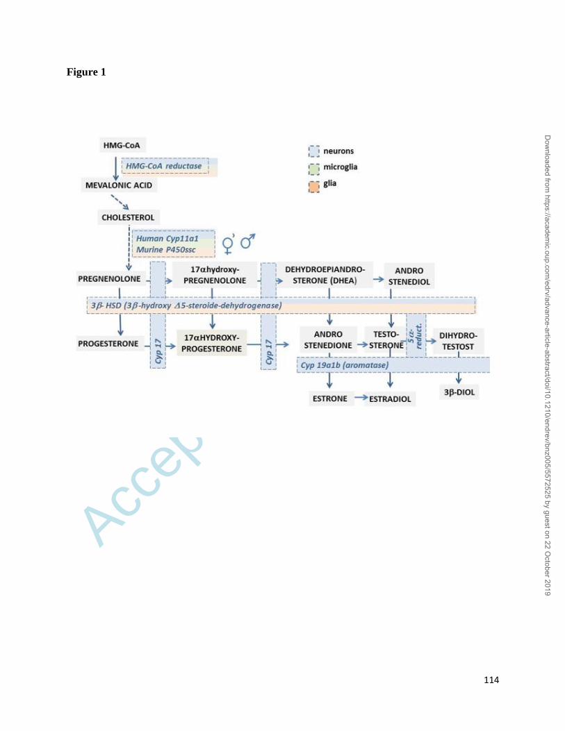
Accep
ted
Man
uscr
ipt
114
Figure 1
Dow
nloaded from https://academ
ic.oup.com/edrv/advance-article-abstract/doi/10.1210/endrev/bnz005/5572525 by guest on 22 O
ctober 2019
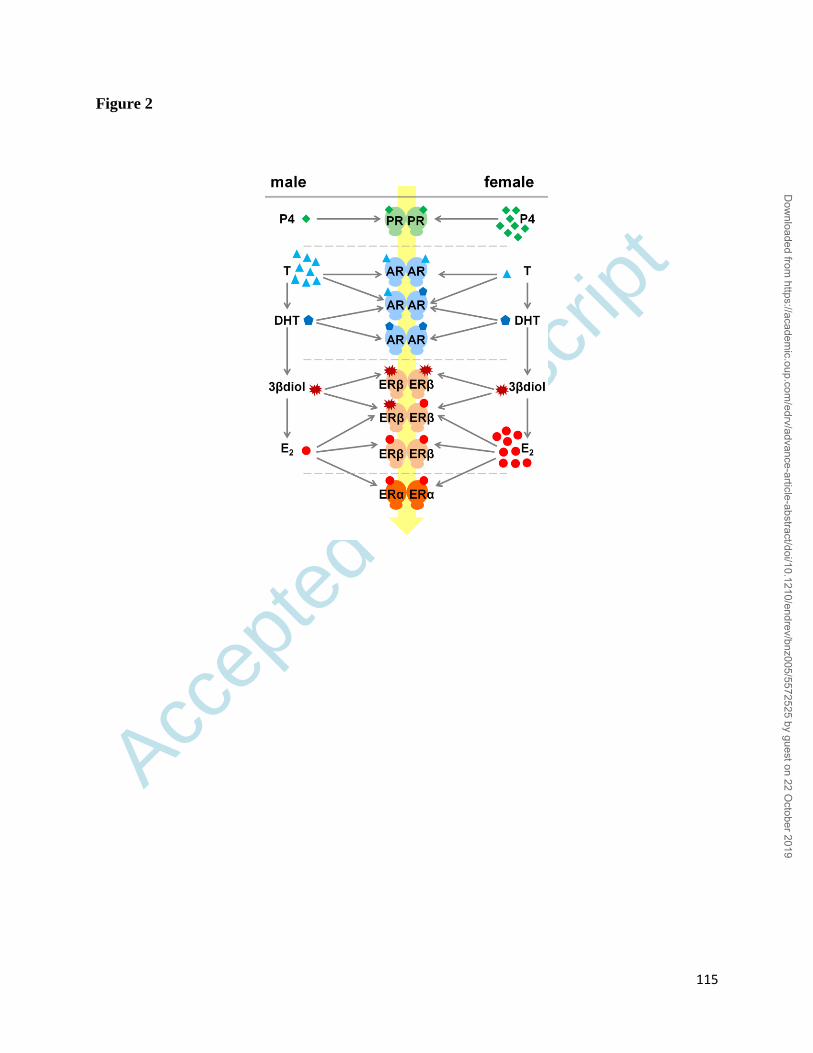
Accep
ted
Man
uscr
ipt
115
Figure 2
Dow
nloaded from https://academ
ic.oup.com/edrv/advance-article-abstract/doi/10.1210/endrev/bnz005/5572525 by guest on 22 O
ctober 2019
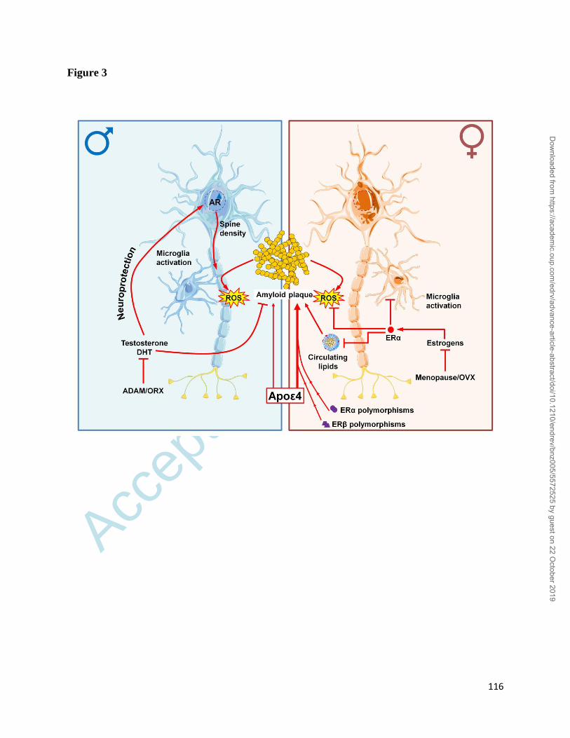
Accep
ted
Man
uscr
ipt
116
Figure 3
Dow
nloaded from https://academ
ic.oup.com/edrv/advance-article-abstract/doi/10.1210/endrev/bnz005/5572525 by guest on 22 O
ctober 2019
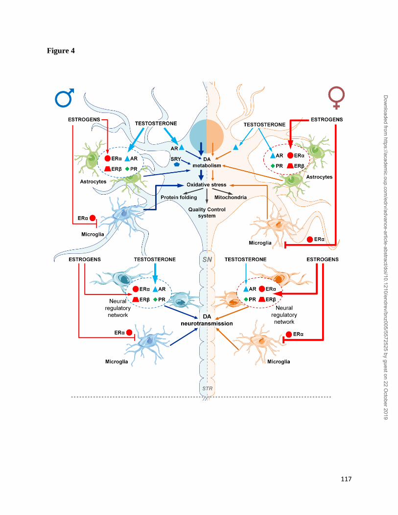
Accep
ted
Man
uscr
ipt
117
Figure 4
Dow
nloaded from https://academ
ic.oup.com/edrv/advance-article-abstract/doi/10.1210/endrev/bnz005/5572525 by guest on 22 O
ctober 2019
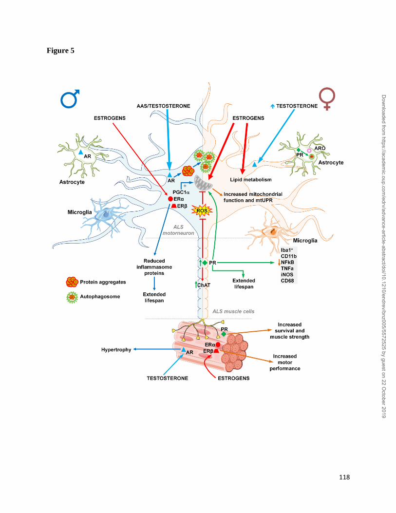
Accep
ted
Man
uscr
ipt
118
Figure 5
Dow
nloaded from https://academ
ic.oup.com/edrv/advance-article-abstract/doi/10.1210/endrev/bnz005/5572525 by guest on 22 O
ctober 2019
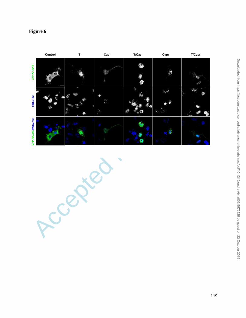
Accep
ted
Man
uscr
ipt
119
Figure 6
Dow
nloaded from https://academ
ic.oup.com/edrv/advance-article-abstract/doi/10.1210/endrev/bnz005/5572525 by guest on 22 O
ctober 2019
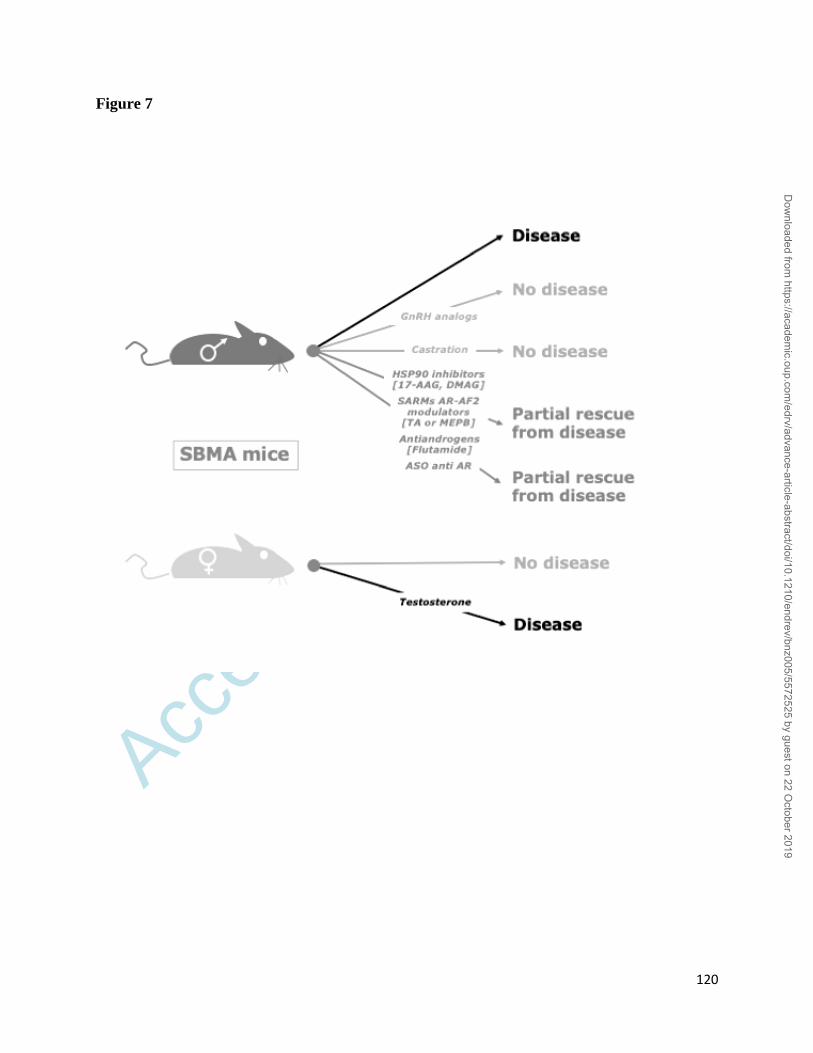
Accep
ted
Man
uscr
ipt
120
Figure 7
Dow
nloaded from https://academ
ic.oup.com/edrv/advance-article-abstract/doi/10.1210/endrev/bnz005/5572525 by guest on 22 O
ctober 2019
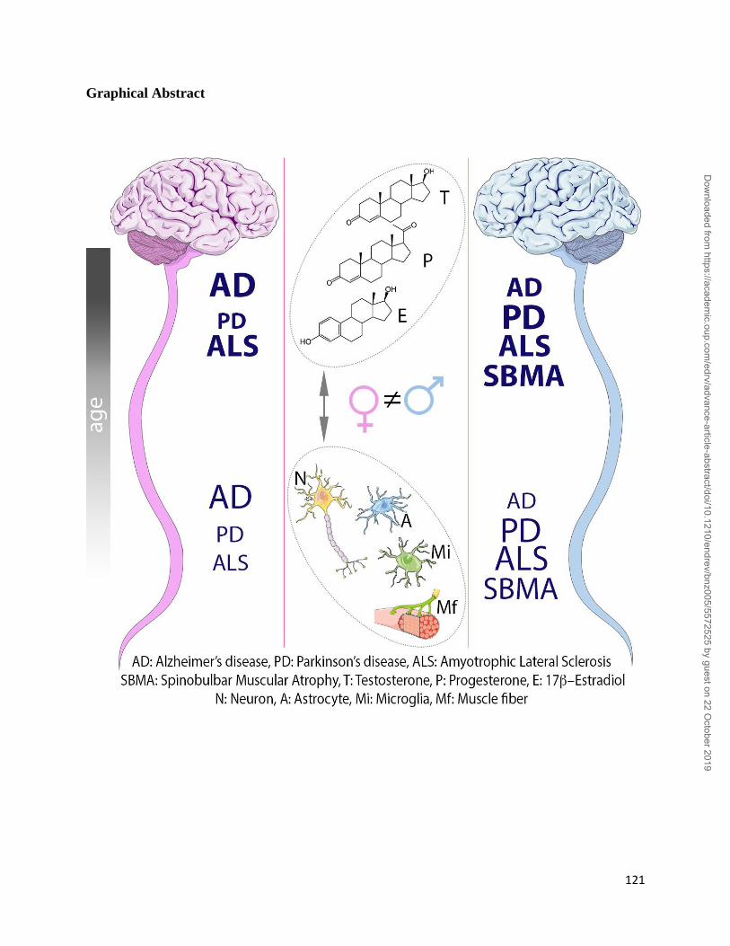
Accep
ted
Man
uscr
ipt
121
Graphical Abstract
Dow
nloaded from https://academ
ic.oup.com/edrv/advance-article-abstract/doi/10.1210/endrev/bnz005/5572525 by guest on 22 O
ctober 2019
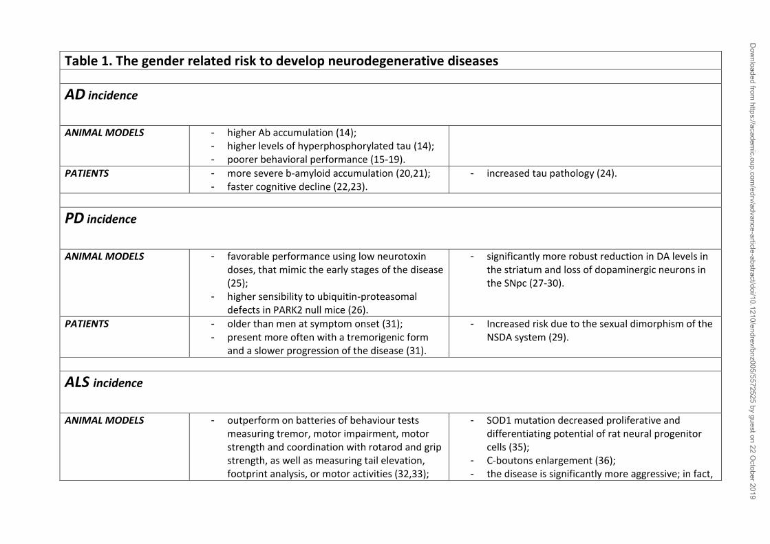
Table 1. The gender related risk to develop neurodegenerative diseases
AD incidence
ANIMAL MODELS - higher Ab accumulation (14); - higher levels of hyperphosphorylated tau (14); - poorer behavioral performance (15-19).
PATIENTS - more severe b-amyloid accumulation (20,21); - faster cognitive decline (22,23).
- increased tau pathology (24).
PD incidence
ANIMAL MODELS - favorable performance using low neurotoxin doses, that mimic the early stages of the disease (25);
- higher sensibility to ubiquitin-proteasomal defects in PARK2 null mice (26).
- significantly more robust reduction in DA levels in the striatum and loss of dopaminergic neurons in the SNpc (27-30).
PATIENTS - older than men at symptom onset (31); - present more often with a tremorigenic form
and a slower progression of the disease (31).
- Increased risk due to the sexual dimorphism of the NSDA system (29).
ALS incidence
ANIMAL MODELS - outperform on batteries of behaviour tests measuring tremor, motor impairment, motor strength and coordination with rotarod and grip strength, as well as measuring tail elevation, footprint analysis, or motor activities (32,33);
- SOD1 mutation decreased proliferative and differentiating potential of rat neural progenitor cells (35);
- C-boutons enlargement (36); - the disease is significantly more aggressive; in fact,
Dow
nloaded from https://academ
ic.oup.com/edrv/advance-article-abstract/doi/10.1210/endrev/bnz005/5572525 by guest on 22 O
ctober 2019
- earlier disease onset in mice co-expressing mutant chromogranin and SOD1 (34).
males ALS mice lose weight and show motor symptoms earlier than females (32,37);
- the disease progression is much shorter in males (32,37).
PATIENTS - older than males at the age of onset (38); - prevalence of the bulbar form (38); - significant vulnerability to develop cognitive
dysfunctions related to ALS (39); - positive association between longer
reproductive time-span and susceptibility and survival of ALS (40).
- prevalence of the forms with limb onset (38); - higher CSF levels of SOD1 (41).
SBMA incidence
ANIMAL MODELS - in tg mouse models females are not affected (42,43);
- testosterone induces SBMA symptoms
PATIENTS - female carriers are asymptomatic or present mild clinical abnormalities (44,45);
- women homozygous did not show any clinical symptom of ND (46).
Dow
nloaded from https://academ
ic.oup.com/edrv/advance-article-abstract/doi/10.1210/endrev/bnz005/5572525 by guest on 22 O
ctober 2019
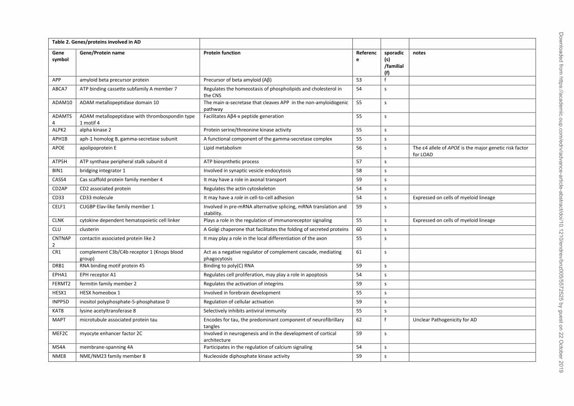
Table 2. Genes/proteins involved in AD
Gene symbol
Gene/Protein name Protein function Reference
sporadic (s) /familial (f)
notes
APP amyloid beta precursor protein Precursor of beta amyloid (Aβ) 53 f
ABCA7 ATP binding cassette subfamily A member 7 Regulates the homeostasis of phospholipids and cholesterol in the CNS
54 s
ADAM10 ADAM metallopeptidase domain 10 The main α-secretase that cleaves APP in the non-amyloidogenic pathway
55 s
ADAMTS4
ADAM metallopeptidase with thrombospondin type 1 motif 4
Facilitates Aβ4-x peptide generation 55 s
ALPK2 alpha kinase 2 Protein serine/threonine kinase activity 55 s
APH1B aph-1 homolog B, gamma-secretase subunit A functional component of the gamma-secretase complex 55 s
APOE apolipoprotein E Lipid metabolism 56 s The ε4 allele of APOE is the major genetic risk factor for LOAD
ATP5H ATP synthase peripheral stalk subunit d ATP biosynthetic process 57 s
BIN1 bridging integrator 1 Involved in synaptic vesicle endocytosis 58 s
CASS4 Cas scaffold protein family member 4 It may have a role in axonal transport 59 s
CD2AP CD2 associated protein Regulates the actin cytoskeleton 54 s
CD33 CD33 molecule It may have a role in cell-to-cell adhesion 54 s Expressed on cells of myeloid lineage
CELF1 CUGBP Elav-like family member 1 Involved in pre-mRNA alternative splicing, mRNA translation and stability.
59 s
CLNK cytokine dependent hematopoietic cell linker Plays a role in the regulation of immunoreceptor signaling 55 s Expressed on cells of myeloid lineage
CLU clusterin A Golgi chaperone that facilitates the folding of secreted proteins 60 s
CNTNAP2
contactin associated protein like 2 It may play a role in the local differentiation of the axon 55 s
CR1 complement C3b/C4b receptor 1 (Knops blood group)
Act as a negative regulator of complement cascade, mediating phagocytosis
61 s
DRB1 RNA binding motif protein 45 Binding to poly(C) RNA 59 s
EPHA1 EPH receptor A1 Regulates cell proliferation, may play a role in apoptosis 54 s
FERMT2 fermitin family member 2 Regulates the activation of integrins 59 s
HESX1 HESX homeobox 1 Involved in forebrain development 55 s
INPP5D inositol polyphosphate-5-phosphatase D Regulation of cellular activation 59 s
KAT8 lysine acetyltransferase 8 Selectively inhibits antiviral immunity 55 s
MAPT microtubule associated protein tau Encodes for tau, the predominant component of neurofibrillary tangles
62 f Unclear Pathogenicity for AD
MEF2C myocyte enhancer factor 2C Involved in neurogenesis and in the development of cortical architecture
59 s
MS4A membrane-spanning 4A Participates in the regulation of calcium signaling 54 s
NME8 NME/NM23 family member 8 Nucleoside diphosphate kinase activity 59 s
Dow
nloaded from https://academ
ic.oup.com/edrv/advance-article-abstract/doi/10.1210/endrev/bnz005/5572525 by guest on 22 O
ctober 2019
PICALM phosphatidylinositol binding clathrin assembly protein
Involved in cellular trafficking and regulation of endocytosis 60 s
PLD3 phospholipase D family member 3 Involved in APP processing 63 s
PSEN1 presenilin 1 Part of the γ-secretase complex, regulating APP processing 64 f
PSEN2 presenilin 2 Part of the γ-secretase complex, regulating APP processing 65 f
PTK2B protein tyrosine kinase 2 beta Enhances signals that regulate neuronal activity 59 s
RIN3 Ras and Rab interactor 3 Involved in the early endocytic pathway 59 s
SORL1 sortilin related receptor 1 A neuronal apolipoprotein E receptor 59 s
TREM2 triggering receptor expressed on myeloid cells 2 Phagocytosis, migration, activation 66 s Exclusively expressed on cells within the myeloid lineage. SNPs increase disease risk by 2- to 4-fold.
TRIP4 thyroid hormone receptor interactor 4 Paly a role in the estrogen receptor and NF-κB transactivation 67 s
UNC5C unc-5 netrin receptor C Direct axon extension and cell migration during neural development
68 s
ZCWPW1
zinc finger CW-type and PWWP domain containing 1 A histone modification reader involved in epigenetic regulation 59 s
Dow
nloaded from https://academ
ic.oup.com/edrv/advance-article-abstract/doi/10.1210/endrev/bnz005/5572525 by guest on 22 O
ctober 2019
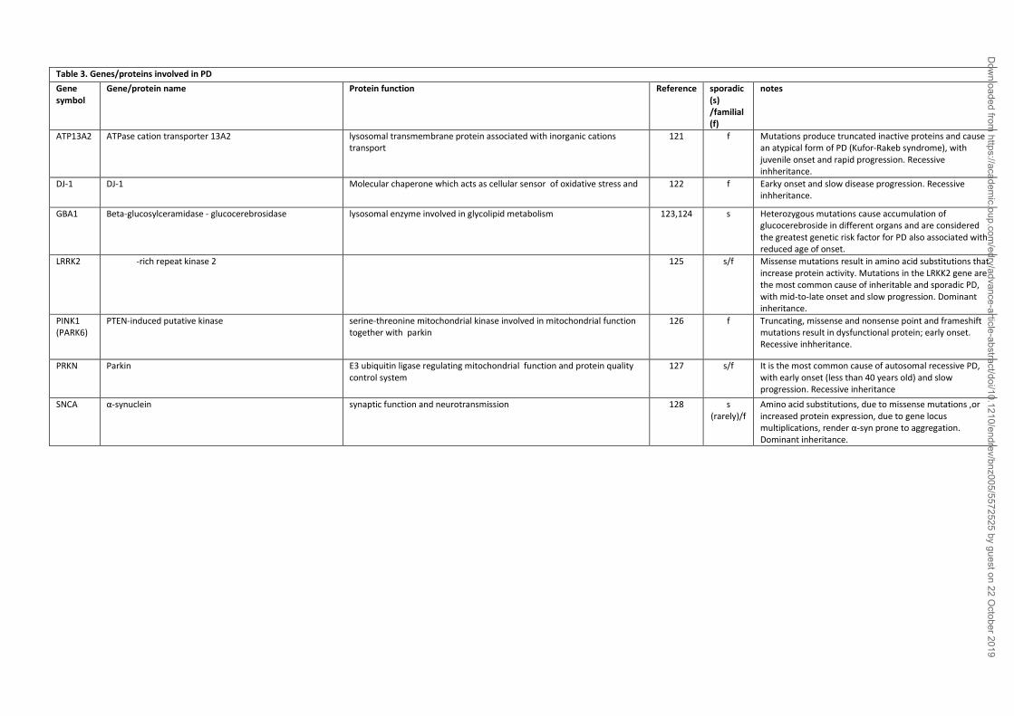
Table 3. Genes/proteins involved in PD
Gene symbol
Gene/protein name Protein function Reference sporadic (s) /familial (f)
notes
ATP13A2 ATPase cation transporter 13A2 lysosomal transmembrane protein associated with inorganic cations transport
121 f Mutations produce truncated inactive proteins and cause an atypical form of PD (Kufor-Rakeb syndrome), with juvenile onset and rapid progression. Recessive inhheritance.
DJ-1 DJ-1 Molecular chaperone which acts as cellular sensor of oxidative stress and protecits mitochondria
122 f Earky onset and slow disease progression. Recessive inhheritance.
GBA1 Beta-glucosylceramidase - glucocerebrosidase lysosomal enzyme involved in glycolipid metabolism 123,124 s Heterozygous mutations cause accumulation of glucocerebroside in different organs and are considered the greatest genetic risk factor for PD also associated with reduced age of onset.
LRRK2 Leucine-rich repeat kinase 2 neurite and synapse formation, membrane trafficking, autophagy 125 s/f Missense mutations result in amino acid substitutions that increase protein activity. Mutations in the LRKK2 gene are the most common cause of inheritable and sporadic PD, with mid-to-late onset and slow progression. Dominant inheritance.
PINK1 (PARK6)
PTEN-induced putative kinase serine-threonine mitochondrial kinase involved in mitochondrial function together with parkin
126 f Truncating, missense and nonsense point and frameshift mutations result in dysfunctional protein; early onset. Recessive inhheritance.
PRKN Parkin E3 ubiquitin ligase regulating mitochondrial function and protein quality control system
127 s/f It is the most common cause of autosomal recessive PD, with early onset (less than 40 years old) and slow progression. Recessive inheritance
SNCA α-synuclein synaptic function and neurotransmission 128 s (rarely)/f
Amino acid substitutions, due to missense mutations ,or increased protein expression, due to gene locus multiplications, render α-syn prone to aggregation. Dominant inheritance.
Dow
nloaded from https://academ
ic.oup.com/edrv/advance-article-abstract/doi/10.1210/endrev/bnz005/5572525 by guest on 22 O
ctober 2019
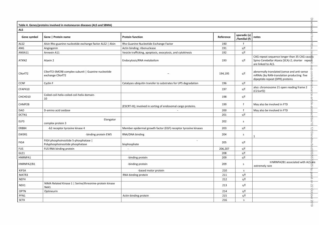
Table 4. Genes/proteins involved in motoneuron diseases (ALS and SBMA)
ALS
Gene symbol Gene | Protein name Protein function Reference sporadic (s) /familial (f)
notes
ALS2 Alsin Rho guanine nucleotide exchange factor ALS2 | Alsin Rho Guanine Nucleotide Exchange Factor 190 f
ANG Angiogenin Actin binding; ribonuclease 191 s/f
ANXA11 Annexin A11 Vesicle trafficking, apoptosis, exocytosis, and cytokinesis 192 s/f
ATXN2 Ataxin 2 Endocytosis/RNA metabolism 193 s/f CAG repeat sequence longer than 35 CAG causes Spino Cerebellar Ataxia (SCA)-2; shorter repeat are linked to ALS.
C9orf72 C9orf72-SMCR8 complex subunit | Guanine nucleotide exchange C9orf72
Guanine nucleotide exchange factor – involved in autophagy 194,195 s/f
Abnormal GGGGCC (G4C2) expansion are abnormally translated (sense and anti-sense mRNAs )by RAN-translation producting five dipeptide-repeat (DPR) proteins
CCNF Cyclin F Catalyzes ubiquitin transfer to substrates for UPS degradation 196 s/f
CFAP410 Cilia and flagella associated protein 410 Regulation of cell morphology and cytoskeletal organization 197 s/f also: chromosome 21 open reading frame 2 (C21orf2)
CHCHD10 Coiled-coil-helix-coiled-coil-helix domain- containing protein 10
Mitochondrial protein 198 s/f
CHMP2B Charged multivesicular body protein 2B Component of the endosomal sorting required for transport complex III (ESCRT-III); involved in sorting of endosomal cargo proteins.
199 f May also be involved in FTD
DAO D-amino acid oxidase Regulates the level of the neuromodulator D-serine in the brain 200 f May also be involved in FTD
DCTN1 Dynactin subunit 1 Component of dynein motor complex 201 s/f May also be involved in FTD
ELP3 Elongator acetyltransferase complex subunit 3 | Elongator complex protein 3
RNA polymerase II component 202 s
ERBB4 Erb-b2 receptor tyrosine kinase 4 Member epidermal growth factor (EGF) receptor tyrosine kinases 203 s/f
EWSR1 EWS RNA binding protein 1 | RNA-binding protein EWS RNA/DNA binding protein 204 s Previous name: Ewing sarcoma breakpoint region 1
FIG4 FIG4 phosphoinositide 5-phosphatase | Polyphosphoinositide phosphatase
Regulates synthesis and turnover of phosphatidylinositol 3,5-bisphosphate
205 s/f
FUS FUS RNA binding protein RNA-binding protein 206,207 s/f
GLE1 GLE1 RNA export mediator | Nucleoporin GLE1 Required for the export of mRNAs from the nucleus into the cytoplasm 208 s/f
HNRNPA1 Heterogeneous nuclear ribonucleoprotein A1 RNA-binding protein 209 s/f
HNRNPA2/B1 Heterogeneous nuclear ribonucleoprotein A2/B1 RNA-binding protein 209 s Mutations in H NRNPA2B1 associated with ALS are extremely rare
KIF5A Kinesin family member 5A | Kinesin heavy chain isoform 5A Microtubule-based motor protein 210 s
MATR3 Matrin 3 RNA-binding protein 211 s/f May also be involved in FTD
NEFH Neurofilament heavy | Neurofilament heavy polypeptide Cytoskeletal component 212 s/f
NEK1 NIMA Related Kinase 1 | Serine/threonine-protein kinase Nek1
Cytoskeletal dynamics 213 s/f
OPTN Optineurin Autophagy adaptor 214 s/f
PFN1 Profilin 1 Actin-binding protein 215 s/f
SETX Senataxin; Probable helicase senataxin DNA/RNA helicase 216 s
Dow
nloaded from https://academ
ic.oup.com/edrv/advance-article-abstract/doi/10.1210/endrev/bnz005/5572525 by guest on 22 O
ctober 2019
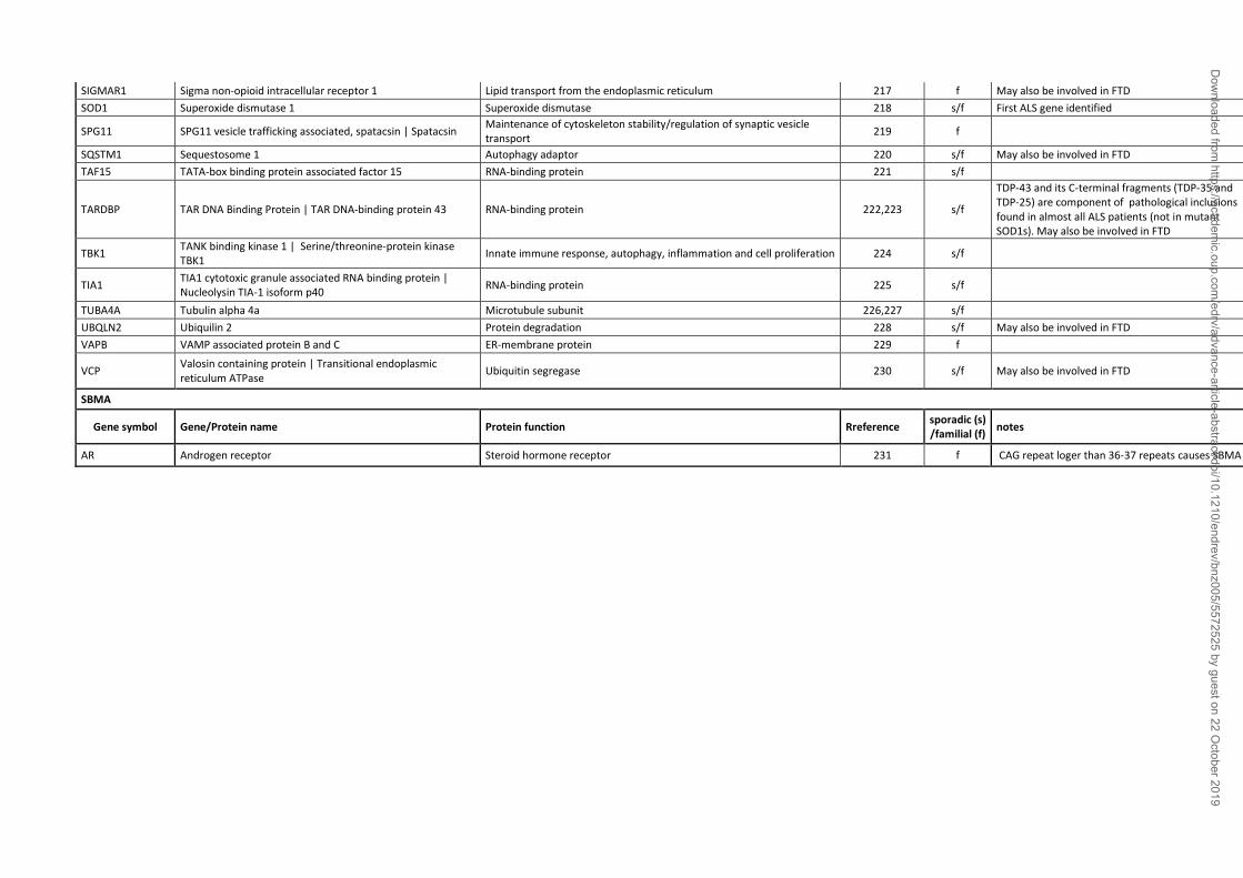
SIGMAR1 Sigma non-opioid intracellular receptor 1 Lipid transport from the endoplasmic reticulum 217 f May also be involved in FTD
SOD1 Superoxide dismutase 1 Superoxide dismutase 218 s/f First ALS gene identified
SPG11 SPG11 vesicle trafficking associated, spatacsin | Spatacsin Maintenance of cytoskeleton stability/regulation of synaptic vesicle transport
219 f
SQSTM1 Sequestosome 1 Autophagy adaptor 220 s/f May also be involved in FTD
TAF15 TATA-box binding protein associated factor 15 RNA-binding protein 221 s/f
TARDBP TAR DNA Binding Protein | TAR DNA-binding protein 43 RNA-binding protein 222,223 s/f
TDP-43 and its C-terminal fragments (TDP-35 and TDP-25) are component of pathological inclusions found in almost all ALS patients (not in mutant SOD1s). May also be involved in FTD
TBK1 TANK binding kinase 1 | Serine/threonine-protein kinase TBK1
Innate immune response, autophagy, inflammation and cell proliferation 224 s/f
TIA1 TIA1 cytotoxic granule associated RNA binding protein | Nucleolysin TIA-1 isoform p40
RNA-binding protein 225 s/f
TUBA4A Tubulin alpha 4a Microtubule subunit 226,227 s/f
UBQLN2 Ubiquilin 2 Protein degradation 228 s/f May also be involved in FTD
VAPB VAMP associated protein B and C ER-membrane protein 229 f
VCP Valosin containing protein | Transitional endoplasmic reticulum ATPase
Ubiquitin segregase 230 s/f May also be involved in FTD
SBMA
Gene symbol Gene/Protein name Protein function Rreference sporadic (s) /familial (f)
notes
AR Androgen receptor Steroid hormone receptor 231 f CAG repeat loger than 36-37 repeats causes SBMA
Dow
nloaded from https://academ
ic.oup.com/edrv/advance-article-abstract/doi/10.1210/endrev/bnz005/5572525 by guest on 22 O
ctober 2019


