Embed Size (px)
Citation preview
The Functional O-Mannose Glycan on α-Dystroglycan Contains a Phospho-ribitol Primed 1 for Matriglycan Addition 2
Jeremy L. Praissman1,*, Tobias Willer3,*, M. Osman Sheikh1,*, Ants Toi4, David Chitayat5,6, 3 Yung-Yao Lin7,8, Hane Lee9, Stephanie Stalnkaker1, Shuo Wang1, Pradeep Kumar Prabhakar1, 4 Stanley F. Nelson9, Derek L. Stemple8, Steven A. Moore10, Kelley W. Moremen1,2, Kevin P. 5 Campbell3,# and Lance Wells1,2,# 6 1Complex Carbohydrate Research Center and 2Department of Biochemistry and Molecular 7 Biology, University of Georgia, Athens, GA 8 3Howard Hughes Medical Institute, Department of Molecular Physiology and Biophysics, 9 Neurology, and Internal Medicine, Carver College of Medicine, University of Iowa, Iowa City, 10 Iowa, USA 11 4Mount Sinai Hospital, Department of Medical Imaging, 600 University Ave., Toronto, Ontario, 12 Canada 13 5Division of Clinical and Metabolic Genetics, The Hospital for Sick Children and University of 14 Toronto, Toronto, Canada. 15 6The Prenatal Diagnosis and Medical Genetics Program, Department of Obstetrics and 16 Gynecology, Mount Sinai Hospital, University of Toronto, Toronto, Canada. 17 7Blizard Institute, Barts and The London School of Medicine and Dentistry, Queen Mary 18 University of London, 4 Newark Street, London E1 2AT, UK 19 8Wellcome Trust Sanger Institute, Wellcome Trust Genome Campus, Hinxton, CB10 1SA, UK 20 9Department of Human Genetics, Pathology and Laboratory Medicine, David Geffen School of 21 Medicine, University of California, Los Angeles, California, USA 22 10Department of Pathology, University of Iowa Roy J. and Lucille A. Carver College of Medicine, 23 Iowa City, Iowa, USA 24
25 * These authors contributed equally to this work 26 # Corresponding Authors: Kevin P. Campbell, [email protected] ; Lance Wells 27 [email protected] 28
Impact Statement: This work delineates a functional O-mannose structure that uses phospho-29 ribitol to bridge between the phosphotrisaccharide attached to the protein and the repeating 30 disaccharide that binds ECM proteins on α-dystroglycan that when disrupted results in multiple 31 forms of congenital muscular dystrophy. 32
Running Title: Functional O-Man Glycosylation of α-DG 33
Keywords: Congenital muscular dystrophy, O-mannosylation, glycosylation, alpha-dystroglycan, 34 ribitol, ISPD, TMEM5, mass spectrometry 35
Major Research Organism: Homo sapiens 36
37
ABSTRACT 38
Multiple glycosyltransferases are essential for the proper modification of alpha-dystroglycan, as 39 mutations in the encoding genes cause congenital/limb-girdle muscular dystrophies. Here we 40 elucidate further the structure of an O-mannose-initiated glycan on alpha-dystroglycan that is 41 required to generate its extracellular matrix-binding polysaccharide. This functional glycan 42 contains a novel ribitol structure that links a phosphotrisaccharide to xylose. ISPD is a CDP-43 ribitol (ribose) pyrophosphorylase that generates the reduced sugar nucleotide for the insertion 44 of ribitol in a phosphodiester linkage to the glycoprotein. TMEM5 is a UDP-xylosyl transferase 45 that elaborates the structure. We demonstrate in a zebrafish model as well as in a human 46 patient that defects in TMEM5 result in muscular dystrophy in combination with abnormal brain 47 development. Thus, we propose a novel structure – a ribitol in a phosphodiester linkage – for 48 the moiety on which TMEM5, B4GAT1, and LARGE act to generate the functional receptor for 49 ECM proteins having LG domains. 50
51
52

INTRODUCTION 53
Twenty-five years ago it was proposed that alpha-dystroglycan (α-DG) played a major role in 54 bridging the extracellular matrix to the muscle plasma membrane and actin cytoskeleton as a 55 component of the multi-protein dystrophin glycoprotein complex (1,2). Defects in the proper 56 formation of this complex have been shown to be causal for various forms of muscular 57 dystrophy (3). However, to date, only 3 patients have been found that have primary defects in 58 the coding sequence of α-DG (4-6). Over fifteen years ago, it was suggested that defects in the 59 glycosyltransferases needed for proper glycosylation of α-DG were causal for a subset of 60 muscular dystrophies, the so-called secondary dystroglycanopathies that can range in severity 61 from mild Limb-Girdle Muscular Dystrophy (LGMD) to severe Walker-Warburg syndrome 62 (WWS) (7-9). In the early 2000’s, it became clear that the defects causal for disease involved 63 enzymes that initiated and elaborated the extended O-mannose (O-Man) glycan structures 64 covalently attached to Ser/Thr residues of α-DG (10,11). Since then, steady progress has been 65 made in elucidating the subset of mammalian O-Man structures that directly interact with 66 extracellular matrix components and the candidate genes necessary for the functional 67 glycosylation of α-DG (12). These include the identification of a subset of phosphorylated O-68 Man structures containing extended, LARGE-dependent, repeating disaccharide polymers, 69 structures that have been recently termed matriglycan (13). 70
O-Mannosylation begins in the endoplasmic reticulum (recently reviewed in (14,15)). The 71 addition of O-Man in an alpha linkage to serine and threonine residues of a select set of 72 mammalian glycoproteins is catalyzed by the POMT1/2 complex using dolichol-73 phosphomannose as the donor (10). At this point, by mechanisms that have yet to be fully 74 elucidated, there is a divergence in elaboration (Figure 1). The vast majority of O-Man sites are 75 extended in the Golgi in a beta-1,2 linkage with N-acetylglucosamine (GlcNAc) by POMGNT1 76 that can then be further branched by GlcNAc and/or elaborated by galactose, fucose, and sialic 77 acid to generate the M1 and M2 glycans (12,16,17). A small subset of O-Man modified sites, 78 apparently exclusively on α-DG, are extended in the endoplasmic reticulum by a GlcNAc in a 79 beta-1,4 linkage by POMGNT2 to generate the M3 glycans (18,19). This is further elaborated 80 into a trisaccharide by the action of a beta-1,3-N-acetylgalactosamine (GalNAc) transferase, 81 B3GALNT2 (19). This trisaccharide is a substrate for POMK that phosphorylates the initiating O-82 Man residue at the 6-position (19). After unknown elaboration of the phosphotrisaccharide in a 83 phosphodiester linkage, presumably by one or more of the as-yet to be assigned CMD-causing 84 gene products (ISPD, TMEM5, Fukutin, and FKRP), B4GAT1 adds a glucuronic acid (GlcA) in 85 a beta-1,4 linkage to an underlying beta-linked xylose (Xyl) (20,21). The addition of the xylose 86 by an unspecified enzyme followed by the action of B4GAT1 serves as a primer for LARGE to 87 synthesize the repeating Xyl-GlcA disaccharide, matriglycan, that serves as the binding site for 88 several extracellular matrix proteins (20,21). 89
Here we report a M3 glycan structure with a phosphodiester linked ribitol that TMEM5, B4GAT1, 90 and LARGE act on to generate the functional receptor for extracellular matrix (ECM) ligands 91 characterized by Laminin G domain-like (LG) protein domains. We demonstrate that ISPD is a 92 CDP-ribitol pyrophosphorylase employed for the synthesis of the required sugar (alcohol) 93 nucleotide needed for ribitol insertion into the M3 glycan. We establish TMEM5 as the candidate 94

xylose transferase and demonstrate the impact of its knockdown in a zebrafish model consistent 95 with a CMD phenotype. We also identify and characterize a novel TMEM5 mutation identified in 96 a WWS family. Finally, we present a model of the functional M3 glycan structure along with the 97 more than a dozen assigned or proposed enzymes required for its synthesis. 98
99
RESULTS 100
Ribitol-Xyl-GlcA is released from α-DG-340 following cleavage of the phosphodiester linkage 101
A truncated, secreted version of α-DG with a COOH-terminal GFP and His tag (α−DG-340 that 102 is only 28 amino acids (313-340) derived from α-DG following endogenous furin cleavage) was 103 overexpressed in HEK293F cells, which express low levels of LARGE, and purified from the 104 media. This 28 amino acid sequence contains only one putative M3 glycan consensus site 105 (TPT, 317-319). We previously demonstrated that the generation of peptides bearing the 106 phosphotrisaccharide could be isolated from α-DG constructs following cleavage of the 107 phoshodiester linkage and that such treatment resulted in loss of IIH6 (an antibody that binds 108 functionally glycosylated α-DG) binding (Figure 2-figure supplement 2) (22). Thus, we decided 109 to interrogate the released glycan portion to further elucidate the functional glycan structure. 110 Following aqueous HF treatment, which selectively cleaves phosphodiesters, released glycans 111 were isolated from α−DG-340 and permethylated. Tandem mass spectrometry of the 112 permethylated glycans revealed the presence of a linear pentitol-Xyl-GlcA (Figure 2) as well as 113 further disaccharide extended structures (pentitol-(Xyl-GlcA)n, Figure 2-figure supplement 1). 114
ISPD is a CDP-Ribitol (ribose) Pyrophosphorylase 115
Given the identification of pentitol in the glycan structure, we investigated whether ISPD might 116 be able to generate CDP-ribitol. Our rationale for this was that mutations in ISPD are causal for 117 CMD (23,24), ISPD is a putative nucleotidyltransferase found in the cytosol (25), and homologs 118 in bacterial systems are involved in the generation of identical, CDP-ribitol (26), or similar 119 structures, 4-CDP-2-C-methyl-D-erythritol(27). ISPD was expressed as a His fusion protein in 120 HEK293F cells and purified from whole cell extracts (Figure 3-figure supplement 1). The 121 enzyme was capable of generating CDP-ribitol or CDP-ribose using CTP and ribitol-5-122 phosphate or ribose-5-phosphate, respectively, but was not able to generate the sugar (alcohol) 123 nucleotides with ribitol or ribose (Figure 3). Thus, ISPD is a CDP-ribitol (ribose) 124 pyrophosphorylase that generates the needed reduced sugar (alcohol) nucleotide for integration 125 of a ribitol into the functional O-Man glycan structure. During the preparation of our manuscript, 126 a study was published demonstrating that CDP-ribose, CDP-ribitol, and CDP-ribulose could all 127 be generated by mammalian ISPD consistent with our present findings (28). 128
Phosphodiester-linked Ribitol is connected to the M3 Glycan 129
Having identified the ribitol directly attached to xylose, we sought to identify whether the ribitol 130 was the bridge between xylose and the phosphotrisaccharide on the polypeptide. We also 131 wanted to confirm the open ring structure enforced by the addition of ribitol, a reduced sugar. In 132 such a linear structure, mild periodate treatment would be expected to cleave vicinal diols and 133 release matriglycan from the polypeptide. Thus, we performed mild periodate cleavage of the 134
purified α-DG-340 protein and demonstrated a loss of reactivity with the IIH6 antibody that 135 recognizes functional LARGE-modified glycoprotein (Figure 4-figure supplement 2). 136 Furthermore, following tryptic cleavage, we examined the resulting α-DG peptides by tandem 137 mass spectrometry (Figure 4-figure supplement 1). We were able to identify multiple 138 glycopeptides where Thr317 (a known site for M3 glycans) was modified to indicate a cleaved 139 ribitol fragment on the phosphotrisaccharide. We observed pairs of essentially identical 140 glycopeptides that showed cleavage between C2 and C3 or between C3 and C4 (Figure 4). We 141 also observed peptides with an additional phosphate suggesting that there can be 2 phosphates 142 on the trisaccharide of which at least one is in a phosphodiester linkage to ribitol (Figure 4). 143 Thus, the ribitol appears most likely to be connected at C1 to the oxygen of a phosphate group 144 on the M3 glycopeptide and at the C5 to xylose (note that ribitol C1 and C5 are indistinguishable 145 due to the nature of the reduced sugar and thus nomenclature for closed-ring standard sugar 146 nucleotides is being used to describe linkage). 147
TMEM5 is a xylose transferase 148
In our previous manuscripts describing B4GAT1 (20,21), we had provided evidence for an 149 underlying xylose in the linker region of matriglycan between the phosphotrisaccharide and the 150 LARGE-dependent Xyl-GlcA repeat disaccharide. Among the uncharacterized genes harboring 151 mutations in patients with CMD, TMEM5 shows sequence similarity to glycosyltransferases (25). 152 TMEM5 was also among the genes uncovered in a screen for loss of IIH6 binding and Lassa 153 virus entry, readouts that are both dependent on functional glycosylation of α-DG (29). Thus, we 154 investigated this enzyme as a candidate xylose transferase. We overexpressed and purified the 155 truncated catalytic domain fused to GFP and a His tag (Figure 5-figure supplement 1A). Using 156 the UDP-Glo assay, we were able to show that transmembrane deleted dTM-TMEM5 can 157 hydrolyze UDP-Xyl, but not other UDP-sugars (Figure 5A) demonstrating selective hydrolytic 158 activity in the absence of acceptor glycans. Furthermore, we were able to show that 159 recombinant full length TMEM5 (Figure 5-figure supplement 1B) can be used to label α-DG-160 Fc340 (Fc-tagged α-DG-340) with radiolabeled UDP-Xyl [Xyl-14C] expressed from TMEM5-161 deficient patient cells compared to α-DG-Fc340 mutated at the M3 site (TPT,317-319, converted 162 to APA) as a negative control (Figure 5B). Also, we were able to show that dTM-TMEM5 can 163 use UDP-Xyl to transfer Xyl to CDP-ribitol as the acceptor but not ribitol, ribitol-5-P, or CMP-164 Neu5Ac suggesting the need for the ribitol to be in a phosphodiester linkage in order to be an 165 acceptor (Figure 5C). In summary, these results provide strong, but not direct, evidence that 166 TMEM5 is the xylosyl transferase enzyme for modification of ribitol that is in a phosphodiester 167 linkage to the M3 glycan on α-DG. 168
TMEM5 knockdown in zebrafish recapitulates WWS phenotype 169
The highly conserved TMEM5 gene, which has formerly been implicated in CMD, encodes a 170 type II transmembrane protein with a predicted glycosyltransferase domain. To investigate the 171 role of TMEM5 in vertebrates, we knocked down the zebrafish (Danio rerio) orthologue using 172 antisense morpholino oligonucleotides (MO). The endogenous zebrafish tmem5 transcript was 173 detected throughout early embryonic development (Figure 6-figure supplement 1). Injection of 174 tmem5 MO specifically inhibited the expression of green fluorescent protein-tagged TMEM5 in a 175

dose-dependent manner (Figure 6-figure supplement 1). Knockdown of tmem5 caused an 176 increased percentage of embryos with mild to severe hydrocephalus (95% in total) and 177 significantly reduced eye size, reminiscent of pathological defects in WWS (Figure 6A). To test 178 whether the brain and eye abnormalities were caused by MO off-target effects mediated through 179 a p53-dependent cell death pathway (30), we inhibited p53 activation by co-injection of p53 MO. 180 88% of embryos still displayed hydrocephalus and significantly reduced eye size was still 181 observed in embryos co-injected with p53 and tmem5 MOs (Figure 6B), suggesting that the 182 brain and eye abnormalities were not caused by MO off-target effects. As knockdown of tmem5 183 also caused reduced motility and lesions in the myotome (data not shown), we assessed the 184 sarcolemma integrity using Evans Blue dye (EBD), which does not penetrate into intact muscle 185 fibers. Muscle fibers were infiltrated by EBD before undergoing degeneration (Figure 6C), 186 suggesting a pathological mechanism in which knockdown of tmem5 leads to compromised 187 sarcolemma integrity. As defective glycosylation of α-DG is a pathological hallmark of WWS, 188 we tested whether knockdown of zebrafish tmem5 would affect the glycosylation of α-DG. 189 Compared to control embryos, knockdown of tmem5 caused a 44% reduction of glycosylated α-190 dystroglycan (IIH6 epitope) on Western blots (Figure 6D). Together, these results clearly 191 illustrate a role for TMEM5 in functional glycosylation of α-DG and knockdown of this enzyme 192 generating a CMD phenotype in vertebrates. 193
Identification of a new TMEM5 missense mutation in a family with WWS 194
Previously, it was reported that mutations in TMEM5 can cause WWS, a congenital form of 195 muscular dystrophy with severe brain involvement (25,29). To investigate if one of the 196 unidentified consanguineous WWS families was affected by a mutation in TMEM5 we 197 performed linkage analysis and whole exome sequencing (WES) in three siblings (Figure 7-198 figure supplement 1A). Genomic DNAs from the three siblings were genotyped and the call 199 rates for the genotyping were 99.7%, 96.0% and 91.9% for P1, 02243-a and 02243-b, 200 respectively. Using ~69K SNPs that overlapped between the two platforms, homozygosity-by-201 descent (HBD) analysis was performed. All three samples had multiple long (>10cM) stretches 202 of homozygous genotypes, confirming that they were descendants of a consanguineous 203 marriage. Four regions that were homozygous in the two affected siblings, but heterozygous or 204 homozygous for the other alleles in the unaffected sibling, were identified (Figure 7-figure 205 supplement 1B). All 6,647 coding exons from these four intervals were subject to targeted 206 sequencing in all three samples. 207
The genomic library was sequenced and variant filter strategies were applied and retained for 208 variants on chromosome 12 and chromosome X. A homozygous, non-reference c.997G>A 209 (p.G333R) sequence variant was found in the TMEM5 gene (Chr. 12) of the two affected 210 siblings (P1 and P2), while the unaffected sibling was homozygous for the wild type sequence 211 (c.997G, p.G333) (Figure 7-figure supplement 1A). The variant was predicted to be damaging 212 by PolyPhen-2 (31) and SIFT (32). Sanger sequencing confirmed this new deleterious variant in 213 exon 6 of TMEM5 (Figure 7-figure supplement 1C). The highly conserved affected amino acid 214 p.G333 is located in a functional domain that is predicted to have glycosyltransferase activity 215 (Figure 7-figure supplement 1D/E). 216

The new p.G333R TMEM5 mutation was identified in a consanguineous family of Pakistani 217 descent. Two out of three pregnancies resulted in fetuses with WWS (P1 and P2), while the 218 third child is normal (Figure 7-figure supplement 1A). Both WWS fetuses had classical brain 219 developmental abnormalities detected by prenatal ultrasound (see Figure 7-figure supplement 220 2A). The first pregnancy (P1) went to term with the child passing away at 4 months of age. The 221 second pregnancy was terminated at 18 weeks (P2); a quadriceps skeletal muscle sample 222 utilized in our studies was obtained after termination. Severe dystrophic features were noted in 223 cryosections of the muscle, and immunofluorescence studies showed an abnormal pattern of 224 dystrophin-glycoprotein complex expression characteristic of a severe dystroglycanopathy 225 (Figure 7-figure supplement 2B). 226
Although patients with TMEM5 mutations have been reported before (25,29) the α-DG 227 glycosylation status in these patients has not been investigated. Immunofluorescence and 228 western blot analysis of skeletal muscle from the 18 week fetus (P2) showed an α-DG 229 glycosylation defect similar to previously described glycosylation-deficient WWS patients (24) 230 with complete loss of both functional glycosylation and laminin binding (Figure 7-figure 231 supplement 2B/C). The loss of receptor function and shift of α-DG core protein to lower 232 molecular weight was confirmed in skeletal muscle (P1, Figure 7-figure supplement 2C), as well 233 as in cultured skin fibroblast samples (P2, Figure 7A). To demonstrate the pathogenicity of the 234 identified TMEM5 mutations, we conducted complementation assays on skin fibroblasts derived 235 from the first child (P1). In the patient cells, expression of wildtype TMEM5 fully restored 236 functional glycosylation while the p.G333R mutant protein did not (Figure 7B). Functional rescue 237 of patient cells supports the interpretation that TMEM5 p.G333R has pathogenic relevance and 238 causes WWS. Furthermore, we determined whether the identified TMEM5 p.G333R variant 239 affects expression and localization of the mutant protein. HA-tagged wildtype and p.G333R 240 TMEM5 constructs were transfected into in HEK293 cells. Immunofluorescence and co-241 localization with a Golgi marker Giantin confirmed that both proteins are expressed and localize 242 to the Golgi apparatus without significant ER mislocaliztion (Figure 7D). This result shows that 243 the loss of TMEM5 function in the WWS patients is not caused by a cellular processing defect, 244 but rather directly affects the catalytic domain and abrogates the proposed xylose 245 glycosyltransferase activity. 246
Model of the Proposed α-DG Functional Glycan Structure and Enzymes that contribute to its 247 synthesis 248
Collectively our data supports a model of a functional glycan structure on α-DG where the 249 phosphorylated trisaccharide, which we previously identified (22), is extended by a ribitol (by an 250 unknown enzyme(s) but presumably Fukutin and/or FKRP) in a phosphodiester linkage followed 251 by the addition of a priming Xyl (added by TMEM5) and GlcA (added by B4GAT1) before 252 extension with the repeating disaccharide matriglycan by LARGE (Figure 8A). Mutations in all of 253 the enzymes in this pathway have been demonstrated to generate secondary 254 dystroglycanopathies in patients (9,13-15,33). Further, ISPD, similar to the DPM1/2/3 enzyme 255 complex that generates dolichol-phosphomannose (Figure 8B) for initial mannosylation, 256 generates CDP-ribitol (Figure 8C) for ribosylation of the phosphotrisaccharide. Both of these 257 processes are involved in the generation of donors for the enzymes involved in functional 258

glycosylation of α-DG and as such CMD resulting from these enzymes should then be referred 259 to as tertiary dystroglycanopathies (25,34). 260
DISCUSSION 261
Initiation and further extension of O-Man glycans on α-DG is required for proper recognition by 262 ECM proteins (13-15,35). Failure of proper glycan elaboration is causal for a significant subset 263 of congenital muscular dystrophies (CMD) referred to as dystroglycanopathies that range from 264 severe Walker-Warburg syndrome (WWS) to the much milder Limb-Girdle muscular dystrophy 265 (LGMD) presumably resulting from the severity of the mutation on enzyme expression, stability, 266 localization, and/or activity (9,33,36). We and others have worked for the last two decades on 267 elucidating the functional O-Man glycan structures that are essential for effective interactions 268 with extracellular matrix proteins and defective in CMD (reviewed recently in (12)). Here, we 269 have further elucidated the functional glycan structure and attempted to assign the enzymes 270 responsible for each step in the biosynthetic pathway. 271
This work extended our previous studies demonstrating a phosphodiester linkage bridging from 272 the phosphotrisaccharide to the extended LARGE-dependent Xyl-GlcA repeat on α-DG (22). 273 We previously identified the M3 phosphotrisaccharide glycan structure following aqueous HF 274 treatment and assigned its sites of modification on the α-DG polypeptide (22). In the present 275 study we examined the structure of the glycan released from polypeptide upon HF cleavage of 276 the phosphodiester linkage (Figure 8). To simplify the analysis, the length of the LARGE-277 dependent repeat was reduced by overexpression of a small IIH6-reactive fragment of α-DG (28 278 amino acids) containing only one M3 site (TPT, 317-319) as a fusion protein in HEK293F cells, 279 which express low levels of LARGE. Following HF release, we were able to identify pentitol-Xyl-280 GlcA by tandem mass spectrometry, as well as further extended versions (Figure 2). 281
We were somewhat surprised to find pentitol (a reduced sugar polyol) attached to the xylose. 282 Assuming that this reduced sugar was likely transferred as an activated sugar nucleotide, we 283 attempted to identify a mammalian CDP-ribitol pyrophosphorylase. ISPD was a reasonable 284 candidate since, as a putative cytosolic nucleotidyltransferase (25), it is mutated in a subset of 285 CMD and LGMD patients (24,37), and bacterial homologs participate in the synthesis of CDP 286 activated alcohols (26,27). Purified recombinant enzyme was able to synthesize CDP-ribitol 287 from ribitol-phosphate and CTP demonstrating that it is indeed a CDP-ribitol pyrophosphorylase 288 (Figure 3). During the preparation of our manuscript, a study was published demonstrating that 289 CDP-ribose, CDP-ribitol, and CDP-ribulose could all be generated by mammalian ISPD 290 consistent with our present results (28). These findings also lend themselves to considering the 291 source of ribitol-P in mammals that likely is generated from ribose-5-phosphate, from the 292 pentose phosphate pathway, by the action of aldose reductase (38). Alternatively, given that 293 ISPD can act on ribitol-phosphate and ribose-phosphate, it is possible that the reduction occurs 294 after the formation of the sugar nucleotide CDP-ribose to form CDP-ribitol. Further investigation 295 of kinetic constants and cellular abundances need to be pursued to determine the order of 296 events. Furthermore, no CDP-ribitol transporter into the secretory pathway has been identified 297 yet. Given the facts that mutations in SLC35A1, a proposed CMP-sialic acid transporter, have 298 been implicated in decreased functional glycosylation of α-DG that is independent from sialic 299
acid (29,39) and that sugar nucleotide transporters often have higher selectivity for the 300 nucleotide than the sugar, it is inviting to speculate that SLC35A1 is a CDP-ribitol transporter 301 though this remains to be formally tested. 302
Given that ISPD could generate CDP-ribose and CDP-ribitol we wanted to confirm that it was 303 ribitol and not ribose that was transferred into the functional O-Man glycan on α-DG. Further, 304 we wanted complementary experimental evidence that would be consistent with the 305 phosphodiester cleavage study that the ribitol was connected directly to the 306 phosphotrisaccharide of α-DG. Both reduced and non-reduced glycans are susceptible to 307 cleavage by mild periodate treatment between vicinal diols (40). If ribitol was indeed inserted 308 between the phosphotrisaccharide and the priming xylose, one would expect loss of IIH6 309 reactivity from the α-DG-340 fusion protein as we observed (Figure 4-figure supplement 2). 310 Furthermore, mass spectrometry analysis of the resulting peptides revealed cleavage between 311 carbons 2-3 and between 3-4 of the ribitol (Figure 4). We also observed phosphotrisaccharide 312 glycopeptides with an additional phosphate moiety suggesting that the M3 trisaccharide can 313 have 2 phosphates connected directly to it (one is at the 6-position of Man and the other we 314 could not resolve). These data are highly suggestive that an M3 phosphate oxygen is 315 connected to the C1 carbon while the xylose is connected to the C5 carbon of the ribitol (note 316 that given the structure of ribitol, C1 and C5 are equivalent and thus we have chosen to use a 317 numbering system that is most consistent with other sugar nucleotides) (Figure 8). 318
Based on our proposed structure, we also predicted a xylose transferase to further elongate the 319 structure and serve as a substrate for B4GAT1 extension with GlcA as a primer for LARGE 320 addition of matriglycan. Given that mutations in TMEM5 have recently been described as being 321 causal for CMD and that the putative enzyme shows sequence homology to 322 glycosyltransferases (25,29), we explored the possibility that TMEM5 was a xylose transferase 323 (Figure 5). We were able to confirm that recombinant TMEM5 was able to hydrolyze UDP-Xyl 324 but not other UDP-sugars. We also observed that while ribitol and ribitol-5-P were not 325 acceptors in vitro that CDP-ribitol was an acceptor for TMEM5 catalyzed transfer of Xyl from 326 UDP-Xyl likely due to mimicking the phosphodiester linkage of the ribitol in the glycopeptide 327 structure. Further, recombinant TMEM5 was able to transfer radiolabeled Xyl from UDP-Xyl to 328 α-DG-Fc340 expressed in a CMD patient cell line with a homozygous mutation in TMEM5 but 329 was not able to effectively transfer to α-DG-340 when the putative M3 sites (TPT,317-319, 330 converted to APA) were eliminated. This data taken together strongly suggests that TMEM5 331 adds Xyl to ribitol when the ribitol is in a phosphodiester linkage. Given the scarcity of studies 332 on TMEM5, we further characterized the functional relevance of TMEM5 in a zebrafish 333 knockdown model and observed a severe CMD phenotype (Figure 6). Further, we identified a 334 novel p.G333R TMEM5 mutation in a consanguineous family with 2 children affected by WWS. 335 We demonstrated that wildtype TMEM5 but not the p.G333R TMEM5 mutant construct could 336 complement the mutation in the patient cell line with regards to functional α-DG (Figure 7B/C). 337 Subcellular localization studies showed normal expression and localization of the mutant protein, 338 suggesting that the p.G333R missense mutation in the predicted catalytic site likely interferes 339 with enzymatic activity (Figure 7D). Thus, we propose that TMEM5 modifies the ribitol in a 340
phosodiester linkage with a xylose and B4GAT1 then extends the structure with a GlcA to serve 341 as a primer for LARGE extension with the repeating Xyl-GlcA disaccharide, matriglycan. 342
In summary, it appears that synthesis of the functional glycan on α-DG requires nearly a dozen 343 enzymes, many of which presumably are exclusively used on a select set of sites on α-DG 344 (Figure 8). While we have further elucidated a functional glycan structure required for 345 interaction with extracellular matrix proteins, several questions remain. For instance, while we 346 have postulated that FKRP and/or Fukutin are involved in the transfer of ribitol using CDP-ribitol 347 as the donor, we have not formally tested this activity due primarily to technical difficulties with 348 expressing these two proteins. Also, while we have confirmed that TMEM5 is a xylose 349 transferase that can add to a ribitol that is in a phosphodiester linkage, we have not 350 experimentally validated its transfer to a ribitol-phosphotrisaccharide modified peptide for the 351 same reasons as above. Furthermore, the generation of the functional glycan in quantities 352 necessary to assign anomeric configurations and confirm linkages in the structure is still 353 required. During the revision of this manuscript, a manuscript was published that addresses 354 many of our outstanding questions (41). In particular, Toda and colleagues were able to 355 establish that Fukutin acts to add a ribitol-P in a phosphodiester linkage to the GalNAc of the 356 phosphotrisaccharide that is then extended with an additional ribitol-P by FKRP before 357 elaboration with Xyl by an unknown enzyme (shown here to be TMEM5) (41). All of our data 358 presented here is completely consistent with the structure proposed by Toda and colleagues. 359
In future studies, we would like to determine if there are other sites of functional glycosylation 360 beyond the 317/319 and 379/381 sites on α-DG that we have previously identified (19,22). It is 361 also puzzling that while many sites on α-DG and even on other proteins, such as cadherins (42), 362 are O-Man modified, only a select few sites on α-DG appear to become modified with M3 363 glycans containing matriglycan for interaction with ECM proteins. The evolution of such a 364 complex biosynthetic pathway for a few sites on a single protein is an enigma. Understanding 365 this exclusivity as well as the role of M1 and M2 glycans in biology is a major future challenge. 366 In closing, we have defined the role of additional enzymes in the O-Mannosylation pathway and 367 further elucidated a functional glycan structure on α-DG for binding to ECM proteins that is 368 lacking in many secondary and tertiary dystroglycanopathies. 369
MATERIALS AND METHODS 370
Materials 371
Chemical reagents were primarily purchased from Sigma-Aldrich (St. Louis, MO) at reagent 372 grade or better. Microcon centrifugal filters were purchased from EMD Millipore (Billerica, MA) 373 as were IIH6C4 antibody stocks. Liquid chromatography and mass spectrometry systems were 374 purchased from Thermo Scientific (Waltham, MA). Glycosidases were purchased from ProZyme 375 (Hayward, CA). Spin columns were purchased from Nest group (Southborough, MA). Software 376 was from Thermo Scientific, Protein Metrics (San Carlos, CA) and EuroCarbDB (43). 377
378
α-DG-340 preparation and purification 379

A truncated form of recombinant α-DG (residues 1-340 of human DAG1, Uniprot Q14118) was 380 expressed by transient transfection of suspension culture HEK293F cells as a soluble secreted 381 fusion protein (lacking amino acids 1-317 following furin cleavage in the secretory pathway). 382 The fusion protein coding region was designed, codon optimized, and chemically synthesized 383 by Life Technologies (ThermoFisher Scientific) and subcloned into a mammalian expression 384 vector (44) that provided a COOH-terminal fusion of the 7 amino acid recognition sequence of 385 the tobacco etch virus (TEV) protease (45), the “superfolder” GFP coding region (46), an 386 AviTag recognition site for in vitro biotinylation (47), followed by an 8xHis tag (construct 387 designated α-DG340-pGEc2). The vector employs a CMV-based promoter and enhancer 388 sequences to drive recombinant protein expression and the NH2-terminal signal sequence of α-389 dystroglycan to target entry into the secretory pathway and secretion from the cell. Recombinant 390 expression of the α-DG340 fusion protein in HEK293F cells (Freestyle 293-F cells, 391 ThermoFisher Scientific, verified by RNA-Seq and tested routinely for mycoplasma 392 contamination by PCR, not on the commonly misidentified cell line registry) and purification of 393 the protein from the conditioned medium by Ni2+-NTA chromatography was carried out as 394 previously described (48). Briefly, cells were pelleted by centrifugation at 1000 x g for 10 395 minutes, 5 days post-transfection, and the cell culture medium containing GFP-α-DG340- was 396 subjected to Ni-NTA chromatography. GFP-hTMEM5 was eluted with 300 mM imidazole. 397 Fractions containing GFP-α-DG340 were pooled, buffer exchanged into phosphate-buffered 398 saline (PBS, pH 7.2) and concentrated to 1 mg/mL using an Amicon Ultra-15 Centrifugal Filter 399 Unit equipped with a 30,000 NMWL membrane (EMD Millipore). 400
α-DG post-phospho moiety release and purification 401
Purified α-DG-340 (~100μg) that only contains AA 318-340 of α-DG was buffer exchanged into 402 Milli-Q water using an EMD Millipore 10kDa NMWL Microcon centrifugal filter according to 403 manufacturer’s instructions (20 minute centrifugation, 13,500 x g, 25°C, final ratio 1:250). Final 404 imidazole concentration and protein concentration were assessed by NanoDrop ND-1000 405 spectroscopy (A230 and A280). The purified protein preparation was dried down in a SpeedVac 406 and treated with cold aqueous 48% hydrofluoric acid (Sigma) on ice at 4°C overnight to cleave 407 phosphodiester bonds. HF was removed by drying with N2 gas on ice. Residual trace HF was 408 removed by resuspending with 100μl Milli-Q H2O several times and SpeedVac drying (22). 409 Released glycans were separated from protein by C18 reverse-phase desalting using Nest 410 Group Macrospin columns with 0.1% formic acid (flow-through and washes containing released 411 glycans). 412
Glycan permethylation 413
Permethylation was carried out as described (49). The dichloromethane and aqueous fractions 414 were each analyzed separately. 415
Full MS and total ion monitoring of permethylated glycans 416
Permethylated glycans were resuspended in ~30μl of 50% MeOH with 1mM NaOH and loaded 417 into a Hamilton syringe for direct infusion at 0.5μl/min into an Orbitrap XL (Thermo Scientific). 418 Orbitrap full MS scans and total ion monitoring (TIM) data were acquired for the organic fraction 419

and separately for the aqueous fraction from the permethylation procedure. Full MS scans were 420 acquired for 30s in m/z range 300-2000 and separately in m/z range 600-2000 with AGC target 421 2e5, spray voltage 2kV in positive mode. The TIM method consisted of ion trap scans in positive 422 mode from 400-2000 with parent mass step 2.0, CID activation, isolation width 2.2, 38 423 normalized collision energy and ITMS MSn AGC target 1e4. Data were analyzed and annotated 424 using a combination of ChemDraw Professional 15.0 with additional structure information from 425 pubchem.ncbi.nlm.nih.gov and Glycoworkbench 2.1 ((43). 426
Mild periodate treatment 427
Mild periodate treatment was carried out according to protocols published previously (40). 428 Purified α-DG-340 (~200μg) was buffer exchanged into PBS pH 7.2 using an EMD Millipore 10 429 kDa NMWL Microcon centrifugal filter as above. An aliquot was removed for mock treatment 430 (omission of periodate only). NaIO4 in Milli-Q H2O was added to achieve a final concentration of 431 2mM and the sample was incubated in the dark at 4°C for 90 minutes. Ethylene glycol was 432 added to a final concentration of 10mM to both samples (for consumption of excess periodate in 433 the periodate treated sample) and incubated for 5 minutes at room temperature. NaBH4 (5M in 434 2.5% NaOH) was then added to both samples to a final concentration of 250mM (pH>13) and 435 the samples were incubated at room temperature for 1 hour. Samples were neutralized with 436 10% acetic acid and then concentrated and buffer exchanged into 40mM NH4HCO3 using EMD 437 Millipore 10 kDa NMWL Microcon centrifugal filters (final ratio 1:100). Flow-through was saved 438 for glycan analysis. Aliquots of buffer exchanged protein were saved for dot blot analysis. 439
Glycosidase treatment 440
Buffer exchanged protein samples as well as C18 purified tryptic peptides were treated with the 441 GlycoPro Enzymatic Deglycosylation Kit along with the prO-LINK Extender from Prozyme 442 (http://prozyme.com/products/gk80115) in 100mM MES pH 6.5. Sialidase A, O-Glycanase, β (1-443 4) Galactosidase and β-N-acetylglucosamindase were applied simultaneously and incubated for 444 six hours at 37°C. 445
Trypsin digestion 446
Prior to glycosidase treatment, samples for MS analysis were digested according to standard 447 protocols (50,51). 448
Dot blots 449
α-DG-340 and processed samples were analyzed by dot blot on PVDF membranes. IIH6C4 450 antibody(Millipore) was used as primary. Dot blots were carried out with serial dilutions and 451 secondary controls alone (not shown). 452
Glycopeptide LC-MSn and data analysis 453
Glycopeptides were analyzed on Orbitrap Fusion and Orbitrap Fusion Lumos instruments with 454 liquid chromatography carried out using an Acclaim PepMap RSLC C18 2μm particle 15cm 455 column on an Ultimate 3000 (Thermo/Dionex). Buffer A was 0.1% formic acid, buffer B was 80% 456 acetonitrile and 0.1% formic acid. The column was heated to 45°C, equilibrated for 5 minutes, 457 and a linear gradient from 5%B to 45%B was run over the course of 120 minutes with a 458

300μl/min flow rate. The column was cleaned after each run by ramping to 99%B for 10 minutes 459 and then returning to 5%B to re-equilibrate. Spray was via a stainless steel emitter with spray 460 voltage set to 2200V, ion transfer tube temperature 280°C. MS methods consisted of full MS 461 scans in the Orbitrap, generally from 500m/z – 1700m/z with quadrupole isolation. Peptide MIPs, 462 charge state selection allowing for states 2-7, dynamic exclusion for 30s after 2 selections with 463 tolerance of 15ppm on each side, and precursor priority of MostIntense were used. All MS2 and 464 MS3 scans were analyzed in the ion trap. One branch consisted of CID scans with pseudo-loss 465 triggered CID and HCD for phosphate, hexose and hexnac combinations with either no charge 466 loss or a charge loss of +1. A second branch consisted of an HCD node leading to product ion 467 triggered ETD given the observation of at least 3 oxonium ions generated by either hexose or 468 hexnac with at least 20% relative intensity. ETD nodes were typically set for reaction times of 469 100-200ms, ETD reagent targets of 2e5 or 4e5 and supplemental activation (primarily EThcD) 470 depending on m/z. Higher m/z ions were subjected to to 40%-45% supplemental activation to 471 disrupt low charge density clusters. Data was analyzed with Preview and Byonic versions 2.6 472 and 2.7 as well as by manual interpretation using Thermo Xcalibur. 473
Expression and purification of human ISPD 474
The DNA coding sequence for human ISPD (Residues 43-451, Uniprot A4D126) was generated 475 by gene synthesis (Life Technologies, ThermoFisher Scientific) with sequences appended to the 476 NH2-terminus comprised of a Kozak sequence followed by an initiating methionine, an 8×His-tag 477 and a TEV-protease cleavage site and the ISPD synthetic gene followed by a termination codon 478 at the end of the ISPD coding region. The resulting sequence was subcloned into the pGEc2 479 vector as described for the α-DG340-pGEc2 construct except the presence of the termination 480 codon precluded the inclusion of the vector encoded COOH-terminal fusion sequences. 481 Suspension culture HEK293 cells (FreeStyle 293-F cells, ThermoFisher Scientific) were 482 transiently transfected with the ISPD-pGEc2 plasmid as described for the α-DG340-pGEc2 483 construct. Cells were harvested by centrifugation at 1000 x g for 10 minutes, 5 days post-484 transfection. The cell pellet was resuspended in lysis buffer [25 mM HEPES-NaOH pH 7.2, 400 485 mM NaCl, 20 mM imidazole, 0.3% Triton X-100, and 1× Protease Inhibitor Cocktail Set V, 486 EDTA-Free (Calbiochem)] on ice, lysed by probe sonication for 3 cycles (15 sec on, 15 sec off 487 at 40% intensity), and the cell lysate was centrifuged at 18,000 x g at 4°C for 30 minutes. The 488 supernatant was subjected to Ni-NTA chromatography, and His8-hISPD was eluted using 300 489 mM imidazole. Fractions containing His8-hISPD were pooled, buffer exchanged into Tris-490 buffered saline (TBS, pH 8.0) and concentrated to 0.3 mg/mL using an Amicon Ultra-15 491 Centrifugal Filter Unit equipped with a 30,000 NMWL (nominal molecular weight limit) 492 membrane (EMD Millipore). 493
Expression and purification of human dTM-TMEM5 494
The DNA coding sequence for human transmembrane deleted dTM-TMEM5 (residues 33-443, 495 Uniprot Q9Y2B1) was generated by gene synthesis (Life Technologies, ThermoFisher 496 Scientific) and subcloned into a mammalian expression vector (pGEn2) containing an amino-497 terminal signal sequence, 8×His-tag, AviTag, and “superfolder” GFP followed by a TEV-498 protease cleavage site (48). Suspension culture HEK293 cells (FreeStyle 293-F cells, 499

Invitrogen) were transiently transfected with the TMEM5-pGEn2 plasmid to generate a soluble 500 secreted GFP-dTM-hTMEM5 and the protein was harvested, purified, and concentrated as 501 described for α-DG340. 502
TMEM5 sugar-nucleotide specificity assay 503
Ultra Pure UDP-Glc, UDP-GlcNAc, UDP-Gal, UDP-GalNAc, and UDP-GlcA, were purchased 504 from Promega. Ultra Pure UDP-Xyl was prepared by incubating 10 μmol UDP-Xylose 505 (Carbosource, University of Georgia) with 3 units of Calf Intestinal Alkaline Phosphatase (CIAP, 506 Promega) in 1× CIAP Buffer (50mM Tris-HCl pH 9.3, 1mM MgCl2, 0.1mM ZnCl2 and 1mM 507 spermidine) at 37°C for 16 hours. CIAP was used to degrade any contaminating nucleotide 508 diphosphates that may contribute to background levels in the downstream UDP-GloTM 509 Glycosyltransferase Assay (Promega). Reactions were allowed to incubate for 16 hours and 510 were stopped by removal of CIAP by filter centrifugation through a Microcon-10kDa Centrifugal 511 Filter Unit with Ultracel-10 membrane (EMD Millipore). 512 513 Specificity of GFP-hTMEM5 sugar-nucleotide hydrolysis was performed by incubation of 3 μM 514 GFP-hTMEM5 with 50 μM of UDP-sugar (Ultra Pure UDP-Glc, UDP-GlcNAc, UDP-Gal, UDP-515 GalNAc, UDP-GlcA, or UDP-Xyl) in the absence of an acceptor substrate in a 20 µL reaction 516 containing 0.1 M MES pH 6.0 and 10 mM MgCl2 at 37°C for 18 hours. Detection of free UDP 517 after hydrolysis of the sugar-nucleotide was performed using the UDP-GloTM 518 Glycosyltransferase Assay Kit (Promega) which detects released UDP by converting UDP to 519 ATP and then light in a luciferase reaction, which can be measured by a luminometer. 520 Luminescence detected is directly proportional to UDP concentration, as determined by a UDP 521 standard curve from 0 to 25 µM. Essentially, each sugar-nucleotide hydrolysis reaction was 522 combined in a ratio of 1:1 (5 µL:5 µL) with the UDP-GloTM Detection Reagent from the assay kit 523 in separate wells of a white, flat bottom 384-well assay plate (Corning) and allowed to incubate 524 at room temperature for 1 hour. Luminescence was measured in triplicate using a Promega 525 GloMax®-Multi+ Microplate Luminometer. 526
Preparation of CDP-ribitol and CDP-ribose 527
Ribitol (adonitol), D-ribose, ribose-5-phosphate, and cytidine 5’triphosphate (CTP) were 528 purchased from Sigma-Aldrich. Ribitol-5-phosphate was prepared essentially as previously 529 described (52). Confirmation of ribitol-5-phosphate was performed by 1D NMR, in addition to 530 observing the complete disappearance of ribose-5-phosphate anomeric signals after reduction 531 by sodium borohydride. Quantification of ribitol-5-phosphate was performed by peak integration 532 of the 1H signals in the 1D 1H NMR spectrum compared to the internal standard 2,2-dimethyl-2-533 silapentane-5-sulfonate (DSS) at 0.00 ppm (data not shown). 534
TMEM5 Glycosyltransferase Assay 535
GFP-dTM-hTMEM5 (from 0 to 2500 nM) was incubated with 1 mM acceptor substrate and 50 536 µM UDP-Xyl donor substrate at 37°C in a reaction containing 0.1 M MES pH 6.0 and 10 mM 537 MgCl2 for 18 hours. Detection of free UDP after hydrolysis of the sugar-nucleotide was 538

performed using the UDP-Glo Glycosyltransferase Assay (Promega) as described above. CMP-539 Neu5Ac was purchased from Sigma-Aldrich. 540
Measuring pyrophosphorylase activity 541
To prepare cytidine diphosphate ribitol (CDP-ribitol) and cytidine diphosphate ribose (CDP-542 ribose), 2 μM His8-hISPD was incubated at 37°C with either 1 mM ribitol-5-phosphate or 1 mM 543 ribose-5-phoshate as the acceptor substrate in a reaction containing 50 mM Tris-HCl pH 7.4, 1 544 mM MgCl2, 1 mM DTT, and 1 mM CTP. Reactions were allowed to incubate for 16-18 hours and 545 were stopped by removal of His8-hISPD by filter centrifugation through a Microcon-10kDa 546 Centrifugal Filter Unit with Ultracel-10 membrane (EMD Millipore). Reaction products were 547 confirmed using a linear ion trap-Fourier transform mass spectrometer (LTQ-Orbitrap Discovery, 548 Thermo-Fisher, San Jose, CA). Reaction products were mixed with an equal volume of 80% 549 acetonitrile and 0.1% formic acid and analyzed by direct infusion in negative ion mode using a 550 nanospray ion source with a fused-silica emitter (360 × 75 × 30 μm, SilicaTip, New Objective) at 551 1.5 kV capillary voltage, 200°C capillary temperature, and a syringe flow rate of 1 μL/min. All 552 products were confirmed by MS/MS ion trap mass spectrometry (ITMS) acquired at 45% 553 collision-induced dissociation (CID) and 2 m/z isolation width. 554
DGFc4 glycosidase and IIH6 assays 555
5μg aliquots of DGFc4 previously purified from the media of HEK293H cells co-transfected with 556 LARGE (22) was incubated overnight with each of the following glycosidases: chondroitinase 557 A,B,C, sialidase A, heparinase I, heparinase II, and β-N-acetylhexosaminidase. Additionally, 558 5μg of DGFc4 was treated with HF as reported previously (22). All samples were later 559 separated by SDS-PAGE, transferred, and immunoblotted with IIH6 for assessment of 560 functionally active α-DG. 561
Cell cultures 562
Cells were maintained at 37°C and 5% CO2 in Dulbecco's modified Eagle's medium (DMEM) 563 plus fetal bovine serum (FBS: 10% in the case of HEK293T cells, 20% in the case of fibroblasts 564 from patient skin) and 2 mM glutamine, 0.5% penicillin-streptomycin (Invitrogen, Carlsbad, CA). 565 Mycoplasma free conditions were verified by PCR and cell lines used are not on the registry of 566 commonly misidentified cells. 567
Cloning of C-terminal HA-tagged TMEM5 wildtype and TMEM5 p.G333R mutant constructs 568
The human TMEM5 coding sequence was PCR amplified and a C-terminal HA epitope-tag was 569 introduced with PCR adapters using the following primer sequences: 570
hTMEM5-HA wildtype (1.3 kb), pTW292: forward (5’-agactcgagaccATGcggctgacgcggaagcg-3’, 571 where the XhoI adapter is bolded and the start ATG codon is shown in capital letters) and 572 reverse (5’- 573 cttgcggccgcCTAAGCGTAGTCTGGGACGTCGTATGGGTAgctagccccacttttattattcattaaaaatg-3’; 574 the NotI adapter is bold and the HA-tag is shown in capital letters). The hTMEM5-HA PCR 575 fragment (TMEM5, NM_014254) was subloned in pIRES-hygromycin. 576

hTMEM5-HA mutant (c.997 G>A, p.G333R) (1.3 kb), pTW293: To generate the human TMEM5-577 HA p.G333R mutant expression construct, we introduced the missense mutation in the wildtype 578 template pTW292 using a QuikChange site-directed mutagenesis kit (Agilent Technologies, 579 Santa Clara, CA) with overlapping primers that included the respective mutation: forward 5’-580 gtgcccggtcAgagtaaacacagaatg-3’ and reverse : 5’-gtgtttactcTgaccgggcacaatgtg -3’. .The 581 introduced mutation is highlighted (capital bold letter). The sequence of the insert DNA was 582 confirmed by Sanger sequencing. 583
Adenovirus generation and gene transfer 584
E1-deficient recombinant adenoviruses (Ad5CMV-α-DG-Fc340, Ad5CMV-α-DG-Fc340 mut 585 (TPT, 317-319 to APA) and Ad5CMV-LARGE/RSVeGFP) were generated by the University of 586 Iowa Gene Transfer Vector Core and have been described previously (53). The constructs used 587 to generate the E1-deficient recombinant adenoviruses Ad5CMV-α-DG-Fc340 and Ad5CMV-α-588 DG-Fc340 mut (TPT, 317-319 to APA) were made from pcDNA3-α-DG-Fc340 and pcDNA3-α-589 DG-Fc340 mut (TPT, 317-319 to APA) (54). pcDNA3-α-DG-Fc340 vectors were digested with 590 KpnI/XbaI, and the resulting fragments were ligated into a KpnI/XbaI-digested pacAd5-CMV-591 NpA vector. To generate Ad5CMV-TMEM5-Myc/RSVeGFP we cloned a 1.3 kb XhoI/SpeI 592 hTMEM5-Myc fragment corresponding to human TMEM5 (TMEM5, NM_014254) from pTW170 593 into the XhoI/SpeI polylinker region of pAd5CMVK-NpA (pTW174). Cultured cells were infected 594 with viral vectors for 12 h, at an MOI of 200. We examined cultures 3–5 d after treatment. 595
We also used nucleofection as a non-viral method for gene transfer into cells. Nucleofection of 596 fibroblasts was performed using the Human Dermal Fibroblast Nucleofector Kit, according to an 597 optimized protocol provided by the manufacturer (Amaxa Biosystems, Germany). 598
599
Glycoprotein enrichment and biochemical analysis 600
Zebrafish embryos (48 h.p.f.) were deyolked, followed by microsome preparation and western 601 blot analysis as previously described (23,55). Relative signal intensity of western blot was 602 quantified using ImageJ software. 603
WGA-enriched glycoproteins from frozen samples and cultured cells were processed as 604 previously described (11). Immunoblotting was carried out on polyvinylidene difluoride (PVDF) 605 membranes as previously described (11). Blots were developed with IR-conjugated secondary 606 antibodies (Pierce Biotechnology, Rockford, IL) and scanned with an Odyssey infrared imaging 607 system (LI-COR Bioscience, Lincoln, NE). 608
The monoclonal antibodies to the fully glycosylated form of α-DG (IIH6) (56), and also the 609 polyclonal antibodies rabbit β-dystroglycan (AP83) (57) were characterized previously. G6317 610 (core-αDG) from rabbit antiserum was raised against a keyhole limpet hemocyanin (KLH)-611 conjugated synthetic peptide of human dystroglycan (24). Mouse monoclonal anti-c-Myc (clone 612 4A6) antibodies were purchased from Millipore (Billerica, MA). 613
Laminin overlay assays 614
Laminin overlay assays were performed on PVDF membranes using standard protocols (11). 615
Immunofluorescence microscopy 616
HEK293T cells were transfected with TMEM5-HA constructs using FuGENE 6 (Roche Applied 617 Science, Penzberg, Germany). After 48 hours cells were fixed with 4% paraformaldehyde in 618 PBS and permeabilized with 0.2% Triton X-100 in PBS for 10 min on ice. After blocking with 3% 619 BSA in PBS, the slides were incubated with monoclonal anti-HA antibody (Clone 16B12, 620 COVANCE, Emeryville, CA) rabbi-polyclonal anti-Giantin (ab24586, Abcam, Cambridge, MA) for 621 18 hours at 4°C. The cells were incubated with an appropriate secondary antibody conjugated 622 to AlexaFluor488 or AlexaFluor555 fluorophore after washing with PBS. 4’,6’-Diamidino-2-623 phenylindole dihydrochloride (DAPI, Sigma, St Louis, MO) was used for nuclear staining. 624 Cryosections (10 μm thick) of the fetal skeletal muscle sample were processed for 625 immunofluorescence as described (57). Mouse monoclonal antibodies included anti-alpha-626 dystroglycan (IIH6 and VIA4-1; Millipore, Billerica, MA), anti-beta-dystroglycan (7D11; DSHB), 627 anti-laminin α2 (anti-merosin, 5H2; Millipore, Billerica, MA), and anti-spectrin (NCL-SPEC1; 628 Leica, Buffalo Grove, IL). A rabbit polyclonal anti-dystrophin C-terminus antibody was from 629 Abcam (ab15278) and a goat polyclonal anti-alpha-dystroglycan core antibody (Gt20) (11) were 630 used. Secondary antibodies were conjugated with AlexaFluor488. Nuclei were stained with 631 DAPI by using ProLong Gold mounting medium (Molecular Probes/ThermoFisher, Waltham, 632 MA). Images were obtained using a Zeiss Zeiss LSM710 confocal microscopy (Carl Zeiss, 633 Thornwood, NY). 634
On-Cell complementation and Western Blot assay 635
The On-Cell complementation assay was performed as described previously (24). In brief, 2x10⁵ 636 cells were seeded into a 48-well dish. The next day the cells were co-infected with 200 MOI of 637 Ad5RSV-DAG1 (53) and Ad5CMV-TMEM5-Myc/RSVeGFP in growth medium. Three days later, 638 the cells were washed in TBS and fixed with 4% paraformaldehyde in TBS for 10min. After 639 blocking with 3% dry milk in TBS + 0.1% Tween (TBS-T), the cells were incubated with primary 640 antibody (glyco α-DG, IIH6) in blocking buffer overnight. To develop the On-Cell Western blots 641 we conjugated goat anti-mouse IgM (Millipore, Billerica, MA) with IR800CW dye (LI-COR 642 Bioscience, Lincoln, NE), subjected the sample to gel filtration, and isolated the labeled antibody 643 fraction. After staining with IR800CW secondary antibody in blocking buffer, we washed the 644 cells in TBS and scanned the 48-well plate using an Odyssey infrared imaging system (LI-COR 645 Bioscience, Lincoln, NE). For cell normalization, DRAQ5 cell DNA dye (Biostatus Limited, 646 United Kingdom) was added to the secondary antibody solution. 647
TMEM5 [Xyl-14C] radioactive sugar donor in vitro assay 648
To generate α-DG-Fc340 and α-DG-Fc340-mut (TPT, 317-319 to APA) acceptor proteins, we 649 infected control and glycosylation-deficient TMEM5-WWS patient skin fibroblasts with Ad5-CMV 650 α-DG-Fc340 adenoviral vectors at an MOI of 400. At 4 days post-infection the secreted proteins 651 were isolated from the culture medium using Protein A-agarose beads (Santa Cruz, Dallas, TX). 652 α-DG-Fc340 bound Protein A-agarose beads were washed three times with TBS and Protein A 653 slurry prebound with ~25 µg α-DG-Fc340 was added to the in vitro TMEM5 assay. Enzyme 654
reactions (25 µl) were carried out at 37°C for 6 hours, with 0.1 µCi UDP-Xyl [Xyl-14C] (final conc. 655 15.8 µM), in 0.1 M MOPS buffer (pH 6.5) supplemented with 10 mM MnCl2, 10 mM MgCl2, 0.2% 656 BSA and 1 µg purified TMEM5 protein (Origene, Rockville, MD). The reaction was terminated by 657 adding 25 µl of 0.1 M EDTA. After four washes with TBS the Protein A-agarose-bound α-DG-658 Fc340 samples were analyzed by scintillation counting. [14C] labeled sugar nucleotides were 659 purchased from ARC (American Radiolabeled Chemicals, St. Louis, MO). 660
Antisense morpholino oligonucleotides (MO) 661
An antisense tmem5 MO targeting the translation start site was designed and ordered from 662 Gene Tools (Philomath, OR). The sequence of tmem5 MO is: 5’-663 CCGGCGAAAAAATCT(CAT)GTTGGAT-3’ (start codon in brackets with the 5’-UTR sequence 664 underlined). Sequences of p53 and dag1 MOs have been described (57,58). MOs were injected 665 into zebrafish embryos by the 2-cell stage with concentration specified in the figures. 666
Molecular cloning of Zebrafish tmem5 and mRNA synthesis 667
Full length tmem5 coding sequence was amplified from IMAGE cDNA clone (7450642) using 668 PCR primers to obtain a PCR product including 7 bases before start codon, yet excluding the 669 stop codon. The PCR product was subsequently cloned into pCS2+_EGFP expression vector 670 using Gateway clonase system (Invitrogen, Carlsbad, CA). Sense tmem5-egfp mRNA 671 containing full tmem5 MO binding site was synthesized using mMESSAGE mMACHINE SP6 kit 672 (Ambion, Austin, TX). 673
Reverse transcription, cDNA synthesis and PCR 674
Wildtype or MO-injected Zebrafish embryos were collected at specific stages and homogenized 675 to extract total RNA using TRIzol (Invitrogen, Carlsbad, CA). First-strand cDNA was synthesized 676 using SuperScript III (Invitrogen, Carlsbad, CA) with either oligo dT or random primers. 677
Statistical analysis in zebrafish 678
Eye width measurements were plotted as mean ± s.d. and statistical significance was 679 determined using one-way ANOVA, followed by Tukey HSD test. A P value smaller than 0.01 680 was considered statistically significant. 681
Evans Blue Dye (EBD) injection 682
0.1% EBD (Sigma) was injected into the blood circulation of zebrafish embryos at least 1 hour 683 before analysis. The sarcolemma integrity was then assessed using confocal and differential 684 interference contrast (DIC) microscopy at 48 h.p.f. 685
Human subjects and samples 686
All tissues and patient cells were obtained and tested according to the guidelines set out by the 687 Human Subjects Institutional Review Board of the University of Iowa; informed consent was 688 obtained from all subjects or their legal guardians. 689
Genotyping and IBD/HBD Analysis 690
High molecular weight genomic DNA samples from three cases were genotyped on Illumina 691 Omni-1 Quad BeadChip at the Southern California Genotyping Consortium (SCGC, 692 http://scgc.genetics.ucla.edu/) or Affymetrix Human Mapping 250K NSP at the UCLA Genome 693 Sequencing Center (http://gsc.ucla.edu/). Homozygosity-by-descent (HBD) analysis was 694 performed using a custom Mathematica script available at Wolfram Research; B. Merriman, 695 http://genome.ucla.edu/~hlee/script_public/HBD_IBD/HBD_IBD_Script.nb and the interval file 696 used is available as Source code. The HBD analysis simply searches for long stretches of 697 homozygous calls within each individual (59). A conservative error rate of 1% was used to allow 698 the algorithm to tolerate possible genotyping errors. Intervals over 10cM with stretches of 699 homozygous genotypes are indications that the individual is a descendent of a consanguineous 700 marriage. To evaluate the sharing between the siblings, SNP positions where the genotypes 701 were not only homozygous in each individual but also the same allele were noted as shared. 702
Capture Array Design and Targeted Sequencing 703
Regions over 3cM that were homozygous in each sample were identified and pairwise 704 comparisons were performed to find subset of the regions where the two affected individuals, 705 TR and 02243-a, were homozygous with the same allele and the unaffected individual, 02243-b, 706 was heterozygous or homozygous with an alternate allele. All coding exon regions within those 707 regions were retrieved using six different exon/gene prediction models (RefSeq (refGene), 708 UCSC (knownGene), Vertebrate Genome Annotation genes (vegaGene), Ensembl genes 709 (ensGene), consensus coding sequence (ccdsGene), and Mammalian Gene Collection genes 710 (mgcGene)). In total, 6,647 coding exons plus the regions extending 10bp on each side of each 711 exon across 4 intervals were subject to capture probe design using Agilent eArray 712 (www.agilent.com). Agilent custom CGH array platform was chosen, requiring the probes to 713 have the melting temperature (Tm) around 80°C (default) with probe trimming allowed. Repeat 714 regions were excluded for probe design by turning on the ‘Avoid Standard Repeat Masked 715 Regions’ option. 716
Genomic DNA was extracted from skin fibroblasts using Qiagen (Hilden, Germany) DNeasy 717 Blood & Tissue Kit was run on Qubit Fluorometer (Invitrogen) and Bioanalyzer (Agilent) for 718 quality check. For each sample, 3µg of high molecular weight genomic DNA was used as 719 starting material, the sequencing library was prepared following Agilent SureSelect SureSelect 720 Target Enrichment System for Illumina Paired-End Sequencing Library Protocol (version 2.0.1). 721 Instead of using the commercial adapter, 3 different custom made bar-coded adapters were 722 used; TR: Forward: ACA CTC TTT CCC TAC ACG ACG CTC TTC CGA TCT TGA GT 723 Reverse: /5Phos/CTC AAG ATC GGA AGA GCG GTT CAG CAG GAA TGC CGA G 724 02243-a: Forward: ACA CTC TTT CCC TAC ACG ACG CTC TTC CGA TCT CAT CT 725 Reverse: /5Phos/GAT GAG ATC GGA AGA GCG GTT CAG CAG GAA TGC CGA G 726 02243-b: Forward: ACA CTC TTT CCC TAC ACG ACG CTC TTC CGA TCT AAT AT 727 Reverse: /5Phos/TAT TAG ATC GGA AGA GCG GTT CAG CAG GAA TGC CGA G. After 728 amplification, samples were pooled at equal molar concentration, captured on one array 729 following an in-house protocol (60) and sequenced on approximately 3/11th lane of Illumina 730 HiSeq2000 as 50bp paired-end reads, following the manufacturer’s protocol. The base-calling 731 was performed by the real time analysis (RTA) software provided by Illumina. 732
Sequence Read Alignment 733

The sequence reads were first de-barcoded using Novobarcode from Novocraft Short Read 734 Alignment Package (http://www.novocraft.com/ index.html) and aligned to the Human reference 735 genome, human_g1k_v37.fasta using Novoalign. The reference genome downloaded from the 736 GATK (The Genome Analysis Toolkit) resource bundle 737 (http://www.broadinstitute.org/gsa/wiki/index.php/Main_Page) in November, 2010 was indexed 738 using novoindex (–k 14 –s 3). The output format was set to SAM and default options for 739 alignment were applied except for the adaptor stripping option (-a) and base quality calibration 740 option (-k). Using SAMtools (http://samtools.sourceforge.net/) version 0.1.15, the SAM file of 741 each sample was converted to BAM file and sorted, and potential PCR duplicates were 742 removed (rmdup) using Picard (http://picard.sourceforge.net/). Local realignment was performed 743 using GATK ‘IndelRealigner’ tool per sample. First, the ‘RealignerTargetCreator’ tool was used 744 to determine the locations that are potentially in need of realignment. The post-rmdup bam file 745 and the known SNP positions (Single Nucleotide Polymorphisms) in dbSNP132 were included 746 as inputs and ‘--mismatchFraction 0.10’ and ‘--realignReadsWithBadMates’ options were used. 747 Using the intervals created, reads were realigned and the mates were fixed using Picard’s 748 ‘FixMateInformation’ tool. Base qualities were recalibrated using GATK ‘TableRecalibration’ tool 749 by analyzing the covariates for the reported base quality score of a base 750 (QualityScoreCovariate), the combination of a base and the previous base (DinucCovariate) and 751 the machine cycle for a base (CycleCovariate). 752
Variant Calling 753
Variants were called using GATK ‘Unified Genotyper’ tool simultaneously for all 8 samples. 754 Small indels were called with the ‘-glm DINDEL’ option. The dbSNP132 file downloaded from 755 the GATK resource bundle was used so that the known SNP positions are annotated in the 756 output VCF (variant call format) file. Variants with phred-scale Qscore of 50.0 or greater were 757 reported as ‘PASS’-ed calls and those with Qscore of 10.0 or greater and less than 50.0 were 758 reported as ‘Low Qual’ calls. Variants with Qscore less than 10.0 were not reported. Only the 759 variants found within the protein coding regions of the captured exons were reported by using 760 the –L option. The interval file used is available as Source code. Using GATK 761 ‘VariantFiltrationWalker’ tool, both the SNPs and INDELs were hard-filtered to filter out low 762 quality variants. The following parameters were used as suggested by GATK as standard 763 filtration: 1) clusterWindowSize 10; 2) MAPQ0 (mapping quality of zero) >40; 3) QD (Quality-by-764 depth) < 5.0; 4) SB (Strand Bias) > -0.10. 765
Variant Annotation 766
The ‘PASS’-ed variants that are not found at dbSNP132 positions were annotated using 767 SeattleSeqAnnotation version 6.16 (http://snp.gs.washington.edu/SeattleSeqAnnotation131/), 768 separately for SNPs and INDELs. Both NCBI full genes and CCDS 2010 gene models were 769 used for the annotation. For variants with multiple annotations, if the ‘functionGVS’ (Genome 770 Variation Server class of function) was different while the ‘genelist’ value was same, the one 771 with protein level change was retained. Variants present in the 1000 Genomes database (March 772 2010 release) or dbSNP131 as well as those resulting in coding-synonymous changes or found 773 outside the coding region were removed from further analysis. 774
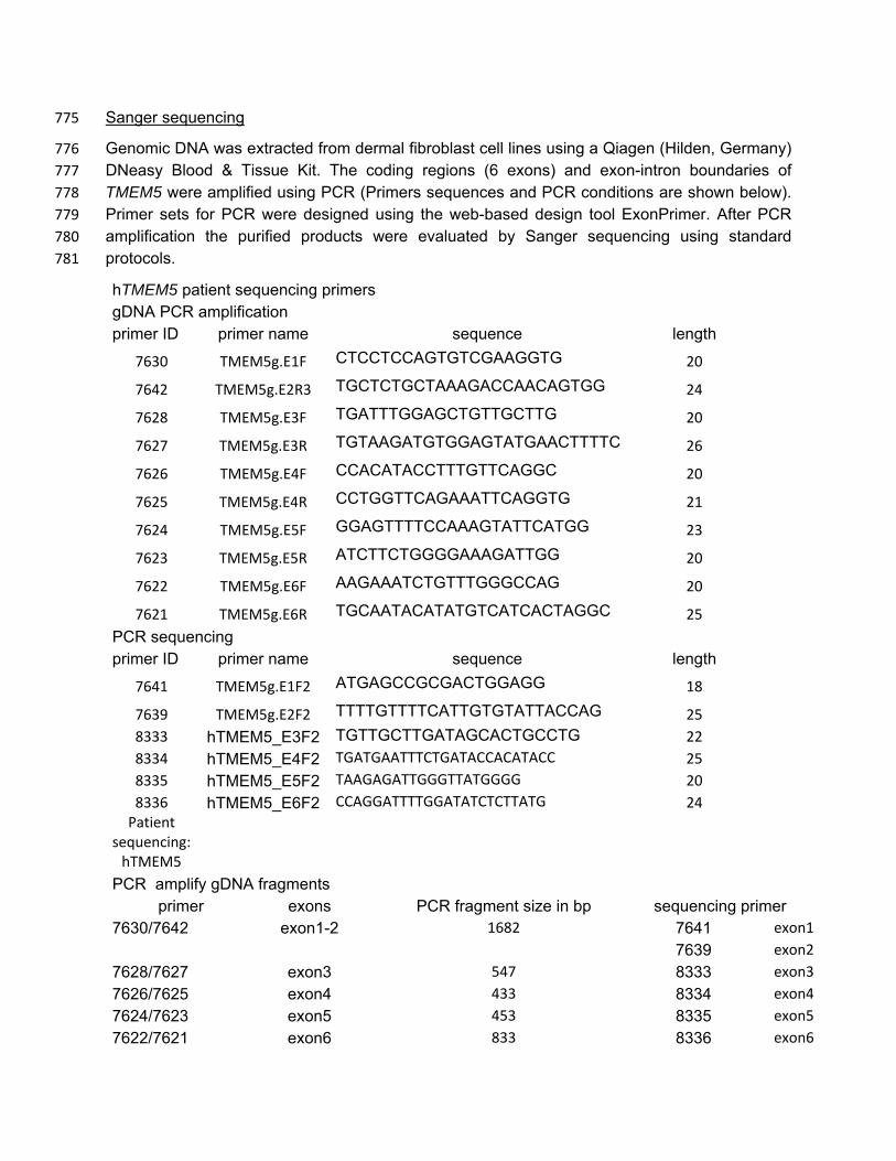
Sanger sequencing 775
Genomic DNA was extracted from dermal fibroblast cell lines using a Qiagen (Hilden, Germany) 776 DNeasy Blood & Tissue Kit. The coding regions (6 exons) and exon-intron boundaries of 777 TMEM5 were amplified using PCR (Primers sequences and PCR conditions are shown below). 778 Primer sets for PCR were designed using the web-based design tool ExonPrimer. After PCR 779 amplification the purified products were evaluated by Sanger sequencing using standard 780 protocols. 781
hTMEM5 patient sequencing primers gDNA PCR amplification primer ID primer name sequence length
7630 TMEM5g.E1F CTCCTCCAGTGTCGAAGGTG 20
7642 TMEM5g.E2R3 TGCTCTGCTAAAGACCAACAGTGG 24
7628 TMEM5g.E3F TGATTTGGAGCTGTTGCTTG 20
7627 TMEM5g.E3R TGTAAGATGTGGAGTATGAACTTTTC 26
7626 TMEM5g.E4F CCACATACCTTTGTTCAGGC 20
7625 TMEM5g.E4R CCTGGTTCAGAAATTCAGGTG 21
7624 TMEM5g.E5F GGAGTTTTCCAAAGTATTCATGG 23
7623 TMEM5g.E5R ATCTTCTGGGGAAAGATTGG 20
7622 TMEM5g.E6F AAGAAATCTGTTTGGGCCAG 20
7621 TMEM5g.E6R TGCAATACATATGTCATCACTAGGC 25 PCR sequencing primer ID primer name sequence length
7641 TMEM5g.E1F2 ATGAGCCGCGACTGGAGG 18
7639 TMEM5g.E2F2 TTTTGTTTTCATTGTGTATTACCAG 25 8333 hTMEM5_E3F2 TGTTGCTTGATAGCACTGCCTG 22 8334 hTMEM5_E4F2 TGATGAATTTCTGATACCACATACC 25 8335 hTMEM5_E5F2 TAAGAGATTGGGTTATGGGG 20 8336 hTMEM5_E6F2 CCAGGATTTTGGATATCTCTTATG 24
Patient sequencing:
hTMEM5
PCR amplify gDNA fragments primer exons PCR fragment size in bp sequencing primer
7630/7642 exon1-2 1682 7641 exon1 7639 exon2
7628/7627 exon3 547 8333 exon3 7626/7625 exon4 433 8334 exon4 7624/7623 exon5 453 8335 exon5 7622/7621 exon6 833 8336 exon6
PCR-Program: Temp. Duration
1) Denaturation 95 3min 2) Cycles 95 30sec 3) Cycles (35x) 53 30sec 4) Cycles 72 1 min 30sec 5) Elongation 72 10min
782
ACKNOWLEDGEMENTS 783
We would like to thank all members of the Wells, Campbell, and Moremen laboratories for 784 helpful discussions. We are indebted to Dr. Michael Tiemeyer for valuable discussions 785 regarding experiments presented here. We would like to thank Allison Schwartz and Traci Toy 786 at the UCLA Genome Sequencing Center (http://gsc.ucla.edu/) for assisting with genotyping and 787 constructing the sequencing libraries and Dr. Suhua Feng at the UCLA Broad Stem Cell 788 Research Center (BSCRC) for assisting in running HiSeq2000. We thank Greg Morgensen and 789 David Venzke for technical support and Christine Blaumueller for critical reading of the 790 manuscript. This work was supported in part by grants from NIGMS/NIH (R01GM111939 to LW, 791 P01GM107012, KWM and LW co-PIs), technology resource grants from NIGMS/NIH 792 (P41GM103490, LW and KWM co-PIs and P41GM103390, KWM, PI), a Paul D. Wellstone 793 Muscular Dystrophy Cooperative Research Center Grant (1U54NS053672, K.P.C., S.A.M and 794 T.W.), a MDA grant (238219, K.P.C. and T.W.) and an ARRA Go Grant (1 RC2 NS069521-01, 795 K.P.C. and T.W.). K.P.C. is an investigator of the Howard Hughes Medical Institute. 796
797
798
799
REFERENCES 800
1. Ervasti, J. M., Ohlendieck, K., Kahl, S. D., Gaver, M. G., and Campbell, K. P. (1990) 801 Deficiency of a glycoprotein component of the dystrophin complex in dystrophic muscle. 802 Nature 345, 315-319, 10.1038/345315a0 803
2. Yoshida, M., and Ozawa, E. (1990) Glycoprotein complex anchoring dystrophin to 804 sarcolemma. Journal of biochemistry 108, 748-752, 805
3. Carmignac, V., and Durbeej, M. (2012) Cell-matrix interactions in muscle disease. The 806 Journal of pathology 226, 200-218, 10.1002/path.3020 807
4. Dong, M., Noguchi, S., Endo, Y., Hayashi, Y. K., Yoshida, S., Nonaka, I., and Nishino, I. 808 (2015) DAG1 mutations associated with asymptomatic hyperCKemia and 809 hypoglycosylation of alpha-dystroglycan. Neurology 84, 273-279, 810 10.1212/WNL.0000000000001162 811
5. Geis, T., Marquard, K., Rodl, T., Reihle, C., Schirmer, S., von Kalle, T., Bornemann, A., 812 Hehr, U., and Blankenburg, M. (2013) Homozygous dystroglycan mutation associated 813 with a novel muscle-eye-brain disease-like phenotype with multicystic leucodystrophy. 814 Neurogenetics 14, 205-213, 10.1007/s10048-013-0374-9 815
6. Hara, Y., Balci-Hayta, B., Yoshida-Moriguchi, T., Kanagawa, M., Beltran-Valero de 816 Bernabe, D., Gundesli, H., Willer, T., Satz, J. S., Crawford, R. W., Burden, S. J., Kunz, 817 S., Oldstone, M. B., Accardi, A., Talim, B., Muntoni, F., Topaloglu, H., Dincer, P., and 818 Campbell, K. P. (2011) A dystroglycan mutation associated with limb-girdle muscular 819 dystrophy. The New England journal of medicine 364, 939-946, 820 10.1056/NEJMoa1006939 821
7. Chiba, A., Matsumura, K., Yamada, H., Inazu, T., Shimizu, T., Kusunoki, S., Kanazawa, 822 I., Kobata, A., and Endo, T. (1997) Structures of sialylated O-linked oligosaccharides of 823 bovine peripheral nerve alpha-dystroglycan. The role of a novel O-mannosyl-type 824 oligosaccharide in the binding of alpha-dystroglycan with laminin. The Journal of 825 biological chemistry 272, 2156-2162, 826
8. Holt, K. H., Crosbie, R. H., Venzke, D. P., and Campbell, K. P. (2000) Biosynthesis of 827 dystroglycan: processing of a precursor propeptide. FEBS letters 468, 79-83, 828
9. Wells, L. (2013) The o-mannosylation pathway: glycosyltransferases and proteins 829 implicated in congenital muscular dystrophy. The Journal of biological chemistry 288, 830 6930-6935, 10.1074/jbc.R112.438978 831
10. Beltran-Valero de Bernabe, D., Currier, S., Steinbrecher, A., Celli, J., van Beusekom, E., 832 van der Zwaag, B., Kayserili, H., Merlini, L., Chitayat, D., Dobyns, W. B., Cormand, B., 833 Lehesjoki, A. E., Cruces, J., Voit, T., Walsh, C. A., van Bokhoven, H., and Brunner, H. G. 834 (2002) Mutations in the O-mannosyltransferase gene POMT1 give rise to the severe 835 neuronal migration disorder Walker-Warburg syndrome. American journal of human 836 genetics 71, 1033-1043, 10.1086/342975 837
11. Michele, D. E., Barresi, R., Kanagawa, M., Saito, F., Cohn, R. D., Satz, J. S., Dollar, J., 838 Nishino, I., Kelley, R. I., Somer, H., Straub, V., Mathews, K. D., Moore, S. A., and 839 Campbell, K. P. (2002) Post-translational disruption of dystroglycan-ligand interactions in 840 congenital muscular dystrophies. Nature 418, 417-422, 10.1038/nature00837 841
12. Praissman, J. L., and Wells, L. (2014) Mammalian O-mannosylation pathway: glycan 842 structures, enzymes, and protein substrates. Biochemistry 53, 3066-3078, 843 10.1021/bi500153y 844
13. Yoshida-Moriguchi, T., and Campbell, K. P. (2015) Matriglycan: a novel polysaccharide 845 that links dystroglycan to the basement membrane. Glycobiology 25, 702-713, 846 10.1093/glycob/cwv021 847

14. Dobson, C. M., Hempel, S. J., Stalnaker, S. H., Stuart, R., and Wells, L. (2013) O-848 Mannosylation and human disease. Cellular and molecular life sciences : CMLS 70, 849 2849-2857, 10.1007/s00018-012-1193-0 850
15. Endo, T. (2015) Glycobiology of alpha-dystroglycan and muscular dystrophy. Journal of 851 biochemistry 157, 1-12, 10.1093/jb/mvu066 852
16. Lee, J. K., Matthews, R. T., Lim, J. M., Swanier, K., Wells, L., and Pierce, J. M. (2012) 853 Developmental expression of the neuron-specific N-acetylglucosaminyltransferase Vb 854 (GnT-Vb/IX) and identification of its in vivo glycan products in comparison with those of 855 its paralog, GnT-V. The Journal of biological chemistry 287, 28526-28536, 856 10.1074/jbc.M112.367565 857
17. Yoshida, A., Kobayashi, K., Manya, H., Taniguchi, K., Kano, H., Mizuno, M., Inazu, T., 858 Mitsuhashi, H., Takahashi, S., Takeuchi, M., Herrmann, R., Straub, V., Talim, B., Voit, T., 859 Topaloglu, H., Toda, T., and Endo, T. (2001) Muscular dystrophy and neuronal migration 860 disorder caused by mutations in a glycosyltransferase, POMGnT1. Dev Cell 1, 717-724, 861
18. Manzini, M. C., Tambunan, D. E., Hill, R. S., Yu, T. W., Maynard, T. M., Heinzen, E. L., 862 Shianna, K. V., Stevens, C. R., Partlow, J. N., Barry, B. J., Rodriguez, J., Gupta, V. A., 863 Al-Qudah, A. K., Eyaid, W. M., Friedman, J. M., Salih, M. A., Clark, R., Moroni, I., Mora, 864 M., Beggs, A. H., Gabriel, S. B., and Walsh, C. A. (2012) Exome sequencing and 865 functional validation in zebrafish identify GTDC2 mutations as a cause of Walker-866 Warburg syndrome. American journal of human genetics 91, 541-547, 867 10.1016/j.ajhg.2012.07.009 868
19. Yoshida-Moriguchi, T., Willer, T., Anderson, M. E., Venzke, D., Whyte, T., Muntoni, F., 869 Lee, H., Nelson, S. F., Yu, L., and Campbell, K. P. (2013) SGK196 is a glycosylation-870 specific O-mannose kinase required for dystroglycan function. Science 341, 896-899, 871 10.1126/science.1239951 872
20. Praissman, J. L., Live, D. H., Wang, S., Ramiah, A., Chinoy, Z. S., Boons, G. J., 873 Moremen, K. W., and Wells, L. (2014) B4GAT1 is the priming enzyme for the LARGE-874 dependent functional glycosylation of alpha-dystroglycan. Elife 3 10.7554/eLife.03943 875
21. Willer, T., Inamori, K., Venzke, D., Harvey, C., Morgensen, G., Hara, Y., Beltran Valero 876 de Bernabe, D., Yu, L., Wright, K. M., and Campbell, K. P. (2014) The 877 glucuronyltransferase B4GAT1 is required for initiation of LARGE-mediated alpha-878 dystroglycan functional glycosylation. Elife 3 10.7554/eLife.03941 879
22. Yoshida-Moriguchi, T., Yu, L., Stalnaker, S. H., Davis, S., Kunz, S., Madson, M., 880 Oldstone, M. B., Schachter, H., Wells, L., and Campbell, K. P. (2010) O-mannosyl 881 phosphorylation of alpha-dystroglycan is required for laminin binding. Science 327, 88-882 92, 10.1126/science.1180512 883
23. Roscioli, T., Kamsteeg, E. J., Buysse, K., Maystadt, I., van Reeuwijk, J., van den Elzen, 884 C., van Beusekom, E., Riemersma, M., Pfundt, R., Vissers, L. E., Schraders, M., 885 Altunoglu, U., Buckley, M. F., Brunner, H. G., Grisart, B., Zhou, H., Veltman, J. A., 886 Gilissen, C., Mancini, G. M., Delree, P., Willemsen, M. A., Ramadza, D. P., Chitayat, D., 887 Bennett, C., Sheridan, E., Peeters, E. A., Tan-Sindhunata, G. M., de Die-Smulders, C. E., 888 Devriendt, K., Kayserili, H., El-Hashash, O. A., Stemple, D. L., Lefeber, D. J., Lin, Y. Y., 889 and van Bokhoven, H. (2012) Mutations in ISPD cause Walker-Warburg syndrome and 890 defective glycosylation of alpha-dystroglycan. Nat Genet 44, 581-585, 10.1038/ng.2253 891
24. Willer, T., Lee, H., Lommel, M., Yoshida-Moriguchi, T., de Bernabe, D. B., Venzke, D., 892 Cirak, S., Schachter, H., Vajsar, J., Voit, T., Muntoni, F., Loder, A. S., Dobyns, W. B., 893 Winder, T. L., Strahl, S., Mathews, K. D., Nelson, S. F., Moore, S. A., and Campbell, K. 894 P. (2012) ISPD loss-of-function mutations disrupt dystroglycan O-mannosylation and 895 cause Walker-Warburg syndrome. Nat Genet 44, 575-580, 10.1038/ng.2252 896
25. Vuillaumier-Barrot, S., Bouchet-Seraphin, C., Chelbi, M., Devisme, L., Quentin, S., Gazal, 897 S., Laquerriere, A., Fallet-Bianco, C., Loget, P., Odent, S., Carles, D., Bazin, A., Aziza, 898
J., Clemenson, A., Guimiot, F., Bonniere, M., Monnot, S., Bole-Feysot, C., Bernard, J. P., 899 Loeuillet, L., Gonzales, M., Socha, K., Grandchamp, B., Attie-Bitach, T., Encha-Razavi, 900 F., and Seta, N. (2012) Identification of mutations in TMEM5 and ISPD as a cause of 901 severe cobblestone lissencephaly. American journal of human genetics 91, 1135-1143, 902 10.1016/j.ajhg.2012.10.009 903
26. Baur, S., Marles-Wright, J., Buckenmaier, S., Lewis, R. J., and Vollmer, W. (2009) 904 Synthesis of CDP-activated ribitol for teichoic acid precursors in Streptococcus 905 pneumoniae. J Bacteriol 191, 1200-1210, 10.1128/JB.01120-08 906
27. Richard, S. B., Bowman, M. E., Kwiatkowski, W., Kang, I., Chow, C., Lillo, A. M., Cane, 907 D. E., and Noel, J. P. (2001) Structure of 4-diphosphocytidyl-2-C- methylerythritol 908 synthetase involved in mevalonate- independent isoprenoid biosynthesis. Nat Struct Biol 909 8, 641-648, 10.1038/89691 910
28. Riemersma, M., Froese, D. S., van Tol, W., Engelke, U. F., Kopec, J., van Scherpenzeel, 911 M., Ashikov, A., Krojer, T., von Delft, F., Tessari, M., Buczkowska, A., Swiezewska, E., 912 Jae, L. T., Brummelkamp, T. R., Manya, H., Endo, T., van Bokhoven, H., Yue, W. W., 913 and Lefeber, D. J. (2015) Human ISPD Is a Cytidyltransferase Required for Dystroglycan 914 O-Mannosylation. Chem Biol 22, 1643-1652, 10.1016/j.chembiol.2015.10.014 915
29. Jae, L. T., Raaben, M., Riemersma, M., van Beusekom, E., Blomen, V. A., Velds, A., 916 Kerkhoven, R. M., Carette, J. E., Topaloglu, H., Meinecke, P., Wessels, M. W., Lefeber, 917 D. J., Whelan, S. P., van Bokhoven, H., and Brummelkamp, T. R. (2013) Deciphering 918 the glycosylome of dystroglycanopathies using haploid screens for lassa virus entry. 919 Science 340, 479-483, 10.1126/science.1233675 920
30. Robu, M. E., Larson, J. D., Nasevicius, A., Beiraghi, S., Brenner, C., Farber, S. A., and 921 Ekker, S. C. (2007) p53 activation by knockdown technologies. PLoS Genet 3, e78, 922 10.1371/journal.pgen.0030078 923
31. Adzhubei, I., Jordan, D. M., and Sunyaev, S. R. (2013) Predicting functional effect of 924 human missense mutations using PolyPhen-2. Curr Protoc Hum Genet Chapter 7, Unit7 925 20, 10.1002/0471142905.hg0720s76 926
32. Sim, N. L., Kumar, P., Hu, J., Henikoff, S., Schneider, G., and Ng, P. C. (2012) SIFT web 927 server: predicting effects of amino acid substitutions on proteins. Nucleic Acids Res 40, 928 W452-457, 10.1093/nar/gks539 929
33. Stalnaker, S. H., Stuart, R., and Wells, L. (2011) Mammalian O-mannosylation: unsolved 930 questions of structure/function. Current opinion in structural biology 21, 603-609, 931 10.1016/j.sbi.2011.09.001 932
34. Lefeber, D. J., Schonberger, J., Morava, E., Guillard, M., Huyben, K. M., Verrijp, K., 933 Grafakou, O., Evangeliou, A., Preijers, F. W., Manta, P., Yildiz, J., Grunewald, S., Spilioti, 934 M., van den Elzen, C., Klein, D., Hess, D., Ashida, H., Hofsteenge, J., Maeda, Y., van 935 den Heuvel, L., Lammens, M., Lehle, L., and Wevers, R. A. (2009) Deficiency of Dol-P-936 Man synthase subunit DPM3 bridges the congenital disorders of glycosylation with the 937 dystroglycanopathies. American journal of human genetics 85, 76-86, 938 10.1016/j.ajhg.2009.06.006 939
35. Endo, T., and Manya, H. (2006) O-mannosylation in mammalian cells. Methods Mol Biol 940 347, 43-56, 10.1385/1-59745-167-3:43 941
36. Muntoni, F., Torelli, S., Wells, D. J., and Brown, S. C. (2011) Muscular dystrophies due 942 to glycosylation defects: diagnosis and therapeutic strategies. Current opinion in 943 neurology 24, 437-442, 10.1097/WCO.0b013e32834a95e3 944
37. Cirak, S., Foley, A. R., Herrmann, R., Willer, T., Yau, S., Stevens, E., Torelli, S., Brodd, 945 L., Kamynina, A., Vondracek, P., Roper, H., Longman, C., Korinthenberg, R., Marrosu, 946 G., Nurnberg, P., Consortium, U. K., Michele, D. E., Plagnol, V., Hurles, M., Moore, S. A., 947 Sewry, C. A., Campbell, K. P., Voit, T., and Muntoni, F. (2013) ISPD gene mutations are 948

a common cause of congenital and limb-girdle muscular dystrophies. Brain 136, 269-281, 949 10.1093/brain/aws312 950
38. Perl, A., Hanczko, R., Telarico, T., Oaks, Z., and Landas, S. (2011) Oxidative stress, 951 inflammation and carcinogenesis are controlled through the pentose phosphate pathway 952 by transaldolase. Trends Mol Med 17, 395-403, 10.1016/j.molmed.2011.01.014 953
39. Riemersma, M., Sandrock, J., Boltje, T. J., Bull, C., Heise, T., Ashikov, A., Adema, G. J., 954 van Bokhoven, H., and Lefeber, D. J. (2015) Disease mutations in CMP-sialic acid 955 transporter SLC35A1 result in abnormal alpha-dystroglycan O-mannosylation, 956 independent from sialic acid. Human molecular genetics 24, 2241-2246, 957 10.1093/hmg/ddu742 958
40. Collins, B. E., Yang, L. J., Mukhopadhyay, G., Filbin, M. T., Kiso, M., Hasegawa, A., and 959 Schnaar, R. L. (1997) Sialic acid specificity of myelin-associated glycoprotein binding. 960 The Journal of biological chemistry 272, 1248-1255, 961
41. Kanagawa, M., Kobayashi, K., Tajiri, M., Manya, H., Kuga, A., Yamaguchi, Y., Akasaka-962 Manya, K., Furukawa, J., Mizuno, M., Kawakami, H., Shinohara, Y., Wada, Y., Endo, T., 963 and Toda, T. (2016) Identification of a Post-translational Modification with Ribitol-964 Phosphate and Its Defect in Muscular Dystrophy. Cell Rep 14, 2209-2223, 965 10.1016/j.celrep.2016.02.017 966
42. Vester-Christensen, M. B., Halim, A., Joshi, H. J., Steentoft, C., Bennett, E. P., Levery, S. 967 B., Vakhrushev, S. Y., and Clausen, H. (2013) Mining the O-mannose glycoproteome 968 reveals cadherins as major O-mannosylated glycoproteins. Proceedings of the National 969 Academy of Sciences of the United States of America 110, 21018-21023, 970 10.1073/pnas.1313446110 971
43. Damerell, D., Ceroni, A., Maass, K., Ranzinger, R., Dell, A., and Haslam, S. M. (2015) 972 Annotation of glycomics MS and MS/MS spectra using the GlycoWorkbench software 973 tool. Methods Mol Biol 1273, 3-15, 10.1007/978-1-4939-2343-4_1 974
44. Barb, A. W., Meng, L., Gao, Z., Johnson, R. W., Moremen, K. W., and Prestegard, J. H. 975 (2012) NMR characterization of immunoglobulin G Fc glycan motion on enzymatic 976 sialylation. Biochemistry 51, 4618-4626, 10.1021/bi300319q 977
45. Tozser, J., Tropea, J. E., Cherry, S., Bagossi, P., Copeland, T. D., Wlodawer, A., and 978 Waugh, D. S. (2005) Comparison of the substrate specificity of two potyvirus proteases. 979 FEBS J 272, 514-523, 10.1111/j.1742-4658.2004.04493.x 980
46. Pedelacq, J. D., Cabantous, S., Tran, T., Terwilliger, T. C., and Waldo, G. S. (2006) 981 Engineering and characterization of a superfolder green fluorescent protein. Nature 982 biotechnology 24, 79-88, 10.1038/nbt1172 983
47. Beckett, D., Kovaleva, E., and Schatz, P. J. (1999) A minimal peptide substrate in biotin 984 holoenzyme synthetase-catalyzed biotinylation. Protein science : a publication of the 985 Protein Society 8, 921-929, 10.1110/ps.8.4.921 986
48. Meng, L., Forouhar, F., Thieker, D., Gao, Z., Ramiah, A., Moniz, H., Xiang, Y., 987 Seetharaman, J., Milaninia, S., Su, M., Bridger, R., Veillon, L., Azadi, P., Kornhaber, G., 988 Wells, L., Montelione, G. T., Woods, R. J., Tong, L., and Moremen, K. W. (2013) 989 Enzymatic basis for N-glycan sialylation: structure of rat alpha2,6-sialyltransferase 990 (ST6GAL1) reveals conserved and unique features for glycan sialylation. The Journal of 991 biological chemistry 288, 34680-34698, 10.1074/jbc.M113.519041 992
49. Kumagai, T., Katoh, T., Nix, D. B., Tiemeyer, M., and Aoki, K. (2013) In-gel beta-993 elimination and aqueous-organic partition for improved O- and sulfoglycomics. Analytical 994 chemistry 85, 8692-8699, 10.1021/ac4015935 995
50. Fakhouri, M., Elalayli, M., Sherling, D., Hall, J. D., Miller, E., Sun, X., Wells, L., and 996 LeMosy, E. K. (2006) Minor proteins and enzymes of the Drosophila eggshell matrix. 997 Dev Biol 293, 127-141, 10.1016/j.ydbio.2006.01.028 998
51. Stalnaker, S. H., Hashmi, S., Lim, J. M., Aoki, K., Porterfield, M., Gutierrez-Sanchez, G., 999 Wheeler, J., Ervasti, J. M., Bergmann, C., Tiemeyer, M., and Wells, L. (2010) Site 1000 mapping and characterization of O-glycan structures on alpha-dystroglycan isolated from 1001 rabbit skeletal muscle. The Journal of biological chemistry 285, 24882-24891, 1002 10.1074/jbc.M110.126474 1003
52. Baddiley, J., Buchanan, J. G., Carss, B., Mathias, A. P., and Sanderson, A. R. (1956) 1004 The isolation of cytidine diphosphate glycerol, cytidine diphosphate ribitol and mannitol 1005 1-phosphate from Lactobacillus arabinosus. Biochem J 64, 599-603, 1006
53. Barresi, R., Michele, D. E., Kanagawa, M., Harper, H. A., Dovico, S. A., Satz, J. S., 1007 Moore, S. A., Zhang, W., Schachter, H., Dumanski, J. P., Cohn, R. D., Nishino, I., and 1008 Campbell, K. P. (2004) LARGE can functionally bypass alpha-dystroglycan glycosylation 1009 defects in distinct congenital muscular dystrophies. Nature medicine 10, 696-703, 1010 10.1038/nm1059 1011
54. Hara, Y., Kanagawa, M., Kunz, S., Yoshida-Moriguchi, T., Satz, J. S., Kobayashi, Y. M., 1012 Zhu, Z., Burden, S. J., Oldstone, M. B., and Campbell, K. P. (2011) Like-1013 acetylglucosaminyltransferase (LARGE)-dependent modification of dystroglycan at Thr-1014 317/319 is required for laminin binding and arenavirus infection. Proceedings of the 1015 National Academy of Sciences of the United States of America 108, 17426-17431, 1016 10.1073/pnas.1114836108 1017
55. Link, V., Shevchenko, A., and Heisenberg, C. P. (2006) Proteomics of early zebrafish 1018 embryos. BMC Dev Biol 6, 1, 10.1186/1471-213X-6-1 1019
56. Ervasti, J. M., and Campbell, K. P. (1991) Membrane organization of the dystrophin-1020 glycoprotein complex. Cell 66, 1121-1131, 1021
57. Duclos, F., Straub, V., Moore, S. A., Venzke, D. P., Hrstka, R. F., Crosbie, R. H., 1022 Durbeej, M., Lebakken, C. S., Ettinger, A. J., van der Meulen, J., Holt, K. H., Lim, L. E., 1023 Sanes, J. R., Davidson, B. L., Faulkner, J. A., Williamson, R., and Campbell, K. P. 1024 (1998) Progressive muscular dystrophy in alpha-sarcoglycan-deficient mice. The Journal 1025 of cell biology 142, 1461-1471, 1026
58. Parsons, M. J., Campos, I., Hirst, E. M., and Stemple, D. L. (2002) Removal of 1027 dystroglycan causes severe muscular dystrophy in zebrafish embryos. Development 129, 1028 3505-3512, 1029
59. Lee, H., Jen, J. C., Cha, Y. H., Nelson, S. F., and Baloh, R. W. (2008) Phenotypic and 1030 genetic analysis of a large family with migraine-associated vertigo. Headache 48, 1460-1031 1467, 10.1111/j.1526-4610.2007.01002.x 1032
60. Lee, H., O'Connor, B. D., Merriman, B., Funari, V. A., Homer, N., Chen, Z., Cohn, D. H., 1033 and Nelson, S. F. (2009) Improving the efficiency of genomic loci capture using 1034 oligonucleotide arrays for high throughput resequencing. BMC Genomics 10, 646, 1035 10.1186/1471-2164-10-646 1036
1037
1038

FIGURE LEGENDS 1039
1040
Figure 1: The 3 subclasses of the O-Mannosylation Pathway. Following addition of Man to 1041 serine/threonine residues on protein substrates by POMT1/2 there is a divergence in 1042 elaboration. Known enzymes are displayed in purple and unknown enzymes prior to this 1043 manuscript are displayed in red. A. POMGnT1 adds a beta-2 linked GlcNAc onto the underlying 1044 Man. Shown is just one potential extended M1 glycan structure. B. Following the action of 1045 POMGnT1, GnT-Vb can add a beta-6 linked GlcNAc onto a M1 glycan structure to convert it to 1046 a M2 glycan structure. Shown is just one potential extended M2 glycan structure. C. Instead of 1047 extension in a beta-2 linkage, M3 glycan structures get the core Man extended by POMGNT2 1048 with a beta-4 linked GlcNAc. This disaccharide is further elaborated to contain a phosphodiester 1049 linkage to an unknown moiety (shown as (X)) that becomes modified with repeats of (Xyl-GlcA). 1050
1051
Figure 2: Ribitol-Xyl-GlcA is released from α-DG upon cleavage of phosphodiester 1052 linkage. Following treatment of purified α-DG-340 with aqueous HF, released glycans were 1053 captured, permethylated and analyzed by tandem mass spectrometry. On the left is a zoom 1054 view of the full MS scan demonstrating the intact mass and to the right the fragmentation pattern 1055 leading to the identification of GlcA-Xyl-pentitol. Experiments were performed in triplicate with 1056 identical findings each time. 1057
1058
Figure 2 – figure supplement 1: Extended Ribitol-(Xyl-GlcA)n Structures are also 1059 observed. Identical analysis as in Figure 1 of a more extended structure (GlcA-Xyl)2-pentitol. 1060
1061
Figure 2 – figure supplement 2: Phosphodiester cleavage but not glycosidase treatment 1062 abolishes IIH6 reactivity on α-DG. A purified C-terminal truncated version of α-DG 1063 (expressed from DGFc4) from HEK293H cells also overexpressing LARGE was treated with a 1064 variety of glycosidases or treated with aqueous HF overnight and then separated by SDS-PAGE 1065 and immunoblotted with IIH6 to detect functionally glycosylated α-DG. Only HF treatment 1066 abolished IIH6 reactivity. One representative result of three is shown. 1067
1068
Figure 3: ISPD synthesizes CDP-Ribitol from CTP and Ribitol-phosphate. A. Purified 1069 recombinant ISPD (+ISPD) or buffer control (-ISPD) was incubated with CTP and either ribitol-5-1070 phosphate (Ribitol-5P) or ribitol (Ribitol) and the donor substrate (CTP, MW 481.98) and/or 1071 product (CDP-ribitol, MW 536.07) was detected by mass spectrometry. CDP-ribitol was only 1072 observed in the presence of ISPD, CTP, and Ribitol-5P. B. Purified recombinant ISPD (+ISPD) 1073 or buffer control (-ISPD) was incubated with CTP and either ribose-5-phosphate (Ribose-5P) or 1074 ribose (Ribose) and the donor substrate (CTP, MW 481.98) and/or product (CDP-ribose, MW 1075 534.05) detected by mass spectrometry. CDP-ribose was only observed in the presence of 1076

ISPD, CTP, and Ribose-5P. Note that CDP-ribose has a molecular weight that is 2 daltons less 1077 than CDP-ribitol. Shown are representative images of an N=3. 1078
1079
Figure 3 – figure supplement 1: Coomassie stained SDS-PAGE gel of purified ISPD. 1080 His8-tagged ISPD was expressed in HEK293F cells and purified from whole cell lysates via 1081 immobilized metal affinity chromatography and the resulting purified protein was separated via 1082 SDS-PAGE and visualized with Coomassie-G250 staining. 1083
1084
Figure 4: Mild periodate cleavage reveals that ribitol is connected to the M3 glycan. 1085 Following treatment of purified α-DG-340 under mild periodate conditions, the protein was 1086 reduced, alkylated, and digested with trypsin. The resulting peptides were analyzed by tandem 1087 mass spectrometry. The α-DG-340 tryptic peptide was detected with multiple glycoforms 1088 containing a phosphotrisaccharide glycopeptide. Specifically, at least 12 tryptic peptides were 1089 identified at less than 9 ppm mass accuracy that contained the phosphotrisaccharide with a 1090 ribitol fragment at threonine 317 (the red T*) in two separate analyses. These peptides differed 1091 in additional glycosylation by either hexose (Hex) or HexNAc sugars at additional hydroxyl 1092 amino acids (indicated by #). We also observed that the N-terminal glutamine was cyclized to 1093 pyroglutamic acid on some peptides (Pyro) and that some glycopeptides were sodiated (Na). 1094 Furthermore, we noticed the presence of a second phosphate on some phosphoglycopeptides. 1095 The 12 peptides observed are grouped by having the same number of additional Hex and 1096 HexNAc glycans (2,3 or 3,3 or 2,4). For all 3 sets of glycopeptides we observed glycopeptides 1097 that had ribitol fragments between C2 and C3 as well as C3 and C4 indicating that C2, C3, and 1098 C4 are not involved in linkages to other moieties. We also observed for each set of 1099 glycopeptides the presence of an additional phosphate. 1100
1101
Figure 4 – figure supplement 1: Tandem MS/MS spectra of glycopeptides. Shown are 1102 MS/MS spectra for 2 of the 12 glycopeptides described in Figure 3. Glycan oxonium ions as 1103 well as b- and y- fragments are labeled on the spectra. The amino acid sequence displays the 1104 observed b- (blue) and y-(red) ions observed as well as highlights the glycan modified amino 1105 acids in red. In all cases the phosphotrisaccharide-ribitol fragment (2-3 or 3-4) is assigned to 1106 Thr-317 (the first red labeled T in the sequence). 1107
1108
Figure 4 – figure supplement 2: Dot blot analysis reveals loss of IIH6 signal for α-DG 1109 treated with mild periodate. Following treatment (or mock treatment with the omission of 1110 NaIO4) of purified α-DG-340 under mild periodate conditions, the protein was spotted at 3 ug or 1111 0.3 ug and immunoblotted with the IIH6 antibody. Mild periodate treatment almost completely 1112 abolishes IIH6 reactivity even at 3 ug spotting. Shown is one of the two blots performed. 1113
1114

Figure 5: TMEM5 is a UDP-Xyl transferase that modifies phosphodiester linked ribitol. A. 1115 Various UDP-sugar nucleotides were incubated with (+) or without (-) purified transmembrane 1116 deleted and tagged TMEM5 and then analyzed for hydrolysis using the UDP-Glo assay (N=3). 1117 The formation of UDP was only observed when dTM-TMEM5 was incubated with UDP-Xyl. B. 1118 Recombinant purified full length TMEM5 and radiolabeled UDP-Xyl [Xyl-14C] was incubated with 1119 either purified α-DG-Fc340 or mutant α-DGFc340 where the sites for M3 addition had been 1120 mutated to alanines (α-DG-Fc340-T317A/T319A) and incorporation of radioactivity into the 1121 proteins were determined by scintillation chromatography (N=3). α-DGFc340 that contains the 1122 sites for M3 addition is the preferred substrate for addition of radiolabeled Xyl by TMEM5. C. 1123 dTM-TMEM5 (2500, 1250, 625, or 312.5 nM) and 50 µM UDP-Xyl donor substrate were 1124 incubated without or with 1mM of the indicated acceptor molecules in 0.1 M MES pH 6.0 and 10 1125 mM MgCl2 for 18 hours at 37°C, and the release of UDP was detected by the UDP-Glo assay. 1126 RLU: Relative Luminescence Units. Data are represented as the mean of three trials and error 1127 bars represent standard deviation. 1128
1129
Figure 5 – figure supplement 1: Analysis of purified TMEM5 constructs. A. GFP/HIS-1130 tagged dTM-TMEM5 was expressed in HEK293F cells and purified from the media via 1131 immobilized metal affinity chromatography and the resulting purified protein was separated via 1132 SDS-PAGE and visualized with Coomassie Brilliant blue and used in experiments shown in 1133 Figure 5A. B. Purified full length TMEM5 C-terminally tagged with Myc/DDK-epitope tag was 1134 purchased from Origene and analyzed by Coomassie Brilliant blue and immunoblotting with 1135 anti-C-Myc following separation via SDS-PAGE and used in experiments shown in Figure 5B. 1136
1137
Figure 5 – figure supplement 2: Specificity of TMEM5 for CDP-ribitol as an acceptor 1138
GFP-dTMTMEM5 was incubated with either 50 µM UDP-Xyl, 1 mM CDP-ribitol, or both in 0.1 M 1139 MES pH 6.0 and 10 mM MgCl2 for 18 hours at 37°C, and the release of UDP (or CDP) was 1140 detected by the UDP-Glo assay. RLU: Relative Luminescence Units. Data are represented as 1141 the mean of three trials and error bars represent standard deviation. 1142
1143
Figure 6: Knockdown of zebrafish tmem5 recapitulates characteristic defects in WWS. A. 1144 Compared with uninjected control and p53 MO injected embryos, embryos injected with tmem5 1145 MO alone or together with p53 MO developed mild to severe hydrocephalus (asterisk) from 32 1146 hour post fertilization (h.p.f.) (95% and 88%, respectively). Each bar represents a combination 1147 of two independent blindly-scored experiments. n= 114-150 embryos. Scale bar, 100 µm. B. 1148 One-way ANOVA analysis revealed that tmem5 MO injected embryos have significantly 1149 reduced eye size (bracket in panel a) at 48 h.p.f. Inhibition of MO off-target effects by co-1150 injection of p53 MO does not rescue the reduced eye size. n= 15 embryos in each group. Error 1151 bars, s.d. ** P< 0.01. NS, not significant. C. Knockdown of tmem5 compromised the 1152 sarcolemma integrity as indicated by EBD infiltrated muscle fibers (arrows) at 48 h.p.f. Muscle 1153 fibers undergoing degeneration pull away from chevron-shaped myosepta. Scale bar, 50 µm. D. 1154

Compared with uninjected control, western blotting with IIH6 antibody showed a 44% reduction 1155 in glycosylated α-dystroglycan in tmem5 MO-injected embryos at 48 h.p.f. Equal loading was 1156 demonstrated by Ponceau S and unknown glycoproteins (~35 kDa) detected by IIH6 in all lanes. 1157
1158
Figure 6 – figure supplement 1: Zebrafish tmem5 temporal gene expression and MO 1159 specificity. A. A schematic representation of the zebrafish tmem5 transcript and protein 1160 domains. White boxes indicate the untranslated region (UTR). Black boxes indicate coding 1161 exons. The translational blocking MO target site and PCR primer binding sites are indicated. 1162 The amino acid sequence of zebrafish Tmem5 is ~73% similar and ~55% identical to that of 1163 human orthologue. Conserved protein domains are boxed: TM, transmembrane domain (7-29 1164 aa); Exostosin (GT-47 homology) domain (254-349 aa). B. The expression of tmem5 transcript 1165 was detected by RT-PCR using primer tmem5_1F and tmem5_3R. Zebrafish tmem5 is 1166 expressed maternally at the 2-cell stage, during the maternal-zygotic transition (6 h.p.f) and 1167 zygotically at later stages (24, 48, 72, 96 and 120 h.p.f.). C. To assess tmem5 MO specificity, 1168 tmem5-egfp mRNA (200 pg) was injected into single cell, followed by injection of tmem5 MO (1 1169 ng or 4 ng) into the yolk flow. Embryos that received only tmem5-egfp mRNA showed a strong 1170 Tmem5-EGFP expression at 30 h.p.f., whereas embryos received a second injection of 1 ng or 1171 4 ng tmem5 MO showed a moderate and strong reduction in Tmem5-EGFP expression, 1172 respectively. These results confirm the specificity of tmem5 MO. n= 20 embryos in each group. 1173 Scale bar, 100 µm. 1174
1175
Figure 7: A missense mutation of TMEM5 in a WWS family leads to a loss of functional α-1176 DG. A. Analysis of α-DG glycosylation status in TMEM5-deficient WWS patient (P1) dermal 1177 fibroblasts. WGA-enriched cell lysates from control and TMEM5 P1 fibroblasts were subjected 1178 to biochemical analysis. α-DG glycosylation status as assessed by western blotting using 1179 antibodies against the glycosylated form of α-DG (IIH6), core α-DG (G6317), and by laminin 1180 overlay assay (N=3). An antibody against β-DG (AP83) was used to assess loading. Apparent 1181 molecular masses are indicated. B. On-Cell western-based complementation assay (N=3) of 1182 TMEM5-WWS patient (P1) fibroblasts after adenoviral gene transfer with wildtype TMEM5 1183 expression construct. Rescue of α-DG functional glycosylation was monitored with α-DG glyco 1184 (IIH6) antibodies. C. On-Cell western-based complementation assay (N=3) of P1 fibroblasts 1185 after nucleofection with a wild-type or mutant (p.G333R) human TMEM5 expression construct. 1186 Rescue of α-dystroglycan functional glycosylation was detected with antibody to glycosylated α-1187 dystroglycan (glyco α-DG, IIH6).. D. Subcellular localization of HA-tagged wildtype and WWS 1188 mutant TMEM5 protein, as assessed by immunofluorescence. HEK293T cells stably expressing 1189 HA-tagged proteins were stained with anti-HA (red), anti-Giantin (Golgi marker, green) and 4 ́,6-1190 diamidino-2-phenylindole (DAPI, nuclei, blue). Individual stainings for HA and Giantin are shown 1191 in greyscale and a merged image is shown in color. Scale bars indicate 10µm. 1192
1193
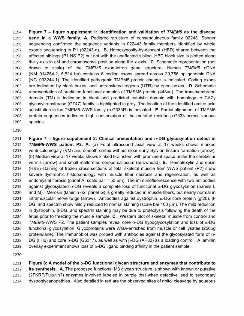
Figure 7 – figure supplement 1: Identification and validation of TMEM5 as the disease 1194 gene in a WWS family. A. Pedigree structure of consanguineous family 02243. Sanger 1195 sequencing confirmed the sequence variants in 022443 family members identified by whole 1196 exome sequencing in P1 (02243-d). B. Homozygosity-by-descent (HBD) shared between the 1197 affected siblings (P1 NS P2) but not with the unaffected sibling. HBD block size is plotted along 1198 the y-axis in cM and chromosomal position along the x-axis. C. Schematic representation (not 1199 drawn to scale) of the TMEM5 exon-intron gene structure. Human TMEM5 cDNA 1200 (NM_014254.2, 5,524 bp) contains 6 coding exons spread across 29,756 bp genomic DNA 1201 (NG_033244.1). The identified pathogenic TMEM5 protein change is indicated. Coding exons 1202 are indicated by black boxes, and untranslated regions (UTR) by open boxes. D. Schematic 1203 representation of predicted functional domains of TMEM5 protein (443aa). The transmembrane 1204 domain (TM) is indicated in black and predicted catalytic domain with homology to CAZy 1205 glycosyltransferase (GT47) family is highlighted in grey. The location of the identified amino acid 1206 substitution in the TMEM5-WWS family (p.G333R) is indicated. E. Partial alignment of TMEM5 1207 protein sequences indicates high conservation of the mutated residue p.G333 across various 1208 species. 1209
1210
Figure 7 – figure supplement 2: Clinical presentation and α-DG glycosylation defect in 1211 TMEM5-WWS patient P2. A. (a) Fetal ultrasound axial view at 17 weeks shows marked 1212 ventriculomegaly (VM) and smooth cortex without clear early Sylvian fissure formation (arrow), 1213 (b) Median view at 17 weeks shows kinked brainstem with prominent space under the cerebellar 1214 vermis (arrow) and small malformed corpus callosum (arrowhead). B. Hematoxylin and eosin 1215 (H&E) staining of frozen cross-sections of fetal skeletal muscle from WWS patient (P2) show 1216 severe dystrophic histopathology with muscle fiber necrosis and regeneration, as well as 1217 endomysial fibrosis (panel A; scale bar = 50 μm). The immunofluorescence with two antibodies 1218 against glycosylated α-DG reveals a complete loss of functional α-DG glycosylation (panels L 1219 and M). Merosin (laminin α2; panel G) is greatly reduced in muscle fibers, but nearly normal in 1220 intramuscular nerve twigs (arrow). Antibodies against dystrophin, α-DG core protein (gt20), β-1221 DG, and spectrin show mildly reduced to normal staining (scale bar 100 μm). The mild reduction 1222 in dystrophin, β-DG, and spectrin staining may be due to proteolysis following the death of the 1223 fetus prior to freezing the muscle sample. C. Western blot of skeletal muscle from control and 1224 TMEM5-WWS P2. The patient samples reveal core α-DG hypoglycosylation and loss of α-DG 1225 functional glycosylation. Glycoproteins were WGA-enriched from muscle or cell lysates (250μg 1226 protein/lane). The immunoblot was probed with antibodies against the glycosylated form of α-1227 DG (IIH6) and core α-DG (G6317), as well as with β-DG (AP83) as a loading control. A laminin 1228 overlay experiment shows loss of α-DG ligand binding affinity in the patient sample. 1229
1230
Figure 8: A model of the α-DG functional glycan structure and enzymes that contribute to 1231 its synthesis. A. The proposed functional M3 glycan structure is shown with known or putative 1232 (?FKRP/Fukutin?) enzymes involved labeled in purple that when defective lead to secondary 1233 dystroglycanopathies. Also detailed in red are the observed sites of ribitol cleavage by aqueous 1234

HF (HF) and mild periodate (NaIO4) treatment. B. The DPM1/2/3 enzyme complex (labeled in 1235 purple) is required for the synthesis of Dolichol-phosphomannose (Dol-P-Man) from GDP-Man 1236 and Dolichol phosphate (Dol-P). . C. ISPD (labeled in purple) is required for the synthesis of 1237 CDP-ribitol from CTP and ribitol-phosphate. Defects in DPM1/2/3 and ISPD are considered 1238 tertiary dystroglycanopathies since they synthesize the sugar donors Dol-P-Man (POMT1/2) and 1239 CDP-ribitol (presumably Fukutin and/or FKRP) for subsequent glycosyltransferases . 1240
1241

M1 M2
C
A BMannose
GlcNAc
Galactose
Xylose
Glucuronic Acid
Key
Sialic Acid
Fucose
α
β2POMGNT1
POMT1/
POMT2α
β2 β6 GnT-Vb
T/S
POMGNT1
POMT1/
POMT2
T/S
GalNAc
β4
β3β4 β3α3 β3α3
POMGNT2
B3GALNT2
POMK
B4GAT1 LARGE and/or LARGE2
Matriglycan
6(X)
Unknown Unknown
M3
T/SαPOMT1/
POMT2
F1

624 626m/z
0
5
10
15
20
25
30
35
40
45
50
55
60
65
70
75
80
85
90
95
100
Rel
ativ
e A
bund
ance
623.2920z=1
624.2952z=1
625.3601z=?
200 250 300 350 400 450 500 550 600m/z
0
5
10
15
20
25
30
35
40
45
50
55
60
65
70
75
80
85
90
95
100
Rel
ativ
e A
bund
ance
415.2
591.4
447.2
391.2 433.2
343.2
593.5563.4521.4
299.2 605.4273.2 581.2559.4448.2 495.3373.3259.1 545.3231.1 465.4393.3 503.3353.3 623.4337.2309.3287.2221.0
MS MS/MS
F2

A. Ribitol-5P – ISPD
Ribitol-5P+ ISPD
CDP-Ribose
CTP CDP-Ribitol
460 480 500 520 540m/z
0
10
20
30
40
50
60
70
80
90
100
Rel
ativ
e A
bund
ance
481.979z=1
508.119z=1
545.130z=1
463.064z=1
CTP + Mg
Ribitol+ ISPD
460 480 500 520 540m/z
0
10
20
30
40
50
60
70
80
90
100R
elat
ive
Abu
ndan
ce536.070
z=1
508.120z=1466.986
z=1
CTP + Mg
460 480 500 520 540m/z
0
10
20
30
40
50
60
70
80
90
100
Rel
ativ
e A
bund
ance
481.978z=1
508.119z=1
466.986z=1 524.944
z=1
CTP
CTP + Mg
B. Ribose-5P – ISPD
Ribose-5P+ ISPD
Ribose+ ISPD
460 480 500 520 540m/z
0
10
20
30
40
50
60
70
80
90
100
Rel
ativ
e A
bund
ance
481.978z=1
508.119z=1
543.113z=1
460.274z=1
CTP
CTP + Mg
460 480 500 520 540m/z
0
10
20
30
40
50
60
70
80
90
100
Rel
ativ
e A
bund
ance
534.054z=1
508.119z=1466.986
z=1481.978
z=1
CTPCTP + Mg
460 480 500 520 540m/z
0
10
20
30
40
50
60
70
80
90
100R
elat
ive
Abu
ndan
ce481.976
z=1
508.117z=1
460.273z=1
540.441z=2
CTP
CTP + Mg
F3

F4
RibitolCleavage Mass ppm Additional
Hex,HexNAc Pyro-Glu Sodiated Additional Phosphate
2-3 4205.953 7.3 2 , 3 N N N
2-3 4285.910 5.2 2 , 3 N N Y
2-3 4228.953 -1.0 2 , 3 N Y N
3-4 4258.963 8.7 2 , 3 N Y N
2-3 4350.968 1.7 3 , 3 Y N N
2-3 4367.992 2.3 3 , 3 N N N
3-4 4397.994 1.0 3 , 3 N N N
3-4 4477.961 1.3 3 , 3 N N Y
2-3 4392.004 5.9 2 , 4 Y N N
2-3 4409.032 6.1 2 , 4 N N N
2-3 4488.9952 5.3 2 , 4 N N Y
3-4 4439.011 -1.4 2 , 4 N N N
(Pyro) QIHAT*PT#PVT#AIGPPT#T#AIQEPPS#R (Na)
* = phosphotrisaccharide-ribitol fragment
# = potential sites of additional sugars

0.00
2000.00
4000.00
6000.00
8000.00
10000.00
12000.00
14000.00
16000.00U
DP-
Glc
UD
P-G
lcN
Ac
UD
P-G
al
UD
P-G
alN
Ac
UD
P-G
lcA
UD
P-Xy
l
UD
P-G
lc
UD
P-G
lcN
Ac
UD
P-G
al
UD
P-G
alN
Ac
UD
P-G
lcA
UD
P-Xy
l
- TMEM5 + TMEM5
Lum
ines
cenc
e (R
LU)
A
B C
0.00E+00
5.00E+03
1.00E+04
1.50E+04
2.00E+04
2.50E+04
3.00E+04
3.50E+04
0 500 1000 1500 2000 2500
Lum
ines
cenc
e (R
LU)
TMEM5 (nM)
TMEM5 Acceptor Substrate Specificity
No Acceptor
Ribitol
Ribitol-5-Phosphate
CDP-ribitol
CMP-Neu5Ac
0
50
100
150
200
250
300
Corr
ecte
d DP
M
-DG-Fc340 -T317A/T319A -DG-Fc340
F5
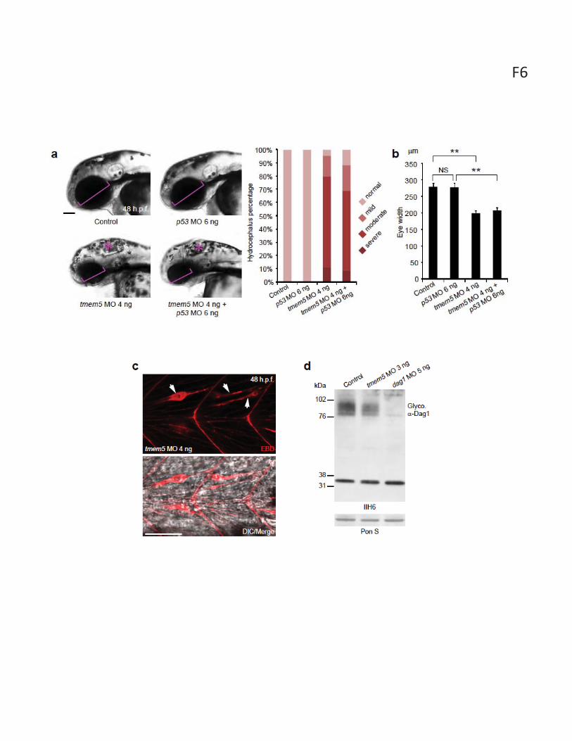
F6
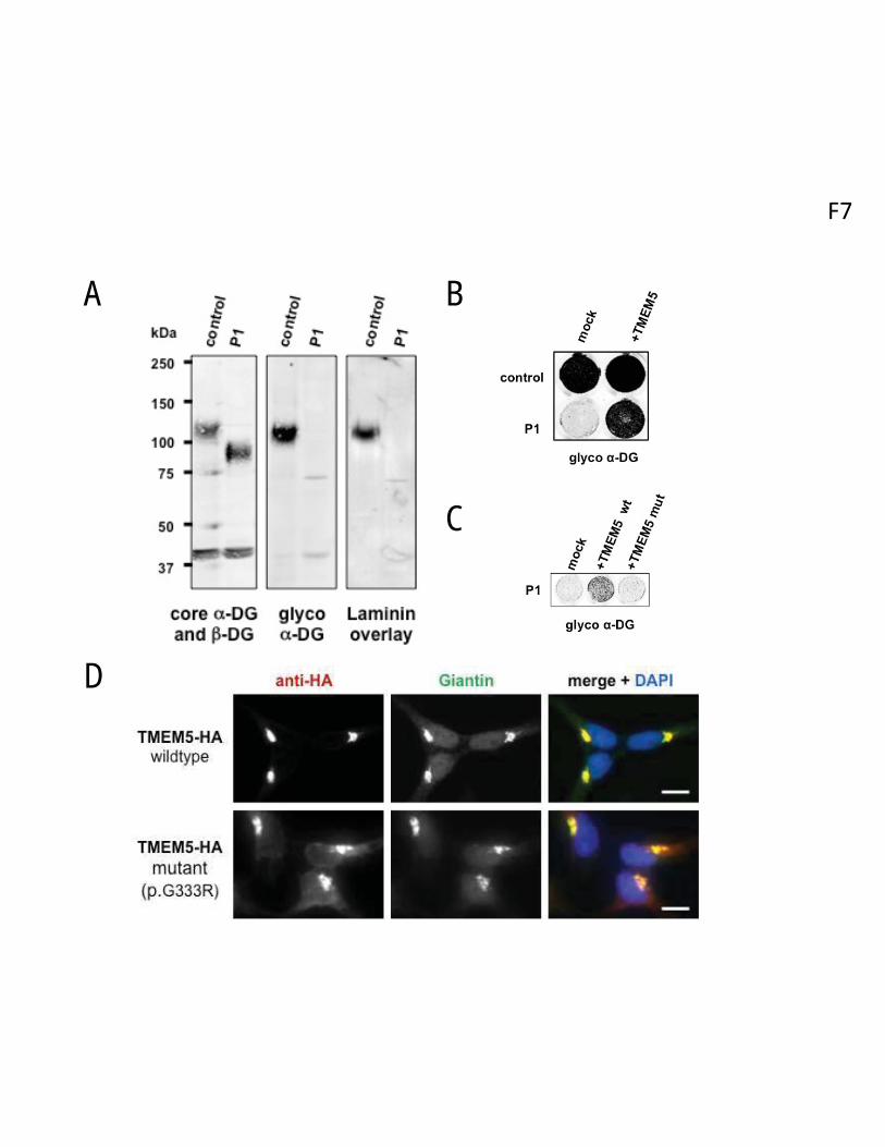
F7
A
D
B
C

Mannose
GlcNAc
GalNAc
Xylose
Glucuronic Acid
Key
A
B C
DPM1/2/3
ISPD
Dol-P Dol-P-Man
β4
β3
POMGNT2
B3GALNT2
POMK
6
T/SαPOMT1/
POMT2
β4 β3α3 β3α3
?FKRP/Fukutin?
TMEM5 B4GAT1 LARGE and/or LARGE2
Matriglycan
HF NaIO4


