Embed Size (px)
Citation preview

Molecular Biology of the CellVol. 16, 824–834, February 2005
Cdc42 and RhoB Activation Are Required for MannoseReceptor-mediated Phagocytosis by Human AlveolarMacrophagesJianmin Zhang,* Jinping Zhu,* Xia Bu,† Melanie Cushion,‡ T. Bernard Kinane,§Hava Avraham,† and Henry Koziel*�
*Division of Pulmonary and Critical Care Medicine and †Division of Experimental Medicine, Department ofMedicine, Beth Israel Deaconess Medical Center and Harvard Medical School, Boston, MA 02115; ‡VAMedical Center, University of Cincinnati, Cincinnati, OH 45220; and §Laboratory of DevelopmentalImmunology, Department of Pediatric Medicine, Massachusetts General Hospital and Harvard MedicalSchool, Boston, MA 02114
Submitted June 8, 2004; Revised November 3, 2004; Accepted November 17, 2004Monitoring Editor: Suzanne Pfeffer
Human alveolar macrophages (AMs) phagocytose Pneumocystis (Pc) organisms predominantly through mannose recep-tors, although the molecular mechanism mediating this opsonin-independent process is not known. In this study, usingAMs from healthy individuals, Pc phagocytosis was associated with focal F-actin polymerization and Cdc42, Rac1, andRho activation in a time-dependent manner. Phagocytosis was primarily dependent on Cdc42 and RhoB activation (asdetermined by AM transfection with Cdc42 and RhoB dominant-negative alleles) and mediated predominantly throughmannose receptors (as determined by siRNA gene silencing of AM mannose receptors). Pc also promoted PAK-1phosphorylation, which was also dependent on RhoGTPase activation. HIV infection of AMs (as a model for reducedmannose receptor expression and function) was associated with impaired F-actin polymerization, reduced Cdc42 and Rhoactivation, and markedly reduced PAK-1 phosphorylation in response to Pc organisms. In healthy AMs, Pc phagocytosiswas partially dependent on PAK activation, but dependent on the Rho effector molecule ROCK. These data provide amolecular mechanism for AM mannose receptor-mediated phagocytosis of unopsonized Pc organisms that appearsdistinct from opsonin-dependent phagocytic receptors. Reduced AM mannose receptor-mediated Cdc42 and Rho activa-tion in the context of HIV infection may represent a mechanism that contributes to the pathogenesis of opportunisticpneumonia.
INTRODUCTION
Opportunistic lung infections such as Pneumocystis pneumo-nia frequently complicate HIV disease, although the under-lying mechanisms of increased susceptibility and diseasepathogenesis remain incompletely understood. Alveolarmacrophages (AMs) are critical components of the pulmo-nary innate immune response to infectious challenge andlikely represent important effector cells in the host responseto Pneumocystis (Limper et al., 1997; Steele et al., 2003). Hu-man AMs express surface innate immune receptors such asmannose receptors (Greenberg and Grinstein, 2002) that canmediate Pneumocystis phagocytosis (Ezekowitz et al., 1991).AMs from asymptomatic HIV-infected individuals at highclinical risk for Pneumocystis pneumonia (peripheral CD4�
T-lymphocyte counts �200 cells/mm3) demonstrate signif-icantly reduced Pneumocystis phagocytosis compared withAMs from healthy individuals (Koziel et al., 1998a). Al-
though the reduced Pneumocystis phagocytosis is in partassociated with reduced AM mannose receptor surface ex-pression (Koziel et al., 1998a), the mechanism of reducedphagocytosis has not been completely established.
The family of small Rho guanosine triphosphatases(GTPases) link membrane receptors to the actin cytoskeletonand represent molecular switches that control a variety ofsignal transduction pathways including phagocytosis (Eti-enne-Manneville and Hall, 2002). Rho GTPases are a sub-group of the Ras superfamily of 20–30-kDa GTP-bindingproteins, and include Cdc42, Rac, and Rho (-A, -B, and -C).Specific Rho GTPase activation may provide the molecularbasis for the observed differences in opsonin-dependent re-ceptor-mediated phagocytosis described for immune globu-lin receptors (Fc�R) and complement receptors (C3R). Fc�R-mediated phagocytosis of appropriately opsonized particlesmay be mediated predominantly by Cdc42 and Rac, whereasC3R-mediated phagocytosis of appropriately opsonized par-ticles may be mediated primarily by Rho (Caron and Hall,1998). However, the role of the actin-based cytoskeleton andthe molecular mechanisms that mediate opsonin-independentphagocytosis of Pneumocystis by human AMs have not beeninvestigated. The purpose of this study was to examine therole of RhoGTPases in opsonin-independent receptor-medi-ated phagocytosis of Pneumocystis organisms by humanAMs and to examine the influence of HIV infection on
Article published online ahead of print in MBC in Press on Decem-ber 1, 2004 (http://www.molbiolcell.org/cgi/doi/10.1091/mbc.E04-06-0463).� Corresponding author. E-mail address: [email protected].
Abbreviations used: GTPases, guanosine triphosphatases; HIV, hu-man immunodeficiency virus; PAK-1, p21-activated kinases; ROCK,Rho-associated coiled-coil kinase.
824 © 2005 by The American Society for Cell Biology

Pneumocystis-mediated AM RhoGTPase expression and ac-tivation.
MATERIALS AND METHODS
Study SubjectsProspectively recruited healthy and asymptomatic HIV seropositive (HIV�)individuals were without evidence for active pulmonary disease and hadnormal spirometry. Healthy individuals were without known risk factors forHIV infection and confirmed to be HIV seronegative by ELISA (ELISA per-formed according to the manufacturers instructions; Abbott Diagnostics; N.Chicago, IL). Demographic characteristics for all participants included age,gender, smoking status, HIV risk factor, prior opportunistic infections, andprescribed antiretroviral medications.
BronchoscopyPulmonary immune cells were obtained by bronchoalveolar lavage (BAL)using standard technique as previously described (Koziel et al., 1998a, 1998b).All procedures are performed on consenting adults following protocols ap-proved by the Beth Israel Deaconess Medical Center institutional reviewboard and Committee for Clinical Investigations. The cells were separatedfrom the pooled BAL fluid as previously described (Koziel et al., 1998a) andcounted on a hemacytometer with light microscopy.
Human AMsAMs were isolated by adherence to plastic-bottom tissue culture plates (1–3 �106 cells/well in six-well plates for Western blot, transfection and immuno-precipitation) or 13-mm round glass coverslips (1–2.5 � 105 cells/slip in24-well plates for phagocytosis and immunofluorescence staining) as previ-ously described (Koziel et al., 1998a). Isolation of AMs from all healthy andHIV� persons yielded cells that were �98% viable as determined by trypanblue dye exclusion and demonstrated �95% positive nonspecific esterasestaining.
In Vitro HIV-1 Infection of AMs from Healthy IndividualsTo assess the direct effect of HIV-1 infection on macrophage RhoGTPaseactivation, AMs from healthy individuals were infected in vitro as previouslydescribed (Koziel et al., 1998a; Salahuddin et al., 1986), utilizing a monocyto-tropic (R5) HIV-1 isolate (HIV Bal). Uninfected macrophages were also main-tained as control conditions. Culture media was changed every 3–4 d, andproductive HIV-1 infection was verified by the measurement of HIV p24antigen in the culture supernatants by ELISA (Dupont, Boston, MA). At 2 wkafter HIV-1 infection, viability was determined on representative cells by
trypan blue dye exclusion, and experiments were performed comparingHIV-1–infected to –uninfected AMs.
AM Innate Immune Receptor StimulationTo examine the role of AM Rho GTPase signaling pathway during opsonin-independent phagocytosis, experiments used PMA (phorbol myristate ace-tate; Sigma Chemical, St. Louis, MO), zymosan (Sigma Chemical) and Pneu-mocystis. PMA is a recognized inducer of Rho GTPase in neutrophils andserved as a positive control. Unopsonized zymosan and Pneumocystis arerecognized by macrophage innate immune receptors (Ezekowitz et al., 1991;Hoffmann et al., 1999). Because sustainable cultivation of Pneumocystis is notpossible, and Pneumocystis derived from human disease (P. jiriveci; Stringer etal., 2002) is rarely available, Pneumocystis organisms were obtained fromchronically immunosuppressed male Lewis rats (University of Cincinnati LabAnimal Medicine Facility, Cincinnati, OH) as previously described (Zhang etal., 2004). Isolated Pneumocystis mixed-life cycle preparations yield �90%trophozoite and 10% cyst forms, and viability was �85% (Chen and Cushion,1994). Pneumocystis preparations were relatively free of contaminating rat-derived proteins (Koziel et al., 1998b), and preparations were endotoxin-free(�1.0 endotoxin units/ml) as determined by E-toxate Limulus polyphemusassay (Sigma Chemical).
Immunofluorescence MicroscopyTo demonstrate intracellular localization of F-actin, Cdc42, Rac1, Rho, and�-tubulin, isolated AMs (2 � 105 cells/coverslip) were rinsed twice withcytoskeletal buffer, fixed with 4% paraformaldehyde (Electron MicroscopySciences, Fort Washington, PA) in phosphate-buffered saline (PBS) for 10min at room temperature (RT), and permeabilized with 2% triton in 1%bovine serum albumin (BSA) in PBS, and then incubated with unconju-gated primary antibodies for 1 h at 37°C. Primary antibodies included thefollowing: anti-Cdc42, anti-Rac1, anti-RhoB (Santa Cruz Biotechnology,Santa Cruz, CA), anti-�-tubulin and anti-�-actin (Sigma-Aldrich). Afterwashing with PBS, cells were incubated a goat anti-rabbit secondaryantibody conjugated either with Cy3 (Sigma Chemical) or a goat anti-mouse secondary antibody conjugated with fluorescein isothiocyanate(FITC; Sigma Chemical) for 1 h at 37°C. Control conditions included cellsstained with primary or secondary antibody only. For select experimentscells are counterstained with the nucleic acid stain DAPI (1:500; MolecularProbes, Eugene, OR; 1:500) for 30 min at RT. For detection of F-actinpolymerization, cells were stained with Rhodamine-phalloidin (MolecularProbes). After washing five times with PBS, coverslips were mounted withMowiol 4 – 88 (Hoechst Celanese, Somerville, NJ), and images were cap-tured using a fluorescence microscope equipped with a digital camera(Nikon Eclipse E800; Diagnostic Instruments; Sterling Heights, MI).
Figure 1. Focal F-actin polymerization duringPneumocystis phagocytosis by AMs from healthyindividuals. Immunofluorescence microscopy im-ages of isolated AMs after incubation with Pneumo-cystis organisms (ratio 10:1 organisms to macro-phages) for 20 min at 37°C in 5% CO2. F-actin wasdetected by rhodamine-phalloidin (red color) asdescribed in Materials and Methods. (A) Unstimu-lated AMs demonstrate cortical distribution of F-actin. (B) Focal accumulation of F-actin (i.e., phago-cytic cup) was evident in AMs at points of contactwith IgG-opsonized Pneumocystis (IgG-Pc; positivecontrol for F-actin polymerization; arrow). (C) Fo-cal accumulation of F-actin was evident in AMs atpoints of contact with unopsonized Pneumocystis(Pc; arrow). Inset demonstrates F-actin polymeriza-tion on the macrophage surface surrounding FITC-labeled Pneumocystis organism (yellow color). NoF-actin polymerization was observed in the pres-ence of cytocholasin B (D; an inhibitor of actinpolymerization), C. difficile toxin B (E; a generalinhibitor of small GTPases), or Y27632 (F; a specificinhibitor of Rho kinase, Rock). Depicted imagesfrom one healthy individual are representative ofAMs from at least four individuals examined.
Cdc42 and RhoB Mediate Mannose Receptor Phagocytosis
Vol. 16, February 2005 825

Western BlotWestern blot was performed as previously described (Zhang et al., 2004) usingpolyclonal anti-Cdc42, anti-Rac, anti-RhoB and anti-PAK, phospho-PAK, andmonoclonal anti-Rho A (Santa Cruz Biotechnology). After blocking with 4%nonfat dry milk, membranes were incubated with appropriate antibodiesovernight at 4°C, washed three times with TTBS, incubated with conjugatedsecondary antibody (1:3000) for 2 h, and washed three times with TTBS, andthen the signal was detected by SuperSignal West Pico protein detectionsystem (Pierce, Rockford, IL). Protein concentrations were determined byBio-Rad protein assay using BSA as standard (Bio-Rad, Hercules, CA). Selectexperiments used the general Rho GTPase inhibitor, Clostridium difficile toxinB (Sigma) or the specific Rho Kinase inhibitor, (�)-R-trans-4-(aminoethyl)-N-4-pyridyl) cyclohexanecarboxamide (Y-27632; Calbiochem, San Diego, CA;Uehata et al., 1997).
Cdc42, Rac, and Rho Activation AssayAM cytoplasmic extracts were prepared, and activation of Cdc42, Rac (Benardet al., 1999), and Rho (Ren et al., 1999) were determined by affinity precipita-tion using GST fusion-protein–based kits according to the manufacturer’sprotocols (Rac1/Cdc42 assay reagent using GST-PBD, and Rhotekin Rhobinding domain using GST-RBD; Upstate Biotechnology, Lake Placid, NY).For Cdc42 and Rac1-GTP, proteins were separated by 12% SDS-PAGE, trans-ferred to nitrocellulose membrane, and blotted for the appropriate specificmonoclonal antibodies (Santa Cruz Biotechnology). To identify Rho-GTP,immobilized Rhotekin (Rho binding domain) was used to selectively precip-itate Rho-GTP (Upstate Biotechnology), and precipitated GTP-Rho was de-tected by immunoblot using polyclonal antibody (-A, -B and -C; UpstateBiotechnology). For all assays, signal was detected by SuperSignal West Picoprotein detection system (Pierce).
Dominant Negative Cdc42, Rac, and Rho AlleleAmplification, Purification, and TransfectionGuanine nucleotide binding-deficient (dominant negative) N-terminal 3�hemaglutinin (HA)-tagged alleles Cdc42 T 17 N, Rac1 T17 N, Rho A T19 N,and Rho B T19 N subcloned in pcDNA 3.1� vector (Guthrie cDNA ResourceCenter, Sayre, PA) were used for transfections. The empty vector pcDNA3.1� (Invitrogen, Carlsbad, CA) was used as a control. Plasmid amplificationwas performed using MAX Efficiency DH5� MAX Competent Escherichia coli(Invitrogen) as previously described (Miralem and Avraham, 2003). DNApurification was performed using Geno Pure Plasmid Maxi Kit (Roche, Indi-anapolis, IN) following the manufacturer’s protocol, and cDNA was con-firmed by resolution on agarose gel and optical density measurement (260nm) by spectrophotometry. Transfection of AMs (1 � 106 cells/well of six-well plate) of each dominant negative allele was performed using EffecteneTransfection Reagent (Qiagen, Valencia, CA) for 60 h.
ImmunoprecipitationTransfected AM cell cytoplasmic protein was prepared using NE-PER kit(Pierce) according to the manufacturer’s protocol. N-terminal 3-� HA–taggeddominant negative Rho GTPase proteins were immunoprecipitated by incu-bation cytoplasmic protein with monoclonal antibody against 3� HA forovernight at 4°C and then the antibody-protein complexes were incubated
with protein A-agrose (Upstate Biotechnology) for 2 h at RT. Each immuno-precipitate was washed three times with PBST. The agrose beads and proteincomplex were solved by SDS-PAGE as described above, and immunoblot wasprobed for 3� HA.
Fluorescein Isothiocyanate Labeling of PneumocystisTo facilitate the identification of Pneumocystis in macrophage phagocytosisstudies, organisms were labeled with FITC (Sigma Chemical), as previouslydescribed (Ezekowitz et al., 1991). The FITC-labeling process stained bothtrophic and cyst form surfaces uniformly and did not affect organism viabilityas determined by flow cytometry (Armstrong et al., 1991).
AM Phagocytosis of PneumocystisThis technique allowed for the discrimination of FITC-labeled Pneumocystisphagocytosed by the macrophages from those only bound to the surface. AMsadherent to round glass cover slips were incubated with FITC-labeled Pneu-mocystis at an organism-to-cell ratio of 5:1 for 60 min at 37°C in 5% CO2. Afterwashing with Hanks’ balanced salt solution containing magnesium and cal-cium (Hanks’ balanced salt solution�), the AM monolayers were fixed in 4%paraformaldehyde overnight at 4°C, and confocal microscopy was used todetermine the phagocytic and binding index as previously described (Kozielet al., 1998a), using a Sarastro 2000 confocal laser scanning microscope (Mo-lecular Dynamics, Sunnyvale, CA) fitted with a 25-mW argon-ion laser.Experimental conditions were performed in duplicate, and the number ofFITC-labeled organisms phagocytosed per 200 macrophages was counted onat least two separate slides.
siRNA Gene Silencing of AM Mannose Receptors andPAK1To determine the specific contribution of AM mannose receptors to Pneumo-cystis-mediated RhoGTPase signaling and to determine the role of PAK1 inPneumocystis phagocytosis, siRNA gene silencing was used to produce func-tional “knock-down” of human AM mannose receptors or PAK1 as previ-ously described (Zhang et al., 2004). Experiments utilized the following oli-gonucleotide (annealed double-stranded siRNA; Qiagen): Human mannosereceptor siRNA (siRNA-MR) sequences: DNA target sequences: AAGTGG-TACGCAGATTGCACG begin from 528 base pairs to 549 base pairs; 5�-GUGGUACGCAGAUUGCACG-3�; and 3�-CGUGCAAUCUGCGUACCAC-5�. Human PAK1 siRNA (siRNA-PAK1) sequences: DNA target sequences:5�-AAGGAGAAGAAAAAGAAGGAC-3�. Sense: 5�-GGAGAAGAAAAA-GAAGGACtt-3�. Antisense: 5�-GUCCUUCUUUUUCUUCUCCtc-3�. MRsiRNA and PAK1 siRNA were individually transfected into AMs usingTransMessenger Transfection reagent (Qiagen).
Detailed procedures were performed following the manufacturer’s pro-tocol. Briefly, 2 �g siRNA of each was mixed with 4 �l enhancer R and 100�l buffer EC-R, incubated at RT for 5 min, and then spun down to collectsupernatant. Then 8 �l transMessenger transfection reagent was added tothis supernatant. The mixture was vortexed and incubated for 10 min atRT. AMs (3 � 106 cells) were washed with sterile PBS once, and 900 �lmedium without serum and antibiotics together with siRNA mixture wereadded into the culture and incubated for 3– 4 h at 37°C/5% CO2. Theculture was washed with sterile PBS once and added back complete
Figure 2. Pneumocystis activates Cdc42,Rac1, and Rho in AMs from healthy individ-uals. AMs are incubated with unopsonizedPneumocystis organisms (10:1) up to 60 min at37°C in 5% CO2, and Western blot on celllysates was performed to detect both total andactivated (�GTP) form of Cdc42, Rac1, andRho. The lane for GTP-� is a positive controlfor the assay. Western blots are shown forAMs from one healthy individual and arerepresentative of experiments from three dif-ferent individuals. The corresponding bargraphs represent densitometry quantitativeanalysis for Cdc42-GTP, Rac1-GTP, and Rho-GTP (n � 3) expressed as relative intensityunits (RIU).
J. Zhang et al.
Molecular Biology of the Cell826

medium 1640 with 10% fetal bovine serum (FBS) and 1% PS/AMs andincubated for 60 h at 37°C in 5% CO2.
PAK1 Protein Phosphorylation DetectionFor PAK-1 protein and phosphorylated protein detection, AMs were incu-bated for 24 h in low FBS (0.4%) complete medium and then incubated withLPS for up to 60 min, or zymosan or Pneumocystis for 15 min. Cytoplasmicproteins were isolated and resolved by Western blot using appropriate spe-cific antibodies. Results were compared with unstimulated AMs. Pneumocys-tis-mediated PAK phosphorylation was examined in the presence and ab-sence of a general small GTPase inhibitor (C. diff B toxin) or specific Rho-associated protein kinase (ROCK) inhibitor, Y-27632 (Uehata et al., 1997).
Statistical AnalysisExperimental conditions were performed in duplicate or triplicate and re-peated with AMs from at least three different individual. Data were analyzedwith an Apple G3 Power PC computer utilizing StatView (SAS Institute, Cary,NC) and INSTAT2 (GraphPad Software, San Diego, CA) statistical software.Nonparametric data were analyzed by Fisher Exact Test or ANOVA. Statis-tical significance was accepted for p � 0.05.
RESULTS
Study SubjectsBronchoscopy was performed on 24 subjects, including 13healthy individuals (mean age 37 11 y, including 4 fe-males, 5 smokers) and 11 asymptomatic HIV� subjects(mean age 34 12 y, including 4 female, 9 smokers). Noneof the individuals had evidence for active respiratory dis-ease. For the HIV� subjects, peripheral blood CD4 lympho-cytes counts were 150-1000 cells/mm3, HIV risk factors in-cluded IVDU and homosexual exposures, all wereprescribed highly active antiretroviral therapy (HAART), allhad undetectable serum viral load (�50 HIV-1 RNA copies/ml), and none experienced a prior opportunistic pneumonia.
AM F-actin Polymerization during Phagocytosis ofUnopsonized Pneumocystis OrganismsActin polymerization is a critical component of phagocytosis(Aderem, 2002), but the role in Pneumocystis phagocytosishas not been previously demonstrated. To determinewhether Pneumocystis phagocytosis induces focal F-actin po-lymerization, AMs from healthy individuals were incubatedwith unopsonized Pneumocystis organisms, and macro-phages were stained with rhodamine-phalloidin (whichstains polymerized actin). As expected, after incubation withIgG-opsonized Pneumocystis, focal F-actin–rich cytoplasmicmembrane was evident at points of contact with the opso-nized Pneumocystis organisms (Figure 1). In comparison,unopsonized Pneumocystis also induced reorganization ofthe actin-based cytoskeleton at points of contact (Figure 1C).No focal F-actin polymerization was evident in response toPneumocystis in the presence of cytocholasin B (an inhibitorof actin polymerization), C. difficile toxin (a general inhibitorof RhoGTPases), or Y27632 (specific inhibitor for Rho kinase,ROCK). These data demonstrate that incubation of unopso-nized Pneumocystis organisms with AMs from healthy indi-viduals induced focal F-actin polymerization, and Pneumo-cystis-induced F-actin assembly was dependent on smallRhoGTPases, especially Rho.
Pneumocystis Phagocytosis Activates Cdc42, Rac1, andRho in AMs from Healthy IndividualsExperiments were next conducted to determine whetherPneumocystis-mediated F-actin polymerization was associ-ated with small RhoGTPase activation, with focus on Cdc42,Rac1, and Rho (Caron and Hall, 1998). AMs were incubatedwith unopsonized Pneumocystis organisms up to 60 min, celllysates prepared, and Western blot determinations wereperformed to detect both total and GTP-(activated) forms of
Cdc42, Rac1, and Rho proteins. Cdc42, Rac1, and Rho pro-teins were detected in all cell lysates examined (Figure 2).Unstimulated AMs demonstrated low constitutive activa-tion of Cdc42 and Rho and absent constitutive phosphory-lation of Rac1. Incubation with unopsonized Pneumocystisresulted in activation of AM Cdc42, Rac1, and Rho. Cdc42was detected within 1 min of incubation with Pneumocystisand peaked by 10 min. Rac1 activation was detected at 3 minand peaked by 15 min. Rho activation demonstrated a bi-modal response, with early detection and peak by 1 min,followed by decline, and another peak by 15 min. These datademonstrate that unopsonized Pneumocystis organisms in-duce AM Cdc42, Rac1, and Rho activation in a time-depen-dent manner, with early activation of Cdc42 and Rho, andrelatively delayed activation of Rac1.
Phagocytosis of Pneumocystis Organisms Impaired byDominant Negative Constructs for Cdc42 and RhoBNext, experiments investigated whether AM phagocytosisof Pneumocystis organisms was dependent on functionalRhoGTPases.
AMs from healthy individuals were transfected with func-tional inhibitors of specific RhoGTPases, including GTP-binding–deficient alleles of Cdc42 (Cdc42T17N), Rac1(Rac1T17N), RhoA (RhoAT19N), or RhoB (RhoBT19N).
Figure 3. Transfection of AMs from healthy individuals with GTP-binding–deficient alleles of Cdc42 (Cdc42T17N), Rac (Rac1T17N),RhoA (RhoAT19N), or RhoB (RhoBT19N). (A) Immunofluorescencemicroscopy images illustrate AMs morphology after transfectionwith empty vector or dominant-negative alleles. Red color indicates3� HA antibody staining of vector used for transfection (arrowsindicate transfected cells stained red). Green staining demonstratesintact cellular tubulin lattice structure, and oval blue-magenta DAPIstain indicates macrophage nuclei. (B) Immunoprecipitation of DNproteins in AMs transfected with individual DN alleles detected byanti-3� HA antibody. (C) Detection of AM Cdc42, Rac1, and Rhoprotein activation by Western blot after incubation with unopso-nized Pneumocystis (15 min at 37°C in 5% CO2) in the presence andabsence of dominant-negative (DN) alleles. The lanes marked GTP�and GDP served as positive and negative controls for the assay.
Cdc42 and RhoB Mediate Mannose Receptor Phagocytosis
Vol. 16, February 2005 827

Transfection of human AMs with these dominant negativeconstructs did not alter cell morphology (Figure 3A), andindividual dominant negative proteins were readily de-tected by immunoprecipitation in AMs transfected with in-dividual dominant-negative alleles (Figure 3B). Incubationof AMs with unopsonized Pneumocystis resulted in activa-tion of Cdc42, Rac1 and Rho (Figure 3C), whereas incubationof dominant-negative transfected AMs with unopsonizedPneumocystis did not result in activation of Cdc42, Rac1, orRho. These data demonstrate successful transfection of hu-man AMs with functional inhibitors of Cdc42, Rac1, and Rho.
Next, to determine the role of specific GTPases in Pneu-mocystis phagocytosis, AMs from healthy individuals trans-fected with individual dominant negative constructs wereincubated with FITC-labeled Pneumocystis organisms andphagocytosis was determined by confocal microscopy aspreviously described (Koziel et al., 1998a). AMs from healthyindividuals could bind and phagocytose Pneumocystis or-ganisms (Figure 4A), and phagocytosis was markedly re-duced in the presence of cytocholasin B (inhibitor of actinpolymerization). Transfection of AMs with an empty con-struct (pcDNA3.1�) did not alter Pneumocystis binding andphagocytosis. In comparison, Pneumocystis binding andphagocytosis was reduced in the presence of functional in-hibitors of Cdc42 and RhoB by qualitative assessment (Fig-ure 4A). Quantitative analysis revealed a significant reduc-tion in Pneumocystis phagocytosis in AMs transfected withCdc42 and RhoB, but not Rac1 (Figure 4B) or RhoA (unpub-lished data). In contrast, Pneumocystis binding was not sig-nificantly influenced by the dominant-negative alleles (Fig-ure 4C). These data demonstrate that phagocytosis of
unopsonized Pneumocystis organisms by human AMs wasdependent on functional Cdc42 and RhoB.
Cdc42 and RhoB Activation Mediated through AMMannose ReceptorsPrior studies demonstrated that transfection of nonphago-cytic COS cells with cDNA for human mannose receptorallowed Pneumocystis phagocytosis, and mannose receptorinhibition markedly reduced Pneumocystis binding andphagocytosis by AMs (Ezekowitz et al., 1991). To determinethe contribution of AM mannose receptors to Pneumocystisphagocytosis and RhoGTPase activation, mannose receptorfunctional “knock-down” experiments were performed bysequence-specific, posttranslational gene silencing usingshort interfering RNA (siRNA) in human AMs.
In general, targeted gene silencing (functional “knock-down”) of the human AM mannose receptors reduced cel-lular mannose receptor protein (Figure 5A), but did notinfluence the expression of Cdc42, Rac1, and Rho proteins,and constitutive activation of Cdc42, Rac1, and Rho werelow in unstimulated cells (Figure 5). Incubation with unop-sonized Pneumocystis did not increase AM Cdc42 activationabove unstimulated conditions (Figure 5A). As control con-ditions, incubation with IgG-opsonized Pneumocystis (recog-nized through Fc-� receptors) increased Cdc42 activationcompared with unstimulated cells. In comparison, Rac1 ac-tivation was increased in response to both unopsonizedPneumocystis and IgG-opsonized Pneumocystis (Figure 5B).Similar to the Cdc42 response, Rho activation was not in-creased in response to unopsonized Pneumocystis, but wasincreased in response to IgG-opsonized Pneumocystis (Figure
Figure 4. Pneumocystis phagocytosis medi-ated by macrophage Cdc42 and RhoB. AMswere incubated with FITC-labeled Pneumo-cystis organisms for 60 min at 37°C in 5%CO2, and phagocytosis was determined asdescribed in Materials and Methods. (A) Rep-resentative confocal microscopy images ofadherent AMs from healthy individuals inthe absence and presence of cytocholasin B(cyto-B), pcDNA3.1� (empty vector), or in-dividual DN RhoGTPase constructs. Pneu-mocystis organisms appear yellow-green.Quantitative analysis for Pneumocystisphagocytosis (B) and binding (C) by AMsfrom healthy individuals. Phagocytic Indexand Binding Index expressed as relative flu-orescence intensity (RFI; values are mean SEM; n � 3) *p � 0.05 compared with con-trol.
J. Zhang et al.
Molecular Biology of the Cell828

5C). These data demonstrated that human AM Cdc42 andRhoB activation in response to unopsonized Pneumocystisorganisms is mediated predominately through mannose re-ceptors.
HIV-1 Infection Impairs Focal F-actin Polymerization inResponse to Unopsonized Pneumocystis OrganismsTo consider the clinical relevance of these findings, experi-ments next examined the role of F-actin polymerization in
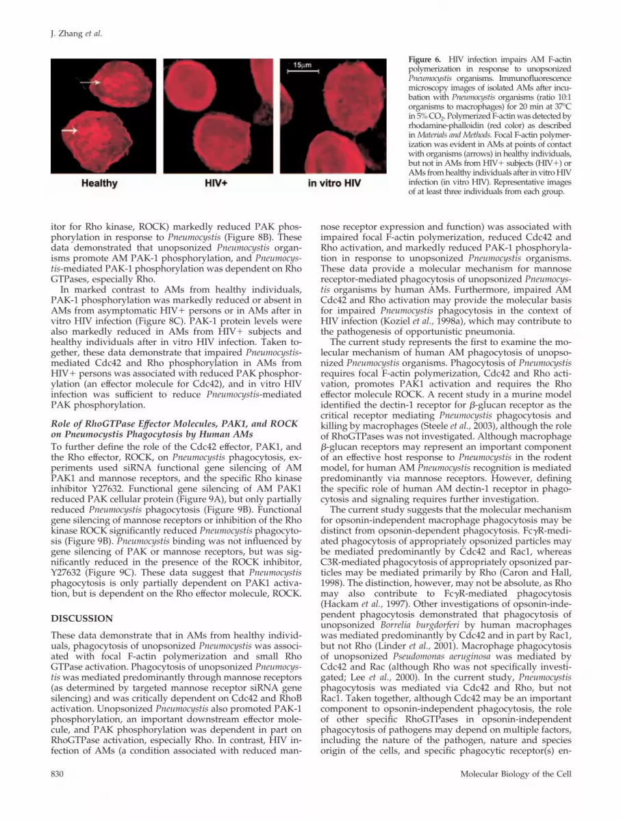
response to nonopsonized Pneumocystis using AMs fromHIV� persons (recognized clinical risk factor for Pneumocys-tis pneumonia). Compared with AMs from healthy individ-uals, incubation of unopsonized Pneumocystis organismswith AMs from asymptomatic HIV� persons did not inducefocal F-actin polymerization at points of contact (Figure 6).Similarly, unopsonized Pneumocystis organisms did not in-duce F-actin polymerization in AMs from healthy individu-als after in vitro HIV infection. These data demonstrate thatPneumocystis-induced F-actin assembly is impaired in AMsfrom HIV� persons, and HIV infection is sufficient to impairPneumocystis-mediated F-actin polymerization in AMs.
HIV Infection Impairs Cdc42 and Rho Activation in AMsin Response to Pneumocystis OrganismsTo test the hypothesis that altered mannose receptor expres-sion may influence Pneumocystis-mediated small GTPase ac-tivation, experiments next examined Cdc42, Rac1, and Rhoactivation in AMs from HIV� subjects, and in AMs fromhealthy individuals after in vitro HIV infection (conditionsassociated with reduced mannose receptor expression andfunction).
Cdc42 protein was detected in the cytoplasm of intactcells and cytosolic extracts from all three groups of AMsstudied (Figure 7). For AMs from healthy individuals,Cdc42 activation was increased in response to unopso-nized Pneumocystis organisms, in addition to zymosan(alternative ligand for mannose receptors) and PMA(mannose receptor-independent ligand). In comparison,AMs from HIV� persons demonstrated reduced Cdc42activation in response to Pneumocystis organisms (andalso reduced in response to zymosan and PMA). After invitro HIV infection, Cdc42 activation was completely in-hibited in response to Pneumocystis organisms and zymo-san and markedly reduced in response to PMA.
In comparison, Rac1 activation was not significantly im-paired in response to Pneumocystis, zymosan, or PMA inAMs from HIV� persons, although Rac1 activation wascompletely impaired in response to Pneumocystis and zymo-san (but not PMA) after in vitro HIV infection. In contrast,Rho activation was not increased in response to Pneumocystisorganisms (or zymosan and PMA) in AMs from HIV� per-sons or after in vitro HIV infection. These data demonstratedthat Cdc42 and Rho activation was reduced in AMs fromHIV� persons in response to unopsonized Pneumocystis or-ganisms, whereas Rac1 activation was not significantly re-duced. Furthermore, in vitro HIV infection of AMs (a con-dition associated with marked reduction in mannosereceptor expression and function; Koziel et al., 1998a) wassufficient to impair Pneumocystis-mediated Cdc42, Rac1, andRho activation.
HIV Infection Impairs p21-activated KinasePhosphorylation in Response to Pneumocystis OrganismsThe p21-activated kinases (PAK) are important serine/thre-onine signaling molecules regulating a variety of cellularfunction (including cytoskeletal dynamics), and PAK arestimulated by activated Cdc42 and Rac (Bokoch, 2003). Todetermine the downstream effects of Pneumocystis-mediatedCdc42, Rac1, and Rho activation, experiments next mea-sured PAK phosphorylation in response to unopsonizedPneumocystis. Incubation of AMs from healthy individualswith unopsonized Pneumocystis organisms increased PAK-1phosphorylation (activated PAK) in a time-dependent man-ner, detected at 10 min and peaked at 15 min (Figure 8A).Pretreatment of the AMs with C. difficile toxin B (generalinhibitor of Rho family GTPases) or Y-27632 (specific inhib-
Figure 5. Cdc42 and RhoB activation mediated through AM mannosereceptors. Targeted AM mannose receptor gene silencing by siRNA fol-lowed by incubation with unopsonized Pneumocystis for 15 min at 37°C in5% CO2. (A) Reduced mannose receptor total cellular protein in AMs aftersiRNA gene silencing. Mannose receptor protein was not influenced bysingle-stranded MR siRNA, random siRNA, or LAM A/C siRNA (servedas negative controls). PAK1 siRNA represents gene silencing of AM PAK1(which did not reduce mannose receptor and demonstrates specificity).(B–D) Influence of Pneumocystis on AM Rho GTPase activation after man-nose receptor gene silencing. Western blot of cell lysates performed todetect both total and activated (�GTP) forms of (B) Cdc42, (C) Rac1, and(D) RhoB. Western blots are shown for AMs from one healthy individualand are representative of experiments from at least three different individ-uals. Corresponding bar graphs represent densitometry quantitative anal-ysis for Cdc42-GTP, Rac1-GTP, and RhoB-GTP (mean SEM; n � 3)expressed as relative intensity units (RIU).
Cdc42 and RhoB Mediate Mannose Receptor Phagocytosis
Vol. 16, February 2005 829
itor for Rho kinase, ROCK) markedly reduced PAK phos-phorylation in response to Pneumocystis (Figure 8B). Thesedata demonstrated that unopsonized Pneumocystis organ-isms promote AM PAK-1 phosphorylation, and Pneumocys-tis-mediated PAK-1 phosphorylation was dependent on RhoGTPases, especially Rho.
In marked contrast to AMs from healthy individuals,PAK-1 phosphorylation was markedly reduced or absent inAMs from asymptomatic HIV� persons or in AMs after invitro HIV infection (Figure 8C). PAK-1 protein levels werealso markedly reduced in AMs from HIV� subjects andhealthy individuals after in vitro HIV infection. Taken to-gether, these data demonstrate that impaired Pneumocystis-mediated Cdc42 and Rho phosphorylation in AMs fromHIV� persons was associated with reduced PAK phosphor-ylation (an effector molecule for Cdc42), and in vitro HIVinfection was sufficient to reduce Pneumocystis-mediatedPAK phosphorylation.
Role of RhoGTPase Effector Molecules, PAK1, and ROCKon Pneumocystis Phagocytosis by Human AMsTo further define the role of the Cdc42 effector, PAK1, andthe Rho effector, ROCK, on Pneumocystis phagocytosis, ex-periments used siRNA functional gene silencing of AMPAK1 and mannose receptors, and the specific Rho kinaseinhibitor Y27632. Functional gene silencing of AM PAK1reduced PAK cellular protein (Figure 9A), but only partiallyreduced Pneumocystis phagocytosis (Figure 9B). Functionalgene silencing of mannose receptors or inhibition of the Rhokinase ROCK significantly reduced Pneumocystis phagocyto-sis (Figure 9B). Pneumocystis binding was not influenced bygene silencing of PAK or mannose receptors, but was sig-nificantly reduced in the presence of the ROCK inhibitor,Y27632 (Figure 9C). These data suggest that Pneumocystisphagocytosis is only partially dependent on PAK1 activa-tion, but is dependent on the Rho effector molecule, ROCK.
DISCUSSION
These data demonstrate that in AMs from healthy individ-uals, phagocytosis of unopsonized Pneumocystis was associ-ated with focal F-actin polymerization and small RhoGTPase activation. Phagocytosis of unopsonized Pneumocys-tis was mediated predominantly through mannose receptors(as determined by targeted mannose receptor siRNA genesilencing) and was critically dependent on Cdc42 and RhoBactivation. Unopsonized Pneumocystis also promoted PAK-1phosphorylation, an important downstream effector mole-cule, and PAK phosphorylation was dependent in part onRhoGTPase activation, especially Rho. In contrast, HIV in-fection of AMs (a condition associated with reduced man-
nose receptor expression and function) was associated withimpaired focal F-actin polymerization, reduced Cdc42 andRho activation, and markedly reduced PAK-1 phosphoryla-tion in response to unopsonized Pneumocystis organisms.These data provide a molecular mechanism for mannosereceptor-mediated phagocytosis of unopsonized Pneumocys-tis organisms by human AMs. Furthermore, impaired AMCdc42 and Rho activation may provide the molecular basisfor impaired Pneumocystis phagocytosis in the context ofHIV infection (Koziel et al., 1998a), which may contribute tothe pathogenesis of opportunistic pneumonia.
The current study represents the first to examine the mo-lecular mechanism of human AM phagocytosis of unopso-nized Pneumocystis organisms. Phagocytosis of Pneumocystisrequires focal F-actin polymerization, Cdc42 and Rho acti-vation, promotes PAK1 activation and requires the Rhoeffector molecule ROCK. A recent study in a murine modelidentified the dectin-1 receptor for �-glucan receptor as thecritical receptor mediating Pneumocystis phagocytosis andkilling by macrophages (Steele et al., 2003), although the roleof RhoGTPases was not investigated. Although macrophage�-glucan receptors may represent an important componentof an effective host response to Pneumocystis in the rodentmodel, for human AM Pneumocystis recognition is mediatedpredominantly via mannose receptors. However, definingthe specific role of human AM dectin-1 receptor in phago-cytosis and signaling requires further investigation.
The current study suggests that the molecular mechanismfor opsonin-independent macrophage phagocytosis may bedistinct from opsonin-dependent phagocytosis. Fc�R-medi-ated phagocytosis of appropriately opsonized particles maybe mediated predominantly by Cdc42 and Rac1, whereasC3R-mediated phagocytosis of appropriately opsonized par-ticles may be mediated primarily by Rho (Caron and Hall,1998). The distinction, however, may not be absolute, as Rhomay also contribute to Fc�R-mediated phagocytosis(Hackam et al., 1997). Other investigations of opsonin-inde-pendent phagocytosis demonstrated that phagocytosis ofunopsonized Borrelia burgdorferi by human macrophageswas mediated predominantly by Cdc42 and in part by Rac1,but not Rho (Linder et al., 2001). Macrophage phagocytosisof unopsonized Pseudomonas aeruginosa was mediated byCdc42 and Rac (although Rho was not specifically investi-gated; Lee et al., 2000). In the current study, Pneumocystisphagocytosis was mediated via Cdc42 and Rho, but notRac1. Taken together, although Cdc42 may be an importantcomponent to opsonin-independent phagocytosis, the roleof other specific RhoGTPases in opsonin-independentphagocytosis of pathogens may depend on multiple factors,including the nature of the pathogen, nature and speciesorigin of the cells, and specific phagocytic receptor(s) en-
Figure 6. HIV infection impairs AM F-actinpolymerization in response to unopsonizedPneumocystis organisms. Immunofluorescencemicroscopy images of isolated AMs after incu-bation with Pneumocystis organisms (ratio 10:1organisms to macrophages) for 20 min at 37°Cin 5% CO2. Polymerized F-actin was detected byrhodamine-phalloidin (red color) as describedin Materials and Methods. Focal F-actin polymer-ization was evident in AMs at points of contactwith organisms (arrows) in healthy individuals,but not in AMs from HIV� subjects (HIV�) orAMs from healthy individuals after in vitro HIVinfection (in vitro HIV). Representative imagesof at least three individuals from each group.
J. Zhang et al.
Molecular Biology of the Cell830

gaged. Further studies are required to completely define therole of specific RhoGTPases in opsonin-independent phago-cytosis.
The current study represents the first to demonstratephagocytic signal transduction pathways mediated by man-nose receptors. Mannose receptors belong to a family of type
Figure 7. HIV infection impairs AM Cdc42and Rho activation in response to unopso-nized Pneumocystis organisms. AMs wereincubated with unopsonized Pneumocystisorganisms (Pc), zymosan (Zy), or PMA for15 min at 37°C in 5% CO2. AMs were fromhealthy individuals, asymptomatic HIV-in-fected persons (HIV�), or healthy individ-uals after in vitro HIV infection (in vitroHIV). Immunofluorescence microscopy im-ages demonstrated similar cellular localiza-tion for (A) Cdc42, (D) Rac1, and (G) Rhowith representative cells for healthy, HIV�,and in vitro HIV. Western blot of cell lysateswas performed to detect both total and ac-tivated (�GPT) forms of Cdc42 (B), Rac1 (E),and Rho (H) for healthy, HIV�, and in vitroHIV. The corresponding bar graphs repre-sent densitometry quantitative analysis forCdc42-GTP (C), Rac1-GTP (F), and Rho-GTP(I) for each group of healthy, HIV�, and invitro HIV. Data are normalized as a ratio ofthe active (�GTP) RhoGTPase over the totalRhoGTPase and are expressed as relativeunits (mean SEM). Data are representa-tive of experiments from three different in-dividuals in each group (n � 3).
Cdc42 and RhoB Mediate Mannose Receptor Phagocytosis
Vol. 16, February 2005 831

I transmembrane C-type lectin receptors that mediate bothendocytosis and phagocytosis (Ezekowitz, 1992). Mannosereceptors contain multiple C-type lectin-like domains eachcapable of binding specific monosaccharides such as man-nose, fucose, and N-acetylglucosamine. Although endocyto-sis requires signaling through a conserved tyrosine-basedFENTLY motif in the cytoplasmic tail of the receptor (Eastand Isacke, 2002), the signaling molecules mediating phago-cytosis and the downstream cell signaling pathways remainundefined (Fraser et al., 1998). The current study demon-strates that mannose receptor-mediated phagocytosis wasassociated with focal F-actin polymerization, dependent onCdc42 and Rho activation, and dependent on the Rho effec-tor molecule, Rock. Rac1 activation was not critical for Pneu-mocystis phagocytosis (as introduction of nonfunctional Rac1dominant-negative allele did not influence Pneumocystisphagocytosis). The relative importance of mannose recep-tors in mediating Pneumocystis phagocytosis by human AMs
was supported by targeted siRNA gene silencing experi-ments and the observations in AMs from HIV-infected per-sons (a disease state characterized by reduced macrophagemannose receptor surface expression; Koziel et al., 1998a).
Findings in the current study suggests that the initialinteraction of unopsonized Pneumocystis organisms withAMs promotes rapid Cdc42 activation, in addition to Rhoactivation (early portion of biphasic response), followed by arelatively delayed Rac1 activation, and further Rho activa-tion (delayed portion of biphasic response). The data sug-gest the early activation of Cdc42 and Rho are mediatedpredominantly through mannose receptors (as supported bythe siRNA gene silencing experiments) and that the delayedresponses of Rac and Rho were mannose receptor indepen-dent (and perhaps mediated by other macrophages recep-tors such as dectin-1; Brown and Gordon, 2001; Steele et al.,2003), although additional experiments are required to fur-ther define these specific events.
Figure 8. HIV infection impairs PAK-1 ac-tivation in response to Pneumocystis organ-isms. (A) AMs from healthy individualswere incubated with unopsonized Pneumo-cystis (5:1) up to 60 min at 37°C in 5% CO2,and cell lysates were examined for phos-phorylated PAK1 (p-PAK1) by Westernblot. LPS served as a positive control forPAK1 phosphorylation. (B) AMs fromhealthy individuals were incubated withunopsonized Pneumocystis (5:1) or zymosan(5:1) in the presence of absence of Y26371(the specific inhibitor of Rho kinase, Rock)or C. diff toxin B (CDT; general RhoGTPaseinhibitor) for 15 min at 37°C in 5% CO2, andcell lysates were examined for total (PAK1)and phosphorylated PAK1 (p-PAK1) byWestern blot using specific antibody. (C)AMs were incubated with Pneumocystis (Pc),zymosan (Zy), or PMA for 15 min at 37°C in5% CO2, and Western blots on cell lysateswere performed to detect both total (PAK1)and phosphorylated PAK1 (p-PAK1) inAMs from healthy, HIV�, and healthy afterin vitro HIV. For all Western blots, the cor-responding bar graphs represent densitom-etry quantitative analysis for p-PAK1 foreach group of healthy, HIV�, and in vitroHIV expressed as relative intensity units(RIU; mean SEM). The depicted Westernblots represent cells from one healthy indi-vidual and are representative of at leastthree different subjects examined.
J. Zhang et al.
Molecular Biology of the Cell832

The mechanism for impaired RhoGTPase activation inHIV infection was not completely defined. HIV infectionwas sufficient to impair RhoGTPase activation in responseto Pneumocystis. Impaired Rho GTPase activation may inpart be attributed to reduced mannose receptor surfaceexpression, based on reduced mRNA (Koziel et al., 1998a)or through increased release of the mannose receptorectodomain (Fraser et al., 2000). Alternatively, impairedRhoGTPase activation may be due to direct influence byHIV gene products that can influence cellular receptorexpression and signal transduction pathways (Emermanand Malim, 1998). HIV-1 tat may down-regulate mannosereceptor transcription (Caldwell et al., 2000). HIV-1 vpuimpedes NF-�B activation by inhibiting I-�B degradation(Bour et al., 2001) in T-cell lines. HIV-1 nef may alter ASK1signal transduction pathways (Geleziunas et al., 2001) anddown-regulate NF-�B signaling in T-cell lines (Bandresand Ratner, 1994), reduce mannose receptor surface ex-pression on dendritic cells (Quaranta et al., 2002), anddown-modulate Fc�-R expression on monocyte-derivedmacrophages (De et al., 1998). The influence of specificHIV-1 gene products on AM RhoGTPase signal transduc-tion pathways has not been previously investigated. Theobserved alteration in Cdc42, Rac1, and Rho in the cur-rent study may thus reflect the influence of one or moreHIV-related factors and represents an area of activeinvestigation.
The role of other receptors implicated in the AM recogni-tion of Pneumocystis, including fibronectin (Pottratz and
Martin, 1990) and immunoglobulin receptors (Masur andJones, 1978; von Behren and Pesanti, 1978) were not specif-ically investigated. The observed Cdc42 and Rac phosphor-ylation in response to IgG-opsonized Pneumocystis in AMswith targeted gene silencing of the mannose receptor sug-gests that the Fc�R pathway was intact. Other limitations ofthese studies include the possibility that the observed re-sponse of human AMs to rodent-derived Pneumocystis mayreflect in part the origin of the organisms. Unfortunately, inthe absence of reliable culturing methods, Pneumocystis ofhuman origin are not available (Sloand et al., 1993). How-ever, the recent observation that rat-derived Pneumocystiselicit similar chemokine and NF-�B responses comparinghuman AMs to rat AMs in part validates the use of thismodel (Zhang et al., 2004). Contaminating rat-derived ad-herent proteins may be in part responsible for the observedRhoGTPase activity, although the Pneumocystis organismsused in these studies are relatively free of host proteins suchas surfactant protein-A (Koziel et al., 1998b). Because thePneumocystis preparation is composed primarily of the tro-phic forms, the current findings may represent the predom-inant macrophage response to the Pneumocystis trophic formand not the cyst or other form. Finally, the possibility that invitro observations may not accurately reflect events in vivoshould be considered.
In conclusion, these data demonstrate that in AMs fromhealthy individuals, phagocytosis of unopsonized Pneumo-cystis was mediated predominantly through mannose recep-tors and was critically dependent on Cdc42 and especially
Figure 9. The role of RhoGTPase effectormolecules, PAK1 and ROCK, on Pneumocystisphagocytosis by human AMs. (A) Westernblot analysis of cellular PAK1 protein in hu-man AMs after siRNA gene silencing. PAK1protein was not influenced by single-strandedPAK siRNA, random siRNA, or LAM A/CsiRNA (served as negative controls). MRsiRNA represents gene silencing of AM man-nose receptor (which did not reduce PAK1and demonstrates specificity). Pneumocystisphagocytic index (B) and binding index (C) ofAMs after functional siRNA gene silencing ofPAK1 (PAK1 siRNA), mannose receptor (MRsiRNA), or specific pharmacological inhibi-tion of the Rho kinase ROCK (by Y27632).Phagocytic Index and Binding Index ex-pressed as relative fluorescence intensity (RFI;values are mean SEM; n � 3) *p � 0.05compared with control.
Cdc42 and RhoB Mediate Mannose Receptor Phagocytosis
Vol. 16, February 2005 833

RhoB activation. These data provide a molecular mechanismfor mannose receptor-mediated phagocytosis of unopso-nized Pneumocystis organisms, and the mechanism is distinctfrom opsonin-dependent phagocytic receptors. Further-more, impaired AM Cdc42 and Rho activation may providethe molecular basis for impaired Pneumocystis phagocytosisin AMs from HIV� persons (Koziel et al., 1998a). Recogniz-ing the spectrum of pathogens recognized by mannose re-ceptors (including Mycobacterium tuberculosis, Cryptococcusneoformans, and Pneumocystis), impaired AM mannose recep-tor-mediated phagocytosis in the context of HIV infectionmay contribute to the pathogenesis of opportunistic pneu-monia.
ACKNOWLEDGMENTS
We acknowledge the participation of all persons who consented to bronchos-copy and the technical assistance of Robert Garland, Rene Andwood, andLorraine Gryniuk. We acknowledge the generous gift of Martine Armstrongand Frank Richards, who provided Pneumocystis organisms for initial studies.We also are indebted to Naimish Patel and Souvenir Tachado for their criticalreview of the manuscript and valuable suggestions. None of the authors haveconflict of interest disclosures regarding the work in this study. This studywas supported by National Institutes of Health research grants RO1 HL63655(H.K.) and F32 HL71372 (J.Z.).
REFERENCES
Aderem, A. (2002). How to eat something bigger than your head. Cell 110,5–8.
Armstrong, M.Y.K., Koziel, H., Rose, R. M., Arena, C., and Richards, F. F.(1991). Indicators of Pneumocystis carinii viability in short-term culture. J.Protozool. 38, 88S–90S.
Bandres, J. C., and Ratner, L. (1994). Human immunodeficiency virus type 1nef protein down-regulates transcription factors NF-kB and AP-1 in human Tcells in vitro after T-cell receptor stimulation. J. Virol. 68, 3243–3249.
Benard, V., Bohl, B. P., and Bokoch, G. M. (1999). Characterization of Rac andCdc42 activation in chemoattractant-stimulated human neutrophils using anovel assay for active GTPases. J. Biol. Chem. 274, 13198–13204.
Bokoch, G. M. (2003). Biology of the p21-activated kinases. Annu. Rev. Bio-chem. 72, 743–781.
Bour, S., Perrin, C., Akari, H., and Strebel, K. (2001). The human immunode-ficiency virus type 1 protein inhibits NF-kB activation by interfering withbeta-TrCP-mediated degradation of IkB. J. Biol. Chem. 276, 15920–15928.
Brown, G. D., and Gordon, S. (2001). A new receptor for beta-glucans. Nature413, 36–37.
Caldwell, R. L., Egan, B. S., and Shephard, V. L. (2000). HIV-1 tat repressestranscription from the mannose receptor promoter. J. Immunol. 165, 7035–7041.
Caron, E., and Hall, A. (1998). Identification of two distinct mechanisms ofphagocytosis controlled by different Rho GTPases. Science 282, 1717–1721.
Chen, F., and Cushion, M. (1994). Use of an ATP bioluminescent assay toevaluate viability of Pneumocystis carinii from rats. J. Clin. Microbiol. 32,2791–2800.
De, S. K., Venkateshan, C.N.S., Seth, P., Gajdusek, D. C., and Gibbs, C. J.(1998). Adenovirus-mediated human immunodeficiency virus-1 nef expres-sion in human monocyte-macrophages and effect of nef on downmodulationof Fc-gamma receptors and expression of monokines. Blood 91, 2108–2117.
East, L., and Isacke, C. M. (2002). The mannose receptor family. Biochim.Biophys. Acta 1572, 364–386.
Emerman, M., and Malim, M. H. (1998). HIV-1 regulatory/accessory genes:keys to unraveling viral and host cell biology. Science 280, 1880–1884.
Etienne-Manneville, S., and Hall, A. (2002). Rho GTPases in cell biology.Nature 420, 629–635.
Ezekowitz, R.A.B. (1992). The mannose receptor and phagocytosis. In: Mono-nuclear Phagocytes, ed. R.v. Furth, Netherlands: Kluwer Academic Publish-ers, 208–213.
Ezekowitz, R.A.B., Williams, D. J., Koziel, H., Armstong, M.Y.K., Warner, A.,Richards, F. F., and Rose, R. M. (1991). Uptake of Pneumocystis carinii medi-ated by the macrophage mannose receptor. Nature 351, 155–158.
Fraser, I. P., Koziel, H., and Ezekowitz, R.A.B. (1998). The serum mannose-binding protein and the macrophage mannose receptor are pattern recogni-tion molecules that link innate and adaptive immunity. Semin. Immunol. 10,363–372.
Fraser, I. P., Takahashi, K., Koziel, H., Fardin, B., Harmsen, A., and Ezekowitz,R.A.B. (2000). Pneumocystis carinii enhances the production of soluble man-nose receptor by macrophages. Microbes Infect. 2, 1305–1310.
Geleziunas, R., Xu, W., Takeda, K., Ichijo, H., and Greene, W. C. (2001). HIV-1Nef inhibits ASK1-dependent death signaling providing a potential mecha-nism for protecting the infected host cell. Nature 410, 834–838.
Greenberg, S., and Grinstein, S. (2002). Phagocytosis and innate immunity.Curr. Opin. Immunol. 14, 136–145.
Hackam, D. J., Rotstein, O. D., Schreiber, A., Zhang, W. J., and Grinstein, S.(1997). Rho is required for the initiation of calcium signaling and phagocytosisby Fc-gamma receptors in macrophages. J. Exp. Med. 186, 955–966.
Hoffmann, J. A., Kafatos, F. C., Janeway, C. A., and Ezekowitz, R.A.B. (1999).Phylogenetic perspectives in innate immunity. Science 284, 1313–1318.
Koziel, H., Eichbaum, Q., Kruskal, B. A., Pinkston, P., Rogers, R. A., Arm-strong, M.Y.K., Richards, F. F., Rose, R. M., and Ezekowitz, R.A.B. (1998a).Reduced binding and phagocytosis of Pneumocystis carinii by alveolar macro-phages from persons infected with HIV-1 correlates with mannose receptordownregulation. J. Clin. Invest. 102, 1332–1344.
Koziel, H., Phelps, D. S., Fishman, J. A., Armstrong, M.Y.K., Richards, F. F.,and Rose, R. M. (1998b). Surfactant protein-A reduces binding and phagocy-tosis of Pneumocystis carinii by human alveolar macrophages in vitro. Am. J.Respir. Cell Mol. Biol. 18, 834–843.
Lee, D. J., Cox, D., Li, J., and Greenberg, S. (2000). Rac1 and Cdc42 requiredfor phagocytosis, but not NF-kB dependent gene expression, in macrophageschallenged with Pseudomonas aeruginosa. J. Biol. Chem. 275, 141–146.
Limper, A. H., Hoyte, J. S., and Standing, J. E. (1997). The role of alveolarmacrophages in Pneumocystis carinii degradation and clearance from the lung.J. Clin. Invest. 99, 2110–2117.
Linder, S., Heimerl, C., Fingerle, V., Aepfelbacher, M., and Wilske, B. (2001).Coiling phagocytosis of Borrelia burgdorferi by primary human macrophages iscontrolled by Cdc42Hs and Rac1 and involves recruitment of Wiskott-AldrichSyndrome Protein and Arp2/3 complex. Infect. Immun. 69, 1739–1746.
Masur, H., and Jones, T. C. (1978). The interaction in vitro of Pneumocystiscarinii with macrophages and L-cells. J. Exp. Med. 147, 157–170.
Miralem, T., and Avraham, H. K. (2003). Extracellular matrix enhancesheregulin-dependent BRCA1 phosphorylation and suppresses BRCA1 ex-pression through its C terminus. Mol. Cell. Biol. 23, 579–593.
Pottratz, S. T., and Martin, W. J. (1990). Mechanism of Pneumocystis cariniiattachment to cultured rat alveolar macrophages. J. Clin. Invest. 86, 1678–1683.
Quaranta, M. G., Tritarelli, E., Giordani, L., and Viora, M. (2002). HIV-1 nefinduces dentritic cell differentiation: a possible mechanism of uninfectedCD4� T cell activation. Exp. Cell Res. 275, 243–254.
Ren, X. D., Kiosses, W. B., and Schwartz, M. A. (1999). Regulation of the smallGTP-binding protein Rho by cell adhesion and the cytoskeleton. EMBO J. 18,578–585.
Salahuddin, S. Z., Rose, R. M., Groopman, J. E., Markham, P. D., and Gallo,R. C. (1986). Human T lymphotropic virus type III infection of human alveolarmacrophages. Blood 68, 281–284.
Sloand, E. et al. (1993). The challenge of Pneumocystis carinii culture. J. Euk.Microbiol. 40, 188–195.
Steele, C., Marrero, L., Swain, S., Harmsen, A. G., Zheng, M., Brown, G. D.,Gordon, S., Shellito, J. E., and Kolls, J. K. (2003). Alveolar macrophage-mediated killing of Pneumocystis carinii f. sp. muris involves molecular rec-ognition by the dectin-1 beta-glucan receptor. J. Exp. Med. 198, 1677–1688.
Stringer, J. R., Beard, C. B., Miller, R. F., and Wakefield, A. E. (2002). A newname (Pneumocystis jiroveci) for Pneumocystis from humans. Emerging Infect.Dis. 8, 891–896.
Uehata, M. et al. (1997). Calcium sensitization of smooth muscle mediated bya Rho-associated protein kinase in hypertension. Nature 389, 990–994.
von Behren, L. A., and Pesanti, E. L. (1978). Uptake and degradation ofPneumocystis carinii by macrophages in vitro. Am. Rev. Respir. Dis. 118,1051–1059.
Zhang, J. M., Zhu, J., Imrich, A., Cushion, M. T., Kinane, B. T., and Koziel, H.(2004). Pneumocystis activates human alveolar macrophage NF-kB signalingthrough mannose receptors. Infect. Immun. 72, 3147–3160.
J. Zhang et al.
Molecular Biology of the Cell834


