Embed Size (px)
Citation preview

Muscle Dystroglycan Organizes the Postsynapse andRegulates Presynaptic Neurotransmitter Release at theDrosophila Neuromuscular JunctionLaurent Bogdanik1,2,3¤, Berenice Framery1,2,3, Andreas Frolich4, Benedicte Franco1,2,3, Dominique
Mornet6, Joel Bockaert1,2,3, Stephan J. Sigrist4,5, Yves Grau1,2,3, Marie-Laure Parmentier1,2,3*
1 CNRS, UMR 5203, Institut de Genomique fonctionnelle, Montpellier, France, 2 INSERM, U661, Montpellier, France, 3 Universite Montpellier,1,2, Montpellier, France,
4 European Neuroscience Institute Gottingen, Gottingen, Germany, 5 Institut fur Klinische Neurobiologie, Rudolf-Virchow Zentrum, Universitat Wurzburg, Wurzburg,
Germany, 6 INSERM, ERI 25, Muscle and Pathologies, Universite de Montpellier1, EA 4202, 34295 Montpellier, France
Abstract
Background: The Dystrophin-glycoprotein complex (DGC) comprises dystrophin, dystroglycan, sarcoglycan, dystrobrevinand syntrophin subunits. In muscle fibers, it is thought to provide an essential mechanical link between the intracellularcytoskeleton and the extracellular matrix and to protect the sarcolemma during muscle contraction. Mutations affecting theDGC cause muscular dystrophies. Most members of the DGC are also concentrated at the neuromuscular junction (NMJ),where their deficiency is often associated with NMJ structural defects. Hence, synaptic dysfunction may also intervene in thepathology of dystrophic muscles. Dystroglycan is a central component of the DGC because it establishes a link between theextracellular matrix and Dystrophin. In this study, we focused on the synaptic role of Dystroglycan (Dg) in Drosophila.
Methodology/Principal Findings: We show that Dg was concentrated postsynaptically at the glutamatergic NMJ, where,like in vertebrates, it controls the concentration of synaptic Laminin and Dystrophin homologues. We also found thatsynaptic Dg controlled the amount of postsynaptic 4.1 protein Coracle and alpha-Spectrin, as well as the relative subunitcomposition of glutamate receptors. In addition, both Dystrophin and Coracle were required for normal Dg concentration atthe synapse. In electrophysiological recordings, loss of postsynaptic Dg did not affect postsynaptic response, but,surprisingly, led to a decrease in glutamate release from the presynaptic site.
Conclusion/Significance: Altogether, our study illustrates a conservation of DGC composition and interactions betweenDrosophila and vertebrates at the synapse, highlights new proteins associated with this complex and suggests anunsuspected trans-synaptic function of Dg.
Citation: Bogdanik L, Framery B, Frolich A, Franco B, Mornet D, et al. (2008) Muscle Dystroglycan Organizes the Postsynapse and Regulates PresynapticNeurotransmitter Release at the Drosophila Neuromuscular Junction. PLoS ONE 3(4): e2084. doi:10.1371/journal.pone.0002084
Editor: Antoni L. Andreu, Hospital Vall d’Hebron, Spain
Received March 3, 2008; Accepted March 17, 2008; Published April 30, 2008
Copyright: � 2008 Bogdanik et al. This is an open-access article distributed under the terms of the Creative Commons Attribution License, which permitsunrestricted use, distribution, and reproduction in any medium, provided the original author and source are credited.
Funding: This work was supported by AFM fellowships to L. Bogdanik and B. Franco, the AFM grant 10808 MNM2 2004 Subvention Groupe C and the ANR grantJC05_56382 to M.-L. Parmentier. Sponsors and funders played no role in the design and conduct of the study, in the collection, analysis, and interpretation of thedata, and in the preparation, review, or approval of the manuscript.
Competing Interests: The authors have declared that no competing interests exist.
* E-mail: [email protected]
¤ Current address: The Jackson Laboratory, Bar Harbor, Maine, United States of America
Introduction
In muscle fibers, the Dystrophin-glycoprotein complex (DGC) is
thought to provide an essential mechanical link between the
intracellular cytoskeleton and the extracellular matrix. This
complex comprises Dystroglycan subunits (a and b), Dystrophin
or Utrophin, sarcoglycans subunits, syntrophin subunits, and
dystrobrevin subunits. Dystroglycan (Dg) is a central component of
the DGC because it establishes the transmembrane link between
Laminins and Dystrophin [1,2]. Dg is encoded by a single gene
(dag1) [3], and is expressed as a propeptide that gets cleaved in two
fragments: a and b-Dg, which associate non-covalently in mature
skeletal muscle. The extracellular Dg a-subunit is heavily
glycosylated, and interacts with extracellular Laminin, whereas
the transmembrane b-subunit interacts with subsarcolemmal
Dystrophin, which itself links the actin network. The maintenance
of this structural link provides stability of the sarcolemma,
especially upon muscle contraction. In Duchenne muscular
dystrophy, mutation and subsequent loss of Dystrophin destabi-
lizes the other DGC members [4], which further leads to
mechanical damage of the muscle cell membrane [5,6]. There
are no known dystrophies associated with a mutation in the
dystroglycan gene, probably because of the importance of the
encoded protein in other cellular types. Indeed, loss of Dg in miceleads to early embryonic death [7]. However, mutations in
enzymes responsible for the glycosylation of a-Dg are the cause of
congenital myopathies such as the Walker-Warburg syndrome, the
muscle-eye-brain disease and Fukuyama-type congenital muscular
dystrophy, for review [8,9].
Most members of the DGC are found to be concentrated at the
cholinergic neuromuscular junction. Moreover, mouse studies
revealed abnormalities of NMJs lacking DGC components [10-15]
PLoS ONE | www.plosone.org 1 April 2008 | Volume 3 | Issue 4 | e2084

suggesting a role for a defective neurotransmission in some
dystrophies [16,17]. For example, NMJs without muscle Dg show
decreased levels of synaptic Laminin and Utrophin, as well as
smaller acetylcholine receptor clusters [17–20]. A more general
function of the DGC in synaptic transmission is further supported
by the localization of DGC components in central brain synapses
[21–23] and the occurrence of mental retardation in myopathic
patients that do not show any major brain structural defect [24]. In
this study we focused on the synaptic role of Dg, using the
Drosophila glutamatergic larval NMJ. This synapse is not only a
neuromuscular junction, but also a well-established model to study
the development, the structure and the function of a glutamatergic
synapse [25]. We showed that Dg is concentrated postsynaptically
at this NMJ. We analyzed the status of Dg partners at this synapse
and found that 1) like in vertebrates, Dg controls synaptic Laminin
and Dystrophin concentration, 2) Dg also controls the amount of
postsynaptic 4.1 protein Coracle, the postsynaptic spectrin
cytoskeleton, and the relative subunit composition of glutamate
receptors, and 3) reciprocally, both Dystrophin and Coracle are
required for Dg concentration at the synapse. Finally, electro-
physiological analysis shows that loss of muscle Dg leads to a
functional defect, i.e. a decrease in presynaptic glutamate release.
Materials and Methods
Fly stocksywCS flies were used as a control in all experiments with dg or cora
mutants. dg-RNAi/+ or UAS-Dg-C/+ flies were used as controls
when studying crosses with these transgenes. The 24B-Gal4 line was
used for all muscle expression experiments [26]. dg323 and dg62
alleles as well as UAS-Dg-C transgenic flies have been previously
described [27]. The genotype of larvae overexpressing Dg-C in our
experiments is 24BGal4/UAS-Dg-C. We also used the piggybac
insertion dge01554 [28]. The dg directed RNAi construct [27]
originally on chromosome 3 was remobilized on chromosome 2
(line nu12), and flies containing the two RNAi transgenes over wild-
type chromosomes were used for all immunocytochemistry
experiments. We created a UAS-Dg-C-GFP construct using the
Gateway system and by inserting the PCR amplified sequence of Dg
(LD11619) in ptWG. The Dys-COOH RNAi transgenic flies were
described in [29]. The hypomorph cora14 allele was described in
[30]. This mutation leads to the replacement of R1607 by a stop
codon. The null cora[k08713] allele was analyzed in [31]. The cora
alleles as well as The P{EPgy2}cora[EY07598] containing stock
were obtained from the Bloomington Drosophila Stock Center.
Green fluorescent protein or Tubby balancers were used to identify
homozygous individuals. All crosses were performed at 25uC except
those with RNAi which were set up at 25uC for three days (to allow
egg-laying) and transferred at 29uC.
Generation of polyclonal antibodyThe Dystroglycan C-terminal polyclonal antibody (LG5) was
raised in New Zealand rabbits by repeated intra dermal injections.
Synthetic peptide of the last 7 C-terminal amino acids of Human
Dystroglycan (PPPVYPP) was conjugated via a cysteine residue
associated to hexanoıc acid to the keyhole limpet hemocyanin and
such KLH-linked peptide was used as antigen according to a
previously described protocol. The resulting polyclonal antibody
was purified and characterized as previously described [32]. Note
the Drosophila 7 C-term sequence is PPPVYSP.
ImmunocytochemistryLarvae were dissected in PBS 1x, EDTA 1mM, and then fixed
in fresh 4% paraformaldehyde (Sigma, L’isle d’Abeau, France) for
20 min. for all stainings, apart from GluRIIA and GluRIIC ones.
For these stainings, preparations were fixed for 15 min. in Bouin’s
fixative (Sigma, L’isle d’Abeau, France). Antibody incubations
were performed in PBS 1x buffer with 0.3% Triton X100 and 0.2
to 1% BSA. The following antibodies were used: Goat anti-HRP
(Sigma, L’isle d’Abeau, France, 1:1000), mouse monoclonal anti
alpha-Spectrin 3A9 (1:25), mouse monoclonal anti-Dlg 4F3
(1:100), mouse monoclonal anti-DGluRIIA 8B4D2 (1:50), mouse
monoclonal anti-Fas2 1D4 (1:20), all four were obtained from the
Developmental Studies Hybridoma Bank (University of Iowa,
Iowa City, IA); rabbit anti-GluRIIC (gift from A. DiAntonio,
1:3000) [33], rabbit anti-Dgex8 (gift from M. Schneider, 1:1000)
[29], rabbit anti-Dys (gift from A. Wodarz, 1:1000) [29], rabbit
anti-Laminin (gift from L. Fessler, 1:1000) [34] and guinea-pig
anti-Cora raised against a ,2KB fragment from the 39 half of the
coding sequence (bp 2193–4225 of cDNA1, see [35]), (gift from R.
Fehon, 1:2500). Alexa 488, Cy3 or Cy5-conjugated anti-rabbit,
anti-guinea pig, anti-goat or anti-mouse were obtained from
Molecular Probes and Jackson ImmunoResearch, (WestGrove,
PA) and used at dilutions ranging from 1:1000 to 1:250. Images
were acquired with a Biorad 1024 or a Zeiss LSM 510 Meta
confocal microscope. Quantification of DGluRIIA and DGluRIIC
staining intensities was performed with imageJ. For each channel,
a threshold was set and used for all images. The sum of pixel
intensities (S) above threshold was calculated for each channel and
the ratio of S (DGluRIIA)/S(DGluRIIC) calculated for each
image. Student’s tests were performed for statistical analysis. 3D
views of confocal image stacks were obtained with the Volocity
software.
Co-immunoprecipitation and immunoblot analysisDrosophila heads from 24B Gal4/+; UAS-Dg-C-GFP/+ flies
where homogenized in lysis buffer (50mM Tris pH8, 100mM
NaCl, 1% NP40, 1mM EDTA, protease inhibitor cocktail) during
459 at 4uC. The lysate was centrifuged at 16000 g at 4uC for
20 min. The supernatant was incubated with anti-GFP antibody
(A6455, Molecular Probes) O/N at 4uC. Next, we added protein-
A conjugated sepharose beads (ProteinA Sepharose CL-4B, GE
Healthcare) at 4uC for 3 hours. After incubation, the complex was
precipitated at 4000g for 2 min., washed 4 times in lysis buffer,
eluted in Laemmli buffer and separated on a 4–12% SDS-Page gel
(4–12% GeBagel, Interchim) followed by transfer onto nitrocellu-
lose membrane (Hybond-C, Amersham Biosciences). Membranes
were incubated overnight at 4uC with primary antibody diluted in
TBS-T (50m M Tris, pH 7.4; 150 mM NaCl and 0.2% Tween 20)
containing 5% nonfat dried milk. Dilutions were 1:10000 for
Guinea-pig anti-Cora, 1:500 for monoclonal anti-alpha Spectrin
3A9 and 1:1000 for monoclonal anti-Shaggy (4G-1E, Upstate).
Membranes were washed three times for 10 min. with TBS-T and
incubated with secondary antibodies for one hour at room
temperature. Membranes were finally washed three times in
TBS-T and signal was detected using SuperSignal West Pico
Chemiluminescent Substrate (Pierce).
For analysis of the specificity of the Cora antibody, Drosophila
larvae from the following genotypes: Canton S, P{EPgy2}
coraEY07598/+; 24B Gal4/+ and P{lacW}corak08713/cora14 were
dissected in PBS 1X, EDTA 1mM and then homogenized in lysis
buffer (50mM Tris pH8, 100mM NaCl, 1% NP40, 1mM EDTA,
protease inhibitor cocktail). Proteins were separated by SDS-
PAGE (8%) and transferred electrophoretically onto nitrocellulose
membranes (Hybond-C, Amersham Biosciences). Membranes
were incubated overnight at 4uC with primary antibodies: anti-
Coracle, Guinea-pig, 1:10000, then anti-Tubulin DM1A, Mouse,
1:1000, and the procedure continued as described above.
Dystroglycan at Drosophila NMJ
PLoS ONE | www.plosone.org 2 April 2008 | Volume 3 | Issue 4 | e2084

Quantitative PCR analysisDg (CG18250) transcript levels were measured to compare the
strength of different dg mutant conditions. The following genotypes
were tested Canton S, dge01554 homozygotes, dge01554/ dg323 and
dge01554 /dg62 transheterozygotes. cDNAs were generated from
1 mg total RNAs treated with DNase I by using random hexamers
and Moloney murine leukemia virus reverse transcriptase (LTI).
Real-time PCR was done using Applied Biosystems (Courtaboeuf,
France) SYBR Green PCR mix according to the manufacturer’s
instructions. PCR was done as follows: 10 minutes at 95uCfollowed by 40 cycles: 15 seconds at 95uC, 60 seconds at 60uC.
Housekeeping genes used to normalize dg expression levels were
RPL13, TBP and PGK. Sequences of the primers are: RPL13
59-AGGAGGCGCAAGAACAAATC and 59-CTTGCTGCGG-
TACTCCTTGAG (amplicon 72 nt), TBP 59-CGTCGCT-
CCGCCAATTC and 59-TTCTTCGCCTGCACTTCCA, PGK
59-TCCTGAAGGTCCTCAACAACATG and 59-TCCACCA-
GTTTCTCGACGATCT, Dg couple 1 59-GAACCGCAGCCG-
GAAGA and 59-GGCCTTGCCCGATGTG. And Dg couple 2
59-GCGACGAAGAGGAGCGCAA and 59-CCTGAAAGAT-
GACGGGAATACC.
ElectrophysiologyTwo-electrode voltage clamp recordings were obtained at 22uC
from VLM 6 in segments A2 and A3, of late third instar larvae, as
previously described in [36]. All cells selected for analysis had
resting potentials between 260 and 270 mV and the input
resistance was $4 MV. Student’s tests were performed for
statistical analysis.
Results
Dystroglycan is a postsynaptic component of theDrosophila NMJ
In order to identify proteins of the Dystrophin-glycoprotein
complex conserved at the Drosophila NMJ, and to study these
molecules in a model organism, we undertook a screen of many
antibodies directed against mammalian DGC proteins, for a specific
staining at the third instar larval NMJ. The LG5 antibody cross-
reacted with Drosophila Dg (Fig. 1A–D). This antibody was directed
against the last 7 C-terminal residues of Human Dystroglycan,
which are identical to the Drosophila Dg C-terminal residues, apart
from the penultimate residue (see Material and Methods). Since the
C-terminal residues are common to the three known Drosophila Dg
isoforms, this antibody certainly recognizes them all. To confirm the
data obtained with LG5, we used a second polyclonal antibody,
Dgex8, directed against the extracellular mucin-like domain of
Drosophila Dg [29]. This domain is subjected to significant levels of
glycosylation [37], and is encoded by an alternatively spliced exon
[27,29,38]. It is present only in the Dg-C isoform, and is not
required for Dg function in epithelial polarity [29]. Both LG5 and
Dgex8 antibodies gave the same result (Fig. 1A and Fig. 1F). They
labelled a large area around the HRP-positive varicosities,
indicating that some of the staining corresponds to postsynaptically
localized Dg. We used transgenic flies that expressed double-
stranded RNA directed against dg sequence present in all dg isoforms
mRNAs (dg-RNAi) [27]. Muscle expression of this dg-RNAi
construct with the 24B Gal4 driver led to a decrease in the NMJ
labelling (Fig. 1B), without significant decrease in the extra-NMJ
staining. This indicated that the antibody staining at the NMJ was
specific, and that Dg was indeed expressed in the muscles. The
presence of Dg at the NMJ was further supported by the analysis of
dg mutants. Since known null mutants of dg, dg62 and dg323 [27] are
lethal at the late embryonic, first instar larval stages, we used a
hypomorph mutation of dg. The Piggybac element insertion
PBac{RB}e01554 in dg led to a 90% decrease in all dg transcripts
(Fig. S1). In third instar larvae transheterozygous for dge01554 and the
dg null allele dg323, the Dg synaptic staining was strongly reduced
with the LG5 antibody (Fig. 1C) and with the Dgex8 antibody as
well (Fig. S1). Finally, when we overexpressed the Dg-C isoform in
muscle cells, we observed a large increase in NMJ Dg staining
(Fig. 1D), as was already previously reported [39], confirming the
synaptic localization of this protein. Overexpressed Dg also
aggregated in discrete patches (arrows in Fig. 1D). Altogether,
these data show that Dystroglycan, notably the Dg-C isoform
containing the mucin-like domain, is endogenously concentrated at
the larval NMJ, mainly on the postsynaptic side.
To look in more detail at the fine synaptic localization of
Dystroglycan, we expressed a Dg-C-GFP tagged isoform in the
muscle. Like the PSD-95 homologue Discs-Large (Dlg) [40], Dg-
C-GFP is partly excluded from the postsynaptic densities facing
the active zones (Fig. 1E). These latter were labelled with an
antibody directed against Bruchpilot (BRP)[36,41] (Fig. 1). This
suggests that Dg-C localization is mainly perisynaptic.
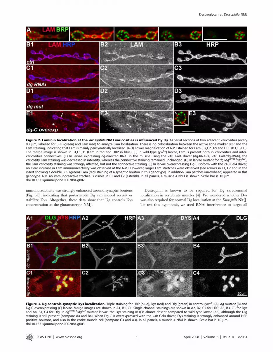
Dystroglycan controls varicosities Laminin concentrationIn vertebrates, loss of Dg in muscle cells was reported to
suppress Laminin concentration at the cholinergic NMJ [19]. We
tested if this was also the case at the Drosophila glutamatergic NMJ.
First, we investigated whether Laminin was indeed present at the
Drosophila NMJ. Laminin is a heterotrimer consisting of three
chains, A, B1 and B2. We used a polyclonal antibody against
Laminin heterotrimer [34]. We could detect some Laminin (Lam)
staining at the muscle surface, like in mammalian muscle cells, and
could detect Lam immunoreactivity at the NMJ (Fig. 2A). At this
level of optical resolution, the NMJ localization of Lam fits with a
trans-synaptic localization of this component of the extracellular
matrix. Costaining with the active zone marker Bruchpilot
revealed that, like Dg, Lam was excluded from synapses (Fig. 2A)
and restricted to the perisynaptic zones. Lam was present around
varicosities as well as around the inter-varicosities connectives
(Fig. 2B). In larvae expressing dg-RNAi in muscles, the Lam
content around the varicosities was decreased so that the
varicosities were less distinguishable compared to the connectives
(Fig. 2C). This situation was similar and even more pronounced in
dg mutants (Fig. 2D). We also analyzed the consequences of Dg
overexpression in the muscle cell on the amount and localization
of Lam. We used the Dg-C isoform, containing the mucin domain,
which is supposed to play a critical role in Lam binding of Dg
[29,42]. Lam patches, never observed in control conditions,
appeared extrasynaptically and colocalized with Dg-C-GFP (Fig.
S2). This indicated that the Dg-C isoform could indeed recruit
Lam in the muscle cell. At the NMJ, the pattern of Lam
localization was more irregular, with some perisynaptic areas
larger, and more intense in Lam immunoreactivity, compared to a
more homogenous situation in controls (Fig. 2E). Altogether, these
data show that Laminin localization around varicosities is partly
dependent on Dg. We then tested the interaction between Dg and
its second well-known partner, Dystrophin, at the synapse.
Dystroglycan controls postsynaptic Dystrophinconcentration
Dg was shown to be partly responsible for the synaptic
concentration of Utrophin at the vertebrate cholinergic NMJ
[18]. In Drosophila, there is only one gene homologous to utrophin
and dystrophin genes, called dystrophin [43]. This gene encodes for
different protein isoforms. Large isoforms are concentrated
postsynaptically at the NMJ [44]. To test whether synaptic
Dystroglycan at Drosophila NMJ
PLoS ONE | www.plosone.org 3 April 2008 | Volume 3 | Issue 4 | e2084

Dystrophin (Dys) localization was dependent on Dg, we used an
antibody directed against a C-terminal sequence of Dys, which
detects all known Dys isoforms [29]. As previously described with
an antibody directed against large isoforms of Dys, we observed a
postsynaptic localization of Dys at the NMJ (Fig. 3A). Dg mutants
displayed a strongly reduced Dys staining (Fig. 3B1-3). This
reduction was not due to a general absence of the postsynaptic
apparatus since postsynaptic marker Discs-Large (Dlg) staining
was present in these mutants (Fig. 3B4). This indicates that Dg is
required for the normal postsynaptic Dys localization. To test
whether Dg was also sufficient for this Dys localization, we
overexpressed postsynaptically Dg-C. In these NMJs, Dystrophin
Figure 1. Localization of Dg at the NMJ. A-D) Dg (A2, B2, C2, D2) and HRP (A3, B3, C3, D3) immunoreactivity at the NMJ of muscle 4. Merge ofboth images with Dg in magenta and HRP in green (A1, B1, C1, D1). The LG5 antibody was used to analyze Dg localization. (A) In wild-type (ywCS) flies,Dg concentration is clearly visible at type Ib boutons. Dg immunoreactivity is much larger than motoneuron-specific HRP immunoreactivity,indicating that Dg is present in the muscle cell, i.e. postsynaptically. (B) In larvae expressing dg-directed RNAi in the muscle using the 24B Gal4 driver,the NMJ LG5 staining was decreased in intensity. (C) In larvae mutant for dg (dge01554/dg323), the NMJ LG5 staining was more strongly affected. (D) Inlarvae overexpressing Dg-C isoform in the muscles using the 24B Gal4 driver, a clear increase in LG5 immunoreactivity was observed (laser intensitywas decreased to avoid too much signal saturation in this genotype) both at the NMJ, and also in distant patches (arrows). (E) Two serial sections(every 0.7 mm) of a synaptic varicosity of a larva expressing a Dg-C-GFP construct in the muscle cells (with the 24B Gal4 driver). GFP fluorescence (E2)is present in the subsynaptic reticulum (SSR) (arrow) and is partially excluded from the sensus-stricto synapses (arrowhead). Synapses are labelledwith the active zone marker Bruchpilot (BRP)(E3). Merge of both images with Dg-GFP in green and BRP in magenta is shown in E1. (F) Dg stainingwith anti-Dgex8 antibody, specific of the Dg-C isoform (F2) and HRP (F3). The Dgex8 immunoreactivity is similar to the LG5 immunoreactivity (A2).Again, Dgex8 immunoreactivity is much larger than motoneuron-specific HRP immunoreactivity (F1), indicating that Dg-C protein is present at thepostsynapse. In all panels, a muscle 4 NMJ is shown. Scale bar is 10 mm in A–D, 5 mm in E and 10 mm in F.doi:10.1371/journal.pone.0002084.g001
Dystroglycan at Drosophila NMJ
PLoS ONE | www.plosone.org 4 April 2008 | Volume 3 | Issue 4 | e2084
immunoreactivity was strongly enhanced around synaptic boutons
(Fig. 3C), indicating that postsynaptic Dg can indeed recruit or
stabilize Dys. Altogether, these data show that Dg controls Dys
concentration at the glutamatergic NMJ.
Dystrophin is known to be required for Dg sarcolemmal
localization in vertebrate muscles [4]. We wondered whether Dys
was also required for normal Dg localization at the Drosophila NMJ.
To test this hypothesis, we used RNAi interference to target all
Figure 2. Laminin localization at the drosophila NMJ varicosities is influenced by dg. A) Serial sections of two adjacent varicosities (every0.7 mm) labelled for BRP (green) and Lam (red) to analyze Lam localization. There is no colocalization between the active zone marker BRP and theLam staining, indicating that Lam is mainly perisynatically localized. B–D) Lower magnification of NMJ stained for Lam (B2,C2,D2) and HRP (B3,C3,D3).The merge image is shown in B1,C1,D1 (Lam in red and HRP in blue). (B) In wild-type (ywCS) larvae, Lam is present both in varicosities and inter-varicosities connectives. (C) In larvae expressing dg-directed RNAi in the muscle using the 24B Gal4 driver (dg-RNAi/+; 24B Gal4/dg-RNAi), thevaricosity Lam staining was decreased in intensity, whereas the connective staining remained unchanged. (D) In larvae mutant for dg (dge01554/dg323),the Lam varicosity staining was strongly affected, but not the connective staining. (E) In larvae overexpressing Dg-C isoform with the 24B Gal4 driver,no clear increase in Lam immunoreactivity was observed at the NMJ. However, larger Lam stretches were observed (see arrows in E1, E2 and in theinsert showing a double BRP (green), Lam (red) staining of a synaptic bouton in this genotype). In addition Lam patches (arrowhead) appeared in thisgenotype. N.B. an immunoreactive trachea is visible in E1 and E2 (asterisk). In all panels, a muscle 4 NMJ is shown. Scale bar is 10 mm.doi:10.1371/journal.pone.0002084.g002
Figure 3. Dg controls synaptic Dys localization. Triple staining for HRP (blue), Dys (red) and Dlg (green) in control (ywCS) (A), dg mutant (B) andDg-C overexpressing (C) larvae. Merge images are shown in A1, B1, C1. Single channel stainings are shown in A2, B2, C2 for HRP, A3, B3, C3 for Dysand A4, B4, C4 for Dlg. In dge01554/dg323 mutant larvae, the Dys staining (B3) is almost absent compared to wild-type larvae (A3), although the Dlgstaining is still present (compare A4 and B4). When Dg-C is overexpressed with the 24B Gal4 driver, Dys staining is strongly enhanced around HRPpositive boutons, and also in the entire muscle cell (compare C3 and A3). In all panels, a muscle 4 NMJ is shown. Scale bar is 10 mm.doi:10.1371/journal.pone.0002084.g003
Dystroglycan at Drosophila NMJ
PLoS ONE | www.plosone.org 5 April 2008 | Volume 3 | Issue 4 | e2084

Dystrophin isoforms in the muscle [29]. Expression of the RNAi
transgene led to a very strong decrease in postsynaptic Dys
immunoreactivity (Fig. 4A–C). This lack of postsynaptic Dystrophin
was accompanied by a decrease in postsynaptic Dg (Fig. 4D, F),
although not a complete loss of Dg. Hence, at the Drosophila NMJ,
Dys partially controls Dg synaptic concentration. Altogether, our
data illustrate the presence of a synaptic Lam/Dg/Dys complex,
like in vertebrate neuromuscular junctions. What can be the
functional consequences of the loss of this complex in absence of
Dg? At the cholinergic NMJ in mammals, Dg is involved in synapse
function through rapsyn-mediated anchoring of the nicotinic
acetylcholine receptors [19,45,46]. We wondered whether Dg is
also required for the clustering of glutamate receptors and if so,
through which cytoplasmic scaffold. We first looked at the synaptic
protein Coracle, reported to control some glutamate receptor
subunits localization at the Drosophila NMJ [31].
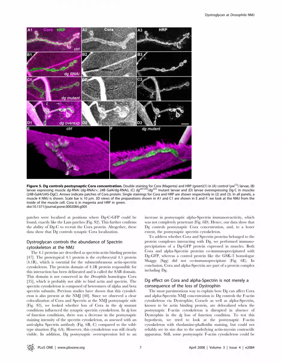
Dystroglycan also controls intracellular Coracle NMJconcentration
Coracle (Cora) is the Drosophila 4.1 protein homologue. It has
been shown to be concentrated at the NMJ and to control the
amount of GluRIIA subunit in postsynaptic glutamate receptors
[31] in late embryos and first instar larvae. The cora gene encodes
four protein isoforms (ranging from 699 to 1698 amino acids) [35].
In Chen et al. [31], the 9C monoclonal antibody directed against
the FERM domain of all Cora isoforms [35] was used, and no
clear Cora immunoreactivity could be detected at the NMJ of late
larval developmental stages. Here, we demonstrated that in third
instar larvae, Cora was concentrated around the NMJ (Fig. 5A),
using a polyclonal guinea-pig antibody directed against a sequence
present only in large Cora isoforms [35]. We tested the specificity
of the stainings we obtained in flies with genetically altered levels
of Cora protein, where the intensity of the staining should parallel
the amount of protein. We used the cora14 hypomorph mutant [30]
and the null mutation corak08713 [31] as well as flies overexpressing
cora. In cora14/corak08713 third instar larvae, the NMJ Cora
staining was strongly reduced, indicating that this staining was
specific and that Cora was actually present postsynaptically in the
domain rich in subsynaptic reticulum (SSR) (Fig. S3). We also
overexpressed Cora in muscles, using the 24B Gal4 driver and the
P element insertion P{EPgy2}cora[EY07598] upstream of the
translation start of all Cora isoforms. This produced an increased
staining in the SSR as well as in the whole sarcolemma (Fig. S3).
These data further confirmed the ability of this antibody to
recognize Cora. We observed that in larvae expressing a dg
directed RNAi and in dg mutants, the synaptic concentration of
Cora was markedly affected (Fig. 5 B,C,E,F). No clear change in
intensity was observed for the sarcolemmal immunoreactivity.
These data showed that Dg was required for the NMJ localization
of Cora. We further tested whether Dg was also sufficient to
concentrate Cora at the synapse, by overexpressing Dg-C in the
muscle. This led to a very large increase in the amount of
postsynaptic Cora (Fig. 5D), but not to an overall homogenous
increase of Cora immunoreactivity at the muscle surface.
However, many cytoplasmic Cora clusters could be found, in a
way similar to Laminin clusters when Dg-C was overexpressed
(arrows in Fig. 5D). In Dg-C-GFP overexpressing larvae, the Cora
Figure 4. Dys controls Dystroglycan postsynaptic concentra-tion. (A–C) Double staining for Fas2 (A green, B), Dys (A magenta, C) oncontrol (1) and UAS-dys-RNAi flies crossed with 24B Gal4 (2). In larvaeexpressing the dys-RNAi (dys-RNAi/+; 24B Gal4/+), there is almost noDys immunoreactivity detectable at the NMJ. (D–G) Triple staining forFas2 (D blue, E), Dystroglycan (D green, F) and Coracle (D red, G) in thesame genotypes. Dg postsynaptic labelling is reduced in absence ofpostsynaptic Dystrophin. Coracle immunoreactivity is also reduced, but
r
to a much lower extent compared to the loss of postsynaptic Dg. (H)alpha-Spectrin immunostaining on the same genotypes. Scale bar is10 mm. In all panels, a muscle 4 NMJ is shown.doi:10.1371/journal.pone.0002084.g004
Dystroglycan at Drosophila NMJ
PLoS ONE | www.plosone.org 6 April 2008 | Volume 3 | Issue 4 | e2084
patches were localized at positions where Dg-C-GFP could be
found, exactly like the Lam patches (Fig. S2). This further confirms
the ability of Dg-C to recruit the Cora protein. Altogether, these
data show that Dg controls synaptic Cora localization.
Dystroglycan controls the abundance of Spectrincytoskeleton at the NMJ
The 4.1 proteins are described as spectrin-actin binding proteins
[47]. The prototypical 4.1 protein is the erythrocytal 4.1 protein
(4.1R), which is essential for the submembranous actin-spectrin
cytoskeleton. The protein domain of 4.1R protein responsible for
this interaction has been delineated and is called the SAB domain.
This domain is not conserved in the Drosophila homologue Cora
[35], which is probably not able to bind actin and spectrin. The
spectrin cytoskeleton is composed of heteromers of alpha and beta
spectrin subunits. Previous studies have shown that this cytoskel-
eton is also present at the NMJ [48]. Since we observed a clear
colocalization of Cora and Spectrin at the NMJ postsynaptic side
(Fig. S3), we looked whether loss of Cora in the dg mutant
conditions influenced the synaptic spectrin cytoskeleton. In dg loss
of function conditions, there was a decrease in the postsynaptic
staining intensity of the spectrin cytoskeleton, as assessed with an
anti-alpha Spectrin antibody (Fig. 6B, C) compared to the wild-
type situation (Fig. 6A). However, this cytoskeleton was still clearly
visible. In addition, Dg postsynaptic overexpression led to an
increase in postsynaptic alpha-Spectrin immunoreactivity, which
was not completely penetrant (Fig. 6D). Hence, our data show that
Dg controls postsynaptic Cora concentration, and, to a lesser
extent, the postsynaptic spectrin cytoskeleton.
To address whether Cora and Spectrin proteins belonged to the
protein complexes interacting with Dg, we performed immuno-
precipitation of a Dg-GFP protein expressed in muscles. Both
Cora and alpha-Spectrin proteins co-immunoprecipitated with
Dg-GFP, whereas a control protein like the GSK-3 homologue
Shaggy (Sgg) did not co-immunoprecipitate (Fig. 6E). In
conclusion, Cora and alpha-Spectrin are part of a protein complex
including Dg.
Dg effect on Cora and alpha-Spectrin is not merely aconsequence of the loss of Dystrophin
The most parsimonious way to explain how Dg can affect Cora
and alpha-Spectrin NMJ concentration is: Dg controls the F-actin
cytoskeleton via Dystrophin; Coracle as well as alpha-Spectrin,
known to be actin binding protein, are delocalized when the
postsynaptic F-actin cytoskeleton is disrupted in absence of
Dystrophin in the dg loss of function condition. To test this
hypothesis, we tried to look at the postsynaptic F-actin
cytoskeleton with rhodamine-phalloidin staining, but could not
reliably see its size due to the underlying actin-myosin contractile
apparatus. Still, some postsynaptic F-actin cytoskeleton could be
Figure 5. Dg controls postsynaptic Cora concentration. Double staining for Cora (Magenta) and HRP (green)(1) in (A) control (ywCS) larvae, (B)larvae expressing muscle dg-RNAi (dg-RNAi/+; 24B Gal4/dg-RNAi), (C) dge01554/dg323 mutant larvae and (D) larvae overexpressing Dg-C in muscles(24B-Gal4/UAS-DgC). Arrows indicate patches of Cora protein. Single stainings for Cora and HRP are shown respectively in (2) and (3). In all panels, amuscle 4 NMJ is shown. Scale bar is 10 mm. 3D views of the preparations shown in A1 and C1 are shown in E and F: we look at the NMJ from theinside of the muscle cell. Cora is in magenta and HRP in green.doi:10.1371/journal.pone.0002084.g005
Dystroglycan at Drosophila NMJ
PLoS ONE | www.plosone.org 7 April 2008 | Volume 3 | Issue 4 | e2084

observed in dg mutants (data not shown). We thus tested whether
the loss of Dystrophin also led to a decrease in Cora and alpha-
Spectrin postsynaptic immunoreactivity. We performed this
experiment, keeping in mind that the loss of Dystrophin also
affected Dg immunoreactivity (Fig. 4). We could see that an almost
complete loss of NMJ Dystrophin obtained when expressing a Dys
RNAi construct always had smaller effects on Cora and a-Spectrin
immunoreactivity compared to a loss of Dg function, which
affected only partially Dys staining (compare Fig. 4 and Fig. 5).
These results suggest that Dg controls postsynaptic Cora and
alpha-Spectrin concentration independently of Dystrophin (Fig.
S4).
Coracle reciprocally controls Dg localizationIf Dg controls in parallel Dys and Cora synaptic concentration,
and Dys controls reciprocally Dg, we wondered whether Cora also
played such a reciprocal function on Dg. We looked at the Dg
NMJ staining in the cora14/corak08713 mutant. This mutant
reproduced the dg phenotype at the NMJ, in that postsynaptic
Cora concentration was markedly affected (Fig. 7B), although
presynaptic HRP staining (Fig. 7A) and postsynaptic Dlg staining
(Fig. 7C) did not change intensity. In this cora hypomorph mutant,
postsynaptic Dg staining was reduced (Fig. 7E). This indicated that
Cora controlled Dg postsynaptic localization and that the
functional interactions between Dg and Cora were reciprocal.
Associated with the postsynaptic Dg loss, we could observe, as
expected, a decrease in alpha-Spectrin immunoreactivity (Fig. 7D)
and in Dystrophin staining (Fig. 7E).
Dg influences the amount of GluRIIA subunits inpostsynaptic glutamate receptor clusters
The Drosophila NMJ is a glutamatergic synapse. The
postsynaptic glutamate channel receptors are thought to be
composed of four subunits: GluRIIC, IID, IIE and either the
GluRIIA or GluRIIB subunit [33,49–52]. Since Cora was shown
to control the amount of GluRIIA, and since the amount of
postsynaptic Cora was largely reduced in dg mutants, we expected
to see a decrease in the amount of GluRIIA subunit present in the
postsynaptic glutamate receptors. We quantified the immunore-
activity for GluRIIA compared to the immunoreactivity for
GluRIIC and could indeed see a significant decrease in the relative
amount of GluRIIA subunit (Fig. 8), although we could never see a
complete loss of this subunit. These data show that Dg influences
the relative subunit composition of glutamate receptors at the
Figure 6. Dg controls postsynaptic Spectrin concentration and both Cora and Spectrin co-immunoprecipitate with Dg. Doublestaining for alpha-Spectrin (Magenta) and HRP (green)(1) in (A) control (ywCS) larvae, (B) larvae expressing muscle dg-RNAi (dg-RNAi/+; 24B Gal4/dg-RNAi), (C) dge01554/dg323 mutant larvae and (D) larvae overexpressing Dg-C isoform in the muscles (24B-Gal4/UAS-DgC). Single stainings for alpha-Spectrin are shown in (2). Scale bar is 10 mm. (E) Co-immunoprecipitation was performed with a polyclonal anti-GFP antibody on protein extractsfrom flies expressing Dg-C-GFP. S corresponds to the supernatant and P to the pellet. Cora and alpha-Spectrin co-immunoprecipitate with Dg-C-GFP,but not Shaggy (Sgg), a cytoplasmic protein kinase.doi:10.1371/journal.pone.0002084.g006
Dystroglycan at Drosophila NMJ
PLoS ONE | www.plosone.org 8 April 2008 | Volume 3 | Issue 4 | e2084

NMJ. We then analyzed electrophysiologically the consequences
of the loss of postsynaptic Dg.
Loss of postsynaptic Dystroglycan leads to a decrease insynaptic quantal content
We performed intracellular, two-electrode voltage clamp
recordings from muscle 6 of segments A3 at the larval NMJs of
WT and dge01554 hypomorphic mutants, or of 24B/+ and dg-
RNAi/+; 24B/+ larvae (Fig. 9). In dg mutants, or in flies expressing
dg-RNAi in muscles, the evoked junctional currents (EJCs) were
decreased by approximately 40% (w control: 73.9563.47 nA,
n = 11; dg mutant: 53.8663.87 nA, n = 10; 24B/+ control:
72.8064.45 nA, n = 10; dg-RNAi/+; 24B/+: 36.0262.75 nA,
n = 11) (Fig. 9A and 9B). This effect could be due either to a
decrease in the sensitivity of the postsynaptic glutamate receptor
field, which should be reflected in a decrease of miniature
junctional currents amplitudes (mEJCs). On the other hand, the
number of vesicles released per action potential (‘‘quantal
content’’) could be reduced. mEJCs amplitude was not diminished
when Dg was decreased (w control: 0.9260.034 nA, n = 11; dg
mutant: 1.1160.064 nA, n = 10; 24B/+ control: 0.8060.024 nA,
n = 9; dg-RNAi/+; 24B/+: 0.7660.036 nA, n = 10) (Fig. 9C). On
the contrary, there was a slight enhancement in the dg mutant, but
not reproduced in dg-RNAi larvae. However, estimation of
quantal content (EJC/mEJC) revealed a reduction of about 40%
in dg mutant and dg-RNAi larvae (w control: 81.164.1, n = 11; dg
mutant: 49.263.8, n = 10; 24B/+ control: 89.866.4, n = 9; dg-
RNAi/+; 24B/+: 50.665.2, n = 10) (Fig. 9D). Hence, postsynaptic
Dg seems to be able to positively control the amount of
presynaptic vesicles released upon stimulation. We further verified
that the effect observed in dg-RNAi/+; 24B/+ flies was not due to
a non specific presynaptic expression of the RNAi, by comparing
this result with flies expressing the RNAi in neurones with the elav-
Gal4 driver. In elav/+; dg-RNAi/+ larvae, the evoked junctional
currents (EJCs) were only slightly decreased compared to control
genotypes (elav/+; dg-RNAi /+: 63.6565.41 nA, n = 9) (Fig. 9B).
In these larvae, the mEJCs amplitude was not affected
(0.8660.025 nA, n = 5) (Fig. 9C) and the quantal content was
not significantly diminished compared to both the w and the 24B/
+ control genotypes (71.86611.40 nA, n = 5) (Fig. 9D). Since
larvae expressing dg-RNAi in motoneurones do not show as strong
a phenotype as larvae expressing dg-RNAi in muscles, this
indicates that postsynaptic Dystroglycan controls presynaptic
vesicle release.
Discussion
Synaptic localization of DgThe widely accepted hypothesis about the function of the DGC
complex is its protective role in the sarcolemma against muscle
contraction induced size changes. Here we analyzed the synaptic
function of a core member of the DGC, Dystroglycan. We showed
that Drosophila Dg was concentrated at the NMJ, and that most Dg
immunoreactivity at the NMJ was postsynaptic. We also showed
that a proportion of synaptic Dg contained the mucin-like domain
(MLD), which is the most heavily glycosylated domain in
vertebrate Dg. Haines et al reported recently that the MLD
containing Drosophila Dg isoform was indeed glycosylated [39].
Thus, like the vertebrate cholinergic NMJ, the Drosophila NMJ is
enriched in Dg, and notably in glycosylated forms of this protein.
These data are in accordance with the previously described
Figure 7. Cora controls Spectrin, Dystroglycan, Dystrophin but not Dlg postsynaptic concentration. (A–C) Triple staining for HRP (A),Cora (B) and Dlg (C) in control (ywCS) (1) and cora14/corak08713 larvae (2). Cora concentration at the NMJ is largely decreased in the cora mutant,whereas synaptic HRP and Dlg stainings show the same signal intensity. This indicates that Cora synaptic loss in the cora mutant is not the directconsequence of a total disruption of NMJ structure. (D–F) Stainings for alpha-Spectrin (D), Dgex8 (E) and Dystrophin (F) in control (1) and cora14/corak08713 larvae (2). (D) In cora mutants, alpha-Spectrin postsynaptic concentration decreases, but does not completely disappear, like in dg mutants.(E) Dystroglycan staining appears thinner in cora mutants. Inserts show higher magnification of a synaptic bouton with presynaptic HRP in magentaand Dg staining in green. The thinner appearance of the NMJ in cora mutants is due to a decrease in postsynaptic Dg staining compared to WTlarvae. (F) In cora mutants, Dys labelling is reduced, but is still visible, contrarily to dg mutants, in which it disappears more strongly. Scale bar is20 mm.doi:10.1371/journal.pone.0002084.g007
Dystroglycan at Drosophila NMJ
PLoS ONE | www.plosone.org 9 April 2008 | Volume 3 | Issue 4 | e2084

concentration of Dystrophin at the Drosophila NMJ [44], suggesting
the presence of all DGC members at the postsynapse.
It is possible that the NMJ defects observed in the dg mutants
used in this study are a consequence of a general muscle
dysfunction, due to the loss of Dg at extrasynaptic sites. Indeed,
muscle dysfunction has been observed in dg null mutants that are
lethal at the embryonic and first instar larval stage [39]. However,
the mutants analyzed here are hypomorphs and the allelic
combination used, dge01554/dg323, is viable. The larvae crawl,
pupate and give rise to fertile adults, which do not show any wing
position phenotype corresponding to flight muscle degeneration.
Although we cannot rule out that there are some subtle muscle
defects at extrasynaptic sites, our data illustrate that synaptic
electrophysiological and morphological defects are already present
in these mild loss of function conditions.
Laminin-Dg-Dys complex is conserved at the Drosophilaglutamatergic NMJ
A function of the lanA gene, encoding a Laminin A subunit, in
stabilizing the initial motoneuron/muscle contact during synapto-
genesis was published earlier [53]. Here, we show that Laminin is
still present during late larval stages, and that it is concentrated
around synapses in varicosities. Our data indicate that, like in mice
where Dg is required for synaptic Utrophin, Laminin a5 and
Laminin c1 concentration [13], Drosophila Dg controls synaptic
Laminin and Dystrophin concentration. In addition, Dystrophin
was known to be required for Dg sarcolemmal localization in
vertebrate muscles [4], and both Dystrophin and Utrophin
account for part of the clustering of Dg at the NMJ [54,55].
Here, we show that Dystrophin also controls synaptic Dg
concentration. Thus the interdependence between Laminin, Dg
and Dystrophin at the NMJ seems to be conserved phylogenet-
ically. Importantly, in dystrophin/utrophin double mutants, a
significant amount of Dg remains at the synapse, indicating that
other proteins control, in parallel, its synaptic localization [54,55].
Our observations indicate that, similarly, the Utrophin-Dystrophin
homologue in flies does not account for the whole synaptic
localization of DG, and we identify Coracle as a new, additional
synaptic anchor for Dg.
The 4.1 protein Cora at the NMJLooking for any new potential partners of Dg, we studied Cora
localization in late larval stages at the NMJ. A previous report
described a function for Cora in early larval stages [31], but did
not show any clear synaptic localization of Cora in late larval
stages, using a monoclonal antibody [35] recognizing all Cora
isoforms. Instead, a strong immunoreactivity in NMJ associated
glial cells was reported. We used a polyclonal antibody recognizing
only the large Cora isoform [35]. With this antibody, we had no
immunoreactivity in any NMJ associated glial cell, but we could
easily detect a postsynaptic concentration of Cora, which partially
disappeared in a cora hypomorph mutant, and was increased when
Cora was overexpressed in the muscle. These data indicated that
the observed staining was indeed Cora. The Localization of
protein 4.1 members in vertebrate muscle fibers is not well
documented. It has been shown that protein 4.1R isoforms were
indeed present in the muscle cells, notably at the cell periphery
(probably the sarcolemma) [56]. Interestingly, in DMD patients,
the peripheral localization of protein 4.1R isoforms was lost,
although the sub-sarcolemmal spectrin cytoskeleton was still
Figure 8. Dg influences glutamate receptor subunit composition. Triple staining for HRP (1), DGluRIIC (2) and DGluRIIA (3) in control (ywCS)(A) and dge01554/dg323 mutant larvae (B). DGluRIIA immunoreactivity is reduced in the dg mutant whereas DGluRIIC and HRP immunoreactivities areunchanged. (C) Quantification of the ratio of DGluRIIA versus DGluRIIC staining intensities (n = 6 for ywCS and dg mutant, and n = 8 for dg-RNAi/+ anddg-RNAi/+; 24B Gal4/+). (* p,0.5; ** p,0.01). Error bars represent SEM.doi:10.1371/journal.pone.0002084.g008
Dystroglycan at Drosophila NMJ
PLoS ONE | www.plosone.org 10 April 2008 | Volume 3 | Issue 4 | e2084

present [56]. This set of data already indicated that protein 4.1
sarcolemmal localization was dependent on the DGC complex.
Here, we show that this is the case at the NMJ, and that Dg is the
principal component involved in Cora localization, since loss of
postsynaptic Dys gave much weaker phenotypes compared to loss
of postsynaptic Dg. In addition, we show that Cora co-
immunoprecipitates with Dg, indicating the presence of the two
proteins in the same complex, although further biochemical
analysis will be required to assess whether they interact directly or
indirectly.
Unexpectedly, we observed that Cora was required for the
normal postsynaptic localization of Dg and, to a lesser extent, of
Dys. This result was observed using a hypomorph cora mutant in
which the C-terminal domain is partially deleted. In this mutant,
synaptic amount of Cora was strongly reduced (Fig. 7 and Fig. S3).
Further structure-function studies will be required to understand 1)
which domain of Cora is required for its synaptic localization and
for its interaction with Dg, 2) which part of Dg C-terminal tail is
involved in Cora interaction. Previous studies have shown that the
juxtamembrane region of the C-terminal Dg tail was interacting
with Ezrin, a protein containing a FERM domain, like Cora
[57,58]. It is possible that the same Dg domain interacts with Cora.
Since Cora was known to control synaptic GluRIIA abundance
[31], an expected consequence of the loss of synaptic Cora in dg
mutant NMJ was a reduction in the amount of GluRIIA subunit at
the NMJ. We found such a reduction, but to a mild degree
(relative quantification to the amount of GluRIIC subunit had to
be done to decrease the effect of preparation variance). This small
effect may be due to the fact that dg-induced reduction of synaptic
Cora is not as strong as a complete cora loss of function, which was
the situation analyzed originally. The small effect observed on
DGluRIIA probably explains why there was no change in the
amplitude of mEJCs in dg loss of functions. Indeed, DGluRIIA is
the dominant subunit compared to DGluRIIB and a significant
loss of DGluRIIA should lead to a decrease in mini amplitude
[51].
Dystroglycan and the spectrin cytoskeletonLoss of synaptic Dystroglycan resulted in a clear decrease in
postsynaptic spectrin cytoskeleton, as assessed with alpha-Spectrin
immunoreactivity. Although the spectrin defect may be a
consequence of the loss of synaptic Cora, a more direct interaction
between Dg and the spectrin cytoskeleton remains a possibility.
Hence, the link between Dg, Cora and spectrin cytoskeleton
remains to be further defined. The postsynaptic spectrin
cytoskeleton was shown to play a role in the repartition of
postsynaptic receptor fields [48]. Indeed, loss of postsynaptic
immunoreactivity for both alpha and beta-Spectrin led to a
disorganization of postsynaptic receptor fields. We looked for such
a defect in the dystroglycan loss of function conditions, but could
not find any. This is probably due to the fact that the loss of
spectrin immunoreactivity in these mutants was not complete.
Presynaptic glutamate release defect in postsynaptic dgdeficient larvae
We demonstrated that Dg played a functional role in
neuromuscular synaptic transmission. Indeed, the glutamate
release was decreased by approximately 40% in absence of muscle
Dg. The main specificity of the insect NMJ, compared to the
vertebrate NMJ is the presence of glutamate as a neurotransmitter
instead of acetylcholine. Hence, these synapses are not only NMJ
models, but also models of glutamatergic synapses, which are by
far the most frequent synapses found in the vertebrate brain. A
previous study of Dg function in brain synapses illustrated an
Figure 9. NMJ electrophysiological defects due to the loss of dgfunction. A) Example traces of evoked postsynaptic currents (EJCs)from NMJs of wild type (w1) and dg mutant dge01554 larvae. B) Mean EJCamplitude for wild type (w1 larvae), dg mutant dge01554, Canton Scrossed with 24B Gal4 (24B Gal4/+), dg-RNAi crossed with 24B Gal4 (dg-RNAi/+;24B Gal4/+) and dg-RNAi crossed with elavC155 Gal4 (elav Gal4/+; dg-RNAi/+). B) mini EJC amplitudes and C) quantal content for thesame genotypes. The number of measured larvae is indicated withineach histogram bar. (* p,0.5; *** p,0.001). Error bars represent SEM.doi:10.1371/journal.pone.0002084.g009
Dystroglycan at Drosophila NMJ
PLoS ONE | www.plosone.org 11 April 2008 | Volume 3 | Issue 4 | e2084

alteration of LTP in DG2CNS mice, but no modification of the
amplitude of synaptic responses evoked by low frequency
stimulation of Schaeffer collaterals, and no changes in paired-
pulse facilitation [59]. Here, we could detect a reduced synaptic
response at low frequency, indicating a function of Dg in basal
glutamatergic synaptic transmission.
One surprising result in our electrophysiology experiments was
the fact that defects in quantal content of the dg mutant were also
present, with the same intensity in flies expressing a 24B Gal4
driven dg-RNAi. This indicated that loss of postsynaptic Dg led to
a functional change in the other synaptic compartment, the
presynapse. Such a presynaptic effect associated with postsynaptic
modifications is not new, since the NMJ function displays
homeostasis, and that decrease in postsynaptic responsiveness is
often associated to increase in neurotransmitter release and vice-
versa in order to maintain constant EJCs [60]. The molecular
mechanisms involved in this homeostatic control are largely
unknown. Here, in dg mutants, homeostatic control is likely absent
since mini amplitude (receptor field) is not altered in absence of
postsynaptic Dg, but glutamate release is modified. This suggests
that Dg-deficient muscles inappropriately signals to the presynap-
tic release machinery. A similar trans-synaptic effect of loss of
muscle Dystrophin onto presynaptic quantal content was observed
[44]. What can be the mechanisms involved? One possibility is
that postsynaptic Dg directly controls the levels of synaptic ECM
molecules such as Laminin. These proteins, by interacting with
presynaptic receptors, would affect the structure of the presynapse,
e.g. the amount, size or molecular composition of active or
periactives zones. This hypothesis is strongly supported by the
finding in mouse, that a synaptic Laminin-calcium channel
interaction organizes active zones in motor nerve terminals [61].
Another presynaptic Laminin receptor could be the synaptic
vesicle protein SV2 [62]. Looking for modifications at the
presynapse, we did not detect any obvious change in the number
and size of active zones, using Bruchpilot immunoreactivity as a
marker (unpublished observations) and did not detect any
modification in the immunoreactivity of the periactive zone
marker Fas2 (unpublished observations). Still, the regulation of
synaptic Laminin by Dg that we demonstrate here, together with
the presynaptic electrophysiological phenotype we observed, make
the hypothesis of Laminin bridging postsynaptic Dg and the
presynapse, at least in periactive zones, very likely.
These findings, i.e. the new components of a Dystroglycan
complex, as well as the unexpected trans-synaptic role of Dg pave
the way for understanding the role of the DGC in the formation,
maintenance and plasticity of glutamatergic synapses.
Supporting Information
Figure S1 dg gene structure, dg mutants and qRT-PCR analysis
A) Gene structure of dg gene compiled from flybase data and [27].
The two null alleles dg323 and dg62 are deletions comprising the
first exon. The Piggybac element PBac{RB}e01554 is localized
within the ninth exon, subjected to alternative splicing. B) To
analyze how the Piggybac element altered dg transcription, we
perfomed qRT-PCR experiments with couples of oligonucleotides
designed against sequences common to all transcripts, either in the
extracellular domain (couple 1), or in the intracellular tail (couple
2). In both the homozygote condition, or in transheterozygote with
the null alleles, the Piggybac element led to a loss of more than
90% of transcripts. C) and D) Specificity of NMJ Dg staining
observed with the anti-Dgex8 antibody on muscle 4 NMJs. Dgex8
(panels 1 and 2) or Discs-Large (panels 1 and 3) immunoreactivity
in ywCS control (C) and dg1554/dg323 mutant larvae (D). The
Dgex8 NMJ immunoreactivity almost completely disappears in the
mutant condition whereas Discs-Large immunoreactivity is still
present. Scale bar is 10 mm.
Found at: doi:10.1371/journal.pone.0002084.s001 (2.12 MB TIF)
Figure S2 Dg-C-GFP can recruit Lam and Cora at extra-
synaptic patches (A-C) Double staining against GFP (A, C) and
Lam (A, B) in larvae overexpressing Dg-C-GFP in the muscles
with the 24B Gal4 driver. In all panels of this figure, a single
confocal optical section, which crosses the sarcolemma, is taken
(D). The bottom right part of each panel corresponds the
extracellular space and the up left part to the sarcoplasma. Lam
colocalizes with Dg-C-GFP patches at the sarcolemma (see arrows
in A). (E-G) Double staining against GFP (E, G) and Cora (E, F) in
larvae overexpressing Dg-C-GFP in the muscles with the 24B Gal4
driver. Cora colocalizes with Dg-C-GFP patches at the sarcolem-
ma (see arrows in E). (H-J) Double staining against Cora (H, I) and
Lam (H, J) in wild-type larvae, without any Dg overexpression.
There are no extrasynaptic patches of Cora and Lam in these
larvae. Scale bar is 10 mm.
Found at: doi:10.1371/journal.pone.0002084.s002 (1.40 MB TIF)
Figure S3 Cora is concentrated at the third instar larval NMJ
and colocalizes with Spectrin. Double staining for Cora (red) and
HRP (blue)(1) on (A) WT larvae, (B) cora14/corak08713 mutant
larvae and (C) P{EPgy2}cora[EY07598]/+; 24B Gal4/+ larvae,
which overexpress all Cora isoforms in muscles. Muscle 4 NMJs
are shown. Single stainings for Cora and HRP are shown
respectively in (2) and (3). Scale bar is 10 mm. (D) Immunoblot
of proteins isolated from ywCS, cora14/corak08713 and P{EP-
gy2}cora[EY07598]/+; 24B Gal4/+ larvae. This blot was stained
with the polyclonal Guinea-pig anti-Cora antibody. The wild-type
larvae display 2 bands of about 210 and 240 kDa. The signal
intensity is clearly reduced in cora hypomorph mutants, whereas it
is enhanced when Cora is overexpressed with the 24B-Gal4 driver.
A second staining of the same blot with an anti-Tubulin antibody
indicated that the protein loading was similar in all lanes. Relative
molecular mass size markers (kDa) are indicated at right. (E) Triple
staining for HRP (blue), Cora (red) and alpha-Spectrin (green) on
WT third instar larvae (E1). A muscle 4 NMJ is shown. Single
stainings for HRP, Cora and alpha-Spectrin are displayed
respectively in E2, E3 and E4. A plot of the intensity of each
labelling along a line crossing a terminal bouton (see in E1) is
represented in E5. Note that the extent of alpha-Spectrin and
Cora stainings around the synaptic bouton -labelled with HRP- is
similar. Scale bar is 10 mm.
Found at: doi:10.1371/journal.pone.0002084.s003 (5.67 MB TIF)
Figure S4 Model of protein interactions with Dg at the
postsynaptic side of the Drosophila NMJ.
Found at: doi:10.1371/journal.pone.0002084.s004 (0.67 MB TIF)
Acknowledgments
We thank M. Schneider, H. Ruohola-Baker and the Bloomington Stock
Center for providing fly stocks. We also thank Aaron DiAntonio, M.
Schneider, A. Wodarz, R. Fehon, L. Fessler and the Developmental
Studies Hybridoma Bank for sending antibodies. All confocal imaging was
performed at the Montpellier Rio Imaging CRIC facility. We gratefully
thank Ashley Stanton and Robert Burgess from the Jackson Laboratory for
helpful comments on the manuscript.
Author Contributions
Conceived and designed the experiments: JB SS MP LB YG. Performed
the experiments: MP LB BF BF AF. Analyzed the data: SS MP LB BF BF
AF. Contributed reagents/materials/analysis tools: DM. Wrote the paper:
MP.
Dystroglycan at Drosophila NMJ
PLoS ONE | www.plosone.org 12 April 2008 | Volume 3 | Issue 4 | e2084

References
1. Ervasti JM, Campbell KP (1993) A role for the dystrophin-glycoprotein complexas a transmembrane linker between laminin and actin. J Cell Biol 122: 809–823.
2. Ibraghimov-Beskrovnaya O, Ervasti JM, Leveille CJ, Slaughter CA, Sernett SW,
et al. (1992) Primary structure of dystrophin-associated glycoproteins linkingdystrophin to the extracellular matrix. Nature 355: 696–702.
3. Ibraghimov-Beskrovnaya O, Milatovich A, Ozcelik T, Yang B, Koepnick K, et
al. (1993) Human dystroglycan: skeletal muscle cDNA, genomic structure, originof tissue specific isoforms and chromosomal localization. Hum Mol Genet 2:
1651–1657.
4. Ohlendieck K, Campbell KP (1991) Dystrophin-associated proteins are greatlyreduced in skeletal muscle from mdx mice. J Cell Biol 115: 1685–1694.
5. Pasternak C, Wong S, Elson EL (1995) Mechanical function of dystrophin in
muscle cells. J Cell Biol 128: 355–361.
6. Petrof BJ, Shrager JB, Stedman HH, Kelly AM, Sweeney HL (1993) Dystrophin
protects the sarcolemma from stresses developed during muscle contraction.
Proc Natl Acad Sci U S A 90: 3710–3714.
7. Williamson RA, Henry MD, Daniels KJ, Hrstka RF, Lee JC, et al. (1997)
Dystroglycan is essential for early embryonic development: disruption of
Reichert’s membrane in Dag1-null mice. Hum Mol Genet 6: 831–841.
8. Lisi MT, Cohn RD (2007) Congenital muscular dystrophies: new aspects of an
expanding group of disorders. Biochim Biophys Acta 1772: 159–172.
9. Sciandra F, Gawlik KI, Brancaccio A, Durbeej M (2007) Dystroglycan: apossible mediator for reducing congenital muscular dystrophy? Trends
Biotechnol 25: 262–268.
10. Adams ME, Kramarcy N, Krall SP, Rossi SG, Rotundo RL, et al. (2000)Absence of alpha-syntrophin leads to structurally aberrant neuromuscular
synapses deficient in utrophin. J Cell Biol 150: 1385–1398.
11. Deconinck AE, Potter AC, Tinsley JM, Wood SJ, Vater R, et al. (1997)Postsynaptic abnormalities at the neuromuscular junctions of utrophin-deficient
mice. J Cell Biol 136: 883–894.
12. Grady RM, Merlie JP, Sanes JR (1997) Subtle neuromuscular defects inutrophin-deficient mice. J Cell Biol 136: 871–882.
13. Grady RM, Zhou H, Cunningham JM, Henry MD, Campbell KP, et al. (2000)
Maturation and maintenance of the neuromuscular synapse: genetic evidencefor roles of the dystrophin–glycoprotein complex. Neuron 25: 279–293.
14. Hosaka Y, Yokota T, Miyagoe-Suzuki Y, Yuasa K, Imamura M, et al. (2002)
Alpha1-syntrophin-deficient skeletal muscle exhibits hypertrophy and aberrantformation of neuromuscular junctions during regeneration. J Cell Biol 158:
1097–1107.
15. Lyons PR, Slater CR (1991) Structure and function of the neuromuscularjunction in young adult mdx mice. J Neurocytol 20: 969–981.
16. Nagel A, Lehmann-Horn F, Engel AG (1990) Neuromuscular transmission in
the mdx mouse. Muscle Nerve 13: 742–749.
17. Taniguchi M, Kurahashi H, Noguchi S, Fukudome T, Okinaga T, et al. (2006)Aberrant neuromuscular junctions and delayed terminal muscle fiber maturation
in alpha-dystroglycanopathies. Hum Mol Genet 15: 1279–1289.
18. Cote PD, Moukhles H, Lindenbaum M, Carbonetto S (1999) Chimaeric micedeficient in dystroglycans develop muscular dystrophy and have disrupted
myoneural synapses. Nat Genet 23: 338–342.
19. Jacobson C, Cote PD, Rossi SG, Rotundo RL, Carbonetto S (2001) The
dystroglycan complex is necessary for stabilization of acetylcholine receptor
clusters at neuromuscular junctions and formation of the synaptic basementmembrane. J Cell Biol 152: 435–450.
20. Tremblay MR, Carbonetto S (2006) An extracellular pathway for dystroglycan
function in acetylcholine receptor aggregation and laminin deposition in skeletalmyotubes. J Biol Chem 281: 13365–13373.
21. Blake DJ, Kroger S (2000) The neurobiology of duchenne muscular dystrophy:
learning lessons from muscle? Trends Neurosci 23: 92–99.
22. Kueh SL, Head SI, Morley JW (2008) GABA(A) RECEPTOR EXPRESSION
AND INHIBITORY POST-SYNAPTIC CURRENTS IN CEREBELLAR
PURKINJE CELLS IN DYSTROPHIN-DEFICIENT mdx MICE. Clin ExpPharmacol Physiol 35: 207–10.
23. Montanaro F, Carbonetto S (2003) Targeting dystroglycan in the brain. Neuron
37: 193–196.
24. Balci B, Uyanik G, Dincer P, Gross C, Willer T, et al. (2005) An autosomal
recessive limb girdle muscular dystrophy (LGMD2) with mild mental retardation
is allelic to Walker-Warburg syndrome (WWS) caused by a mutation in thePOMT1 gene. Neuromuscul Disord 15: 271–275.
25. Collins CA, DiAntonio A (2007) Synaptic development: insights from
Drosophila. Curr Opin Neurobiol 17: 35–42.
26. Brand AH, Perrimon N (1993) Targeted gene expression as a means of altering
cell fates and generating dominant phenotypes. Development 118: 401–415.
27. Deng WM, Schneider M, Frock R, Castillejo-Lopez C, Gaman EA, et al. (2003)Dystroglycan is required for polarizing the epithelial cells and the oocyte in
Drosophila. Development 130: 173–184.
28. Thibault ST, Singer MA, Miyazaki WY, Milash B, Dompe NA, et al. (2004) Acomplementary transposon tool kit for Drosophila melanogaster using P and
piggyBac. Nat Genet 36: 283–287.
29. Schneider M, Khalil AA, Poulton J, Castillejo-Lopez C, Egger-Adam D, et al.(2006) Perlecan and Dystroglycan act at the basal side of the Drosophila
follicular epithelium to maintain epithelial organization. Development 133:
3805–3815.
30. Lamb RS, Ward RE, Schweizer L, Fehon RG (1998) Drosophila coracle, a
member of the protein 4.1 superfamily, has essential structural functions in the
septate junctions and developmental functions in embryonic and adult epithelial
cells. Mol Biol Cell 9: 3505–3519.
31. Chen K, Merino C, Sigrist SJ, Featherstone DE (2005) The 4.1 protein coracle
mediates subunit-selective anchoring of Drosophila glutamate receptors to the
postsynaptic actin cytoskeleton. J Neurosci 25: 6667–6675.
32. Pons F, Robert A, Fabbrizio E, Hugon G, Califano JC, et al. (1994) Utrophin
localization in normal and dystrophin-deficient heart. Circulation 90: 369–374.
33. Marrus SB, Portman SL, Allen MJ, Moffat KG, DiAntonio A (2004) Differential
localization of glutamate receptor subunits at the Drosophila neuromuscular
junction. J Neurosci 24: 1406–1415.
34. Fessler LI, Campbell AG, Duncan KG, Fessler JH (1987) Drosophila laminin:
characterization and localization. J Cell Biol 105: 2383–2391.
35. Fehon RG, Dawson IA, Artavanis-Tsakonas S (1994) A Drosophila homologue
of membrane-skeleton protein 4.1 is associated with septate junctions and is
encoded by the coracle gene. Development 120: 545–557.
36. Kittel RJ, Wichmann C, Rasse TM, Fouquet W, Schmidt M, et al. (2006)
Bruchpilot promotes active zone assembly, Ca2+ channel clustering, and vesicle
release. Science 312: 1051–1054.
37. Winder SJ (2001) The complexities of dystroglycan. Trends Biochem Sci 26:
118–124.
38. Shcherbata HR, Yatsenko AS, Patterson L, Sood VD, Nudel U, et al. (2007)
Dissecting muscle and neuronal disorders in a Drosophila model of muscular
dystrophy. Embo J 26: 481–493.
39. Haines N, Seabrooke S, Stewart BA (2007) Dystroglycan and Protein O-
Mannosyltransferases 1 and 2 Are Required to Maintain Integrity of Drosophila
Larval Muscles. Mol Biol Cell.
40. Schmid A, Qin G, Wichmann C, Kittel RJ, Mertel S, et al. (2006) Non-NMDA-
type glutamate receptors are essential for maturation but not for initial assembly
of synapses at Drosophila neuromuscular junctions. J Neurosci 26:
11267–11277.
41. Wagh DA, Rasse TM, Asan E, Hofbauer A, Schwenkert I, et al. (2006)
Bruchpilot, a protein with homology to ELKS/CAST, is required for structural
integrity and function of synaptic active zones in Drosophila. Neuron 49:
833–844.
42. Kanagawa M, Saito F, Kunz S, Yoshida-Moriguchi T, Barresi R, et al. (2004)
Molecular recognition by LARGE is essential for expression of functional
dystroglycan. Cell 117: 953–964.
43. Greener MJ, Roberts RG (2000) Conservation of components of the dystrophin
complex in Drosophila. FEBS Lett 482: 13–18.
44. van der Plas MC, Pilgram GS, Plomp JJ, de Jong A, Fradkin LG, et al. (2006)
Dystrophin is required for appropriate retrograde control of neurotransmitter
release at the Drosophila neuromuscular junction. J Neurosci 26: 333–344.
45. Bartoli M, Ramarao MK, Cohen JB (2001) Interactions of the rapsyn RING-H2
domain with dystroglycan. J Biol Chem 276: 24911–24917.
46. Marchand S, Stetzkowski-Marden F, Cartaud J (2001) Differential targeting of
components of the dystrophin complex to the postsynaptic membrane.
Eur J Neurosci 13: 221–229.
47. Gimm JA, An X, Nunomura W, Mohandas N (2002) Functional characteriza-
tion of spectrin-actin-binding domains in 4.1 family of proteins. Biochemistry 41:
7275–7282.
48. Pielage J, Fetter RD, Davis GW (2006) A postsynaptic spectrin scaffold defines
active zone size, spacing, and efficacy at the Drosophila neuromuscular junction.
J Cell Biol 175: 491–503.
49. DiAntonio A, Petersen SA, Heckmann M, Goodman CS (1999) Glutamate
receptor expression regulates quantal size and quantal content at the Drosophila
neuromuscular junction. J Neurosci 19: 3023–3032.
50. Featherstone DE, Rushton E, Rohrbough J, Liebl F, Karr J, et al. (2005) An
essential Drosophila glutamate receptor subunit that functions in both central
neuropil and neuromuscular junction. J Neurosci 25: 3199–3208.
51. Petersen SA, Fetter RD, Noordermeer JN, Goodman CS, DiAntonio A (1997)
Genetic analysis of glutamate receptors in Drosophila reveals a retrograde signal
regulating presynaptic transmitter release. Neuron 19: 1237–1248.
52. Qin G, Schwarz T, Kittel RJ, Schmid A, Rasse TM, et al. (2005) Four different
subunits are essential for expressing the synaptic glutamate receptor at
neuromuscular junctions of Drosophila. J Neurosci 25: 3209–3218.
53. Prokop A, Martin-Bermudo MD, Bate M, Brown NH (1998) Absence of PS
integrins or laminin A affects extracellular adhesion, but not intracellular
assembly, of hemiadherens and neuromuscular junctions in Drosophila embryos.
Dev Biol 196: 58–76.
54. Deconinck AE, Rafael JA, Skinner JA, Brown SC, Potter AC, et al. (1997)
Utrophin-dystrophin-deficient mice as a model for Duchenne muscular
dystrophy. Cell 90: 717–727.
55. Grady RM, Teng H, Nichol MC, Cunningham JC, Wilkinson RS, et al. (1997)
Skeletal and cardiac myopathies in mice lacking utrophin and dystrophin: a
model for Duchenne muscular dystrophy. Cell 90: 729–738.
Dystroglycan at Drosophila NMJ
PLoS ONE | www.plosone.org 13 April 2008 | Volume 3 | Issue 4 | e2084

56. Delhommeau F, Dalla Venezia N, Moriniere M, Collin H, Maillet P, et al.
(2005) Protein 4.1R expression in normal and dystrophic skeletal muscle.
C R Biol 328: 43–56.
57. Batchelor CL, Higginson JR, Chen YJ, Vanni C, Eva A, et al. (2007)
Recruitment of Dbl by ezrin and dystroglycan drives membrane proximal
Cdc42 activation and filopodia formation. Cell Cycle 6: 353–363.
58. Spence HJ, Chen YJ, Batchelor CL, Higginson JR, Suila H, et al. (2004) Ezrin-
dependent regulation of the actin cytoskeleton by beta-dystroglycan. Hum Mol
Genet 13: 1657–1668.
59. Moore SA, Saito F, Chen J, Michele DE, Henry MD, et al. (2002) Deletion of
brain dystroglycan recapitulates aspects of congenital muscular dystrophy.Nature 418: 422–425.
60. Macleod GT, Zinsmaier KE (2006) Synaptic homeostasis on the fast track.
Neuron 52: 569–571.61. Nishimune H, Sanes JR, Carlson SS (2004) A synaptic laminin-calcium channel
interaction organizes active zones in motor nerve terminals. Nature 432:580–587.
62. Son YJ, Scranton TW, Sunderland WJ, Baek SJ, Miner JH, et al. (2000) The
synaptic vesicle protein SV2 is complexed with an alpha5-containing laminin onthe nerve terminal surface. J Biol Chem 275: 451–460.
Dystroglycan at Drosophila NMJ
PLoS ONE | www.plosone.org 14 April 2008 | Volume 3 | Issue 4 | e2084


