Embed Size (px)
Citation preview
A
cscnu©
K
1
tfifiostdpRm
eismfto
f
0d
Progress in Organic Coatings 57 (2006) 91–97
The fine dispersion of functionalized carbonnanotubes in acrylic latex coatings�
P. Vandervorst a, C.-H. Lei a, Y. Lin c, O. Dupont b, A.B. Dalton a, Y.-P. Sun c, J.L. Keddie a,∗a Department of Physics, University of Surrey, Guildford, Surrey GU2 7XH, UK
b Cytec Surface Specialties, Anderlecht Str. 33, B-1620 Drogenbos, Belgiumc Department of Chemistry and Centre for Advanced Engineering Fibers and Films, Clemson University, Clemson, SC 29634-0973, USA
bstract
Nanocomposites of a polymer and carbon nanotubes exhibit high electrical and thermal conductivity and enhanced mechanical properties inomparison to the polymer alone. Film formation from latex dispersions is an ideal way to create nanocomposite coatings with the advantages of
olvent-free processing and a high uniformity of dispersion. It is shown here that carbon nanotubes functionalised with poly(vinyl alcohol) (PVA)an be blended with two types of acrylic latex to create stable colloidal dispersions without the need for added surfactant or emulsifier. Waterborneanocomposite films with optical transparency can be formed. Microscopic analysis shows that the PVA-functionalized nanotubes are finely andniformly dispersed in the polymer matrix.2006 Elsevier B.V. All rights reserved.
mic fo
poc
(oehJhbpobci
o
eywords: Carbon nanotubes; Optical absorption; Latex; Nanocomposite; Ato
. Introduction
As a result of efforts worldwide by theorists and experimen-alists, a very good understanding of the mechanisms of latexlm formation has been achieved. There has been a clear identi-cation of the factors that influence the uniformity of the dryingf latex films in both directions: lateral [1] (i.e. parallel to theubstrate) and vertical [2,3] (i.e. normal to the substrate). Fur-hermore, there is now an enhanced understanding of the particleeformation mechanisms because of insight gained from therocess model of latex film formation, proposed by Routh andussell [4]. Experimental results are broadly consistent with theodel’s predictions [5].This understanding of latex film formation can now be
xploited to underpin the processing of new types of coat-ngs and adhesives. In particular, latex particles can be con-idered to be the “building blocks” in the creation of nano-aterials. For instance, through careful control of the film
ormation conditions, and through the use of core-shell [6] orwo-phase particles, the material’s structure can be controlledn the scale of tens of nanometers [7]. The blending of latex
� This paper is part of the special issue “Coatings Science International Con-erence Program 2005” which was published in February 2006 in issue 55/2.∗ Corresponding author. Tel.: +44 1483 686803; fax: +44 1483 686781.
E-mail address: [email protected] (J.L. Keddie).
pv[ttwan
300-9440/$ – see front matter © 2006 Elsevier B.V. All rights reserved.oi:10.1016/j.porgcoat.2006.07.005
rce microscopy
articles and inorganic nanoparticles provides a facile meansf ensuring dispersion at the nanometer scale in compositeoatings.
Ever since the first scientific report of carbon nanotubesCNTs) in 1991, these materials have attracted enormous interestwing to their potential applications in field-emission devices,lectronics, fibers, composites, sensors, detectors, capacitors,ydrogen storage media, and fuel cells, among others [8]. As ofanuary of 2004, 152 patents relating to nanotube applicationsad been issued, and another 274 were pending. Recent worky numerous groups has demonstrated how nanocomposites ofolymers and CNTs offer the advantages of polymers, such asptical clarity, viscoelasticity and good barrier properties, com-ined with the strength [9] and the high thermal and electricalonductivity [10] of CNTs. Seven patents have already beenssued for CNT nanocomposites and fibers.
Four of the most common processing routes for this classf nanocomposites are (1) in situ polymerization [11], (2) meltrocessing and extrusion [12], (3) casting from a common sol-ent [13], and (4) functionalisation of the CNT by polymers14]. Recently, nanocomposite films were prepared from CNTshat were functionalized with poly(vinyl alcohol) (PVA) to make
hem dispersible in water [15]. The PVA-functionalized CNTsere then dispersed in an aqueous PVA solution and cast to cre-te a nanocomposite film with a PVA matrix. This approach doesot require emulsifiers or surfactants to disperse the CNTs. How-

9 n Org
ea
cCdciaaCspuaaissaoecccis
2
2
baom(sTaoa5
ftSaScp1CsAm
aioPp
2
tocLtatl5
ubmtau42oewutc
3
3
otsaslpili
Cvot
2 P. Vandervorst et al. / Progress i
ver, the final nanocomposite film is water-soluble, of course,nd of limited applicability as a protective coating.
Within the past few years, it has been realised that the pro-essing of latex dispersions offers an opportunity for blendingNTs with polymers at the nanometer scale to achieve a goodispersion in a coating matrix that is not water-soluble. The keyhallenge in the processing of waterborne nanocomposite coat-ngs is to achieve the low percolation thresholds of CNTs thatre predicted by theory [16]. In one of the first such reports [17],styrene-butyl acrylate latex was blended with multi-walledNTs that had been suspended in water using sodium dodecyl
ulfate (SDS). More recently, Regev et al. [18] created nanocom-osite films of poly(styrene) and poly(methyl methacrylate)sing compression molding. They likewise did not function-lize the CNTs but dispersed them in water using SDS or gumrabic. A possible drawback of this approach is that it couldncrease the hydrophilicity of the coating. Grunlan et al. [19] pre-ented evidence that they had achieved a highly-uniform disper-ion of single-wall carbon nanotubes (SWNTs) in a poly(vinylcetate) latex film. The SWNTs were stabilized with either GAr poly(vinyl pyrrolidone). According to their measurements oflectrical conductivity, percolation of the CNTs occurred at aoncentration of 0.04 wt.%. In this present work, we show thatrack-free, water-resistant, and optically clear nanocompositeoatings can be created using latex processing and functional-zed SWNTs. In this approach, there is no requirement for addedurfactant or emulsifier.
. Materials and techniques
.1. Materials
Two types of latex were used. The first type is an acrylicased on a random copolymer of 2-ethyl hexyl acrylate, butylcrylate (BA), ethyl acrylate, methyl methacrylate (MMA), andther monomers. It was prepared by semi-batch emulsion poly-erization. The polymer has a low glass transition temperature
Tg = −45 ◦C) and has applications as a pressure-sensitive adhe-ive. The latex dispersion has an average particle size of 190 nm.he second latex polymer is a random copolymer of BA, MMAnd methacrylic acid (MAA) in a molar ratio of BA:MMA:MAAf 85:10:5. With its Tg of 20 ◦C, this latex has applicationss a protective coating. The dispersion has a solids content of0 wt.% and an average particle size of 270 nm.
SWNTs, from two different sources and with two differentunctionalities, were used. One type was produced by the elec-ric arc-discharge of Ni–Y catalyzed graphite electrodes (Carbonolutions Inc., CA, USA). The material was purified with nitriccid and left in a highly-functionalized form. The resultingWNTs contain 4–6 at.% carboxylic acid (COOH) and have aarbonaceous purity of 80–90%. These COOH-CNTs were dis-ersed in dimethyl formamide (DMF) with ultrasonication for0 min to create a dispersion containing 0.55 wt.% CNT. The
OOH-CNT/DMF dispersion was blended with the latex disper-ions while stirring, relying on the solubility of DMF and water.second type of SWNT was produced via the arc-discharge
ethod at Clemson University. The nanotubes were purified
aaaa
anic Coatings 57 (2006) 91–97
nd then functionalized with poly(vinyl alcohol) via an ester-fication reaction, as described elsewhere [15]. The weight ratiof PVA:CNT is approximately 10:1. A dispersion of 1.5 wt.%VA-CNTs in water was mixed with the concentrated latex dis-ersions in various proportions while stirring.
.2. Techniques
With two types of latex (low-Tg and high-Tg) and with twoypes of SWNT (COOH and PVA), nanocomposite films basedn the four different combinations were created. The final con-entrations of CNTs were as high as 3.5 wt.% on the polymer.atex films were cast on clean, glass plates with a cube applica-
or having a gap of 100 �m. The high-Tg latex films were driedt room temperature and then heated to 90 ◦C for 30 min. Theyypically had a final film thickness of 35–40 �m. The low-Tgatex films were dried at room temperature and were typically0 �m thick.
The optical absorption coefficients of the films were obtainedsing a UV–vis spectrophotometer (Camspec, UK). The distri-ution of CNTs in the films was determined using atomic forceicroscopy (NT-MDT Ntegra Laboratory, Moscow, Russia) in
he intermittent contact mode. High tapping free amplitudes andrelatively stiff cantilever (spring constant k = 46 N/m) were
sed. Electron microscopy was performed using an Hitachi S-000 cold field emission scanning electron microscope using a0 kV beam with an emission current of 10 �A. Cross-sectionsf the high-Tg latex were prepared for analysis by scanninglectron microscopy (SEM), as follows. The latex dispersionere cast on sheets of poly(propylene) and film-formed in thesual way. The films were immersed in liquid nitrogen, and thenhe brittle films were fractured. The cross-section surfaces wereoated with a thin film of Au/Pd prior to SEM analysis.
. Results
.1. Colloidal stability
Each of the two types of CNT could be blended with eachf the two types of latex dispersion. The latex dispersions con-aining PVA-CNTs were found to be highly stable. There is noignificant amount of phase separation or sedimentation overperiod of weeks. Similarly, the COOH-CNTs exhibited good
tability in the low-Tg latex. However, in blends with the high-Tgatex, the COOH-CNTs were seen to rise to the top of the dis-ersion slowly over a period of several weeks. The differencesn the stability of the two types of blends might be because theow Tg latex exhibits some adhesion with the COOH-CNTs thats not found with the high Tg latex.
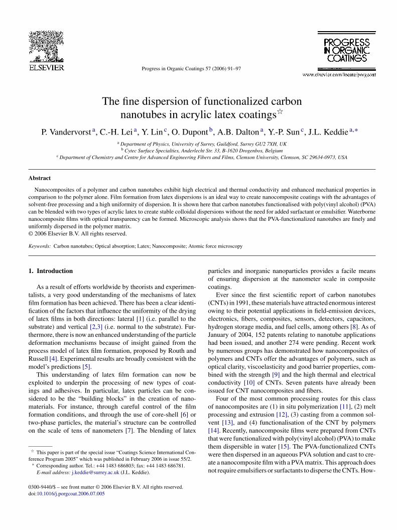
The colloidal interactions between the latex particles and theNTs were further explored using atomic force microscopy. Aery dilute blend of the low-Tg latex and COOH-CNTs was castnto freshly-cleaved mica sheets. Fig. 1 shows the observed dis-ribution of CNTs and latex particles. Nanotube structures with
verage lengths of ca. 1 �m and heights of ca. 2 nm (typical forsingle SWNT) are observed. The resolution of the technique,s determined by the AFM tip geometry, does not enable a reli-ble measurement of the diameter of the structure, however. The
P. Vandervorst et al. / Progress in Organic Coatings 57 (2006) 91–97 93
F righm SWN
va
aaalatttcoal
urhra
3
arawmmtitt3
3
φ
idawnaaatb[
l1taPSs1pd
3
ii
ig. 1. AFM images (height image on the left and phase contrast image on theica surface. The arrows indicate where three latex particles are clustered on a
alue of the height is consistent with what is expected if exfoli-tion of the SWNTs had been achieved in the dispersion.
There is also evidence for attraction between the two phases,s some latex particles can be observed in Fig. 1 to cluster aroundSWNT. In most instances, one or more latex particle was
ttached to the SWNTs. Further indication of the clustering ofatex particles around the CNTs was given by the visual appear-nce of the dispersions. When the low-Tg latex was blended withhe CNTs, the dispersion became blue. Although the reasons forhis observation are not known with certainty, it is likely to behe result of the diffraction of light from the latex particles. Theenter-to-center distance of the particles in the clusters is on therder of one-half the wavelength of blue light. If the particlesre held in place at this separation distance, they would diffractight.
In further support of this explanation, other investigationssing latex with a larger particle size found that the color wased-shifted as the latex diameter was increased. Furthermore, theigh Tg latex became gray with the additions of SWNTs. Oneeason for this result is that these latex particles are less adhesivend do not cluster around the SWNTs.
.2. Film formation
Nanocomposite films with good film integrity and notice-ble tack were obtained from the low-Tg latex when cast atoom temperature. When casting the blend of the high-Tg acrylicnd COOH-CNT solution at room temperature, crack-free filmsere obtained, whereas as-cast films from the parent latex hadicro-cracking and were opaque. The quality of the film for-ation improved with the addition of the COOH-CNT because
he dimethyl formamide used in the processing has a plasticiz-ng effect on the acrylic. As the PVA-CNTs were waterborne,hey had no plasticizing effect. Hence, nanocomposite films ofhe high-Tg latex and the PVA-CNTs were heated at 90 ◦C for0 min to achieve good film formation.
eacSe
t) of a dilute blend of the low-Tg latex and COOH-CNTs (0.4 wt.%) cast on aT. Area of image is 6 �m × 6 �m.
.3. Dispersion of SWNTs
Percolation theories predict the volume fraction of nanotubes,c, above which they can create a percolating network in an
sotropic matrix. This theory treats carbon nanotubes as cylin-rical rods that do not interact with each other and are randomlyrranged in the matrix. It is known from the theory [20], asell as from computer simulations [16], that φc for carbonanotubes is inversely proportional to the tube’s aspect ratio,, defined as the ratio of its length, L, to its diameter, d. Asn example, for a nanotube with L = 2 �m and with d = 2 nm,= 103. Then φc is predicted to be less than 10−3. Because of
he high aspect ratio of CNTs, percolation in latex films haseen observed elsewhere at concentrations as low as 0.04 wt.%18].
According to AFM analysis, both types of SWNT ranged inength from L = 0.25 to 2 �m, with the majority being less than�m in length. The average L of the COOH-SWNTs was greater
han that of the PVA-SWNTs. The values of d ranged between 2nd 6 nm for the COOH-SWNTs and between 2 and 7 nm for theVA-SWNTs. The slightly larger d for the PVA-functionalizedWNTs is attributed to a thin layer of PVA on the nanotubeurfaces. The aspect ratios thus spanned the range from about02 to 103. In order to achieve the very low φc values that areredicted by theory, the nanotubes must be finely and randomlyispersed in the polymer matrix.
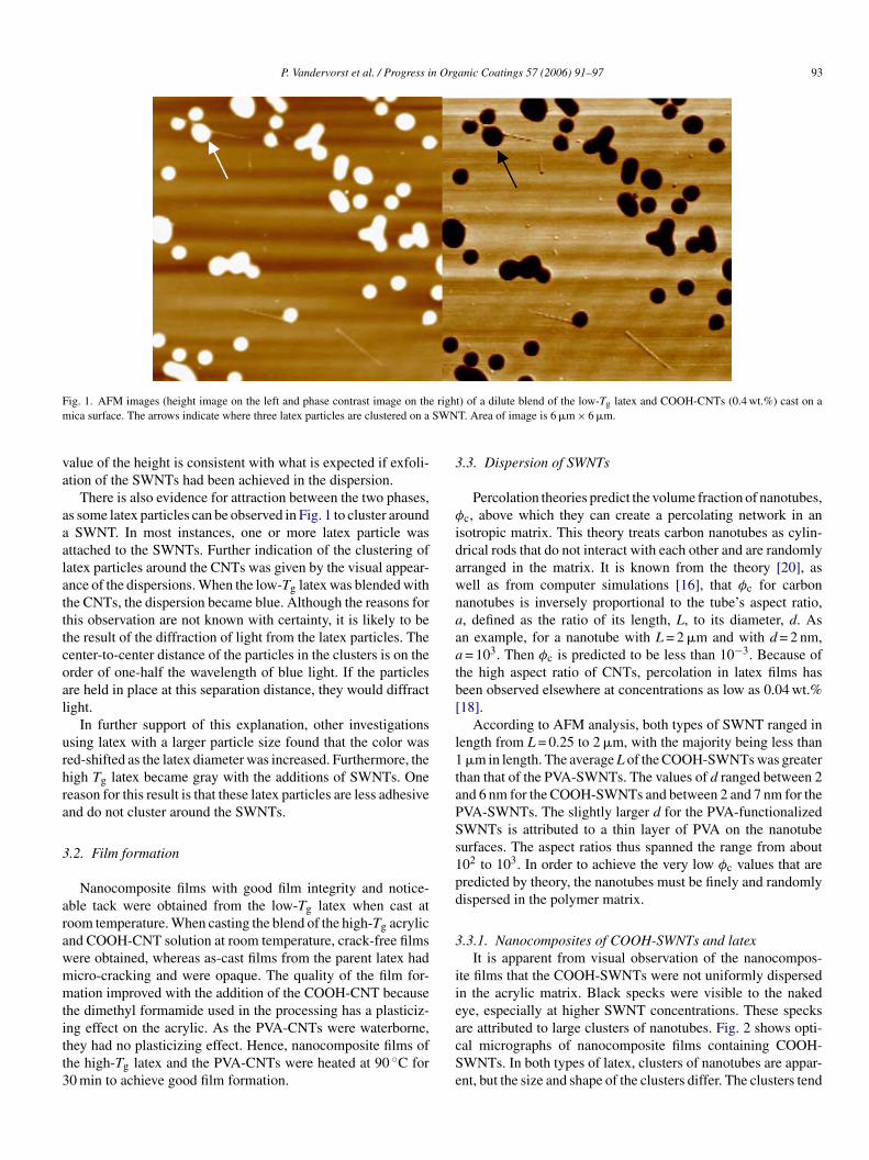
.3.1. Nanocomposites of COOH-SWNTs and latexIt is apparent from visual observation of the nanocompos-
te films that the COOH-SWNTs were not uniformly dispersedn the acrylic matrix. Black specks were visible to the nakedye, especially at higher SWNT concentrations. These specks
re attributed to large clusters of nanotubes. Fig. 2 shows opti-al micrographs of nanocomposite films containing COOH-WNTs. In both types of latex, clusters of nanotubes are appar-nt, but the size and shape of the clusters differ. The clusters tend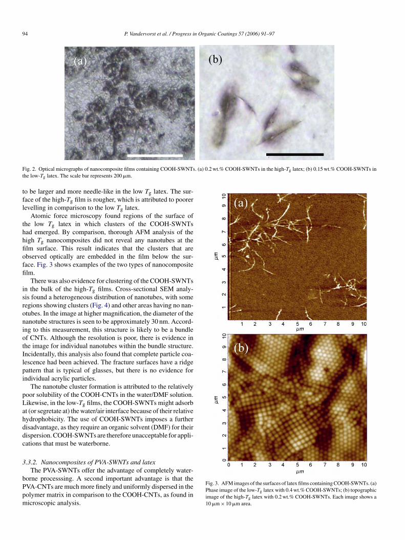
94 P. Vandervorst et al. / Progress in Organic Coatings 57 (2006) 91–97
F . (a) 0.2 wt.% COOH-SWNTs in the high-Tg latex; (b) 0.15 wt.% COOH-SWNTs int
tfl
thhfioffi
isroniotIlpi
pLahddc
3
bPpm
ig. 2. Optical micrographs of nanocomposite films containing COOH-SWNTshe low-Tg latex. The scale bar represents 200 �m.
o be larger and more needle-like in the low Tg latex. The sur-ace of the high-Tg film is rougher, which is attributed to poorerevelling in comparison to the low Tg latex.
Atomic force microscopy found regions of the surface ofhe low Tg latex in which clusters of the COOH-SWNTsad emerged. By comparison, thorough AFM analysis of theigh Tg nanocomposites did not reveal any nanotubes at thelm surface. This result indicates that the clusters that arebserved optically are embedded in the film below the sur-ace. Fig. 3 shows examples of the two types of nanocompositelm.
There was also evidence for clustering of the COOH-SWNTsn the bulk of the high-Tg films. Cross-sectional SEM analy-is found a heterogeneous distribution of nanotubes, with someegions showing clusters (Fig. 4) and other areas having no nan-tubes. In the image at higher magnification, the diameter of theanotube structures is seen to be approximately 30 nm. Accord-ng to this measurement, this structure is likely to be a bundlef CNTs. Although the resolution is poor, there is evidence inhe image for individual nanotubes within the bundle structure.ncidentally, this analysis also found that complete particle coa-escence had been achieved. The fracture surfaces have a ridgeattern that is typical of glasses, but there is no evidence forndividual acrylic particles.
The nanotube cluster formation is attributed to the relativelyoor solubility of the COOH-CNTs in the water/DMF solution.ikewise, in the low-Tg films, the COOH-SWNTs might adsorbt (or segretate at) the water/air interface because of their relativeydrophobicity. The use of COOH-SWNTs imposes a furtherisadvantage, as they require an organic solvent (DMF) for theirispersion. COOH-SWNTs are therefore unacceptable for appli-ations that must be waterborne.
.3.2. Nanocomposites of PVA-SWNTs and latexThe PVA-SWNTs offer the advantage of completely water-
orne processsing. A second important advantage is that theVA-CNTs are much more finely and uniformly dispersed in theolymer matrix in comparison to the COOH-CNTs, as found inicroscopic analysis.
Fig. 3. AFM images of the surfaces of latex films containing COOH-SWNTs. (a)Phase image of the low-Tg latex with 0.4 wt.% COOH-SWNTs; (b) topographicimage of the high-Tg latex with 0.2 wt.% COOH-SWNTs. Each image shows a10 �m × 10 �m area.
P. Vandervorst et al. / Progress in Organic Coatings 57 (2006) 91–97 95
F ex an
ott(fis
Fat
cfioa
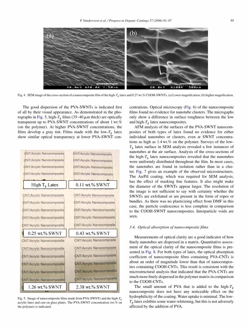
ig. 4. SEM image of the cross-section of a nanocomposite film of the high-Tg lat
The good dispersion of the PVA-SWNTs is indicated firstf all by their visual appearance. As demonstrated in the pho-ographs in Fig. 5, high-Tg films (35–40 �m thick) are opticallyransparent up to PVA-SWNT concentrations of about 1 wt.%
on the polymer). At higher PVA-SWNT concentrations, thelms develop a gray tint. Films made with the low-Tg latexhow similar optical transparency at lower PVA-SWNT con-ig. 5. Image of nanocomposite films made from PVA-SWNTs and the high-Tg
crylic latex and cast on glass plates. The PVA-SWNT concentration (wt.% onhe polymer) is indicated.
pitTntwttThttSbcts
3
fimscaimmt
nhTa
d 0.27 wt.% COOH-SWNTs. (a) Lower magnification; (b) higher magnification.
entrations. Optical microscopy (Fig. 6) of the nanocompositelms found no evidence for nanotube clusters. The microgaphsnly show a difference in surface roughness between the lownd high-Tg latex nanocomposites.
AFM analysis of the surfaces of the PVA-SWNT nanocom-osites of both types of latex found no evidence for eitherndividual nanotubes or clusters, even at SWNT concentra-ions as high as 1.4 wt.% on the polymer. Surveys of the low-g latex surface in SEM analysis revealed a few instances ofanotubes at the air surface. Analysis of the cross-sections ofhe high-Tg latex nanocomposites revealed that the nanotubesere uniformly distributed throughout the film. In most cases,
he nanotubes are found in isolation rather than in a clus-er. Fig. 7 gives an example of the observed microstructures.he Au/Pd coating, which was required for SEM analysis,as the effect of masking fine features. It also might makehe diameter of the SWNTs appear larger. The resolution ofhe image is not sufficient to say with certainty whether theWNTs are exfoliated or are present in the form of ropes orundles. As there was no plasticizing effect from DMF in thisase, the particle coalescence is less complete in comparisono the COOH-SWNT nanocomposites. Interparticle voids areeen.
.4. Optical absorption of nanocomposite films
Measurements of optical clarity are a good indicator of hownely nanotubes are dispersed in a matrix. Quantitative assess-ent of the optical clarity of the nanocomposite films is pre-
ented in Fig. 8. For both types of latex, the optical absorptionoefficient of nanocomposite films containing PVA-CNTs isbout an order of magnitude lower than that of nanocompos-tes containing COOH-CNTs. This result is consistent with the
icrostructural analysis that indicated that the PVA-CNTs areuch more finely dispersed in the polymer matrix in comparison
o the COOH-CNTs.
The small amount of PVA that is added to the high-Tganocomposite does not have any noticeable effect on theydrophilicity of the coating. Water uptake is minimal. The low-g latex exhibits some water-whitening, but this is not adverselyffected by the addition of PVA.

96 P. Vandervorst et al. / Progress in Organic Coatings 57 (2006) 91–97
Fig. 6. Optical micrographs of nanocomposite films containing PVA-SWNTs. (a) 2.5 wt.% PVA-SWNTs in the high-Tg latex; (b) 3.1 wt.% PVA-SWNTs in thelow-Tg latex. The scale bar represents 200 �m.
Fig. 7. SEM image of the cross-section of a nanocomposite film of the high-Tg latexThe cracks that appear in (b) are in the Au coating.
Fig. 8. Optical absorption coefficients at a wavelength of 600 nm for four typesof nanocomposite film. Triangles indicate nanocomposites containing COOH-Cl
4
p
tnsioneo
mcwobn
ptio
NTs; squares indicate those with PVA-CNTs. The filled symbols are for theow-Tg latex; the open symbols are for the high-Tg latex.
. Conclusions
Single-wall carbon nanotubes were functionalized witholy(vinyl alcohol) to render them water-soluble. The CNT solu-
A
t
and 1.26 wt.% PVA-CNTs. (a) Lower magnification; (b) higher magnification.
ions were blended with acrylic latex dispersions to produceanocomposite wateborne coatings. Analysis by microscopyhowed that the PVA-functionalized CNTs were finely dispersedn the polymer matrix. The nanocomposite films exhibited lowptical absorption coefficients. It is expected that this type ofanocomposite coating will potentially exhibit high thermal andlectrical conductivity along with high wear-resistance whileffering the advantage of solvent-free processing.
COOH-functionalized CNTs were dispersed in dimethyl for-amide and were similarly blended with latex dispsersions. In
ontrast to the PVA-functionalized CNTs, this type of nanotubeas not as uniformly dispersed in the latex, resulting in higherptical absorption. Microscopy provided evidence for nanotubeundles. In some cases, larger clusters were even apparent to theaked eye.
Further study of the mechanical properties of the nanocom-osites is underway in our laboratory. Because the use of surfac-ants or other dispersants are avoided, the coating’s hydrophilic-ty and barrier properties are expected to be comparable to thatf the parent latex coating.
cknowledgements
P.V.’s stay at the University of Surrey was supported throughhe SOCRATES student exchange programme. A travel grant
n Org
faivC2
R
[[[[[[
P. Vandervorst et al. / Progress i
rom the Royal Society enabled J.L.K. to present this workt the Coatings Science International meeting (COSI 2005)n Noordwijk, The Netherlands. Support for C.-H.L. was pro-ided through the NsHAPe project funded by the Europeanommission’s Framework 6 Programme (Contract NMP3-CT-004-505442).
eferences
[1] J.M. Salamanca, et al., Langmuir 17 (2001) 3202.[2] A.F. Routh, W.B. Zimmerman, Chem. Eng. Sci. 59 (2004) 2961.[3] J.-P. Gorce, et al., Eur. Phys. J. E 8 (2002) 421.[4] A.F. Routh, W.B. Russel, Langmuir 15 (1999) 7762.[5] A.F. Routh, W.B. Russel, Ind. Eng. Chem. Res. 40 (2001) 4302.
[[[[[
anic Coatings 57 (2006) 91–97 97
[6] D. Juhue, J. Lang, Macromolecules 28 (1995) 1306.[7] J.L. Keddie, Mater. Sci. Eng. Rep. 21 (1997) 101.[8] W.A. de Heer, MRS Bull. 29 (2004) 281.[9] A.B. Dalton, et al., J. Mater. Chem. 14 (2004) 1.10] R. Andrews, et al., Appl. Phys. Lett. 75 (1999) 1329.11] J.K.W. Sandler, et al., Polymer 44 (2003) 5893.12] J.K.W. Sandler, et al., Polymer 45 (2004) 2001.13] B.P. Grady, et al., J. Phys. Chem. B 106 (2002) 5852.14] R. Sainz, et al., Nanotechnology 16 (2005) S150.15] Y. Lin, et al., Macromolecules 36 (2003) 7199.
16] M. Foygel, et al., Phys. Rev. B 71 (2005) 104201.17] A. Dufresne, et al., J. Mater. Sci. 37 (2002) 3915.18] O. Regev, et al., Adv. Mater. 16 (2004) 248.19] J.C. Grunlan, et al., Adv. Mater. 16 (2004) 150.20] I. Balberg, et al., Phys. Rev. B 30 (1984) 3933.