Embed Size (px)
Citation preview
processes
Article
Functionalized N-Pyridinylmethyl EngraftedBisarylmethylidenepyridinones as Anticancer Agents
Dhaifallah M. Al-thamili, Abdulrahman I. Almansour, Natarajan Arumugam ,Faruq Mohammad and Raju Suresh Kumar *
Department of Chemistry, College of Science, King Saud University, P.O. Box 2455, Riyadh 11451, Saudi Arabia;[email protected] (D.M.A.-t.); [email protected] (A.I.A.); [email protected] (N.A.);[email protected] (F.M.)* Correspondence: [email protected] or [email protected]
Received: 25 July 2020; Accepted: 11 September 2020; Published: 15 September 2020�����������������
Abstract: Structurally interesting N-pyridinylmethyl engrafted bisarylmethylidenepyridinones withhigh functionality have been constructed in good yield. The structural interpretation of thesecompounds has been done with the aid of spectroscopic analysis and further established by singlecrystal X-ray diffraction studies. Following physical characterization, the synthesized compoundswere tested for their in vitro anticancer activity against HepG2 cancer cells and it was found that allof the compounds exhibited some level of activity. We observed a significant level of cell viabilitylosses to the cancer cells, while only smaller losses to the non-cancer cells were observed. Besides,the mechanistic investigation of toxicology revealed that the cancer cells were undergoing apoptoticpathway, induced by the generation of oxidative stress and the involvement of caspases. The analysisprovides preliminary evidence for the successful control of cancer cells with a minimal effect onhealthy normal cells because of the high IC50 levels and cell death mechanisms.
Keywords: functionalized unsaturated ketones; anticancer studies; apoptosis; caspase-3;ROS generation
1. Introduction
Cancer is caused by a series of events, such as the abnormal growth of tumor cells, distortionof normal tissue functions, and migration of mutated cells to different sites of the body in the laterstages. The migration of diseased cells to distant sites, called metastasis, is an almost uncontrollablestage of cancer phase as it may become difficult to control the growth of mutated cells in tissueswhich are deep inside the body [1]. Despite the enormous progress achieved in cancer treatmentmethodologies through the development of new diagnosis tools and the involvement of anticancerdrugs, the treatment process still suffers from the limitations of poor efficacy, non-targeting ability,drug resistance, association with side effects, etc. [2]. Hence, it is highly important to fully understandalready available drugs’ therapeutic efficacies and invent new drugs with improved efficacy againstcancer diseased cells and reduced side effects towards healthy cells.
Compounds owning α,β-unsaturated carbonyl units are the fundamental core of numerouscompounds exhibiting attractive biological properties [3–6]. Since α,β-unsaturated ketones possesssome privileged electrophilic behaviors towards thiol groups as compared to those of amino orhydroxyl groups [7,8], they maintain some resistance from the attacking of nucleic acids with no thiolfunctionality, and thus the mutagenic and carcinogenic potential of these compounds are observedto be low [9]. In addition, there have been a number of studies that deal with the cytotoxic activitiesof α,β-unsaturated ketones which are being designed as thiol-alkylators [10–12]. In one study, forexample, acyclic α,β-unsaturated ketones were found to be nearly bereft of cytotoxic activity following
Processes 2020, 8, 1154; doi:10.3390/pr8091154 www.mdpi.com/journal/processes
Processes 2020, 8, 1154 2 of 15
their tests against P388 lymphocytic leukemia in mice [13–15]. Later studies dealt with the cytotoxicactivity of 2,6-bis(phenylmethylene)-cyclohexanone against murine P388/MRI leukemic cells wherethe IC50 value disclosed to be 36.5 nM [16]. Some N-unsubstituted 3,5-bis(arylidene)-4-piperidonesand N-acyl-3,5-bis(benzylidene)-4-piperidones resembled the compounds of the present study andexhibited IC50 values (concentration required to observe 50% cell death) in the low micromolar tosubmicromolar range towards a number of malignant cell lines [17,18]. Among these compounds,N-acyl analogues exhibited superior cytotoxic activity than their N-unsubstituted analogues towardsthe P388/MRI cells [16]. Based on the analysis of these compounds’ behavior towards the cells,the hepatic glutathione concentrations were observed to be low in mice, thereby providing evidencefor their mechanistic action as a thiol alkylator.
In some studies, it has been observed that compounds maintaining two or more alkylation sitespossessed more selective toxicity against neoplastic cells as compared to healthy normal cells [19].The observation of such activity against neoplastic cells can be attributed to the increased chemicalinsult offered by the presence of more alkylation sites, making the neoplasms more sensitive duringa second or third attack [20,21]. Additionally, some other studies have revealed the dependency ofenhanced tumor sensitivity with respect to antineoplastic drugs by the potentiators, and so compoundswith two or more thiolation sites are of utmost importance in cancer therapy [22–24]. In addition, somestudies on piperidone derivatives indicated that compounds with a higher number of unsaturatedgroups can offer greater potency because of their steric impedance, hence the thiol attack might beweaker as compared to that of a compound with low or no unsaturated groups. Besides, it is noteworthyto mention that substituted piperidine moiety is an integral part of many natural products and a fewdrugs. In addition, these compounds maintain fluorescent properties along with an antitumor capacity,which can be specifically beneficial to tumor cell studies, thereby making them potential fluorescentantitumor drug candidates [7]. Taking these facts into consideration, developing a series of cancercell-targeting compounds seems to be judicious; in that view, the current study dealt with the efficientsynthesis of targeted pyridinone analogues enclosing α,β-unsaturated ketone structural motifs asthey can attack the thiol groups of nucleic acids better than amino or hydroxy groups. Further,the antiproliferative activity of the synthesized compounds was assessed against two different cell lines:HepG2 161 and L929 cells. Finally, the mechanism of toxicity against the cancer cells was performedwith assays of apoptosis, oxidative stress, and by the involvement of caspases.
2. Materials and Methods
2.1. Chemistry
All reagents and solvents used in the present study were procured from commercial suppliers.The reaction progress was observed by thin layer chromatography (TLC) analysis. The melting pointsof the synthesized compounds were measured using open capillary tubes and were uncorrected. FT-IRspectra of these compounds were documented on a PerkinElmer system 2000 FT-IR instrument (KBr).A Jeol 500 MHz instrument was employed to measure the NMR spectra of the synthesized compoundswhile a DART-ToF-MS mass spectrometer was used to record the molecular mass. X-ray data setsof the representative compounds were collected from a STOE IPDS 2 Diffractometer, and structurerefinement was done using SHELXL2018/3 (Sheldrick, 2018) Program(s).
2.2. General Procedure for the Synthesis of N-Pyridinylmethyl-Bisarylmethylidenepyridinones 5(a–h)
To 20 mL of acetonitrile, N-unsubstituted pyridinones 3 (1 mmol) and K2CO3 (3 mmol) wereadded and stirred at ambient temperature for 30 min, followed by the gradual addition of pyridinehydrochloride 4 (1.1 mmol), and then the whole reaction mixture was allowed to reflux. Afterthe complete disappearance of the starting materials, as evidenced by the TLC analysis, the reactionmixture was allowed to cool to room temperature. The filtrate collected after filtration was evaporated,and the residue partitioned between 20 mL of CHCl3 and 10 mL of water. The organic phase was
Processes 2020, 8, 1154 3 of 15
separated, dried (Na2SO4), and evaporated. Crystallization of the crude product in acetonitrile affordedcompound 5 in pure form.
2.2.1. N-1-(2-Pyridinylmethyl)-3,5-bis[(E)-Phenylmethylidene]Tetrahydro-4(1H)-Pyridinone (5a)
Obtained as yellow solid, (1.76 g, 88%); mp = 85–88 ◦C; IR (KBr): 1671, 1612, 1586 cm−1; 1H NMR(500 MHz, CDCl3): δH 3.87 (2H, s, 7′-CH2), 3.92–3.93 (4H, m, 2′-CH2 and 6′-CH2), 7.08–7.11 (1H, m,ArH), 7.33–7.38 (11H, m, ArH), 7.50–7.54 (1H, m, ArH), 7.81 (2H, s, ArH), 8.48–8.50 (1H, m, ArH). 13CNMR (125 MHz, CDCl3): δC 54.89, 63.21, 122.27, 122.98, 128.53, 128.97, 130.37, 133.21, 135.15, 136.52,136.60, 149.12, 157.79, 187.54. Mass: 367 [M+]. Anal. calcd for C25H22N2O: C, 81.94; H, 6.05; N, 7.64%;found: C, 81.82; H, 6.23; N, 7.75%.
2.2.2. N-1-(2-Pyridinylmethyl)-3,5-bis[(E)-(2-Methylphenyl)Methylidene]Tetrahydro-4(1H)-Pyridinone (5b)
Obtained as yellow solid, (1.8 g, 90%); mp = 96–98 ◦C; IR (KBr): 1672, 1615, 1589 cm−1; 1H NMR(500 MHz, CDCl3): δH 2.34 (6H, s, CH3), 3.73–3.75 (6H, m, 2-CH2′ , 6-CH2′ and 7′-CH2), 7.05–7.25(10H, m, ArH), 7.47–7.50 (1H, m, ArH), 7.97 (2H, s, ArH), 8.42–8.43 (1H, m, ArH). 13C NMR (125 MHz,CDCl3): δC 20.02, 54.58, 62.78, 122.15, 122.94, 125.50, 128.78, 130.24, 133.35, 134.20, 136.16, 136.38, 138.05,149.13, 157.74, 187.41. Mass: 395 [M+]. Anal. calcd for C27H26N2O: C, 82.20; H, 6.64; N, 7.10%; found:C, 82.33; H, 6.49; N, 7.31%.
2.2.3. N-1-(2-Pyridinylmethyl)-3,5-bis[(E)-(2-Methoxylphenyl)Methylidene]Tetrahydro-4(1H)-Pyridinone (5c)
Obtained as yellow solid, yield = 85%; mp = 120–122 ◦C; IR (KBr): 1670, 1614, 1586 cm−1; 1HNMR (500 MHz, CDCl3): δH 3.78 (3H, s, OCH3), 3.81 (3H, s, OCH3), 3.82–3.83 (6H, m, 2′-CH2, 6-CH2′
and 7′-CH2), 6.86–6.90 (4H, m, ArH), 7.02–7.12 (3H, m, ArH), 7.23–7.29 (3H, m, ArH), 7.43–7.46 (1H, m,ArH), 8.06 (2H, s, ArH), 8.44–8.45 (1H, m, ArH). 13C NMR (125 MHz, CDCl3): δC 54.60, 55.19, 62.35,110.37, 119.75, 121.78, 122.68, 124.06, 129.94, 130.11, 132.26, 132.90, 136.120, 148.80, 157.89, 158.08, 187.34.Mass: 427 [M+]. Anal. calcd for C27H26N2O3: C, 76.03; H, 6.14; N, 6.57%; found: C, 76.24; H, 6.02; N,6.70%.
2.2.4. N-1-(2-Pyridinylmethyl)-3,5-bis[(E)-(3-Nitrophenyl)Methylidene]Tetrahydro-4(1H)-Pyridinone (5d)
Obtained as yellow solid, (1.70 g, 85%); mp = 158–160 ◦C; IR (KBr): 1668, 1615, 1589 cm−1; 1HNMR (500 MHz, CDCl3): δH 3.88 (2H, s, 7′-CH2), 3.91–3.92 (4H, m, 2′-CH2 and 6′-CH2), 7.10–7.12 (1H,m, ArH), 7.29 (1H, d, J = 7.5 Hz, ArH), 7.55–7.65 (6H, m, ArH), 7.80 (2H, s, ArH), 8.17–8.19 (3H, m,ArH), 8.48–8.50 (1H, m, ArH). 13C NMR (125 MHz, CDCl3): δC 54.40, 63.13, 123.35, 123.67, 124.53,129.76, 134.20, 135.26, 135.96, 136.61, 136.81, 148.42, 149.51, 156.92, 186.52. Mass: 457 [M+]. Anal. calcdfor C25H20N4O5: C, 65.78; H, 4.42; N, 12.27%; found: C, C, 65.91; H, 4.59; N, 12.15%.
2.2.5. N-1-(2-Pyridinylmethyl)-3,5-bis[(E)-(4-Methylphenyl)Methylidene]Tetrahydro-4(1H)-Pyridinone (5e)
Obtained as yellow solid, (1.80 g, 90%); mp = 130–133 ◦C; IR (KBr): 1670, 1618, 1586 cm−1; 1HNMR (500 MHz, CDCl3): δH 2.35 (6H, s, CH3), 3.87 (2H, s, 7′-CH2), 3.90–3.91 (4H, m, 2′-CH2 and6′-CH2), 7.08–7.10 (1H, m, ArH), 7.16–7.18 (3H, m, ArH), 7.24–7.26 (5H, m, ArH), 7.31 (1H, d, J = 7.5 Hz,ArH), 7.50–7.53 (1H, m, ArH), 7.77 (2H, s, ArH), 8.48–8.50 (1H, m, ArH). 13C NMR (125 MHz, CDCl3):δC 21.39, 55.01, 63.28, 122.25, 122.99, 129.30, 130.50, 132.38, 136.43, 136.64, 139.30, 149.10, 158.51, 187.53.Mass: 395 [M+]. Anal. calcd for C27H26N2O: C, 82.20; H, 6.64; N, 7.10%; found: C, 82.35; H, 6.76; N,7.02%.
Processes 2020, 8, 1154 4 of 15
2.2.6. N-1-(2-Pyridinylmethyl)-3,5-Bis[(E)-(4-Chlorophenyl)Methylidene]Tetrahydro-4(1H)-Pyridinone (5f)
Obtained as yellow solid, (1.76 g, 88%); mp = 129–131 ◦C; IR (KBr): 1671, 1617, 1587 cm−1; 1HNMR (500 MHz, CDCl3): δH 3.84–3.88 (6H, m, 2′-CH2, 6′-CH2 and 7′-CH2), 7.11–7.13 (1H, m, ArH),7.25–7.35 (9H, m, ArH), 7.53–7.56 (1H, m, ArH), 7.72 (2H, s, ArH), 8.48–8.50 (1H, m, ArH). 13C NMR(125 MHz, CDCl3): δC 54.73, 63.20, 122.44, 123.03, 128.85, 131.55, 133.49, 135.11, 135.31, 136.70, 149.20,157.51, 187.06. Mass: 436 [M+]. Anal. calcd for C25H20Cl2N2O: C, 68.97; H, 4.63; N, 6.43%; found: C,68.81; H, 4.51; N, 6.62%.
2.2.7. N-1-(2-Pyridinylmethyl)-3,5-Bis[(E)-(4-Fluorophenyl)Methylidene]Tetrahydro-4(1H)- Pyridinone(5g)
Obtained as yellow solid, yield = 88%; mp = 110–112 ◦C; IR (KBr): 1668, 1615, 1588 cm−1; 1HNMR (500 MHz, CDCl3): δH 3.84–3.87 (6H, m, 2′-CH2, 6′-CH2 and 7′-CH2), 7.03–7.11 (5H, m, ArH),7.28–7.33 (5H, m, ArH), 7.51–7.54 (1H, m, ArH), 7.74 (2H, s, ArH), 8.48–8.49 (1H, m, ArH). 13C NMR(125 MHz, CDCl3): δC 54.73, 63.23, 115.60, 115.78, 122.35, 122.99, 131.23, 131.24, 132.30, 132.77, 135.36,136.62, 149.16, 157.62, 161.86, 163.86, 187.19. Mass: 403 [M+]. Anal. calcd for C25H20F2N2O: C, 74.61;H, 5.01; N, 6.96%; found: C, 74.77; H, 5.14; N, 6.85%.
2.2.8. N-1-(2-Pyridinylmethyl)-3,5-Bis[(E)-(2,4-Dichlorophenyl)Methylidene]Tetrahydro-4(1H)-Pyridinone (5h)
Obtained as yellow solid, (1.80 g, 90%); mp = 105–107 ◦C; IR (KBr): 1672, 1615, 1584 cm−1; 1HNMR (500 MHz, CDCl3): δH 3.71–3.76 (4H, m, 2′-CH2 and 6′-CH2), 3.95 (2H, s, 7′-CH2), 7.05–7.25(5H, m, ArH), 7.42–7.52 (4H, m, ArH), 7.89 (2H, s, ArH), 8.44–8.45 (1H, m, ArH). 13C NMR (125 MHz,CDCl3): δC 53.86, 62.07, 122.08, 122.20, 126.55, 126.58, 129.52, 130.60, 131.66, 132.83, 134.42, 134.45,135.56, 136.27, 148.97, 157.09, 186.12. Mass: 505 [M+]. Anal. calcd for C25H18Cl4N2O: C, 59.55; H, 3.60;N, 5.56%; found: C, 59.67; H, 3.49; N, 5.65%.
2.3. Biology
2.3.1. Cell Culture and Maintenance
For the testing of the anticancer efficacy of our synthesized compounds, two different cell lines,one cancerous and the other non-cancerous, were selected. The cancer cell lines included HepG2cells of human hepatic cells, and the selected non-cancer cells were L929 cells of mouse subcutaneousconnective tissue, obtained from National Centre for Cell Science (NCCS), Pune, India. Both cell typeswere cultured individually in Eagle’s Minimum Essential Medium (EMEM) supplemented with 10%fetal bovine serum (FBS) containing the antibiotics streptomycin (100 U/mL) and penicillin (100 U/mL).For culturing, the cells were suspended in cell culture flasks and incubated at 37 ◦C in a 5% CO2
humidified atmosphere. The cells were extracted when the growth reached 80% confluency, andthe cell counting was done with the aid of a hemocytometer; the corresponding assays were performedafter calculating the cell viability and seeding.
2.3.2. MTT Cytotoxicity Assay
To understand the anticancer efficacy of the synthesized cage-like compounds 5(a–h),the 3-(4,5-dimethylthiazol-2-yl)-2,5-diphenyltetrazolium bromide (MTT) assay was performed, andwas based on the reduction of MTT dye by the cells. The HepG2 cells having the suspension of2 × 104 cells/well/200 µL of EMEM were added to 96-well plates and allowed to grow for another 12 h.After this period, different concentrations of the test compounds in the range of 0–200 µg/mL wereadded to the wells and incubated for another 24 h in a 5% CO2 atmosphere. Then, the medium wasremoved, and the fresh medium containing 2% FBS was added and incubated for another 2 h. Afterthis period, 20 µL (5 mg/mL) of MTT reagent was added to the cells and incubated in CO2 atmospheric
Processes 2020, 8, 1154 5 of 15
air for another 2 h. At the end of incubation, the formazan crystals formed were dissolved in 100 µL ofdimethylsufoxide (DMSO) and the absorbance was measured at 570 nm using an ELISA microplatereader. The cells without any testing sample treatment were taken as the negative control with 100%viability and Melphalan (Mel) of 15 µg/mL concentration was taken as the positive control where the %(percentage) viability of the cells was determined from the following Equation (1).
% viability =Mean absorbanceo f test sample
Mean absorbanceo f negative control× 100 (1)
Similarly, IC50 values were determined for all of the testing samples (the IC50 value for eachsample was determined based on the linear regression equation generated from the plot of logconcentration vs. cell viability). From the analysis of the results provided by the MTT assay and IC50
values, the highly effective samples 5(a,g) were determined, and cell viability studies were performedusing the non-cancer L929 cell line so as to understand the active samples’ behavior towards healthynormal cells.
2.3.3. Apoptosis Assay
The apoptotic assay was carried out on the HepG2 cells using the testing sample of 5g, for whichthe Annexin V/FITC kit (BD Biosciences; Catalog no. 556547) was employed. For the assay, the cells ata density of 1 × 10⁶ cells/well were plated and allowed to settle for 12 h, and after the period, the wellswere treated with IC50 concentrations of the testing compound (5g) and the positive control of Meland incubated for another 24 h. The flow cytometric analysis was used to estimate the fluorescenceintensities generated by the cells due to the involvement of FITC-conjugated Annexin V and PI(propidium iodide) dyes. For the analysis, the samples of treated cells were collected, washed witha PBS buffer, and resuspended in Annexin V-FITC buffer. The suspension was incubated at 25 ◦C for10 min in the dark with 5 µL of Annexin V-FITC (provided by the manufacturer). Then, the cell pelletwas collected using centrifugation and suspended again in an Annexin V-FITC buffer, and 5 µL of PIwas added in an ice bath and further analyzed on a flow cytometry instrument that operates on a CellQuest software (Becton Dickinson Biosciences). Each experiment was repeated thrice for accuracy andthe average of each individual fluorescence experiment was taken as the final where the data shownare the mean ± SD (n = 3).
2.3.4. Oxidative Stress Assay
The generation of oxidative stress due to the treatment of compound 5g in the HepG2 cells wasstudied by analyzing the reactive oxygen species (ROS) formation. For the testing, the cells at a densityof 1 × 10⁵ cells/well were first seeded in 6-well plates, allowed to adhere, and when the cell growthreached its 80% confluence, they were treated with the IC50 values of the testing compounds (5g andMel), and incubated for another 24 h. After this period, the medium was removed, and the cells werewashed with D-PBS twice and incubated with 10 µM of H2DFCDA (provided by the manufacturer,Invitrogen, catalog number: D399) in a dark condition at room temperature for 30 min. All of the assayprotocols were followed as indicated by the manufacturer, i.e., washing of the cultured cells withD-PBS twice, pellet collection using centrifugation, resuspension of the obtained pellet in 500 µL ofD-PBS, and further analysis of the cells by BD FACS Calibur. The compound 5g results were comparedwith those of Mel and the cells of without any treatment.
2.3.5. Caspase-3 Expression Assay
Similar to the earlier assays, the HepG2 cells at density of 1 × 10⁵ cells/well were seeded in6-well culturing plates, allowed to grow for 12 h, treated with respective test compounds, andallowed to incubate for 24 h. After the incubation period, all steps were followed in accordance withthe manufacturer’s instruction, i.e., removal of the medium, washing of the cells with D-PBS twice,
Processes 2020, 8, 1154 6 of 15
and fixing of the cells with 2% paraformaldehyde. The cells were then permeabilized with 0.01%Tween 20 in D-PBS and, having 1% bovine serum albumin (BSA), incubated with 50 µL of Caspase3–FITC (Abcam, ab65613) in the dark at room temperature for another 60 min. Finally, the cells werewashed, suspended in a PBS solution, and analyzed for the expression of caspases by an EVOS FLAUTO fluorescence microscope.
2.3.6. Statistical Analysis
Each sample was tested thrice for the accuracy of its biological efficacy, and the average of all ofthe obtained values was considered as final, where the data shown are the mean ± SD. The statisticalanalysis was based on the Student’s t-test, and, from the analysis, the significant values (p < 0.05) areshown by * and the highly significant values (p < 0.01) are shown as ** against the untreated controlmeasurements run using the GraphPad prism software (version 6).
3. Results and Discussion
3.1. Chemistry
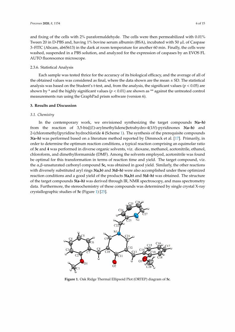
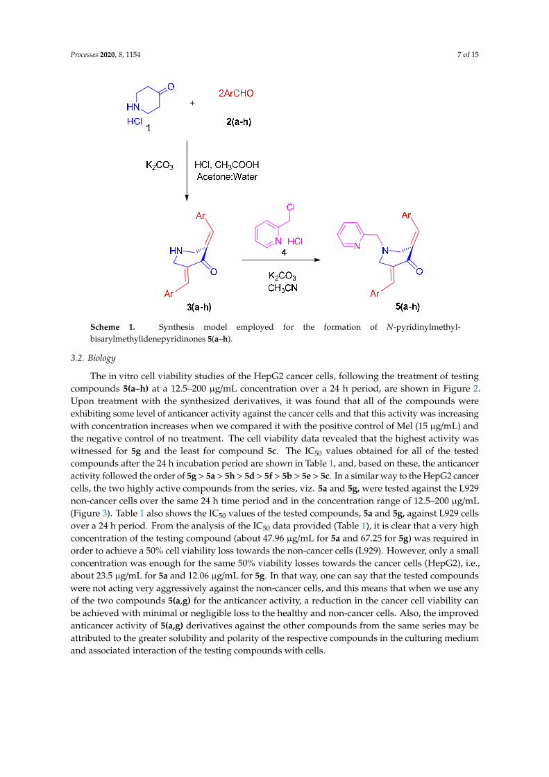
In the contemporary work, we envisioned synthesizing the target compounds 5(a–h)from the reaction of 3,5-bis[(E)-arylmethylidene]tetrahydro-4(1H)-pyridinones 3(a–h) and2-(chloromethyl)pyridine hydrochloride 4 (Scheme 1). The synthesis of the prerequisite compounds3(a–h) was performed based on a literature method reported by Dimmock et al. [17]. Primarily, inorder to determine the optimum reaction conditions, a typical reaction comprising an equimolar ratioof 3c and 4 was performed in diverse organic solvents, viz. dioxane, methanol, acetonitrile, ethanol,chloroform, and dimethylformamide (DMF). Among the solvents employed, acetonitrile was foundbe optimal for this transformation in terms of reaction time and yield. The target compound, viz.the α,β-unsaturated carbonyl compound 5c, was obtained in good yield. Similarly, the other reactionswith diversely substituted aryl rings 3(a,b) and 3(d–h) were also accomplished under these optimizedreaction conditions and a good yield of the products 5(a,b) and 5(d–h) was obtained. The structureof the target compounds 5(a–h) was derived through IR, NMR spectroscopy, and mass spectrometrydata. Furthermore, the stereochemistry of these compounds was determined by single crystal X-raycrystallographic studies of 5c (Figure 1) [25].Processes 2020, 8, x FOR PEER REVIEW 7 of 15
Figure 1. Oak Ridge Thermal Ellipsoid Plot (ORTEP) diagram of 5c.
3.2. Biology
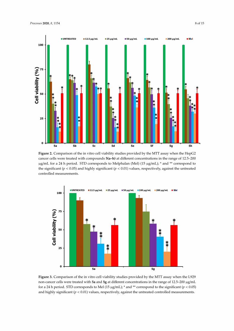
The in vitro cell viability studies of the HepG2 cancer cells, following the treatment of testing
compounds 5(a–h) at a 12.5–200 μg/mL concentration over a 24 h period, are shown in Figure 2. Upon
treatment with the synthesized derivatives, it was found that all of the compounds were exhibiting
some level of anticancer activity against the cancer cells and that this activity was increasing with
concentration increases when we compared it with the positive control of Mel (15 μg/mL) and the
negative control of no treatment. The cell viability data revealed that the highest activity was
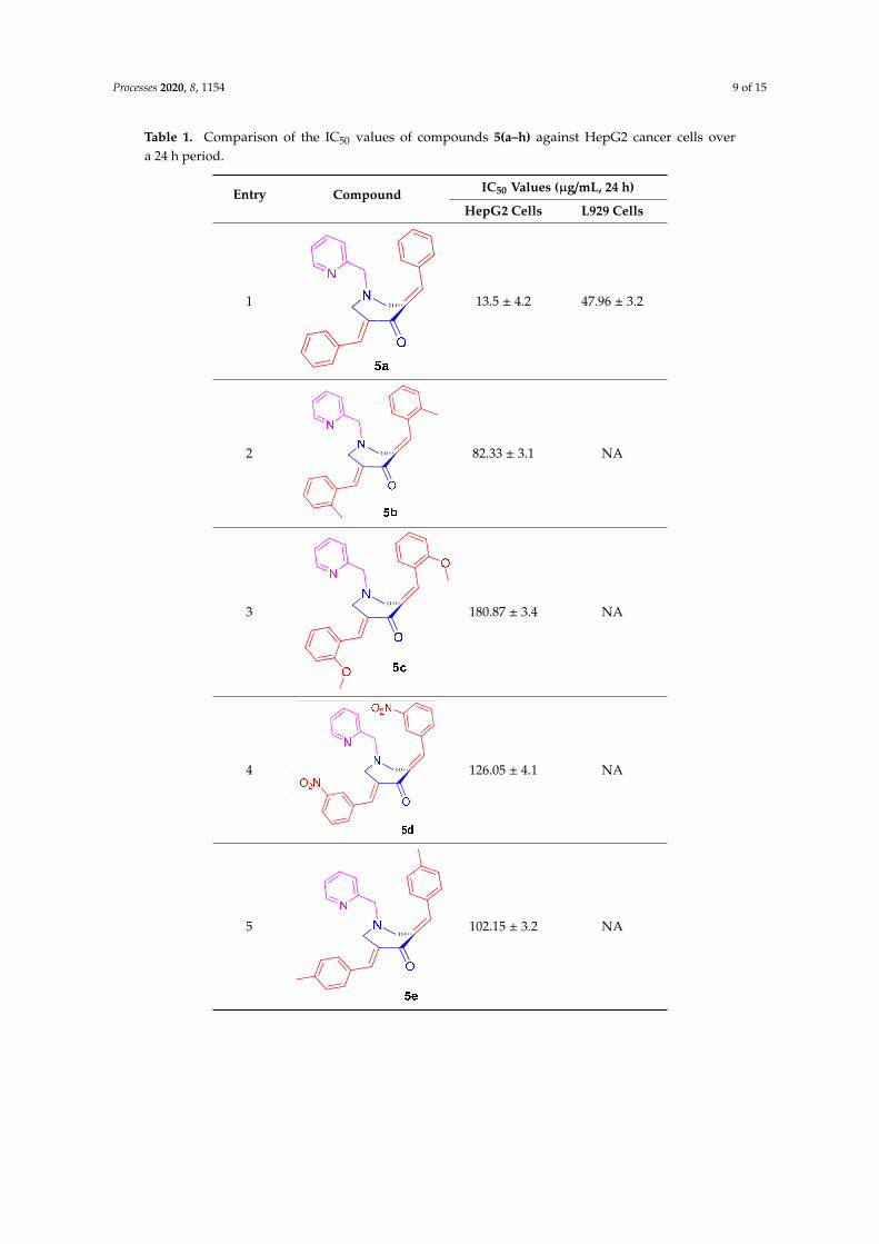
witnessed for 5g and the least for compound 5c. The IC50 values obtained for all of the tested
compounds after the 24 h incubation period are shown in Table 1, and, based on these, the anticancer
activity followed the order of 5g > 5a > 5h > 5d > 5f > 5b > 5e > 5c. In a similar way to the HepG2
cancer cells, the two highly active compounds from the series, viz. 5a and 5g, were tested against the
L929 non‐cancer cells over the same 24 h time period and in the concentration range of 12.5–200
μg/mL (Figure 3). Table 1 also shows the IC50 values of the tested compounds, 5a and 5g, against L929
cells over a 24 h period. From the analysis of the IC50 data provided (Table 1), it is clear that a very
high concentration of the testing compound (about 47.96 μg/mL for 5a and 67.25 for 5g) was required
in order to achieve a 50% cell viability loss towards the non‐cancer cells (L929). However, only a small
concentration was enough for the same 50% viability losses towards the cancer cells (HepG2), i.e.,
about 23.5 μg/mL for 5a and 12.06 μg/mL for 5g. In that way, one can say that the tested compounds
were not acting very aggressively against the non‐cancer cells, and this means that when we use any
of the two compounds 5(a,g) for the anticancer activity, a reduction in the cancer cell viability can be
achieved with minimal or negligible loss to the healthy and non‐cancer cells. Also, the improved
anticancer activity of 5(a,g) derivatives against the other compounds from the same series may be
attributed to the greater solubility and polarity of the respective compounds in the culturing medium
and associated interaction of the testing compounds with cells.
Figure 1. Oak Ridge Thermal Ellipsoid Plot (ORTEP) diagram of 5c.
Processes 2020, 8, 1154 7 of 15
Processes 2020, 8, x FOR PEER REVIEW 6 of 15
suspended in a PBS solution, and analyzed for the expression of caspases by an EVOS FL AUTO
fluorescence microscope.
2.3.6. Statistical Analysis
Each sample was tested thrice for the accuracy of its biological efficacy, and the average of all of
the obtained values was considered as final, where the data shown are the mean ± SD. The statistical
analysis was based on the Student’s t‐test, and, from the analysis, the significant values (p < 0.05) are
shown by * and the highly significant values (p < 0.01) are shown as ** against the untreated control
measurements run using the GraphPad prism software (version 6).
3. Results and Discussion
3.1. Chemistry
In the contemporary work, we envisioned synthesizing the target compounds 5(a–h) from the
reaction of 3,5‐bis[(E)‐arylmethylidene]tetrahydro‐4(1H)‐pyridinones 3(a–h) and 2‐
(chloromethyl)pyridine hydrochloride 4 (Scheme 1). The synthesis of the prerequisite compounds
3(a–h) was performed based on a literature method reported by Dimmock et al. [17]. Primarily, in
order to determine the optimum reaction conditions, a typical reaction comprising an equimolar ratio
of 3c and 4 was performed in diverse organic solvents, viz. dioxane, methanol, acetonitrile, ethanol,
chloroform, and dimethylformamide (DMF). Among the solvents employed, acetonitrile was found
be optimal for this transformation in terms of reaction time and yield. The target compound, viz. the
α,β‐unsaturated carbonyl compound 5c, was obtained in good yield. Similarly, the other reactions
with diversely substituted aryl rings 3(a,b) and 3(d–h) were also accomplished under these optimized
reaction conditions and a good yield of the products 5(a,b) and 5(d–h) was obtained. The structure
of the target compounds 5(a–h) was derived through IR, NMR spectroscopy, and mass spectrometry
data. Furthermore, the stereochemistry of these compounds was determined by single crystal X‐ray
crystallographic studies of 5c (Figure 1) [25].
Scheme 1. Synthesis model employed for the formation of N‐pyridinylmethyl‐
bisarylmethylidenepyridinones 5(a–h).
Scheme 1. Synthesis model employed for the formation of N-pyridinylmethyl-bisarylmethylidenepyridinones 5(a–h).
3.2. Biology
The in vitro cell viability studies of the HepG2 cancer cells, following the treatment of testingcompounds 5(a–h) at a 12.5–200 µg/mL concentration over a 24 h period, are shown in Figure 2.Upon treatment with the synthesized derivatives, it was found that all of the compounds wereexhibiting some level of anticancer activity against the cancer cells and that this activity was increasingwith concentration increases when we compared it with the positive control of Mel (15 µg/mL) andthe negative control of no treatment. The cell viability data revealed that the highest activity waswitnessed for 5g and the least for compound 5c. The IC50 values obtained for all of the testedcompounds after the 24 h incubation period are shown in Table 1, and, based on these, the anticanceractivity followed the order of 5g > 5a > 5h > 5d > 5f > 5b > 5e > 5c. In a similar way to the HepG2 cancercells, the two highly active compounds from the series, viz. 5a and 5g, were tested against the L929non-cancer cells over the same 24 h time period and in the concentration range of 12.5–200 µg/mL(Figure 3). Table 1 also shows the IC50 values of the tested compounds, 5a and 5g, against L929 cellsover a 24 h period. From the analysis of the IC50 data provided (Table 1), it is clear that a very highconcentration of the testing compound (about 47.96 µg/mL for 5a and 67.25 for 5g) was required inorder to achieve a 50% cell viability loss towards the non-cancer cells (L929). However, only a smallconcentration was enough for the same 50% viability losses towards the cancer cells (HepG2), i.e.,about 23.5 µg/mL for 5a and 12.06 µg/mL for 5g. In that way, one can say that the tested compoundswere not acting very aggressively against the non-cancer cells, and this means that when we use anyof the two compounds 5(a,g) for the anticancer activity, a reduction in the cancer cell viability canbe achieved with minimal or negligible loss to the healthy and non-cancer cells. Also, the improvedanticancer activity of 5(a,g) derivatives against the other compounds from the same series may beattributed to the greater solubility and polarity of the respective compounds in the culturing mediumand associated interaction of the testing compounds with cells.
Processes 2020, 8, 1154 8 of 15Processes 2020, 8, x FOR PEER REVIEW 8 of 15
Figure 2. Comparison of the in vitro cell viability studies provided by the MTT assay when the HepG2
cancer cells were treated with compounds 5(a–h) at different concentrations in the range of 12.5–200
μg/mL for a 24 h period. STD corresponds to Melphalan (Mel) (15 μg/mL); * and ** correspond to the
significant (p < 0.05) and highly significant (p < 0.01) values, respectively, against the untreated
controlled measurements.
Figure 3. Comparison of the in vitro cell viability studies provided by the MTT assay when the L929
non‐cancer cells were treated with 5a and 5g at different concentrations in the range of 12.5–200
μg/mL for a 24 h period. STD corresponds to Mel (15 μg/mL); * and ** correspond to the significant (p
< 0.05) and highly significant (p < 0.01) values, respectively, against the untreated controlled
measurements.
Figure 2. Comparison of the in vitro cell viability studies provided by the MTT assay when the HepG2cancer cells were treated with compounds 5(a–h) at different concentrations in the range of 12.5–200µg/mL for a 24 h period. STD corresponds to Melphalan (Mel) (15 µg/mL); * and ** correspond tothe significant (p < 0.05) and highly significant (p < 0.01) values, respectively, against the untreatedcontrolled measurements.
Processes 2020, 8, x FOR PEER REVIEW 8 of 15
Figure 2. Comparison of the in vitro cell viability studies provided by the MTT assay when the HepG2
cancer cells were treated with compounds 5(a–h) at different concentrations in the range of 12.5–200
μg/mL for a 24 h period. STD corresponds to Melphalan (Mel) (15 μg/mL); * and ** correspond to the
significant (p < 0.05) and highly significant (p < 0.01) values, respectively, against the untreated
controlled measurements.
Figure 3. Comparison of the in vitro cell viability studies provided by the MTT assay when the L929
non‐cancer cells were treated with 5a and 5g at different concentrations in the range of 12.5–200
μg/mL for a 24 h period. STD corresponds to Mel (15 μg/mL); * and ** correspond to the significant (p
< 0.05) and highly significant (p < 0.01) values, respectively, against the untreated controlled
measurements.
Figure 3. Comparison of the in vitro cell viability studies provided by the MTT assay when the L929non-cancer cells were treated with 5a and 5g at different concentrations in the range of 12.5–200 µg/mLfor a 24 h period. STD corresponds to Mel (15 µg/mL); * and ** correspond to the significant (p < 0.05)and highly significant (p < 0.01) values, respectively, against the untreated controlled measurements.
Processes 2020, 8, 1154 9 of 15
Table 1. Comparison of the IC50 values of compounds 5(a–h) against HepG2 cancer cells overa 24 h period.
Entry Compound IC50 Values (µg/mL, 24 h)
HepG2 Cells L929 Cells
1
Processes 2020, 8, x FOR PEER REVIEW 9 of 15
Table 1. Comparison of the IC50 values of compounds 5(a–h) against HepG2 cancer cells over a 24 h
period.
Entry Compound IC50 Values (μg/mL, 24 h)
HepG2 Cells L929 Cells
1
13.5 ± 4.2 47.96 ± 3.2
2
82.33 ± 3.1 NA
3
180.87 ± 3.4 NA
4
126.05 ± 4.1 NA
5
102.15 ± 3.2 NA
6
56.72 ± 2.9 NA
13.5 ± 4.2 47.96 ± 3.2
2
Processes 2020, 8, x FOR PEER REVIEW 9 of 15
Table 1. Comparison of the IC50 values of compounds 5(a–h) against HepG2 cancer cells over a 24 h
period.
Entry Compound IC50 Values (μg/mL, 24 h)
HepG2 Cells L929 Cells
1
13.5 ± 4.2 47.96 ± 3.2
2
82.33 ± 3.1 NA
3
180.87 ± 3.4 NA
4
126.05 ± 4.1 NA
5
102.15 ± 3.2 NA
6
56.72 ± 2.9 NA
82.33 ± 3.1 NA
3
Processes 2020, 8, x FOR PEER REVIEW 9 of 15
Table 1. Comparison of the IC50 values of compounds 5(a–h) against HepG2 cancer cells over a 24 h
period.
Entry Compound IC50 Values (μg/mL, 24 h)
HepG2 Cells L929 Cells
1
13.5 ± 4.2 47.96 ± 3.2
2
82.33 ± 3.1 NA
3
180.87 ± 3.4 NA
4
126.05 ± 4.1 NA
5
102.15 ± 3.2 NA
6
56.72 ± 2.9 NA
180.87 ± 3.4 NA
4
Processes 2020, 8, x FOR PEER REVIEW 9 of 15
Table 1. Comparison of the IC50 values of compounds 5(a–h) against HepG2 cancer cells over a 24 h
period.
Entry Compound IC50 Values (μg/mL, 24 h)
HepG2 Cells L929 Cells
1
13.5 ± 4.2 47.96 ± 3.2
2
82.33 ± 3.1 NA
3
180.87 ± 3.4 NA
4
126.05 ± 4.1 NA
5
102.15 ± 3.2 NA
6
56.72 ± 2.9 NA
126.05 ± 4.1 NA
5
Processes 2020, 8, x FOR PEER REVIEW 9 of 15
Table 1. Comparison of the IC50 values of compounds 5(a–h) against HepG2 cancer cells over a 24 h
period.
Entry Compound IC50 Values (μg/mL, 24 h)
HepG2 Cells L929 Cells
1
13.5 ± 4.2 47.96 ± 3.2
2
82.33 ± 3.1 NA
3
180.87 ± 3.4 NA
4
126.05 ± 4.1 NA
5
102.15 ± 3.2 NA
6
56.72 ± 2.9 NA
102.15 ± 3.2 NA
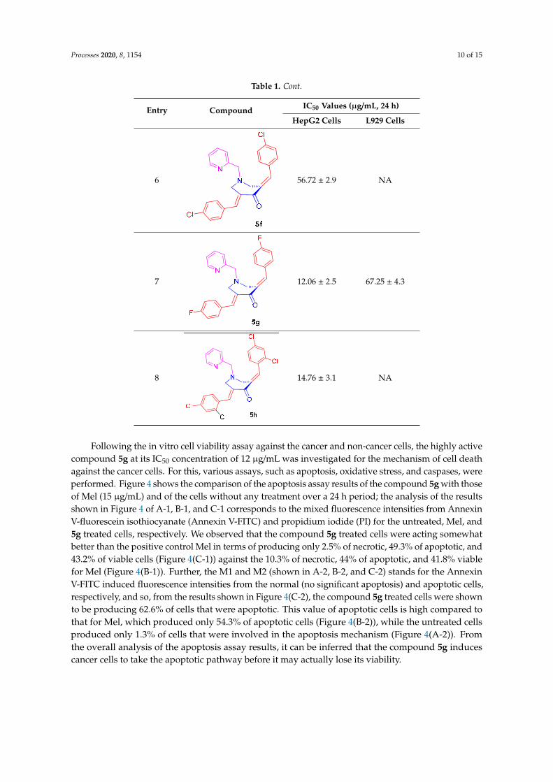
Processes 2020, 8, 1154 10 of 15
Table 1. Cont.
Entry Compound IC50 Values (µg/mL, 24 h)
HepG2 Cells L929 Cells
6
Processes 2020, 8, x FOR PEER REVIEW 9 of 15
Table 1. Comparison of the IC50 values of compounds 5(a–h) against HepG2 cancer cells over a 24 h
period.
Entry Compound IC50 Values (μg/mL, 24 h)
HepG2 Cells L929 Cells
1
13.5 ± 4.2 47.96 ± 3.2
2
82.33 ± 3.1 NA
3
180.87 ± 3.4 NA
4
126.05 ± 4.1 NA
5
102.15 ± 3.2 NA
6
56.72 ± 2.9 NA 56.72 ± 2.9 NA
7
Processes 2020, 8, x FOR PEER REVIEW 10 of 15
7
12.06 ± 2.5 67.25 ± 4.3
8
14.76 ± 3.1 NA
Following the in vitro cell viability assay against the cancer and non‐cancer cells, the highly
active compound 5g at its IC50 concentration of 12 μg/mL was investigated for the mechanism of cell
death against the cancer cells. For this, various assays, such as apoptosis, oxidative stress, and
caspases, were performed. Figure 4 shows the comparison of the apoptosis assay results of the
compound 5g with those of Mel (15 μg/mL) and of the cells without any treatment over a 24 h period;
the analysis of the results shown in Figure 4 of A‐1, B‐1, and C‐1 corresponds to the mixed
fluorescence intensities from Annexin V‐fluorescein isothiocyanate (Annexin V‐FITC) and
propidium iodide (PI) for the untreated, Mel, and 5g treated cells, respectively. We observed that the
compound 5g treated cells were acting somewhat better than the positive control Mel in terms of
producing only 2.5% of necrotic, 49.3% of apoptotic, and 43.2% of viable cells (Figure 4(C‐1)) against
the 10.3% of necrotic, 44% of apoptotic, and 41.8% viable for Mel (Figure 4(B‐1)). Further, the M1 and
M2 (shown in A‐2, B‐2, and C‐2) stands for the Annexin V‐FITC induced fluorescence intensities from
the normal (no significant apoptosis) and apoptotic cells, respectively, and so, from the results shown
in Figure 4(C‐2), the compound 5g treated cells were shown to be producing 62.6% of cells that were
apoptotic. This value of apoptotic cells is high compared to that for Mel, which produced only 54.3%
of apoptotic cells (Figure 4(B‐2)), while the untreated cells produced only 1.3% of cells that were
involved in the apoptosis mechanism (Figure 4(A‐2)). From the overall analysis of the apoptosis assay
results, it can be inferred that the compound 5g induces cancer cells to take the apoptotic pathway
before it may actually lose its viability.
12.06 ± 2.5 67.25 ± 4.3
8
Processes 2020, 8, x FOR PEER REVIEW 10 of 15
7
12.06 ± 2.5 67.25 ± 4.3
8
14.76 ± 3.1 NA
Following the in vitro cell viability assay against the cancer and non‐cancer cells, the highly
active compound 5g at its IC50 concentration of 12 μg/mL was investigated for the mechanism of cell
death against the cancer cells. For this, various assays, such as apoptosis, oxidative stress, and
caspases, were performed. Figure 4 shows the comparison of the apoptosis assay results of the
compound 5g with those of Mel (15 μg/mL) and of the cells without any treatment over a 24 h period;
the analysis of the results shown in Figure 4 of A‐1, B‐1, and C‐1 corresponds to the mixed
fluorescence intensities from Annexin V‐fluorescein isothiocyanate (Annexin V‐FITC) and
propidium iodide (PI) for the untreated, Mel, and 5g treated cells, respectively. We observed that the
compound 5g treated cells were acting somewhat better than the positive control Mel in terms of
producing only 2.5% of necrotic, 49.3% of apoptotic, and 43.2% of viable cells (Figure 4(C‐1)) against
the 10.3% of necrotic, 44% of apoptotic, and 41.8% viable for Mel (Figure 4(B‐1)). Further, the M1 and
M2 (shown in A‐2, B‐2, and C‐2) stands for the Annexin V‐FITC induced fluorescence intensities from
the normal (no significant apoptosis) and apoptotic cells, respectively, and so, from the results shown
in Figure 4(C‐2), the compound 5g treated cells were shown to be producing 62.6% of cells that were
apoptotic. This value of apoptotic cells is high compared to that for Mel, which produced only 54.3%
of apoptotic cells (Figure 4(B‐2)), while the untreated cells produced only 1.3% of cells that were
involved in the apoptosis mechanism (Figure 4(A‐2)). From the overall analysis of the apoptosis assay
results, it can be inferred that the compound 5g induces cancer cells to take the apoptotic pathway
before it may actually lose its viability.
14.76 ± 3.1 NA
Following the in vitro cell viability assay against the cancer and non-cancer cells, the highly activecompound 5g at its IC50 concentration of 12 µg/mL was investigated for the mechanism of cell deathagainst the cancer cells. For this, various assays, such as apoptosis, oxidative stress, and caspases, wereperformed. Figure 4 shows the comparison of the apoptosis assay results of the compound 5g with thoseof Mel (15 µg/mL) and of the cells without any treatment over a 24 h period; the analysis of the resultsshown in Figure 4 of A-1, B-1, and C-1 corresponds to the mixed fluorescence intensities from AnnexinV-fluorescein isothiocyanate (Annexin V-FITC) and propidium iodide (PI) for the untreated, Mel, and5g treated cells, respectively. We observed that the compound 5g treated cells were acting somewhatbetter than the positive control Mel in terms of producing only 2.5% of necrotic, 49.3% of apoptotic, and43.2% of viable cells (Figure 4(C-1)) against the 10.3% of necrotic, 44% of apoptotic, and 41.8% viablefor Mel (Figure 4(B-1)). Further, the M1 and M2 (shown in A-2, B-2, and C-2) stands for the AnnexinV-FITC induced fluorescence intensities from the normal (no significant apoptosis) and apoptotic cells,respectively, and so, from the results shown in Figure 4(C-2), the compound 5g treated cells were shownto be producing 62.6% of cells that were apoptotic. This value of apoptotic cells is high compared tothat for Mel, which produced only 54.3% of apoptotic cells (Figure 4(B-2)), while the untreated cellsproduced only 1.3% of cells that were involved in the apoptosis mechanism (Figure 4(A-2)). Fromthe overall analysis of the apoptosis assay results, it can be inferred that the compound 5g inducescancer cells to take the apoptotic pathway before it may actually lose its viability.
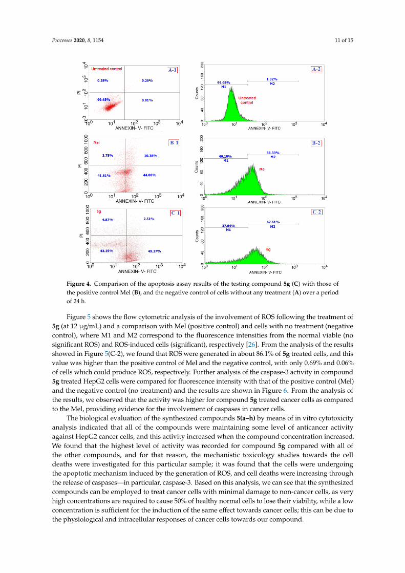
Processes 2020, 8, 1154 11 of 15Processes 2020, 8, x FOR PEER REVIEW 11 of 15
Figure 4. Comparison of the apoptosis assay results of the testing compound 5g (C) with those of the
positive control Mel (B), and the negative control of cells without any treatment (A) over a period of
24 h.
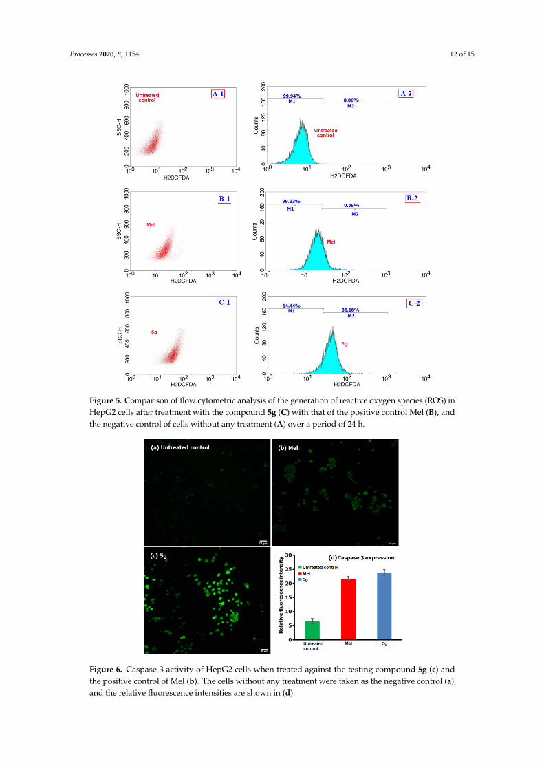
Figure 5 shows the flow cytometric analysis of the involvement of ROS following the treatment
of 5g (at 12 μg/mL) and a comparison with Mel (positive control) and cells with no treatment
(negative control), where M1 and M2 correspond to the fluorescence intensities from the normal
viable (no significant ROS) and ROS‐induced cells (significant), respectively [26]. From the analysis
of the results showed in Figure 5(C‐2), we found that ROS were generated in about 86.1% of 5g treated
cells, and this value was higher than the positive control of Mel and the negative control, with only
0.69% and 0.06% of cells which could produce ROS, respectively. Further analysis of the caspase‐3
activity in compound 5g treated HepG2 cells were compared for fluorescence intensity with that of
the positive control (Mel) and the negative control (no treatment) and the results are shown in Figure
6. From the analysis of the results, we observed that the activity was higher for compound 5g treated
cancer cells as compared to the Mel, providing evidence for the involvement of caspases in cancer
cells.
Figure 4. Comparison of the apoptosis assay results of the testing compound 5g (C) with those ofthe positive control Mel (B), and the negative control of cells without any treatment (A) over a periodof 24 h.
Figure 5 shows the flow cytometric analysis of the involvement of ROS following the treatment of5g (at 12 µg/mL) and a comparison with Mel (positive control) and cells with no treatment (negativecontrol), where M1 and M2 correspond to the fluorescence intensities from the normal viable (nosignificant ROS) and ROS-induced cells (significant), respectively [26]. From the analysis of the resultsshowed in Figure 5(C-2), we found that ROS were generated in about 86.1% of 5g treated cells, and thisvalue was higher than the positive control of Mel and the negative control, with only 0.69% and 0.06%of cells which could produce ROS, respectively. Further analysis of the caspase-3 activity in compound5g treated HepG2 cells were compared for fluorescence intensity with that of the positive control (Mel)and the negative control (no treatment) and the results are shown in Figure 6. From the analysis ofthe results, we observed that the activity was higher for compound 5g treated cancer cells as comparedto the Mel, providing evidence for the involvement of caspases in cancer cells.
The biological evaluation of the synthesized compounds 5(a–h) by means of in vitro cytotoxicityanalysis indicated that all of the compounds were maintaining some level of anticancer activityagainst HepG2 cancer cells, and this activity increased when the compound concentration increased.We found that the highest level of activity was recorded for compound 5g compared with all ofthe other compounds, and for that reason, the mechanistic toxicology studies towards the celldeaths were investigated for this particular sample; it was found that the cells were undergoingthe apoptotic mechanism induced by the generation of ROS, and cell deaths were increasing throughthe release of caspases—in particular, caspase-3. Based on this analysis, we can see that the synthesizedcompounds can be employed to treat cancer cells with minimal damage to non-cancer cells, as veryhigh concentrations are required to cause 50% of healthy normal cells to lose their viability, while a lowconcentration is sufficient for the induction of the same effect towards cancer cells; this can be due tothe physiological and intracellular responses of cancer cells towards our compound.
Processes 2020, 8, 1154 12 of 15Processes 2020, 8, x FOR PEER REVIEW 12 of 15
Figure 5. Comparison of flow cytometric analysis of the generation of reactive oxygen species (ROS)
in HepG2 cells after treatment with the compound 5g (C) with that of the positive control Mel (B),
and the negative control of cells without any treatment (A) over a period of 24 h.
The biological evaluation of the synthesized compounds 5(a–h) by means of in vitro cytotoxicity
analysis indicated that all of the compounds were maintaining some level of anticancer activity
against HepG2 cancer cells, and this activity increased when the compound concentration increased.
We found that the highest level of activity was recorded for compound 5g compared with all of the
other compounds, and for that reason, the mechanistic toxicology studies towards the cell deaths
were investigated for this particular sample; it was found that the cells were undergoing the apoptotic
mechanism induced by the generation of ROS, and cell deaths were increasing through the release of
caspases—in particular, caspase‐3. Based on this analysis, we can see that the synthesized compounds
can be employed to treat cancer cells with minimal damage to non‐cancer cells, as very high
concentrations are required to cause 50% of healthy normal cells to lose their viability, while a low
concentration is sufficient for the induction of the same effect towards cancer cells; this can be due to
the physiological and intracellular responses of cancer cells towards our compound.
Figure 5. Comparison of flow cytometric analysis of the generation of reactive oxygen species (ROS) inHepG2 cells after treatment with the compound 5g (C) with that of the positive control Mel (B), andthe negative control of cells without any treatment (A) over a period of 24 h.
Processes 2020, 8, x FOR PEER REVIEW 13 of 15
Figure 6. Caspase‐3 activity of HepG2 cells when treated against the testing compound 5g (c) and the
positive control of Mel (b). The cells without any treatment were taken as the negative control (a), and
the relative fluorescence intensities are shown in (d).
4. Conclusions
In the present work, some structurally interesting N‐pyridinylmethyl engrafted
bisarylmethylidenepyridinones with high functionality were synthesized with good yield. The
structural interpretation of these compounds was done through spectroscopic analysis. Following
the physical characterization, the compounds 5(a–h) were tested for their anticancer activities by
making use of HepG2 cells up to a 200 μg/mL concentration over a 24 h period. From the comparative
analysis of synthesized compounds against Mel (15 μg/mL) and cells without any treatment, we
observed that most of the derivatives exhibited certain levels of anticancer activity by significantly
decreasing the cells’ viability. The highest levels of activity against the cancer cells were found to be
exhibited by compounds 5a,g, and with the same compounds, when tested with the non‐cancer cells,
we observed only limited cell losses, and the IC50 values were found to be relatively very high. On
consideration of the structure–activity relationship for the two compounds, the highest level of
activity was observed for the one with two fluoro substitutions at the para position (5g), followed by
the derivative having no other substitution than that of the naked core (5a). The observation of such
activity by the two derivatives can be linked to the solubility in the culturing medium influenced by
the polarity, steric hindrance, and associated interaction with the intracellular proteins. Further
analysis of the cancer cell deaths showed that the cells were taking the apoptotic pathway, induced
by the generation of ROS and through the release of caspases. This analysis provides preliminary
evidence for the effective controlling of cancer cells with no or minimal damage to the healthy cells
because of the variation of toxicity doses and cell death mechanisms. Finally, the results generated in
this work can be used for designing new therapeutic drugs or improving on the traditional drugs by
developing a better restriction capacity over the fast growing cancer cells, while simultaneously
protecting the moderately proliferating non‐cancer cells and thereby paving the way towards
sustainable cancer treatment processes.
Figure 6. Caspase-3 activity of HepG2 cells when treated against the testing compound 5g (c) andthe positive control of Mel (b). The cells without any treatment were taken as the negative control (a),and the relative fluorescence intensities are shown in (d).
Processes 2020, 8, 1154 13 of 15
4. Conclusions
In the present work, some structurally interesting N-pyridinylmethyl engraftedbisarylmethylidenepyridinones with high functionality were synthesized with good yield.The structural interpretation of these compounds was done through spectroscopic analysis. Followingthe physical characterization, the compounds 5(a–h) were tested for their anticancer activities bymaking use of HepG2 cells up to a 200 µg/mL concentration over a 24 h period. From the comparativeanalysis of synthesized compounds against Mel (15 µg/mL) and cells without any treatment, weobserved that most of the derivatives exhibited certain levels of anticancer activity by significantlydecreasing the cells’ viability. The highest levels of activity against the cancer cells were found tobe exhibited by compounds 5a,g, and with the same compounds, when tested with the non-cancercells, we observed only limited cell losses, and the IC50 values were found to be relatively very high.On consideration of the structure–activity relationship for the two compounds, the highest level ofactivity was observed for the one with two fluoro substitutions at the para position (5g), followedby the derivative having no other substitution than that of the naked core (5a). The observation ofsuch activity by the two derivatives can be linked to the solubility in the culturing medium influencedby the polarity, steric hindrance, and associated interaction with the intracellular proteins. Furtheranalysis of the cancer cell deaths showed that the cells were taking the apoptotic pathway, induced bythe generation of ROS and through the release of caspases. This analysis provides preliminary evidencefor the effective controlling of cancer cells with no or minimal damage to the healthy cells because ofthe variation of toxicity doses and cell death mechanisms. Finally, the results generated in this workcan be used for designing new therapeutic drugs or improving on the traditional drugs by developinga better restriction capacity over the fast growing cancer cells, while simultaneously protectingthe moderately proliferating non-cancer cells and thereby paving the way towards sustainable cancertreatment processes.
Author Contributions: Conceptualization, R.S.K.; Writing—original draft preparation and supervision, R.S.K.;Investigation, D.M.A.-t.; Investigation, supervision, and funding acquisition, A.I.A.; Investigation, N.A.;Writing—original draft preparation, F.M. All authors have read and agreed to the published version ofthe manuscript.
Funding: This research was funded by Deanship of Scientific Research, King Saud University, research group (No.RGP-026).
Acknowledgments: The authors would like to extend their sincere appreciation to the Deanship of ScientificResearch at King Saud University, Riyadh, Saudi Arabia for funding this work through the research grantnumber RGP-026.
Conflicts of Interest: The authors declare no conflict of interest.
References
1. Harris, C.C. Chemical and physical carcinogenesis: Advances and perspectives for the 1990s. Cancer Res.1991, 51, 5023–5044.
2. Wong, C.F.; Guminski, A.; Saunders, N.A.; Burgess, A.J. Exploiting novel cell cycle targets in the developmentof anticancer agents. Curr. Cancer Drug Targets 2005, 5, 85–102. [CrossRef] [PubMed]
3. Lunardi, F.; Guzela, M.; Rodrigues, A.T.; Corrêa, R.; Eger-Mangrich, I.; Steindel, M.; Grisard, E.C.; Assreuy, J.;Calixto, J.B.; Santos, A.R. Trypanocidal and leishmanicidal properties of substitution-containing chalcones.Antimicrob. Agents Chemother. 2003, 47, 1449–1451. [CrossRef] [PubMed]
4. Chimenti, F.; Fioravanti, R.; Bolasco, A.; Chimenti, P.; Secci, D.; Rossi, F.; Yanez, M.; Orallo, F.; Ortuso, F.;Alcaro, S. Chalcones: A Valid Scaffold for Monoamine Oxidases Inhibitors. J. Med. Chem. 2009, 52, 2818–2824.[CrossRef] [PubMed]
5. Zarghi, A.; Zebardast, T.; Hakimion, F.; Shirazi, F.H.; Rao, P.N.; Knaus, E.E. Synthesis and biologicalevaluation of 1,3-diphenylprop-2-en-1-ones possessing a methanesulfonamido or an azido pharmacophoreas cyclooxygenase-1/-2 inhibitors. Bioorg. Med. Chem. 2006, 14, 7044–7050. [CrossRef]
Processes 2020, 8, 1154 14 of 15
6. Bag, S.; Ramar, S.; Degani, M.S. Synthesis and biological evaluation of α, β-unsaturated ketone as potentialantifungal agents. Med. Chem. Res. 2009, 18, 309–316. [CrossRef]
7. Das, U.; Pati, H.N.; Sakagami, H.; Hashimoto, K.; Kawase, M.; Balzarini, J.; De Clercq, E.; Dimmock, J.R.3,5-Bis(benzylidene)-1-[3-(2-hydroxyethylthio)propanoyl]piperidin-4-ones: A novel cluster of potenttumor-selective cytotoxins. J. Med. Chem. 2011, 54, 3445–3449. [CrossRef]
8. Dimmock, J.R.; Kumar, P.; Chen, M.; Quail, J.W.; Yang, J.; Allen, T.M.; Kao, G.Y. Synthesis and cytotoxicevaluation of mesna adducts of some 1-aryl-4,4-dimethyl-5-(1-piperidino)-1-penten-3-one hydrochlorides.Pharmazie 1995, 50, 449–453. [CrossRef]
9. Okey, A.B.; Harper, P.A. Chemical carcinogenesis. In Principles of Medical Pharmacology, 7th ed.; Galant, H.,Grant, D.M., Mitchell, J., Eds.; Elsevier: Toronto, ON, Canada, 2007; p. 902.
10. Cheng, C.C. Progress in Medicinal Chemistry, Volume 25; Ellis, G.P., Wesl, G.B., Eds.; Elsevier Science Publishers:Amsterdam, The Netherlands, 1988; pp. 35–83.
11. Dimmock, J.R.; Erciyas, E.; Sidhu, K.K.; Luo, X.; Mezey, P.G.; Allen, T.M.; Murray, L. Charge densities ofatoms of conjugated styryl ketones having activity against L1210 leukemia cells. Drug Des Deliv. 1990, 7,45–49. [PubMed]
12. Dimmock, J.R.; Wong, M.C.L. Bioactivities and potential uses in drug design of acyclic alpha, beta-unsaturatedketones. Can. J. Pharm. Sci. 1976, 11, 35–53.
13. Dimmock, J.R.; Smith, L.M. Syntheses and evaluation of ketals, hemithioketals, and dithioketals of conjugatedstyryl ketones principally for antineoplastic activity. J. Pharm. Sci. 1980, 69, 575–580. [CrossRef] [PubMed]
14. Dimmock, J.R.; Nyathi, C.B.; Smith, P.J. Syntheses and bioactivities of 1-(hydroxyphenyl)-1-nonen-3-onesand related ethers and esters. J. Pharm. Sci. 1979, 68, 1216–1221. [CrossRef] [PubMed]
15. Dimmock, J.R.; Kirkpatrick, D.L.; Webb, N.G.; Cross, B.M. Synthesis and evaluation of some alkoxy-,chloro-, and acyloxy-conjugated styryl ketones against P-388 lymphocytic leukemia and an examination ofthe metabolism and toxicological effects of 1-(m-ethoxymethyloxyphenyl)-1-nonen-3-one in rats. J. Pharm.Sci. 1982, 71, 1000–1007. [CrossRef] [PubMed]
16. Dimmock, J.R.; Arora, V.K.; Wonko, S.L.; Hamon, N.W.; Quail, J.W.; Jia, Z.; Warrington, R.C.; Fang, W.D.;Lee, J.S. 3,5-Bis-benzylidene-4-piperidones and related compounds with high activity towards P388 leukemiacells. Drug Des. Deliv. 1990, 6, 183–194.
17. Dimmock, J.R.; Padmanilayam, M.P.; Puthucode, R.N.; Nazarali, A.J.; Motaganahalli, N.L.; Zello, G.A.;Quail, J.W.; Oloo, E.O.; Kraatz, H.B.; Prisciak, J.S.; et al. A conformational and structure-activity relationshipstudy of cytotoxic 3,5-bis(arylidene)-4-piperidones and related N-acryloyl analogues. J. Med. Chem. 2001, 44,586–593. [CrossRef]
18. Pati, H.N.; Das, U.; Quail, J.W.; Kawase, M.; Sakagami, H.; Dimmock, J.R. Cytotoxic3,5-bis(benzylidene)piperidin-4-ones and N-acyl analogs displaying selective toxicity for malignant cells.Eur. J. Med. Chem. 2008, 43, 1–7. [CrossRef]
19. Dimmock, J.R.; Kandepu, N.M.; Nazarali, A.J.; Motaganahalli, N.L.; Kowalchuk, T.P.; Pugazhenthi, U.;Prisciak, J.S.; Quail, J.W.; Allen, T.M.; LeClerc, R.; et al. Sequential cytotoxicity: A theory evaluated usingnovel 2-[4-(3-aryl-2-propenoyloxy)phenylmethylene]cyclohexanones and related compounds. J. Med. Chem.2000, 43, 3933–3940. [CrossRef]
20. Tsutsui, K.; Komuro, C.; Ono, K.; Nishidai, T.; Shibamoto, Y.; Takahashi, M.; Abe, M. Chemosensitization bybuthionine sulfoximine in vivo. Int. J. Radiat. Oncol. Biol. Phys. 1986, 12, 1183–1186. [CrossRef]
21. Mitchell, J.B.; Russo, A. The role of glutathione in radiation and drug induced cytotoxicity. Br. J. Cancer Suppl.1987, 8, 96–104.
22. Brandes, L.J.; Queen, G.M.; LaBella, F.S. N,N-diethyl-2-[4-(phenylmethyl)phenoxy] ethanamine (DPPE)a chemopotentiating and cytoprotective agent in clinical trials: Interaction with histamine at cytochromeP450 3A4 and other isozymes that metabolize antineoplastic drugs. Cancer Chemother. Pharmacol. 2000, 45,298–304. [CrossRef]
23. Griffin, R.J.; Arris, C.E.; Bleasdale, C.; Boyle, F.T.; Calvert, A.H.; Curtin, N.J.; Dalby, C.; Kanugula, S.;Lembicz, N.K.; Newell, D.R.; et al. Resistance-modifying agents. 8. Inhibition of O(6)-alkylguanine-DNAalkyltransferase by O(6)-alkenyl-, O(6)-cycloalkenyl-, and O(6)-(2-oxoalkyl)guanines and potentiation oftemozolomide cytotoxicity in vitro by O(6)-(1-cyclopentenylmethyl)guanine. J. Med. Chem. 2000, 43,4071–4083. [CrossRef] [PubMed]
Processes 2020, 8, 1154 15 of 15
24. Vahrmeijer, A.L.; van Dierendonck, J.H.; Schutrups, J.; van de Velde, C.J.; Mulder, G.J. Potentiationof the cytostatic effect of melphalan on colorectal cancer hepatic metastases by infusion of buthioninesulfoximine (BSO) in the rat: Enhanced tumor glutathione depletion by infusion of BSO in the hepatic artery.Cancer Chemother. Pharmacol. 1999, 44, 111–116. [CrossRef] [PubMed]
25. Al-thamili, D.M.; Almansour, A.I.; Arumugam, N.; Kansız, S.; Dege, N.; Soliman, S.M.; Azam, M.; Kumar, R.S.Highly functionalized N-1-(2-pyridinylmethyl)-3,5-bis[(E)-arylmethylidene]tetrahydro-4(1H)-pyridinones:Synthesis, characterization, crystal structure and DFT studies. J. Mol. Struct. 2020, 1222, 128940. [CrossRef]
26. Mishra, T.; Arya, R.K.; Meena, S.; Joshi, P.; Pal, M.; Meena, B.; Upreti, D.K.; Rana, T.S.; Datta, D. Isolation,characterization and anticancer potential of cytotoxic triterpenes from Betula utilis bark. PLoS ONE 2016, 11,e0159430. [CrossRef]
© 2020 by the authors. Licensee MDPI, Basel, Switzerland. This article is an open accessarticle distributed under the terms and conditions of the Creative Commons Attribution(CC BY) license (http://creativecommons.org/licenses/by/4.0/).