Embed Size (px)
Citation preview
of August 20, 2015.This information is current as
T Cells+Lines by EBV Nuclear Antigen-Specific CD4
the Recognition of EBV-Transformed B Cell A Role for Intercellular Antigen Transfer in
Martin Larsen, Jill Brooks and Alan B. RickinsonGraham S. Taylor, Heather M. Long, Tracey A. Haigh,
http://www.jimmunol.org/content/177/6/3746doi: 10.4049/jimmunol.177.6.3746
2006; 177:3746-3756; ;J Immunol
Referenceshttp://www.jimmunol.org/content/177/6/3746.full#ref-list-1
, 41 of which you can access for free at: cites 56 articlesThis article
Subscriptionshttp://jimmunol.org/subscriptions
is online at: The Journal of ImmunologyInformation about subscribing to
Permissionshttp://www.aai.org/ji/copyright.htmlSubmit copyright permission requests at:
Email Alertshttp://jimmunol.org/cgi/alerts/etocReceive free email-alerts when new articles cite this article. Sign up at:
Print ISSN: 0022-1767 Online ISSN: 1550-6606. Immunologists All rights reserved.Copyright © 2006 by The American Association of9650 Rockville Pike, Bethesda, MD 20814-3994.The American Association of Immunologists, Inc.,
is published twice each month byThe Journal of Immunology
by guest on August 20, 2015
http://ww
w.jim
munol.org/
Dow
nloaded from
by guest on August 20, 2015
http://ww
w.jim
munol.org/
Dow
nloaded from
A Role for Intercellular Antigen Transfer in the Recognition ofEBV-Transformed B Cell Lines by EBV NuclearAntigen-Specific CD4� T Cells1
Graham S. Taylor,2 Heather M. Long,2 Tracey A. Haigh, Martin Larsen, Jill Brooks, andAlan B. Rickinson3
The CD4� T cell response to EBV may have an important role in controlling virus-driven B lymphoproliferation because CD4�
T cell clones to a subset of EBV nuclear Ag (EBNA) epitopes can directly recognize virus-transformed lymphoblastoid cell lines(LCLs) in vitro and inhibit their growth. In this study, we used a panel of EBNA1, 2, 3A, and 3C-specific CD4� T cell clones tostudy the route whereby endogenously expressed EBNAs access the HLA class II-presentation pathway. Two sets of results spokeagainst a direct route of intracellular access. First, none of the clones recognized cognate Ag overexpressed in cells from vacciniavectors but did recognize Ag fused to an endo/lysosomal targeting sequence. Second, focusing on clones with the strongest LCLrecognition that were specific for EBNA2- and EBNA3C-derived epitopes LCL recognition was unaffected by inhibiting autoph-agy, a postulated route for intracellular Ag delivery into the HLA class II pathway in LCL cells. Subsequently, using these sameepitope-specific clones, we found that Ag-negative cells with the appropriate HLA-restricting allele could be efficiently sensitizedto CD4� T cell recognition by cocultivation with Ag-positive donor lines or by exposure to donor line-conditioned culture medium.Sensitization was mediated by a high m.w. antigenic species and required active Ag processing by recipient cells. We infer thatintercellular Ag transfer plays a major role in the presentation of EBNA-derived CD4 epitopes by latently infected targetcells. The Journal of Immunology, 2006, 177: 3746–3756.
E pstein-Barr virus is a B lymphotrophic herpesvirus whichhas potent B cell growth-transforming activity and islinked to several B cell malignancies, yet is carried by the
great majority of individuals as an asymptomatic infection. A rolefor the host T cell response in the long-term control of persistentEBV infection is clear from clinical observation. Thus, T cell-immunocompromised patients, in particular transplant recipientsgiven high doses of T cell-suppressive drugs, are at greatly in-creased risk of EBV-positive posttransplant lymphoproliferativedisease (PTLD)4 (1–3). Most PTLD lesions express the full spec-trum of EBV latent proteins, that is the EBV nuclear Ags (EBNAs)1, 2, 3A, 3B, 3C, and LP and the latent membrane proteins 1 and2, and in this respect resemble the lymphoblastoid cell lines(LCLs) generated when EBV transforms normal B cells in vitro.Such PTLD tumors are susceptible to immunological attack byadoptively transferred T cell populations produced in vitro by LCLstimulation of EBV latent-Ag-specific memory T cells either fromthe patient or from a HLA-matched donor (1–3). Virus-specific
CD8� T cells are thought to be the main effectors in this regardand indeed LCL-stimulated populations tend to be dominated bycytotoxic CD8� T cell clones reactive to one or more immuno-dominant epitopes, most often derived from the EBNA 3A, 3B, 3Csubset of proteins (4, 5). However, these same populations alsocontain CD4� T cells and this has stimulated increasing interest inlatent-Ag-specific CD4� T cell responses. Such responses mayhave a dual role. First, they are likely to be important in the main-tenance of virus-specific CD8� T cell surveillance in the host (6);second, because virally transformed B cells express HLA class IImolecules and have HLA class II-processing function (7), EBV-specific CD4� T cells may be capable of recognizing latently in-fected cells directly and thereby acting as effectors in their ownright.
The first CD4� T cell clones to EBV latent proteins, specific forEBNA1-and EBNA2-derived epitopes respectively, were identi-fied as rare components of LCL-reactivated memory T cell prep-arations (8, 9). Of these, only the EBNA2- specific clone appearedto be capable of recognizing LCL cells directly in cytotoxicityassays (9). Since then, CD4� recall responses to more epitopeshave been generated by a variety of protocols. Most work in thisarea has focused on EBNA1 as a CD4� T cell target and hasproduced conflicting reports as to the ability of Ag-specific CD4�
clones to recognize LCL cells endogenously expressing theEBNA1 protein from the resident EBV genome (10, 11). In a re-cent study, we widened the analysis to include responses against arange of epitopes in the EBNA1, EBNA2, EBNA3A, andEBNA3C proteins, and found that the capacity for LCL recogni-tion was highly epitope specific (12). Indeed, for any one of theabove Ags, responses to individual epitopes differed markedly intheir level of LCL recognition. Thus, while EBV infection natu-rally elicits CD4� T cell responses to a range of different EBNA-derived epitopes, only a subset of these responses are likely to have
Cancer Research U.K. Institute for Cancer Studies, University of Birmingham, Bir-mingham, United Kingdom
Received for publication September 8, 2005. Accepted for publication June 26, 2006.
The costs of publication of this article were defrayed in part by the payment of pagecharges. This article must therefore be hereby marked advertisement in accordancewith 18 U.S.C. Section 1734 solely to indicate this fact.1 This work was supported by Cancer Research U.K.2 G.S.T. and H.M.L. contributed equally to the work.3 Address correspondence and reprint requests to Prof. A. B. Rickinson, Cancer Re-search U.K. Institute for Cancer Studies, University of Birmingham, Vincent Drive,Edgbaston, Birmingham, B15 2TT, U.K. E-mail address: [email protected] Abbreviations used in this paper: PTLD, posttransplant lymphoproliferative disease;LCL, lymphoblastoid cell line; EBNA, EBV nuclear Ag; 3-MA, 3-methyladenine; Ii,invariant chain; GAr, glycine-alanine repeat; moi, multiplicity of infection; MVA,modified vaccinia Ankara; CytC, cytochrome c; BL, Burkitt’s lymphoma.
The Journal of Immunology
Copyright © 2006 by The American Association of Immunologists, Inc. 0022-1767/06/$02.00
by guest on August 20, 2015
http://ww
w.jim
munol.org/
Dow
nloaded from
direct therapeutic potential as effectors against EBV-driven lym-phoproliferations in vivo.
Given these findings, we set out to study the processing mech-anisms that lead to LCL sensitization. There are multiple exampleswhere indicator Ags have been expressed endogenously withinLCL cells and appear to have gained direct intracellular entry intothe HLA class II-processing pathway, apparently bypassing thenormal exogenous pathway in which Ag is taken up from the ex-tracellular milieu before being processed in endo/lysosomal com-partments. Many of these examples involve membrane or secretedproteins (13–16) which are thought to engage immature MHCclass II molecules during transit through the endoplasmic reticu-lum. However, others involve long-lived cytoplasmic proteins, sta-bly expressed in cells by gene transfection, which appear to enterthe endosomal compartment either by a specialized chaperone-mediated route (17) or by a pathway that was blocked by 3-methy-ladenine (3-MA) (18), a known inhibitor of autophagy (19). Mostrecently, two reports have suggested that nuclear Ags may alsoaccess the MHC class II-processing pathway by an autophagicroute. In one case, boosting autophagy in LCL cells by nutrientstarvation increased the representation of nuclear protein-derivedpeptides complexed with HLA class II molecules on the cell sur-face (20). In another, evidence was presented that in LCL cells, theendogenously expressed EBNA1 protein was processed and pre-sented to EBNA1-specific CD4� T cell clones via a 3-MA-sensi-tive autophagic route (21). In this study, we use CD4� T cellclones to a panel of EBNA-derived epitopes and show that, forthose epitopes mediating the strongest LCL recognition and there-fore representing the best targets for T cell-directed therapy, mostif not all of this recognition depended upon intercellular Ag trans-fer occurring within the LCL culture.
Materials and MethodsTarget cell lines and T cell clones
EBV-transformed LCLs were generated from normal B cells using theprototype 1 strain B95.8 or the prototype 2 strain Ag876, or a B95.8 re-combinant virus lacking the immediate early gene BZLF1 and thereforeincapable of lytic virus replication (22); all LCLs expressed the full panelof EBV latent proteins. The EBV (type 1)-positive Burkitt’s lymphoma(BL) lines Kem-BL and Oku-BL express, respectively, EBNA1 only andEBNAs 1, 3A, 3B, and 3C as described (23); the BL41 line is EBV genomenegative. All lines were routinely cultured in RPMI 1640 (Invitrogen LifeTechnologies) supplemented with 2 mM glutamine, 100 IU penicillin/ml,100 �g streptomycin/ml, and 10% FCS. All cell lines and T cell cloneswere regularly checked using immunofluorescence (Ridascreen) and theMycoalert Mycoplasma Detection kit (Cambrex) to confirm the absence ofmycoplasma contamination. CD4� and CD8� T cell clones specific fordefined epitopes within EBNA 1, 2, 3A, or 3C were generated as described(12, 24). Overall, the experiments involved CD4� T cell clones to ninedifferent EBNA-derived epitopes identified along with their HLA classII-restricting alleles in Table I. Also shown for each epitope is the effi-ciency with which epitope-specific CD4� T cells recognize the autologousLCL target; this is determined by IFN-� release and expressed as a per-centage of the IFN-� release induced by the same target LCL loaded withan optimal concentration of epitope peptide (12). T cell recognition exper-iments also included (as internal controls) CD8� T cell clones to the fol-lowing epitopes: the HLA-B35-restricted EBNA1 407–417 epitope HPV(25), the HLA-B38-restricted EBNA 2 14–23 epitope YHL (26), the HLA-B35-restricted EBNA3A 458–466 epitope YPL (27), the HLA-B27-re-stricted EBNA3C 258–266 epitope RRI (28), and the HLA-A11-restrictedEBNA3B 399–408 and 416–424 epitopes AVF and IVT (29).
Synthetic peptides and protein preparations
Epitope peptides were synthesized using 9-fluorenylmethoxycarbonylchemistry (Alta Bioscience; University of Birmingham, Birmingham,U.K.), dissolved in DMSO, and their concentrations determined by biuretassay. The 9-mer TAMRA fluorescently labeled peptide was a gift from J.Fox (Alta Bioscience, University of Birmingham, Birmingham, U.K.). Pro-tein preparations, provided by Dr. F. Grasser (Institut fur Mikrobiologie
und Hygiene, Homburg/Saar, Germany), consisted of lysates from insectcells infected with control or EBNA2 (B95.8 strain) expressingbaculoviruses.
MVA recombinants
EBNA1, 2, 3A, and 3C coding sequences (from the B95.8 EBV strain)were recombined into the modified vaccinia Ankara (MVA) genome usingthe pSC11 vaccinia virus shuttle vector as before (5); MVA recombinedwith the empty pSC11 vector served as a control. The HLA class II-tar-geted Ag constructs were made for EBNA1 by fusion with an N-terminalsignal sequence and a C-terminal LAMP1 sequence (30, 31) and forEBNAs 1, 2, 3A, and 3C by fusion with aa 1–82 of the p33 isoform of theinvariant chain (Ii) (32) at the N terminus. Note that all constructs con-taining EBNA1 were deleted for the glycine-alanine repeat (GAr) domainbecause the GAr is refractory to expression from vaccinia vectors. Exper-iments used recombinant virus preparations purified by sucrose gradientcentrifugation (33) to minimize contamination of virus stocks with non-virion proteins (in particular, the EBNA protein) present in infected cellsduring production of stocks. Expression of the relevant EBNA proteins inthe target cells for T cell recognition assays was confirmed by immuno-blotting of protein extracts from rMVA-infected LCLs (multiplicity of in-fection (moi) 10; 18 h postinfection) probed with mAbs 1H4 to EBNA1,PE2 to EBNA2, E3cA10 to EBNA3C (23) and a polyclonal sheep serumto EBNA3A (Exalpha Biologicals).
T cell assays involving MVA recombinants
Target LCLs with the relevant HLA-restricting allele were exposed for 60min to MVA virus preparations at a moi of 10, and then washed well. Insome experiments, these infected LCL cells were then immediately incu-bated in V-bottom microtest plate wells (50,000 targets/well) with clonedCD4� or CD8� T cells specific for epitopes within the same EBNA Ag(500 T cells/well), and the assay supernatants were harvested after a totalof 18 h coculture. These supernatants were then assayed for IFN-� contentby ELISA as described in earlier work (12). In other experiments, Ag876-transformed LCL cells with the relevant HLA-restricting allele were in-fected as above and then cultured for 24 h before addition to a microtestplate (50,000 targets/well) for a further 18 h incubation with T cells (2,500T cells/well); therefore, these target cells had been infected for a total of42 h overall. These longer assays also included B95.8-transformed HLA-mismatched LCL cells, infected as above and then 24 h later cocultured fora further 18 h with T cells, with or without the addition of uninfectedAg876-transformed LCL cells with the relevant HLA-restricting allele(50,000/well) before the 18 h assay. All such MVA assays included, ascontrol targets, uninfected cells both from HLA-matched and HLA-mis-matched LCLs either prepulsed for 1 h with 5 �M epitope peptide or withan equivalent concentration of DMSO solvent as a control, then washedwell and used immediately in the assay.
Autophagy inhibitor experiments
The reported ability of 3-MA to inhibit autophagy in LCL cells (18, 21)was first checked in experiments in which the LCL cells were nucleofected(Amaxa Biosystems) with a plasmid pINCO-NeoR-GFP (provided by Dr.J. Mautner, GSF-National Research Centre for Environment and Health,Munich, Germany). After 2 days to allow expression of the neomycin
Table I. Summary of epitope-specific CD4� T cell clones
Ag EpitopeEpitope
CoordinatesHLA
Restriction% Recognition
of LCLa
EBNA1 PQC 529–543 DR14 0NPK 475–494 DP 0
EBNA2 PAQ 301–320 DR17 3PRS 276–295 DR52b 35
EBNA3A GPW 780–799 DR1 1EDL 364–383 DR15 4
EBNA3C ILC 141–155 DR13 0PHD 100–119 DR16 3SDD 386–400 DQ5 7
a Recognition of the unmanipulated autologous LCL, as measured by IFN-� re-lease, is expressed as a percentage of the IFN-� release value seen in the same assayagainst the same LCL optimally loaded with epitope peptide. The value for eachepitope is the mean from assays on several epitope-specific clones (except for PHDwhere only one clone was available).
3747The Journal of Immunology
by guest on August 20, 2015
http://ww
w.jim
munol.org/
Dow
nloaded from
phosphotransferase II-GFP (NeoR-GFP) fusion protein, the cells were in-cubated for a further 1–4 days either in culture medium alone or in thepresence of a range of concentrations (2.5–10 mM) of 3-MA. Levels ofNeoR-GFP, a known target for autophagy (18), were analyzed by flowcytometry (Coulter Epics Excel Flow Cytometer; Coulter) gating on livecells. In parallel experiments, LCLs were maintained in control medium orin the presence of 3-MA as above for 3–4 days, then washed three times,fixed for 60 s in 0.05% glutaraldehyde (stopped by quenching in excessglycine), washed a further three times and used as targets in CD4� T cellrecognition assays. In additional control experiments conducted to deter-mine the half-life of pre-existing HLA class II/epitope complexes on thecell surface of such targets, LCL cells carrying EBV strains that naturallylack the relevant epitope sequences or BL cells that lack the relevant Agwere exposed for 1 h with a limiting concentration (10�7 M) of epitopepeptide, washed well, and then cultured in normal medium for periods ofup to 7 days before being used as targets in standard CD4� T cell recog-nition assays (12).
Ag transfer experiments
In cell mixing experiments, donor and recipient cell lines were seeded asa 1:1 mixture at low initial cell densities and then grown in coculture forup to 12 days without further feeding; cells were then harvested, washed,and used as targets in IFN-� ELISAs as above. In other experiments, con-ditioned medium was harvested from donor line cultures (3 days postsub-culture), centrifuged at 2000 rpm for 5 min, and filtered through a 0.2-�mmembrane. Recipient cells were then grown in this conditioned medium,where necessary refeeding every 3 days, before washing and immediatelytesting as targets as above. In later experiments of this type, recipient cellswere exposed overnight to concentrated conditioned medium prepared asabove from donor lines growing in serum-free AIM-V lymphocyte mediumbut then concentrated with a Centricon centrifugal device (Amicon) with a10-kDa molecular mass cutoff. Cells were then washed and used as targetswith or without prepulsing for 1 h with 5 �M epitope peptide as above. Insome cases, recipient cells were first prefixed by 1 min exposure to 0.05%glutaraldehyde (as above), then washed before exposure to concentratedconditioned medium or, as a positive control, to an EBNA2 protein prep-aration. In other cases, recipient cells were pre-exposed for 2 h to a rangeof concentrations of the cathepsin inhibitors E-64 or leupeptin (Sigma-Aldrich), then exposed overnight to concentrated medium in the continuedpresence of the inhibitors, the recipient cells were fixed as above, washed,and used as targets in T cell assays. In a final series of experiments, con-centrated conditioned medium was fractionated on a Sephadex G-75 col-umn (Amersham Biosciences) pre-equilibrated with PBS. Fractions fromthe column were mixed with an equal volume of 2� DMEM (InvitrogenLife Technologies) containing 20% FCS; recipient cells were then exposedto these fractions, washed and tested as targets in T cell assays. PurifiedBSA and cytochrome c (CytC) were separated under the same conditionsto serve as m.w. markers; in addition, the 20-mer PRS epitope peptide anda fluorescent 9-mer marker peptide were separated under the same condi-tions and detected in fractions by T cell assays and fluorometry,respectively.
ResultsCD4� T cell clones do not recognize cognate Ag overexpressedendogenously in LCL cells from vaccinia virus vectors
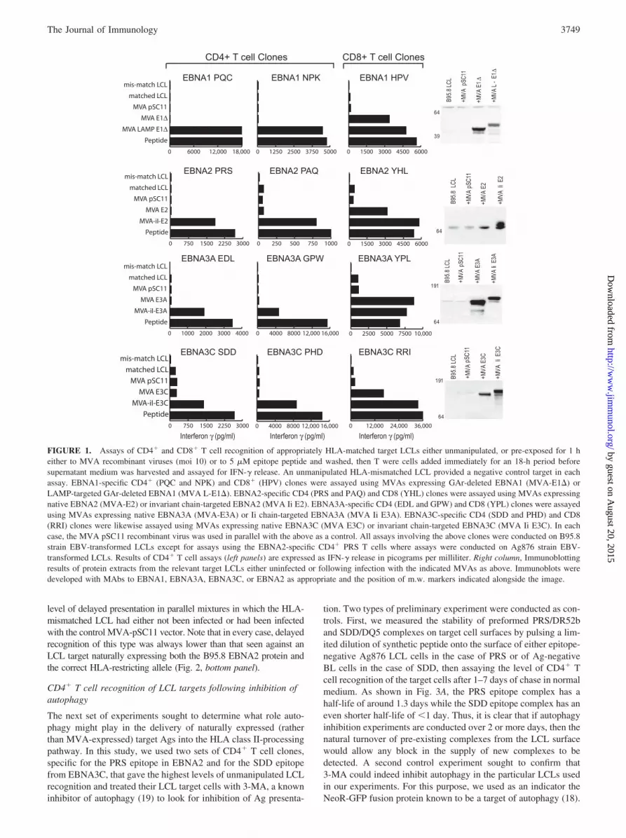
In a first series of experiments, we looked for evidence that endo-genously expressed EBNA Ags could directly access the HLAclass II presentation pathway in LCL cells by overexpressing in-dividual Ags from the vaccinia virus vector MVA. Thus, we gen-erated a panel of MVA recombinants encoding EBV B95.8 strainEBNA1 (GAr deleted), EBNA2, EBNA3A, and EBNA3C in theirnative, nuclear-localizing, form. As positive controls, MVA re-combinants were also constructed to express these same proteinsfused to LAMP or Ii-targeting sequences that deliver the Ag di-rectly to endosomes/lysosomes and therefore into the HLA classII-processing pathway. Fig. 1, right panels, shows Western blotswhere protein extracts made from LCL cells 18 h postinfectionwith the relevant MVA recombinants were probed with EBNA-specific Abs. Note that the MVA vectors significantly increase thelevel of Ag above that already being expressed from the residentEBV genome. The MVA-coded EBNA1 protein runs at �50 kDa,as expected for the GAr-deleted form, while the LAMP-targeted
form runs at a slightly higher m.w.; both are significantly smallerthan the EBV-coded native EBNA1 protein containing the GArdomain. The MVA-coded EBNA2, EBNA3A, and EBNA3C areexpressed as proteins of the expected size and the Ii-chain-targetedforms are slightly larger.
We then used such LCL cells infected with appropriate MVA-EBNA constructs (or with the MVA-pSC11 control virus) as tar-gets for CD4� T cells clones specific for each of the differentEBNA-derived T cell epitopes shown in Table I, assaying T cellrecognition by IFN-� release. Note that although this panel of Tcell clones includes some that are capable of directly recognizingthe autologous B95.8 virus-transformed LCL, in almost everycase, the baseline level of unmanipulated LCL recognition is suf-ficiently low as to allow any incremental recognition on MVA-infected targets to be easily detected. The exceptions are clonesspecific for the EBNA2-derived PRS epitope, which show thehighest baseline recognition of B95.8 virus-transformed LCLs; inthis case, we used target LCLs transformed with the EBV strainAg876 where multiple sequence changes in the PRS epitope re-duce baseline recognition to zero (9). Representative results areshown in Fig. 1 using CD4� T cell clones to two epitopes inEBNA1 (PQC and NPK), two in EBNA2 (PAQ and PRS), two inEBNA3A (EDL and GPW), and two in EBNA3C (SDD and PHD).These include clones that either fail to see the unmanipulated LCL(e.g., PQC) and others that naturally exhibit low (e.g., GPW), mod-erate (e.g., SDD) or high levels (e.g., PRS) of LCL recognition. Aconsistent pattern of results was obtained throughout. All clonesfailed to show any increased recognition of LCL cells overexpress-ing the native form of the Ag. By contrast cells expressing theHLA class II-targeted form were clearly recognized, up to levelsthat in many cases approached the optimal level seen using thesame LCLs exogenously loaded with peptide. To ensure that thetarget cells expressing MVA-encoded native Ag could indeed berecognized by T cells, we included in the same experiments CD8�
T cell clones specific for defined epitopes in the EBNA1, 2, 3A,and 3C proteins. Where an LCL with the appropriate class I- andclass II-restricting alleles was available, we used exactly the sametarget cells in both the CD4 and CD8 assays; otherwise, we useda different LCL for the CD8 assay but infected this in parallel withthe CD4 assay target. Although CD8� effectors will invariablyrecognize the unmanipulated LCL target in such assays, the levelof recognition seen by IFN-� release is again such as to allow anyincremental increase to be observed. Importantly, all of the CD8�
T cell clones showed significantly increased recognition of LCLsoverexpressing the relevant native EBNA protein from the MVAvector.
To cover the possibility that endogenous Ag processing via theHLA class II pathway may take longer to detect, we extended thetimeframe of the experiment from 18 to 42 h by delaying the additionof T cells until 24 h postinfection of the targets and then measuringIFN-� release over the next 18 h of coculture. Fig. 2 shows the resultsfrom one representative experiment, in this case using CD4� T cellsagainst the EBNA2-derived PRS epitope. Now, we did detect somelow level IFN-� release from T cells exposed to the MVA-EBNA2-infected HLA-matched target cells; this clearly reflected specificrecognition because similarly infected HLA-mismatched LCL targetsremained negative. However, this delayed presentation of the PRSepitope appeared to be occurring via Ag release from infected cellsfollowed by subsequent uptake and processing in neighboring cellswithin the culture. Thus, if uninfected cells of the HLA-matchedAg876 LCL were added to the MVA-EBNA2-infected mismatchedLCL targets for the last 24 h of the experiment (including the 18-h Tcell assay period), we observed similar low level recognition byCD4� T cells. Indeed, we even observed a lower but still significant
3748 CD4� T CELL RECOGNITION OF EBV-INFECTED B CELLS
by guest on August 20, 2015
http://ww
w.jim
munol.org/
Dow
nloaded from
level of delayed presentation in parallel mixtures in which the HLA-mismatched LCL had either not been infected or had been infectedwith the control MVA-pSC11 vector. Note that in every case, delayedrecognition of this type was always lower than that seen against anLCL target naturally expressing both the B95.8 EBNA2 protein andthe correct HLA-restricting allele (Fig. 2, bottom panel).
CD4� T cell recognition of LCL targets following inhibition ofautophagy
The next set of experiments sought to determine what role auto-phagy might play in the delivery of naturally expressed (ratherthan MVA-expressed) target Ags into the HLA class II-processingpathway. In this study, we used two sets of CD4� T cell clones,specific for the PRS epitope in EBNA2 and for the SDD epitopefrom EBNA3C, that gave the highest levels of unmanipulated LCLrecognition and treated their LCL target cells with 3-MA, a knowninhibitor of autophagy (19) to look for inhibition of Ag presenta-
tion. Two types of preliminary experiment were conducted as con-trols. First, we measured the stability of preformed PRS/DR52band SDD/DQ5 complexes on target cell surfaces by pulsing a lim-ited dilution of synthetic peptide onto the surface of either epitope-negative Ag876 LCL cells in the case of PRS or of Ag-negativeBL cells in the case of SDD, then assaying the level of CD4� Tcell recognition of the target cells after 1–7 days of chase in normalmedium. As shown in Fig. 3A, the PRS epitope complex has ahalf-life of around 1.3 days while the SDD epitope complex has aneven shorter half-life of �1 day. Thus, it is clear that if autophagyinhibition experiments are conducted over 2 or more days, then thenatural turnover of pre-existing complexes from the LCL surfacewould allow any block in the supply of new complexes to bedetected. A second control experiment sought to confirm that3-MA could indeed inhibit autophagy in the particular LCLs usedin our experiments. For this purpose, we used as an indicator theNeoR-GFP fusion protein known to be a target of autophagy (18).
FIGURE 1. Assays of CD4� and CD8� T cell recognition of appropriately HLA-matched target LCLs either unmanipulated, or pre-exposed for 1 heither to MVA recombinant viruses (moi 10) or to 5 �M epitope peptide and washed, then T were cells added immediately for an 18-h period beforesupernatant medium was harvested and assayed for IFN-� release. An unmanipulated HLA-mismatched LCL provided a negative control target in eachassay. EBNA1-specific CD4� (PQC and NPK) and CD8� (HPV) clones were assayed using MVAs expressing GAr-deleted EBNA1 (MVA-E1�) orLAMP-targeted GAr-deleted EBNA1 (MVA L-E1�). EBNA2-specific CD4 (PRS and PAQ) and CD8 (YHL) clones were assayed using MVAs expressingnative EBNA2 (MVA-E2) or invariant chain-targeted EBNA2 (MVA Ii E2). EBNA3A-specific CD4 (EDL and GPW) and CD8 (YPL) clones were assayedusing MVAs expressing native EBNA3A (MVA-E3A) or Ii chain-targeted EBNA3A (MVA Ii E3A). EBNA3C-specific CD4 (SDD and PHD) and CD8(RRI) clones were likewise assayed using MVAs expressing native EBNA3C (MVA E3C) or invariant chain-targeted EBNA3C (MVA Ii E3C). In eachcase, the MVA pSC11 recombinant virus was used in parallel with the above as a control. All assays involving the above clones were conducted on B95.8strain EBV-transformed LCLs except for assays using the EBNA2-specific CD4� PRS T cells where assays were conducted on Ag876 strain EBV-transformed LCLs. Results of CD4� T cell assays (left panels) are expressed as IFN-� release in picograms per milliliter. Right column, Immunoblottingresults of protein extracts from the relevant target LCLs either uninfected or following infection with the indicated MVAs as above. Immunoblots weredeveloped with MAbs to EBNA1, EBNA3A, EBNA3C, or EBNA2 as appropriate and the position of m.w. markers indicated alongside the image.
3749The Journal of Immunology
by guest on August 20, 2015
http://ww
w.jim
munol.org/
Dow
nloaded from
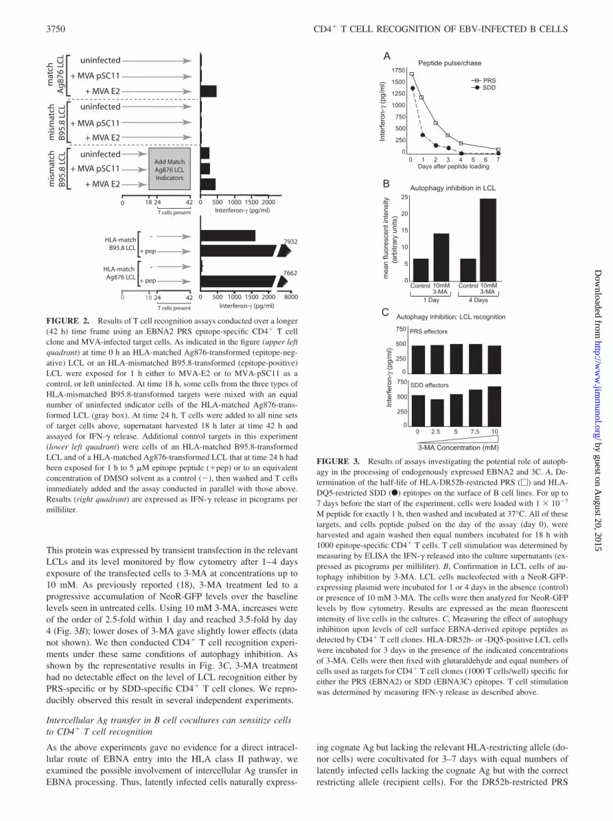
This protein was expressed by transient transfection in the relevantLCLs and its level monitored by flow cytometry after 1–4 daysexposure of the transfected cells to 3-MA at concentrations up to10 mM. As previously reported (18), 3-MA treatment led to aprogressive accumulation of NeoR-GFP levels over the baselinelevels seen in untreated cells. Using 10 mM 3-MA, increases wereof the order of 2.5-fold within 1 day and reached 3.5-fold by day4 (Fig. 3B); lower doses of 3-MA gave slightly lower effects (datanot shown). We then conducted CD4� T cell recognition experi-ments under these same conditions of autophagy inhibition. Asshown by the representative results in Fig. 3C, 3-MA treatmenthad no detectable effect on the level of LCL recognition either byPRS-specific or by SDD-specific CD4� T cell clones. We repro-ducibly observed this result in several independent experiments.
Intercellular Ag transfer in B cell cocultures can sensitize cellsto CD4� T cell recognition
As the above experiments gave no evidence for a direct intracel-lular route of EBNA entry into the HLA class II pathway, weexamined the possible involvement of intercellular Ag transfer inEBNA processing. Thus, latently infected cells naturally express-
ing cognate Ag but lacking the relevant HLA-restricting allele (do-nor cells) were cocultivated for 3–7 days with equal numbers oflatently infected cells lacking the cognate Ag but with the correctrestricting allele (recipient cells). For the DR52b-restricted PRS
FIGURE 2. Results of T cell recognition assays conducted over a longer(42 h) time frame using an EBNA2 PRS epitope-specific CD4� T cellclone and MVA-infected target cells. As indicated in the figure (upper leftquadrant) at time 0 h an HLA-matched Ag876-transformed (epitope-neg-ative) LCL or an HLA-mismatched B95.8-transformed (epitope-positive)LCL were exposed for 1 h either to MVA-E2 or to MVA-pSC11 as acontrol, or left uninfected. At time 18 h, some cells from the three types ofHLA-mismatched B95.8-transformed targets were mixed with an equalnumber of uninfected indicator cells of the HLA-matched Ag876-trans-formed LCL (gray box). At time 24 h, T cells were added to all nine setsof target cells above, supernatant harvested 18 h later at time 42 h andassayed for IFN-� release. Additional control targets in this experiment(lower left quadrant) were cells of an HLA-matched B95.8-transformedLCL and of a HLA-matched Ag876-transformed LCL that at time 24 h hadbeen exposed for 1 h to 5 �M epitope peptide (�pep) or to an equivalentconcentration of DMSO solvent as a control (�), then washed and T cellsimmediately added and the assay conducted in parallel with those above.Results (right quadrant) are expressed as IFN-� release in picograms permilliliter.
FIGURE 3. Results of assays investigating the potential role of autoph-agy in the processing of endogenously expressed EBNA2 and 3C. A, De-termination of the half-life of HLA-DR52b-restricted PRS (�) and HLA-DQ5-restricted SDD (F) epitopes on the surface of B cell lines. For up to7 days before the start of the experiment, cells were loaded with 1 � 10�7
M peptide for exactly 1 h, then washed and incubated at 37°C. All of thesetargets, and cells peptide pulsed on the day of the assay (day 0), wereharvested and again washed then equal numbers incubated for 18 h with1000 epitope-specific CD4� T cells. T cell stimulation was determined bymeasuring by ELISA the IFN-� released into the culture supernatants (ex-pressed as picograms per milliliter). B, Confirmation in LCL cells of au-tophagy inhibition by 3-MA. LCL cells nucleofected with a NeoR-GFP-expressing plasmid were incubated for 1 or 4 days in the absence (control)or presence of 10 mM 3-MA. The cells were then analyzed for NeoR-GFPlevels by flow cytometry. Results are expressed as the mean fluorescentintensity of live cells in the cultures. C, Measuring the effect of autophagyinhibition upon levels of cell surface EBNA-derived epitope peptides asdetected by CD4� T cell clones. HLA-DR52b- or -DQ5-positive LCL cellswere incubated for 3 days in the presence of the indicated concentrationsof 3-MA. Cells were then fixed with glutaraldehyde and equal numbers ofcells used as targets for CD4� T cell clones (1000 T cells/well) specific foreither the PRS (EBNA2) or SDD (EBNA3C) epitopes. T cell stimulationwas determined by measuring IFN-� release as described above.
3750 CD4� T CELL RECOGNITION OF EBV-INFECTED B CELLS
by guest on August 20, 2015
http://ww
w.jim
munol.org/
Dow
nloaded from
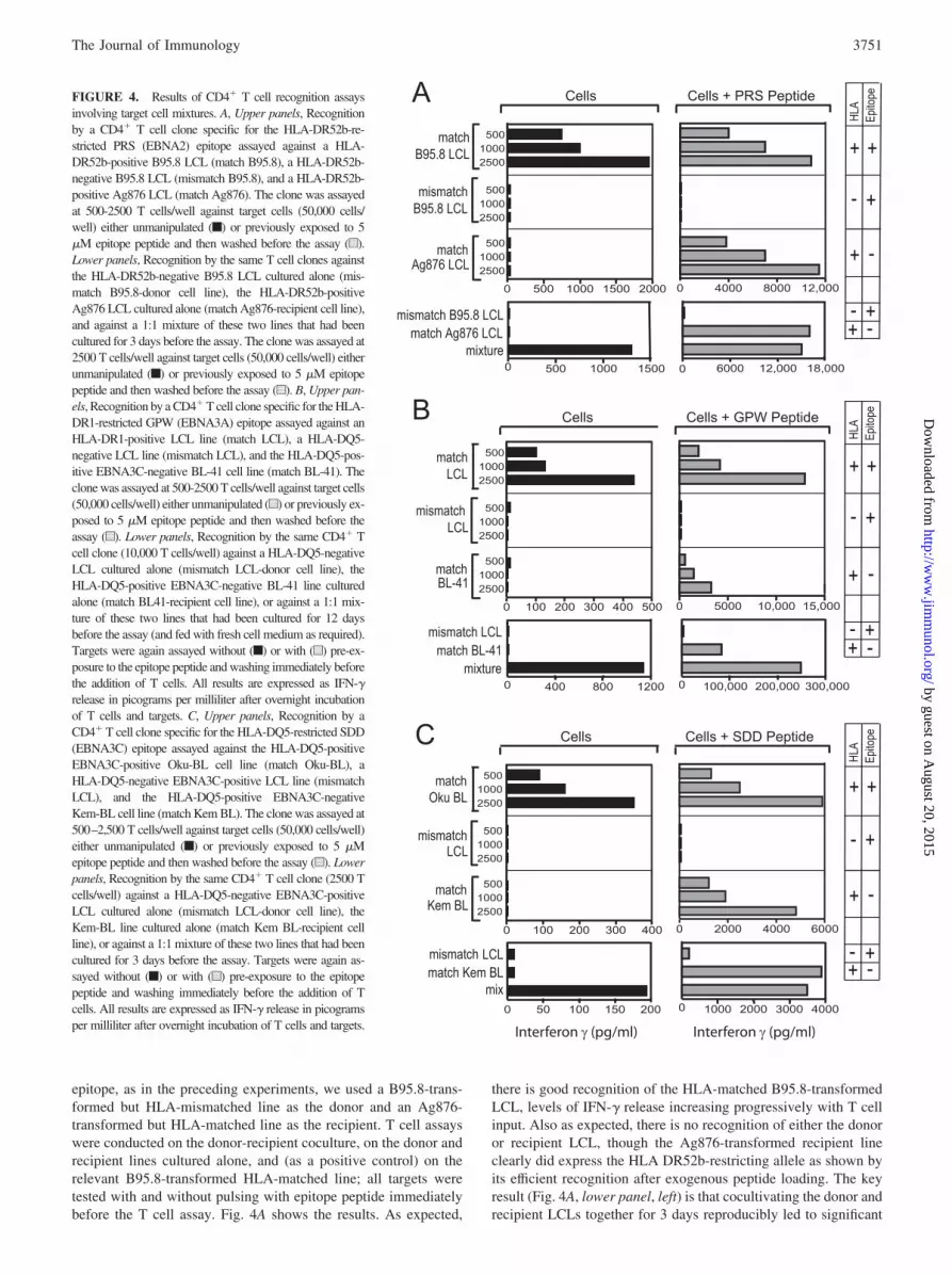
epitope, as in the preceding experiments, we used a B95.8-trans-formed but HLA-mismatched line as the donor and an Ag876-transformed but HLA-matched line as the recipient. T cell assayswere conducted on the donor-recipient coculture, on the donor andrecipient lines cultured alone, and (as a positive control) on therelevant B95.8-transformed HLA-matched line; all targets weretested with and without pulsing with epitope peptide immediatelybefore the T cell assay. Fig. 4A shows the results. As expected,
there is good recognition of the HLA-matched B95.8-transformedLCL, levels of IFN-� release increasing progressively with T cellinput. Also as expected, there is no recognition of either the donoror recipient LCL, though the Ag876-transformed recipient lineclearly did express the HLA DR52b-restricting allele as shown byits efficient recognition after exogenous peptide loading. The keyresult (Fig. 4A, lower panel, left) is that cocultivating the donor andrecipient LCLs together for 3 days reproducibly led to significant
FIGURE 4. Results of CD4� T cell recognition assaysinvolving target cell mixtures. A, Upper panels, Recognitionby a CD4� T cell clone specific for the HLA-DR52b-re-stricted PRS (EBNA2) epitope assayed against a HLA-DR52b-positive B95.8 LCL (match B95.8), a HLA-DR52b-negative B95.8 LCL (mismatch B95.8), and a HLA-DR52b-positive Ag876 LCL (match Ag876). The clone was assayedat 500-2500 T cells/well against target cells (50,000 cells/well) either unmanipulated (f) or previously exposed to 5�M epitope peptide and then washed before the assay (u).Lower panels, Recognition by the same T cell clones againstthe HLA-DR52b-negative B95.8 LCL cultured alone (mis-match B95.8-donor cell line), the HLA-DR52b-positiveAg876 LCL cultured alone (match Ag876-recipient cell line),and against a 1:1 mixture of these two lines that had beencultured for 3 days before the assay. The clone was assayed at2500 T cells/well against target cells (50,000 cells/well) eitherunmanipulated (f) or previously exposed to 5 �M epitopepeptide and then washed before the assay (u). B, Upper pan-els, Recognition by a CD4� T cell clone specific for the HLA-DR1-restricted GPW (EBNA3A) epitope assayed against anHLA-DR1-positive LCL line (match LCL), a HLA-DQ5-negative LCL line (mismatch LCL), and the HLA-DQ5-pos-itive EBNA3C-negative BL-41 cell line (match BL-41). Theclone was assayed at 500-2500 T cells/well against target cells(50,000 cells/well) either unmanipulated (u) or previously ex-posed to 5 �M epitope peptide and then washed before theassay (u). Lower panels, Recognition by the same CD4� Tcell clone (10,000 T cells/well) against a HLA-DQ5-negativeLCL cultured alone (mismatch LCL-donor cell line), theHLA-DQ5-positive EBNA3C-negative BL-41 line culturedalone (match BL41-recipient cell line), or against a 1:1 mix-ture of these two lines that had been cultured for 12 daysbefore the assay (and fed with fresh cell medium as required).Targets were again assayed without (f) or with (u) pre-ex-posure to the epitope peptide and washing immediately beforethe addition of T cells. All results are expressed as IFN-�release in picograms per milliliter after overnight incubationof T cells and targets. C, Upper panels, Recognition by aCD4� T cell clone specific for the HLA-DQ5-restricted SDD(EBNA3C) epitope assayed against the HLA-DQ5-positiveEBNA3C-positive Oku-BL cell line (match Oku-BL), aHLA-DQ5-negative EBNA3C-positive LCL line (mismatchLCL), and the HLA-DQ5-positive EBNA3C-negativeKem-BL cell line (match Kem BL). The clone was assayed at500–2,500 T cells/well against target cells (50,000 cells/well)either unmanipulated (f) or previously exposed to 5 �Mepitope peptide and then washed before the assay (u). Lowerpanels, Recognition by the same CD4� T cell clone (2500 Tcells/well) against a HLA-DQ5-negative EBNA3C-positiveLCL cultured alone (mismatch LCL-donor cell line), theKem-BL line cultured alone (match Kem BL-recipient cellline), or against a 1:1 mixture of these two lines that had beencultured for 3 days before the assay. Targets were again as-sayed without (f) or with (u) pre-exposure to the epitopepeptide and washing immediately before the addition of Tcells. All results are expressed as IFN-� release in picogramsper milliliter after overnight incubation of T cells and targets.
3751The Journal of Immunology
by guest on August 20, 2015
http://ww
w.jim
munol.org/
Dow
nloaded from
recognition at levels which approached those shown by the sameCD4� T cell clones tested against the B95.8-transformed HLA-matched positive control.
A similar approach was used to study presentation of the DR1-restricted GPW epitope from EBNA3A; clones to this epitope dorecognize unmanipulated LCL targets but at much lower levels(see Table I). This epitope is antigenically conserved among EBVstrains and thus no GPW epitope-negative LCL was available to beused as a recipient cell line. Instead, we used the HLA-DR1-ex-pressing BL line BL41 derived from an EBV-negative Burkitt tu-mor and known to be capable of processing exogenous Ag as ef-ficiently as LCL cells (34); this was cocultured with a B95.8-transformed but DR1-negative donor LCL. As shown in Fig. 4B(lower panels), this donor-recipient cell mixture again led to sig-nificant recognition after coculture; however, sensitization was notas strong as that illustrated earlier using PRS-specific clones andrequired a greater number of T cells in the assay. Again, donor andrecipient lines alone were never recognized.
Fig. 4C shows the results from a third such experiment usingCD4� T cell clones against the HLA-DQ5-restricted SDD epitopein EBNA3C. The recipient cell line in this experiment was theDQ5-positive BL cell line Kem BL that lacks EBNA3C expres-sion; the donor line was an HLA-mismatched LCL. Again, weobserved CD4� T cell recognition of the cell mixture but not ofeither line individually. Note that the positive control in this ex-periment was the DQ5-positive Oku-BL line which is unusual inexpressing the EBNA3 proteins as well as EBNA1 (23). Interest-ingly, the SDD-specific clone recognized unmanipulated Oku-BLcells to levels similar to those described earlier for DQ5-posi-tive LCLs; this supports the view that BL lines, though knownto be deficient in the HLA class I presentation pathway (35, 36),are equivalent to LCLs in their susceptibility to EBNA-specificCD4� T cell recognition providing the BL cells express thecognate Ag (10).
Detection of antigenic species in medium harvested from LCLcultures
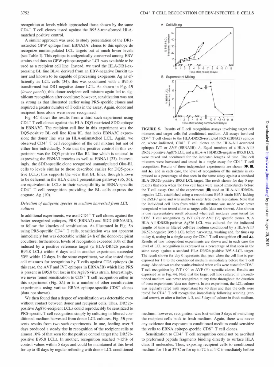
In additional experiments, we used CD4� T cell clones against thebetter recognized epitopes, PRS (EBNA2) and SDD (EBNA3C),to follow the kinetics of sensitization. As illustrated in Fig. 5Ausing PRS-specific CD4� T cells, sensitization was not apparentimmediately but was detectable within 24 h of the donor-recipientcoculture; furthermore, levels of recognition exceeded 30% of thatinduced by a positive reference target (a HLA-DR52b positiveB95.8 LCL) within 6 days and in another experiment exceeded50% within 12 days. In the same experiment, we also tested thesecell mixtures for recognition by T cells against CD8 epitopes (inthis case, the AVF and IVT epitopes in EBNA3B) which like PRSis present in B95.8 but lost in the Ag876 virus strain. Interestingly,we never found sensitization to CD8� T cell recognition either inthis experiment (Fig. 5A) or in a number of other cocultivationexperiments using various EBNA epitope-specific CD8� clones(data not shown).
We then found that a degree of sensitization was detectable evenwithout contact between donor and recipient cells. Thus, DR52b-positive Ag876-recipient LCLs could reproducibly be sensitized toPRS-specific T cell recognition simply by culturing in filtered con-ditioned medium harvested from donor LCL cultures. Fig. 5B pre-sents results from two such experiments. In one, feeding over 5days produced a steady rise in recognition of the recipient cells toalmost 10% of that seen for the positive control target (the DR52b-positive B95.8 LCL). In another, recognition reached �15% ofcontrol values within 5 days and could be maintained at this levelfor up to 40 days by regular refeeding with donor-LCL conditioned
medium; however, recognition was lost within 3 days of switchingthe recipient cells back to fresh medium. Again, there was neverany evidence that exposure to conditioned medium could sensitizethe cells to EBNA epitope-specific CD8� T cell clones.
Sensitization to CD4� T cell recognition could not be ascribedto preformed peptide fragments binding directly to surface HLAclass II molecules. Thus, exposing recipient cells to conditionedmedium for 1 h at 37°C or for up to 72 h at 4°C immediately before
FIGURE 5. Results of T cell recognition assays involving target cellmixtures and target cells fed conditioned medium. All assays involvedCD4� T cell clones to the HLA-DR52b-restricted PRS (EBNA2) epitopeor, where indicated, CD8� T cell clones to the HLA-A11-restrictedepitopes IVT or AVF (EBNA3B). A, Equal numbers of a HLA-A11/DR52b-positive Ag876 LCL and a HLA-A11/DR52b-negative B95.8 LCLwere mixed and cocultured for the indicated lengths of time. The cellmixtures were harvested and tested in a single assay for CD4� T cellrecognition. Results of three independent experiments are shown (F, f,and Œ), and in each case, the level of recognition of the mixture is ex-pressed as a percentage of that seen in the same assay against a standardHLA-DR52b-positive B95.8 LCL target. The result shown for day 0 rep-resents that seen when the two cell lines were mixed immediately beforethe T cell assay. One of the experiments (f) used an HLA-A11/DR52b-negative LCL established using a recombinant B95.8 strain EBV lackingthe BZLF1 gene and was unable to enter lytic cycle replication. Note thatthe individual cell lines from which the mixture was made were neverrecognized when tested alone as target cells (data not shown). Also shownis one representative result obtained when cell mixtures were tested forCD8� T cell recognition by IVT (�) or AVF (ƒ) specific clones. B, AHLA-A11/DR52b-positive Ag876 LCL was cultured for the indicatedlengths of time in filtered cell-free medium conditioned by a HLA-A11/DR52b-negative B95.8 LCL before harvesting, washing and, for times upto day 6, testing in a single assay for CD4� T cell recognition (F and Œ).Results of two independent experiments are shown and in each case thelevel of LCL recognition is expressed as a percentage of that seen in thesame assay against a standard HLA-DR52b-positive B95.8-LCL target.The result shown for day 0 represents that seen when the cell line is pre-exposed for 1 h to the conditioned medium immediately before the T cellassay. Also shown are the results obtained when cells were tested for CD8�
T cell recognition by IVT (�) or AVF (ƒ) -specific clones. Results areexpressed as in Fig. 4A. Note that the target cell line cultured in uncondi-tioned medium was never recognized at any time throughout the durationof these experiments (data not shown). In one experiment, the LCL culturewas regularly refed with supernatant for 40 days and then the cells weretested for CD4� T cell recognition immediately following washing (ver-tical arrow), or after a further 1, 3, and 5 days of culture in fresh medium.
3752 CD4� T CELL RECOGNITION OF EBV-INFECTED B CELLS
by guest on August 20, 2015
http://ww
w.jim
munol.org/
Dow
nloaded from
the assay did not result in any recognition by CD4� T cell clones,whereas parallel treatments with culture medium spiked with alimiting dose of synthetic epitope peptide clearly were sensitizing(data not shown). It is also important to note that sensitization,either by the donor LCL or by its supernatant medium, did notdepend upon the presence of a small number of cells in the donorLCL undergoing EBV lytic replication leading to cell death. Thus,Fig. 5 includes data from experiments using donor LCLs trans-formed with a B95.8 recombinant strain deleted for the BZLF1gene and therefore incapable of entering lytic cycle (22).
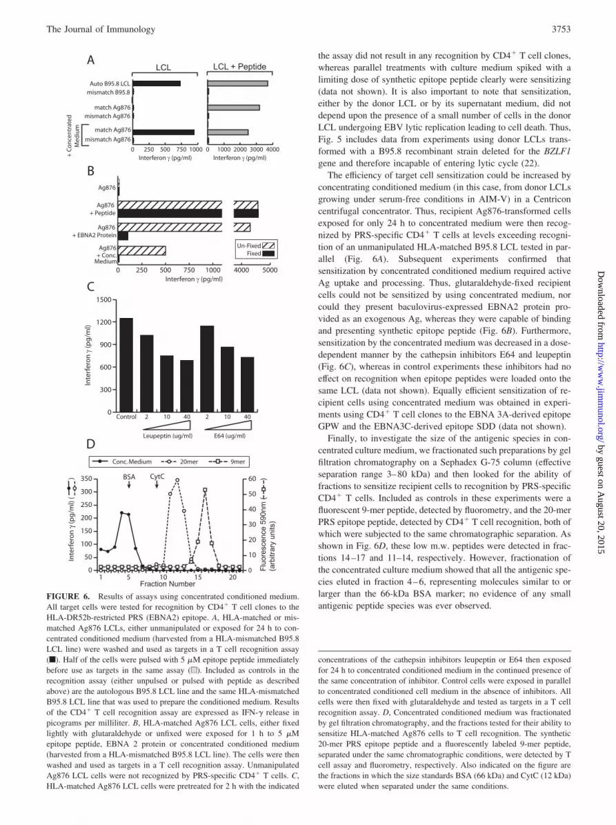
The efficiency of target cell sensitization could be increased byconcentrating conditioned medium (in this case, from donor LCLsgrowing under serum-free conditions in AIM-V) in a Centriconcentrifugal concentrator. Thus, recipient Ag876-transformed cellsexposed for only 24 h to concentrated medium were then recog-nized by PRS-specific CD4� T cells at levels exceeding recogni-tion of an unmanipulated HLA-matched B95.8 LCL tested in par-allel (Fig. 6A). Subsequent experiments confirmed thatsensitization by concentrated conditioned medium required activeAg uptake and processing. Thus, glutaraldehyde-fixed recipientcells could not be sensitized by using concentrated medium, norcould they present baculovirus-expressed EBNA2 protein pro-vided as an exogenous Ag, whereas they were capable of bindingand presenting synthetic epitope peptide (Fig. 6B). Furthermore,sensitization by the concentrated medium was decreased in a dose-dependent manner by the cathepsin inhibitors E64 and leupeptin(Fig. 6C), whereas in control experiments these inhibitors had noeffect on recognition when epitope peptides were loaded onto thesame LCL (data not shown). Equally efficient sensitization of re-cipient cells using concentrated medium was obtained in experi-ments using CD4� T cell clones to the EBNA 3A-derived epitopeGPW and the EBNA3C-derived epitope SDD (data not shown).
Finally, to investigate the size of the antigenic species in con-centrated culture medium, we fractionated such preparations by gelfiltration chromatography on a Sephadex G-75 column (effectiveseparation range 3–80 kDa) and then looked for the ability offractions to sensitize recipient cells to recognition by PRS-specificCD4� T cells. Included as controls in these experiments were afluorescent 9-mer peptide, detected by fluorometry, and the 20-merPRS epitope peptide, detected by CD4� T cell recognition, both ofwhich were subjected to the same chromatographic separation. Asshown in Fig. 6D, these low m.w. peptides were detected in frac-tions 14–17 and 11–14, respectively. However, fractionation ofthe concentrated culture medium showed that all the antigenic spe-cies eluted in fraction 4–6, representing molecules similar to orlarger than the 66-kDa BSA marker; no evidence of any smallantigenic peptide species was ever observed.
FIGURE 6. Results of assays using concentrated conditioned medium.All target cells were tested for recognition by CD4� T cell clones to theHLA-DR52b-restricted PRS (EBNA2) epitope. A, HLA-matched or mis-matched Ag876 LCLs, either unmanipulated or exposed for 24 h to con-centrated conditioned medium (harvested from a HLA-mismatched B95.8LCL line) were washed and used as targets in a T cell recognition assay(f). Half of the cells were pulsed with 5 �M epitope peptide immediatelybefore use as targets in the same assay (u). Included as controls in therecognition assay (either unpulsed or pulsed with peptide as describedabove) are the autologous B95.8 LCL line and the same HLA-mismatchedB95.8 LCL line that was used to prepare the conditioned medium. Resultsof the CD4� T cell recognition assay are expressed as IFN-� release inpicograms per milliliter. B, HLA-matched Ag876 LCL cells, either fixedlightly with glutaraldehyde or unfixed were exposed for 1 h to 5 �Mepitope peptide, EBNA 2 protein or concentrated conditioned medium(harvested from a HLA-mismatched B95.8 LCL line). The cells were thenwashed and used as targets in a T cell recognition assay. UnmanipulatedAg876 LCL cells were not recognized by PRS-specific CD4� T cells. C,HLA-matched Ag876 LCL cells were pretreated for 2 h with the indicated
concentrations of the cathepsin inhibitors leupeptin or E64 then exposedfor 24 h to concentrated conditioned medium in the continued presence ofthe same concentration of inhibitor. Control cells were exposed in parallelto concentrated conditioned cell medium in the absence of inhibitors. Allcells were then fixed with glutaraldehyde and tested as targets in a T cellrecognition assay. D, Concentrated conditioned medium was fractionatedby gel filtration chromatography, and the fractions tested for their ability tosensitize HLA-matched Ag876 cells to T cell recognition. The synthetic20-mer PRS epitope peptide and a fluorescently labeled 9-mer peptide,separated under the same chromatographic conditions, were detected by Tcell assay and fluorometry, respectively. Also indicated on the figure arethe fractions in which the size standards BSA (66 kDa) and CytC (12 kDa)were eluted when separated under the same conditions.
3753The Journal of Immunology
by guest on August 20, 2015
http://ww
w.jim
munol.org/
Dow
nloaded from
DiscussionThe CD4� T cell response to EBV latent cycle proteins has at-tracted much interest because of its potential for direct recognitionof latently infected cells. However, although a number of CD4� Tcell epitopes have been identified in latent cycle proteins, only asubset of these epitopes appear to be presented at sufficient levelson the surface of EBV-transformed LCL cells to allow direct rec-ognition by in vitro-reactivated CD4� T cell clones. That subset isnot limited to epitopes derived from one particular Ag; thus, theliterature contains examples of both well-presented and poorly pre-sented epitopes derived from EBNA1 (8, 10–12, 37–39), fromEBNA2 (9, 12, 40), from EBNA3A (12), and from EBNA3C (12,41, 42). As a first step toward understanding these differences,here, we set out to study how endogenously expressed EBNA pro-teins are being processed for CD4� T cell recognition in latentlyinfected cells. From the literature, there are several potential routeswhereby endogenously expressed Ags might directly enter theMHC class II pathway. Some of these appear to be restricted tomembrane or secreted proteins which naturally intercept nascentMHC class II molecules in the endoplasmic reticulum (13–16).Others can involve cytoplasmic and even nuclear proteins beingdirectly delivered to the endosomal MHC class II-loading com-partment (18, 21, 43–46). This can occur by incorporation of cel-lular components into autophagosomes which then fuse to endo-somal/lysosomal vesicles, a pathway induced under conditions ofstress but active to some degree under conventional culture con-ditions (20), or by a specialized chaperone-mediated pathwaytransporting peptide fragments generated by the proteasome orother cytoplasmic proteinases (17, 45).
Several mechanistic studies of endogenous Ag presentation toCD4� T cells have successfully used vaccinia vectors to expressthe Ag within target cells, whether in murine models using amouse B cell line (47) or in human systems using LCLs as thetargets for vaccinia infection (44, 48–51). Therefore, we reasonedthat if EBNAs did enjoy direct intracellular access to the HLAclass II pathway in LCL cells, such access should be exaggeratedif the native Ag is overexpressed in these cells from a vacciniavector. This would therefore allow LCL recognition by CD4� Tcell clones to be increased over baseline levels or (for clones withno baseline recognition) to be revealed for the first time. Vacciniasexpressing a class II-targeted form of the Ag were generated toserve as a positive control in such experiments. We deliberatelyused the MVA vaccinia strain as a vector for the relevant EBNAconstructs because, unlike other strains, this does not encode adecoy IFN-� receptor (52) and so does not introduce artifacts intoIFN-� release assays. Furthermore, MVA does not replicate inhuman cells and thus avoids potential complications from spreadof the infection within the assay culture. The results were remark-ably consistent for CD4� T cell clones to all nine epitopes testedin standard assays. There was never any detectable recognition ofthe vector-expressed native Ag yet the corresponding class II-tar-geted protein was always well-recognized. By contrast, vector-expressed native Ag was efficiently processed for recognition byEBV-specific CD8� T cell clones, often using the very same targetcells as in the CD4� T cell assays. Interestingly, in each case theHLA class II-targeted protein was also efficiently processed forrecognition by CD8 T cells, an observation noted in other CD8� Tcell systems (30, 31). All these assays were conducted over an18-h timeframe which in other studies using influenza matrix pro-tein expressed from a vaccinia vector (48) was much longer thanrequired to allow HLA class II presentation in LCL cells. How-ever, to allow for the possibility that EBNAs can directly accessboth the HLA class I and class II pathways but at different rates, we
allowed the vaccinia infection to proceed for 24 h before the 18 hT cell assay was initiated. Then, we did notice a low level ofpresentation to CD4� T cells in cells expressing native Ag, but theinclusion of uninfected recipient LCL as an indicator showed thatthis reflected Ag release and reprocessing within the extendedtimeframe of the assay.
In view of recent reports identifying autophagy as an intracel-lular route for Ag feeding into the HLA class II pathway (21), weset up a new series of experiments using 3-MA to inhibit the au-tophagic process. Using as an indicator protein NeoR-GFP, aknown substrate of autophagy (18), we confirmed earlier findingsthat treatment with 3-MA did inhibit autophagy in LCL cells (21)and that the blockade could be maintained for several days. Wethen used these same conditions to look for evidence of 3-MA-induced inhibition of LCL recognition by CD4� T cell clonesagainst the two epitopes, PRS from EBNA2 and SDD fromEBNA3C, mediating the highest baseline levels of recognition.Note that these epitopes were shown to have half-lives of 1.3 and�1 day, respectively, on the cell surface, and so any blockade ofde novo epitope supply to the HLA class II pathway would easilybe detectable within the 3-day time scale of the experiment. In fact,there was never any effect of 3-MA treatment on LCL recognitionby either PRS- or SDD-specific T cells.
Given these negative results, we turned to the possibility (firstraised by the 42-h MVA infection experiments) that the EBNAswere accessing the HLA class II pathway by a slower and lessdirect route involving intercellular Ag transfer. This possibilitywas examined by coculturing HLA-matched recipient cells lackingthe Ag or epitope in question with HLA-mismatched donor cellsexpressing the Ag at physiologic levels from the resident EBVgenome. For all three epitopes tested (PRS, SDD and GPW), sen-sitivity to T cell recognition was slowly but cumulatively acquiredby recipient cells with increasing time of coculture. This did notinvolve superinfection of the recipient with virus released fromdonor cells because donor LCLs carrying a replication-deficientEBV strain gave the same results. We infer that such intercellulartransfer of antigenic species must be happening continuously instandard LCL cultures. Furthermore, this transfer did not requirecell contact because recipient cells could be sensitized to CD4� Tcell recognition by exposure to cell free-conditioned medium. Thiseffect was accelerated using concentrated medium and assays us-ing this as an Ag source confirmed that sensitization required theactive uptake and processing of antigenic species by recipientcells.
Several reports in other systems have looked for the presence ofAg transfer in cocultivation or conditioned medium feeding ex-periments and have failed to detect significant recognition (37, 43,47, 48, 51). However, these very often involve overnight or 24-hincubation times. In our system, although we can see low levelsensitization with conditioned medium within 24 h, the effect ismore apparent at later times and in other systems may have beenmissed if assays were not conducted over longer periods. It wasinteresting that both the coculture and conditioned medium proto-cols reproducibly sensitized cells to recognition by EBNA epitope-specific CD4� but not CD8� T cell clones. This cannot be as-cribed to differential sensitivity because CD8� T cells areconsistently the more avid, often detecting peptide in the 10�9 to10�11 M range (24, 25, 29) compared with the 10�7 to 10�9 Mrange typically shown by the present CD4� T cells (12). We inferthat, although LCL cells are capable of processing exogenouslyacquired Ag via the HLA class I pathway if Ag is provided at highconcentrations experimentally (25), the levels of exogenous Agavailable within LCL cultures are only ever sufficient to charge theHLA class II pathway. Indeed from recent work, this also seems to
3754 CD4� T CELL RECOGNITION OF EBV-INFECTED B CELLS
by guest on August 20, 2015
http://ww
w.jim
munol.org/
Dow
nloaded from
be true of exogenously acquired EBV lytic cycle Ags. Thus, co-culture between appropriate mixtures of semipermissive HLA-mismatched (donor) and nonpermissive HLA-matched (recipient)LCL never led to recognition by lytic epitope-specific CD8� Tcells (53). By contrast, the recognition of semipermissive LCLcells by virus-structural Ag-specific CD4� T cells was found to bedependent upon the intercellular transfer of virions within the cul-ture (54). Furthermore, a similar example of LCL recognition byCD4� T cell clones against the nonstructural lytic cycle proteinBHRF1 has been ascribed to slow charging of the HLA class IIpathway by Ag released from lytically infected cells (55).
The identity of the latent cycle antigenic species being trans-ferred in the present work remains to be determined. Gel filtrationchromatography demonstrated that the molecular mass of the sen-sitizing species exceeded 66 kDa in the case of EBNA2 (an 85-kDa protein in its native form) and was clearly much larger thanthe 20-mer epitope peptide run under the same conditions. Thesefindings are consistent with the transfer of intact EBNA2 Ag butalso with several other possibilities, for example the transfer of Agor antigenic fragments either complexed with other proteins oreven as components of exosomes, structures known to be shed inabundance from LCL cells (56). Whatever the nature of the trans-ferred Ag, the levels of sensitization achieved in cell mixing andconditioned medium feeding experiments identify intercellular Agtransfer as the major route whereby at least three EBNA proteins,EBNA2, 3A, and 3C, gain access to the HLA class II pathway inLCL cells. Our results provide no evidence for the existence of asecond pathway providing direct intracellular access. If such apathway does exist, it must be a minor contributor to the presen-tation of EBNA2-, 3A-, and 3C-derived epitopes on the LCL sur-face and, moreover, must involve a mechanism that is not opera-tional in MVA-infected cells. We can make no definitive statementregarding EBNA1 Ag processing in the type of cell mixing andconditioned medium feeding experiments described here becausesufficiently sensitive CD4� T cell clones were not available. Re-cently, it has been reported by one group that EBNA1 is processedintracellularly by an autophagosomal pathway in LCL cells (21).In contrast, another group who first described autophagosome-me-diated processing of an indicator cytoplasmic Ag in LCL cells (18)could find no evidence for endogenous EBNA1 presentation bythis or any other route (11). Further work will be needed to deter-mine whether EBNA1 is somehow different from the other EBNAsin its intracellular processing for CD4� T cell recognition.
AcknowledgmentsWe thank Elisabeth Houssaint and Elise Landais (Institut National de laSante et de la Recherche Medicale, Unite 463, Institut de Biologie, Nantes,France) for supplying the CD4� T cell clone specific for the EBNA3C-derived peptide PHD.
DisclosuresThe authors have no financial conflict of interest.
References1. Rooney, C. M., C. A. Smith, C. Y. Ng, S. K. Loftin, J. W. Sixbey, Y. Gan,
D. K. Srivastava, L. C. Bowman, R. A. Krance, M. K. Brenner, and H. E. Heslop.1998. Infusion of cytotoxic T cells for the prevention and treatment of Epstein-Barr virus-induced lymphoma in allogeneic transplant recipients. Blood 92:1549–1555.
2. Khanna, R., S. Bell, M. Sherritt, A. Galbraith, S. R. Burrows, L. Rafter,B. Clarke, R. Slaughter, M. C. Falk, J. Douglass, et al. 1999. Activation andadoptive transfer of Epstein-Barr virus-specific cytotoxic T cells in solid organtransplant patients with posttransplant lymphoproliferative disease. Proc. Natl.Acad. Sci. USA 96: 10391–10396.
3. Haque, T., C. Taylor, G. M. Wilkie, P. Murad, P. L. Amlot, S. Beath,P. J. McKiernan, and D. H. Crawford. 2001. Complete regression of posttrans-plant lymphoproliferative disease using partially HLA-matched Epstein Barr vi-rus-specific cytotoxic T cells. Transplantation 72: 1399–1402.
4. Khanna, R., S. R. Burrows, M. G. Kurilla, C. A. Jacob, I. S. Misko, T. B. Sculley,E. Kieff, and D. J. Moss. 1992. Localization of Epstein-Barr virus cytotoxic T cellepitopes using recombinant vaccinia: implications for vaccine development.J. Exp. Med. 176: 169–176.
5. Murray, R. J., M. G. Kurilla, J. M. Brooks, W. A. Thomas, M. Rowe, E. Kieff,and A. B. Rickinson. 1992. Identification of target antigens for the human cyto-toxic T cell response to Epstein-Barr virus (EBV): implications for the immunecontrol of EBV-positive malignancies. J. Exp. Med. 176: 157–168.
6. Rocha, B., and C. Tanchot. 2004. Towards a cellular definition of CD8� T-cellmemory: the role of CD4� T-cell help in CD8� T-cell responses. Curr. Opin.Immunol. 16: 259–263.
7. Young, L. S., and A. B. Rickinson. 2004. Epstein-Barr virus: 40 years on. Nat.Rev. Cancer 4: 757–768.
8. Khanna, R., S. R. Burrows, P. M. Steigerwald-Mullen, S. A. Thomson,M. G. Kurilla, and D. J. Moss. 1995. Isolation of cytotoxic T lymphocytes fromhealthy seropositive individuals specific for peptide epitopes from Epstein-Barrvirus nuclear antigen 1: implications for viral persistence and tumor surveillance.Virology 214: 633–637.
9. Khanna, R., S. R. Burrows, S. A. Thomson, D. J. Moss, P. Cresswell,L. M. Poulsen, and L. Cooper. 1997. Class I processing-defective Burkitt’s lym-phoma cells are recognized efficiently by CD4� EBV-specific CTLs. J. Immunol.158: 3619–3625.
10. Paludan, C., K. Bickham, S. Nikiforow, M. L. Tsang, K. Goodman,W. A. Hanekom, J. F. Fonteneau, S. Stevanovic, and C. Munz. 2002. Epstein-Barr nuclear antigen 1-specific CD4� Th1 cells kill Burkitt’s lymphoma cells.J. Immunol. 169: 1593–1603.
11. Mautner, J., D. Pich, F. Nimmerjahn, S. Milosevic, D. Adhikary, H. Christoph,K. Witter, G. W. Bornkamm, W. Hammerschmidt, and U. Behrends. 2004. Ep-stein-Barr virus nuclear antigen 1 evades direct immune recognition by CD4� Thelper cells. Eur. J. Immunol. 34: 2500–2509.
12. Long, H. M., T. A. Haigh, N. H. Gudgeon, A. M. Leen, C. W. Tsang, J. Brooks,E. Landais, E. Houssaint, S. P. Lee, A. B. Rickinson, and G. S. Taylor. 2005.CD4� T-cell responses to Epstein-Barr virus (EBV) latent-cycle antigens and therecognition of EBV-transformed lymphoblastoid cell lines. J. Virol. 79:4896–4907.
13. Aichinger, G., L. Karlsson, M. R. Jackson, M. Vestberg, J. H. Vaughan,L. Teyton, R. I. Lechler, and P. A. Peterson. 1997. Major histocompatibilitycomplex class II-dependent unfolding, transport, and degradation of endogenousproteins. J. Biol. Chem. 272: 29127–29136.
14. Oxenius, A., M. F. Bachmann, P. G. Ashton-Rickardt, S. Tonegawa,R. M. Zinkernagel, and H. Hengartner. 1995. Presentation of endogenous viralproteins in association with major histocompatibility complex class II: on the roleof intracellular compartmentalization, invariant chain and the TAP transportersystem. Eur. J. Immunol. 25: 3402–3411.
15. Polydefkis, M., S. Koenig, C. Flexner, E. Obah, K. Gebo, S. Chakrabarti,P. L. Earl, B. Moss, and R. F. Siliciano. 1990. Anchor sequence-dependent en-dogenous processing of human immunodeficiency virus 1 envelope glycoproteingp160 for CD4� T cell recognition. J. Exp. Med. 171: 875–887.
16. Weiss, S., and B. Bogen. 1991. MHC class II-restricted presentation of intracel-lular antigen. Cell 64: 767–776.
17. Zhou, D., P. Li, Y. Lin, J. M. Lott, A. D. Hislop, D. H. Canaday,R. R. Brutkiewicz, and J. S. Blum. 2005. Lamp-2a facilitates MHC class IIpresentation of cytoplasmic antigens. Immunity 22: 571–581.
18. Nimmerjahn, F., S. Milosevic, U. Behrends, E. M. Jaffee, D. M. Pardoll,G. W. Bornkamm, and J. Mautner. 2003. Major histocompatibility complex classII-restricted presentation of a cytosolic antigen by autophagy. Eur. J. Immunol.33: 1250–1259.
19. Seglen, P. O., and P. B. Gordon. 1982. 3-Methyladenine: specific inhibitor ofautophagic/lysosomal protein degradation in isolated rat hepatocytes. Proc. Natl.Acad. Sci. USA 79: 1889–1892.
20. Dengjel, J., O. Schoor, R. Fischer, M. Reich, M. Kraus, M. Muller,K. Kreymborg, F. Altenberend, J. Brandenburg, H. Kalbacher, et al. 2005. Au-tophagy promotes MHC class II presentation of peptides from intracellular sourceproteins. Proc. Natl. Acad. Sci. USA 102: 7922–7927.
21. Paludan, C., D. Schmid, M. Landthaler, M. Vockerodt, D. Kube, T. Tuschl, andC. Munz. 2005. Endogenous MHC class II processing of a viral nuclear antigenafter autophagy. Science 307: 593–596.
22. Feederle, R., M. Kost, M. Baumann, A. Janz, E. Drouet, W. Hammerschmidt, andH. J. Delecluse. 2000. The Epstein-Barr virus lytic program is controlled by theco-operative functions of two transactivators. EMBO J. 19: 3080–3089.
23. Kelly, G., A. Bell, and A. Rickinson. 2002. Epstein-Barr virus-associated Burkittlymphomagenesis selects for downregulation of the nuclear antigen EBNA2. Nat.Med. 8: 1098–1104.
24. Lee, S. P., R. J. Tierney, W. A. Thomas, J. M. Brooks, and A. B. Rickinson. 1997.Conserved CTL epitopes within EBV latent membrane protein 2: a potentialtarget for CTL-based tumor therapy. J. Immunol. 158: 3325–3334.
25. Blake, N., S. Lee, I. Redchenko, W. Thomas, N. Steven, A. Leese,P. Steigerwald-Mullen, M. G. Kurilla, L. Frappier, and A. Rickinson. 1997. Hu-man CD8� T cell responses to EBV EBNA1: HLA class I presentation of the(Gly-Ala)-containing protein requires exogenous processing. Immunity 7:791–802.
26. Chapman, A. L., A. B. Rickinson, W. A. Thomas, R. F. Jarrett, J. Crocker, andS. P. Lee. 2001. Epstein-Barr virus-specific cytotoxic T lymphocyte responses inthe blood and tumor site of Hodgkin’s disease patients: implications for a T-cell-based therapy. Cancer Res. 61: 6219–6226.
3755The Journal of Immunology
by guest on August 20, 2015
http://ww
w.jim
munol.org/
Dow
nloaded from

27. Burrows, S. R., J. Gardner, R. Khanna, T. Steward, D. J. Moss, S. Rodda, andA. Suhrbier. 1994. Five new cytotoxic T cell epitopes identified within Epstein-Barr virus nuclear antigen 3. J. Gen. Virol. 75: 2489–2493.
28. Brooks, J. M., R. J. Murray, W. A. Thomas, M. G. Kurilla, and A. B. Rickinson.1993. Different HLA-B27 subtypes present the same immunodominant Epstein-Barr virus peptide. J. Exp. Med. 178: 879–887.
29. Gavioli, R., M. G. Kurilla, P. O. de Campos-Lima, L. E. Wallace, R. Dolcetti,R. J. Murray, A. B. Rickinson, and M. G. Masucci. 1993. Multiple HLA A11-restricted cytotoxic T-lymphocyte epitopes of different immunogenicities in theEpstein-Barr virus-encoded nuclear antigen 4. J. Virol. 67: 1572–1578.
30. Bonini, C., S. P. Lee, S. R. Riddell, and P. D. Greenberg. 2001. Targeting antigenin mature dendritic cells for simultaneous stimulation of CD4� and CD8� Tcells. J. Immunol. 166: 5250–5257.
31. Wu, T. C., F. G. Guarnieri, K. F. Staveley-O’Carroll, R. P. Viscidi, H. I. Levitsky,L. Hedrick, K. R. Cho, J. T. August, and D. M. Pardoll. 1995. Engineering anintracellular pathway for major histocompatibility complex class II presentationof antigens. Proc. Natl. Acad. Sci. USA 92: 11671–11675.
32. Chaux, P., V. Vantomme, V. Stroobant, K. Thielemans, J. Corthals, R. Luiten,A. M. Eggermont, T. Boon, and P. van der Bruggen. 1999. Identification ofMAGE-3 epitopes presented by HLA-DR molecules to CD4� T lymphocytes.J. Exp. Med. 189: 767–778.
33. Joklik, W. K. 1962. The purification of four strains of poxvirus. Virology 18:9–18.
34. Khanna, R., S. R. Burrows, P. M. Steigerwald-Mullen, D. J. Moss, M. G. Kurilla,and L. Cooper. 1997. Targeting Epstein-Barr virus nuclear antigen 1 (EBNA1)through the class II pathway restores immune recognition by EBNA1-specificcytotoxic T lymphocytes: evidence for HLA-DM-independent processing. Int.Immunol. 9: 1537–1543.
35. Khanna, R., S. R. Burrows, V. Argaet, and D. J. Moss. 1994. Endoplasmic re-ticulum signal sequence facilitated transport of peptide epitopes restores immu-nogenicity of an antigen processing defective tumour cell line. Int. Immunol. 6:639–645.
36. Rowe, M., R. Khanna, C. A. Jacob, V. Argaet, A. Kelly, S. Powis, M. Belich,D. Croom-Carter, S. Lee, S. R. Burrows, et al. 1995. Restoration of endogenousantigen processing in Burkitt’s lymphoma cells by Epstein-Barr virus latent mem-brane protein-1: coordinate up-regulation of peptide transporters and HLA-classI antigen expression. Eur. J. Immunol. 25: 1374–1384.
37. Munz, C., K. L. Bickham, M. Subklewe, M. L. Tsang, A. Chahroudi,M. G. Kurilla, D. Zhang, M. O’Donnell, and R. M. Steinman. 2000. HumanCD4� T lymphocytes consistently respond to the latent Epstein-Barr virus nu-clear antigen EBNA1. J. Exp. Med. 191: 1649–1660.
38. Leen, A., P. Meij, I. Redchenko, J. Middeldorp, E. Bloemena, A. Rickinson, andN. Blake. 2001. Differential immunogenicity of Epstein-Barr virus latent-cycleproteins for human CD4� T-helper 1 responses. J. Virol. 75: 8649–8659.
39. Voo, K. S., T. Fu, H. E. Heslop, M. K. Brenner, C. M. Rooney, and R. F. Wang.2002. Identification of HLA-DP3-restricted peptides from EBNA1 recognized byCD4� T cells. Cancer Res. 62: 7195–7199.
40. Omiya, R., C. Buteau, H. Kobayashi, C. V. Paya, and E. Celis. 2002. Inhibitionof EBV-induced lymphoproliferation by CD4� T cells specific for an MHC classII promiscuous epitope. J. Immunol. 169: 2172–2179.
41. Rajnavolgyi, E., N. Nagy, B. Thuresson, Z. Dosztanyi, A. Simon, I. Simon,R. W. Karr, I. Ernberg, and E. Klein. 2000. A repetitive sequence of Epstein-Barrvirus nuclear antigen 6 comprises overlapping T cell epitopes which induce HLA-DR-restricted CD4� T lymphocytes. Int. Immunol. 12: 281–293.
42. Landais, E., X. Saulquin, E. Scotet, L. Trautmann, M. A. Peyrat, J. L. Yates,W. W. Kwok, M. Bonneville, and E. Houssaint. 2004. Direct killing of Epstein-Barr virus (EBV)-infected B cells by CD4 T cells directed against the EBV lyticprotein BHRF1. Blood 103: 1408–1416.
43. Chen, M., M. Shirai, Z. Liu, T. Arichi, H. Takahashi, and M. Nishioka. 1998.Efficient class II major histocompatibility complex presentation of endogenouslysynthesized hepatitis C virus core protein by Epstein-Barr virus-transformed B-lymphoblastoid cell lines to CD4� T cells. J. Virol. 72: 8301–8308.
44. Gueguen, M., and E. O. Long. 1996. Presentation of a cytosolic antigen by majorhistocompatibility complex class II molecules requires a long-lived form of theantigen. Proc. Natl. Acad. Sci. USA 93: 14692–14697.
45. Lich, J. D., J. F. Elliott, and J. S. Blum. 2000. Cytoplasmic processing is aprerequisite for presentation of an endogenous antigen by major histocompati-bility complex class II proteins. J. Exp. Med. 191: 1513–1524.
46. Mukherjee, P., A. Dani, S. Bhatia, N. Singh, A. Y. Rudensky, A. George, V. Bal,S. Mayor, and S. Rath. 2001. Efficient presentation of both cytosolic and endog-enous transmembrane protein antigens on MHC class II is dependent on cyto-plasmic proteolysis. J. Immunol. 167: 2632–2641.
47. Bartido, S. M., S. Diment, and C. S. Reiss. 1995. Processing of a viral glycop-rotein in the endoplasmic reticulum for class II presentation. Eur. J. Immunol. 25:2211–2219.
48. Jaraquemada, D., M. Marti, and E. O. Long. 1990. An endogenous processingpathway in vaccinia virus-infected cells for presentation of cytoplasmic antigensto class II-restricted T cells. J. Exp. Med. 172: 947–954.
49. Malnati, M. S., S. Ceman, M. Weston, R. DeMars, and E. O. Long. 1993. Pre-sentation of cytosolic antigen by HLA-DR requires a function encoded in theclass II region of the MHC. J. Immunol. 151: 6751–6756.
50. Malnati, M. S., M. Marti, T. LaVaute, D. Jaraquemada, W. Biddison, R. DeMars,and E. O. Long. 1992. Processing pathways for presentation of cytosolic antigento MHC class II-restricted T cells. Nature 357: 702–704.
51. Jin, Y., W. K. Shih, and I. Berkower. 1988. Human T cell response to the surfaceantigen of hepatitis B virus (HBsAg). Endosomal and nonendosomal processingpathways are accessible to both endogenous and exogenous antigen. J. Exp. Med.168: 293–306.
52. Blanchard, T. J., A. Alcami, P. Andrea, and G. L. Smith. 1998. Modified vacciniavirus Ankara undergoes limited replication in human cells and lacks several im-munomodulatory proteins: implications for use as a human vaccine. J. Gen. Virol.79: 1159–1167.
53. Pudney, V. A., A. M. Leese, A. B. Rickinson, and A. D. Hislop. 2005. CD8�
immunodominance among Epstein-Barr virus lytic cycle antigens directly reflectsthe efficiency of antigen presentation in lytically infected cells. J. Exp. Med. 201:349–360.
54. Adhikary, D., U. Behrends, A. Moosmann, K. Witter, G. W. Bornkamm, andJ. Mautner. 2006. Control of Epstein-Barr virus infection in vitro by T helpercells specific for virion glycoproteins. J. Exp. Med. 203: 995–1006.
55. Landais, E., X. Saulquin, M. Bonneville, and E. Houssaint. 2005. Long-termMHC class II presentation of the EBV lytic protein BHRF1 by EBV latentlyinfected B cells following capture of BHRF1 antigen. J. Immunol. 175:7939–7946.
56. Raposo, G., H. W. Nijman, W. Stoorvogel, R. Liejendekker, C. V. Harding,C. J. Melief, and H. J. Geuze. 1996. B lymphocytes secrete antigen-presentingvesicles. J. Exp. Med. 183: 1161–1172.
3756 CD4� T CELL RECOGNITION OF EBV-INFECTED B CELLS
by guest on August 20, 2015
http://ww
w.jim
munol.org/
Dow
nloaded from


