Embed Size (px)
Citation preview
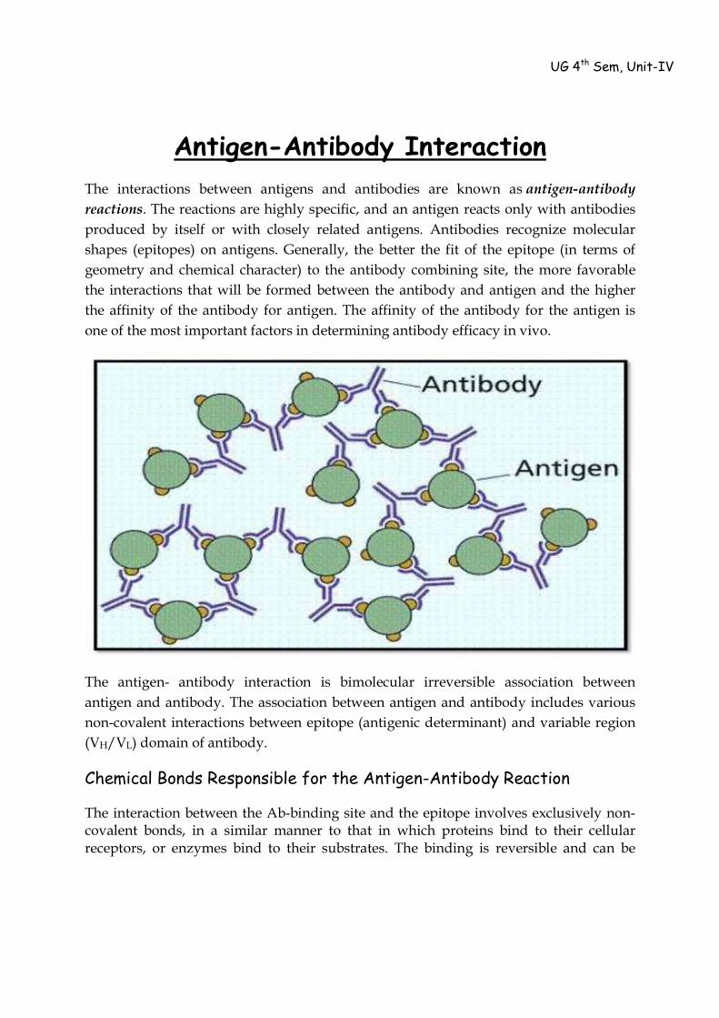
Antigen-Antibody Interaction The interactions between antigens and antibodies are known as antigen-antibody reactions. The reactions are highly specific, and an antigen reacts only with antibodies produced by itself or with closely related antigens. Antibodies recognize molecular shapes (epitopes) on antigens. Generally, the better the fit of the epitope (in terms of geometry and chemical character) to the antibody combining site, the more favorable the interactions that will be formed between the antibody and antigen and the higher the affinity of the antibody for antigen. The affinity of the antibody for the antigen is one of the most important factors in determining antibody efficacy in vivo.
The antigen- antibody interaction is bimolecular irreversible association between antigen and antibody. The association between antigen and antibody includes various non-covalent interactions between epitope (antigenic determinant) and variable region (VH/VL) domain of antibody.
Chemical Bonds Responsible for the Antigen-Antibody Reaction
The interaction between the Ab-binding site and the epitope involves exclusively non-covalent bonds, in a similar manner to that in which proteins bind to their cellular receptors, or enzymes bind to their substrates. The binding is reversible and can be
UG 4th Sem, Unit-IV
prevented or dissociated by high ionic strength or extreme pH. The following intermolecular forces are involved in Ag–Ab binding:
1. Electrostatic bonds: This result from the attraction between oppositely charged ionic groups of two protein side chains; for example, an ionized amino group (NH4+) on a lysine in the Ab, and an ionized carboxyl group (COO_) on an aspartate residue in the Ag.
2. Hydrogen bonding: When the Ag and Ab are in very close proximity, relatively weak hydrogen bonds can be formed between hydrophilic groups (e.g., OH and C=O, NH and C=O, and NH and OH groups).
3. Hydrophobic interactions: Hydrophobic groups, such as the side chains of valine, leucine, and phenylalanine, tend to associate due to Van der Waals bonding and coalesce in an aqueous environment, excluding water molecules from their surroundings. As a consequence, the distance between them decreases, enhancing the energies of attraction involved. This type of interaction is estimated to contribute up to 50% of the total strength of the Ag–Ab bond.
4. Van der Waals bonds: These forces depend upon interactions between the “electron clouds” that surround the Ag and Ab molecules. The interaction has been compared to that which might exist between alternating dipoles in two molecules, alternating in such a way that, at any given moment, oppositely oriented dipoles will be present in closely apposed areas of the Ag and Ab molecules.
Each of these non-covalent interactions operates over very short distance (generally about 1 Å) so, Ag-Ab interactions depends on very close fit between antigen and antibody.
Strength of Ag-Ab interaction:
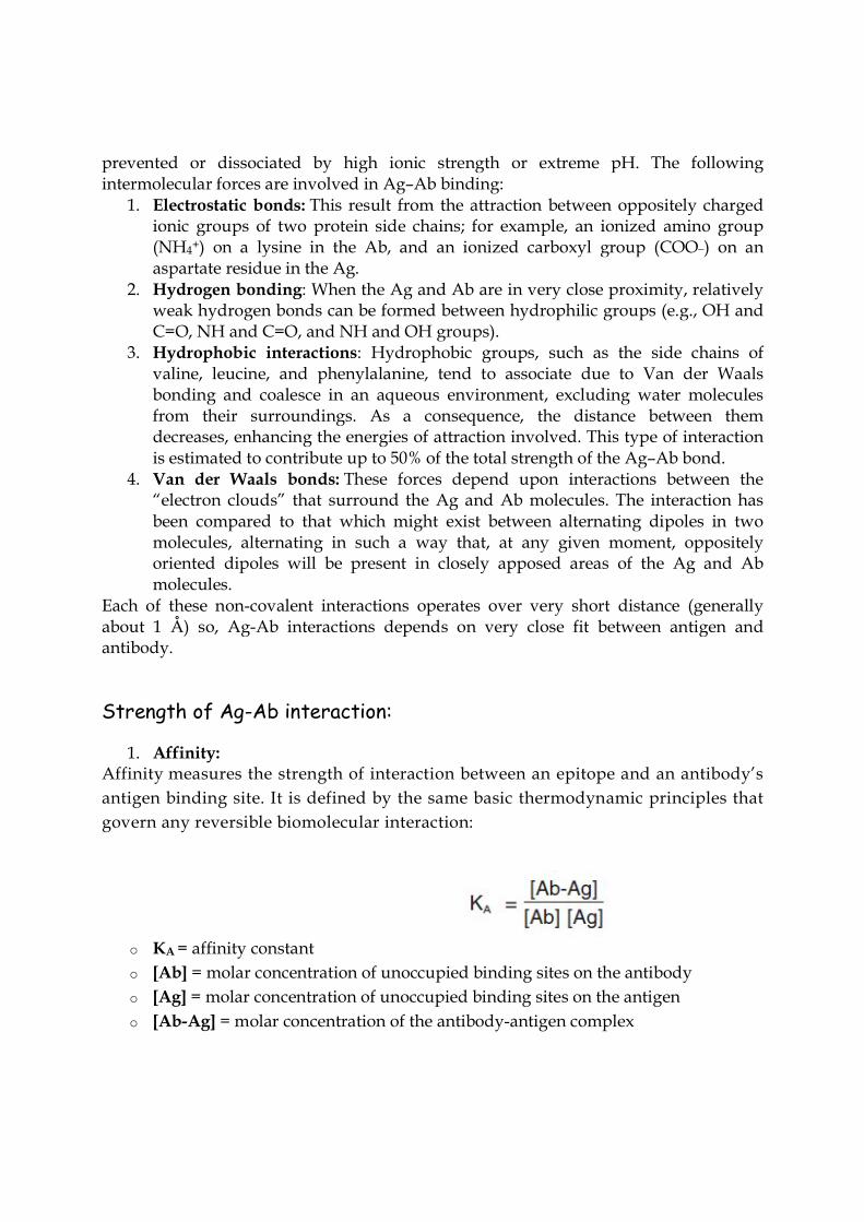
1. Affinity: Affinity measures the strength of interaction between an epitope and an antibody’s antigen binding site. It is defined by the same basic thermodynamic principles that govern any reversible biomolecular interaction:
o KA = affinity constant o [Ab] = molar concentration of unoccupied binding sites on the antibody o [Ag] = molar concentration of unoccupied binding sites on the antigen o [Ab-Ag] = molar concentration of the antibody-antigen complex
In other words, KA describes how much antibody-antigen complex exists at the point when equilibrium is reached. The time taken for this to occur depends on rate of diffusion and is similar for every antibody. However, high-affinity antibodies will bind a greater amount of antigen in a shorter period of time than low-affinity antibodies. KA can therefore vary widely for antibodies from below 105 mol-1 to above 1012 mol-1, and can be influenced by factors including pH, temperature and buffer composition.
Combined strength of total non-covalent interactions between single Ag- binding
site of Ab and single epitope is affinity of Ab for that epitope. Low affinity Ab: Bind Ag weakly and dissociates readily. High affinity Ab: Bind Ag tightly and remain bound longer.
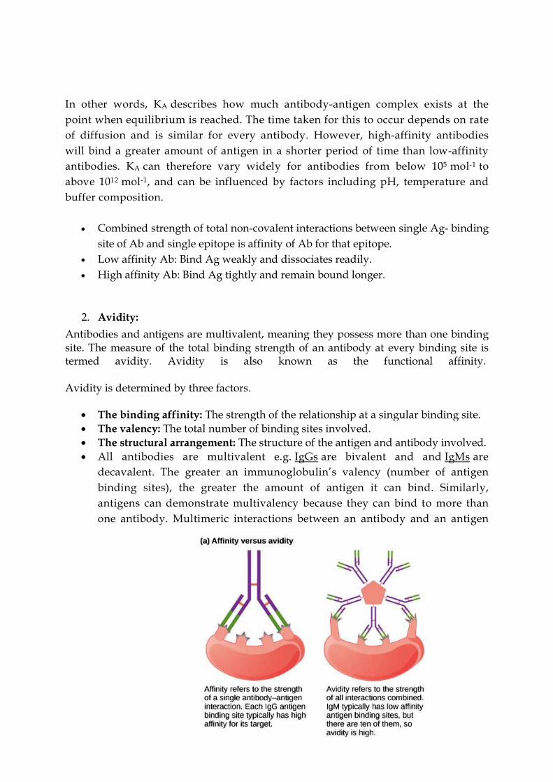
2. Avidity: Antibodies and antigens are multivalent, meaning they possess more than one binding site. The measure of the total binding strength of an antibody at every binding site is termed avidity. Avidity is also known as the functional affinity. Avidity is determined by three factors.
The binding affinity: The strength of the relationship at a singular binding site. The valency: The total number of binding sites involved. The structural arrangement: The structure of the antigen and antibody involved. All antibodies are multivalent e.g. IgGs are bivalent and and IgMs are
decavalent. The greater an immunoglobulin’s valency (number of antigen binding sites), the greater the amount of antigen it can bind. Similarly, antigens can demonstrate multivalency because they can bind to more than one antibody. Multimeric interactions between an antibody and an antigen
help their stabilization. A favorable structural arrangement of antibody and antigen can also lead to a
more stable antibody-antigen complex
Strength of multiple interactions between multivalent Ab and Ag is avidity. Avidity is better measure of binding capacity of antibody than affinity. High avidity can compensate low affinity.
3. Cross reactivity:
Antibody elicited by one Ag can cross react with unrelated Ag if they share
identical epitope or have similar chemical properties.
Types of Ag-Ab reactions:
1. Agglutination 2. Precipitation 3. Complement Fixation 4. Enzyme linked Immunosorbent Assay 5. RadioImmuno Assay 6. Western Blotting
Agglutination
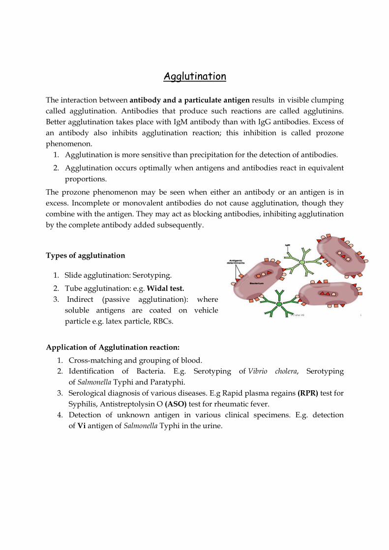
The interaction between antibody and a particulate antigen results in visible clumping called agglutination. Antibodies that produce such reactions are called agglutinins. Better agglutination takes place with IgM antibody than with IgG antibodies. Excess of an antibody also inhibits agglutination reaction; this inhibition is called prozone phenomenon.
1. Agglutination is more sensitive than precipitation for the detection of antibodies.
2. Agglutination occurs optimally when antigens and antibodies react in equivalent proportions.
The prozone phenomenon may be seen when either an antibody or an antigen is in excess. Incomplete or monovalent antibodies do not cause agglutination, though they combine with the antigen. They may act as blocking antibodies, inhibiting agglutination by the complete antibody added subsequently.
Types of agglutination
1. Slide agglutination: Serotyping.
2. Tube agglutination: e.g. Widal test. 3. Indirect (passive agglutination): where
soluble antigens are coated on vehicle particle e.g. latex particle, RBCs.
Application of Agglutination reaction:
1. Cross-matching and grouping of blood. 2. Identification of Bacteria. E.g. Serotyping of Vibrio cholera, Serotyping
of Salmonella Typhi and Paratyphi. 3. Serological diagnosis of various diseases. E.g Rapid plasma regains (RPR) test for
Syphilis, Antistreptolysin O (ASO) test for rheumatic fever. 4. Detection of unknown antigen in various clinical specimens. E.g. detection
of Vi antigen of Salmonella Typhi in the urine.
Precipitation
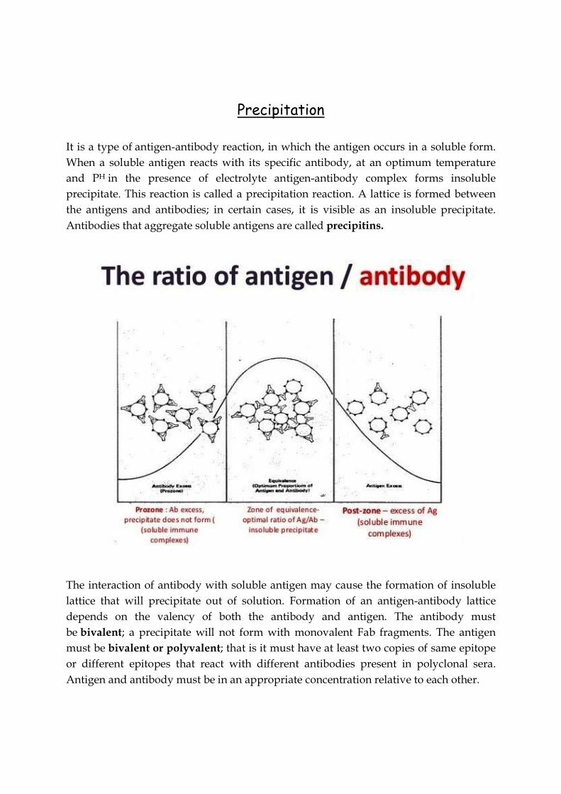
It is a type of antigen-antibody reaction, in which the antigen occurs in a soluble form. When a soluble antigen reacts with its specific antibody, at an optimum temperature and PH in the presence of electrolyte antigen-antibody complex forms insoluble precipitate. This reaction is called a precipitation reaction. A lattice is formed between the antigens and antibodies; in certain cases, it is visible as an insoluble precipitate. Antibodies that aggregate soluble antigens are called precipitins.
The interaction of antibody with soluble antigen may cause the formation of insoluble lattice that will precipitate out of solution. Formation of an antigen-antibody lattice depends on the valency of both the antibody and antigen. The antibody must be bivalent; a precipitate will not form with monovalent Fab fragments. The antigen must be bivalent or polyvalent; that is it must have at least two copies of same epitope or different epitopes that react with different antibodies present in polyclonal sera. Antigen and antibody must be in an appropriate concentration relative to each other.
1. Antigen access: Too much antigen prevents efficient crosslinking/lattice formation.
2. Antibody access: Too much antibody prevents efficient crosslinking/lattice formation.
3. Equivalent Antigen and Antibody: Maximum amount of lattice (Precipitate) is formed.
Application of Precipitation reaction:
1. Detection of unknown antibody to diagnose infection e.g. VDRL test for syphilis. 2. Standardization of toxins and antitoxins. 3. Identification of Bacteria e.g. Lancified grouping of streptococci. 4. Identification of bacterial component e.g Ascoli’s thermoprecipitin test
for Bacillus anthracis.
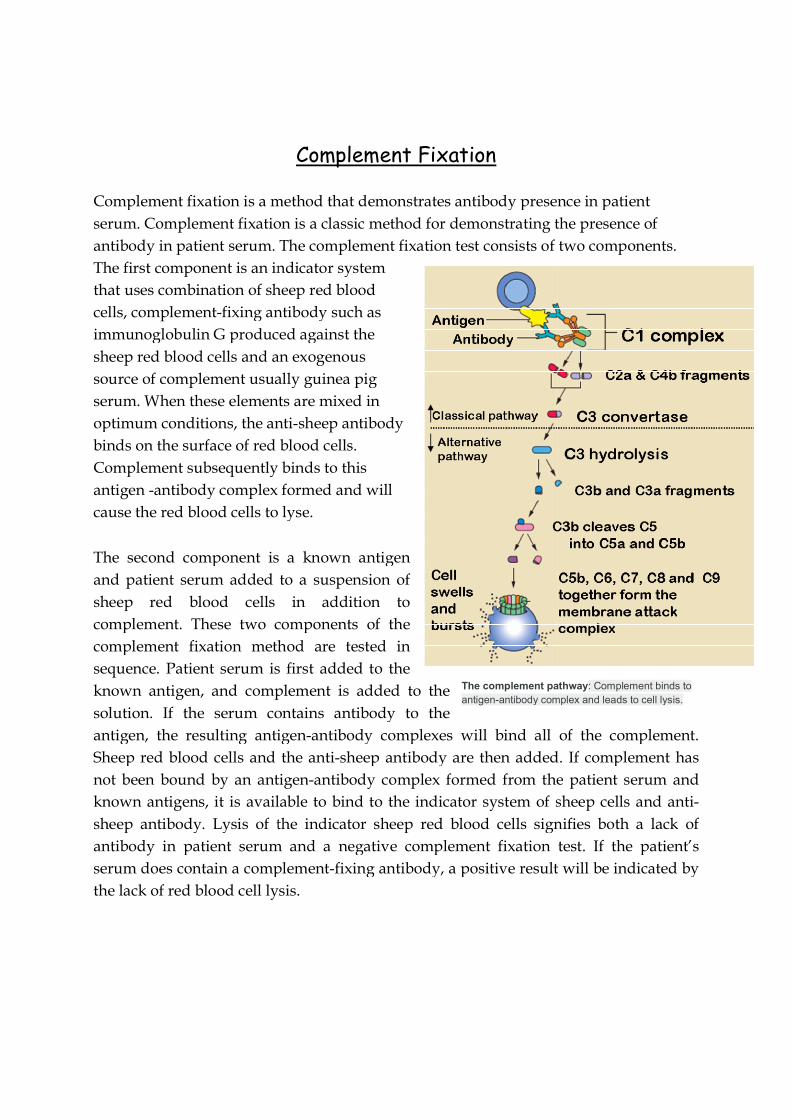
Complement fixation is a method that demonstrates antibody presence in patient serum. Complement fixation is a classic method for demonstrating the presence of antibody in patient serum. The complement fixation test consists of two components. The first component is an indicator system that uses combination of sheep red blood cells, complement-fixing antibody such as immunoglobulin G produced against the sheep red blood cells and an exogenous source of complement usually guinea pig serum. When these elements are mixed in optimum conditions, the antibinds on the surface of red blood cells. Complement subsequently binds to this antigen -antibody complex formed and will cause the red blood cells to lyse
The second component is a known antigen and patient serum added to a suspension of sheep red blood cells in addition to complement. These two components of the complement fixation method are tested in sequence. Patient serum is first added to the known antigen, and complement is added to the solution. If the serum contains antibody to the antigen, the resulting antigenSheep red blood cells and the antinot been bound by an antigenknown antigens, it is available to bind to the indicator system of sheep cells and antisheep antibody. Lysis of the indicator sheep red blood cells signifies both a lack of antibody in patient serum and a negative complement fixation test. If the patient’s serum does contain a complementthe lack of red blood cell lysis
The complement pathwaantigen-antibody complex and leads to cell lysis
Complement Fixation
Complement fixation is a method that demonstrates antibody presence in patient Complement fixation is a classic method for demonstrating the presence of
antibody in patient serum. The complement fixation test consists of two components. omponent is an indicator system
that uses combination of sheep red blood fixing antibody such as
immunoglobulin G produced against the sheep red blood cells and an exogenous source of complement usually guinea pig
ts are mixed in optimum conditions, the anti-sheep antibody binds on the surface of red blood cells. Complement subsequently binds to this
antibody complex formed and will cause the red blood cells to lyse.
second component is a known antigen and patient serum added to a suspension of sheep red blood cells in addition to complement. These two components of the complement fixation method are tested in sequence. Patient serum is first added to the
n, and complement is added to the the serum contains antibody to the
antigen, the resulting antigen-antibody complexes will bind all of the complement. Sheep red blood cells and the anti-sheep antibody are then added. If complement has
n bound by an antigen-antibody complex formed from the patient serum and known antigens, it is available to bind to the indicator system of sheep cells and antisheep antibody. Lysis of the indicator sheep red blood cells signifies both a lack of
in patient serum and a negative complement fixation test. If the patient’s serum does contain a complement-fixing antibody, a positive result will be indicated by the lack of red blood cell lysis.
pathway: Complement binds to antibody complex and leads to cell lysis.
Complement fixation is a method that demonstrates antibody presence in patient Complement fixation is a classic method for demonstrating the presence of
antibody in patient serum. The complement fixation test consists of two components.
antibody complexes will bind all of the complement. sheep antibody are then added. If complement has
antibody complex formed from the patient serum and known antigens, it is available to bind to the indicator system of sheep cells and anti-sheep antibody. Lysis of the indicator sheep red blood cells signifies both a lack of
in patient serum and a negative complement fixation test. If the patient’s fixing antibody, a positive result will be indicated by


