Embed Size (px)
Citation preview

Cellular/Molecular
Synaptically Released Zinc Triggers Metabotropic Signalingvia a Zinc-Sensing Receptor in the Hippocampus
Limor Besser,1,3* Ehud Chorin,1,3* Israel Sekler,2,3 William F. Silverman,1,3 Stan Atkin,4 James T. Russell,4 andMichal Hershfinkel1,3
Departments of 1Morphology and 2Physiology and 3Zlotowski Center, Ben Gurion University, Beer-Sheva 84105, Israel, and 4Section for Cell Biology andSignal Transduction, National Institute of Child Health and Human Development–National Institutes of Health, Bethesda, Maryland 20892-4480
Zn 2� is coreleased with glutamate from mossy fiber terminals and can influence synaptic function. Here, we demonstrate that synapti-cally released Zn 2� activates a selective postsynaptic Zn 2�-sensing receptor (ZnR) in the CA3 region of the hippocampus. ZnR activationinduced intracellular release of Ca 2�, as well as phosphorylation of extracellular-regulated kinase and Ca 2�/calmodulin kinase II.Blockade of synaptic transmission by tetrodotoxin or CdCl inhibited the ZnR-mediated Ca 2� rises. The responses mediated by ZnR werelargely attenuated by the extracellular Zn 2� chelator, CaEDTA, and in slices from mice lacking vesicular Zn 2�, suggesting that synapti-cally released Zn 2� triggers the metabotropic activity. Knockdown of the expression of the orphan G-protein-coupled receptor 39(GPR39) attenuated ZnR activity in a neuronal cell line. Importantly, we observed widespread GPR39 labeling in CA3 neurons, suggestinga role for this receptor in mediating ZnR signaling in the hippocampus. Our results describe a unique role for synaptic Zn 2� acting as thephysiological ligand of a metabotropic receptor and provide a novel pathway by which synaptic Zn 2� can regulate neuronal function.
IntroductionHistochemically reactive or “free” Zn 2� in the brain is mostlylocalized within glutamatergic synaptic vesicles in the hippocam-pus and in a variety of forebrain areas (Frederickson and Mon-crieff, 1994). Uptake of Zn 2� ions into synaptic vesicles is medi-ated by the Zn 2� transporter, zinc transporter 3 (ZnT3), anddeletion of the gene encoding it results in the elimination of thesynaptic Zn 2� pool (Palmiter et al., 1996; Cole et al., 1999). Atmossy fiber synapses, Zn 2� is coreleased with glutamate in anactivity-dependent manner (Vogt et al., 2000; Qian and Noebels,2005). The hippocampal mossy fibers, by virtue of their highconcentration of synaptic Zn 2� and their well defined circuitry,have been the focus of many of the studies on the role of Zn 2� inthe CNS.
While increases in intracellular free Zn 2� (Sensi et al., 1999)have been associated with neuronal death (Aizenman et al., 2000;McLaughlin et al., 2001; Sensi and Jeng, 2004; Zhang et al., 2004),a physiological role for synaptically released Zn 2� is still largelyunknown. Prior work has focused primarily on the function ofsynaptic Zn 2� as a modulator of ionotropic receptors. For exam-ple, it has been shown that Zn 2� modifies the activity of postsyn-aptic NMDA receptors through interactions with specific low
and high affinity binding sites (Paoletti et al., 1997; Rachline et al.,2005; Izumi et al., 2006). Additionally, zinc ions have been shownto block the activity of GABAA receptors (Hosie et al., 2003; Ruizet al., 2004). Synaptic plasticity has thus been reported to beaffected by Zn 2� by shaping NMDA (Vogt et al., 2000) or GABAsynaptic potentials (Ruiz et al., 2004), by modulating assembly ofthe postsynaptic density (Gundelfinger et al., 2006), or by regu-lating long-term potentiation (LTP) (Li et al., 2001; Huang et al.,2008). Despite these findings, Zn 2�-dependent allosteric regula-tion of ionotropic pathways may not fully explain how endoge-nous Zn 2� is linked to all these physiological processes (Paolettiet al., 2009).
Metabotropic receptors play a critical role in synaptic trans-mission (Nicholls et al., 2006). These receptors mediate slow syn-aptic potentials, activate intracellular signaling pathways, and in-directly modulate ion channels and transporters. Metabotropicreceptors (mGluRs) are also involved in changes in synaptic plas-ticity via activation of the mitogen-activated protein (MAP) ki-nase pathways and in the regulation of gene expression (Volk etal., 2006). Activation of the mossy fibers has been shown to in-duce intracellular Ca 2� release from postsynaptic CA3 neuronsvia a metabotropic pathway (Kapur et al., 2001). Although syn-aptic Zn 2� is released in this region, a role for this ion in metabo-tropic signaling has heretofore not been demonstrated.
We previously characterized a Zn 2�-sensing receptor, ZnR, inepithelial cells, which induces release of intracellular Ca 2� medi-ated by a G�q-protein through the IP3 pathway (Hershfinkel etal., 2001). In the present study, we demonstrate that synapticZn 2� specifically triggers metabotropic activity in the CA3 hip-pocampal region through activation of a ZnR. Our results alsosuggest that the orphan G-protein-coupled receptor 39 (GPR39)mediates neuronal ZnR activity.
Received Oct. 22, 2008; revised Dec. 23, 2008; accepted Dec. 26, 2008.This work was supported by United States–Israel Binational Science Foundation Grant 2003201 (M.H., J.T.R.), a
Rich Foundation grant, and Israel Science Foundation Grant 585/05 (M.H.). We thank Dr. Richard Palmiter for theZnT3 KO mice, Dr. Meredin Stoltenberg for advice on the ZnT3 KO experiments, and Drs. Elias Aizenman and EdiBarkai for valuable discussions and critical reading of this manuscript. We thank Astellas Pharma Inc. for generouslyproviding the G�q inhibitor YM-254890.
*L.B. and E.C. contributed equally to this work.Correspondence should be addressed to Michal Hershfinkel, Department of Morphology, Faculty of Health Sci-
ences, Ben Gurion University, P.O. Box 653, Beer-Sheva 84105, Israel. E-mail: [email protected]:10.1523/JNEUROSCI.5093-08.2009
Copyright © 2009 Society for Neuroscience 0270-6474/09/292890-12$15.00/0
2890 • The Journal of Neuroscience, March 4, 2009 • 29(9):2890 –2901

Materials and MethodsSlice preparation. Experimental procedures were performed in accor-dance with protocols approved by the committee for the Ethical Care andUse of Animals in Research at Ben Gurion University. After decapitation,brains were quickly removed and placed into ice-cold (4°C) artificial CSF(ACSF). Coronal slices, 400 �m thick, from mice postnatal day 8 (P8) toP16, were produced on a vibrating microtome (Campden Instruments)as described previously (Qian and Noebels, 2005; Frederickson et al.,2006). Slices were maintained at room temperature in ACSF for 1 h,containing the following (in mM): 124 NaCl, 3 KCl, 1.25 NaH2PO4, 26NaHCO3, 10 dextrose, 2 MgSO4, and 2 CaCl2, saturated with 95% O2 and5% CO2.
Cell culture and GPR39 silencing. Human neuroblastoma cells, SH-SY5Y, were cultured in DMEM as described previously (Kan et al., 2007).Cells were seeded on 10 mm glass coverslips to 80% confluency andimaged after 24 h as described below. For gene silencing experiments,cells were cotransfected with the silencing plasmids, 3 �g of shGPR39 orshT1R3, and 1 �g YFP plasmid in 35 mm plates, using Lipofectamine2000 as described by the manufacturer (Invitrogen), and imaged 48 hafter transfection. The target sequence of the GPR39 for shRNA wasCCATGGAGTTCTACAGCATtt and that of T1R3 was CUUAGGAUG-AAGGGGGACUtt.
Fluorescence imaging. Slices were loaded with the Ca 2� indicatorFura-2 A.M. (25 �M, TefLabs) for imaging experiments, or Fluo-4 A.M.(25 �M, Invitrogen), for confocal analysis, in the presence of 0.02% plu-ronic acid for 20 min, as described previously (Beierlein et al., 2002). Theimaging system consisted of an upright fluorescence microscope (Olym-pus BX51; Olympus Optical), Polychrome II monochromator (TILLPhotonics) and a SensiCam cooled charge-coupled device (PCO). Fluo-rescence imaging measurements were acquired with Imaging Work-bench 2 (Axon Instruments) as described previously (Hershfinkel et al.,2001). The ratio of the Fura-2 fluorescence response excited at 340 and380 nm was determined. Representative traces from each experiment,averaged over �60 cells from at least 3 slices � SEM are shown. In someexperiments, the intracellular Ca 2� concentration was determined bycalibration of intracellular Ca 2� in slices superfused with the calciumionophore, Bromo-A23187, in the presence of 5 mM Ca 2� (Rmax) or 5mM EGTA (Rmin), as described previously (Grynkiewicz et al., 1985;Hershfinkel et al., 2001), using a Kd of 220 nM (for Fig. 1, Rmax � 1.56 andRmin � 0.84, and for Fig. 6, Rmax � 1.62 and Rmin � 0.89).
Because calcium stores are often rapidly depleted, and store refillingmay take several seconds, and because ZnR activity may undergo rapiddesensitization, repetitive application of Zn 2� or electrical stimuli wereavoided. We therefore monitored Ca 2� responses triggered by only asingle stimulation per slice. If required, cells were preincubated in ACSFcontaining Zn 2� (for the desensitization protocol) or inhibitors beforeimaging. Slices were imaged within 15–30 min after loading with theintracellular dye, except for the desensitization experiment which re-quired extended time for the recovery of the intracellular Ca 2�. In someof the experiments, the fluorescence response after stimulation remainedat an elevated plateau or was followed by a slow rise, an effect that wasmore apparent in the presence of inhibitors, since the signal was attenu-ated but this nonrelated, elevated plateau persisted. This elevated fluo-rescence may arise from several factors, including activation of the storeoperated channels, continuous release of Ca 2� from the IP3 sensitivestore (Bianchi et al., 1999; Nakamura et al., 2000), or slow permeation ofZn 2�, or Cd 2� (see Fig. 6 A) via voltage sensitive Ca 2� channels, leadingto Fura-2 fluorescence (Hinkle et al., 1987).
The change in the fluorescence response, “�R,” was determined bysubtracting the maximal signal after stimulation from the baseline signal.The �R (the number of slices for each experiment is indicated in thefigure legend) was averaged and is presented in the bar graphs. Statisticalanalyses were performed using a student’s t test, compared with controlor ANOVA with post hoc comparisons as relevant.
For studying the metal selectivity of the receptor, the �R was obtainedafter application of each heavy metal in the presence or absence of theG�q inhibitor. The �R obtained in the presence of the inhibitor wassubtracted from the �R measured in its absence, to eliminate the fluo-
rescence response triggered via Gq-independent pathways. Thus, we de-termined the residual G�q-dependent fluorescent response triggered bythe ions, which was marked as �RGq inhibitor.
Confocal imaging analysis was performed using slices loaded with theCa 2� indicator, Fluo-4 (5 �M). Images were acquired by a Zeiss LSM 510system using a 20� objective (488 nm excitation). Changes in Fluo-4signal were monitored after the application of Zn 2� and subsequentlyslices were washed and stained on stage with Sulforhodamine 101, SR101(Sigma, 1 �M in ACSF) (Nimmerjahn et al., 2004) for 2 min. The sliceswere subsequently rinsed in ACSF and imaged at an excitation wave-length of 540 nm.
Fluorescence imaging of cultured cells was performed as describedpreviously (Hershfinkel et al., 2001). Cells were loaded with Fura-2 (3�M) for 20 min and allowed to recover for 30 min. Transfected cells wereidentified by monitoring YFP fluorescence (excitation at 480 nm) andCa 2� signals were monitored and averaged only on the transfected cells.
Mossy fiber stimulation. Transverse slices were prepared from mice atP8 –P16 as described above, and imaged within 60 –90 min to avoidsynaptic Zn 2� depletion (Yin et al., 2002; Frederickson et al., 2006).Slices were loaded with Fura-2 as described above and intracellular Ca 2�
imaging was performed by digitally acquiring the fluorescent imagesevery 3 s. A bipolar tungsten electrode placed near the hilus of the dentategyrus was used to stimulate mossy fibers. Electrical stimuli (66 Hz, 100�A for 150 ms, total of 10 pulses) were delivered to the mossy fibers usingMaster-8 stimulator unit (A.M.P.I.), and intracellular Ca 2� was moni-tored in the CA3 hippocampal region.
Desensitization protocol. Slices were pretreated with 75 �M Zn 2� for 15min, washed, and allowed to recover in ACSF for 60 –100 min. Controlslices were treated with ACSF for the same time period. Slices were thenloaded with Fura-2 and imaged using the same protocol as above. Finally,Zn 2� (300 �M) was reapplied and the Ca 2� response compared betweencontrol and Zn 2�-pretreated slices.
Immunohistochemical analysis. For GPR39 labeling, mice were per-fused intracardially with 1% sodium sulfide in PBS (50 ml) followed by4% paraformaldehyde in PBS (50 ml) and 5 �m paraffin-embeddedsections were prepared. Sections were then deparaffinized in xylene andrehydrated in decreasing concentrations of ethanol. Antigen retrieval wasachieved by boiling the slide-mounted sections in 0.01 M citric acidbuffer, pH 6.0, in a microwave for 10 min. The sections were then rinsedin PBS, incubated in blocking solution for 30 min in room temperature,and incubated overnight in primary antibody. The following dilutionswere used: rabbit anti-GPR39 (Novus Biological, 1:1000); NeuN (NeuNAlexa 488, Millipore, 1:500) and MAP2 (Santa Cruz Biotechnology,1:200), at room temperature. After rinsing in PBS, the sections wereincubated for 1 h with affinity-purified cy2 or cy3-conjugated goat anti-rabbit IgG (The Jackson Laboratory), rinsed in PBS, and mounted withanti-fade solution (Kierrkegaard). The tissue was imaged in a confocalmicroscope (Zeiss LSM 510) with 488 nm or 543 nm excitation wave-length for cy2 and cy3 staining, respectively.
For kinase phosphorylation analysis, 400 �m slices were incubatedwith Zn 2� (100 �M for 90 or 180 s). Some slices were pretreated with theG�q inhibitor (1 �M, YM-254890, 10 min) or the ionotropic glutamatereceptor (iGluR) inhibitors, CNQX (20 �M) and AP5 (50 �M), and theCa 2� channel blocker, nimodipine (1 �M). Immunohistochemical anal-ysis was performed as described previously (Sekler et al., 2002). Briefly,slices were frozen in OCT embedding media (Tissue-TEK), sectioned ona cryostat (10 �m) and fixed in 4% paraformaldehyde at room temper-ature (20 min). Sections were rinsed in PBS, incubated in blocking solu-tion for 30 min at room temperature, and then incubated for 3 h withanti-phosphorylated extracellular regulated kinase (pERK1/2) (Sigma-Aldrich) at 1:100 or overnight with anti-phosphorylated Ca 2�/calmod-ulin kinase (pCaMKII) (Santa Cruz Biotechnology, 1:200). This was fol-lowed by 1 h incubation in Cy3-conjugated secondary antibody (TheJackson Laboratory). Immunolabeled sections were mounted withDAPI-containing mounting medium (Immunomount) and viewed in aZeiss Axiomat microscope equipped with appropriate fluorescence bar-rier and excitation filters or in a Zeiss LSM 510 confocal microscope.Images were analyzed and digitally captured into the ImageJ analysissoftware to quantify pERK1/2 or pCaMKII phosphorylation, and nor-
Besser et al. • Metabotropic Zinc-Sensing Receptor in CA3 J. Neurosci., March 4, 2009 • 29(9):2890 –2901 • 2891
malized to the respective DAPI staining to con-trol for the number of cells in the region of in-terest. The images were assigned false colorscales and superimposed with Adobe Photo-shop 7.0 software (Adobe Systems). Graphsrepresent mean of the normalized fluorescencedetermined from at least 3 regions obtainedfrom three independent experiments � SEM.
ResultsA metabotropic response is triggeredby Zn 2�
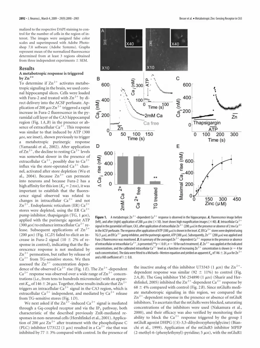
To determine if Zn 2� activates metabo-tropic signaling in the brain, we used coro-nal hippocampal slices. Cells were loadedwith Fura-2 and treated with Zn 2� by di-rect delivery into the ACSF perfusate. Ap-plication of 200 �M Zn 2� triggered a rapidincrease in Fura-2 fluorescence in the py-ramidal cell layer of the CA3 hippocampalregion (Fig. 1A,B) in the presence or ab-sence of extracellular Ca 2�. This responsewas similar to that induced by ATP (300�M, see inset), shown previously to triggera metabotropic purinergic response(Yamazaki et al., 2002). After applicationof Zn 2�, the decline to resting Ca 2� levelswas somewhat slower in the presence ofextracellular Ca 2�, possibly due to Ca 2�
influx via the store-operated Ca 2� chan-nel, activated after store depletion (Wu etal., 2004). Because Zn 2� can permeateinto neurons and because Fura-2 has ahigh affinity for this ion (Kd � 2 nM), it wasimportant to establish that the fluores-cence signal observed was related tochanges in intracellular Ca 2� and notZn 2�. Endoplasmic reticulum (ER) Ca 2�
stores were depleted, using the ER Ca 2�
pump inhibitor, thapsigargin (TG, 1 �M),applied with the purinergic agonist ATP(300 �M) to enhance intracellular Ca 2� re-lease. Subsequent applications of Zn 2�
(200 �M) (Fig. 1C,D) failed to elicit an in-crease in Fura-2 signal (10 � 2% of re-sponse in control), indicating that the flu-orescence response is not mediated byZn 2� permeation, but rather by release ofCa 2� from TG-sensitive stores. We thenassessed the Zn 2� concentration depen-dence of the observed Ca 2� rise (Fig. 1E). The Zn 2�-dependentCa 2� response was observed over a wide range of Zn 2� concen-trations (i.e., from tens to hundreds micromolar) with an appar-ent Km of 146 � 26 �M. Together, these results indicate that Zn 2�
triggers an intracellular Ca 2� signal in the CA3 region, which isextracellular Ca 2� independent, and mediated by Ca 2� releasefrom TG-sensitive stores (Fig. 1D).
We next asked if the Zn 2�-induced Ca 2� signal is mediatedthrough a Gq-coupled receptor and via the IP3 pathway, bothcharacteristic of the described previously ZnR-mediated re-sponses in non-neuronal cells (Hershfinkel et al., 2001). Applica-tion of 200 �M Zn 2� after treatment with the phospholipase C(PLC) inhibitor U73122 (1 �M) resulted in a Ca 2� rise that wasinhibited by 77 � 3% compared with control. In the presence of
the inactive analog of this inhibitor U73343 (1 �M) the Zn 2�-dependent response was similar (92 � 11%) to control (Fig.2A,B). The G�q inhibitor YM-254890 (1 �M) (Sharir and Her-shfinkel, 2005) inhibited the Zn 2�-dependent Ca 2� response by68 � 4% compared with control (Fig. 2B). Since mGluRs medi-ate metabotropic signaling in this region, we compared theZn 2�-dependent response in the presence or absence of mGluRinhibitors. To ascertain that the mGluRs were blocked, saturatingconcentrations of the inhibitors were used (Nakamura et al.,2000), and their efficacy was also verified by monitoring theirability to block the Ca 2� response triggered by the group ImGluR agonist DHPG ((S)-3,5-dihydroxyphenylglycine) (Bian-chi et al., 1999). Application of the mGluR5 inhibitor MPEP(2-methyl-6-(phenylethynyl)-pyridine; 5 �M), with the mGluR1
Figure 1. A metabotropic Zn 2�-dependent Ca 2� response is observed in the hippocampus. A, Fluorescence image before(left), and after (right) application of 200 �M zinc (�10). Inset shows high magnification images (�40). B, Intracellular Ca 2�
signal in the pyramidal cell layer, CA3, after application of extracellular Zn 2� (200 �M) in the presence or absence of 2 mM Ca 2�
in the ACSF perfusate. The response after application of ATP (300 �M) is shown in the inset. C, ER Ca 2� stores were depleted usingTG (1 �M), an ER Ca 2� pump inhibitor, and the purinergic agonist, ATP (300 �M). Subsequently, Zn 2� (200 �M) was applied andFura-2 fluorescence was monitored. D, A summary of the averaged Zn 2�-dependent Ca 2� response in the presence or absenceof extracellular or intracellular Ca 2�, is presented (**p � 0.01, n � 10 for each treatment). E, Zn 2� was applied at the indicatedconcentrations, and the calibrated intracellular Ca 2� level as a function of increasing Zn 2� concentration is shown (n � 4 foreach concentration). The data were fitted to a Michaelis–Menten equation and yielded an apparent Km of 146 � 26 �M for Zn 2�
and a Hill coefficient of 1 � 0.8.
2892 • J. Neurosci., March 4, 2009 • 29(9):2890 –2901 Besser et al. • Metabotropic Zinc-Sensing Receptor in CA3

inhibitor AIDA (1-aminoindan-1,5-dicarboxylic acid; 500 �M),attenuated the Ca 2� signal induced by 100 �M DHPG by 62 �6% (n � 8), while 10 �M MPEP in the presence of 1 mM AIDAreduced the DHPG-dependent response by 69 � 5% (n � 8). Incontrast, the Zn 2�-dependent Ca 2� signal was not reduced ateither concentration of the mGluR inhibitors ( p � 0.3) (Fig. 2B),suggesting that the Zn 2�-triggered response is not related tomGluRs, but is mediated via a distinct metabotropic pathway.The metal selectivity of the metabotropic response was then as-sessed, focusing on Cd 2� and Pb 2�. Cd 2� is a group IIB metal,like Zn 2�, which binds with high affinity to Zn 2� binding sites onproteins such as metallothionein (Henkel and Krebs, 2004). Pb 2�
is a divalent metal that has been shown to alter synaptic function(White et al., 2007). To avoid background effects mediated bypermeation of the ions, and to resolve if these ions trigger themetabotropic Ca 2� response, we determined the difference be-tween the maximal Fura-2 signal, triggered by Cd 2�, Pb 2� orZn 2�, in the presence or absence of the G�q inhibitor(�RGq inhibitor, see Materials and Methods). As shown in Figure2C, the Zn2�-dependent Fura-2 response was largely blocked by theG�q inhibitor (1 �M), yielding a �RGq inhibitor � 0.65 � 0.05. Incontrast, the inhibitor did not significantly reduce Cd2� or Pb2�-
dependent fluorescence (�RGq inhibitor �0.05 � 0.09 or 0.1 � 0.05, respectively). Thisindicates that Cd2� and Pb2� do not inducea metabotropic Ca2� rise, and that the ZnRresponse is selective for Zn2�.
Previous work in our lab has shownthat the epithelial ZnR undergoes pro-found functional desensitization after pro-longed exposure to Zn 2� at concentra-tions which can activate the ZnR (Azriel-Tamir et al., 2004; Sharir and Hershfinkel,2005). Such a regulatory mechanismwould be especially relevant in the CNS,considering the pathophysiological impli-cations of sustained activation of the IP3
pathway (Tang et al., 2003), and the poten-tial excessive release of synaptic Zn 2� dur-ing intense excitatory activity (Weiss et al.,2000). To determine if Zn 2�-dependentsignaling in neurons undergoes desensiti-zation, slices were pretreated with 75 �M
Zn 2� for 15 min and then washed in ACSF(see Materials and Methods). We subse-quently applied 300 �M Zn 2� while mon-itoring the Ca 2� response. The Zn 2� andATP-dependent Ca 2� response observedin the control slices (pretreated withACSF) is smaller than that shown in Figure2B, and may result from the prolonged in-terval before imaging. The Zn 2�-dependent Ca 2� response in slices thatwere pre-exposed to Zn 2� was only 15 �2% compared with control (Fig. 2D)(�R � 0.33 � 0.02 in control comparedwith 0.05 � 0.01 in the Zn 2� pretreatedslices), while application of ATP triggereda robust Ca 2� rise (Fig. 2D). As shown,ATP-induced Ca 2� responses were some-what larger in Zn 2�-desensitized prepara-tions, likely due to the fact that intracellu-lar stores had not been depleted by a ZnR-
mediated response. Together, these results indicate that theZn 2�-dependent metabotropic response triggered in the CA3region is mediated through a Gq-coupled receptor via the IP3
pathway, and undergoes a Zn 2�-dependent desensitization sim-ilar to that described previously for the epithelial ZnR.
To determine whether neurons or glia exhibit metabotropicZn 2�-dependent Ca 2� release, Zn 2� was applied as before andthe Ca 2� response monitored in a confocal microscope (Fig. 3A).Slices were subsequently stained with the astroglial marker SR101(Sulforhodamine 101) (Nimmerjahn et al., 2004). As shown inFigure 3B, cells which exhibited a Zn 2�-dependent Ca 2� rise(white arrows) were not labeled with the astrocyte marker, SR101suggesting that ZnR activity is not found in astroglia. To furtheraddress this issue, we asked if ZnR activity is observed in anestablished neuronal cell line, SH-SY5Y, a subclone of the SK-N-SH cell line. SH-SY5Y cells were previously used to study neu-rite outgrowth, cell growth and glutamate toxicity (Canals et al.,2005; Smith et al., 2009). As shown in Figure 3C, application of200 �M Zn 2�, a concentration which triggered robust ZnR activ-ity in the slices (Fig. 1), was also followed by an intracellular Ca 2�
rise in the SH-SY5Y cells. When SH-SY5Y cells were treated withthe G�q inhibitor, YM-254890 (1 �M), the Zn 2�-induced Ca 2�
Figure 2. The Ca 2� rise triggered by Zn 2� is mediated by a Gq- and PLC-dependent pathway. A, Slices were pretreated (asmarked) with the PLC inhibitor U73122 (1 �M, active form) or its inactive derivative U73343 (1 �M, control) and Zn 2� (200 �M)was applied. As control, ATP (300 �M) was subsequently applied. The Ca 2� signal (Fura-2) is shown. B, The averaged Ca 2� risetriggered by Zn 2� in the presence of the inhibitors of PLC (U73122, 1 �M) or G�q (YM-254890, 1 �M) or the mGluR1 and 5inhibitors (AIDA and MPEP, respectively) at the indicated concentrations (**p � 0.01, n � 20 for control and n � 9 for all othertreatments). C, The fluorescent response observed after application of Cd 2� and Pb 2� were monitored in the absence orpresence of the G�q inhibitor (1 �M, YM-254890). The difference (�RGq inhibitor) between the response in the presence or absenceof the inhibitor is shown (**p�0.01, n�5). D, Slices were pretreated with 75 �M Zn 2� or ACSF for 15 min, washed, and allowedto recover in ACSF for 60 –100 min. Zn 2� (300 �M) was then reapplied while monitoring the Ca 2� response. As control,ATP (300 �M) was subsequently applied (**p � 0.01, n � 7).
Besser et al. • Metabotropic Zinc-Sensing Receptor in CA3 J. Neurosci., March 4, 2009 • 29(9):2890 –2901 • 2893

response was inhibited by 95 � 3% com-pared with control (Fig. 3C) (�R � 0.61 �0.15 in control and 0.03 � 0.01 in the pres-ence of the inhibitor), indicating that theZn 2�-dependent Ca 2� response is medi-ated via a Gq-dependent pathway.
MAP and CAM kinase pathways areactivated by metabotropicZn 2�-dependent signalingActivation of extracellular regulated ki-nase (ERK1/2) and Ca 2�/calmodulin-dependent protein kinase (CaMKII) playfundamental roles in hippocampal synap-tic plasticity and neuronal survival (Han-sen et al., 2003; Giovannini, 2006; Luo andDeFranco, 2006; Cohen-Matsliah et al.,2007). Because intracellular Ca 2� rise canactivate these pathways (Berkeley andLevey, 2003), we sought to determine if theZn 2�-dependent metabotropic responsecould trigger ERK1/2 or CaMKII phos-phorylation in neurons. As shown in Fig-ure 4, A and B, application of Zn 2� (100�M, 90 s) led to a fourfold increase inpERK1/2 in the CA3 region comparedwith control. Colocalization with DAPIusing confocal microscopy indicated thatpERK1/2 staining was abundant in the nu-clear and perinuclear regions (Fig. 4A,bottom). Application of blockers of theiGluRs (20 �M CNQX and 50 �M AP5)(Fig. 4B), and the Ca 2� channel blocker,nimodipine (1 �m), did not attenuate theZn 2�-dependent phosphorylation ofERK1/2, indicating that it is mediated byextracellular Zn 2� and not by its potentialpermeation through these pathways. As illustrated in Figure 4, Cand D, application of Zn 2� also resulted in an increase of �50%in phosphorylated CaMKII. Finally, to assess the role of themetabotropic pathway in mediating Zn 2�-dependent kinasephosphorylation, slices were treated with the G�q inhibitor YM-254890 (1 �M) before application of Zn 2�. As shown in Figure 4,application of the G�q inhibitor reduced the Zn 2�-dependentphosphorylation of both ERK1/2 and CaMKII to baseline levels.Together, our results indicate that a G�q-coupled pathway me-diates Zn 2�-dependent ERK1/2 and CaMKII phosphorylation inthe CA3 region of the hippocampus.
GPR39 mediates neuronal Zn 2�-dependent signalingIt was recently suggested that a member of the ghrelin receptor fam-ily of GPCRs, namely GPR39, can be activated by serum Zn2� (Ya-suda et al., 2007). The physiological significance of the Zn2�-dependent signal mediated by GPR39 activation remains unclear,however (Holst et al., 2007; Yasuda et al., 2007). We hypothesizedthat GPR39 might be the ZnR itself and thus sought to determine ifit mediates neuronal Zn2�-dependent signaling. The endogenousexpression of GPR39 was initially determined by immunoblot anal-ysis of the neuronal SH-SY5Y cells (Fig. 5A). This cell line is partic-ularly useful for ectopic expression of genes and RNA constructsbecause of the high transfection efficiency that is typically achieved.A short hairpin RNA (shGPR39) construct targeted to silenceGPR39 efficiently lowered GPR39 expression in these cells, as deter-
mined by immunoblot analysis (Fig. 5A). As a control, a hairpinconstruct targeted against T1R3, a closely related GPCR, did not alterGPR39 expression in SH-SY5Y cells (Fig. 5A). The Ca2� response toapplication of Zn2� was then monitored in SH-SY5Y cells trans-fected with shGPR39 or in cells transfected with either shT1R3 orwith an empty vector. As shown in Figure 5, B and C, the Zn2�-dependent Ca2� response was similar in vector and shT1R3-expressing cells, but significantly attenuated (by 73 � 7%) in cellstransfected with shGPR39. We then asked whether GPR39 was en-dogenously expressed in the hippocampus. Immunofluorescent la-beling of GPR39 (Fig. 5D) was observed as punctuate staining at theperiphery of cells expressing the neuronal nuclear marker (NeuN) inCA3. Some punctuate staining was also observed near dendrites inthe CA3 region (Fig. 5D, inset). This suggests that GPR39 is presenton the cells that exhibit Zn2�-dependent metabotropic activity.These results are consistent with the hypothesis that GPR39 maymediate ZnR signaling in CA3 neurons.
Synaptic Zn 2� triggers ZnR-dependent response in theCA3 regionWhile the experiments described above strongly support the no-tion that a metabotropic receptor is activated in neurons in theCA3 region by exogenous Zn 2�, induction of the Ca 2� responseby synaptically released Zn 2� is required to demonstrate thephysiological significance of this phenomenon. To address thisissue, synaptic Zn 2� release was induced by electrical stimulation
Figure 3. Zn 2�-dependent Ca 2� rise is monitored in CA3 neurons but not in astrocytes. A, Confocal microscope analysis ofslices that were loaded with the Ca 2� indicator, Fluo-4 (5 �M). The Ca 2� rise after application of Zn 2� in two representative cellsis shown. B, Cells that showed Zn 2�-dependent Ca 2� rise were indicated (arrows) and subsequently SR101 (1 �M) was added tomark astroglia cells (red). C, Zn 2� (200 �M) was applied to SH-SY5Y neuronal cells loaded with Fura-2 in the absence or presenceof the G�q inhibitor (1 �M YM-254890) (**p � 0.01, n � 9).
2894 • J. Neurosci., March 4, 2009 • 29(9):2890 –2901 Besser et al. • Metabotropic Zinc-Sensing Receptor in CA3

of the mossy fibers using a protocol consisting of 10 pulses at 66Hz (Fig. 6A) while monitoring intracellular Ca 2� changes in CA3neurons. As shown in Figure 6B, a robust increase in intracellularCa 2� concentration was triggered after the electrical stimulation.
This response was blocked by applicationof the voltage-gated Na�-channel blockertetrodotoxin (1 �M, 91 � 3% inhibitioncompared with control) (Fig. 6B,D). Wealso applied CdCl (200 �M) at a concentra-tion that did not attenuate a metabotropicsignal triggered by ATP (data not shown)yet is known to effectively inhibit theCa 2�-channels that govern synaptic re-lease (Hinkle et al., 1987; Rosenmund andStevens, 1996). Similar to the effect of te-trodoxin (TTX), application of CdCl in-hibited the fluorescence response in theCA3 cells after stimulation of the mossyfibers (Fig. 6B,D) (86 � 5% inhibitioncompared with control), suggesting thatthe metabotropic Ca 2� response in theCA3 region is triggered by synaptic trans-mission. Application of the G�q inhibitorYM-254890 (1 �M) also resulted in a 92 �4% inhibition of the responses, comparedwith control slices (Fig. 6B,D). The inhib-itory action of YM-254890 indicates that aGq-coupled receptor triggers the Ca 2� re-sponse in the CA3 neurons after mossy fi-ber stimulation. The observed Ca 2� sig-nals occurred over a range of severalseconds, characteristic of metabotropicreceptor-mediated physiological re-sponses (Fig. 1) (Kapur et al., 2001). Inhib-itors of iGluRs, namely CNQX (20 �M)and AP5 (50 �M), failed to attenuate theCa 2� rise (Fig. 6C). Finally, in slices thatwere treated with AIDA and MPEP, inhib-itors, respectively, of mGluR1 andmGluR5, the Ca 2� response triggered bythe electrical stimulation was reduced by�50%, compared with the response incontrol slices (Fig. 6D). A similar inhibi-tory effect was observed in slices treatedwith both the mGluR and iGluR inhibitors(1 mM AIDA, 10 �M MPEP, 20 �M CNQXand 50 �M AP5). Thus, a significant, re-sidual metabotropic Ca 2� response per-sisted in the presence of mGluR inhibi-tors, while the G�q inhibitor completelyinhibited this response (Fig. 6 D). Thissuggests that after synaptic release a re-ceptor distinct from the mGluRs is re-sponsible for a substantial metabotropicCa 2� response in CA3.
To assess the role of Zn 2� in triggeringthe synaptically mediated metabotropicCa 2� response, electrical stimulation wasperformed in the presence of the cell-impermeable Zn 2� chelator, CaEDTA,which does not significantly affect extra-cellular Ca 2� concentration (Qian andNoebels, 2005). As shown in Figure 7, A
and B, in the presence of 10 � CaEDTA, the Ca 2� responsewithin the CA3 layer was only slightly lower than that in control.However, an inhibitory effect was observed in the presence ofeither 150 �M or 1 mM CaEDTA (Fig. 7B) (inhibition of 51 � 5%
Figure 4. The metabotropic Zn 2�-dependent response is followed by phosphorylation of ERK1/2 and CaMKII in the CA3region. A, Zn 2� (100 �M, 90 s) was applied to slices in the presence or absence of the G�q inhibitor (1 �M, YM-254890). Sliceswere then reacted with pERK1/2 antibody, shown in red, and DAPI (blue). Images acquired at �10 (top) and �63 (confocal,bottom) are shown. B, Quantitative analysis of ERK1/2 phosphorylation normalized to DAPI staining in the CA3 region. Shown isthe phosphorylation after application of Zn 2� in the absence or presence of the ionotropic glutamate inhibitors (CNQX, 20 �M andAP5, 50 �M) and Ca 2� channel blocker (nimodipine 1 �M), or the G�q inhibitor (1 �M YM-254890) (**p � 0.01, n � 6). C, Slicestreated with Zn 2� (100 �M, 3 min), in the presence or absence of the G�q inhibitor, were then reacted with pCaMKII antibodies(red) and DAPI (blue). D, Quantitative analysis of pCaMKII staining normalized to DAPI staining (**p � 0.01, n � 6).
Besser et al. • Metabotropic Zinc-Sensing Receptor in CA3 J. Neurosci., March 4, 2009 • 29(9):2890 –2901 • 2895

or 54 � 7%, respectively, compared withcontrol). This indicates that synapticallyreleased Zn 2� mediates a major part of themetabotropic Ca 2� response. The rela-tively short time required to induce themetabotropic effect indicates that CaE-DTA is unlikely to affect this signaling byregulating the protein expression pattern,as was previously demonstrated for GluR2activation (Calderone et al., 2004). Finally,we asked if the mGluR inhibitors togetherwith the chelator have an additive effect inblocking the metabotropic Ca 2� response.As shown in Figure 7B, the metabotropicsignal after electrical stimulation of themossy fibers in the presence of the mGluRinhibitors (1 mM AIDA and 10 �M MPEP)and CaEDTA (150 �M) was attenuated by84 � 6%. Thus, the coapplication of thechelator together with the mGluR inhibi-tors yielded a significantly smaller re-sponse than that monitored in the pres-ence of either CaEDTA ( p � 0.05) or theinhibitors alone ( p � 0.05).
Mice deficient in ZnT3 lack synapticZn2� while maintaining apparently normalsynaptic activity, including release of gluta-mate and GABA (Cole et al., 2000, 2001; Lo-pantsev et al., 2003). In agreement with pre-vious studies (Qian and Noebels, 2005), weobserved that stimulation of the mossy fiberswas followed by a significantly lower fluores-cence signal by the extracellular Zn2�-sensitive dye, Newport green (1 �M), in slicesfrom ZnT3 knock-out (KO) mice comparedwith wild-type controls (data not shown).These mice can thus serve as a useful modelto determine the role of synaptic Zn2� inactivating the ZnR and subsequent metabo-tropic Ca2� signaling. The Ca2� responseafter stimulation of the mossy fibers in slicesfrom ZnT3 KO mice was 56 � 7% of theresponse in slices from wild-type (WT) mice(Fig. 7C). Thus, in slices from mice lackingsynaptic Zn2�, we observed �45% inhibi-tion of the metabotropic signaling in CA3(Fig. 7D), similar to the reduction of themetabotropic signal by CaEDTA in WTmice (Fig. 7B). Application of exogenousZn2� to slices from the ZnT3 KO mice, however, was followed by aCa2� rise (108 � 7%) similar to WT controls (Fig. 7D). This Ca2�
response triggered by exogenous Zn2� in slices from ZnT3 KO miceindicates that the lack of synaptic Zn2�, rather than a deficiency ofZnR, is responsible for the attenuated metabotropic response. Ap-plication of CaEDTA (150 �M) to slices from ZnT3 KO mice yieldeda Ca2� response that did not significantly differ from the responseobtained in nontreated ZnT3 KO slices (Fig. 7E,F). Finally, applica-tion of the mGluR inhibitors, using the same experimental paradigmdescribed in Figure 6D, was followed by a Ca2� response that wasmuch smaller than the response in the nontreated, ZnT3 KO slices(Fig. 7F) (73 � 3% inhibition). Thus, the mGluR inhibitors exhibitsignificantly ( p � 0.01) greater inhibition of the response in ZnT3KO slices than their effect in the WT slices. The inhibitory effect of
the extracellular Zn2� chelator in slices from the WT, but not theZnT3 KO mice, together with the substantially attenuated metabo-tropic Zn2� response in the ZnT3 KO mice, support the conclusionthat synaptic Zn2�, released from mossy fiber terminals, activates aZnR and triggers Ca2� signaling in CA3 neurons of thehippocampus.
DiscussionPrevious studies have suggested that synaptic Zn 2� is a neuro-modulator, acting allosterically at ionotropic synaptic receptors(Vogt et al., 2000; Smart et al., 2004; Paoletti et al., 2009). Thepresent work demonstrates, for the first time, that Zn 2� specifi-cally activates metabotropic signaling in neurons, and does so viaa Zn 2�-sensing receptor. This finding is supported by the atten-
Figure 5. ZnR signaling is mediated by GPR39. A, Western blot analysis of GPR39 expression in SH-SY5Y cells transfected withcontrol vector, shGPR39 or shT1R3 plasmids. B, The Ca 2� response after application of Zn 2� (100 �M) in cells transfected withthe shGPR39 or vector (control). C, Quantitative analysis of the Zn 2�-dependent Ca 2� rise in cells transfected with the shGPR39,shT1R3 or control (**p � 0.01, n � 9). D, Confocal images of GPR39 labeling (red) and the neuronal marker NeuN (green) in theCA3. A combined image of both indicates that GPR39 is expressed in neuronal cells (bottom left). Bright field image is also shown(bottom right). Insert shows staining of the dendritic marker MAP2 (green) combined with GPR39 labeling (red) in the CA3neurons (scale bar, 10 �m).
2896 • J. Neurosci., March 4, 2009 • 29(9):2890 –2901 Besser et al. • Metabotropic Zinc-Sensing Receptor in CA3

uation of the Zn 2�-dependent Ca 2� rise after depletion of Ca 2�
stores in the ER, and after inhibition of the IP3 pathway using aG�q inhibitor or PLC blocker. A role for the IP3 pathway in ZnRsignaling is consistent with a previous study suggesting that
metabotropic signaling in the CA3 regionis IP3-dependent rather than rayonidine-receptor dependent (Kapur et al., 2001).The lack of a Zn 2�-dependent Ca 2� rise incells labeled by the astroglial marker,SR101, suggests that although glial cells ex-press metabotropic receptors, the Zn 2�-dependent metabotropic response is me-diated primarily by neurons.
Our results indicate that the transientmetabotropic Ca 2� response in CA3 neu-rons is triggered by synaptically releasedZn 2� after mossy fiber stimulation. TheCa 2� response triggered by electrical stim-ulation was inhibited by blockade of syn-aptic transmission. Furthermore, the re-sponse is attenuated in the presence of anextracellular Zn 2� chelator, CaEDTA, orin slices from mice lacking synaptic Zn 2�.The synaptic Zn 2�-dependent Ca 2� re-sponse was also mediated via a Gq-coupled mechanism. We conclude, there-fore, that in addition to glutamate,metabotropic signaling in the CA3 cells ismediated by synaptically released Zn 2�.
We used CaEDTA at concentrationssufficient to remove Zn 2� from the extra-cellular milieu, based on steady state con-ditions (http://www.stanford.edu/�cpat-ton/webmaxc/webmaxcS.htm) and theexpected concentration of released-Zn 2�
(Vogt et al., 2000; Qian and Noebels,2005). At low concentration of the chela-tor (10 �M), no significant effect on ZnRactivity was apparent. This is consistentwith the fact that ZnR is not activated bybaseline concentrations of extracellularZn 2�, because of its relative low affinity(Km �150 �M) (Fig. 1), unlike the highaffinity Zn 2� binding sites on the NMDAreceptor (Qian and Noebels, 2005). In thepresence of 150 �M or 1 mM CaEDTA,however, the ZnR response was reduced.In previous studies, CaEDTA, at similarconcentrations, did not effectively reducea transient rise in extracellular Zn 2� be-cause of its slow chelation activity (Qianand Noebels, 2005). The effective inhibi-tion of the ZnR response is probably re-lated to the relatively low affinity of theZnR to Zn 2�, suggesting such that evenpartial reduction of extracellular Zn 2� willsuffice to lower its concentration belowthe threshold of ZnR. Such reduction inextracellular Zn 2� may not be apparentusing high affinity Zn 2�-sensitive dyes. Inaddition, metabotropic receptors are oftendistantly located from the release site ofneurotransmitters (Knopfel and Uusi-
saari, 2008), as we also show for the GPR39 (Fig. 5). Thus, pro-longed diffusion time of Zn 2� toward its receptor may enablemore effective chelation of Zn 2� by CaEDTA.
While some studies have suggested that synaptic Zn 2� does
Figure 6. Mossy fiber stimulation triggers a metabotropic Ca 2� rise that is partially independent of the mGluRs. A, Schematic repre-sentationoftheexperimentalsetupforelectricalstimulation.Astimulatingelectrodewasplacedatthemossyfiberaxons,nearthedentategyrus (DG) and the Fura-2 signal was monitored in the CA3 pyramidal cell layer. Shown is a representative time lapse Ca 2� signal acquiredfrom a single cell, after electrical stimulation (66 Hz, 100 �A for 150 ms, total of 10 pulses) of the mossy fibers at the marked time. B, TheCa 2� response in the CA3 region after electrical stimulation of the mossy fibers in the presence or absence of the G�q inhibitor (1 �M,YM-254890), with the voltage-gated Na �-channel blocker TTX (1 �M), or with the voltage-gated Ca 2� channel blocker CdCl (200 �M).Representative responses averaged over 25 cells in 1 slice. C, Slices were treated with the iGluR inhibitors (CNQX, 20 �M and AP5, 50 �M)subsequently the mossy fibers were stimulated and the Ca 2� response is shown. Representative responses averaged over 24 cells in 1slice. D, The averaged responses of the Ca 2� rise triggered after electrical stimulation of the mossy fibers in control slices (n � 23) or inslices treated with TTX (n � 6), CdCl (n � 6), the G�q inhibitor (n � 6), the mGluR1 and 5 inhibitors (AIDA and MPEP, respectively, n �6) or the mGluRs inhibitors together with the iGluRs inhibitors (1 mM AIDA, 10�M MPEP, 20�M CNQX and 50�M AP5, n�4) (*p�0.05,**p � 0.01; NS, nonsignificant).
Besser et al. • Metabotropic Zinc-Sensing Receptor in CA3 J. Neurosci., March 4, 2009 • 29(9):2890 –2901 • 2897

not affect activity-dependent plasticity,but rather, reshapes the NMDA response(Vogt et al., 2000), others support an effectof synaptic Zn 2� on modulation of long-term synaptic changes in the hippocampus(Xie and Smart, 1994; Li et al., 2001; Izumiet al., 2006). Furthermore, synaptic Zn 2�
was shown previously to facilitate LTP infear conditioning pathways by attenuatingGABAergic inhibition (Kodirov et al.,2006). Recently, synaptic Zn 2� was sug-gested to enhance mossy fiber LTP by ac-tivating intracellular signaling of TrkB re-ceptors (Huang et al., 2008; Nagappan etal., 2008). Interestingly, TrkB signaling isstrongly regulated by GPCRs, via signalingpathway similar to that activated by ZnR(Hwang et al., 2005; Chen et al., 2007). Ithas also been reported that TrkB is acti-vated by extracellular Zn 2� through regu-lation of metalloproteases (Hwang et al.,2005). An intriguing question remainswhether ZnR links synaptic Zn 2� to regu-lation of LTP.
Our results indicate that extracellularZn 2�, via the ZnR, activates mitogen-activated protein kinase (MAPK) andCaMKII pathways. Activation of theMAPK pathway by intracellular Zn 2� rise(Seo et al., 2001; Harris et al., 2004), orafter oxidative or nitrosative stress(McLaughlin et al., 2001; Du et al., 2003;Zhang et al., 2004, 2007), is associated withneuronal death. In contrast, activation ofMAPK by extracellular Zn 2�, in neurons,has been shown to be anti-apoptotic (An etal., 2005). Activation of CaMKII has alsobeen linked to neuronal survival, and inac-tivation of CaMKII during seizure in-creases neuronal cell death (Hansen et al.,2003). Zinc has been shown to activateCaMKII (Lengyel et al., 2000), while an ex-tracellular Zn 2� chelator suppressedCaMKII activity in the hippocampus (Tanand Chen, 1997). In the absence of synap-tic Zn 2�, hippocampal CA3 neurons aremore vulnerable to seizure-induced celldeath (Domínguez et al., 2003; Cote et al.,2005), consistent with a prosurvival rolefor ZnR-dependent activation of MAPKand CaMKII. Finally, both the MAPK andCaMKII pathways are critical players insynaptic plasticity, involved in hippocam-pal learning and memory (Rosenblum etal., 2002; Miyamoto, 2006). Our resultsshow that Zn 2�, applied for a shortdura-tion (i.e., 90 s), induces translocation ofpERK1/2 to the nucleus.
Changes in expression and function of metabotropic gluta-mate receptors appear to be critical factors in the etiology ofepileptic seizures (Pitsch et al., 2007). Interestingly, chelation ofsynaptic Zn 2�, as well as dietary Zn 2� deficiency, are associatedwith increased susceptibility to seizures (Fukahori and Itoh,
1990; Domínguez et al., 2003). These effects complement thephenotype of the ZnT3 knock-out mice, which are more suscep-tible to kainate-induced seizures (Cole et al., 2000). At present, itis unclear how synaptic Zn 2� is related to seizure activity. Theinvolvement of Zn 2� in modulating GABA and NMDA receptorsdoes not dictate either an inhibitory or excitatory role for this
Figure 7. The metabotropic Ca 2� response is attenuated in the absence of synaptic Zn 2�. A, The Ca 2� response in cells in theCA3 region after electrical stimulation of the mossy fibers in the presence or absence of the extracellular Zn 2� chelator CaEDTA atthe indicated concentrations. Representative responses averaged over 28 cells in 1 slice. B, The averaged Ca 2� response in thepresence or absence of CaEDTA (n � 9) at the indicated concentration, or 150 �M CaEDTA in the presence of the mGluR inhibitors(n � 6), or CaEDTA with the mGluR and iGluR inhibitors (n � 4) (*p � 0.05 compared with control and #p � 0.05 compared withCaEDTA alone; NS, nonsignificant). The control is the same as in Figure 6 D. C, The response of cells in the CA3 region after electricalstimulation of the mossy fibers in slices obtained from ZnT3 KO versus WT, control, mice. Representative responses averaged over23 cells in 1 slice. D, Summary of the Ca 2� responses triggered in slices from the ZnT3 KO and WT mice after electrical stimulationor the application of exogenous Zn 2� using the paradigm described in Figure 1 (**p � 0.01, n � 11 for each treatment, the WTcontrol is the same as in Fig. 6 D for the stimulation induced response and to Fig. 2 B for the exogenous Zn 2� application). E, TheCa 2� rise triggered in slices from the ZnT3 KO mice in the presence or absence of CaEDTA. Representative response averaged over26 cells in 1 slice. F, Averaged response after electrical stimulation of the mossy fibers in slices from ZnT3 KO mice in the presenceof CaEDTA (n � 7) or the mGluR inhibitors (500 �M AIDA, 5 �M MPEP, n � 7) (**p � 0.01).
2898 • J. Neurosci., March 4, 2009 • 29(9):2890 –2901 Besser et al. • Metabotropic Zinc-Sensing Receptor in CA3

metal, and thus, provides no clear basis for assigning it a role inseizure generation. As such, ZnR function in the hippocampusmay represent a novel contributor to the overall regulation ofneuronal excitability by Zn 2�.
Effects of Zn 2� on synaptic plasticity and neuronal excitabil-ity, and effects of Zn 2� deficiency on learning and memory inmice and humans are well documented (Cole et al., 2000; Lopant-sev et al., 2003; Smart et al., 2004; Kodirov et al., 2006). SinceZnT3 is considered the principal transport system for the loadingof Zn 2� ions into synaptic vesicles, and knockout of this trans-porter completely eliminates Zn 2� from these vesicles (Fred-erickson et al., 2006; Linkous et al., 2008), the relatively mildphenotype characterizing ZnT3 KO mice has been somewhat un-expected. Our finding that the metabotropic Ca 2� response inslices from ZnT3 KO mice was unaffected by CaEDTA, suggeststhat it is not mediated via the ZnR.
The metabotropic Zn 2�-dependent activity we observed inCA3 neurons is remarkably similar to the extracellular Zn 2�-dependent response we previously characterized in epithelial cells(Hershfinkel et al., 2001; Sharir and Hershfinkel, 2005; Dubi etal., 2008). The molecular identity of the receptor mediating thisresponse had been unknown. The orphan GPCR GPR39 is amember of the ghrelin receptor family (McKee et al., 1997).Obestatin was initially proposed as the putative ligand for GPR39(Zhang et al., 2005), yet, subsequent studies failed to confirm this(Lauwers et al., 2006). It was subsequently suggested that Zn 2�
may directly interact with this receptor and enhance its signaling(Holst et al., 2004). The results presented here suggest thatGPR39 may indeed directly mediate Zn 2�-dependent signaling.The metal selectivity and dose dependence of the neuronal ZnRpresent in the hippocampus (Fig. 2), and those described forGPR39 (Yasuda et al., 2007) are strikingly similar. It has beenreported, moreover, that GPR39 mRNA is expressed in the CNS,and specifically in the hippocampus (Jackson et al., 2006). Con-sistent with these findings, our immunofluorescence analysis in-dicates that GPR39 expression is highly enriched in the CA3 neu-rons that are postsynaptic to the Zn 2�-rich mossy fibers, thesame cells that mediate synaptically released Zn 2�-dependentmetabotropic activity. Thus, we propose that GPR39 is a criticalcomponent of ZnR metabotropic signaling, and, most likely, thatGPR39 and ZnR are one and the same receptor.
In conclusion, we have identified Zn 2�-dependent metabo-tropic activity in hippocampal CA3 neurons. This activity inbrain areas rich in synaptic Zn 2� may represent the long-soughtlink between dynamic changes in extracellular Zn 2� and neuro-nal signaling mediated by this metal. Our data suggest that spe-cific Zn 2�-sensing receptor activity, putatively mediated byGPR39, triggers synaptic Zn 2�-dependent metabotropic signal-ing in the CA3 region. This is the first evidence that Zn 2� is notonly a modulatory ion, but acts via a specific postsynaptic recep-tor to trigger neuronal metabotropic signaling.
ReferencesAizenman E, Stout AK, Hartnett KA, Dineley KE, McLaughlin B, Reynolds IJ
(2000) Induction of neuronal apoptosis by thiol oxidation: putative roleof intracellular zinc release. J Neurochem 75:1878 –1888.
An WL, Pei JJ, Nishimura T, Winblad B, Cowburn RF (2005) Zinc-inducedanti-apoptotic effects in SH-SY5Y neuroblastoma cells via the extracellu-lar signal-regulated kinase 1/2. Brain Res Mol Brain Res 135:40 – 47.
Azriel-Tamir H, Sharir H, Schwartz B, Hershfinkel M (2004) Extracellular zinctriggers ERK-dependent activation of Na�/H�exchange in colonocytes me-diated by the zinc-sensing receptor. J Biol Chem 279:51804–51816.
Beierlein M, Fall CP, Rinzel J, Yuste R (2002) Thalamocortical bursts triggerrecurrent activity in neocortical networks: layer 4 as a frequency-dependent gate. J Neurosci 22:9885–9894.
Berkeley JL, Levey AI (2003) Cell-specific extracellular signal-regulated ki-nase activation by multiple G protein-coupled receptor families in hip-pocampus. Mol Pharmacol 63:128 –135.
Bianchi R, Young SR, Wong RK (1999) Group I mGluR activation causesvoltage-dependent and -independent Ca2� rises in hippocampal pyra-midal cells. J Neurophysiol 81:2903–2913.
Calderone A, Jover T, Mashiko T, Noh KM, Tanaka H, Bennett MV, Zukin RS(2004) Late calcium EDTA rescues hippocampal CA1 neurons fromglobal ischemia-induced death. J Neurosci 24:9903–9913.
Canals M, Angulo E, Casado V, Canela EI, Mallol J, Vinals F, Staines W,Tinner B, Hillion J, Agnati L, Fuxe K, Ferre S, Lluis C, Franco R (2005)Molecular mechanisms involved in the adenosine A and A receptor-induced neuronal differentiation in neuroblastoma cells and striatal pri-mary cultures. J Neurochem 92:337–348.
Chen MJ, Nguyen TV, Pike CJ, Russo-Neustadt AA (2007) Norepinephrineinduces BDNF and activates the PI-3K and MAPK cascades in embryonichippocampal neurons. Cell Signal 19:114 –128.
Cohen-Matsliah SI, Brosh I, Rosenblum K, Barkai E (2007) A novel role forextracellular signal-regulated kinase in maintaining long-term memory-relevant excitability changes. J Neurosci 27:12584 –12589.
Cole TB, Wenzel HJ, Kafer KE, Schwartzkroin PA, Palmiter RD (1999)Elimination of zinc from synaptic vesicles in the intact mouse brain bydisruption of the ZnT3 gene. Proc Natl Acad Sci U S A 96:1716 –1721.
Cole TB, Robbins CA, Wenzel HJ, Schwartzkroin PA, Palmiter RD (2000)Seizures and neuronal damage in mice lacking vesicular zinc. Epilepsy Res39:153–169.
Cole TB, Martyanova A, Palmiter RD (2001) Removing zinc from synapticvesicles does not impair spatial learning, memory, or sensorimotor func-tions in the mouse. Brain Res 891:253–265.
Cote A, Chiasson M, Peralta MR 3rd, Lafortune K, Pellegrini L, Toth K(2005) Cell type-specific action of seizure-induced intracellular zinc ac-cumulation in the rat hippocampus. J Physiol 566:821– 837.
Domínguez MI, Blasco-Ibanez JM, Crespo C, Marques-Marí AI, Martínez-Guijarro FJ (2003) Zinc chelation during non-lesioning overexcitationresults in neuronal death in the mouse hippocampus. Neuroscience116:791– 806.
Du L, Zhang X, Han YY, Burke NA, Kochanek PM, Watkins SC, Graham SH,Carcillo JA, Szabo C, Clark RS (2003) Intra-mitochondrial poly(ADP-ribosylation) contributes to NAD� depletion and cell death induced byoxidative stress. J Biol Chem 278:18426 –18433.
Dubi N, Gheber L, Fishman D, Sekler I, Hershfinkel M (2008) Extracellularzinc and zinc-citrate, acting through a putative zinc sensing receptor,regulate growth and survival of prostate cancer cells. Carcinogenesis29:1692–1700.
Frederickson CJ, Moncrieff DW (1994) Zinc-containing neurons. Biol Sig-nals 3:127–139.
Frederickson CJ, Giblin LJ 3rd, Balaji RV, Rengarajan B, Masalha R, Fred-erickson CJ, Zeng Y, Lopez EV, Koh JY, Chorin U, Besser L, HershfinkelM, Li Y, Thompson RB, Krezel A (2006) Synaptic release of zinc frombrain slices: factors governing release, imaging, and accurate calculationof concentration. J Neurosci Methods 154:19 –29.
Fukahori M, Itoh M (1990) Effects of dietary zinc status on seizure suscep-tibility and hippocampal zinc content in the El (epilepsy) mouse. BrainRes 529:16 –22.
Giovannini MG (2006) The role of the extracellular signal-regulated kinasepathway in memory encoding. Rev Neurosci 17:619 – 634.
Grynkiewicz G, Poenie M, Tsien RY (1985) A new generation of Ca2� in-dicators with greatly improved fluorescence properties. J Biol Chem260:3440 –3450.
Gundelfinger ED, Boeckers TM, Baron MK, Bowie JU (2006) A role for zincin postsynaptic density asSAMbly and plasticity? Trends Biochem Sci31:366 –373.
Hansen MR, Bok J, Devaiah AK, Zha XM, Green SH (2003) Ca2�/calmodulin-dependent protein kinases II and IV both promote survivalbut differ in their effects on axon growth in spiral ganglion neurons.J Neurosci Res 72:169 –184.
Harris FM, Brecht WJ, Xu Q, Mahley RW, Huang Y (2004) Increased tauphosphorylation in apolipoprotein E4 transgenic mice is associated withactivation of extracellular signal-regulated kinase: modulation by zinc.J Biol Chem 279:44795– 44801.
Henkel G, Krebs B (2004) Metallothioneins: zinc, cadmium, mercury, and
Besser et al. • Metabotropic Zinc-Sensing Receptor in CA3 J. Neurosci., March 4, 2009 • 29(9):2890 –2901 • 2899

copper thiolates and selenolates mimicking protein active site features–structural aspects and biological implications. Chem Rev 104:801– 824.
Hershfinkel M, Moran A, Grossman N, Sekler I (2001) A zinc-sensing re-ceptor triggers the release of intracellular Ca2� and regulates ion trans-port. Proc Natl Acad Sci U S A 98:11749 –11754.
Hinkle PM, Kinsella PA, Osterhoudt KC (1987) Cadmium uptake and toxicityvia voltage-sensitive calcium channels. J Biol Chem 262:16333–16337.
Holst B, Holliday ND, Bach A, Elling CE, Cox HM, Schwartz TW (2004)Common structural basis for constitutive activity of the ghrelin receptorfamily. J Biol Chem 279:53806 –53817.
Holst B, Egerod KL, Schild E, Vickers SP, Cheetham S, Gerlach LO, Storjo-hann L, Stidsen CE, Jones R, Beck-Sickinger AG, Schwartz TW (2007)GPR39 signaling is stimulated by zinc ions but not by obestatin. Endocri-nology 148:13–20.
Hosie AM, Dunne EL, Harvey RJ, Smart TG (2003) Zinc-mediated inhibi-tion of GABA(A) receptors: discrete binding sites underlie subtype spec-ificity. Nat Neurosci 6:362–369.
Huang YZ, Pan E, Xiong ZQ, McNamara JO (2008) Zinc-mediated trans-activation of TrkB potentiates the hippocampal mossy fiber-CA3 pyramidsynapse. Neuron 57:546 –558.
Hwang JJ, Park MH, Choi SY, Koh JY (2005) Activation of the Trk signalingpathway by extracellular zinc: role of metalloproteinases. J Biol Chem280:11995–12001.
Izumi Y, Auberson YP, Zorumski CF (2006) Zinc modulates bidirectionalhippocampal plasticity by effects on NMDA receptors. J Neurosci26:7181–7188.
Jackson VR, Nothacker HP, Civelli O (2006) GPR39 receptor expression inthe mouse brain. Neuroreport 17:813– 816.
Kan I, Ben-Zur T, Barhum Y, Levy YS, Burstein A, Charlow T, Bulvik S,Melamed E, Offen D (2007) Dopaminergic differentiation of humanmesenchymal stem cells– utilization of bioassay for tyrosine hydroxylaseexpression. Neurosci Lett 419:28 –33.
Kapur A, Yeckel M, Johnston D (2001) Hippocampal mossy fiber activityevokes Ca2� release in CA3 pyramidal neurons via a metabotropic glu-tamate receptor pathway. Neuroscience 107:59 – 69.
Knopfel T, Uusisaari M (2008) Modulation of excitation by metabotropicglutamate receptors. Results Probl Cell Differ 44:163–175.
Kodirov SA, Takizawa S, Joseph J, Kandel ER, Shumyatsky GP, Bolshakov VY(2006) Synaptically released zinc gates long-term potentiation in fearconditioning pathways. Proc Natl Acad Sci U S A 103:15218 –15223.
Lauwers E, Landuyt B, Arckens L, Schoofs L, Luyten W (2006) Obestatindoes not activate orphan G protein-coupled receptor GPR39. BiochemBiophys Res Commun 351:21–25.
Lengyel I, Fieuw-Makaroff S, Hall AL, Sim AT, Rostas JA, Dunkley PR (2000)Modulation of the phosphorylation and activity of calcium/calmodulin-dependent protein kinase II by zinc. J Neurochem 75:594 – 605.
Li Y, Hough CJ, Frederickson CJ, Sarvey JM (2001) Induction of mossyfiber3CA3 long-term potentiation requires translocation of synapticallyreleased Zn 2�. J Neurosci 21:8015– 8025.
Linkous DH, Flinn JM, Koh JY, Lanzirotti A, Bertsch PM, Jones BF, Giblin LJ,Frederickson CJ (2008) Evidence that the ZNT3 protein controls thetotal amount of elemental zinc in synaptic vesicles. J Histochem Cyto-chem 56:3– 6.
Lopantsev V, Wenzel HJ, Cole TB, Palmiter RD, Schwartzkroin PA (2003)Lack of vesicular zinc in mossy fibers does not affect synaptic excitabilityof CA3 pyramidal cells in zinc transporter 3 knockout mice. Neuroscience116:237–248.
Luo Y, DeFranco DB (2006) Opposing roles for ERK1/2 in neuronal oxida-tive toxicity: distinct mechanisms of ERK1/2 action at early versus latephases of oxidative stress. J Biol Chem 281:16436 –16442.
McKee KK, Tan CP, Palyha OC, Liu J, Feighner SD, Hreniuk DL, Smith RG,Howard AD, Van der Ploeg LH (1997) Cloning and characterization oftwo human G protein-coupled receptor genes (GPR38 and GPR39) re-lated to the growth hormone secretagogue and neurotensin receptors.Genomics 46:426 – 434.
McLaughlin B, Pal S, Tran MP, Parsons AA, Barone FC, Erhardt JA, Aizen-man E (2001) p38 activation is required upstream of potassium currentenhancement and caspase cleavage in thiol oxidant-induced neuronalapoptosis. J Neurosci 21:3303–3311.
Miyamoto E (2006) Molecular mechanism of neuronal plasticity: inductionand maintenance of long-term potentiation in the hippocampus. J Phar-macol Sci 100:433– 442.
Nagappan G, Woo NH, Lu B (2008) Ama “zinc” link between TrkB trans-activation and synaptic plasticity. Neuron 57:477– 479.
Nakamura T, Nakamura K, Lasser-Ross N, Barbara JG, Sandler VM, Ross WN(2000) Inositol 1,4,5-trisphosphate (IP3)-mediated Ca 2� release evokedby metabotropic agonists and backpropagating action potentials in hip-pocampal CA1 pyramidal neurons. J Neurosci 20:8365– 8376.
Nicholls RE, Zhang XL, Bailey CP, Conklin BR, Kandel ER, Stanton PK(2006) mGluR2 acts through inhibitory Galpha subunits to regulatetransmission and long-term plasticity at hippocampal mossy fiber-CA3synapses. Proc Natl Acad Sci U S A 103:6380 – 6385.
Nimmerjahn A, Kirchhoff F, Kerr JN, Helmchen F (2004) Sulforhodamine101 as a specific marker of astroglia in the neocortex in vivo. Nat Methods1:31–37.
Palmiter RD, Cole TB, Quaife CJ, Findley SD (1996) ZnT-3, a putativetransporter of zinc into synaptic vesicles. Proc Natl Acad Sci U S A93:14934 –14939.
Paoletti P, Ascher P, Neyton J (1997) High-affinity zinc inhibition ofNMDA NR1-NR2A receptors. J Neurosci 17:5711–5725.
Paoletti P, Vergnano AM, Barbour B, Casado M (2009) Zinc at glutamater-gic synapses. Neuroscience 158:126 –136.
Pitsch J, Schoch S, Gueler N, Flor PJ, van der Putten H, Becker AJ (2007)Functional role of mGluR1 and mGluR4 in pilocarpine-induced tempo-ral lobe epilepsy. Neurobiol Dis 26:623– 633.
Qian J, Noebels JL (2005) Visualization of transmitter release with zinc flu-orescence detection at the mouse hippocampal mossy fibre synapse.J Physiol 566:747–758.
Rachline J, Perin-Dureau F, Le Goff A, Neyton J, Paoletti P (2005) Themicromolar zinc-binding domain on the NMDA receptor subunit NR2B.J Neurosci 25:308 –317.
Rosenblum K, Futter M, Voss K, Erent M, Skehel PA, French P, Obosi L, JonesMW, Bliss TV (2002) The role of extracellular regulated kinases I/II inlate-phase long-term potentiation. J Neurosci 22:5432–5441.
Rosenmund C, Stevens CF (1996) Definition of the readily releasable poolof vesicles at hippocampal synapses. Neuron 16:1197–1207.
Ruiz A, Walker MC, Fabian-Fine R, Kullmann DM (2004) Endogenous zincinhibits GABA(A) receptors in a hippocampal pathway. J Neurophysiol91:1091–1096.
Sekler I, Moran A, Hershfinkel M, Dori A, Margulis A, Birenzweig N, NitzanY, Silverman WF (2002) Distribution of the zinc transporter ZnT-1 incomparison with chelatable zinc in the mouse brain. J Comp Neurol447:201–209.
Sensi SL, Jeng JM (2004) Rethinking the excitotoxic ionic milieu: the emerg-ing role of Zn(2�) in ischemic neuronal injury. Curr Mol Med 4:87–111.
Sensi SL, Yin HZ, Carriedo SG, Rao SS, Weiss JH (1999) Preferential Zn2�influx through Ca2�-permeable AMPA/kainate channels triggers pro-longed mitochondrial superoxide production. Proc Natl Acad Sci U S A96:2414 –2419.
Seo SR, Chong SA, Lee SI, Sung JY, Ahn YS, Chung KC, Seo JT (2001)Zn2�-induced ERK activation mediated by reactive oxygen speciescauses cell death in differentiated PC12 cells. J Neurochem 78:600 – 610.
Sharir H, Hershfinkel M (2005) The extracellular zinc-sensing receptor me-diates intercellular communication by inducing ATP release. BiochemBiophys Res Commun 332:845– 852.
Smart TG, Hosie AM, Miller PS (2004) Zn2� ions: modulators of excitatoryand inhibitory synaptic activity. Neuroscientist 10:432– 442.
Smith IF, Wiltgen SM, Parker I (2009) Localization of puff sites adjacent tothe plasma membrane: functional and spatial characterization of Ca(2�)signaling in SH-SY5Y cells utilizing membrane-permeant caged IP(3).Cell Calcium 45:65–76.
Tan SE, Chen SS (1997) The activation of calcium/calmodulin-dependentprotein kinase II after glutamate or potassium stimulation in hippocam-pal slices. Brain Res Bull 43:269 –273.
Tang TS, Tu H, Chan EY, Maximov A, Wang Z, Wellington CL, Hayden MR,Bezprozvanny I (2003) Huntingtin and huntingtin-associated protein 1influence neuronal calcium signaling mediated by inositol-(1,4,5)triphosphate receptor type 1. Neuron 39:227–239.
Vogt K, Mellor J, Tong G, Nicoll R (2000) The actions of synaptically re-leased zinc at hippocampal mossy fiber synapses. Neuron 26:187–196.
Volk LJ, Daly CA, Huber KM (2006) Differential roles for group 1 mGluRsubtypes in induction and expression of chemically induced hippocampallong-term depression. J Neurophysiol 95:2427–2438.
2900 • J. Neurosci., March 4, 2009 • 29(9):2890 –2901 Besser et al. • Metabotropic Zinc-Sensing Receptor in CA3

Weiss JH, Sensi SL, Koh JY (2000) Zn(2�): a novel ionic mediator of neuralinjury in brain disease. Trends Pharmacol Sci 21:395– 401.
White LD, Cory-Slechta DA, Gilbert ME, Tiffany-Castiglioni E, Zawia NH,Virgolini M, Rossi-George A, Lasley SM, Qian YC, Basha MR (2007)New and evolving concepts in the neurotoxicology of lead. Toxicol ApplPharmacol 225:1–27.
Wu X, Zagranichnaya TK, Gurda GT, Eves EM, Villereal ML (2004) ATRPC1/TRPC3-mediated increase in store-operated calcium entry is re-quired for differentiation of H19 –7 hippocampal neuronal cells. J BiolChem 279:43392– 43402.
Xie X, Smart TG (1994) Modulation of long-term potentiation in rat hip-pocampal pyramidal neurons by zinc. Pflugers Arch 427:481– 486.
Yamazaki Y, Fujii S, Nakamura T, Miyakawa H, Kudo Y, Kato H, Ito K(2002) Changes in [Ca2�]. (i) during adenosine triphosphate-inducedsynaptic plasticity in hippocampal CA1 neurons of the guinea pig. Neu-rosci Lett 324:65– 68.
Yasuda S, Miyazaki T, Munechika K, Yamashita M, Ikeda Y, Kamizono A(2007) Isolation of Zn2� as an endogenous agonist of GPR39 from fetalbovine serum. J Recept Signal Transduct Res 27:235–246.
Yin HZ, Sensi SL, Ogoshi F, Weiss JH (2002) Blockade of Ca 2�-permeableAMPA/kainate channels decreases oxygen-glucose deprivation-inducedZn 2� accumulation and neuronal loss in hippocampal pyramidal neu-rons. J Neurosci 22:1273–1279.
Zhang JV, Ren PG, Avsian-Kretchmer O, Luo CW, Rauch R, Klein C, HsuehAJ (2005) Obestatin, a peptide encoded by the ghrelin gene, opposesghrelin’s effects on food intake. Science 310:996 –999.
Zhang Y, Wang H, Li J, Jimenez DA, Levitan ES, Aizenman E, Rosenberg PA(2004) Peroxynitrite-induced neuronal apoptosis is mediated by intracellu-lar zinc release and 12-lipoxygenase activation. J Neurosci 24:10616–10627.
Zhang Y, Aizenman E, DeFranco DB, Rosenberg PA (2007) Intracellularzinc release, 12-lipoxygenase activation and MAPK dependent neuronaland oligodendroglial death. Mol Med 13:350 –355.
Besser et al. • Metabotropic Zinc-Sensing Receptor in CA3 J. Neurosci., March 4, 2009 • 29(9):2890 –2901 • 2901