Embed Size (px)
Citation preview
Neuron, Vol. 21, 1453–1463, December, 1998, Copyright 1998 by Cell Press
PDZ Proteins Bind, Cluster, and SynapticallyColocalize with Eph Receptorsand Their Ephrin Ligands
complex (reviewed by Kennedy, 1997; Ziff, 1997; Cravenand Bredt, 1998). Critical links in this complex have beenfound to be proteins containing PDZ domains, thusdubbed for the first three proteins for which this domainwas defined: the PSD-95 postsynaptic density protein
Richard Torres,* Bonnie L. Firestein,† Hualing Dong,‡Jeff Staudinger,§ Eric N. Olson,‖ Richard L. Huganir,‡David S. Bredt,† Nicholas W. Gale,*and George D. Yancopoulos*#*Regeneron Pharmaceuticals
(Cho et al., 1992), the DlgA Drosophila discs-large imagi-Tarrytown, New York 10591-6707nal disc protein (Woods and Bryant, 1991), and the ZO-1†Department of Physiologytight junction protein (Itoh et al., 1993). PDZ proteins areUniversity of Californiaalso involved in clustering and localizing cell surfaceSan Francisco, California 94143receptors outside of the synapse (reviewed by Kim,‡Howard Hughes Medical Institute1995; Craven and Bredt, 1998).Department of Neuroscience
PDZ proteins are modular proteins that can act asThe Johns Hopkins Universityadaptors, attaching via certain of their PDZ domains toSchool of Medicinethe C termini of membrane receptors and channels,Baltimore, Maryland 21205while also binding to cytoskeletal or signaling proteins§Department of Molecular Endocrinologyvia other PDZ domains or other modules (reviewed byGlaxoWellcomeCraven and Bredt, 1998). The initial consensus C-termi-Research Triangle Park, North Carolina 27709nal peptide sequence shown to be required for inter-‖Department of Molecularacting with PDZ domains was (T/S)XV (Kim et al., 1995;Biology and OncologyKornau et al., 1995), though there appear to be additionalThe University of Texasdeterminants of binding, because different PDZ do-Southwestern Medical Centermains appear to be able to distinguish between bindingDallas, Texas 75235partners containing the same consensus motif; relatedC-terminal motifs such as VKI and YYV have also beenshown to bind to PDZ domains (Hata et al., 1996; DongSummaryet al., 1997).
Receptor tyrosine kinases remain a receptor classLocalizing cell surface receptors to specific subcellu-that has not been shown to interact with PDZ-containing
lar positions can be critical for their proper functioning,proteins in vertebrates. However, such a link has been
as most notably demonstrated at neuronal synapses.made in the pathway that controls vulval induction in C.
PDZ proteins apparently play critical roles in such pro-elegans. Several genetic mutants in this pathway have
tein localizations. Receptor tyrosine kinases have notbeen characterized, including ones in PDZ-containing
been previously shown to interact with PDZ proteins proteins known as Lin-2A, Lin-7, and Lin-10 (Hoskins etin vertebrates. We report that Eph receptors and their al., 1996; Simske et al., 1996; Kaech et al., 1998) as wellmembrane-linked ligands all contain PDZ recognition as in a receptor tyrosine kinase known as Let-23 (Aroianmotifs and can bind and be clustered by PDZ proteins. et al., 1990). Current evidence suggests that a complexIn addition, we find that Eph receptors and ligands involving Lin-2A, Lin-7, Lin-10, and Let-23 ensures thatcolocalize with PDZ proteins at synapses. Thus, PDZ the receptor is localized to the basolateral cell surfaceproteins may play critical roles in localizing vertebrate so as to have access to its ligand and accordingly medi-receptor tyrosine kinases and/or their ligands and may ate its signaling role; mutation of Lin-2A, Lin-7, or Lin-be particularly important for Eph function in guidance 10 mislocalizes the Let-23 receptor and results in a sig-or patterning or at the synapse. naling defect (Hoskins et al., 1996; Simske et al., 1996;
Kaech et al., 1998). The localization of growth factorIntroduction receptors to the basolateral or the apical membrane of
polarized epithelial cells has led to the suggestion thatThe localization and clustering of cell surface receptors similar requirements for receptor localization, mediatedand channels to specific subcellular positions can be by PDZ-containing proteins, may be occurring in verte-critical for their proper functioning. The best studied brates (Simske et al., 1996; Bredt, 1998).examples of such localizations occur at synapses, The Eph family of receptor tyrosine kinases, as wellwhere neuronal processes interconnect with each other as their membrane-linked ligands, seem like ideal candi-or couple to their nonneuronal targets; membrane pro- dates for localization by PDZ-containing proteins. Theteins critical in sending and receiving synaptic signals Eph family is the largest known family of receptor tyro-are often highly concentrated at pre- and postsynaptic sine kinases, with at least 14 members, and eightsites. It has recently been appreciated that synaptic “ephrin” ligands for this family have been described tomembrane proteins are physically linked to an array date (reviewed by Gale and Yancopoulos, 1997; Flana-of cytoskeletal components and intracellular signaling gan and Vanderhaeghen, 1998; Holland et al., 1998). Themolecules, forming a large synaptic macromolecular Eph receptors and their ligands can broadly be divided
into two subclasses based on structural homologies andbinding specificities, with a great deal of redundancy# To whom correspondence should be addressed (e-mail: gdy@
regpha.com). within a subclass in terms of receptor/ligand binding

Neuron1454
specificities. The ligands in one subclass (referred to Thus, the role of PDZ-containing proteins can be ex-tended to the clustering of receptor tyrosine kinasesas the ephrin-A subclass) are linked to the membrane
via a glycosylphosphatidylinositol (GPI) linkage, while and their ligands, perhaps recruiting them to macromo-lecular complexes that include additional membranethe members of the other subclass (referred to as the
ephrin-B subclass) are transmembrane proteins; ephrin-A components, such as neurotransmitters or ion channels.Our findings provide for a new modality of regulatedligands preferentially bind to receptors of the EphA sub-
class, while ephrin-B ligands preferentially bind to re- signaling in such complexes and also suggest that Ephreceptor tyrosine kinases may play as important a role atceptors of the EphB subclass (Brambilla et al., 1995;
Gale et al., 1996a, 1996b). Unlike most other ligands for central synapses as the muscle-specific kinase (MuSK)receptor tyrosine kinase has recently been shown to doreceptor tyrosine kinases, the ephrins cannot act as
soluble mediators. The ephrins are cell surface–bound at the neuromuscular junction (NMJ) (Valenzuela et al.,1995b; DeChiara et al., 1996; Glass et al., 1996).proteins that require membrane linkage to somehow
cluster them and thus to allow them to activate Ephreceptors on adjacent cells; soluble versions of the li-
Resultsgands are inactive unless artificially clustered (Davis etal., 1994). Thus, it was proposed that the ephrins might
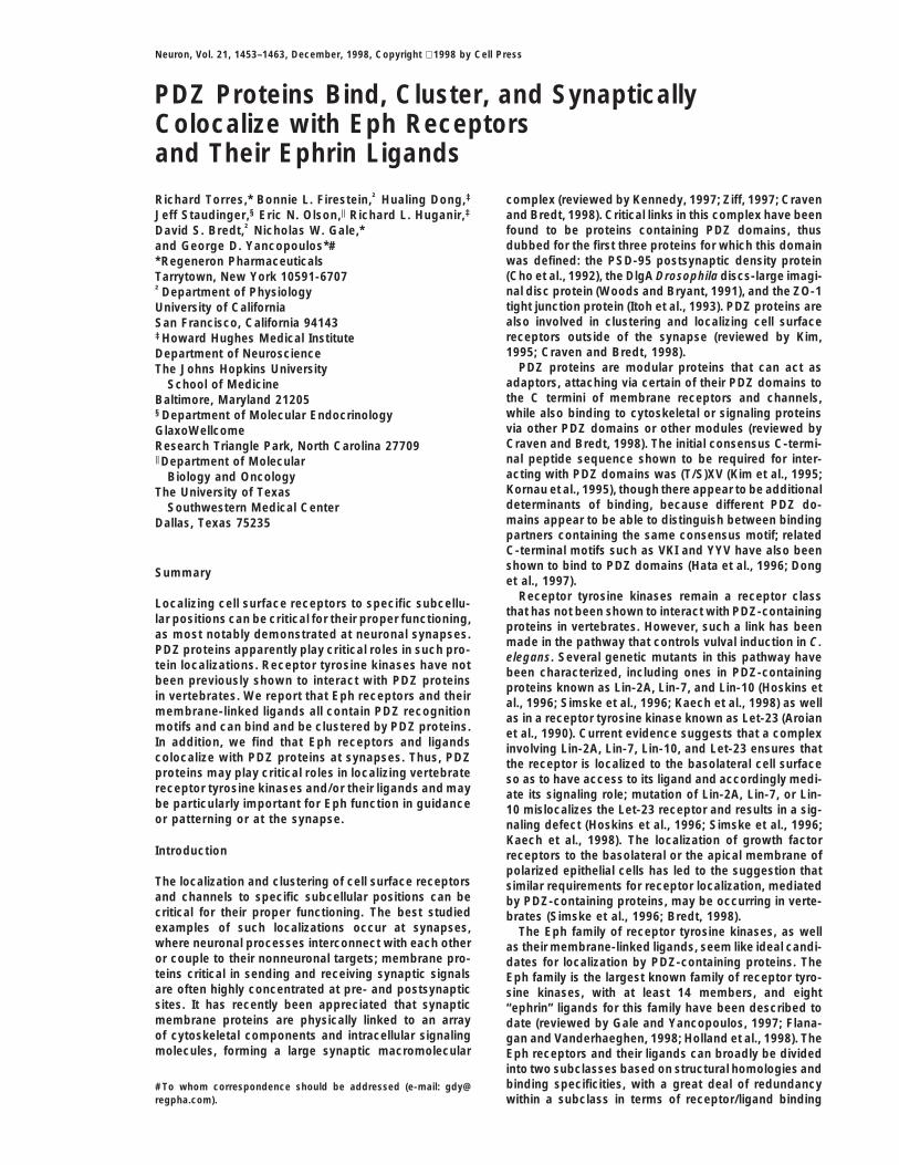
Isolation of PDZ-Containing Proteins that Interactprovide key cues in a cell-to-cell contact–dependentwith the C Termini of Eph Family Receptorsfashion and that specific mechanisms are involved inand Ligands in a Yeast Two-Hybrid Systemclustering the ligands and therefore allowing them toTo determine if the Eph receptors and their ligands couldactivate their receptors (Davis et al., 1994); recent evi-potentially interact with PDZ-containing proteins, we ex-dence suggests that such clustering might be induciblyamined their C-terminal sequences (Figure 1). Remark-regulated by various growth factors and the protein ki-ably, we found that almost all of these sequences endednase C (PKC) pathway (Bruckner et al., 1997; Stein etin a valine, as do most known consensus sequences foral., 1998). Remarkably, there appears to be reciprocalPDZ domain binding (Songyang et al., 1997). However,signaling between ligands and receptors in the B sub-while many of the Eph receptors (of both the A and Bclass in that ephrin-B ligands not only activate theirsubclasses) ended in the consensus sequence VXV (orrespective receptors but are in turn activated upon en-a variation in which the valine at the 23 position wasgaging their receptors, as judged by tyrosine phosphor-replaced by an alternate hydrophobic residue) (Figuresylation of the ephrin-B cytoplasmic domains (Holland et1A and 1C), the ephrin-B ligands ended in the consensusal., 1996; Bruckner et al., 1997); once again, clusteringYKV (Figure 1B). The VXV sequence was most similarof receptors seems important for this ligand activation.to the VKI C termini of the GluR2 and GluR3 subunitsConsistent with important roles for receptor and ligandof the AMPA receptor, which are known to interact withclustering, Eph receptors have been visualized on devel-the synaptic protein GRIP, which contains seven PDZoping neuritic processes by electron microscopy, re-domains (Dong et al., 1997), while the YKV consensusvealing clustered receptors at points of process contactwas most similar to the YYV C termini of the neurexins,(Henkemeyer et al., 1994). It thus seems likely that regu-also known to be PDZ binders (Hata et al., 1996). Exami-lated processes involved in localizing and clustering Ephnation of the cytoplasmic tails of other receptor tyrosinefamily receptors and/or ligands are crucial to ensure thekinases revealed that ending in a VXV consensus wassignaling processes required for the biological pro-the exception rather than the rule (Figures 1C and 1D).cesses in which the Eph family is involved, such as inRemarkably, the MuSK receptor tyrosine kinase, whichrepulsive interactions between neuritic processes dur-is localized to the NMJ and is critical for its formationing axon guidance or during the establishment of bound-(Valenzuela et al., 1995b; DeChiara et al., 1996; Glassaries between neighboring cell compartments in the ner-et al., 1996), has a similar C-terminal motif that is con-vous system or the vasculature (Gale and Yancopoulos,served from man to torpedo fish (Figure 1C). Interest-1997; Flanagan and Vanderhaeghen, 1998; Holland etingly, the C terminus of ErbB2 also conforms to thisal., 1998; Wang et al., 1998; Yancopoulos et al., 1998).consensus (Figure 1D), and this receptor is known toIt seems possible that PDZ proteins are involved in suchlocalize to the muscle side of the NMJ (Zhu et al., 1995),regulated clustering of Eph receptors and their ligands.as MuSK does.Here, we report that many of the Eph receptors and
We next focused on the VXV tails of the Eph receptorstheir ephrin-B ligands contain consensus C-terminalto see if they could indeed bind to PDZ domains bymotifs reminiscent of those binding PDZ domains. Wescreening for such binding using the yeast two-hybridfurther identify PDZ-containing proteins that can specifi-system. Baits were generated that encoded the C terminically bind and cluster both Eph receptors and their li-of mouse EphB2 (the last 96 residues) (Henkemeyer etgands in heterologous cells and that are tyrosine phos-al., 1994) or rat EphA7 (the last 98 residues) (Valenzuelaphorylated upon association with the Eph receptors. Weet al., 1995a), fused downstream to the LexA DNA–also show that such interactions can be detected inbinding domain; corresponding “control baits” werevivo, suggesting that PDZ-mediated clustering may begenerated in which the last 3 residues (either VEV orimportant for previously defined developmental roles ofIQV; see Figure 1A) were deleted. The first two baitsthe Eph family in neural guidance and tissue patterning.were then introduced into the L40 double reporter (yeastInterestingly, we find that Eph receptors and ligands canHIS3 and bacterial lacZ genes) yeast strain and utilizedbe colocalized with PDZ-containing proteins at neuro-in two-hybrid screens with a mouse embryonic day 11nal synapses, suggesting a new role for these develop-
mentally important molecules during synaptic plasticity. (E11) cDNA library that was fused in-frame downstream
PDZ Proteins Bind and Cluster Ephs and Ephrins1455
Figure 1. C Termini Alignment of Various Re-ceptor Tyrosine Kinases and Ephrins RevealsPotential PDZ Binding Motifs
Examination of the C termini of Eph familyreceptors (A), Ephrin-B ligands (B), MuSK or-thologs (C), and other receptor tyrosine ki-nases (D) for potential PDZ-binding motifs.Letters in front of each sequence signify spe-cies (c, chicken; h, human; m, mouse; r, rat;t, torpedofish; and Zf, zebrafish). Within thealignments, dots define amino acid residueswith identity with the consensus sequence atthe top of each panel, dashes denote gapsintroduced to maximize the alignment, and“x” denotes any amino acid. The two Ephreceptors used below as baits, EphB2 andEphA7, are highlighted in red, and the VXVconsensus sequence is highlighted in blue.
of the GAL4 transcription activation domain (GAL4- noted to control baits such as LexA alone or LexA fu-sions to Lamin or non-C-terminal regions of receptorTAD). cDNA clones that resulted in a HIS31/LacZ1 phe-
notype were then screened for interaction with the con- tyrosine kinases (Figure 2B and data not shown).Since GRIP has multiple PDZ domains, further two-trol baits containing the C-terminal deletions to ensure
the isolation of clones that specifically interacted with hybrid assays were performed with smaller GRIP fusionproteins (Figure 2C), which revealed that binding to boththe desired VEV or IQV C termini. From the approxi-
mately 6 3 106 transformants that were screened with Eph receptors and ephrin-B ligands depended uponPDZ domains 6 and 7 of GRIP; interestingly, the AMPAthe EphB2 bait, two distinct cDNA clones were isolated
that interacted in a VEV-dependent manner. One of the receptor binds to PDZ domains 4 and 5 of GRIP (Donget al., 1997), leaving open the possibility of simultaneoustwo clones contained the entire open reading frame of
mouse PICK1, a single PDZ domain–containing protein binding of GRIP to multiple partners.that was previously identified as a PKC-a-interactingprotein (Staudinger et al., 1995, 1997) (Figure 2A). The Confirmation that Interactions between
PDZ-Containing Proteins and Eitherother clone encoded a protein fragment that displayed.99% amino acid identity to the portion of rat GRIP Eph Receptors or Ephrins Occur
in Mammalian Cells(Dong et al., 1997) containing the last four PDZ domainsand was thus assumed to represent the mouse GRIP The above interactions were next explored via “pull-
down” assays in which glutathione S-transferase (GST)counterpart (Figure 2A). Approximately 9 3 106 trans-formants were screened with the EphA7 bait, and from fusion proteins bearing the above identified PDZ do-
mains were used to pull down various full length receptorthe HIS31/LacZ1 clones that were analyzed, only onedistinct cDNA clone was obtained that required the IQV tyrosine kinases expressed in mammalian cells, veri-
fying the same specificity of interactions for GRIP andfor interaction. This clone encoded the entire open read-ing frame of what appeared to be the mouse counterpart syntenin as noted in yeast (Figure 2D). As expected, in
these assays, we did not observe binding to a controlof the recently described human Syndecan-interactingprotein (89.6% amino acid identity), designated syntenin EphA5 receptor in which the C-terminal PDZ recognition
motif was destroyed by addition of a C-terminal “myc”(Grootjans et al., 1997), which contains two PDZ do-mains (Figure 2A). tag. Next, we examined the interactions in mammalian
cells by cotransfecting expression constructs for bothTo characterize the above PDZ domain–containingproteins further in terms of their binding specificity and the PDZ-containing proteins as well as their potential
partners into the same COS7 cells. When PICK1 wasto determine if they could bind ephrin-B ligands as wellas Eph receptors, we directly compared the binding of coexpressed with EphB2 (flag tagged at its N terminus),
ephrin-B1, and EphA7, coimmunoprecipitation of PICK1all three to the above baits as well as to a bait containingthe C terminus of ephrin-B1 (Davis et al., 1994) and and each of its potential partners was obvious, indicat-
ing complex formation (Figures 3A, lanes 1–3; 3B; andto a number of control baits (Figure 2B). Under theseconditions, the EphA7 and ephrin-B1 C termini were 3C). Coexpression using deleted versions of EphB2 or
ephrin-B1 lacking their 3 C-terminal residues preventedfound to bind to all three PDZ-containing proteins, whileEphB2 bound only to PICK1 and GRIP PDZs 4–7; in all complex formation (Figure 3A, lane 4 and data not
shown), indicating that the noted interactions dependedcases tested, this binding absolutely depended uponthe last three residues of the baits, and binding was not upon the C-terminal residues of Eph family members.

Neuron1456
Figure 2. Identification and Characterizationof PICK1, Syntenin, and GRIP Interactionswith the C Termini of EphB2, EphA7, andEphrin-B1 in Yeast Two-Hybrid Screens
(A) Schematic representation of the regionsof PICK1, syntenin, and GRIP that were iso-lated as fusion proteins (to the GAL4 tran-scription activation domain GAL4-TAD) inyeast two-hybrid screens for interaction withthe C termini of Eph receptors.(B) The specificity of interaction of these fu-sion proteins for various baits (fused to theLexA DNA binding domain LexA-DBD), as in-dicated.(C) Determination of which PDZ domain(s) ofGRIP is required for interaction with the vari-ous Eph family members using different GRIPdeletion mutants as indicated. The numberof plus signs indicates qualitative evaluationof strength of interaction and ND indicatesinteraction not determined.(D) GST pulldown assays showing that GSTfusion proteins to GRIP (PDZ domains 6 and7) or syntenin (PDZ domains 1 and 2) cancoprecipitate EphB2 and EphA7 with thesame specificity as exhibited in yeast two-hybrid screens. A mutant form of the EphA5receptor, in which the C-terminal PDZ rec-ognition motif is destroyed by appending aC-terminal myc tag, is used as a negativecontrol. Lysates from COS cells transfectedwith each of the indicated receptors wasmixed with the GST fusion protein as indi-cated, precipitations were performed, andimmunoblotting was done with the indicatedantibodies to detect pulldown products. Im-munoblotting of 10% of total lysates utilizedin binding assays is shown.
Similarly, coexpression of GRIP with its potential part- phosphorylated when in complex with EphB2 nor thatPICK1 was tyrosine phosphorylated when complexedners in COS7 cells demonstrated coprecipitation, and
thus complex formation, with the EphB2 and EphA7 with the ephrin ligands. Thus, at least some PDZ-con-taining proteins become tyrosine phosphorylated whenreceptors as well as the ephrin-B ligands but not with
a variety of control receptors (Figure 3D and data not complexed to receptor tyrosine kinases.shown). Confirmation of specific interaction betweensyntenin and EphA7 and ephrin-B1 was also obtained PICK1 Induces Clustering of EphB2
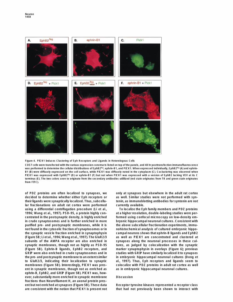
and Ephrin-B Ligands(data not shown).Our efforts to identify PDZ interactions for Eph familymembers was inspired by observations that Eph recep-PICK1 Gets Tyrosine Phosphorylated
When Complexed with EphB2 tors and ligands were found in clusters in vivo and thatclustering was apparently required for their ability toThe demonstration that PDZ-containing proteins di-
rectly interact with the Eph receptors defines a new class activate their partners (Davis et al., 1994; Henkemeyeret al., 1994). We thus were motivated to explore whetherof proteins that can associate with vertebrate receptor
tyrosine kinases. Many of the previously identified inter- PDZ-containing proteins could actually cause clusteringof Eph family members. COS7 cells have been utilizedactors with receptor tyrosine kinases have been shown
to be substrates of the kinase, and their tyrosine phos- in the past to demonstrate that PSD-95 family membersform coclusters when coexpressed with either NMDAphorylation often regulates their activity in some way
(reviewed by van der Geer et al., 1994). To examine receptors or Shaker-type K1 channels, as dramaticallydemonstrated by double immunofluorescence micros-whether PDZ-containing proteins could be substrates
for the Eph receptors, we determined whether PICK1 copy (for example, Kim et al., 1995, 1996). We thus per-formed similar experiments using Eph receptors, ephrins,was tyrosine phosphorylated upon its association with
the EphB2 receptor. Indeed, levels of PICK1 tyrosine and PDZ-containing proteins. When PICK1, EphB2, andephrin-B1 are individually expressed in COS7 cells, aphosphorylation precisely correlated not with the levels
of its expression but only with whether it was associated diffuse staining pattern is detected for all three proteinsafter visualization with TR or FITC-coupled secondarywith EphB2 (Figure 3A, lane 2; note that levels of PICK1
tyrosine phosphorylation correlate not with PICK1 pro- antibodies (Figures 4A, 4B, and 4C). When PICK1 iscoexpressed with either EphB2 or ephrin-B1, dramatictein levels but with association with EphB2). Compara-
ble experiments did not reveal that GRIP was tyrosine coclustering is observed (Figures 4D and 4F), though in
PDZ Proteins Bind and Cluster Ephs and Ephrins1457
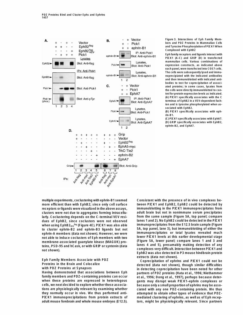
Figure 3. Interactions of Eph Family Mem-bers and PDZ Proteins in Mammalian Cellsand Tyrosine Phosphorylation of PICK1 WhenComplexed with EphB2
Eph family receptors and ligands interact withPICK1 (A–C) and GRIP (D) in transfectedmammalian cells. Various combinations ofexpression constructs, as indicated aboveeach panel, were transfected into COS7 cells.The cells were subsequently lysed and immu-noprecipiated with the indicated antibodiesand then immunoblotted with indicated anti-bodies to test for coprecipitation of associ-ated proteins; in some cases, lysates fromthe cells were directly immunoblotted to con-trol for protein expression levels as indicated.(A) PICK1 specifically associates with the Cterminus of EphB2 in a VEV-dependent fash-ion and is tyrosine phosphorylated when as-sociated with EphB2.(B) PICK1 specifically associates with eph-rin-B1.(C) PICK1 specifically associates with EphA7.(D) GRIP specifically associates with EphB2,ephrin-B2, and EphA7.
multiple experiments, coclustering with ephrin-B1 seemed Consistent with the presence of in vivo complexes be-tween PICK1 and EphB2, EphB2 could be detected bymore efficient than with EphB2; since only cell surfaceimmunoblotting in the PICK1 immunoprecipitates fromreceptors or ligands were visualized in the above assays,adult brain but not in nonimmune serum precipitatesclusters were not due to aggregates forming intracellu-from the same sample (Figure 5A, top panel; comparelarly. Coclustering depends on the C-terminal VEV resi-lanes 1 and 2). No EphB2 could be detected in the PICK1dues of EphB2, since coclusters were not observedimmunoprecipitates from the E12.5 brain sample (Figurewhen using EphB2VEV
flag (Figure 4E). PICK1 was also able5A, top panel, lane 3), but immunoblotting of either theto cluster ephrin-B2 and ephrin-B3 ligands but notimmunoprecipitates or total lysates revealed muchephrin-A members (data not shown). However, we werelower PICK1 levels at this earlier developmental stagenot able to induce coclusters of Eph members with two(Figure 5A, lower panel; compare lanes 1 and 3 andmembrane-associated guanylate kinase (MAGUK) pro-lanes 4 and 5), presumably making detection of anyteins, PSD-95 and hCask, or with GRIP or syntenin (datacomplexes very difficult. Interaction between PICK1 andnot shown).EphB2 was also detected in P3 mouse hindbrain proteinextracts (data not shown).
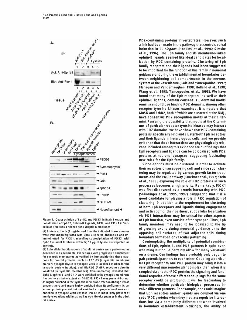
Eph Family Members Associate with PDZ Coprecipitation of ephrins and PICK1 could not beProteins in the Brain and Colocalize detected (data not shown), though similar difficultieswith PDZ Proteins at Synapses in detecting coprecipitation have been noted for otherHaving demonstrated that associations between Eph partners of PDZ proteins (Hata et al., 1996; Niethammerfamily members and PDZ-containing proteins can occur et al., 1996; Dong et al., 1997), perhaps because deter-when these proteins are expressed in heterologous gents may disrupt weak PICK1–ephrin complexes orcells, we next decided to explore whether these associa- because only a small proportion of ephrins may be asso-tions are physiologically relevant by examining whether ciated with any one PDZ-containing protein. We thusthey normally occur in vivo. We thus performed anti- attempted to obtain other types of evidence that PDZ-PICK1 immunoprecipitations from protein extracts of mediated clustering of ephrins, as well as of Eph recep-
tors, might be physiologically relevant. Since partnersadult mouse forebrain and whole mouse embryos (E12.5).
Neuron1458
Figure 4. PICK1 Induces Clustering of Eph Receptors and Ligands in Heterologous Cells
COS7 cells were transfected with the various expression constructs listed on top of the panels, and 48 hr posttransfection immunofluorescencewas performed to determine the cellular distributions of EphB2flag, ephrin-B1, and PICK1. When expressed individually, EphB2flag (A) and ephrin-B1 (B) were diffusely expressed on the cell surface, while PICK1 was diffusely noted in the cytoplasm (C). Coclustering was observed whenPICK1 was expressed with EphB2flag (D) or ephrin-B1 (F) but not when PICK1 was expressed with a version of EphB2 lacking VEV at its Cterminus (E). The two colors seen to originate from the secondary antibodies utililized (red stain originates from TR and green stain originatesfrom FITC).
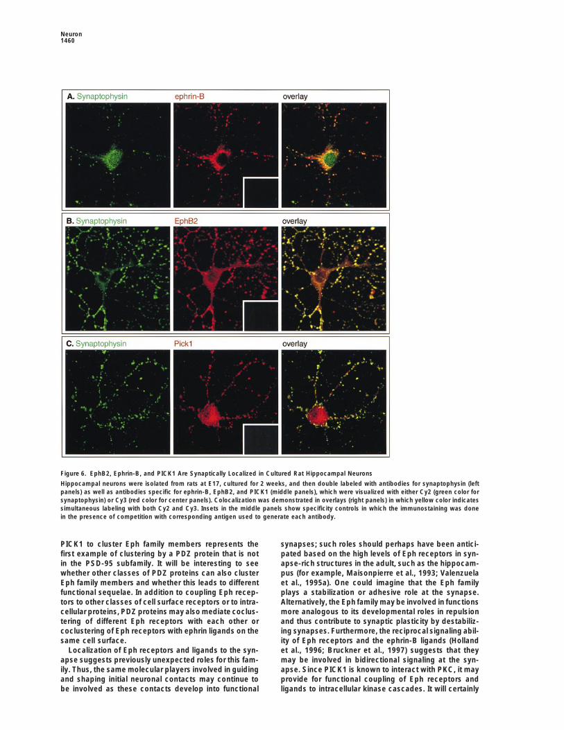
of PDZ proteins are often localized to synapses, we only at synapses but elsewhere in the adult rat cortexas well. Similar studies were not performed with syn-decided to determine whether either Eph receptors or
their ligands were synaptically localized. Thus, subcellu- tenin, as immunoblotting antibodies for syntenin are notcurrently available.lar fractionations on adult rat cortex were performed
using a differential centrifugation procedure (Li et al., To localize the Eph family members and PDZ proteinsat a higher resolution, double-labeling studies were per-1996; Wang et al., 1997). PSD-95, a protein highly con-
centrated in the postsynaptic density, is highly enriched formed using confocal microscopy on low-density em-bryonic hippocampal neuronal cultures. Consistent within crude synaptosomes and is further enriched in more
purified pre- and postsynaptic membranes, while it is the above subcellular fractionation experiments, immu-nohistochemical analysis of cultured embryonic hippo-not found in the cytosolic fraction of synaptosomes or in
the synaptic vesicle fraction enriched in synaptophysin campal neurons shows that ephrin-B ligands and EphB2as well as PICK1 are concentrated and clustered at(Figure 5B; Li et al., 1996; Wang et al., 1997). The GluR2/3
subunits of the AMPA receptor are also enriched in synapses along the neuronal processes in these cul-tures, as judged by colocalization with the synapticsynaptic membranes, though not as highly as PSD-95
(Figure 5B). Ephrin-B ligands, EphB2 receptors, and marker synaptophysin in overlays (Figure 6); previousstudies with GRIP have similarly localized it to synapsesGRIP were also enriched in crude synaptosomes and in
the pre- and postsynaptic membrane to an extent similar in embryonic hippocampal neuronal cultures (Dong etal., 1997). Thus, Eph receptors and ligands seem toto GluR2/3, indicating their localization to synaptic
membranes (Figure 5B). Interestingly, PICK1 was pres- colocalize with PDZ proteins in adult rat cortex as wellas in embryonic hippocampal neuronal cultures.ent in synaptic membranes, though not as enriched as
ephrin-B, EphB2, and GRIP (Figure 5B). PICK1 was, how-ever, substantially more enriched in synaptic membrane Discussionfractions than Neurofilament H, an axonal protein pres-ent but not enriched at synapses (Figure 5B). These data Receptor tyrosine kinases represented a receptor class
that had not previously been shown to interact withare consistent with the notion that PICK1 is present not
PDZ Proteins Bind and Cluster Ephs and Ephrins1459
PDZ-containing proteins in vertebrates. However, sucha link had been made in the pathway that controls vulvalinduction in C. elegans (Hoskins et al., 1996; Simskeet al., 1996). The Eph family and its membrane-linkedephrin-B ligands seemed like ideal candidates for local-ization by PDZ-containing proteins. Clustering of Ephfamily receptors and their ligands had been suggestedto be important for the function of this family in neuronalguidance or during the establishment of boundaries be-tween neighboring cell compartments in the nervoussystem or the vasculature (Gale and Yancopoulos, 1997;Flanagan and Vanderhaeghen, 1998; Holland et al., 1998;Wang et al., 1998; Yancopoulos et al., 1998). We havefound that many of the Eph receptors, as well as theirephrin-B ligands, contain consensus C-terminal motifsreminiscent of those binding PDZ domains. Among otherreceptor tyrosine kinases examined, it is notable thatMuSK and ErbB2, both of which are clustered at the NMJ,have consensus PDZ recognition motifs at their C ter-mini. Pursuing the possibility that motifs at the C termi-nus of particular receptor tyrosine kinases may interactwith PDZ domains, we have shown that PDZ-containingproteins specifically bind and cluster both Eph receptorsand their ligands in heterologous cells, and we provideevidence that these interactions are physiologically rele-vant. Included among this evidence are our findings thatEph receptors and ligands can be colocalized with PDZproteins at neuronal synapses, suggesting fascinatingnew roles for the Eph family.
Since ephrins must be clustered in order to activatetheir receptors on an apposing cell, and since such clus-tering may be regulated by various growth factor treat-ments and the PKC pathway (Bruckner et al., 1997; Steinet al., 1998), exploring the role of PDZ proteins in suchprocesses becomes a high priority. Remarkably, PICK1was first discovered as a protein interacting with PKC(Staudinger et al., 1995, 1997), suggesting that it is agood candidate for playing a role in PKC regulation ofclustering. In addition to the requirement for clusteringof both Eph receptors and ligands during engagementand activation of their partners, subcellular localizationvia PDZ interactions may be critical for other aspects
Figure 5. Coassociation of EphB2 and PICK1 in Brain Extracts and of Eph function, even outside of the synapse. Thus, EphLocalization of EphB2, Ephrin-B Ligands, GRIP, and PICK1 in Sub- family members may need to be localized to the tipcellular Fractions Enriched for Synaptic Membranes of growing axons during neuronal guidance or to the(A) Protein extracts (3 mg) derived from the indicated tissue sources apposing cell surfaces of two adjacent cells duringwere immunoprecipitated with EphB2-specific antibodies and im-
boundary formation or vascular patterning.munoblotted for PICK1, revealing coprecipitation of PICK1 withContemplating the multiplicity of potential combina-EphB2 in adult forebrain extracts; 50 mg of lysate are depicted as
controls. tions of Eph, ephrin-B, and PDZ partners is quite over-(B) Subcellular fractionations of adult rat cortex were performed as whelming but could certainly allow for many variationsdescribed in Experimental Procedures with progressive enrichment on a theme. Our findings have probably only begun tofor synaptic membranes as verified by immunoblotting these frac-
pair potential partners to each other. Coupling a particu-tions for control proteins, such as PSD-95 (a synaptic membranelar Eph receptor to one PDZ protein may bring it into amarker), synaptophysin (a synaptic vesicle localized marker for the
synaptic vesicle fraction), and GluR2/3 (AMPA receptor subunits very different macromolecular complex than when it islocalized to synaptic membranes). Immunoblotting revealed that coupled via another PDZ protein; the signaling and func-EphB2, ephrin-B, and GRIP were enriched in the synaptic membrane tional sequelae of these different couplings for the samefraction to a similar extent as GluR2/3. PICK1 was present but not
receptor could be profound. It will be fascinating toas highly enriched in the synaptic membrane fraction (though it wasdetermine whether particular biological processes in-present there and more highly enriched than Neurofilament H, anvolve different partners. For example, one could imagineaxonal protein present but not enriched at synapses) and was also
enriched in synaptic vesicles; thus, PICK1 is most likely present at that Eph receptors and/or ligands are coupled via onemultiple locations within, as well as outside of, synapses in the adult set of PDZ proteins when they mediate repulsive interac-rat cortex. tions but via a completely different set when involved
in boundary establishment. Strikingly, the ability of
Neuron1460
Figure 6. EphB2, Ephrin-B, and PICK1 Are Synaptically Localized in Cultured Rat Hippocampal Neurons
Hippocampal neurons were isolated from rats at E17, cultured for 2 weeks, and then double labeled with antibodies for synaptophysin (leftpanels) as well as antibodies specific for ephrin-B, EphB2, and PICK1 (middle panels), which were visualized with either Cy2 (green color forsynaptophysin) or Cy3 (red color for center panels). Colocalization was demonstrated in overlays (right panels) in which yellow color indicatessimultaneous labeling with both Cy2 and Cy3. Insets in the middle panels show specificity controls in which the immunostaining was donein the presence of competition with corresponding antigen used to generate each antibody.
PICK1 to cluster Eph family members represents the synapses; such roles should perhaps have been antici-pated based on the high levels of Eph receptors in syn-first example of clustering by a PDZ protein that is not
in the PSD-95 subfamily. It will be interesting to see apse-rich structures in the adult, such as the hippocam-pus (for example, Maisonpierre et al., 1993; Valenzuelawhether other classes of PDZ proteins can also cluster
Eph family members and whether this leads to different et al., 1995a). One could imagine that the Eph familyplays a stabilization or adhesive role at the synapse.functional sequelae. In addition to coupling Eph recep-
tors to other classes of cell surface receptors or to intra- Alternatively, the Eph family may be involved in functionsmore analogous to its developmental roles in repulsioncellular proteins, PDZ proteins may also mediate coclus-
tering of different Eph receptors with each other or and thus contribute to synaptic plasticity by destabiliz-ing synapses. Furthermore, the reciprocal signaling abil-coclustering of Eph receptors with ephrin ligands on the
same cell surface. ity of Eph receptors and the ephrin-B ligands (Hollandet al., 1996; Bruckner et al., 1997) suggests that theyLocalization of Eph receptors and ligands to the syn-
apse suggests previously unexpected roles for this fam- may be involved in bidirectional signaling at the syn-apse. Since PICK1 is known to interact with PKC, it mayily. Thus, the same molecular players involved in guiding
and shaping initial neuronal contacts may continue to provide for functional coupling of Eph receptors andligands to intracellular kinase cascades. It will certainlybe involved as these contacts develop into functional
PDZ Proteins Bind and Cluster Ephs and Ephrins1461
be of interest to determine the list of other molecules prove to be as important as those served by other recep-tor tyrosine kinases at the NMJ or those played by thethat are linked to the Eph receptors and ligands at the
synapse and to learn whether the Eph family provides Eph family during development.a signaling capability that can modify the activity of
Experimental Proceduresassociated synaptic components, such as neurotrans-mitter receptors, ion channels, adhesion molecules, and
Yeast Two-Hybrid Screensintracellular signaling molecules. The presence of dis-To identify mammalian proteins that interact with the C termini oftinct receptor types within the same complexes is cer-Eph family members, a mouse E11 Matchmaker cDNA library (Clon-
tainly suggested by the recent observation that PICK1— tech) was transformed into the Saccharomyces cerevisiae L40 re-like GRIP—can bind and cluster AMPA receptors (Xia porter strain bearing either the LexA–EphB2–C term or LexA–
EphA7–C term fusion plasmids; cDNA clones that resulted in aet al., submitted) as well as Eph family members. InHIS31/LacZ1 phenotype were then screened for interaction withaddition, Eph family members in large complexes mayseveral additional baits, including the LexA-EphB2(VEV) andotherwise impact intracellular signaling. Recent studiesLexA-EphA7(IQV) baits and the LexA-ephrin-B1 bait, as well as con-have identified a synaptic ras GTPase–activating proteintrol baits such as LexA-lamin. Standard yeast manipulations, yeast
(synGAP) that associates with the PDZ domains of the two-hybrid screens, and the HIS3 and lacZ reporter assays werePSD-95 protein family (Chen et al., 1998; Kim et al., performed as previously described (Vojtek et al., 1993; Schreiber-
Agus et al., 1995).1998), and it has been reported that a rasGAP also asso-ciates with EphB2 (Holland et al., 1997). Proteins (such
GST Pulldown Assaysas extracellular signal–related protein kinase 2– [ERK2-]Five micrograms of GST or GST fusion proteins were incubated withtype mitogen-activated protein kinase [Suzuki et al.,500 mg of COS7 cell lysates expressing the indicated receptors, and
1995], a calcium/calmodulin-dependent kinase II [aCaM- binding asssays were performed as indicated in Hannon et al. (1993).KII] [Goldenring et al., 1984; Kelly et al., 1984], and CRIPT Following separation on SDS-PAGE gels and transfer to nylon mem-[Niethammer et al., 1998]) that regulate phosphorylation branes, immunoblotting was performed with the indicated anti-
bodies.of cytoskeletal proteins or the interaction of proteinswith the cytoskeleton are enriched in synapses. Thus,
Antibodiesthe clustering of ephrins and their receptors may reflectThe affinity-purified rabbit polyclonal PICK1 antibody utilized fortheir role in synapse remodeling via regulation of thethe immunoprecipitations with COS7 lysates (Figure 3) and for the
synaptic cytoskeleton. It is also interesting to consider clustering assay in COS7 cells (Figure 4) was generated with athe possibility that by inducing tyrosine phosphorylation peptide (ADLDYDIEEDKLGIP) corresponding to the N-terminal re-of associated PDZ proteins, as we have shown can be gion of mPICK1. The anti-PICK1 affinity-purified rabbit polyclonal
utilized in Figure 5 was generated against a longer peptide con-the case with PICK1, Eph receptors may modify thetaining residues 2–31 of mPICK1. The anti-GRIP antibody is an affin-coupling functions of these proteins and thus alter theity-purified rabbit polyclonal antibody raised against a peptide toassembled macromolecular complex itself.the C terminus of GRIP. The anti-EphA7 affinity-purified rabbit poly-
There are many analogies between synapses in the clonal antibody was raised against the KADQEGDEELYFHFKFPGTKcentral nervous system and the most studied individual TYID peptide from the juxtamembrane region of EphA7. The anti-synapse of all, i.e., the NMJ. Recent studies have dem- myc 9E10 mouse monoclonal antibody was obtained from Calbio-
chem. The mouse monoclonal anti-phosphotyrosine antibody (4G10)onstrated crucial roles for receptor tyrosine kinases atwas obtained from UBI. The anti-EphB2 rabbit polyclonal antibodythe NMJ. In particular, agrin activation of the MuSKwas kindly provided by Sacha Holland and Tony Pawson. The anti-receptor tyrosine kinase is responsible for initiating allephrin-B(C-18) antibody was obtained from Santa Cruz. The mouse
aspects of NMJ formation (DeChiara et al., 1996; Glass monoclonal anti-PSD-95 antibody was obtained from ABR. Theet al., 1996), and MuSK remains highly localized to the mouse monoclonal anti-synaptophysin antibody was obtained frompostsynaptic muscle membrane in the adult (Valenzuela Sigma.et al., 1995b). In addition, ErbB receptors are localized
COS7 Expression Studies, Immunoprecipitations,to the postsynaptic side of the NMJ (Zhu et al., 1995),and Immunoblottingwhere they play key roles in regulating levels of acetyl-The PICK1 expression construct was generated by subcloning thecholine receptor expression in response to nerve-derivedfull length mouse pick1 cDNA into the pMT21 mammalian expres-
neuregulin. It does not seem too surprising that we have sion vector. The GRIP expression construct was generated by sub-found that MuSK and ErbB receptors have PDZ-binding cloning the full length rat GRIP cDNA into the pBK mammalianmotifs at their C termini; indeed, we have recently found expression vector. EphB2flag was constructed in pMT21 with a flag
tag at the N terminus, and EphB2VEVflag was constructed by introduc-that PDZ proteins, including PICK1, associate with the
ing a stop codon at the 23 amino acid position of the C terminus,C terminus of MuSK (R. T. et al., unpublished data).therefore deleting the last 3 residues (-VEV). The EphA7 constructHowever, it does seem quite surprising that, despite thewas generated by subcloning the rat ephA7 cDNA into pMT21.
importance of receptor tyrosine kinases at the NMJ, EphA5-myc was constructed by introducing a triple myc tag at thenot much attention has been paid to the role of such C terminus of the rat ephA5. COS7 cell transfections, cell lysis, tissuereceptors in central synapses. We now provide the first lysis, immunoprecipitations, and immunoblotting were performed as
previously described (Henkemeyer et al., 1994; Holland et al., 1997).demonstration of a family of receptor tyrosine kinaseswhose members are localized to central synapses within
ImmunohistochemistryPDZ-containing complexes. Since this is the largestClustering Assay in COS7 Cellsfamily of receptor tyrosine kinases, it may have the diver-COS7 cells plated on coverslips in 35 mM wells were transfectedsity required to differentially act at different types ofwith each of the indicated expression constructs. Forty-eight hours
synapses, particularly when coupled to the diversity of posttransfection, cells were washed with PBS and fixed with 4%choice possible with PDZ-containing partners. The roles paraformaldehyde at room temperature for 15 min. The anti-flag
mouse monoclonal M2 antibody (to detect EphB2flag or EphB2Sflag)of the Eph family at central synapses may ultimately
Neuron1462
(Kodak Scientific) and EphB2-Fc (to detect ephrin-B1) were then vulval induction encodes a tyrosine kinase of the EGF receptorsubfamily. Nature 348, 693–699.utilized followed by permeabilization of the cells and incubation with
the anti-PICK1 antibody; this protocol allowed one to detect only Banker, G.A., and Gowan, W.M. (1977). Rat hippocampal neuronscell surface receptors and ligands. Goat anti-mouse conjugated to in dispersed cell culture. Brain Res. 126, 397–425.TR (to detect M2), goat anti-human (to detect EphB2-Fc), or goat Brambilla, R., Schnapp, A., Casagranda, F., Labrador, J.P., Berge-anti-rabbit conjugated to FITC (to detect PICK1) were utilized as mann, A.D., Flanagan, J.G., Pasquale, E.B., and Klein, R. (1995).secondary antibodies to visualize the proteins. All secondary anti- Membrane-bound LERK2 ligand can signal through three differentbodies were obtained from Jackson ImmunoResearch Laboratories. Eph-related receptor tyrosine kinases. EMBO J. 14, 3116–3126.Photos were taken under fluorescence microscopy at 403 objective.
Bredt, D.S. (1998). Sorting out genes that regulate epithelial andRat Hippocampal Culturesneuronal polarity. Cell 94, 691–694.E17 rat hippocampal cultures were prepared as previously de-Bruckner, K., Pasquale, E.B., and Klein, R. (1997). Tyrosine phos-scribed (Banker and Cowan, 1977; Goslin and Banker, 1991) andphorylation of transmembrane ligands for Eph receptors. Scienceplated on coverslips. After 10–14 days in vitro, cells were washed275, 1640–1643.with PBS twice and fixed with methanol at 2208C for 15 min. Cells
were washed three times with PBS 1 0.1% triton (PBST) and incu- Chen, H.-J., Rojas-Soto, M., Oguni, A., and Kennedy, M.B. (1998).bated with the various affinity-purified primary antibodies (anti- A synaptic ras-GTPase activating protein (p135 SynGAP) inhibitedPICK1, anti-EphB2, anti-ephrin-B, and mouse anti-synaptophysin) by CaM Kinase II. Neuron 20, 895–904.for 2 hr at 258C. Cells were washed with PBST three times and Cho, K., Hunt, C.A., and Kennedy, M.B. (1992). The rat brain postsyn-incubated with secondary antibodies (donkey anti-mouse Cy2 and aptic density fraction contains a homolog of the Drosophila discs-goat anti-rabbit Cy3) for 2 hr at 258C to visualize the proteins. Cells large tumor suppressor protein. Neuron 9, 929–942.were washed three times with PBST and mounted by using Fluoro-
Craven, S.E., and Bredt, D.S. (1998). PDZ proteins organize synapticmount-G. Secondary antibodies conjugated to Cy2 or Cy3 were
signaling pathways. Cell 93, 495–498.purchased from Jackson Laboratories. Fluoromount-G was pur-
Davis, S., Gale, N.W., Aldrich, T.H., Maisonpierre, P.C., Lhotak, V.,chased from Southern Biotechnology Associates (Birmingham, AL).Pawson, T., Goldfarb, M., and Yancopoulos, G.D. (1994). LigandsPhotos were taken with confocal microscopy, model MRC1024 fromfor the EPH-related receptor tyrosine kinases that require membraneBiorad, at the 603 objective.attachment or clustering for activity. Science 266, 816–819.
DeChiara, T.M., Bowen, D.C., Valenzuela, D.M., Simmons, M.V.,Synaptosomal FractionationPoueymirou, W.T., Thomas, S., Kinetz, E., Compton, D.L., Rojas, E.,Synaptosomes were prepared as described by Li et al. (1996) withPark, J.S. et al. (1996). The receptor tyrosine kinase MuSK is requiredmodification. Four adult rat cortices were homogenized in 36 mlfor neuromuscular junction formation in vivo. Cell 85, 501–512.homogenization buffer (HB; 320 mM sucrose, 4 mM HEPES [pH 7.4],
1 mM EGTA, and 1 mM PMSF) with ten strokes at 900 rpm of a loose- Dong, H., O’Brien, R.J., Fung, E.T., Lanahan, A.A., Worley, P.F.,fitting glass Teflon homogenizer (Kontes, #22). The homogenate was and Huganir, R.L. (1997). GRIP: a synaptic PDZ domain–containingcentrifuged at 1000 g for 10 min. The supernatant was collected protein that interacts with AMPA receptors. Nature 386, 279–284.and centrifuged at 12,000 g for 15 min, and the second pellet was Flanagan, J.G., and Vanderhaeghen, P. (1998). The ephrins and Ephresuspended in 24 ml HB and centrifuged at 13,000 g for 15 min. receptors in neural development. Annu. Rev. Neurosci. 21, 309–345.The resulting pellet represented a crude synaptosomal fraction. This Gale, N.W., Flenniken, A., Compton, D.C., Jenkins, N., Copeland,crude fraction was lysed by osmotic shock and homogenized by N.G., Gilbert, D.J., Davis, S., Wilkinson, D.G., and Yancopoulos, G.D.three strokes in a glass Teflon homogenizer at 2000 rpm, and the (1996a). Elk-L3, a novel transmembrane ligand for the Eph family ofhomogenate was spun at 33,000 g for 20 min to yield a supernatant receptor tyrosine kinase, expressed in embryonic floor plate, roofand pellet (pre- and postsynaptic membranes). The supernatant plate and hindbrain segments. Oncogene 13, 1343–1352.was spun at 251,000 g for 2 hr. The resulting supernatant (cytosol)
Gale, N.W., Holland, S.J., Valenzuela, D.M., Flenniken, A., Pan, L.,contained soluble proteins, and the pellet (synaptic vesicles) con-Ryan, T.E., Henkemeyer, M., Strebhardt, K., Hirai, H., Wilkinson,tained synaptic vesicle proteins. Ten micrograms of each fractionD.G. et al. (1996b). Eph receptors and ligands comprise two majorwas separated on SDS-PAGE gels, and Western blot analyses werespecificity subclasses and are reciprocally compartmentalized dur-performed with the indicated antibodies.ing embryogenesis. Neuron 17, 9–19.
Gale, N.W., and Yancopoulos, G.D. (1997). Ephrins and their recep-Acknowledgmentstors: a repulsive topic? Cell Tissue Res. 290, 227–241.
Glass, D.J., Bowen, D.C., Stitt, T.N., Radziejewski, C., Bruno, J.,We thank Drs. L. P. Schleifer and P. Roy Vagelos for a stimulatingRyan, T.E., Gies, D.R., Shah, S., Mattsson, K., Burden, S.J. et al.scientific environment and many Regeneron scientists for discus-(1996). Agrin acts via a MuSK receptor complex. Cell 85, 513–523.sions, advice, and reagents. We also thank Dr. R. A. DePinho for
stimulating discussions at the inception of this project and for his Goldenring, J.R., McGuire, J.J., and DeLorenzo, R.J. (1984). Identifi-support, D. S. Hintz and L. T. Esau for expert technical assistance, cation of the major postsynaptic density protein as homologous withDrs. P. Hickmott and P. Steen for excellent technical help with the the major calmodulin-binding subunit of a calmodulin-dependentconfocal imaging, and C. R. Murphy and E. J. Burrows for excellent protein kinase. J. Neurochem. 42, 1077–1084.graphics work. We thank S. J. Holland and T. Pawson for the EphB2 Goslin, K., and Banker, G. (1991). Culturing Nerve Cells, K. Goslinantibody, R. A. DePinho for the cask plasmid, J. Trimmer for advice and G. Banker, eds. (Cambridge, MA: MIT Press).with the COS7 clustering assay, and S. M. Hollenberg for yeast two-
Grootjans, J.J., Zimmerman, P., Reekmans, G., Smets, A., Degeest,hybrid plasmids and the L40 yeast strain. B. L. F and D. S. B. thank
G., and Durr, J. (1997). Syntenin, a PDZ protein that binds SyndecanJ. F. Kilbridge for expert technical assistance. B. L. F. is supported
cytoplasmic domains. Proc. Natl. Acad. Sci. USA 94, 13683–13688.by a National Institutes of Health National Research Service Award.
Hannon, G.J., Demetrick, D., and Beach, D. (1993). Isolation of theD. S. B. is supported by the National Institutes of Health, the SearlesRb-related p130 through its interaction with CDK2 and cyclins.Scholars Program, the Culpeper Foundation, the Beckman Founda-Genes Dev. 7, 2378–2391.tion, and the Lucille P. Markey Charitable Trust. E. N. O is supportedHata, Y., Butz, S., and Sudhof, T.C. (1996). Cask: a novel dlg/PSD95by the National Institutes of Health and the Robert A. Welch Foun-homolog with an N-terminal calmodulin-dependent protein kinasedation.domain identified by interaction with Neurexins. J. Neurosci. 16,2488–2494.Received August 6, 1998; revised October 19, 1998.Henkemeyer, M., Marengere, L.E.M., McGlade, J., Olivier, J.P., Con-
References lon, R.A., Holmyard, D.P., Letwin, K., and Pawson, T. (1994). Immu-nolocalization of the Nuk receptor tyrosine kinase suggests roles in
Aroian, R.V., Koga, M., Mendel, J.E., Ohshima, Y., and Sternberg, segmental patterning of the brain and axonogenesis. Oncogene 9,1001–1014.P.W. (1990). The let-23 gene necessary for Caenorhabditis elegans

PDZ Proteins Bind and Cluster Ephs and Ephrins1463
Holland, S.J., Gale, N.W., Mbamalu, G., Yancopoulos, G.D., Henke- Songyang, Z., Fanning, A.S., Fu, C., Xu, J., Marfatia, S.M., Chishti,A.H., Crompton, A., Chan, A.C., Anderson, J.M., and Cantley, L.C.meyer, M., and Pawson, T. (1996). Bidirectional signaling through
the EPH-family receptor Nuk and its transmembrane ligands. Nature (1997). Recognition of unique carboxyl-terminal motifs by distinctPDZ domains. Science 275, 73–77.383, 722–725.
Holland, S.J., Gale, N.W., Gish, G.D., Roth, R.A., Songyang, Z., Cant- Staudinger, J., Zhou, J., Burgess, R., Elledge, S.J., and Olson, E.N.(1995). PICK1: A perinuclear binding protein and substrate for pro-ley, L.C., Henkemeyer, M., Yancopoulos, G.D., and Pawson, T.
(1997). Juxtamembrane tyrosine residues couple the Eph family re- tein kinase C isolated by the yeast two-hybrid system. J. Cell Biol.128, 263–271.ceptor EphB2/Nuk to specific SH2 domain proteins in neuronal cells.
EMBO J. 16, 3877–3888. Staudinger, J., Lu, J., and Olson, E.N. (1997). Specific interaction ofthe PDZ domain protein PICK1 with the COOH terminus of proteinHolland, S.J., Peles, E., Pawson, T., and Schlessinger, J. (1998).
Cell-contact-dependent signaling in axon growth and guidance: Eph kinase C-a. J. Biol. Chem. 272, 32019–32024.receptor tyrosine kinases and receptor protein tyrosine phospha- Stein, E., Lane, A.A., Cerretti, D.P., Schoecklmann, H.O., Schroff,tase beta. Curr. Opin. Neurobiol. 8, 117–127. A.D., Van Etten, R.L., and Daniel, T.O. (1998). Eph receptors discrimi-Hoskins, R., Hajnal, A.F., Harp, S.A., and Kim, S.K. (1996). The C. nate specific ligand oligomers to determine alternative signalingelegans vulval induction gene lin-2 encodes a member of the MA- complexes, attachment, and assembly responses. Genes Dev. 12,GUK family of cell junction proteins. Development 122, 97–111. 667–678.
Itoh, M., Nagafuchi, A., Yonemura, S., Kitani-Yasuda, T., Tsukita, S., Suzuki, T., Okumura, N.K., and Nishida, E. (1995). ERK2-type mito-and Tsukita, S. (1993). The 220 kD protein colocalizing with Cadher- gen-activated protein kinase (MAPK) and its substrates in postsyn-ins in non-epithelial cells is identical to ZO-1, a tight junction-associ- aptic density fractions from the rat brain. Neurosci. Res. 22, 277–285.ated protein in epithelial cells: cDNA cloning and immunoelectron Valenzuela, D.M., Rojas, E., Griffiths, J.A., Compton, D.L., Gisser,microscopy. J. Cell Biol. 121, 491–502. M., Ip, N.Y., Goldfarb, M., and Yancopoulos, G.D. (1995a). Identifica-Kaech, S.M., Whitfield, C.W., and Kim, S.K. (1998). The LIN-2/LIN- tion of full-length and truncated forms of Ehk-3, a novel member of7/LIN-10 complex mediates basolateral membrane localization of the Eph receptor tyrosine kinase family. Oncogene 10, 1573–1580.the C. elegans EGF receptor LET-23 in vulval epithelial cells. Cell Valenzuela, D.M., Stitt, T.N., DiStefano, P.S., Rojas, E., Mattsson,94, 761–771. K., Compton, D.L., Nunez, L., Park, J.S., Stark, J.L., Gies, D.R. et al.Kelly, P.T., McGuinness, T.L., and Greengard, P. (1984). Evidence (1995b). Receptor tyrosine kinase specific for the skeletal musclethat the major postsynaptic density protein is a component of a lineage: expression in embryonic muscle, at the neuromuscularCa21/calmodulin-dependent protein kinase. Proc. Natl. Acad. Sci. junction, and after injury. Neuron 15, 573–584.USA 81, 945–949. van der Geer, P., Hunter, T., and Lindberg, R.A. (1994). ReceptorKennedy, K.M. (1997). The postsynaptic density at glutamatergic proteins–tyrosine kinases and their signal transduction pathways.synapses. Trends Neurosci. 20, 264–268. Annu. Rev. Cell Biol. 10, 251–337.Kim, E., Niethammer, M., Rothchild, A., Jan, Y.N., and Sheng, M. Vojtek, A.B., Hollenberg, S.M., and Cooper, J.A. (1993). Mammalian(1995). Clustering of Shaker-type K1 channels by interaction with Ras interacts directly with the serine/threonine kinase Raf. Cell 74,a family of membrane-associated guanylate kinases. Nature 378, 205–214.85–88.
Wang, H.U., Chen, Z., and Anderson, D.J. (1998). Molecular distinc-Kim, E., Cho, K., Rothschild, A., and Sheng, M. (1996). Heteromultim- tion and angiogenic interaction between embryonic arteries anderization and NMDA receptor–clustering activity of Chapsyn-110, a veins revealed by ephrin-B2 and its receptor EphB4. Cell 93,member of the PSD-95 family of proteins. Neuron 17, 103–113. 741–753.Kim, J.H., Dezhi, L., Lau, L.F., and Huganir, R. (1998). SynGAP: a Wang, Y., Okamoto, M., Schmitz, F., Hofmann, K., and Sudhof, T.C.synaptic RasGAP that associates with the PSD-95/SAP90 protein (1997). Rim is a putative Rab3 effector in regulating synaptic–vesiclefamily. Neuron 20, 683–691. fusion. Nature 388, 593–598.Kim, S.K. (1995). Tight junctions, membrane-associated guanylate Woods, D.F., and Bryant, P.J. (1991). The discs-large tumor suppres-kinases and cell signaling. Curr. Opin. Cell Biol.7, 641–649. sor gene of Drosophila encodes a guanylate kinase homolog local-Kornau, H., Schenker, L.T., Kennedy, M.B., and Seeburg, P.H. (1995). ized at septate junctions. Cell 66, 451–464.Domain interaction between NMDA receptor subunits and the post- Yancopoulos, G.D., Klagsbrun, M., and Folkman, J. (1998). Vasculo-synaptic density protein PSD-95. Science 269, 1737–1740. genesis, angiogenesis, and growth factors: ephrins enter the frayLi, X.J., Sharp, A.H., Li, S.H., Dawson, T.M., Snyder, S.H., and Ross, at the border. Cell 93, 661–664.C.A. (1996). Huntingtin-associated protein (HAP1): discrete neuronal Zhu, X., Lai, C., Thomas, S., and Burden, S.J. (1995). Neuregulinlocalizations in the brain resemble those of neuronal nitric oxide receptors, erbB3 and erbB4, are localized at neuromuscular syn-synthase. Proc. Natl. Acad. Sci. USA 93, 4839–4844. apses. EMBO J. 14, 5842–5848.Maisonpierre, P.C., Barrezueta, N.X., and Yancopoulos, G.D. (1993). Ziff, E.B. (1997). Enlightening the postsynaptic density. Neuron 19,Ehk-1 and Ehk-2: two novel members of the Eph receptor–like tyro-
1163–1174.sine kinase family with distinctive structures and neuronal expres-sion. Oncogene 8, 3277–3288.
Note Added in ProofNiethammer, M., Kim, E., and Sheng, M. (1996). Interaction betweenthe C terminus of NMDA receptor subunits and multiple members Following submission of this manuscript, a report by Hock et al.of the PSD-95 family of membrane-associated guanylate kinases. revealed that the Eph family of RTKs is capable of interacting withJ. Neurosci. 16 , 2157–2163. the PDZ-containing protein AF6: Hock, B., Bohme, B., Karn, T.,Niethammer, M., Valtschanoff, J.G., Kapoor, T.M., Allison, D.W., Yamamoto, T., Kaibuchi, K., Holtrich, U., Holland, S., Pawson, T.,Weinberg, R.J., Craig, A.M., and Sheng, M. (1998). CRIPT, a novel Rubsamen-Waigmann, H., and Strebhardt, K. PDZ-domain-medi-postsynaptic protein that binds to the third PDZ domain of PSD- ated interaction of the Eph-related receptor tyrosine kinase EphB395/SAP90. Neuron 20, 693–707. and the ras-binding protein AF6 depends on the kinase activity of
the receptor. Proc. Natl. Acad. Sci. USA 95, 9779–9784.Schreiber-Agus, N., Chin, L., Chen, K., Torres, R., Rao, G., Guida,P., Skoultchi, A.I., and DePinho, R.A. (1995). An amino-terminal do-main of Mxi1 mediates anti-myc oncogenic activity and interactswith a homolog of the yeast transcriptional repressor SIN3. Cell 80,777–786.
Simske, J.S., Kaech, S.M., Harp, S.A., and Kim, S.A. (1996). LET-23receptor localization by the cell junction protein LIN-7 during C.elegans vulval induction. Cell 85, 195–204.


