Embed Size (px)
Citation preview
Supporting Information
A PDA-DTC/Cu-MnO2 Nanoplatform for MR Imaging and Multi-Therapy of Triple-Negative Breast Cancer
Xiaochun Hu‡a, Yonglin Lu‡a, Wenrong Zhaob, Menglin Sunb, Ruihao Lia, Lei Fenga, Tianming Yaoa, Chunyan Dong*b and Shuo Shi*a
‡ Xiaochun Hu and Yonglin Lu contributed equally to this work.
a. Breast Cancer Center, Shanghai East Hospital, School of Medicine, Shanghai Key Laboratory of Chemical Assessment and
Sustainability, School of Chemical Science and Engineering. Tongji University. Shanghai 200120, P. R. China
b. Breast Cancer Center,Shanghai East Hospital, Nanjing Medical University. Shanghai 200120, P. R. China
Corresponding authors:
E-mail: [email protected] (Chunyan Dong); [email protected] (Shuo Shi).
Experimental Section
Materials: Dopamine hydrochloride was purchased from sigma. Copper (II) chloride dihydrate (CuCl2·2H2O),
diethyldithiocarbamate (DTC), 5,5'-Dithiobis-(2-nitrobenzoic acid) (DTNB), methylene blue (MB) and Rhodamine (RhB) were
purchased from Aladdin. Fetal bovine serium (FBS), phosphate-buffered saline (PBS), Dulbecco’s modified Eagle medium
(DMEM), penicillin streptomycin (PS), Cell counting Kit-8 (CCK-8) and trypsin were ordered from KeyGEN BioTECH. The used
antibodies were purchased from Servicebio. Unless otherwise noted, all chemicals were used without further purification. MDA-
MB-231 human breast cancer cells were cultured from cells originally purchased from Chinese Academy of Sciences Cell Library.
Female BALB/c nude mice were purchased from Slac Experimental Animal Centre (Shanghai, China). All experiments in vivo were
conducted in compliance with the guidelines for the Care and Use of Laboratory Animals of Tongji University Experimental
Animal Center and the ethical approval number was TJBB00721102.
Characterizations: TEM images were obtained with a TEOL JEM-2100 transmission electron microscope. Zeta-potential
measurements were conducted on a Malvern Zetasizer Nano ZS instrument. Confocal fluorescence imaging was performed on
a laser confocal scanning microscope (Leica TCS SP5). 808 nm NIR lasers (Changchun Laser) were used to carry out the PTT study.
The camera (DALI TECHNOLOGY) was utilized to monitor the photothermal conversion.
Synthesis of PDA-DTC/Cu-MnO2:PDA NPs were synthesized by a reported method.1 Typically, 50 mg of dopamine
hydrochloride was dissolved in 100 mL Tris-HCl buffer (10mM, pH=10.5). 20 mg DTC was added to the as-synthesized PDA colloid
formed after 5 h of polymerization. After 20 h incubation at room temperature, the DTC- loaded PDA NPs were washed with
water for several times to remove the unloaded DTC. 20 mg CuCl2 was added in 20 mL of a DTC-loaded PDA NPs dispersion (1
mg/mL) under sonication, after that, the mixture solution was kept for further 2 h under drastic agitation. Thereafter, PDA-DTC-
Cu NPs were collected by centrifugation and washed with water for three times. 12.5 mL of KMnO4 (0.2 mg/mL) was dropwise
added into 5 mL of PDA-DTC/Cu suspension (1 mg/mL) under sonication. And then, after stirring for 5 min, the PDA-DTC/Cu-
MnO2 was obtained by centrifugation. S, Cu and Mn elements were quantified by ICP-OES.
Electronic Supplementary Material (ESI) for Chemical Communications.This journal is © The Royal Society of Chemistry 2021
According to reported method,2, 3 RhB labeled PDA-DTC/Cu-MnO2 was obtained for cellular uptake and biodistribution studies in vitro
and in vivo. In brief, 1 mL PDA-DTC/Cu-MnO2 solution (2 mg/mL) and 1 mg RhB were stirred overnight. Then, the precipitate was
collected by centrifugation (10000 rpm, 10 min), and washed with water to remove the excess dye. The release process of RhB was
monitored using UV-vis absorbance measurements. In the presence of GSH (10 mM), the release of GSH percent sharply increased to
44% within 4 h and reached 83% at 24 h (Figure S7), which could be attributed to the biodegradation of MnO2 by reacting with GSH.4
DTC release profiles from PDA-DTC/Cu-MnO2: Briefly, 2 mg lyophilized NPs were put in a centrifuge tube and redispersed in 2 mL
phosphate buffer solution (PBS, pH 7.4, containing 0.1% w/v Tween 80, with or without 10 mM H2O2). The tube was put into a shaker
and vibrated at 200 rpm at 37 °C. At designated time intervals, the tube was taken out and centrifuged at 10000 rpm for 5 min. Then,
the supernatant was transferred into a test tube and dealt with aqua regia for ICP analysis. The pellet was resuspended in 2 mL fresh
PBS solution and put back into the shaker for subsequent measurement. The cumulative release of DTC from NPs was plotted against
time.
·OH generation by Mn2+-mediated Fenton-like reaction:In vitro ·OH generation measurement was carried out using
methylene blue as an indicator.5 25 mM NaCO3/5% CO2 buffer solution containing 10 μg/mL MB, 8 mM H2O2, and 0.5 mM MnCl2
was allowed to incubate at 37 °C for 30 min. The ·OH-induced MB degradation was measured by the absorbance change at 665
nm.
Scavenging effect of GSH on ·OH: 25 mM NaCO3/5% CO2 buffer solution containing 10 μg/mL MB, 8 mM H2O2, 10 mM GSH and
0.5 mM MnCl2 or PDA-DTC/Cu-MnO2 (0.5 mM by Mn) was allowed to incubate at 37 °C for 30 min. The ·OH-induced MB
degradation was measured by the absorbance change at 665 nm.
GSH depletion measurements: 5,5’-Dithiobis (2-nitrobenzoic acid) (DTNB) was used to detect GSH consumption of PDA-
DTC/Cu-MnO2.6 1 mg PDA-DTC/Cu-MnO2 was dispersed in 10 mL of GSH solution (1 mM) at room temperature. After determined
time (10 min, 1 h, 2 h, 3 h), the supernatant was collected. Then, DTNB solution was added into supernatant, and the mixture
contained for another 10 min before UV-vis measurement. PBS and pure PDA were used as the control.
In vitro photothermal performance: In vitro photothermal performance of PDA-DTC/Cu-MnO2 was investigated by measuring
the temperature changes of its suspensions with the concentrations ranged from 0 to 400 µg/mL upon 808 nm laser illumination
for 5 min. The changed temperature was recorded and imaged simultaneously with an infrared thermal imaging camera.
Cell lines: MDA-MB-231 human breast cancer cells were cultured in DMEM supplemented with 10% FBS and 1% PS solution at
37 °C in an incubator containing 5% carbon dioxide.
Cytotoxicity assessment: MDA-MB-231 Cells were seeded into 96-well plates at a density of 7 × 10³ cells with 100 μL of DMEM
per well. After incubation for 24 h, the cells were treated by different concentrations of PDA-DTC/Cu-MnO2 (0, 2.5, 5, 10, 20
μg/mL) for 24 h. For PTT groups, MDA-MB-231 cells were irradiated under 808 nm laser for 5 min.
Detection of intracellular GSH:For in vitro GSH detection, MDA-MB -231 cells were seeded into culture plates and treated with
(a) PBS as a control; (b) PDA; (c) PDA-DTC/Cu; (d) PDA-DTC-MnO2 and (e) PDA-DTC/Cu-MnO2 for 6 h, respectively. The
intracellular GSH content was confirmed by GSH assay kits (Beyotime) according to the manufacturer’s instructions.
Detection of insoluble fraction NPL4: According to the reported method,7 immobilization of endogenous NPL4 was detected by
western blot. Briefly, for in vitro insoluble fraction detection, MDA-MB -231 cells were seeded into culture plates at a density of
5 × 10⁶ cells per dish and treated with (a) PBS as a control; (b) PDA; (c) PDA-MnO2; (d) DTC/Cu and (e) PDA-DTC/Cu-MnO2 for 4
h, respectively. Cells were washed with cold PBS, and lysis buffer (50 mM HEPES pH 7.4, 150 mM NaCl, 2 mM MgCl2, 10% glycerol,
0.5% Triton X-100, protease inhibitor cocktail by Servicebio) was added, and the plate was agitated gently for 10 min at 4 °C.
Then, cells were scraped into tubes and insoluble fraction was extracted. Protein concentration was determined by the
bicinchoninic acid (BCA) method. Protein of different groups were added into the sodium dodecyl sulfate (SDS)-polyacrylamide
gels, and transferred to polyvinylidene fluoride (PVDF) membrane. The PVDF membrane was proved with anti-NPL4 (1:1,000;
Abcam, ab224435), followed by HRP-conjugated secondary antibody. The protein bands of different groups were detected by
the ECL kit.
Cellular uptake measured by confocal microscopy: To investigate the cellular internalization of PDA-DTC-MnO2, MDA-MB-231
cells (1 × 10⁵ cells per well) were cultured in confocal dishes with DMEM for 24 h. For the confocal imaging, after incubating for
24 h, the cells were treated with RhB labelled PDA-DTC-MnO2 (10, 20, 40 μg/mL) for another 6 h. The cells were then washed
three times with ice-cold PBS and visualized immediately by using a confocal microscope with a 63 × oil-immersion objective
lens.
In vivo photothermal/fluorescence/MR imaging: All experiments in vivo were conducted in compliance with the guidelines of
the Laboratory Animal Center of Tongji University (Shanghai, China). Tumor xenograft model was established by breast fat pad
orthotopic transplantation on the BALB/c nude mice (5-6 weeks old, female, ~20g). 5 × 10⁵ MDA-MB- 231 cells were injected
into the right mammary fat pad for each mouse. After tumor volume reached 200-300 mm3, the fluorescent imaging was
performed by NightOWL LB 983 IN VIVO imaging system. Mice injected with RhB or PDA-DTC/Cu-MnO2-RhB were observed at 1
h, 3 h, 6 h and 24 h, respectively. Finally, all mice were sacrificed to excise main organs (heart, lung, liver, spleen and kidney)
and the tumors for further observation.
For in vivo photothermal effect evaluation, tumor-bearing mice were administrated with 100 μL of 5 mg/mL PDA-DTC/Cu-MnO2
suspension and PBS respectively, and then irradiated with 808 nm NIR laser at the power density of 1 W/cm2 for 10 min at 24 h
post intravenous injection. The tumor temperatures of mice were also recorded with the IR camera thermographic system.
T1 relaxivity of PDA-DTC/Cu-MnO2 suspension with different concentration was assessed via 1.5 T medical superconducting MR
T1-weighted MRI system (Bruker, USA). Before scanning, concentration of Mn2+ was determined by ICP-OES. In vivo T1-weighted
MR imaging dynamic evaluations was performed by a 3.0 T clinical MRI scanner (GE 750plus 3.0 T system, USA). The images of
tumor-bearing mice were collected at 3 h and 6 h post intravenous injection of PDA-DTC/Cu-MnO2 and Mn2+ (concentration of
Mn2+: 0.5 mM) with a rat coil (50 × 41 × 26 cm). The parameters of MRI scanner are as follows: field of view (FOV) = 90 × 60 mm2;
matrix = 256 × 256; repetition time (TR) = 500 ms; echo time (TE) = 20 ms; slice thickness = 4 mm; slices = 15; spacing = 0.5 mm.
In vivo antitumor efficiency evaluation: When the average tumor volume approached 100 mm3, the tumor-bearing mice were
randomly divided into 7 groups (n = 5): PBS, PDA-MnO2, DTC/Cu, PDA + 808 nm laser, PDA-DTC/Cu, PDA-DTC/Cu-MnO2, PDA-
DTC/Cu-MnO2 + 808 nm laser. Intravenous injection of PDA-DTC/Cu-MnO2 suspension was conducted at a 3-day interval. The
tumor sites were illuminated with 808 nm laser post injection for 24 h (1 W/cm2, 10 min). Tumor sizes and mice body weight
were recorded every 2 days. Tumor weight was measured after all mice were sacrificed at day 14. Tumor volume was calculated
using the following formula:
Tumor volume = 0.5 × length × width2
Statistical analysis: One-way single factorial of variance (ANOVA) was performed for all data analysis and values were presented
as means ± standard deviation. P < 0.05 (*), P < 0.01 (**) and P < 0.001(***) mean significant difference. All the data was analysed
by SPSS 20.0.
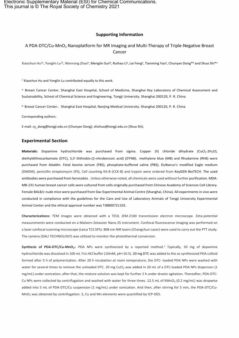
Figure S1. The energy variation of DTC and PDA before combination and after combination. The DFT calculation was carried out by
Gaussian 09 with D3-b3lyp/6-31g* (PCM) method.
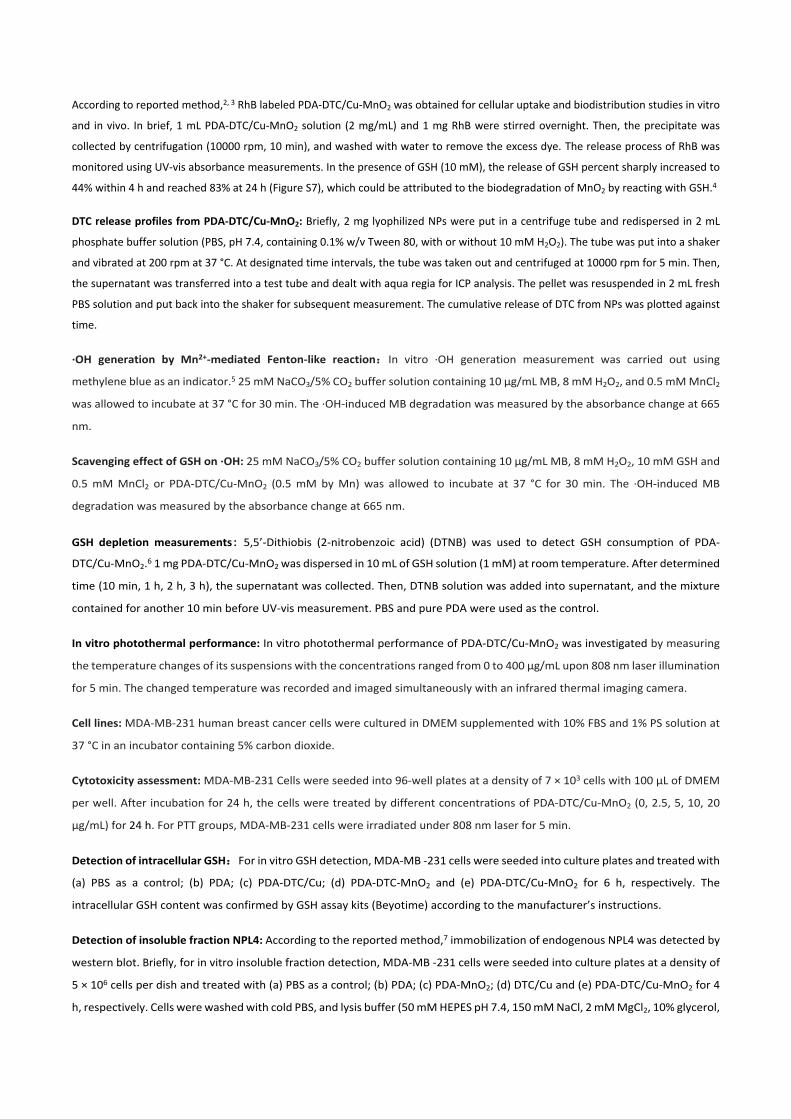
Figure S2. (a) Hydrodynamic diameters and (b) PDI of nanoparticles. (c) TEM-elemental mapping of PDA-DTC/Cu-MnO2.
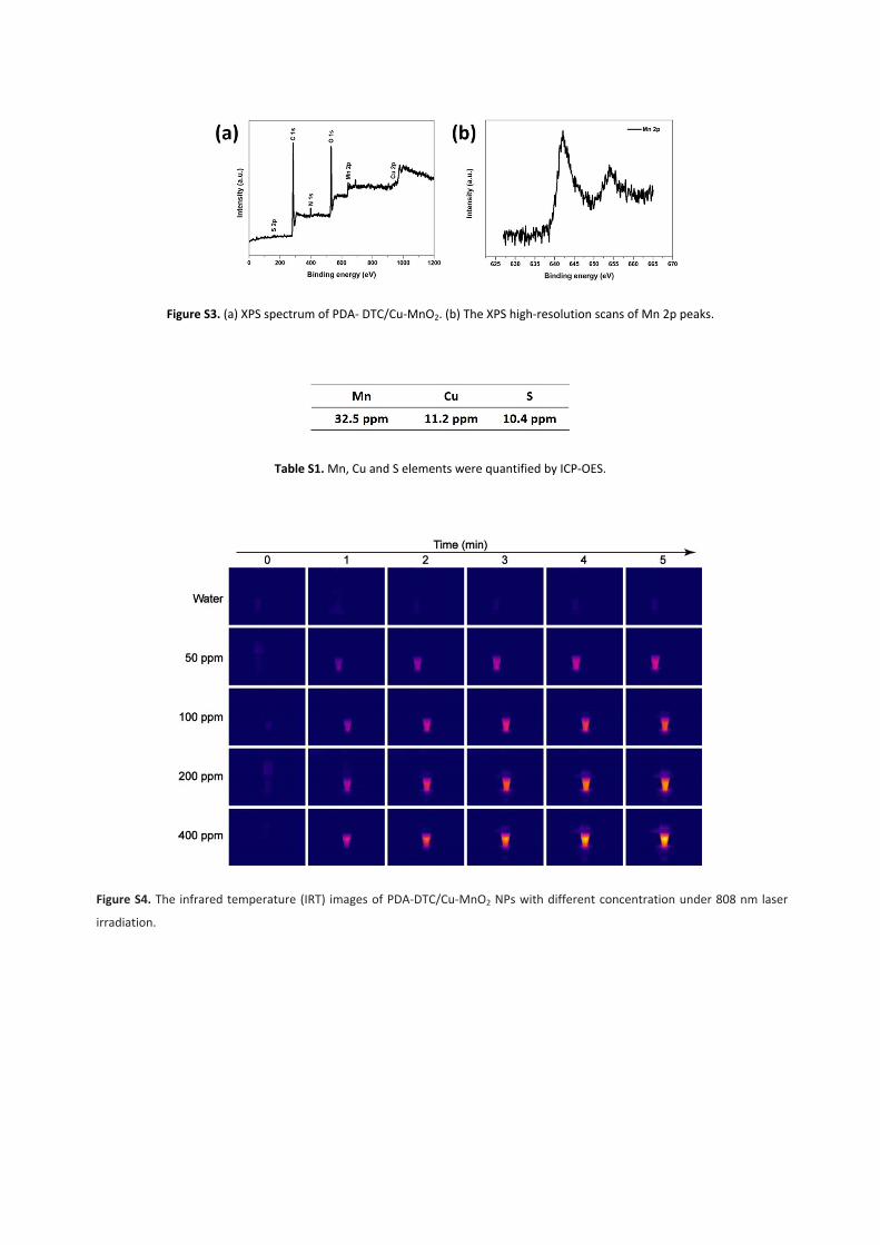
Figure S3. (a) XPS spectrum of PDA- DTC/Cu-MnO2. (b) The XPS high-resolution scans of Mn 2p peaks.
Table S1. Mn, Cu and S elements were quantified by ICP-OES.
Figure S4. The infrared temperature (IRT) images of PDA-DTC/Cu-MnO2 NPs with different concentration under 808 nm laser
irradiation.
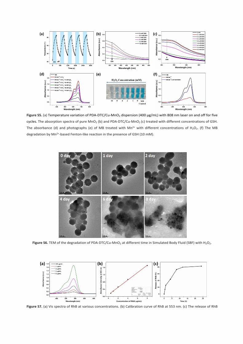
Figure S5. (a) Temperature variation of PDA-DTC/Cu-MnO2 dispersion (400 μg/mL) with 808 nm laser on and off for five
cycles. The absorption spectra of pure MnO2 (b) and PDA-DTC/Cu-MnO2 (c) treated with different concentrations of GSH.
The absorbance (d) and photographs (e) of MB treated with Mn2+ with different concentrations of H2O2. (f) The MB
degradation by Mn2+-based Fenton-like reaction in the presence of GSH (10 mM).
Figure S6. TEM of the degradation of PDA-DTC/Cu-MnO2 at different time in Simulated Body Fluid (SBF) with H2O2.
Figure S7. (a) Vis spectra of RhB at various concentrations. (b) Calibration curve of RhB at 553 nm. (c) The release of RhB
from the PDA-DTC/Cu-MnO2-RhB in the presence of GSH.
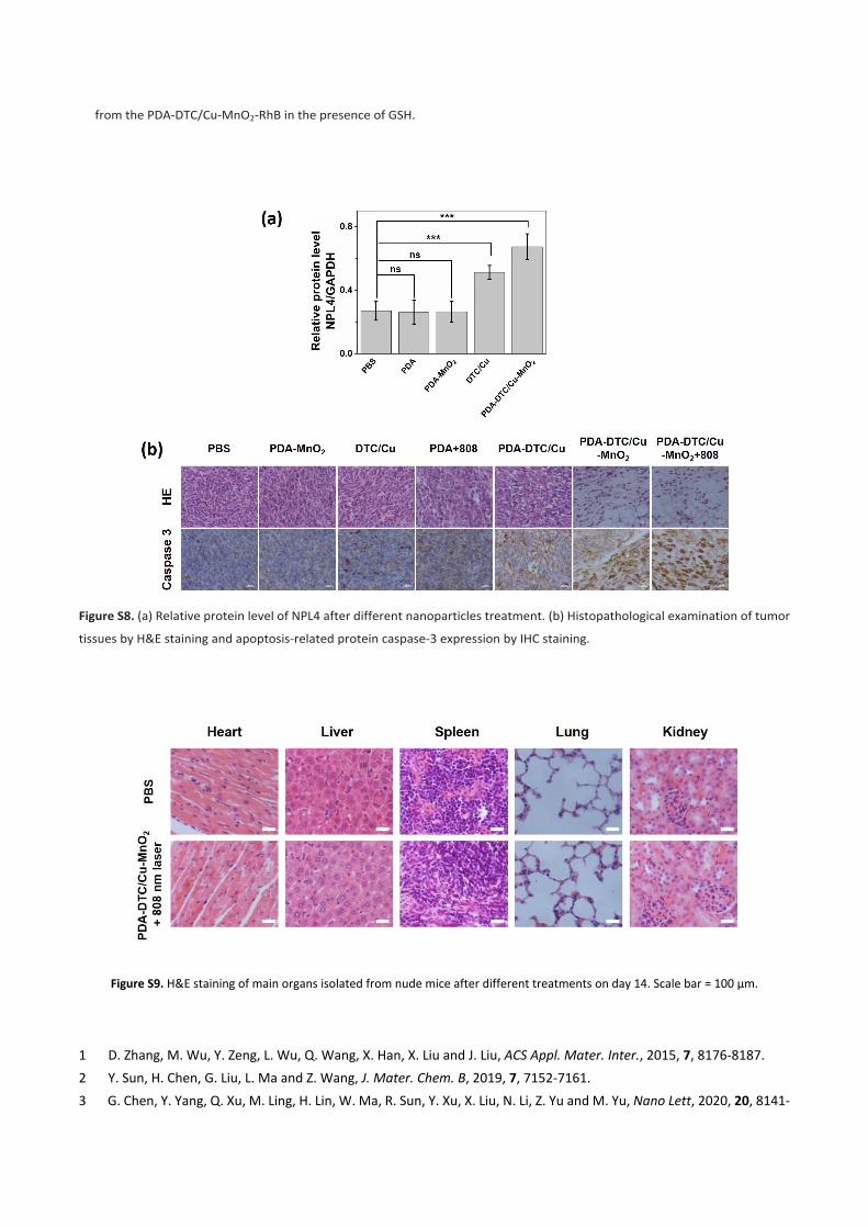
Figure S8. (a) Relative protein level of NPL4 after different nanoparticles treatment. (b) Histopathological examination of tumor
tissues by H&E staining and apoptosis-related protein caspase-3 expression by IHC staining.
Figure S9. H&E staining of main organs isolated from nude mice after different treatments on day 14. Scale bar = 100 μm.
1 D. Zhang, M. Wu, Y. Zeng, L. Wu, Q. Wang, X. Han, X. Liu and J. Liu, ACS Appl. Mater. Inter., 2015, 7, 8176-8187.2 Y. Sun, H. Chen, G. Liu, L. Ma and Z. Wang, J. Mater. Chem. B, 2019, 7, 7152-7161.3 G. Chen, Y. Yang, Q. Xu, M. Ling, H. Lin, W. Ma, R. Sun, Y. Xu, X. Liu, N. Li, Z. Yu and M. Yu, Nano Lett, 2020, 20, 8141-

8150.4 T. Zhang, C. Xu, W. Zhao, Y. Gu, X. Li, J. Xu and H. Chen, Chem. Sci., 2018, 9, 6749-6757.5 L. S. Lin, J. Song, L. Song, K. Ke, Y. Liu, Z. Zhou, Z. Shen, J. Li, Z. Yang, W. Tang, G. Niu, H. H. Yang and X. Chen, Angew. Chem., 2018, 57, 4902-4906.6 Z. Wang, B. Liu, Q. Sun, S. Dong, Y. Kuang, Y. Dong, F. He, S. Gai and P. Yang, ACS Appl. Mater. Inter., 2020, 12, 17254-17267.7 Z. Skrott, M. Mistrik, K. K. Andersen, S. Friis, D. Majera, J. Gursky, T. Ozdian, J. Bartkova, Z. Turi, P. Moudry, M. Kraus, M. Michalova, J. Vaclavkova, P. Dzubak, I. Vrobel, P. Pouckova, J. Sedlacek, A. Miklovicova, A. Kutt, J. Li, J. Mattova, C. Driessen, Q. P. Dou, J. Olsen, M. Hajduch, B. Cvek, R. J. Deshaies and J. Bartek, Nature, 2017, 552, 194-199.


![Oxidation of iodide and iodine on birnessite ([delta]-MnO2) in the pH range 4-8](https://img.dokumen.tips/doc/110x75/6331d8c6576b626f850d21a5/oxidation-of-iodide-and-iodine-on-birnessite-delta-mno2-in-the-ph-range-4-8.jpg)


