Embed Size (px)
Citation preview

research papers
824 doi:10.1107/S090744490601804X Acta Cryst. (2006). D62, 824–832
Acta Crystallographica Section D
BiologicalCrystallography
ISSN 0907-4449
Structure of 8Sa globulin, the major seed storageprotein of mung bean
Takafumi Itoh,a Roberta N.
Garcia,b Motoyasu Adachi,a‡
Yukie Maruyama,a Evelyn Mae
Tecson-Mendoza,b Bunzo
Mikamia and Shigeru Utsumia*
aFood Quality Design and Development,
Graduate School of Agriculture, Kyoto
University, Gokasho, Uji, Kyoto 611-0011,
Japan, and bInstitute of Plant Breeding, College
of Agriculture, University of the Philippines Los
Banos, College, Laguna 4031, Philippines
‡ Present address: Center for Neutron Science,
Tokai Research Establishment, Japan Atomic
Energy Research Institute, 2-4 Shirakata,
Tokai-mura, Ibaraki 319-1195, Japan.
Correspondence e-mail:
# 2006 International Union of Crystallography
Printed in Denmark – all rights reserved
The 8S globulins of mung bean [Vigna radiata (L.) Wilczek]
are vicilin-type seed storage globulins which consist of three
isoforms: 8S�, 8S�0 and 8S�. The three isoforms have high
sequence identities with each other (around 90%). The
structure of 8S� globulin has been determined for the first
time by X-ray crystallographic analysis and refined at 2.65 A
resolution with a final R factor of 19.6% for 10–2.65 A
resolution data. The refined 8S� globulin structure consisted
of 366 of the 423 amino-acid residues (one subunit of the
biological trimer). With the exception of several disordered
regions, the overall 8S� globulin structure closely resembled
those of other seed storage 7S globulins. The 8S� globulin
exhibited the highest degree of sequence identity (68%) and
structural similarity (a root-mean-square deviation of 0.6 A)
with soybean �-conglycinin � (7S globulin). Their surface
hydrophobicities are also similar to each other, although their
solubilities differ under alkaline conditions at low ionic
strength. This difference seems to be a consequence of
charge–charge interactions and not hydrophobic interactions
of the surfaces, based on a comparison of the electrostatic
potentials of the molecular surfaces. The thermal stability of
8S� globulin is lower than that of soybean �-conglycinin �.
This correlates with the cavity size derived from the crystal
structure, although other structural features also have a small
effect on the protein’s thermal stability.
Received 10 March 2006
Accepted 16 May 2006
PDB Reference: 8S� globulin,
2cv6, r2cv6sf.
1. Introduction
Mung beans [Vigna radiata (L.) Wilczek] are a popular food
crop in Asia, South America, Australia and the USA and are
similar to other legumes such as soybeans, jack beans and
kidney beans. Mung bean seeds contain about 20–25% protein
and are a major source of protein, especially in developing
countries.
The major seed proteins of mung bean are storage globulins
of the basic 7S type (�3%), vicilin type (8S; �90%) and
legumin type (11S; �8%) (Tecson-Mendoza et al., 2001). The
8S globulins, which are the major storage globulins of mung
bean, have molecular weights of about 150 kDa and consist of
three homologous isoforms, 8S�, 8S�0 and 8S�, which exhibit
high homology (about 90% identity) to each other and have a
molecular weight of about 49 kDa, indicating that the native
8S globulin consists of heterotrimers (Tecson-Mendoza et al.,
2001; Bernardo et al., 2004). Like other 7S globulins, they have
no disulfide linkages (Bernardo et al., 2004). Their amino-acid
sequences exhibit similarity (about 68%) to soybean �-con-
glycinin � (a 7S globulin). It has been reported that soybean
seed globulins (�-conglycinin and glycinin) have high nutri-
tional value, with a high content of the essential amino acid
lysine, and have useful physicochemical properties for food
systems, such as heat-induced gel-forming and emulsifying
abilities (Utsumi, 1992; Utsumi et al., 1997; Friedman &
Brandon, 2001). Therefore, soybean globulins have been used
in the production of a variety of processed foods. It has also
recently been reported that the soybean globulins have several
biological activities, such as a hypocholesterolaemic effect
(Carroll & Kurowska, 1995), triglyceride-lowering function
(Aoyama et al., 2001) and an appetite-suppressing effect
(Nishi et al., 2003). It is expected that further studies on mung
bean globulins may result in the enhancement of their char-
acteristics corresponding to those of soybeans and will expand
the food utility of mung beans.
In the improvement of the nutritional qualities and func-
tional properties of proteins as food materials, their structural
analysis is indispensable. The crystal structures of seed storage
globulins such as soybean �-conglycinin �0c (core), consisting
of only the core region of �0 (Maruyama, Maruyama et al.,
2004), and �-conglycinin � (Maruyama et al., 2001), soybean
proglycinin A1aB1b (Adachi et al., 2001, 2004; Adachi, Okuda
et al., 2003), mature glycinin A3B4 (Adachi, Kanamori et al.,
2003), jack bean canavalin (McPherson, 1980; Ko et al., 1993)
and kidney bean phaseolin (Lawrence et al., 1994) have been
determined. Moreover, the structure–function relationships of
soybean proteins have been studied (Maruyama et al., 1998,
1999, 2002a,b; Maruyama, Prak et al., 2004; Prak et al., 2005,
2006; Tandang et al., 2005). Although these soybean proteins
have similar three-dimensional structures, the physicochem-
ical properties of each protein are different and unique.
Physicochemical properties are important features for the
utilization of these proteins as food materials. The gel-forming
ability of the recombinant soybean 11S proglycinin A1aB1b
subunit was enhanced by the introduction of cysteine residues
and disulfide bonds (Adachi et al., 2004). A large amount of a
physiologically active peptide was also introduced into the 11S
proglycinin A1aB1b subunit (Prak et al., 2006). The peptides
were released by in vitro digestion with trypsin. This protein
engineering was based on the three-dimensional structure.
Thus, the 8S� globulin structure will provide useful informa-
tion for enhancing its qualities for use in food production, e.g.
its gelation characteristics, and for expanding the food utili-
zation, e.g. the production of valuable peptides, of mung bean
proteins. Therefore, we need to elucidate the structure of the
mung bean 8S globulin in order to be able to make precise
designs to improve the food quality of mung bean through
protein and genetic engineering.
We have expressed the major isoform 8S� of mung bean 8S
globulin in Escherichia coli and have successfully crystallized
it (Bernardo et al., 2004). Recently, several physicochemical
and functional properties of native and recombinant 8S�globulin have been clarified and will be described. This article
describes the crystal structure of 8S� globulin determined by
X-ray crystallography.
2. Materials and methods
2.1. Crystallization and X-ray diffraction
Recombinant 8S� globulin was overexpressed in E. coli,
purified and crystallized as described previously (Bernardo et
al., 2004). The hanging-drop vapour-diffusion method was
used to crystallize the recombinant 8S� globulin. The hanging
drop (6 ml) contained 3 ml protein solution and 3 ml reservoir
solution consisting of 12% PEG 1000, 0.2 M NaCl and 0.1 M
MES pH 6.0 and was equilibrated against 1 ml reservoir
solution. The protein concentration was 5 mg ml�1 and crys-
tallization was allowed to proceed at 293 K. X-ray diffraction
images of the 8S� globulin crystal in a capillary were collected
at 293 K with a Bruker Hi-Star multiwire area detector using
Cu K� radiation generated by a MacScience M18XHF
rotating-anode generator and were processed with SADIE
and SAINT software (Bruker, Karlsruhe, Germany) to a
resolution of 2.61 A (Table 1).
2.2. Structure determination and refinement
The 8S� globulin crystal structure was determined by the
molecular-replacement method as implemented in CNS v.1.1
(Brunger et al., 1998). The refined crystal structure of the
recombinant soybean �-conglycinin � model was used as the
probe structure (PDB code 1ipk). Model building and
refinement were performed using TURBO-FRODO (AFMB-
CNRS, Marseille, France) and CNS, respectively, on a Silicon
Graphics Octane computer. Fo � Fc and 2Fo � Fc maps were
used to locate the correct model. Several rounds of positional
and B-factor refinement followed by manual model building
research papers
Acta Cryst. (2006). D62, 824–832 Itoh et al. � 8S� globulin 825
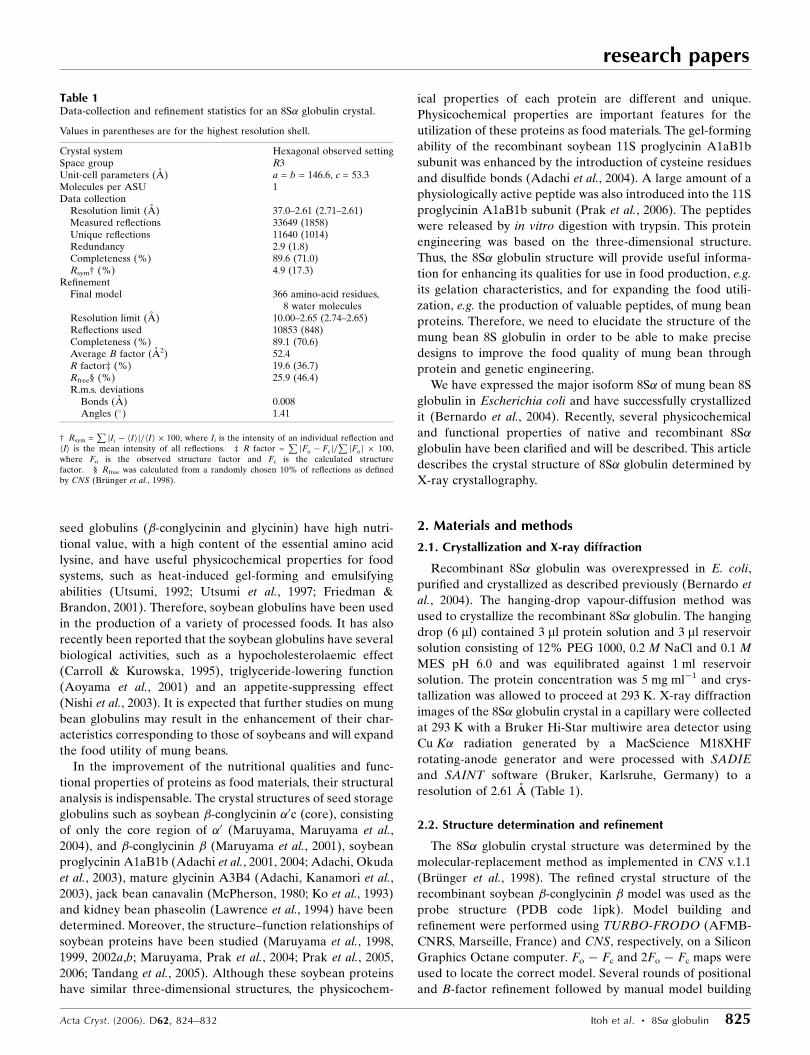
Table 1Data-collection and refinement statistics for an 8S� globulin crystal.
Values in parentheses are for the highest resolution shell.
Crystal system Hexagonal observed settingSpace group R3Unit-cell parameters (A) a = b = 146.6, c = 53.3Molecules per ASU 1Data collection
Resolution limit (A) 37.0–2.61 (2.71–2.61)Measured reflections 33649 (1858)Unique reflections 11640 (1014)Redundancy 2.9 (1.8)Completeness (%) 89.6 (71.0)Rsym† (%) 4.9 (17.3)
RefinementFinal model 366 amino-acid residues,
8 water moleculesResolution limit (A) 10.00–2.65 (2.74–2.65)Reflections used 10853 (848)Completeness (%) 89.1 (70.6)Average B factor (A2) 52.4R factor‡ (%) 19.6 (36.7)Rfree§ (%) 25.9 (46.4)R.m.s. deviations
Bonds (A) 0.008Angles (�) 1.41
† Rsym =PjIi � hIij=hIi � 100, where Ii is the intensity of an individual reflection and
hIi is the mean intensity of all reflections. ‡ R factor =PjFo � Fcj=
PjFoj � 100,
where Fo is the observed structure factor and Fc is the calculated structurefactor. § Rfree was calculated from a randomly chosen 10% of reflections as definedby CNS (Brunger et al., 1998).

were performed to improve the model by increasing the data
to a resolution of 2.65 A. Water molecules were incorporated
where the difference density exceeded the mean by 3.0� or
more and the 2Fo � Fc map showed a density exceeding 1.0�.
The final R factor was 19.6% for 10 853 data points in the
resolution range 10.0–2.65 A (89.1% completeness). The free
R value calculated for a randomly separated 10% of the data
was 25.9%. The stereo quality of the model was assessed using
PROCHECK (Laskowski et al., 1993) and WHAT-CHECK
(Hooft et al., 1996). Structural similarity was searched for in
the PDB (Berman et al., 2000) using DALI (Holm & Sander,
1993). The coordinates of soybean �-conglycinin � (1ipk), jack
bean canavalin (1dgw), kidney bean phaseolin (2phl), soybean
11S proglycinin A1aB1b (1fxz) and oxalate decarboxylase
Yvrk from Bacillus subtilis sp. 168 (1uw8) were taken from the
PDB. These models were superimposed by a fitting program in
TURBO-FRODO. Ribbon plots were prepared using
MOLSCRIPT (Kraulis, 1991) and RASTER3D (Merritt &
Murphy, 1994). The accessible surface areas (ASAs) were
calculated with NACCESS (Hubbard & Thornton, 1993),
which uses the algorithm of Lee & Richards (1971). The probe
was taken to be a water molecule of 1.4 A. Electrostatic
surface potential was calculated using GRASP (Nicholls et al.,
1991). The salt concentration was changed to 0.1 M from the
default setting. The file full.crg was used for charge
assignment, where the histidine residue has no charge. The
cavity size was estimated by CASTp (Liang et al., 1998). The
aliphatic index was estimated by the ProtParam tool from the
ExPASy Proteomics Server (Gasteiger et al., 2003).
3. Results and discussion
3.1. Crystallization and structure determination
The recombinant 8S� globulin of mung bean is a trimer of
three identical subunits, each with a molecular weight of about
49 kDa and consisting of 423 amino-acid residues without a
signal peptide (Bernardo et al., 2004) (Fig. 1). 8S� globulin
crystals (0.4 � 0.3 � 0.2 mm) were obtained by the hanging-
drop vapour-diffusion method as described previously
(Bernardo et al., 2004). The space group was determined to be
R3, with unit-cell parameters a = b = 146.6, c = 53.3 A, and the
solvent content was 47% for one subunit per asymmetric unit.
Results of data collection are summarized in Table 1. The
structure of the protein was determined by the molecular-
replacement method using the structure of the homologous
(68% identity) soybean recombinant �-conglycinin � as the
starting model (Fig. 1) and was refined by the simulated-
annealing and restrained least-squares methods (Table 1).
3.2. Quality of the refined model
The refined model of 8S� globulin consisted of 366 amino-
acid residues (one subunit of the trimer) and eight water
molecules. The electron densities of the main chain and side
chain were generally very well defined in the 2Fo � Fc map,
except for the five disordered regions (described below).
Water molecules were also well fitted. The final overall R
factor for the refined model was 19.6%, with 10 853 unique
reflections in the resolution range 10.0–2.65 A. The final free R
factor was 25.9%. The final r.m.s. deviations from standard
geometry were 0.008 A for bond lengths and 1.41� for bond
angles. Based on theoretical curves in the plot calculated
according to Luzzati (1952), the absolute positional error was
estimated to be close to 0.33 A at a resolution of 5.0–2.65 A.
Most nonglycine residues (81.0%) lie within the most
favoured regions and most other residues (18.1%) lie within
the additionally allowed regions of the Ramachandran plot as
research papers
826 Itoh et al. � 8S� globulin Acta Cryst. (2006). D62, 824–832
Figure 1Amino-acid sequence alignment of 8S� globulin and other seed storage7S globulins obtained using ClustalW (http://align.genome.jp/). 8S, 8S�globulin of mung bean (accession No. PRF3021374A); Con�, soybean �-conglycinin � (accession No. P25974); Cana, jack bean canavalin(accession No. P50477); Pha, kidney bean phaseolin (accession No.AAC04316). Identical or similar amino-acid residues among the fourproteins are indicated by asterisks or dots, respectively. �-Helices areindicated by black boxes and �-strands by arrows. The surrounding fiveamino-acid regions are disordered regions.

defined in PROCHECK (Laskowski et al., 1993). However,
two residues (0.6%), Arg22 (’ = �175, = �44�) and Asp325
(’ = 85, = �16�), are in generously allowed regions and one
residue (0.3%), Gln399 (’ = 51, =�65�), which exhibits well
defined density in the 2Fo � Fc map, is in a
disallowed region. Arg22 and Asp325 were
observed in distorted type-I �-turns, while
Gln399 was located in a sharp bend of the
loop near the C-terminal amino acid. The
averaged B factor was 52.4 A2, which is
relatively high (Table 1), probably owing to
the loose crystal packing of the trimer in the
unit cell.
3.3. Overall 8Sa globulin structure
The overall structure of 8S� globulin is
shown as a ribbon model in Fig. 2. There is
one subunit in the asymmetric unit of the
crystal. The trimer consisted of three iden-
tical subunits related by a threefold axis and
has approximate dimensions of 96 � 97 �
48 A, similar to other seed storage 7S
globulins (described below; Figs. 2a and 2b).
The monomer (one subunit) can be divided
into two similar modules, the N- and
C-terminal modules, related by a pseudo-
twofold symmetry axis (Fig. 2c). Each
module of the subunit consisted of a core
�-barrel (jelly-roll) domain and an extended
loop domain containing two helices. The
secondary-structure elements were named
according to the strands in soybean
�-conglycinin � (Maruyama et al., 2001).
Each core �-barrel domain consisted of two
�-sheets, A0ABIDG and J0JCHEF.
The 8S� globulin model had five regions
that could not be seen in the electron-
density maps (the six N-terminal amino
acids 1–6; the 20 C-terminal amino acids 404–423; 11 residues
in the ‘internal I’ region, 181–191; 11 residues in the ‘internal
II’ region, 214–224; nine residues in the ‘internal III’ region,
302–310) (Figs. 1 and 2). Although the total averaged B factor
research papers
Acta Cryst. (2006). D62, 824–832 Itoh et al. � 8S� globulin 827
Figure 2Overall structure of 8S� globulin. (a) The 8S�globulin trimer is seen along a threefold axis. Thethree subunits are shown in red, magenta andyellow. Each subunit contains two similar modules(N- and C-terminal modules) consisting of a core�-barrel and extended loop domain. (b) View after90� rotation around the vertical axis of (a). (c) The8S� globulin subunit. Colours denote secondary-structure elements (blue, �-helices; red, �-strands;yellow, loops and coils). The broken line in thefigure is the pseudo-twofold axis of the N- andC-terminal modules. (d) Intersubunit interface of8S� globulin. The structure is represented as a redand magenta ribbon model of 8S� globulin subunits.In the ribbon model, yellow or blue denotes theresidues participating in the hydrophobic inter-actions. Stick models (green and cyan) designate theresidues participating in the hydrogen bond. Blackbroken lines show the hydrogen bonds of two ionpairs (Arg55–Glu311, Arg95–Glu371).

was 52.4 A2 and relatively high (Table 1), the B factors at both
termini of the invisible regions were higher than the mean
value. The values are 79.6 A2 for the N-terminal amino acid,
75.7 A2 for internal I, 72.3 A2 for internal II, 78.0 A2 for
internal III and 62.5 A2 for the C-terminal amino acid. The N-
terminal amino-acid sequence and molecular size indicated by
SDS–PAGE of the recombinant 8S� globulin were consistent
with the deduced amino-acid sequence from the cDNA as
described previously (Bernardo et al., 2004). Therefore, the
invisible regions are thought to be disordered.
The ASAs of one subunit and the trimer, using a probe
radius of 1.4 A, were 18 470 and 39 714 A2, respectively. 28.3%
of the ASA of the subunit is used for formation of the trimer.
The association of subunits is dominated by the extended loop
domain and the J0JCHEF �-sheet (Fig. 2). The nonpolar atoms
comprise 67.8% of the intersubunit interface. The number of
residues participating in the hydrophobic
interactions (C—C contacts <4 A) were 42
in the N-terminal module and 34 in the C-
terminal module, respectively. 20 hydrogen
bonds (<3.2 A) were found. In particular,
two ion pairs among these interactions were
observed between two modules (Arg55–
Glu311, 2.7 A; Arg95–Glu371, 3.2 A)
(Fig. 2d).
3.4. Structural comparison
The 8S� globulin structure consisted of
two jelly-roll folds and exhibited similarity
to four other reported seed storage 7S
globulin structures that have amino-acid
sequence similarities to 8S� globulin, i.e.
soybean �-conglycinin �0c (Maruyama,
Maruyama et al., 2004), �-conglycinin �(Maruyama et al., 2001), jack bean canavalin
(McPherson, 1980) and kidney bean
phaseolin (Lawrence et al., 1994). The
amino-acid sequence similarities between
8S� globulin and these 7S globulins are
about 68% for �-conglycinin �0c and �,
about 52% for canavalin and about 58% for
phaseolin (Fig. 1). In addition to seed
storage 7S globulins, this basic fold is
common to 11S globulins [soybean pro-
glycinin A1aB1b (Adachi et al., 2001) and
mature glycinin A3B4 (Adachi, Kanamori et
al., 2003)], plant germine (an Mn-binding
protein with oxalate oxidase and superoxide
dismutase activities; Woo et al., 2000), plant
auxin-binding protein (Woo et al., 2002) and
the bacterial oxalate decarboxylase YvrK
(Anand et al., 2002) in the SCOP database
(http://scop.mrc-lmb.cam.ac.uk/scop; Murzin
et al., 1995).
Fig. 3(a) shows the superimposition of
8S� globulin on other 7S globulins. The
r.m.s. deviation was 0.6 A for the super-
imposition of 355 common C� atoms of
�-conglycinin �, 0.8 A for 343 C� atoms of
jack bean canavalin and 0.9 A for 342 C�
atoms of kidney bean phaseolin calculated
by the RIGID program implemented in
TURBO-FRODO. These structures had
almost similar topologies, consisting of the
research papers
828 Itoh et al. � 8S� globulin Acta Cryst. (2006). D62, 824–832
Figure 3Structural comparison of overall structures. (a) 7S globulins (green, soybean �-conglycinin �;blue, jack bean canavalin; yellow, kidney bean phaseolin) superimposed onto 8S� globulin(red). (b) Other structural similar protein and enzyme [yellow, soybean 11S proglycinin(A1aB1b); blue, oxalate decarboxylase Yvrk from B. subtilis sp. 168] also superimpose onto8S� globulin (red). The black broken line surrounds the site near the N-terminal amino-acidpeptide and the N-terminal extended loop region, which forms a different formation in eachglobulin. The coordinates of �-conglycinin � (1ipk), canavalin (1dgw), phaseolin (2phl), 11Sproglycinin (1fxz) and Yvrk (1uw8) were taken from the PDB (Berman et al., 2000).

two core �-barrels and extended loop domains. The N- or C-
terminal amino acids are disordered in other 7S globulins
(Fig. 3a). Internal I is in the extended loop region of the N-
terminal module, participating in the intersubunit interface
(Fig. 2), and was longer than those of any other 7S globulins
(Fig. 1). This site is also disordered in �-conglycinin �.
However, in phaseolin and canavalin, this site exists as a short
loop (Fig. 3a). Internal II is in the region connecting the N-
and C-terminal modules near the two core �-barrels and does
not take part in trimer formation (Fig. 2). This loop is also
disordered in the 7S globulins, except for �-conglycinin �.
Superimposition of this site was not well fitted in comparison
with that of the overall structure (Fig. 3a). Therefore, this site
is highly flexible within these globulins. Internal III is a
neighbour of the loop near the N-terminal amino acid (Fig. 2).
Although this site exists as a short loop and a turn in cana-
valin, this site is also disordered in other
globulins (Fig. 3a).
Fig. 3(b) shows the superimposition
of 8S� globulin on soybean proglycinin
A1aB1b and oxalate decarboxylase
YvrK from B. subtilis sp. 168, which also
exhibited a high degree of similarity to
8S� globulin in the PDB (Berman et al.,
2000) as observed using DALI (Holm &
Sander, 1993). The r.m.s. deviation was
1.8 A for the superimposition of 306
common C� atoms of A1aB1b and 1.8 A
for 235 C� atoms of Yvrk. Yvrk is a
manganese-dependent enzyme that
catalyzes the conversion of oxalate to
formate and carbon dioxide and has a
hexameric conformation consisting of
two trimers on top of one another,
differing from mung bean 8S globulins.
The overall structures of their subunits
resemble each other well. However,
some differences exist in the extended
loop region located in an intersubunit
interface of the N-terminal module and
the N-terminal amino-acid residues
(Fig. 3b). Although the extended loop
regions were superimposed well in 7S
and 8S globulins (Fig. 3a), these regions
were not well fitted in 8S� globulin, 11S
glycinin and YvrK. In Yvrk only, the
loop located in the N-terminal amino-
acid residues is longer and protrudes to
the outside (blue C� model in Fig. 3b).
The manganese and four manganese-
binding residues (three histidines and
one glutamate) of Yvrk are not
observed in the 8S� globulin structure.
These residues are not conserved in the
other seed storage globulins. Further-
more, to the best of our knowledge,
there are no reports of any metalloen-
zymatic activity of these seed storage
globulins.
3.5. Molecular surface andphysicochemical properties
Owing to their useful physicochem-
ical properties, such as gel-forming and
research papers
Acta Cryst. (2006). D62, 824–832 Itoh et al. � 8S� globulin 829
Figure 4The molecular surface of (a) 8S� globulin and (b) soybean �-conglycinin �. Structures arerepresented as white molecular-surface models. Hydrophobic residues are green. The electrostaticpotential surface of (c) 8S� globulin and (d) soybean �-conglycinin �. Structures are represented aswhite molecular-surface models. The electrostatic potential surface are drawn in the range from�15kBT (red) to +15kBT (blue), where kB is Boltzmann’s constant and T is the absolutetemperature (K).

emulsifying abilities, various seed storage globulins are
important for utilization as food materials (Utsumi, 1992;
Utsumi et al., 1997). The surface hydrophobicity of a protein is
significantly related to its solubility (Nakai & Li-Chan, 1988).
Solubility is one of the most important features for the
physicochemical properties of these globulins. The ASA ratio
of hydrophobic residues (Ala, Val, Leu, Ile, Pro, Met, Phe, Tyr
and Trp) on the molecular surface of the trimer was 4957/
39714 A2/A2 (12.5%; Fig. 4a). This value was very close to that
of soybean �-conglycinin � trimer (5494/40755 A2/A2; 13.5%;
Fig. 4b), with which mung bean 8S globulin � has high amino-
acid sequence and structural similarities (Figs. 1 and 3a).
Therefore, the globulins would be expected to exhibit similar
hydrophobic properties. Indeed, both exhibited similar
retention times on hydrophobic (butyl or phenyl Sepharose)
column chromatography (data not shown). The globulin is
soluble in salt solution but not in water. 8S� globulin and
soybean �-conglycinin � were soluble in the examined pH
range at high ionic strength (� = 0.5). This is because elec-
trostatic interactions between polypeptides are suppressed by
the presence of the salt. However, their solubilities differ
under alkaline conditions (pH > 7) at low ionic strength
(� = 0.08) (data not shown). 8S� globulin is highly soluble at
pH > 7 and � = 0.08, while soybean �-conglycinin � is almost
insoluble under these conditions. Considering the close simi-
larity in surface hydrophobicity (Figs. 4a and 4b), the differ-
ence in the solubility under alkaline conditions at low ionic
strength may arise from the difference in charge–charge
interactions and not the hydrophobic interactions of protein–
protein contacts through the molecular surface. Figs. 4(c) and
4(d) show the electrostatic potential on the molecular surface.
In the case of 8S� globulin, although one surface was covered
by mainly negative potentias, the other was not mainly
covered by positive potential (Fig. 4c). On the other hand,
positive or negative potential was closely distributed at the
centre of each surface of �-conglycinin � (Fig. 4d). It is
thought that the predominantly and concentrated electrostatic
potential (positive or negative) surfaces lead to charge–charge
interactions in �-conglycinin �. Owing to this undesirable
interaction through the molecular contacts, �-conglycinin �would precipitate under alkaline conditions (pH > 7) and low
ionic strength. Although the effect of the disordered regions
discussed above cannot be determined, the internal II dis-
ordered region (214–224), which exists on the surface and is
visible in �-conglycinin �, might affect the solubility.
Thermal stability is also one of the most important physi-
cochemical properties of these globulins related to food
functions such as heat-induced gelation. The thermal stability
of 8S� globulin indicated by DSC analysis is Tm = 350.6 K at
ionic strength � = 0.5 and pH 7.6 (Table 2) (data not shown).
For soybean �-conglycinin isoforms, these values under the
same conditions are 351.7 K for �, 350.4 K for �c, 355.8 K for
�0, 356.4 K for �0c and 363.9 K for � (Maruyama et al., 1999).
The difference in thermal stability between �-conglycinin �0c,
which consists only of the core region without the long
N-terminal extension region (141 residues), and � has been
accounted for by a combination of several structural features,
such as cavities, number of hydrogen bonds, intersubunit salt
bridges, surface hydrophobicity, number of proline residues
and loop region (Maruyama, Maruyama et al., 2004). The Tm
values of deletion mutants �c (deletion of the N-terminal
extension region; 125 residues) and �0c (deletion of the
N-terminal extension region; 141 residues) are very close to
those of the � and �0 subunits (Table 2; Maruyama et al., 1999).
Thus, the thermal stabilities of �-conglycinins are conferred by
the core regions. The cavity, which is inside the molecule and
which decreases the thermal stability of the 8S� globulin, was
6441 A3 in volume. This value is higher than that for any other
�-conglycinin (5463.5 A3 for �0c and 4753.7 A3 for �; Table 2).
Furthermore, the order of these values (8S� globulin > �0c >
�) is correlated with their respective thermal stability values
(8S� globulin < �0c < �). The numbers of hydrogen bonds and
salt bridges of 8S� globulin are greater than those of �(Table 2). Although the hydrogen bonds and salt bridges are
thought to be important features for stable packing, this is not
consistent with the DSC experimental data. The surface
hydrophobicities were very close to each other as described
above. The number of proline residues, which decreases the
entropy of the denatured structure, is 19 in the 8S� globulin
subunit (Table 2). Although � and �0 have a higher number of
proline residues than any other globulin, they do not exhibit
the highest thermal stability. Also, �c and �0c have a similar
number of proline residues to 8S� globulin and �. Thus, the
proline residues in the N-terminal extension region of � or �0
have little effect on thermal stability. Shorter loops have been
shown to be another stabilizing feature (Maruyama, Maru-
yama et al., 2004; Chakravarty & Varadarajan, 2002). The
effect of disordered regions cannot be discussed completely.
The long disordered (or flexible) regions of the seed storage
globulins (the N-terminal extension region of �-conglycinin
and the hypervariable regions of 11S glycinin) have little
influence on their thermal stabilities, as described above
(Maruyama et al., 1999; Prak et al., 2005). For soybean
�-conglycinins and mung bean 8S� globulin, there are long
disordered regions (internal I, II and III) which are rich in
charged residues, e.g. glutamic acid and lysine residues, and
the amino-acid sequences of the regions are almost conserved
between the proteins (Fig. 1). This might affect the thermal
research papers
830 Itoh et al. � 8S� globulin Acta Cryst. (2006). D62, 824–832
Table 2Thermal stability and structural features.
�-Conglycinin
8S� globulin �† �c† �0† �0c �
Molecular weight (kDa) 49 63 48 67 50 48Tm‡ (K) 350.6 351.7 350.4 355.8 356.4 363.9Cavity§ (A3) 6440.5 — — — 5463.5 4753.7No. of hydrogen bonds} 766 (2) — — — 704 (0) 729 (1)No. of Pro residues 19 38 20 33 19 21Aliphatic index†† 80.6 70.9 88.6 64.7 84.1 85.8
† There are no usable three-dimensional structures. ‡ DSC measurement at ionicstrength (� = 0.5) and pH 7.6 (Maruyama et al., 1999). § Cavity volume was calculatedby CASTp (Liang et al., 1998). } The values in parentheses represent the number ofsalt bridges of the intersubunit. †† This value was calculated using the ProtParam tool(Gasteiger et al., 2003).

stability a little. The influence of the water molecules on the
thermal stability of the storage proteins cannot be discussed
here, because the resolutions of the seed storage globulin
structures, at around 2.6 A, are generally too low to observe
the water molecules in the model (Maruyama et al., 2001;
McPherson, 1980; Ko et al., 1993; Lawrence et al., 1994). To
discuss the contribution of water molecules to the difference
in the thermal stability among �-conglycinins and 8S�globulin, higher resolution will be needed. Furthermore, the
aliphatic index, which is the relative volume occupied by
aliphatic side chains (Ala, Val, Ile and Leu), has been shown to
explain the difference in the thermal stability of several 11S
globulins (Molina et al., 2004). According to this report, the
higher the aliphatic index, the higher the thermal stability.
However, this trend is not exhibited among �-conglycinin and
8S globulins (Table 2).
4. Conclusion
The refined 8S� globulin structure consisted of one subunit of
the biological trimer and two jelly-roll folds. There were five
invisible regions. The overall structure, with the exception of
these disordered regions, very much resembled those of other
seed storage 7S globulins, soybean proglycinin A1aB1b and
the oxalate decarboxylase Yvrk from B. subtilis sp. 168. The
surface hydrophobicities of 8S� globulin and soybean
�-conglycinin � were similar to each other. However, their
solubilities are different under alkaline conditions at low ionic
strength. The difference seems to be a consequence of charge–
charge interactions and not of the hydrophobic interactions of
the surfaces. The difference in the thermal stability between
8S� globulin and �-conglycinin � was accounted for by several
other structural features. In particular, the cavity correlated
with this difference.
Computation time was provided by the Supercomputer
Laboratory, Institute for Chemical Research, Kyoto Univer-
sity. This work was supported in part by a grant for the
National Project on Protein Structural and Functional
Analyses from the Ministry of Education, Culture, Sports,
Science and Technology of Japan.
References
Adachi, M., Chunying, H. & Utsumi, S. (2004). J. Agric. Food Chem.52, 5717–5723.
Adachi, M., Kanamori, J., Masuda, T., Yagasaki, K., Kitamura, K.,Mikami, B. & Utsumi, S. (2003). Proc. Natl Acad. Sci. USA, 100,7395–7400.
Adachi, M., Okuda, E., Kaneda, Y., Hashimoto, A., Shutov, A. D.,Becker, C., Muntz, K. & Utsumi, S. (2003). J. Agric. Food Chem. 51,4633–4639.
Adachi, M., Takenaka, Y., Gidamis, A. B., Mikami, B. & Utsumi, S.(2001). J. Mol. Biol. 305, 291–305.
Anand, R., Dorrestein, P. C., Kinsland, C., Begley, T. P. & Ealick, S. E.(2002). Biochemistry, 41, 7659–7669.
Aoyama, T., Kohno, M., Saito, T., Fukui, K., Takamatsu, K.,Hashimoto, T., Yamamoto, Y., Hirotsuka, M. & Kito, M. (2001).Biosci. Biotechnol. Biochem. 65, 1071–1075.
Berman, H. M., Westbrook, J., Feng, Z., Gilliland, G., Bhat, T. N.,Weissig, H., Shindyalov, I. N. & Bourne, P. E. (2000). Nucleic AcidsRes. 28, 235–242.
Bernardo, A. E., Garcia, R. N., Adachi, M., Angeles, J. G., Kaga, A.,Ishimoto, M., Utsumi, S. & Tecson-Mendoza, E. M. (2004). J. Agric.Food Chem. 52, 2552–2560.
Brunger, A. T., Adams, P. D., Clore, G. M., DeLano, W. L., Gros, P.,Grosse-Kunstleve, R. W., Jiang, J.-S., Kuszewski, J., Nilges, M.,Pannu, N. S., Read, R. J., Rice, L. M., Simonson, T. & Warren, G. L.(1998). Acta Cryst. D54, 905–921.
Carroll, K. K. & Kurowska, E. M. (1995). J. Nutr. 125, 594S–597S.
Chakravarty, S. & Varadarajan, R. (2002). Biochemistry, 41, 8152–8161.
Friedman, M. & Brandon, D. L. (2001). J. Agric. Food Chem. 49,1069–1086.
Gasteiger, E., Gattiker, A., Hoogland, C., Ivanyi, I., Appel, R. D. &Bairoch, A. (2003). Nucleic Acids Res. 31, 3784–3788.
Holm, L. & Sander, C. (1993). J. Mol. Biol. 233, 123–138.Hooft, R. W., Vriend, G., Sander, C. & Abola, E. E. (1996). Nature
(London), 381, 272.Hubbard, S. J. & Thornton, J. M. (1993). NACCESS Computer
Program. Department of Biochemistry and Molecular Biology,University College, London.
Ko, T.-P., Ng, J. D. & McPherson, A. (1993). Plant Physiol. 101,729–744.
Kraulis, P. J. (1991). J. Appl. Cryst. 24, 946–950.Laskowski, R. A., MacArthur, M. W., Moss, D. S. & Thornton, J. M.
(1993). J. Appl. Cryst. 26, 283–291.Lawrence, M. C., Izard, T., Beuchat, M., Blagrove, R. J. & Colman,
P. M. (1994). J. Mol. Biol. 238, 748–776.Lee, B. & Richards, F. M. (1971). J. Mol. Biol. 55, 379–400.Liang, J., Edelsbrunner, H. & Woodward, C. (1998). Protein Sci. 7,
1884–1897.Luzzati, V. (1952). Acta Cryst. 5, 802–810.McPherson, A. (1980). J. Biol. Chem. 255, 10472–10480.Maruyama, N., Adachi, M., Takahashi, K., Yagasaki, K., Kohno, M.,
Takenaka, Y., Okuda, E., Nakagawa, S., Mikami, B. & Utsumi, S.(2001). Eur. J. Biochem. 268, 3595–3604.
Maruyama, N., Katsube, T., Wada, Y., Oh, M. H., Barba De La Rosa,A. P., Okuda, E., Nakagawa, S. & Utsumi, S. (1998). Eur. J.Biochem. 258, 854–862.
Maruyama, N., Mohamad Ramlan, M. S., Takahashi, K., Yagasaki, K.,Goto, H., Hontani, N., Nakagawa, S. & Utsumi, S. (2002a). J. Am.Oil Chem. Soc. 79, 139–144.
Maruyama, N., Mohamed Ramlan, M. S., Takahashi, K., Yagasaki, K.,Goto, H., Hontani, N., Nakagawa, S. & Utsumi, S. (2002b). J. Agric.Food Chem. 50, 4323–4326.
Maruyama, N., Prak, K., Motoyama, S., Choi, S. K., Yagasaki, K.,Ishimoto, M. & Utsumi S. (2004). J. Agric. Food Chem. 52, 8197–8201.
Maruyama, N., Sato, R., Wada, Y., Matsumura, Y., Goto, H., Okuda,E., Nakagawa, S. & Utsumi, S. (1999). J. Agric. Food Chem. 47,5278–5284.
Maruyama, Y., Maruyama, N., Mikami, B. & Utsumi, S. (2004). ActaCryst. D60, 289–297.
Merritt, E. A. & Murphy, M. E. P. (1994). Acta Cryst. D50, 869–873.
Molina, M. I., Petruccelli, S. & Anon, C. (2004). J. Agric. Food. Chem.52, 6023–6029.
Murzin, A. Z., Brenner, S. E., Hubbard, T. J. & Chothia, C. (1995). J.Mol. Biol. 247, 536–540.
Nakai, S. & Li-Chan, E. (1988). Hydrophobic Interactions in FoodSystems, pp. 43–61. Boca Raton, FL, USA: CRC Press.
Nicholls, A., Sharp, K. & Honig, B. (1991). Proteins, 11, 281–296.Nishi, T., Hara, H. & Tomita, F. (2003). J. Nutr. 133, 352–357.Prak, K., Maruyama, Y., Maruyama, N. & Utsumi, S. (2006). Peptides,
27, 1179–1186.
research papers
Acta Cryst. (2006). D62, 824–832 Itoh et al. � 8S� globulin 831
Prak, K., Nakatani, K., Katsube-Tanaka, T., Adachi, M., Maruyama,N. & Utsumi S. (2005). J. Agric. Food Chem. 53, 3650–3657.
Tandang, M. R., Atsuta, N., Maruyama, N., Adachi, M. & Utsumi, S.(2005). J. Agric. Food. Chem. 53, 8736–8744.
Tecson-Mendoza, E. M., Adachi, M., Bernardo, A. E. & Utsumi, S.(2001). J. Agric. Food Chem. 49, 1552–1558.
Utsumi, S. (1992). Adv. Food Nutr. Res. 36, 89–208.
Utsumi, S., Matsumura, Y. & Mori, T. (1997). Food Proteins and TheirApplications, edited by S. Damodaran & A. Paraf, pp. 257–291.New York: Marcel Dekker.
Woo, E. J., Dunwell, J. M., Goodenough, P. W., Marvier, A. C. &Pickersgill, R. W. (2000). Nature Struct. Biol. 7, 1036–1040.
Woo, E. J., Marshall, J., Bauly, J., Chen, J. G., Venis, M., Napier, R. M.& Pickersgill, R. W. (2002). EMBO J. 21, 2877–2885.
research papers
832 Itoh et al. � 8S� globulin Acta Cryst. (2006). D62, 824–832


