Embed Size (px)
Citation preview

Available online at www.sciencedirect.com
www.elsevier.com/locate/jmbbm
j o u r n a l o f t h e m e c h a n i c a l b e h a v i o r o f b i o m e d i c a l m a t e r i a l s 2 9 ( 2 0 1 4 ) 6 8 – 8 0
1751-6161/$ - see frohttp://dx.doi.org/10.
nCorresponding autE-mail address: x
Research Paper
Stress relaxation behavior of tessellated cartilagefrom the jaws of blue sharks
Xiaoxi Liua,n, Mason N. Deanb, Hamed Youssefpoura, Adam P. Summersc,James C. Earthmana
aDepartment of Chemical Engineering and Materials Science, 916 Engineering Tower, University of California, Irvine,CA 92697-2575, USAbDepartment of Biomaterials, Max Planck Institute of Colloids and Interfaces, 14424 Potsdam, GermanycFriday Harbor Labs, University of Washington, Friday Harbor, WA 98250, USA
a r t i c l e i n f o
Article history:
Received 9 April 2013
Received in revised form
8 August 2013
Accepted 15 August 2013
Available online 26 August 2013
Keywords:
Shark tessellated cartilage
Relaxation test
Transversely isotropic
biphasic model
Generalized Maxwell model
Equilibrium modulus
nt matter & 2013 Elsevie1016/j.jmbbm.2013.08.014
hor. Tel.: þ1 949 335 [email protected] (X. Liu).
a b s t r a c t
Much of the skeleton of sharks, skate and rays (Elasmobranchii) is characterized by a
tessellated structure, composed of a shell of small, mineralized plates (tesserae) joined by
intertesseral ligaments overlaying a soft cartilage core. Although tessellated cartilage is a
defining feature of this group of fishes, the significance of this skeletal tissue type –
particularly from a mechanical perspective – is unknown. The aim of the present work was
to perform stress relaxation experiments with tessellated cartilage samples from the jaws
of blue sharks to better understand the time dependent behavior of this skeletal type.
In order to facilitate this aim, the resulting relaxation behavior for different loading
directions were simulated using the transversely isotropic biphasic model and this model
combined with generalized Maxwell elements to represent the tessellated layer. Analysis
of the ability of the models to simulate the observed experimental behavior indicates that
the transversely isotropic biphasic model can provide good predictions of the relaxation
behavior of the hyaline cartilage. However, the incorporation of Maxwell elements into this
model can achieve a more accurate simulation of the dynamic behavior of calcified
cartilage when the loading is parallel to the tessellated layer. Correlation of experimental
data with present combined composite models showed that the equilibrium modulus of
the tessellated layer for this loading direction is about 45 times greater than that for
uncalcified cartilage. Moreover, tessellation has relatively little effect on the viscoelasticity
of shark cartilage under loading that is normal to the tessellated layer.
& 2013 Elsevier Ltd. All rights reserved.
r Ltd. All rights reserved.
; fax: þ1 949 824 2541.
1. Introduction
Cartilage is both stiff and resilient, an ideal contour filler andbearing surface that minimizes contact stresses generatedduring compressive loading and contributes to lubrication
mechanisms in the joint (Hall, 2005; Hunziker, 1999). In mostvertebrates, whereas bones provide a rigid framework for thebody, cartilage exists in much smaller quantity, providingflexible and elastic structural support, primarily in the joints.One exception is found in the elasmobranch fishes (sharks,

j o u r n a l o f t h e m e c h a n i c a l b e h a v i o r o f b i o m e d i c a l m a t e r i a l s 2 9 ( 2 0 1 4 ) 6 8 – 8 0 69
rays and their relatives), whose skeletons are made entirelyof cartilage, which therefore must perform both the functionscarried out by mammalian bone as well as typical functionsof mammalian cartilage (Dean et al. 2009; Macesic andSummers, 2012). However, possession of a fully cartilaginousskeleton clearly does not compromise behaviors, as elasmo-branchs are among the fastest and largest animals in theoceans, and many species can feed on prey larger thanthemselves or protected by turtle or mollusk shell, whichcan be harder than cortical bone. Furthermore, the skeleton iscyclically loaded, perhaps 106–1010 times over an animal′slifetime without failure (Ashhurst, 2004). A key aspect thatapparently makes this high level of performance possible isthe “tessellation” of the shark skeleton, a composite ofmineralized blocks of hydroxyapatite (tesserae) joined byintertesseral ligaments over a core of uncalcified cartilage(Clement, 1992; Dingerkus et al., 1991; Kemp and Westrin,1979; Moss, 1977; Dean and Summers, 2006). An example ofthis tissue is illustrated in Fig. 1 with a correspondingphotograph (inset).
The specific role of tessellation in the high performanceand safety factors of elasmobranch skeletons is unknown andto date has only been addressed theoretically. Wroe et al.(2008) multi-material finite-element (FE) models of bite forcein the white shark (Carcharodon carcharias) took into accountthe composite nature of the skeleton, When compared with ahypothetical shark with bony jaws, the modeled cartilaginousjaws exhibited lower stress, but greater deformation andstrain; however the use of cartilage rather than bone showedno apparent impediment to the generation of high bite forces.Liu et al. (2010) developed an analytical composite model tosimulate the bending behavior of tessellated cartilage, incor-porating anatomical dimensions for tesserae and intertesseral
Fig. 1 – The tessellated cartilage of elasmobranch fishes. (A) CTsection of the skeleton (B) shows uncalcified cartilage [UC] overl[PC]; the inset image provides a schematic view of the tissue reThe bottom left of the image shows a perspective on tesserae intesseral covering is more obvious in a CT scan of a jaw joint (C)how tesserae cover the entire surface of the jaws, even on comhalleri; jaw joint CT scan in C from Urobatis jamaicensis.).
joints, as well as the preliminary material property data fortissue components used in Wroe et al. (2008)′s FE-models. Liuet al.′s two-dimensional cross-sectional models demonstratethat the high fatigue resistance of elasmobranch skeletonscould be accomplished in part by the tessellated layer′sdifferential response to tensile vs. compressive loading.The modeled movement of the joints within the tesseraemat would allow the skeleton to manage stresses to avoiddamage, ensuring that the majority of loading stresses areconsolidated in the tesserae on the side of the skeleton loadedunder compression, a mode more resistant to fatigue damage(Malzahn and Schultz, 1986; Hacker and Ansell, 2001). It isclear that the heterogeneity of this tissue (the combination ofmineralized and unmineralized cartilage and fibrous material)and its tessellation play primary roles in the skeleton′smechanical properties, so both must be accounted for to trulycharacterize this composite tissue′s response to loading andunderstand the contribution of individual components.
As elasmobranch uncalcified cartilage is surely highlyviscoelastic (like mammalian cartilage), an effective methodfor understanding the effects of tessellation on cartilage is todescribe the contributions of tissue compositeness and ani-sotropy (loading direction) to the viscoelastic response. Likemammalian articular cartilage, water is the most abundantcomponent of the uncalcified portion of elasmobranch carti-lage, constituting up to 90% of its wet weight, while collagenand proteoglycans are the most abundant structural macro-molecule in ECM, comprising up to 50% and 70% of the dryweight of the uncalcified cartilage, respectively, for severalshark species (Porter et al., 2013). Given these broad composi-tional similarities between uncalcified cartilage in mammalsand elasmobranchs, previous viscoelastic analyses of mam-malian articular cartilage can provide vital reference for the
scan of the blue shark, P. glauca, used in this study. A cross-ain by mineralized tesserae [T(c)] and fibrous perichondriumlationships, as well as the fibrous joints between tesserae.surface aspect [T(s)], covered with perichondrium. The tiled, where the perichondrium is not visualized, and illustratesplex surfaces. (Cryo-SEM image in B from jaws of Urobatis
j o u r n a l o f t h e m e c h a n i c a l b e h a v i o r o f b i o m e d i c a l m a t e r i a l s 2 9 ( 2 0 1 4 ) 6 8 – 8 070
effective modeling of the unmineralized phase of the tessel-lated skeleton. In Mow et al. (1980)′s biphasic theory, articularcartilage is treated as a binary mixture of an intrinsicallyincompressible solid phase, representing primarily the col-lagen fibers, proteoglycans, and chondrocytes, and an intrin-sically incompressible fluid phase representing interstitialwater. The time dependent deformation behavior of cartilageis thought to result primarily from the dissipative drag ofinterstitial fluid flowing through a porous solid matrix con-sisting of different fibers. Mow et al. (1980,1989), Mak andMow (1987) first used a linear biphasic model to describe bothcreep and relaxation behaviors of articular cartilage in con-fined and unconfined compressive tests and indentation test.Their studies assert that the frictional forces acting on thesolid phase, which correspond to the flow of the liquid phase,are governed by the tissue permeability, tissue thickness andinterstitial hydro-static fluid pressure drop. However, the trueanisotropic and inhomogeneous nature of the tissue andnonlinearities in finite deformation were not accounted for.To address these complexities, Cohen et al. (1993, 1998)demonstrated that a linear transversely isotropic biphasictheory could provide successful simulations of stress relaxa-tion tests, suggesting that difficulties encountered with theisotropic model could be overcome by modeling tissue aniso-tropy. Alternatively, Mak (1986) proposed a biphasic porovis-coelastic model that accounts for the intrinsic viscoelasticityof the solid phase of the tissue. Suh and Bai (1998) were ableto successfully use this approach in a finite element model topredict the relaxation behaviors of articular cartilage.
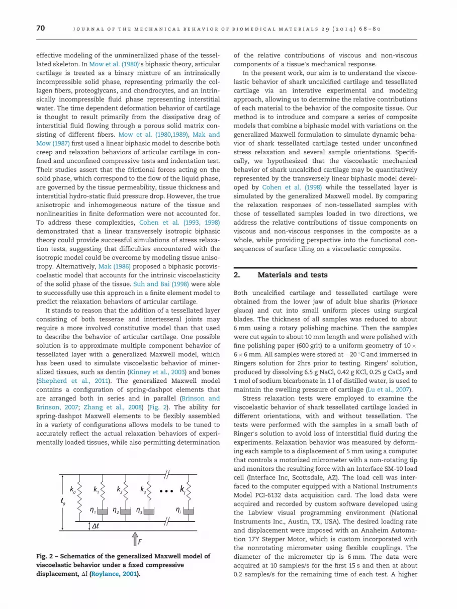
It stands to reason that the addition of a tessellated layerconsisting of both tesserae and intertesseral joints mayrequire a more involved constitutive model than that usedto describe the behavior of articular cartilage. One possiblesolution is to approximate multiple component behavior oftessellated layer with a generalized Maxwell model, whichhas been used to simulate viscoelastic behavior of miner-alized tissues, such as dentin (Kinney et al., 2003) and bones(Shepherd et al., 2011). The generalized Maxwell modelcontains a configuration of spring-dashpot elements thatare arranged both in series and in parallel (Brinson andBrinson, 2007; Zhang et al., 2008) (Fig. 2). The ability forspring-dashpot Maxwell elements to be flexibly assembledin a variety of configurations allows models to be tuned toaccurately reflect the actual relaxation behaviors of experi-mentally loaded tissues, while also permitting determination
Fig. 2 – Schematics of the generalized Maxwell model ofviscoelastic behavior under a fixed compressivedisplacement, Δl (Roylance, 2001).
of the relative contributions of viscous and non-viscouscomponents of a tissue′s mechanical response.
In the present work, our aim is to understand the viscoe-lastic behavior of shark uncalcified cartilage and tessellatedcartilage via an interative experimental and modelingapproach, allowing us to determine the relative contributionsof each material to the behavior of the composite tissue. Ourmethod is to introduce and compare a series of compositemodels that combine a biphasic model with variations on thegeneralized Maxwell formulation to simulate dynamic beha-vior of shark tessellated cartilage tested under unconfinedstress relaxation and several sample orientations. Specifi-cally, we hypothesized that the viscoelastic mechanicalbehavior of shark uncalcified cartilage may be quantitativelyrepresented by the transversely linear biphasic model devel-oped by Cohen et al. (1998) while the tessellated layer issimulated by the generalized Maxwell model. By comparingthe relaxation responses of non-tessellated samples withthose of tessellated samples loaded in two directions, weaddress the relative contributions of tissue components onviscous and non-viscous responses in the composite as awhole, while providing perspective into the functional con-sequences of surface tiling on a viscoelastic composite.
2. Materials and tests
Both uncalcified cartilage and tessellated cartilage wereobtained from the lower jaw of adult blue sharks (Prionaceglauca) and cut into small uniform pieces using surgicalblades. The thickness of all samples was reduced to about6 mm using a rotary polishing machine. Then the sampleswere cut again to about 10 mm length and were polished withfine polishing paper (600 grit) to a uniform geometry of 10�6�6 mm. All samples were stored at �20 1C and immersed inRingers solution for 2hrs prior to testing. Ringers’ solution,produced by dissolving 6.5 g NaCl, 0.42 g KCl, 0.25 g CaCl2 and1 mol of sodium bicarbonate in 1 l of distilled water, is used tomaintain the swelling pressure of cartilage (Lu et al., 2007).
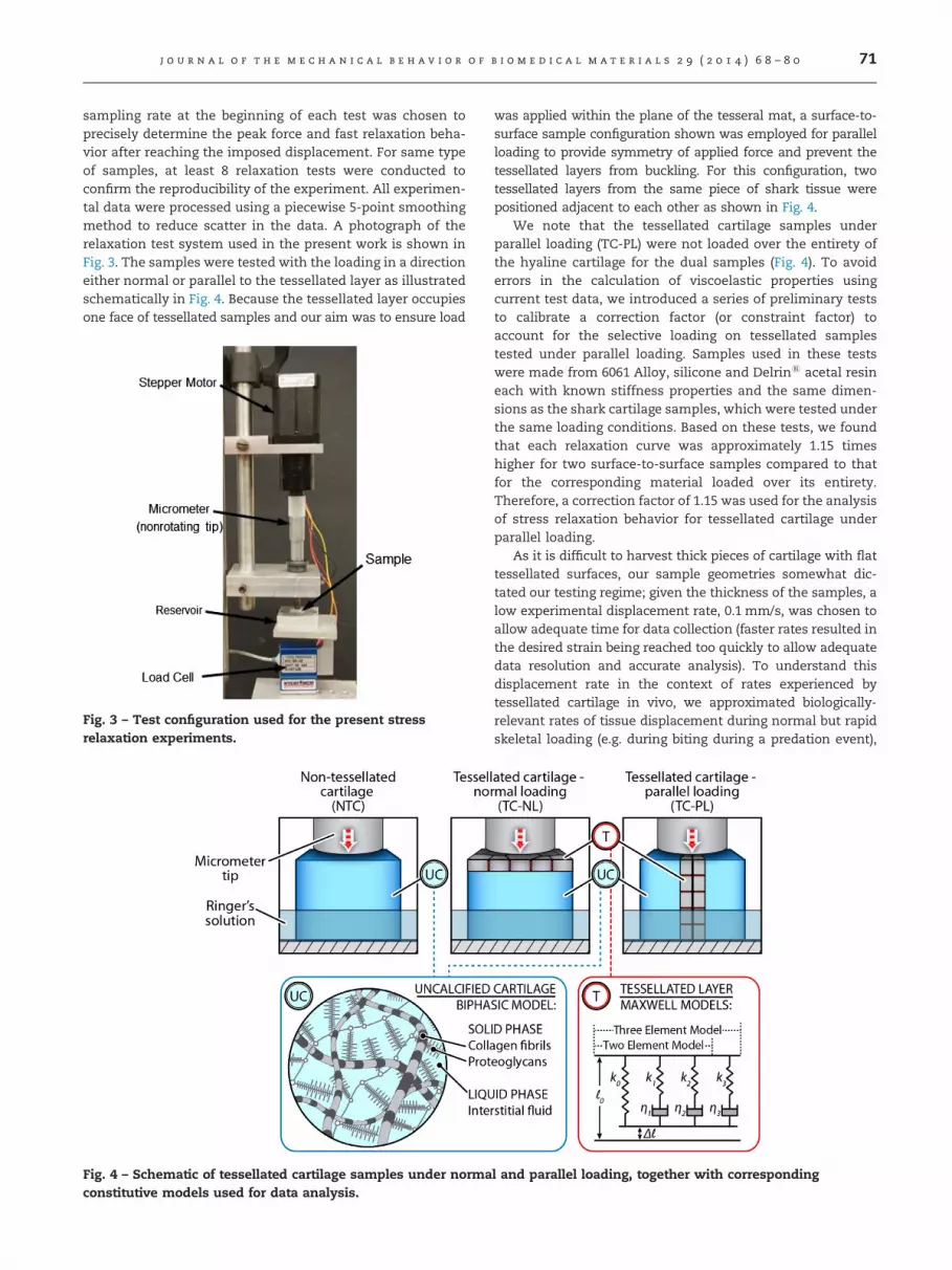
Stress relaxation tests were employed to examine theviscoelastic behavior of shark tessellated cartilage loaded indifferent orientations, with and without tessellation. Thetests were performed with the samples in a small bath ofRinger′s solution to avoid loss of interstitial fluid during theexperiments. Relaxation behavior was measured by deform-ing each sample to a displacement of 5 mm using a computerthat controls a motorized micrometer with a non-rotating tipand monitors the resulting force with an Interface SM-10 loadcell (Interface Inc, Scottsdale, AZ). The load cell was inter-faced to the computer equipped with a National InstrumentsModel PCI-6132 data acquisition card. The load data wereacquired and recorded by custom software developed usingthe Labview visual programming environment (NationalInstruments Inc., Austin, TX, USA). The desired loading rateand displacement were imposed with an Anaheim Automa-tion 17Y Stepper Motor, which is custom incorporated withthe nonrotating micrometer using flexible couplings. Thediameter of the micrometer tip is 6 mm. The data wereacquired at 10 samples/s for the first 15 s and then at about0.2 samples/s for the remaining time of each test. A higher
j o u r n a l o f t h e m e c h a n i c a l b e h a v i o r o f b i o m e d i c a l m a t e r i a l s 2 9 ( 2 0 1 4 ) 6 8 – 8 0 71
sampling rate at the beginning of each test was chosen toprecisely determine the peak force and fast relaxation beha-vior after reaching the imposed displacement. For same typeof samples, at least 8 relaxation tests were conducted toconfirm the reproducibility of the experiment. All experimen-tal data were processed using a piecewise 5-point smoothingmethod to reduce scatter in the data. A photograph of therelaxation test system used in the present work is shown inFig. 3. The samples were tested with the loading in a directioneither normal or parallel to the tessellated layer as illustratedschematically in Fig. 4. Because the tessellated layer occupiesone face of tessellated samples and our aim was to ensure load
Fig. 3 – Test configuration used for the present stressrelaxation experiments.
Fig. 4 – Schematic of tessellated cartilage samples under normaconstitutive models used for data analysis.
was applied within the plane of the tesseral mat, a surface-to-surface sample configuration shown was employed for parallelloading to provide symmetry of applied force and prevent thetessellated layers from buckling. For this configuration, twotessellated layers from the same piece of shark tissue werepositioned adjacent to each other as shown in Fig. 4.
We note that the tessellated cartilage samples underparallel loading (TC-PL) were not loaded over the entirety ofthe hyaline cartilage for the dual samples (Fig. 4). To avoiderrors in the calculation of viscoelastic properties usingcurrent test data, we introduced a series of preliminary teststo calibrate a correction factor (or constraint factor) toaccount for the selective loading on tessellated samplestested under parallel loading. Samples used in these testswere made from 6061 Alloy, silicone and Delrins acetal resineach with known stiffness properties and the same dimen-sions as the shark cartilage samples, which were tested underthe same loading conditions. Based on these tests, we foundthat each relaxation curve was approximately 1.15 timeshigher for two surface-to-surface samples compared to thatfor the corresponding material loaded over its entirety.Therefore, a correction factor of 1.15 was used for the analysisof stress relaxation behavior for tessellated cartilage underparallel loading.
As it is difficult to harvest thick pieces of cartilage with flattessellated surfaces, our sample geometries somewhat dic-tated our testing regime; given the thickness of the samples, alow experimental displacement rate, 0.1 mm/s, was chosen toallow adequate time for data collection (faster rates resulted inthe desired strain being reached too quickly to allow adequatedata resolution and accurate analysis). To understand thisdisplacement rate in the context of rates experienced bytessellated cartilage in vivo, we approximated biologically-relevant rates of tissue displacement during normal but rapidskeletal loading (e.g. during biting during a predation event),
l and parallel loading, together with corresponding

j o u r n a l o f t h e m e c h a n i c a l b e h a v i o r o f b i o m e d i c a l m a t e r i a l s 2 9 ( 2 0 1 4 ) 6 8 – 8 072
using existing anatomical, material property and feedingperformance data from two shark species with very differentecologies: the horn shark (Heterodontus francisci), a hard preyspecialist, and the lemon shark (Negaprion brevirostris), apiscivore (Huber et al., 2005; Huber, 2006). We calculatedthe average rate of force development (300–470 N/s) duringthe closing phase of an anterior bite (the front of the jaw)from the rising slope of force vs. time curves for feedingevents. The stress rate (4.46–5.97�106 Pa/s) was then calcu-lated by dividing the force rate by the cross-sectional area ofthe jaws at the bite point, measured from CT scans ofanimals using Amira software (VSG, Burlington, MA, USA).The stress rate was then divided by the stiffness of the jawtissue for these species (4.30–5.60�107 Pa, determined fromstress relaxation tests of cylindrical plugs of uncalcified jawtissue, Huber, unpublished data), to calculate strain rate(0.10–0.11 Pa/s), which was converted to displacement rate(0.87–1.65 mm/s) by multiplying by the height of the jaws atthe bite point.
These calculated tissue displacement rates during feedingare several times higher than our experimental displacementrate, however it is important to note that the incorporated,species-specific values for tissue stiffness do not take intoaccount the tessellated layer. Although the volume of thetessellated layer is small in comparison with the uncalcifiedcore of the skeleton, Liu et al. (2010)′s anatomically-basedanalytical model indicates that the tesserae play a major rolein determining overall skeletal stiffness. Given that prelimin-ary nanoindentation tests show that the mineralized tissue oftesserae is as stiff as bone (10–30 GPa, determined fromnanoindentation tests for Urobatis halleri jaw tesserae; Fix,et al., 2013), the Young′s modulus of the tessellated skeletaltissue (the uncalcified and calcified cartilage composite)would be one to two orders of magnitude higher than thespecies-specific stiffness values for uncalcified cartilage listedabove. With this higher stiffness, we would then expect ourcalculated species-specific displacement rates to be propor-tionately smaller (i.e. �1.25�10�1–1.25�10�2 mm/s); ourexperimental rates are therefore likely biologically reasonablefor tissue displacements during normal skeletal movementslike biting.
3. Modeling
3.1. Transversely isotropic biphasic model foruncalicified cartilage
Cohen et al. (1998) developed a biphasic model for describingthe viscoelastic behavior of hyaline cartilage, with the axis oftransverse isotropy in the direction aligned with the collagenfiber. In transverse isotropy, the fourth order stiffness tensor,C, is characterized by five independent elastic constants: theelastic properties are Young′s modulus and Poisson′s ratio inthe transverse plane (E1 and ν21, respectively) and out-of-plane (E3 and ν31, respectively) and the out-of-plane shearmodulus (G31), which is not used for the case of uniaxialcompression, since no shear deformation occurs in theloading direction in the present models. The equations fortotal axial stress were derived from governing equations and
solved for the stress relaxation test using Laplace transformsand modified Bessel functions.
The loading history in the current stress relaxation experi-ments can be divided into two segments for which strainfunctions are defined. These functions are:
εðtÞ ¼ _εðt�t0Þ for the ramp segment ð1Þ
εðtÞ ¼ _ε t1 for the relaxation segment ð2Þ
where _ε is the constant strain rate during the ramp segment, tis the duration of the ramp segment, and t0¼0 for the presenttest. The ramp displacement was terminated at time and thestrain, given by Eq. (2), was held constant thereafter duringthe relaxation segment. The stress during the ramp portion ofexperiment described by a transversely isotropic biphasicmodel is
sCðtÞ ¼ E3 _εtþ E1_εa2
C11kΔ3
18� ∑
1
n ¼ 1
expðAntÞBn
� �ð3Þ
and that during the relaxation portion (for t4t1) is given by
sTðtÞ ¼ E3 _εt1�E1_εa2
C11kΔ3 ∑
1
n ¼ 1
expðAntÞ�expðAnðt�t1ÞÞBn
� �ð4Þ
where k is the permeability, which is indicative of the flowcharacteristics of the interstitial fluid, and a is the diameter ofthe loading surface for the present experiments (Cohen et al.,1998). The constants in Eqs. (3) and (4) are defined as
C11 � E1Δ2=Δ1; ð5Þ
Δ1 � 1�v21�2v231E1=E3; ð6Þ
Δ2 � ð1�v231E1=E3Þ=ð1þ v21Þ; ð7Þ
Δ3 � ð1�2v231ÞΔ2=Δ1; ð8Þ
An ��α2nC11k=a2; ð9Þ
Bn � α2n½Δ22α
2n�Δ1=ð1þ v21Þ�; ð10Þ
and αn are roots of the equation:
J1ðxÞ�1�v231E1=E3Þ
1�v21�2v231E1=E3
!xJ0ðxÞ ¼ 0: ð11Þ
where J0 and J1 are Bessel functions of the first kind, andx¼αn. Then the relaxation elasticity of hyaline cartilage canbe calculated by differentiating Eq. (3) with respect to strain,which gives
ECðtÞ ¼ E3�E1a2
C11kΔ3 ∑
1
n ¼ 1
AnexpðAntÞBn
: ð12Þ
The specimens tested in the present work were rectangu-lar instead of cylindrical to avoid difficulties in the standar-dization of specimen preparation. Rectangular specimengeometries have been modeled successfully using this bipha-sic approach. For example, a biphasic and transversely iso-tropic model was applied to rectangular meniscus samplesusing a finite element approach (LeRoux and Setton, 2002).Their model provided good predictions of experimental datafor stress relaxation tests, with coefficients of determination,R240.9.
j o u r n a l o f t h e m e c h a n i c a l b e h a v i o r o f b i o m e d i c a l m a t e r i a l s 2 9 ( 2 0 1 4 ) 6 8 – 8 0 73
3.2. Generalized Maxwell model for tessellated layer
The relaxation function of a generalized Maxwell solid istypically determined using a Prony series (Chen, 2000):
YðtÞ ¼Y0 1� ∑n
i ¼ 1Pið1�e�ðt=τiÞÞ
!; ð13Þ
where Pi is the ith Prony constant (i¼1, 2, ….), τi is the ithrelaxation time constant (i¼1, 2, …), n is the number ofMaxwell elements and Y0 is the instantaneous response ofthe material, which is a constant for a given tissue, loadingdirection temperature and imposed strain. We assume thatthe relaxation elasticity of the tessellated layer can berepresented by Prony′s series as indicated by Eq. (13). Itfollows that the time dependent elastic modulus is given by
ETðtÞ ¼ E0 1� ∑n
i ¼ 1Pið1�e�ðt=τiÞÞ
!: ð14Þ
where E0 is the instantaneous modulus. For time t¼0, ET(0)¼E0 and for t¼1, ET(1)¼E0 (1�Σ Pi). E(1) is the equilibriummodulus, which is the intrinsic time independent stiffness ofthe viscoelastic material (Lu et al., 2007; Armstrong et al.,1984).
3.3. Composite model for shark tessellated cartilage
To account for the additional complexity introduced by thecalcified tessellated layer, we iteratively added additionalMaxwell elements to the biphasic model described above(Fig. 4) to determine whether they improve the accuracy ofsimulating the relaxation behavior of this tissue. For viscoe-lastic materials, a superposition of hereditary integrals can beused to describe a time dependent response. If a specimen isdeformation free prior to the time t¼0 at which a strain ε(t) isapplied, the stress for time t40 is given by Chen (2000)
s¼Z t
0Eðt�δÞdεðδÞ
dtdδ: ð15Þ
where dε(δ)/dt is the strain rate. We note that normal loadingshould predominantly result in isostress conditions whileparallel loading should predominantly result in isostrainconditions between the tessellated and uncalcified cartilagelayers as illustrated in Fig. 4. The equations governing theeffective modulus for these two loading direction are
Eef f ðtÞisotress ¼f TET
þ ð1�f TÞEC
� ��1
¼ EC
1þ ððEC=ETÞ�1Þ � f Tð16Þ
Eef f ðtÞisotrain ¼ f TET þ ð1�f TÞEC; ð17Þ
where fT is the volume fraction of tessellated layer, which isET is the elastic modulus of the tessellated layer, and EC is theelastic modulus of the uncalcified cartilage. The volumefraction of the tessellated layer (fT) in the structure can bemeasured from the thickness ratio of tessellated layer tooverall cartilage, which is approximately 0.1 for presentsamples. Substituting Eq. (12) (transversely isotropic biphasicmodel) for ECðtÞ and Eq. (14) (generalized Maxwell model) forETðtÞ into Eqs. (16) and (17) and then substituting the resultingrelation for the effective modulus under isostress conditioninto Eq. (15), the stress on tessellated cartilage under normal
loading is given by: 0otot1 :
sðtÞ ¼Z t
0
E3�E1ða2=C11kÞΔ3∑1n ¼ 1ðAnexpðAnðt�δÞÞ=BnÞ
1þ E3�E1ða2=C11kÞΔ3∑1n ¼ 1ðAnexpðAnðt�δÞÞ=BnÞ
E0ð1�∑ni ¼ 1
Pið1�e�ððt�δÞ=τi Þ ÞÞ �1� �
� f T
_εdδ;
ð18Þt4t1:
sðtÞ ¼Z t1
0
E3�E1ða2=C11kÞΔ3∑1n ¼ 1ðAnexpðAnðt�δÞÞ=BnÞ
1þ E3�E1ða2=C11kÞΔ3∑1n ¼ 1ðAnexpðAnðt�δÞÞ=BnÞ
E0ð1�∑ni ¼ 1
Pið1�e�ððt�δÞ=τi Þ ÞÞ �1� �
� f T
_εt1dδ
ð19ÞSimilarly, the mean stress for tessellated cartilage under
parallel loading is then: 0otot1:
sðtÞ ¼ f T E3 _εtþ E1_εa2
C11kΔ3
18� ∑
1
n ¼ 1
expðAntÞBn
� �� �þð1�f TÞE0 _εðt�∑n
i ¼ 1pitþ∑ni ¼ 1piτi þ∑n
i ¼ 1piτiexpð�ðt=τiÞÞÞ;ð20Þ
t4t1:
sðtÞ ¼ f T E3 _εt1�E1 _εa2
C11kΔ3 ∑
1
i ¼ 1
expðAntÞ�expðAnðt�t1ÞÞBn
( ) !
þð1�f TÞE0 _εðt1�∑ni ¼ 1pit1 þ∑n
i ¼ 1piτiexpð�ððt�t1Þ=τiÞÞ�∑n
i ¼ 1piτiexpð�ðt=τiÞÞÞ: ð21ÞThe correlations between experimental data and model
results were determined using non-linear least squares regres-sion analysis (p¼0.05). For uncalcified cartilage, we note thatfrom Eqs. (3) and (4) that there are five material coefficients: v21,v31, E1, E3 and k. These material parameters were determinedas follows. First, the equilibrium modulus, E3, was calculatedusing the equilibrium stress and strain values (when t43000 sfor current relaxation tests). Next, a nonlinear least-squaresregression method was used to fit the remaining materialparameters v21, v31, E1 and k to for both ramp and relaxation.A master table for the roots of xJ0(x) and J1(x) was then created.Based on this table, the bisection method was used to calculateeach αn where eight terms were used (Yin and Elliott, 2004). Fortessellated cartilage, we substituted the biphasic properties ofthe uncalcified cartilage samples for this component in thecomposite model except for the permeability, which we assumecan be influenced by the introduction of the tessellated layer.
For faster ramps, both the ramp and the early portion ofthe relaxation should not be included in regression analysisdue to transient effects (Lakers and Vanderby, 1999; Yin andElliott, 2004). Based on the 2.5 s ramp time, we chose to modelthe relaxation starting at 5 s to ensure that transient effectsdid not have a dominant influence on our data. In orderto achieve a full representation of both short term andlong term relaxing mechanisms of viscoelastic materials,one-element, two-element and three-element generalizedMaxwell terms were incorporated to describe the observedbehavior.
4. Results
Both load ramp and relaxation curves are plotted in Fig. 5aand b for all three test configurations. The stress relaxationdata for uncalcified cartilage and tessellated cartilage undernormal loading were very similar while the stress for parallel

j o u r n a l o f t h e m e c h a n i c a l b e h a v i o r o f b i o m e d i c a l m a t e r i a l s 2 9 ( 2 0 1 4 ) 6 8 – 8 074
loading (in-plane direction) for tessellated cartilage indicatedsubstantially higher stress resulting from a greater overallstiffness. The average regression results for the relaxationbehavior of uncalcified non-tessellated cartilage (NTC) usingthe transversely isotropic biphasic model are listed in Table 1.The accuracy of this model (dashed) for a representativerelaxation response (solid) is demonstrated in Fig. 6a. Themodel described the majority of variation in experimentaldata, with a coefficient of determination, R2, greater than0.987 (Table 1). Thus, it appears that the time dependentresponse of the shark hyaline cartilage can be simulatedreasonably well by the transversely isotropic biphasic model.The average instantaneous modulus (E0) and equilibrium
Fig. 5 – Ramp (a) and stress relaxation (b) curves fortessellated and uncalcified cartilage. NTC: non-tessellatedcartilage, TC: tessellated cartilage, NL: loading normal to thetessellated layer, PL: loading parallel to the tessellated layer.
Table 1 – Non-linear regression results for the transversely isocartilage and tessellated cartilage. NTC: non-tessellated cartilatessellated layer, PL: loading parallel to the tessellated layer. Tmeans of the best fit values for at least eight replicate experim
NTC T
E3 (MPa) 1.9770.04E1 (MPa) 0.1570.05E0 (MPa) 5.5670.44
ν21 0.9870.02ν31 0.4670.05
k (m2/Ns) 8.79� 10�1672.95� 10�16 1R2 0.992
modulus (E3) of shark hyaline cartilage were calculated tobe about 5.6 MPa and 2 MPa, respectively.
Predication of relaxation data for tessellated cartilage undernormal loading (TC NL) was first attempted using the presentMaxwell/biphasic composite model based on the isostresscondition using Eq. (17). Although high correlation coefficients(R240.99; Table 2) were determined between the entire stressrelaxation data sets and models, the calculated moduli of thetessellated layer exhibited high standard deviations, indicatingthat the Maxwell/biphasic composite model designed in thepresent work is not able to provide a consistent estimation ofmaterials properties of the tessellated layer under normalloading. Rather, the biphasic model used to model non-tessellated cartilage seems to be applicable in this case, giventhe similarity in the shape of the relaxation curves for non-tessellated cartilage and tessellated cartilage under normalloading (Fig. 5). The accuracy of the biphasic model (dashed)for a representative relaxation response of tessellated cartilageunder normal loading is demonstrated in Fig. 6b and associatedregression results listed in Table 1.
Simulation of the time dependent response for tessellatedcartilage under parallel loading (TC PL) was attempted usingboth the transversely isotropic biphasic model and thecomposite model derived in present work for isostrain con-ditions. Representative experimental data for the stressrelaxation phase are plotted in Fig. 6(c) and Fig. 7(a)–(c), withcorresponding regression results for composite models withone Maxwell element, two Maxwell elements and threeMaxwell elements. We note that the plots in Fig. 7 focus onthe first 500 s of stress relaxation where the most rapidtransitions occur. The coefficients of determination listed inTables 1 and 2 indicate that both the Maxwell/biphasic compo-site model and biphasic model exhibit good agreement withexperimental data. However, the permeability predicted by thebiphasic model (9.93�10�13 m4/Ns) is approximately threeorders of magnitude higher than that for typical mammalianhyaline cartilage and highly variable as indicated by therelatively large standard deviation (10.7�10�13 m4/Ns). Thus,it appears that tessellated cartilage under parallel loading doesnot deform in a manner as mammalian hyaline cartilage that isdescribed well by biphasic theory alone. Instead, moduli andpermeabilities estimated from Maxwell/biphasic compositemodels are statistically more self-consistent as indicated byrelatively small standard deviations compared to their corre-sponding mean values (Table 2). A comparison of the threeiterations of composite models with the first 500 s of a
tropic biphasic model for both shark non-tessellatedge, TC: tessellated cartilage, NL: loading normal to thehe values and standard deviations listed correspond to theents for each specimen type and loading direction.
C-NL TC-PL
2.7570.25 10.9170.360.1170.03 2.0170.876.1370.73 12.0570.271.0170.01 1.5170.160.3470.05 0.4370.09
.28� 10�1570.39� 10�15 9.93�10�13710.70� 10�13
0.990 0.985

represen
tativestress
relaxation(Fig.7),in
dicates
that
theth
ree-elem
ent
Maxw
ell/biphasic
mod
elis
betterat
simulatin
gth
etim
e-dep
enden
tbeh
aviorof
tessellatedcartilage
under
parallel
loading
overth
emost
dyn
amic
part
ofth
eload
ing
history.
Bycon
trast,th
eon
e-elemen
tMaxw
ell/biphasic
mod
elgives
pred
ictionsth
athave
arelatively
poor
correlationwith
experim
ental
data
and
the
two-elem
ent
Maxw
ell/biphasic
mod
elunderestim
atesth
einitial
respon
seby
5%.Theaverage
instan
taneou
san
deq
uilibriu
mmod
uli
ofth
etessellated
Fig.6–Rep
resentative
relaxation
respon
sefor
shark
(a)non
-tessellated
cartilage,(b)
tessellatedcartilage
under
parallel
loadingan
d(c)
tessellatedcartilage
under
parallel
loading
alongwith
non
-linear
regressionfits
forth
etran
sverselyisotrop
icbip
hasic
mod
el.
Table 2 – Non-linear regression results for composite models that combine a biphasic description of the hyaline component with a generalized Maxwell model of thetessellated layer. TC: tessellated cartilage, NL: loading normal to the tessellated layer, PL: loading parallel to the tessellated layer. The values and standard deviations listedcorrespond to the means of the best fit values for at least eight replicate experiments for each specimen type and loading direction.
TC NL TC PL
One Maxwell element Two Maxwell elements Three Maxwell elements One Maxwell element Two Maxwell elements Three Maxwell elements
E0 (MPa) 3.8572.28 12.1679.42 12.3679.95 99.3173.61 101.173.34 102.873.24E1 (MPa) 1.9371.20 1.9371.24 1.8871.28 91.6774.20 91.4374.03 91.3173.94
P1 0.4670.16 0.6270.21 0.6470.13 0.0870.01 0.0770.01 0.0570.01P2 0.1870.14 0.1470.07 0.0270.02 0.0470.01P3 0.0370.04 0.0270.01
k (m2/Ns) 2.84� 10�1671.02� 10�16 2.82� 10�1671.03� 10�16 2.75� 10�1671.03� 10�16 2.01� 10�1170.81� 10�1 2.02� 10�1170.81� 10�11 1.34� 10�1170.27� 10�11
τ1 132.7756.6 2.3971.72 1.7370.36 68.55745.68 17.6676.24 5.8170.84τ2 148.5759.4 121.4774.1 466.87287.7 51.49719.96τ3 683.77369.1 870.87549.5R2 0.998 0.998 0.998 0.963 0.988 0.989
journal
of
the
mechanical
behavior
of
biom
edical
materials
29
(2014)68–80
75
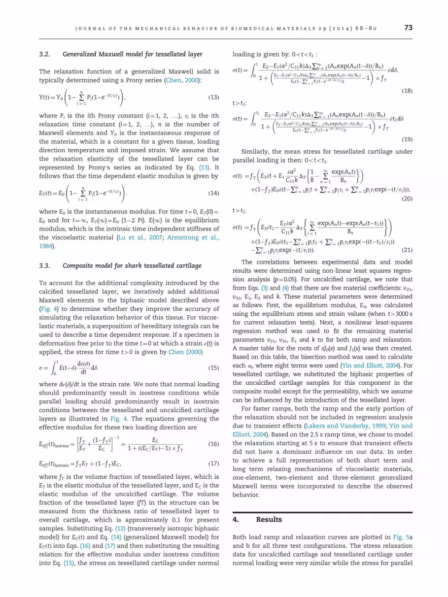
Fig. 7 – The first 500 s of a representative relaxationresponse for shark tessellated cartilage under parallelloading along with regression fits for the present compositemodel with (a) one Maxwell element, (b) two Maxwellelements and (c) three Maxwell elements.
j o u r n a l o f t h e m e c h a n i c a l b e h a v i o r o f b i o m e d i c a l m a t e r i a l s 2 9 ( 2 0 1 4 ) 6 8 – 8 076
layer predicted by the three-element composite models aregreater than 101 MPa and 91 MPa, respectively, which areapproximately 18 times and 45 times higher than those fornon-tessellated cartilage. In addition, the average value forpermeability of the tessellated cartilage predicted by thecomposite model was approximately by four orders of magni-tude greater than that for typical hyaline cartilage (10�15 to10�16 m4/Ns) (Mansour, 2003).
5. Discussion
The present correlations between model predictions andexperimental data demonstrated that the transversely isotro-pic biphasic model developed by Cohen et al. (1998) is adequatefor an accurate representation of the viscoelastic behavior ofuncalcified shark cartilage and tessellated cartilage undernormal loading in stress relaxation tests. However, the biphasicmodel is not optimum for describing the behavior of tessellatedcartilage when parallel loading is applied. Incorporation of thebiphasic model into a generalized Maxwell model was found tobe more effective for simulating the behavior of the tessellatedcartilage for this loading direction. This finding can be under-stood by examining the differences in roles of the calcified anduncalcified cartilage under normal and parallel loading condi-tions. Since we would expect the tessellated layer and uncalci-fied cartilage to deform to different degrees in normal loading,and therefore to correspond to isostress conditions (Eq. (16)),and since the tessellated layer comprises a small portion of theoverall tissue (volume fraction �0.1), it follows that theeffective elastic modulus of the tissue would be dominatedby the less stiff hyaline cartilage. This explains the effectivesimilarity in time-dependent properties for uncalcified carti-lage and tessellated cartilage under normal loading (i.e. simi-larly accurate nonlinear regressions of experimental data bythe biphasic model). The equilibrium and instantaneous mod-ulus for tessellated cartilage under normal loading seem to beonly slightly higher and the predicted Poisson′s ratios andpermeability are also similar. For this loading direction, thetessellated tissue essentially behaves as uncalcified cartilage inmost aspects.
In contrast to normal loading, parallel loading gives rise toisostrain conditions governed by Eq. (17) which dictate thatthe substantially stiffer tessellated layer contributes more tothe overall composite modulus. As a result, a compositemodel, combining the biphasic model with generalized Max-well components (representing the hyaline cartilage andtessellated layer, respectively), more accurately describedthe relaxation behavior of the composite tissue. A compositemodel combining three-Maxwell elements with the biphasicmodel provided better agreement with experimental datathan composite models with only one or two Maxwellelements describing the tessellated layer (Fig. 7(a)–(c)). Theaccuracy of the three-Maxwell element composite modelcould be a function of the three material types contributingto the viscoelastic relaxation behavior of the tessellated layer:the mineralized tissue in tesserae, the fiber joints betweenthe tesserae, and the interstitial fluid distributed throughboth the mineralized and fibrous phases. It is also worth tonotice that the stress relaxation curve for tessellated cartilageunder parallel loading appears to oscillate somewhat irra-tionally, which is possibly a consequence of the settlement ofjoints in the tessellated layer. Liu et al. (2010) demonstratedthat joints vary significantly in dynamic response within thetessellated layer and their influences on the overall relaxa-tion behavior could be significant. So it is likely that that therelaxation response of joints may be responsible for theobserved oscillations in the data. More work is needed toexplore this possibility.

j o u r n a l o f t h e m e c h a n i c a l b e h a v i o r o f b i o m e d i c a l m a t e r i a l s 2 9 ( 2 0 1 4 ) 6 8 – 8 0 77
The present model also indicates that tessellated cartilageunder parallel loading exhibits a much greater permeabilityand therefore reveals that the interstitial fluid is extrudedmore rapidly in the tessellated cartilage under parallel load-ing. In addition, although the average permeability value foruncalcified blue shark cartilage was determined to be8.79�10�16 m4/Ns, within the range of typical values (10�15
to 10�16 m4/Ns) for mammalian cartilage, the average perme-ability of the tessellated cartilage predicted by the compositemodel was approximately by four orders of magnitudegreater than that for typical hyaline cartilage (Mansour,2003). These results indicate that the drag forces betweenthe hyaline cartilage matrix and interstitial fluid are greatlydiminished for deformation under parallel loading. Thisincrease of permeability can be attributed to the fact thatthe tessellated layer supports most of the resulting stressunder parallel loading, with the high permeability of tessel-lated cartilage in parallel loading likely a reflection of the highpermeability of the tessellated layer itself: anisotropy in thefreedom of fluid movement (in normal vs. parallel loading) issupported by a recent anatomical study revealing a networkof passageways (canaliculi) extending through tesserae, con-necting the cells entombed in the mineralized matrix (Deanet al., 2010). These intratesseral canaliculi radiate out fromthe center of each tessera, predominantly within the plane ofthe tesseral mat, not perpendicular to it (i.e. canals connectadjacent cells, not those above or below). As the canalsperforate the mineralized tissue completely, opening intothe joints connecting adjacent tesserae, interstitial fluid couldconceivably flow easily within the plane of the tesseral mat,an arrangement that would increase the permeability oftessellated cartilage in parallel loading, and could also bevital for communication and the distribution of nutrientsamong cells.
There are currently no published data on elasmobranchunmineralized cartilage material properties, making it diffi-cult to ground-truth our data with other studies. Our valuesfor the elastic modulus of blue shark uncalcified cartilage(2 MPa), however, are similar to those reported for mamma-lian cartilages (0.45–19 MPa; Athanasiou et al., 1991; Mansour,2003; Silver et al., 1992). Two as of yet unpublished datasetson elasmobranch cartilage report values, an order of magni-tude higher than ours, from uncalcified jaw cartilage fromtwo shark species tested in stress relaxation (N. brevirostris, H.francisci; 43–56MPa; Huber, unpublished. data) and uncalcifiedjaw and chondrocranial cartilage plugs tested in unconfinedcompression (Squalus acanthias; 20–78 MPa for jaws, 116–775 MPa for chondrocranium; Porter et al., 2013). This sug-gests perhaps that the structure and modulus of uncalcifiedcartilage may vary to some degree across species andbetween different skeletal elements; investigations of spe-cies- and element-specific variation in tissue properties andrelation to animal ecology (e.g. diet, swimming mode, habitat)will provide valuable insight into properties relevant toskeletal performance in this system.
The substantial difference between the modulus of the blueshark uncalcified cartilage and the tessellated layer indicated bythe present models is reasonable considering the materialsproperties and volume fractions of tissues involved in thetessellated layer (Fung, 1993): the mineral component of the
tesserae (carbonated apatite; Applegate, 1967) has a modulusthat can exceed 100 GPa and the soft joint tissue between thetesserae only occupies only about 2% of tessellated layer (Deanet al., 2009; Liu et al., 2010). Tesserae exhibit a variety ofgeometric shapes, but are predominantly hexagonal (Deanet al., 2009); we note that only tesseral joints that are arrangedin series with respect to the loading direction have a significantimpact on the effective modulus of the tessellated layer (Liuet al., 2010). Accordingly, we substitute the effective equilibriummodulus of the tessellated layer (EeffE91 MPa) and the refer-ence values of the elastic modulus of tesserae as well as thevolume fraction of joints into Eq. (16). Rearranging terms, theresulting elastic modulus of the interstitial joints is calculatedto be 1.8 MPa, in the lower end of the range of moduli formammalian fibrous tissues (e.g. ligament; Donahue et al., 2003)and well beneath the threshold modulus predicted by Liuet al.'s (2010) analytical model for maintaining adequate flex-ibility in the tessellated layer to re-distribute damaging stressesaway from tension-loaded areas of the skeleton.
Perhaps the most striking contribution the tessellatedlayer provides to the overall performance of the tissue is thatit strongly increases both the instantaneous and equilibriummodulus of the composite structure, however only underparallel loading. This effect would result in much greaterstiffness for loading in this direction; it is interesting to notein cross-sections of elasmobranch skeletons, particularlythose with more limited loading directions (e.g. jaws, Fig. 8),material is typically arranged to maximize the portion of thetessellated layer under parallel loading orientation and mini-mize the portion arranged under normal loading. Such tissuearrangement would be particularly advantageous for portionsof the skeleton experiencing relatively anisotropic and/orrapid loading: for example, the greater dynamic stiffnessprovided by the tessellated layer in parallel loading wouldbe advantageous for mastication of prey, particularly thosethat are protected with a hard shell. By contrast, the dynamicstiffness normal to the tessellated layer is practically as lowas that for hyaline cartilage so that a predatory or defensivestrike in this direction would result in relatively modeststresses and therefore, have less potential for inducingdamage to the tissue. The anisotropic stress relaxationresponse of tessellated shark cartilage therefore results inan inherent, “smart” management of loading stresses,likely extremely valuable to the fatigue resistance of thisskeletal type.
Given the mechanical anisotropy we have shown for thetessellated cartilage system, the arrangement of tessellatedtissue in a skeletal cross-section should provide clues to theloading regimes experienced by the animal in life. Given thelack of gravity experienced by cartilaginous fishes, we ima-gine their skeletons’ primary loading mode to be bending.From an engineering standpoint, beams (or skeletal ele-ments) with circular cross-sections provide the best resis-tance to multi-axial bending, whereas beams with ellipticalcross-sections exhibit asymmetrical bending resistance andare stiffer when bending is in line with the cross-section′slongest axis (as in the familiar example of an architecturalI-beam) (Weaver and Ashby, 1997). Flexural stiffness ofan element can also be increased through use of stifferconstituent materials (Weaver and Ashby, 1997); in the case

Fig. 8 – Tesseral surface orientations in the jaws of the blueshark, Prionace glauca. In digital CT scan cross-sections fromtwo locations in the lower jaw (left: from beneath the teeth;right: from area of muscle adductor attachment),mineralized material in the tessellated layer is white andthe radiolucent uncalcified cartilage has been colored blue.Loading orientations applied in this study correspond toloading scenarios in vivo: in regions where the tessellatedareas are oriented more horizontally, bite force would beapplied in a direction “normal” to the tessellated layer(top schematic image), whereas regions with more verticalorientation would experience more in-plane “parallel”loading (bottom schematic image). (For interpretation of thereferences to color in this figure legend, the reader isreferred to the web version of this article.)
j o u r n a l o f t h e m e c h a n i c a l b e h a v i o r o f b i o m e d i c a l m a t e r i a l s 2 9 ( 2 0 1 4 ) 6 8 – 8 078
of composite tissues with anisotropic bending resistance,however, a given tissue morphology can provide a range offlexural stiffnesses dependent on the tissue arrangementrelative to the primary direction of loading.
If the inherent anisotropy of the tesserae-cartilage com-posite plays an in vivo role in skeletal stiffening, we wouldexpect regions with anisotropic loading direction to exhibitelliptical skeletal cross-sections, with the predominant load-ing direction being the one where the most tesserae areloaded in the plane of the tesseral mat (“parallel loading”orientation). Such an arrangement would exploit the aniso-tropy of tessellated cartilage at the tissue level, while alsoincreasing the second moment of area and bending resis-tance of the whole cross-section by arranging the majority ofmaterial in line with skeletal bending. This hypothesis issupported by Macesic and Summers’ study, showing that thecross-sectional shape of tessellated pelvic skeletal elementsin ray species varied predictably according to the degree towhich they use the elements to push along the seafloor, withmore elliptical cross-sections reflecting a predominant push-ing direction for species that rely more on this particular
locomotory mode. Observed differences in skeletal flexuralstiffness across the five species examined were dictated bythe geometric arrangement (and to some degree, the level ofmineralization) of the tessellated layer, rather than bychanges in thickness of tesserae (Macesic and Summers,2012). This demonstration of the link between morphologyand skeletal performance underlines the importance of thebasic tissue anisotropy, shown by our data, in determinationof whole element biomechanics in the tessellated skeletalsystem.
Such a fundamental structure–function relationship –
wherein gross skeletal geometry reflects loading direction – iswell-known for bone, where skeletal architecture, geometry andcomposition can adapt in response to both the magnitude andorientation of mechanical loads (Burr et al., 2002; Ruff et al.,2006; Seeman, 2009). Tessellated cartilage, however, apparentlycannot perform the fundamental remodeling and modelingprocesses that allow bones to adapt to loading conditions;therefore (non-pathologic) skeletal geometries are geneticallypredetermined, not environmentally shaped (Summers, 2000;Ashhurst, 2004). Given this ontogenetic constraint on skeletalshape, in the case of elliptical skeletal cross-sections (e.g. theextremely compressiform jaw section to the right in Fig. 8),potentially dangerous off-axis loads must be kept to a mini-mum, either by stereotyped muscle contraction patterns and/orinherent aspects of skeletal architecture (e.g. specific tesseraltiling patterns allowing some skeletal deformations but notothers). Further analyses of tessellated cartilage morphologyand mechanics in the contexts of organismal performance andbehavior will provide vital insight into the roles of the tissuephases in this tiled composite material.
6. Conclusion
Stress relaxation data were generated for tessellated anduncalcified cartilage tissue from the jaws of blue sharks.The experimental results for the uncalcified cartilage samplewere found to agree reasonably well with behavior predictedby a transversely isotropic biphasic model. Incorporation ofthis biphasic approach into a generalized Maxwell modelprovides more accurate simulations of the behavior of tessel-lated tissue when loaded parallel to the plane of the tesserae.This is a reasonable finding considering the tessellated layer′scontribution to stiffness and constraint of the soft tissueunder parallel loading. However, the generalized Maxwellmodel does not provide a better description of the behaviorof calcified cartilage under loading normal to the plane of thetessellated layer. This result is consistent with the fact thatthe role of the tessellated layer is much less significant underloading in this direction. As a result, there was a relativelysmall difference between the viscoelastic behaviors of tessel-lated cartilage under loading normal to the plane of the tissuecompared to uncalcified cartilage. In sum, the present resultsindicate that the tessellated layer provides anisotropic com-pressive stiffness and that the tissue may be arranged inways to take the most advantage of this effect, with parallel-loaded tesserae positioned in areas requiring relatively highforces (e.g. beneath the teeth of the jaw) and normal-loaded

j o u r n a l o f t h e m e c h a n i c a l b e h a v i o r o f b i o m e d i c a l m a t e r i a l s 2 9 ( 2 0 1 4 ) 6 8 – 8 0 79
tesserae, which have little effect in increasing stiffness, inareas where load-damping may be more important.
Acknowledgment
The present work was supported by the National ScienceFoundation (Award No. 0616322). Assistance in fabricating thetest system by William Dang and Ted Ediss is greatlyappreciated. We would also like to thank John Dunlop andRon Shahar for valuable discussion on the work; PepijnKamminga and Kerin Claeson for providing CT scan datafor Fig. 1; and Dan Huber for his help in revisiting his data tocalculate in vivo tissue displacement rates.
r e f e r e n c e s
Applegate, S.P., 1967. A survey of shark hard parts. In: Gilbert, P.W.,Mathewson, R.F., Rall, D.P. (Eds.), Sharks, Skates and Rays.Johns Hopkins Press, Maryland, pp. 37–66.
Armstrong, C.G., Lai, W.M., Mow, V.C., 1984. An analysis of theunconfined compression of articular cartilage. Journal ofBiomechanical Engineering 106, 165–173.
Ashhurst, D.E., 2004. The cartilaginous skeleton of anelasmobranch fish does not heal. Matrix Biology 23, 15–22.
Athanasiou, K.A., Rosenwasser, M.P., Buckwalter, J.A., Malinin, T.I.,Mow, V.C., 1991. Interspecies comparisons of in situ intrinsicmechanical properties of distal femoral cartilage. Journal ofOrthopaedic Research 9, 330–340.
Brinson, H.F., Brinson, L.C., 2007. Engineering Science andViscoelasticity. Springer, New York171–172.
Burr, D.B., Robling, A.G., Turner, C.H., 2002. Effects ofbiomechanical stress on bones in animals. Bone 30, 781–786.
Chen, T., 2000. Determining a Prony Series for a ViscoelasticMaterial from Time Strain Data. NASA/TM-2000-210123.
Clement, J.G., 1992. Re-examination of fine structure ofnendoskeletal mineralization in chondricthyes: implicationsfor growth, aging, and calcium Homeostasis. AustralianJournal of Marine & Freshwater Research 43, 157–181.
Cohen, B., Lai, W.M., Mow, V.C., 1998. A transversely isotropicbiphasic model for unconfined compression of growth plateand chondroepiphysis. Journal of Biomechanical Engineering120, 491–496.
Cohen, B., Gardner, T.R., Ateshian, G.A., 1993. The influence oftransverse isotropy on cartilage indentation behavior. A studyon the human humeral head. Transactions of OrthopaedicResearch Society 18, 185.
Dean, M.N., Summers, A.P., 2006. Mineralized cartilage incondrichthyan fishes. Zoology 109, 164–168.
Dean, M.N., Mull, C.G., Gorb, S.N., Summers, A.P., 2009. Ontogenyof the tessellated skeleton: insight from the skeletal growth ofthe round stingray Urobatis halleri. Journal of Anatomy 215,227–239.
Dean, M.N., Socha, J.J., Hall, B.K., Summers, A.P., 2010. Canaliculiin the tessellated skeleton of cartilaginous fishes. Journal ofApplied Ichthyology 26, 263–267.
Dingerkus, G., Seret, B., Guilbert, E., 1991. Multiple prismaticcalcium phosphate layers in the jaws present-day sharks.Experientia 47, 38–40.
Donahue, T.L.H., Hulla, M.L., Rashid, M.M., Jacobs, C.R., 2003. Howthe stiffness of meniscal attachments and meniscal materialproperties affect tibio-femoral contact pressure computedusing a validated finite element model of the human kneejoint. Journal of Biomechanics 36, 19–34.
Fix, D., Weinkamer, R., Fratzl, P., Dean, M.N., 2013. Micrometer-scale material characterization of mineralized elasmobranchcartilage: a novel combination of nanoindentation andscanning acoustic microscopy, in preparation.
Fung, Y.C., 1993. Biomechanics: Mechanical Properties of LivingTissues. Springer, New York503.
Hacker, C.L., Ansell, M.P., 2001. Fatigue damage and hysteresis inwood-epoxy laminates. Journal of Materials Science 36, 609–621.
Hall, B.K., 2005. Bones and Cartilage: Developmental SkeletalBiology. Elsevier/Academic Press, London.
Huber, D.R., 2006. Cranial biomechanics and feeding performanceof sharksDepartment of Biology, University of South Florida,Tampa, FL250.
Huber, D.R., Eason, T.G., Hueter, R.E., Motta, P.J., 2005. Analysis ofthe bite force and mechanical design of the feedingmechanism of the durophagous horn shark Heterodontusfrancisci. Journal of Experimental Biology 208, 3553–3571.
Hunziker, E.B., 1999. Articular cartilage repair: are the intrinsicbiological constraints undermining this process insuperable?Osteoarthritis Cartilage 7, 15–28.
Kemp, N.E., Westrin, S.K., 1979. Ultrastructure of calcifiedcartilage in the endoskeletal tesserae of sharks. Journal ofMorphology 160, 75–102.
Kinney, J.H., Marshall, S.J., Marshall, G.W., 2003. The mechanicalproperties of human dentin: a critical review and re-evaluation of the dental literature. Critical Reviews in OralBiology & Medicine 14 (1), 13–29.
Lakers, R.S., Vanderby, R., 1999. Interrelation of creep andrelaxation: a modeling approach for ligaments. Journal ofBiomechanics 121 (6), 612–615.
LeRoux, M.A., Setton, L.A., 2002. Experimental and biphasic FEMdeterminations of the material properties and hydraulicpermeability of the meniscus in tension. Journal ofBiomechanics 124, 315–321.
Liu, X., Dean, M.N., Summer, A.P., Earthman, J.C., 2010. Compositemodel of the shark′s skeleton in bending: a novel architecturefor biomimetic design of functional compression bias.Materials Science and Engineering C 30, 1077–1084.
Lu, X.L., Miller, C., Chen, F.H., Guo, X.E., Mow, V.C., 2007. Thegeneralized triphasic correspondence principle forsimultaneous determination of the mechanical properties andproteoglycan content of articular cartilage by indentation.Journal of Biomechanics 40, 2434–2441.
Macesic, L.J., Summers, A.P., 2012. Flexural stiffness andcomposition of the batoid propterygium as predictors ofpunting ability. The Journal of Experimental Biology 215,2003–2012.
Mak, A.F., 1986. The apparent viscoelastic behavior of articularcartilage—the contributions from the intrinsic matrixviscoelasticity and interstitial fluid flows. Journal ofBiomechanical Engineering 108, 123–130.
Mak, A.F., Mow, V.C., 1987. Biphasic indentation of articularcartilage-1. Theoretical analysis. Journal of Biomechanics 20,703–714.
Malzahn, J.C., Schultz, J.M., 1986. Tension–tension andcompression–compression fatigue behavior of an injection-molded short-glass-fiber/poly(ethylene terephthalate)composite. Composites Science and Technology 27, 253–289.
Mansour, J.M., 2003. Biomechanics of cartilage. In: Oatis, C.A.(Ed.), Kinesiology: the Mechanics and Pathomechanics ofHuman Movement. Lippincott Williams and Wilkins,Philadelphia (Chapter 5), pp. 66–79.
Mow, V.C., Gibbs, M.C., Lai, W.M., Zhu, W.B., Athanasiou, K.A.,1989. Biphasic indentation of articular Cartilage-2. Anumerical algorithm and an experimental study. Journal ofBiomechanics 22, 853–861.
Mow, V.C., Kuei, S.C., Lai, W.M., Armstrong, C.G., 1980. Biphasiccreep and stress relaxation of articular cartilage in

j o u r n a l o f t h e m e c h a n i c a l b e h a v i o r o f b i o m e d i c a l m a t e r i a l s 2 9 ( 2 0 1 4 ) 6 8 – 8 080
compression: theory and experiments. Journal ofBiomechanical Engineering 102, 73–84.
Moss, S.A., 1977. Skeletal tissues in sharks. American Zoologist17, 335–342.
Porter, M.E., Beltrán, J.L., Kajiura, S.M., Koob, T.J., Summers, A.P.,2013. Stiffness without mineral: material properties andbiochemical components of jaws and chondrocrania in theElasmobranchii (sharks, skates, and rays). PeerJ., Submitted.
Roylance, D., 2001. Engineering Viscoelasticity, p. 14–15.Ruff, C., Holt, B., Trinkaus, E., 2006. Who′s afraid of the big bad
Wolff?: ‘Wolff′s law’ and bone functional adaptation.American Journal of Physical Anthropology 129, 484–498.
Seeman, E., 2009. Bone modeling and remodeling. CriticalReviews in Eukaryotic Gene Expression 19, 219–230.
Shepherd, T.N., Zhang, J., Ovaert, T.C., Roeder, R.K., Niebur, G.L.,2011. Direct comparison of nanoindentation and macroscopicmeasurements of bone viscoelasticity. Journal of theMechanical Behavior of Biomedical Materials 4, 2055–2062.
Silver, F.H., Kato, Y.P., Ohno, M., Wasserman, A.J., 1992. Analysisof mammalian connective tissue: relationship betweenhierarchical structures and mechanical properties. Journal ofLong-Term Effects of Medical Implants 2, 165–198.
Suh, J.K., Bai, S., 1998. Finite element formulation of biphasic
poroviscoelastic model for articular cartilage. Journal of
Biomechanical Engineering 120, 195–201.Summers, A.P., 2000. Stiffening the stingray skeleton - An
investigation of durophagy in myliobatid stingrays
(Chondrichthyes, Batoidea, Myliobatidae). Journal of
Morphology 243, 113–126.Weaver, P.M., Ashby, M.F., 1997. Material limits for shape
efficiency. Progress in Materials Science 41, 61–128.Wroe, S., Huber, D.R., Lowry, M.B., McHenry, C.R., Moreno, K.,
Clausen, P.D., Ferrara, T.L., 2008. Three-dimensional computer
analysis of white shark jaw mechanics: how hard can a great
white bite? Zoology 276, 336–342.Yin, L., Elliott, D.M., 2004. A biphasic and transversely isotropic
mechanical model for tendon: application to mouse tail
fascicles in uniaxial tension. Journal of Biomechanics 22
(927-916).Zhang, M., Guo, X., Kassab, G.S., 2008. A generalized Maxwell
model for creep behavior of artery opening angle. Journal of
Biomechanical Engineering 130, 054502.