Embed Size (px)
Citation preview
Cartilage Tissue Engineering – Comparison of Articular Cartilage
Progenitor Cells and Mesenchymal Stromal Cells in Agarose and
Hyaluronic Acid-Based Hydrogels
-
Tissue Engineering von Knorpel – Vergleich von Gelenkknorpel-
Vorläuferzellen und mesenchymalen Stromazellen in Agarose-
und Hyaluronsäure-basierten Hydrogelen
Doctoral thesis for a doctoral degree
at the Graduate School of Life Sciences,
Julius-Maximilians-Universität Würzburg,
Section Biomedicine
submitted by
Stefanie Schmidt
from
Landsberg am Lech
Würzburg, 2021
This document is licensed under the Creative Commons Attribution-NonCommercial-NoDerivatives 4.0 International License (CC BY-NC-ND 4.0): http://creativecommons.org/licenses/by-nc-nd/4.0 This CC license does not apply to third party material (attributed to another source) in this publication.
Submitted on: ………………………………………………………
Members of the Thesis Committee
Chairperson: Prof. Dr. Carmen Villmann
Primary Supervisor: Prof. Dr. Torsten Blunk
Supervisor (Second): Dr. habil. Jörg Teßmar
Supervisor (Third): Dr. Marietta Herrmann
Date of Public Defence: ………………………………………
Date of Receipt of Certificates: ………………………………
Table of contents
Summary ................................................................................................................................... 1
Zusammenfassung .................................................................................................................... 3
1 Introduction .......................................................................................................................... 7
1.1 Articular cartilage .......................................................................................................... 9
1.1.1 Structure of synovial joints and function of articular cartilage ............................ 9
1.1.2 Articular cartilage composition and structure .................................................... 10
1.1.3 Articular cartilage defects and clinical treatments ............................................. 12
1.2 Important aspects of articular cartilage tissue engineering ......................................... 15
1.2.1 Different cell sources ......................................................................................... 15
1.2.2 Oxygen partial pressure ...................................................................................... 19
1.2.3 Zonal structure .................................................................................................... 21
1.2.4 Scaffold material ................................................................................................ 22
1.3 Hyaluronic acid ........................................................................................................... 25
1.3.1 Characteristics and functions of HA in the human body ................................... 25
1.3.2 Applications of HA in articular cartilage tissue engineering ............................. 27
1.4 Goals of the thesis ....................................................................................................... 31
1.4.1 Comparison of chondrogenesis of ACPCs and MSCs in different
hydrogels ...................................................................................................................... 31
1.4.2 Evaluation of the contribution of hyaluronan to chondrogenic gene
expression of MSCs ............................................................................................................... 33
2 Material ............................................................................................................................... 35
2.1 Instruments .................................................................................................................. 37
2.2 Consumables ............................................................................................................... 38
2.3 Chemicals .................................................................................................................... 40
2.4 Hydrogel components ................................................................................................. 41
2.5 Antibodies ................................................................................................................... 42
2.6 Primers ........................................................................................................................ 43
2.7 Cell culture media ....................................................................................................... 44
2.8 Cells ............................................................................................................................. 45
2.9 Buffers and solutions ................................................................................................... 45
2.10 Software .................................................................................................................... 47
3 Methods ............................................................................................................................... 49
3.1 Cell culture .................................................................................................................. 51
3.1.1 Cell isolation ...................................................................................................... 51
3.1.2 2D cell expansion ............................................................................................... 52
3.1.3 Treatment of cells in 2D monolayer with HA .................................................... 52
3.1.4 Treatment of cells in 3D pellet cultures with HA .............................................. 52
3.1.5 3D agarose hydrogel culture............................................................................... 53
3.1.6 3D HA-SH hydrogel culture............................................................................... 53
3.1.6.1 HA-SH/P(AGE-co-G) hydrogels .............................................................. 53
3.1.6.2 HA-SH /PEGDA/PEG-allyl/Irgacure2959 hydrogels ............................... 54
3.1.7 Preparation of zonal hydrogels ........................................................................... 54
3.2 Staining of cells and tissue sections ............................................................................ 55
3.2.1 Cell viability assay ............................................................................................. 55
3.2.2 Sectioning of 3D constructs ............................................................................... 55
3.2.2.1 Cryo-sectioning ......................................................................................... 55
3.2.2.2 Paraffin sectioning ..................................................................................... 55
3.2.3 Safranin O staining ............................................................................................. 56
3.2.4 Picrosirius red staining ....................................................................................... 56
3.2.5 Immunohistochemistry ....................................................................................... 56
3.3 Biochemical analysis ................................................................................................... 57
3.3.1 Papain digestion ................................................................................................. 57
3.3.2 DNA assay .......................................................................................................... 58
3.3.3 Glycosaminoglycan assay .................................................................................. 58
3.3.4 Hydroxyproline assay ......................................................................................... 58
3.3.5 Alkaline phosphatase activity assay ................................................................... 58
3.4 Quantitative real-time PCR analysis ........................................................................... 59
3.5 Flow cytometry ........................................................................................................... 60
3.6 Statistical analysis ....................................................................................................... 60
4 Results and discussion ........................................................................................................ 61
4.1 Articular cartilage tissue engineering with ACPCs and MSCs ................................... 63
4.1.1 Chondrogenesis of ACPCs and MSCs in agarose hydrogel............................... 64
4.1.1.1 Comparison of chondrogenesis of ACPCs and MSCs in normoxia .......... 66
4.1.1.2 Influence of hypoxia on chondrogenesis of ACPCs and MSCs ................ 76
4.1.1.3 Summary of chondrogenesis of ACPCs and MSCs in agarose hydrogel.. 86
4.1.2 Chondrogenesis of ACPCs and MSCs in a HA-based hydrogel ........................ 88
4.1.3 Influence of different hydrogels on chondrogenesis of ACPCs and MSCs ....... 95
4.2 Contribution of hyaluronan to chondrogenic gene expression of MSCs .................... 97
4.2.1 Influence of hyaluronan on HA-receptors CD44 and CD168 ............................ 99
4.2.1.1 Basal expression of CD44 and CD168 ...................................................... 99
4.2.1.2 Effects of HA on CD44 and CD168 ........................................................ 101
4.2.2 Influence of hyaluronan on stemness markers ................................................. 103
4.2.3 Influence of hyaluronan on different transcription factors ............................... 105
4.2.3.1 Influence on SOX9, SOX5 and SOX6 .................................................... 106
4.2.3.2 Influence on RUNX2 and PPARG .......................................................... 108
4.2.4 Influence of hyaluronan on chondrogenic markers ACAN and COL2A1 ....... 110
4.2.5 Summary of the effects of hyaluronan on MSCs ............................................. 113
5 Conclusion ......................................................................................................................... 117
6 Bibliography ..................................................................................................................... 121
Annex ..................................................................................................................................... 147
A.1 List of figures ............................................................................................................ 149
A.2 List of tables .............................................................................................................. 153
A.3 List of abbreviations .................................................................................................. 155
A.4 Statement on Copyright and Self-plagiarism ............................................................ 159
A.5 Affidavit .................................................................................................................... 161
A.6 Acknowledgement ..................................................................................................... 163
A.7 Curriculum Vitae ....................................................................................................... 165

1
Summary
Articular cartilage damage caused by sports accidents, trauma or gradual wear and tear can lead
to degeneration and the development of osteoarthritis because cartilage tissue has only limited
capacity for intrinsic healing. Osteoarthritis causes reduction of mobility and chronic pain and
is one of the leading causes of disability in the elderly population. Current clinical treatment
options can reduce pain and restore mobility for some time, but the formed repair tissue has
mostly inferior functionality compared to healthy articular cartilage and does not last long-term.
Articular cartilage tissue engineering is a promising approach for the improvement of the qual-
ity of cartilage repair tissue and regeneration.
In this thesis, a promising new cell type for articular cartilage tissue engineering, the so-called
articular cartilage progenitor cell (ACPC), was investigated for the first time in the two different
hydrogels agarose and HA-SH/P(AGE-co-G) in comparison to mesenchymal stromal cells
(MSCs). In agarose, ACPCs´ and MSCs´ chondrogenic capacity was investigated under
normoxic (21 % oxygen) and hypoxic (2 % oxygen) conditions in monoculture constructs and
in zonally layered co-culture constructs with ACPCs in the upper layer and MSCs in the lower
layer. In the newly developed hyaluronic acid (HA)-based hydrogel HA-SH/P(AGE-co-G),
chondrogenesis of ACPCs and MSCs was also evaluated in monoculture constructs and in zon-
ally layered co-culture constructs like in agarose hydrogel. Additionally, the contribution of the
bioactive molecule hyaluronic acid to chondrogenic gene expression of MSCs was investigated
in 2D monolayer, 3D pellet and HA-SH hydrogel culture.
It was shown that both ACPCs and MSCs could chondrogenically differentiate in agarose and
HA-SH/P(AGE-co-G) hydrogels. In agarose hydrogel, ACPCs produced a more articular car-
tilage-like tissue than MSCs that contained more glycosaminoglycan (GAG), less type I colla-
gen and only little alkaline phosphatase (ALP) activity. Hypoxic conditions did not increase
extracellular matrix (ECM) production of ACPCs and MSCs significantly but improved the
quality of the neo-cartilage tissue produced by MSCs. The creation of zonal agarose constructs
with ACPCs in the upper layer and MSCs in the lower layer led to an ECM production in zonal
hydrogels that lay in general in between the ECM production of non-zonal ACPC and MSC
hydrogels. Even though zonal co-culture of ACPCs and MSCs did not increase ECM produc-
tion, the two cell types influenced each other and, for example, modulated the staining intensi-
ties of type II and type I collagen in comparison to non-zonal constructs under normoxic and
hypoxic conditions. In HA-SH/P(AGE-co-G) hydrogel, MSCs produced more ECM than
2
ACPCs, but the ECM was limited to the pericellular region for both cell types. Zonal HA-
SH/P(AGE-co-G) hydrogels resulted in a native-like zonal distribution of ECM as MSCs in the
lower zone produced more ECM than ACPCs in the upper zone. It appeared that chondrogen-
esis of ACPCs was supported by hydrogels without biological attachment sites such as agarose,
and that chondrogenesis of MSCs benefited from hydrogels with biological cues like HA.
As HA is an attractive material for cartilage tissue engineering, and the HA-based hydrogel
HA-SH/P(AGE-co-G) appeared to be beneficial for MSC chondrogenic differentiation, the con-
tribution of HA to chondrogenic gene expression of MSCs was investigated. An upregulation
of chondrogenic gene expression was found in 2D monolayer and 3D pellet culture of MSCs in
response to HA supplementation, while gene expression of osteogenic and adipogenic tran-
scription factors was not upregulated. MSCs, encapsulated in a HA-based hydrogel, showed
upregulation of gene expression for chondrogenic, osteogenic and adipogenic differentiation
markers as well as for stemness markers. In a 3D bioprinting process, using the HA-based hy-
drogel, gene expression levels of MSCs mostly did not change. Nevertheless, expression of
three tested genes (COL2A1, SOX2, CD168) was downregulated in printed in comparison to
cast constructs, underscoring the importance of closely monitoring cellular behaviour during
and after the printing process.
In summary, it was confirmed that ACPCs are a promising cell source for articular cartilage
engineering with advantages over MSCs when they were cultured in a suitable hydrogel like
agarose. The performance of the cells was strongly dependent on the hydrogel environment
they were cultured in. The different chondrogenic performance of ACPCs and MSCs in agarose
and HA-SH/P(AGE-co-G) hydrogels highlighted the importance of choosing suitable hydrogels
for the different cell types used in articular cartilage tissue engineering. Hydrogels with high
polymer content, such as the investigated HA-SH/P(AGE-co-G) hydrogels, can limit ECM dis-
tribution to the pericellular area and should be developed further towards less polymer content,
leading to more homogenous ECM distribution of the cultured cells. The influence of HA on
chondrogenic gene expression and on the balance between differentiation and maintenance of
stemness in MSCs was demonstrated. More studies should be performed in the future to further
elucidate the signalling functions of HA and the effects of 3D bioprinting in HA-based hydro-
gels.
Taken together, the results of this thesis expand the knowledge in the area of articular cartilage
engineering with regard to the rational combination of cell types and hydrogel materials and
open up new possible approaches to the regeneration of articular cartilage tissue.
3
Zusammenfassung
Gelenkknorpeldefekte, die durch Sportverletzungen, Unfälle oder graduelle Abnutzung entste-
hen, können zu Degeneration des Gewebes und zur Entstehung von Arthrose führen, da Knor-
pelgewebe nur über eine eingeschränkte Fähigkeit zur Selbstheilung verfügt. Arthrose reduziert
die Beweglichkeit und verursacht chronische Schmerzen. Sie ist vor allem bei älteren Menschen
einer der häufigsten Gründe für körperliche Behinderung. Die zurzeit verfügbaren operativen
Behandlungsmöglichkeiten können die Symptome meist für einige Zeit lindern, aber das dabei
gebildete Ersatzgewebe zeigt meistens nur eingeschränkte Funktionalität im Vergleich zu na-
türlichem gesunden Knorpelgewebe und bleibt nur für eine begrenzte Zeit stabil. Tissue Engi-
neering von Gelenkknorpelgewebe ist ein vielversprechender Ansatz, um die Qualität des Er-
satzgewebes und der Knorpelregeneration zu verbessern.
Diese Arbeit untersuchte einen neuen vielversprechenden Zelltyp für das Tissue Engineering
von Knorpelgewebe, sogenannte Gelenkknorpel-Vorläuferzellen (ACPCs). Diese Zellen wur-
den erstmals in zwei verschiedenen Hydrogelen, Agarose und HA-SH/P(AGE-co-G), mit
mesenchymalen Stromazellen (MSCs) verglichen. Die chondrogene Kapazität von ACPCs und
MSCs in Agarose wurde unter normoxischen (21 % Sauerstoff) und hypoxischen (2 % Sauer-
stoff) Bedingungen in Monokultur und zonal geschichteter Kokultur untersucht. In den zonalen
Kokulturen befanden sich ACPCs in einer oberen Schicht und MSCs in einer unteren Schicht.
In dem neu entwickelten Hyaluronsäure (HA)-basierten Hydrogel HA-SH/P(AGE-co-G) wurde
die chondrogene Differenzierung von ACPCs und MSCs ebenfalls in Monokultur und in zonal
geschichteter Kokultur, wie im Agarose-Hydrogel, analysiert. Außerdem wurde der Beitrag des
biologisch aktiven Moleküls Hyaluronsäure zur chondrogenen Genexpression von MSCs in
2D-, 3D-Pellet- und HA-SH-Hydrogel-Kulturen untersucht.
Diese Arbeit zeigte, dass sowohl ACPCs als auch MSCs in Agarose- und HA-SH/P(AGE-co-
G)-Hydrogelen chondrogen differenzieren konnten. ACPCs produzierten im Agarose-Hydrogel
ein Gewebe, das dem Gelenkknorpel ähnlicher war als das von MSCs produzierte Gewebe, da
es mehr Glykosaminoglykane (GAG), weniger Typ I Kollagen und nur geringe Aktivität der
Alkalinen Phosphatase (ALP) aufwies. Hypoxische Bedingungen konnten die Produktion von
extrazellulärer Matrix (ECM) durch ACPCs und MSCs nicht erhöhen, aber sie verbesserten die
Qualität des von MSCs produzierten Gewebes. Die Herstellung von zonalen Agarose-Kon-
strukten mit ACPCs in der oberen Schicht und MSCs in der unteren Schicht führte zu einer
ECM-Produktion in zonalen Hydrogelen, die im Allgemeinen zwischen der ECM-Produktion
4
der ACPC-Monokultur und der MSC-Monokultur lag. Zonale Kokultur von ACPCs und MSCs
führte zwar nicht zu einer erhöhten ECM-Produktion, allerdings beeinflussten die beiden Zell-
typen sich gegenseitig und modulierten zum Beispiel die Intensitäten der Typ II und Typ I
Kollagen Färbungen im Vergleich zu Monokulturen unter normoxischen und hypoxischen Be-
dingungen. Im HA-SH/P(AGE-co-G)-Hydrogel produzierten die MSCs mehr ECM als die
ACPCs, allerdings war die Verteilung der gebildeten ECM bei beiden Zelltypen auf den peri-
zellulären Bereich beschränkt. Zonale HA-SH/P(AGE-co-G)-Hydrogele führten zu einer zon-
alen Verteilung von ECM, die der natürlichen Struktur von Gelenkknorpel ähnlich war, da die
MSCs in der unteren Schicht mehr ECM produzierten als die ACPCs in der oberen Schicht.
Anscheinend wurde die chondrogene Differenzierung von ACPCs von Hydrogelen unterstützt,
die, so wie Agarose, keine biologischen Bindestellen aufwiesen, und die Chondrogenese von
MSCs profitierte von Hydrogelen mit biologischen Signalen wie HA.
Da HA ein attraktives Material für Tissue Engineering von Knorpel darstellt und das HA-ba-
sierte Hydrogel HA-SH/P(AGE-co-G) anscheinend die chondrogene Differenzierung von
MSCs begünstigte, wurde der Beitrag von HA zur chondrogenen Genexpression in MSCs un-
tersucht. Eine Hochregulation der chondrogenen Genexpression ließ sich in 2D- und 3D-Pellet-
Kulturen von MSCs als Reaktion auf HA beobachten, während die Genexpression von osteo-
genen oder adipogenen Transkriptionsfaktoren nicht hochreguliert wurde. Der Einschluss von
MSCs in einem HA-basierten Hydrogel führte zu einer Erhöhung der Genexpression von
chondrogenen, osteogenen, adipogenen und Stemness-Markern. Ein 3D-Druck-Prozess mit
dem HA-basierten Hydrogel veränderte die Genexpression von MSCs in den meisten Fällen
nicht. Dennoch wurde die Expression von drei getesteten Genen (COL2A1, SOX2, CD168) in
gedruckten im Vergleich zu gegossenen Konstrukten herunterreguliert. Dies unterstrich die
Wichtigkeit einer genauen Kontrolle des Verhaltens der Zellen während und nach dem Druck-
Prozess.
Zusammenfassend ließen sich ACPCs als vielversprechender neuer Zelltyp für das Tissue En-
gineering von Gelenkknorpelgewebe bestätigen. ACPCs haben Vorteile gegenüber MSCs, vor
allem, wenn sie in einem passenden Hydrogel wie Agarose kultiviert werden. Die Leistung der
Zellen war stark von den verschiedenen Hydrogelen und der Umgebung beeinflusst, die diese
den Zellen darboten. Die unterschiedliche chondrogene Leistung von ACPCs und MSCs in
Agarose- und HA-SH/P(AGE-co-G)-Hydrogelen zeigte deutlich die übergeordnete Relevanz
der Auswahl von passenden Hydrogelen für die verschiedenen Zelltypen, die im Tissue Engi-
neering von Gelenkknorpel Verwendung finden. Hydrogele mit einem hohen Polymergehalt,
5
wie das eingesetzte HA-SH/P(AGE-co-G)-Hydrogel, können die Verteilung der gebildeten
ECM auf den perizellulären Bereich beschränken und sollten weiterentwickelt werden, um ei-
nen niedrigeren Polymergehalt und damit eine homogenere ECM-Verteilung durch die kulti-
vierten Zellen zu erreichen. Der Einfluss von HA auf die chondrogene Genexpression und auf
die Balance zwischen Differenzierung und Erhaltung der Stemness in MSCs ließ sich aufzei-
gen. In Zukunft sollten weitere Studien die Signalfunktionen von HA und den Einfluss des 3D-
Drucks in HA-basierten Hydrogelen genauer zu untersuchen.
Zusammengenommen erweitern die Ergebnisse dieser Arbeit das Wissen im Bereich des Tissue
Engineerings von Gelenkknorpelgewebe, vor allem in Bezug auf eine rationale Kombination
von Zelltypen und Hydrogel-Materialien, und eröffnen neue Ansätze zur Knorpelregeneration.

6

7
Introduction

8
9
1 Introduction
1.1 Articular cartilage
There are three different types of cartilage in the human body: Elastic cartilage, fibrocarti-
lage, and hyaline (articular) cartilage. Elastic cartilage contains much elastin and is a flexi-
ble sort of cartilage that can be found in non-load bearing body parts like the auricle [1].
Fibrocartilage contains thick collagen fibres and is the only cartilage that contains type I
collagen additionally to type II collagen in its mature state. An example for fibrocartilage
is the meniscus [2]. Hyaline articular cartilage is a connective tissue that covers articulating
surfaces of bone in synovial joints, such as the knee or the hip [3,4].
1.1.1 Structure of synovial joints and function of articular cartilage
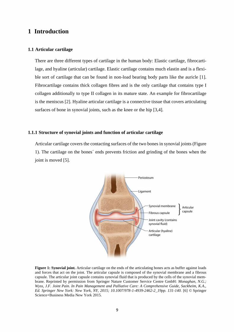
Articular cartilage covers the contacting surfaces of the two bones in synovial joints (Figure
1). The cartilage on the bones´ ends prevents friction and grinding of the bones when the
joint is moved [5].
Figure 1: Synovial joint. Articular cartilage on the ends of the articulating bones acts as buffer against loads
and forces that act on the joint. The articular capsule is composed of the synovial membrane and a fibrous
capsule. The articular joint capsule contains synovial fluid that is produced by the cells of the synovial mem-
brane. Reprinted by permission from Springer Nature Customer Service Centre GmbH: Monaghan, N.G.;
Wyss, J.F. Joint Pain. In Pain Management and Palliative Care: A Comprehensive Guide, Sackheim, K.A.,
Ed. Springer New York: New York, NY, 2015; 10.1007/978-1-4939-2462-2_19pp. 131-140. [6] © Springer
Science+Business Media New York 2015.
10
The joint cavity is filled with synovial fluid that is produced by cells of the synovial mem-
brane. The synovial membrane surrounds the joint cavity and the articulating bone surfaces
and keeps the synovial fluid in the joint capsule (Figure 1). Synovial fluid has two main
functions. It contributes to further reduction of friction between the joint bones as it is a
thick, lubricating fluid. Additionally, it provides nutrients for chondrocytes, the resident
cells in articular cartilage, as cartilage does not have a blood supply [3]. The fluid is
squeezed out of the tissue when the joint is loaded and flows back in with fresh nutrients
when the pressure is gone. The main function of articular cartilage in a joint is to provide
smooth movement of the bones´ ends against each other and to withstand and buffer the
loads and forces that act on a weight-bearing joint like the knee [3,5].
1.1.2 Articular cartilage composition and structure
Hyaline articular cartilage is a tissue with low cell density. Chondrocytes, the resident cells,
have no direct contact with each other and amount to only 2 % of the total cartilage volume.
The tissue also has no blood or lymphatic vessels and no innervation [4,5]. Extracellular
matrix (ECM) that is produced by chondrocytes makes up the largest part of articular carti-
lage. Its main components are water, collagens and proteoglycans. Type II collagen is with
90-95 % the main collagen in articular cartilage and forms a fibril network that is stabilized
by less abundant collagens (types I, IV, V, VI, IX and XI). Proteoglycans are proteins that
carry one or several chains of glycosaminoglycans (GAGs). Glycosaminoglycans are long
linear chains of repeating disaccharides, for example chondroitin sulfate, keratan sulfate or
hyaluronic acid. The most prominent proteoglycan in articular cartilage is aggrecan. Aggre-
can carries over 100 glycosaminoglycan chains, mainly chondroitin sulfate but also keratan
sulfate. This already large proteoglycan forms huge aggregates with hyaluronic acid through
non-covalent bonds and the stabilizing function of the link protein. These aggregates draw
water into the tissue because they contain many negatively charged residues that draw coun-
ter-ions, and they provide a hydrated gel like structure. The water is pressed out of the tissue
during loading of the joint and seeps back in after relieving the load. Intertwined collagens
and proteoglycans form a gel-like network that is viscoelastic and resilient and provides the
tensile (type II collagen) and compressive (aggrecan) strength of cartilage tissue that is
needed to withstand the continuous loads and shear forces that synovial joints are exposed
to [3-5].
11
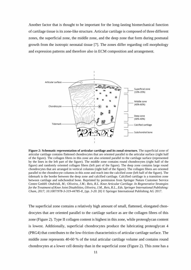
Another factor that is thought to be important for the long-lasting biomechanical function
of cartilage tissue is its zone-like structure. Articular cartilage is composed of three different
zones, the superficial zone, the middle zone, and the deep zone that form during postnatal
growth from the isotropic neonatal tissue [7]. The zones differ regarding cell morphology
and expression patterns and therefore also in ECM composition and arrangement.
Figure 2: Schematic representation of articular cartilage and its zonal structure. The superficial zone of
articular cartilage contains flattened chondrocytes that are oriented parallel to the articular surface (right half
of the figure). The collagen fibres in this zone are also oriented parallel to the cartilage surface (represented
by the lines in the left part of the figure). The middle zone contains round chondrocytes (right half of the
figure) and randomly oriented collagen fibres (left part of the figure). The deep zone contains large round
chondrocytes that are arranged in vertical columns (right half of the figure). The collagen fibres are oriented
parallel to the chondrocyte columns in this zone and reach into the calcified zone (left half of the figure). The
tidemark is the border between the deep zone and calcified cartilage. Calcified cartilage is a transition zone
between cartilage and subchondral bone. Reprinted by permission from Springer Nature Customer Service
Centre GmbH: Ondrésik, M.; Oliveira, J.M.; Reis, R.L. Knee Articular Cartilage. In Regenerative Strategies
for the Treatment of Knee Joint Disabilities, Oliveira, J.M., Reis, R.L., Eds. Springer International Publishing:
Cham, 2017; 10.1007/978-3-319-44785-8_1pp. 3-20. [8] © Springer International Publishing AG 2017.
The superficial zone contains a relatively high amount of small, flattened, elongated chon-
drocytes that are oriented parallel to the cartilage surface as are the collagen fibres of this
zone (Figure 2). Type II collagen content is highest in this zone, while proteoglycan content
is lowest. Additionally, superficial chondrocytes produce the lubricating proteoglycan 4
(PRG4) that contributes to the low-friction characteristics of articular cartilage surface. The
middle zone represents 40-60 % of the total articular cartilage volume and contains round
chondrocytes at a lower cell density than in the superficial zone (Figure 2). This zone has a
12
high content of proteoglycans and type II collagen fibres, and the collagen fibres are ran-
domly arranged. Middle and deep zone are mainly responsible for resistance to compressive
forces. The deep zone contains circa 30-40 % of total cartilage volume and has the highest
concentration of proteoglycan and collagen. Chondrocytes from the deep zone are large and
round and arranged in vertical columns (Figure 2). Collagen fibres are organized parallel to
these chondrocyte columns, reach into the calcified layer and thereby attach cartilage to
bone. The tidemark represents the border between deep cartilage and calcified layer. The
calcified layer serves as a transition between cartilage and bone, and the chondrocytes it
contains are hypertrophic and express hypertrophy markers like collagen type X or alkaline
phosphatase (ALP) [4,5,7].
1.1.3 Articular cartilage defects and clinical treatments
Cartilage is a complex and highly specialized tissue that can withstand great mechanical
forces and cyclic loading without being damaged [5,9]. However, when load limits are
breached for example by a sports accident or a traumatic injury, cartilage tissue can be dam-
aged. Another frequent cause of cartilage damage is wearing down the tissue over many
years which is often the case in older patients. Due to its composition, especially the lacking
blood supply, cartilage tissue has a limited capacity for self-regeneration [4,5]. Therefore,
injuries mostly do not heal by themselves and can evolve into osteoarthritis (OA), a degen-
erative disease of cartilage that can lead to pain and disability [10,11]. OA is the most com-
mon joint disease worldwide and circa 10 % of men and 18 % of women suffer from it [12].
Hip and knee OA was even ranked one of the main contributors to global disability [13]. In
the end-stage of the disease, artificial joint replacement is the last option for patients. How-
ever, there are clinical treatments for repair of beginning or less severe cartilage injuries.
Additionally, much research is done with the goal to someday reproduce normal healthy
articular cartilage tissue for cartilage defects.
One of the most used clinical treatments is microfracture, a bone marrow stimulation tech-
nique. Microfracture is a simple, fast and inexpensive way to treat small cartilage defects.
In this minimally invasive procedure, holes are made into subchondral bone to stimulate the
bone marrow to flow into the cartilage defect and form a blood clot containing mesenchymal
stem cells. These cells form fibrocartilaginous tissue to close the defect. That leads to pain
reduction in patients and is stable for several years, however, this tissue often is
13
biomechanically inferior to hyaline cartilage, and the use of microfracture is mostly limited
to defects smaller than 2-4 cm2 [14-16].
Another clinical approach for small (< 4 cm2) cartilage defects is Osteochondral Autograft
Transfer (OAT). Osteochondral plugs are harvested from non-weight bearing cartilage of
the patient and transplanted directly in the defect. Donor site morbidity can be a problem
with this technique because new tissue defects are being introduced [15,17].
For larger defects, Osteochondral Allograft Transfer (OCA) can be used. Instead of trans-
planting the patient’s own tissue, fresh grafts from another person are used. Tissue availa-
bility and graft failure are main problems of this treatment. OAT and OCA provide mature
cartilage tissue for the treated defect that can bear loads earlier after surgery and thereby
reduce the recovery time in comparison to other surgical treatments [15,18].
PACI (particulate articular cartilage implantation) is mostly used for smaller defects. For
this method, autologous or allogeneic cartilage is being crushed into small particles and then
implanted into the cartilage defect. Long-term follow up data are needed for this method to
rigorously evaluate its promising results [19].
A cell-based method for treating large (> 4 cm2) cartilage defects is autologous chondrocyte
implantation (ACI). For this treatment, the patient´s own chondrocytes are harvested from
non-weight-bearing regions of the joint, expanded in vitro for four to six weeks and then re-
implanted into the cartilage defect. ACI has led to hyaline-like cartilage production that was
stable for several years also in larger defects. However, the biggest disadvantages are long
recovery times and the two needed surgeries that lead to higher costs and increased burden
for the patient [15,16,20,21].
However, none of the currently known surgical techniques can consistently and completely
regenerate hyaline articular cartilage tissue. Additionally, incomplete defect filling and poor
integration with the surrounding tissue can lead to failed regeneration. One step to improve
the outcome of cartilage defect treatments was to introduce the use of scaffolds [16]. Scaf-
folds can be used in combination with cells or alone and give the implant more stability
from the beginning. They provide an easier way to implant cells and can additionally pro-
mote desired cell behaviour and instruct and organize development and distribution of ECM
[22]. Scaffold-plus-chondrocytes approaches are called MACT (matrix assisted chondro-
cyte transplantation) and are tissue engineered treatment options for the clinic. BioSeed®-
C, CaReS® and NOVOCART® 3D are three examples that are already available in some
European countries [19,23]. BioSeed®-C (BioTissue Technologies GmbH, Freiburg, Ger-
many) uses expanded autologous chondrocytes like ACI. Autologous serum is used as a
14
stimulus for expanding cells. The scaffold is a polyglactin 910/poly-p-dioxanone fleece that
is seeded with the expanded chondrocytes suspended in fibrin glue. The implant can be
sutured or glued into the cartilage defect [23]. CaReS® (Arthro Kinetics Biotechnology,
Krems, Austria) uses a type I collagen hydrogel as cell carrier and primary autologous chon-
drocytes. After cells are embedded in the hydrogel, they are cultured in vitro for 10-13 days.
Autologous serum is used as stimulus in that time. Then the construct can be implanted
[19,23]. NOVOCART® 3D (TETEC AG, Reutlingen, Germany) is a biphasic construct that
is seeded with P1 autologous chondrocytes. Therefore, isolated cells are expanded in 2D
until they reach confluence and are then seeded in the scaffold. One layer of the scaffold is
a dense collagen-based membrane that cells cannot pass, and the other layer is a porous
sponge consisting of type I collagen and chondroitin sulfate that is carrying the cells
[19,23,24]. Those tissue engineered approaches showed promising results in their clinical
studies, but the formed tissue was often partially fibrocartilaginous, and the results were
often not consistent between individuals or studies [19,25-30].
Summing up, despite many improvements, innovations and research that have happened in
the field of cartilage regeneration over the last 20 years, we are still looking for a way to
restore native articular cartilage function completely. There is still a lot that we do not know,
and careful, yet innovative research is still needed to improve clinical therapy. Tissue engi-
neering still appears to be the method of choice for cartilage repair. Several important as-
pects that are critical for the successful engineering of cartilage tissue have been identified
and are presented in the following section of the introduction. Consideration of these aspects
in cartilage engineering has been suggested to lead to improvement of the regeneration of
articular cartilage tissue.
15
1.2 Important aspects of articular cartilage tissue engineering
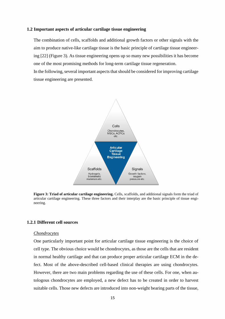
The combination of cells, scaffolds and additional growth factors or other signals with the
aim to produce native-like cartilage tissue is the basic principle of cartilage tissue engineer-
ing [22] (Figure 3). As tissue engineering opens up so many new possibilities it has become
one of the most promising methods for long-term cartilage tissue regeneration.
In the following, several important aspects that should be considered for improving cartilage
tissue engineering are presented.
Figure 3: Triad of articular cartilage engineering. Cells, scaffolds, and additional signals form the triad of
articular cartilage engineering. These three factors and their interplay are the basic principle of tissue engi-
neering.
1.2.1 Different cell sources
Chondrocytes
One particularly important point for articular cartilage tissue engineering is the choice of
cell type. The obvious choice would be chondrocytes, as those are the cells that are resident
in normal healthy cartilage and that can produce proper articular cartilage ECM in the de-
fect. Most of the above-described cell-based clinical therapies are using chondrocytes.
However, there are two main problems regarding the use of these cells. For one, when au-
tologous chondrocytes are employed, a new defect has to be created in order to harvest
suitable cells. Those new defects are introduced into non-weight bearing parts of the tissue,

16
but the risk of donor site morbidity is still an issue [31]. Only small parts of cartilage can
be harvested to avoid too much damage, and therefore, the harvested chondrocytes have to
be expanded in vitro in 2D culture. This results in the second problem, as chondrocytes tend
to dedifferentiate when they are expanded in 2D [32,33]. When chondrocytes dedifferenti-
ate, they lose their ability to produce and maintain healthy articular cartilage ECM. This
results often in the production of biomechanically inferior fibrocartilage. It has been shown
that chondrocytes can maintain their chondrogenic phenotype better when they are cultured
in 3D [32,34], but then harvested cells cannot be expanded as much as in 2D culture.
MSCs
Mesenchymal stromal cells (MSCs) on the other hand can be differentiated into chondro-
cyte-like cells in vitro [35-37]. They can be expanded in 2D without losing their chondro-
genic differentiation potential like chondrocytes. Therefore, MSCs are a promising alterna-
tive cell source for articular cartilage repair strategies. MSCs can be isolated from different
tissues, for example bone marrow, adipose tissue, synovium or umbilical cord blood
[38,39]. Human MSCs have to fulfil at least three minimal criteria, according to The Inter-
national Society for Cellular Therapy: They have to adhere to plastic surfaces, carry the
surface antigens CD73, CD90 and CD105, while they do not carry the surface antigens
CD14, CD19, CD34, CD45 and HLA-DR, and they have to be able to differentiate in the
chondrogenic, adipogenic and osteogenic direction [40].
In recent years, MSCs have received increasing attention in cartilage regenerative medicine.
Autologous and allogeneic MSCs have been used for injection into the joint with or without
additional substances [41-56] especially for the treatment of osteoarthritis. The results of
these studies were promising regarding pain relief in patients but were controversial regard-
ing regeneration of articular cartilage [54,55]. More standardized studies and comparisons
would be needed to fully reveal the potential of injected MSCs [54,57]. Recently, several
studies could show that extracellular vesicles, so-called exosomes, that are produced by
MSCs are responsible for the positive effects on injured and inflamed cartilage tissue, and
that the main course of action of injected MSCs in the joint is a paracrine one [58-60]. This
is a promising application for MSCs and their exosomes in cartilage regeneration, addition-
ally to their use in cartilage tissue engineering. MSCs also have been implanted into carti-
lage defects with or without a scaffold in several studies [53-56,61-69]. Nejadnik et al.
could, for example, show that the implantation of MSCs led to similar results as common
ACI with chondrocytes [61]. However, the results differed between the studies. Most
showed improvement compared to controls, but the quality of the formed tissue varied
17
between fibrocartilage and hyaline-like cartilage [56]. For this treatment, as for the injection
of MSCs into the joint, more comparable studies, also long-term follow-ups, are needed to
evaluate the full potential of these therapeutical approaches.
MSC-based articular cartilage tissue engineering is additionally limited. Several studies
have shown that especially MSCs from the bone marrow (BMSCs) that are often used for
cartilage regeneration tend to form hypertrophic cartilage when differentiated in common
in vitro chondrogenic culture systems [35,70,71]. That can lead to endochondral ossification
of the tissue. It has been proposed that these bone-marrow-derived MSCs follow a natural
line of differentiation and form only transient cartilage tissue that then progresses towards
the formation of bone [70,72], similar to the longitudinal growth of long bones or bone
fracture healing. However, in the regeneration of cartilage tissue, this process is highly un-
desirable as bone tissue of course cannot fulfil the functions of healthy articular cartilage.
Common markers for hypertrophy and endochondral ossification are type X collagen, alka-
line phosphatase (ALP), matrix metalloproteinase 13 (MMP13), runt-related transcription
factor II (RUNX2) and increased volume of the cells [71,73,74]. There are several ap-
proaches to control the differentiation of MSCs and to steer it towards a more stable chon-
drogenic phenotype, for example by co-culture with chondrocytes [75-78], low oxygen ten-
sion (hypoxia) [79-82], scaffolds [83-85], inhibition or activation of signalling pathways
[86-88] or changes in in vitro differentiation protocols [89,90].
Due to the difficulties with chondrocytes and MSCs for articular cartilage tissue engineer-
ing, there is an ongoing search for alternative promising cell sources. Induced pluripotent
stem cells (iPSCs) or embryonic stem cells (ESCs) have been used, but high costs, tumor-
igenicity and the ethical problems associated with ESCs have limited their use in cartilage
regeneration so far [19,72,91].
ACPCs
However, some years ago, chondroprogenitor cells were detected in articular cartilage
[92,93]. These cells, also called articular cartilage progenitor cells (ACPCs), are a subpop-
ulation of chondrocytes that reside mostly in the superficial layer of articular cartilage [93].
ACPCs can differentiate into chondrogenic, osteogenic and adipogenic direction [92,94].
Alsalameh et al. isolated them from cartilage tissue by selecting cells that were positive for
CD105 and CD166. The co-expression of these two surface markers was proposed to define
a bone-marrow-derived mesenchymal stem cell population [92,95]. Dowthwaite et al. iso-
lated ACPCs from the superficial layer of articular cartilage using differential adhesion to
serum fibronectin and a high colony forming efficiency after an initially low cell seeding
18
density [93]. Other isolation methods of cartilage progenitor cells have also been used after
their discovery by different groups [96-99]. Unfortunately, it has not been possible to define
one specific marker for ACPC identification yet, and differently isolated progenitor cells
are probably not the same cell populations [100]. The present work focuses mostly on AC-
PCs that were identified, isolated and characterized in the way the Archer group established
in their previous work [93,94,101].
Interestingly, when in vitro monolayer expansion of ACPCs and normal chondrocytes was
compared, it was found that ACPCs could still form cartilage in high-density pellet culture
after 30 population doublings, while chondrocytes were defined as dedifferentiated after
only 21 population doublings. Additionally, ACPCs showed longer telomere-length at 22
population doublings than chondrocytes at 21 population doublings. Telomerase activity in
ACPCs was 2,6-fold higher than in freshly isolated chondrocytes, while dedifferentiated
chondrocytes showed no detectable telomerase activity at all [101]. The maintenance of
telomere length indicated the stem cell or progenitor character of ACPCs [94]. Clonally
derived ACPCs also maintained SOX9 expression after monolayer expansion of up to 45
population doublings, while chondrocytes usually lose SOX9 expression due to dedifferen-
tiation after only a few population doublings [94,101]. Several studies have also shown that
ACPCs seem to form cartilage tissue without tendencies towards hypertrophy or endochon-
dral ossification [94,102,103]. These findings make ACPCs a promising cell type for carti-
lage tissue engineering, as they can be expanded in 2D culture to achieve enough cells, do
not lose their chondrogenic potential during monolayer expansion, and form stable cartilage
tissue without the risk of endochondral bone formation. There have been several animal
studies and even one pilot clinical study in humans to test performance of ACPCs in carti-
lage tissue repair [94,98,104-106]. The number of studies is of course still limited, and no
final conclusion can be drawn from them. However, the first results were promising as it
could be shown that ACPCs were able to repair a cartilage lesion in a goat model with
similar results as the current gold-standard chondrocytes [94], that autologous ACPCs out-
performed fibrin-only constructs in an equine cartilage defect model [104] and that ACPC-
scaffold constructs formed cartilage-like tissue without chondrogenic induction when they
were implanted subcutaneously into nude mice [105]. In the latter study, the compare-group
with bone marrow derived stem cells became vascularized after six weeks [105]. In another
study, allogeneic ACPCs were injected intraarticularly together with hyaluronic acid, using
a rabbit knee model. This treatment had no adverse effects but did not yield better results
than a treatment with hyaluronic acid only [106]. In the pilot clinical study in humans,
19
ACPC implantation (MACT procedure) was performed in 15 patients. The results were
promising, as no graft failures occurred and after one year, all patients reported good quality
of life and no moderate or severe limitations. The study results were judged by the authors
to be similar or better than previously reported results from chondrocyte implantations [98].
The number of studies comparing chondrogenesis of ACPCs and MSCs directly in hydro-
gels for cartilage tissue engineering is also limited [103,107]. Levato et al. compared ACPC-
hydrogel constructs for the first time with MSC- and chondrocyte-hydrogel constructs. The
used hydrogel was gelatin methacryloyl (GelMA), derived from porcine gelatin. Cells were
chondrogenically differentiated as monocultures or zonally layered co-cultures in the hy-
drogel. ACPCs outperformed chondrocytes but not MSCs regarding cartilage ECM produc-
tion but showed significantly lower levels of type X collagen than the other cell types. Co-
culture of MSCs and ACPCs yielded the highest glycosaminoglycan amount in comparison
to the other co-cultures [103]. Mouser et al., from the same research group, tested chondro-
genesis of ACPCs, MSCs and chondrocytes against each other in two GelMA-based hydro-
gels: GelMA/gellan and GelMA/gellan/HAMA [107]. The results were similar to the pre-
vious study [103] with ACPCs outperforming chondrocytes but not MSCs. A zonally lay-
ered construct with ACPCs in the upper and MSCs in the lower zone was 3D bio-printed
and successfully chondrogenically differentiated, but the printing process decreased the
quality of the formed ECM [107]. However, these studies show only the beginning use of
ACPC in cartilage repair. There is still a lot that we do not know about this promising cell
type, and further research studies are needed to fully characterize the potential of ACPCs in
articular cartilage tissue engineering.
1.2.2 Oxygen partial pressure
As was described before, articular cartilage does not have a blood supply. Oxygen is usually
transported to the organs in the body via vasculature, and the oxygen partial pressure is
specifically adapted to the needs of the specific organs [108,109]. The levels of oxygen in
the body are lower than those in the air (21 %) [109]. Cartilage tissue gets oxygen the same
way it gets nutrients, via the synovial fluid that is pressed out and flows into the tissue when
the joint is loaded and unloaded. Synovial fluid itself is relatively hypoxic, and therefore,
physioxia (the physiological oxygen partial pressure) in cartilage tissue lies between 1 and
5 % [110-112]. This chronically low oxygen pressure strongly influences development and
integrity of native articular cartilage [113,114]. Cells can sense the level of oxygen in their
20
surroundings and can respond to it with different actions. This is mediated mainly by the
hypoxia inducible factors (HIFs) HIF-1α and HIF-2α. When oxygen pressure is high, HIF-
α subunits are hydroxylated by specific prolyl hydroxylases that need oxygen for this pro-
cedure. As result of this hydroxylation, HIF-α subunits are degraded by the proteasome.
However, when oxygen levels are low, hydroxylation is inhibited by the lack of oxygen and
HIF-α is not degraded. Instead, it forms heterodimers with the constitutively expressed HIF-
β. These heterodimers translocate to the nucleus and activate the transcription of specific
hypoxia responsive elements (HREs) that can, for example, regulate survival and metabo-
lism in cartilage [115,116].
Despite the important roles of hypoxia in native cartilage, many in vitro tissue engineering
studies are performed under 21 % oxygen that represent hyperoxic conditions for normal
articular cartilage and chondrocytes and can influence the results in unwanted ways [109].
Therefore, oxygen partial pressure that is relevant for in vivo situations is an aspect in car-
tilage tissue engineering that should be considered. It has been shown that using hypoxic
(for cartilage physioxic) oxygen partial pressure (1-5 %) instead of normoxic air conditions
(21 %) can have beneficial effects on the performance of the cells restoring cartilage tissue.
There are several studies that report an increase in matrix deposition and a downregulation
of catabolic factors in isolated and passaged (dedifferentiated) chondrocytes by hypoxia in
contrast to normoxic air conditions [115,117-121].
Additionally, low oxygen tension can have beneficial effects on the differentiation of MSCs
towards cartilage tissue. An increase in chondrogenic gene expression and matrix deposi-
tion in hypoxia-differentiated MSCs (in pellets or scaffolds) has been reported several times
[81,122-128]. However, there are also studies that found no effect of hypoxia on chondro-
genic gene expression [129] or matrix production [130-132]. It was suggested by Anderson
et al. [133] that donor variability might play a role in these contradictory results. They found
that MSCs with low chondrogenic capacity in normoxia showed a stronger reaction to hy-
poxia with upregulation of chondrogenic markers than MSCs with initially high chondro-
genic capacity in normoxia [133].
When MSCs are chondrogenically differentiated, hypoxia can steer their phenotype to a
more permanent articular cartilage by inhibiting hypertrophy and thereby avoiding the more
growth plate-like cartilage phenotype that can result in endochondral ossification in the tis-
sue. Several studies have shown downregulation of hypertrophy markers in MSC derived
cartilage tissue formed under hypoxic compared to normoxic conditions [81,82,130,134].
There are hints that this could be a similar process than in embryonic limb development as
21
there is an oxygen gradient with lower oxygen levels where permanent articular cartilage is
developing and higher oxygen levels (because of beginning vascularisation) where transient
cartilage is undergoing hypertrophy and endochondral ossification [81].
Considering the possible modulating influences of hypoxic cell culture, it becomes clear
that the oxygen partial pressure is an important factor that should be considered in cartilage
tissue engineering.
1.2.3 Zonal structure
Native articular cartilage contains three distinct zones, the superficial zone, the middle zone
and the deep zone, as was described before. Cell morphology and gene expression as well
as matrix composition are distinct between the zones. The biomechanical function of artic-
ular cartilage is strongly correlated with its zonal architecture [5,135,136]. The lack of this
specific zonal structure in repair tissue is suggested to be one of the main reasons for insuf-
ficient cartilage regeneration of most treatments currently available [137,138]. Therefore,
research has been trying to recapitulate a zonal organization that is similar to native articular
cartilage structure. The aim was to mimic some of the properties that make native zonal
cartilage more stable than non-zonal repair tissue [136,137]. Several approaches have been
employed to isolate chondrocytes from distinct zones and to use them to build up tissue
engineered zonal constructs [136,139-142]. Compared to constructs with non-zonal, full-
thickness isolated chondrocytes, zonal constructs showed similar or higher matrix produc-
tion and biomechanical properties [141,142]. However, the use of zonally isolated chondro-
cytes for reconstruction of zonal constructs is limited because it is difficult to isolate clini-
cally relevant cell numbers and to obtain pure zonal chondrocyte populations with the cur-
rent methods [136]. Different other cell types (for example MSCs or ACPCs) were sug-
gested and used for the generation of different zones in combination with or as alternative
for chondrocytes [103,107,136-138]. Similar to general tissue engineering of articular car-
tilage, approaches with and without scaffolds and materials were investigated, each with
their own advantages and disadvantages [137]. Other approaches are working with different
materials and physical or chemical gradients that can influence the encapsulated cells or
have zonal properties in themselves [137,143-146]. The layered osteochondral structure
may also be reproduced by zonal tissue engineering [136,147,148].
Biofabrication and 3D bioprinting have established themselves in the generation of zonal
tissue engineered constructs as they provide the possibility to deposit different materials
22
and/or cells as specific structures and layer by layer. Even complex, personalized implant
structures may be produced this way [103,136,137,149,150]. However, the effects of the
printing process on the used cells should be evaluated as cells are exposed to shear forces
when they are printed. At high shear forces, cells can be harmed and killed. However, when
cells survive the printing process, they can still be changed afterwards. For example, it has
been shown before that the 3D bioprinting process can alter the gene expression profile of
MSCs [107]. Therefore, it has to be made sure that the printing process does not affect the
used cells in unwanted ways.
In principle, zonal tissue engineering for articular cartilage is a promising approach for bet-
ter functional repair tissue but much research is still needed to find the optimal conditions
to produce a tissue that mimics native zonal structure and function of cartilage.
1.2.4 Scaffold material
One main aspect of tissue engineering is the material that is used to encapsulate or carry the
cells. General requirements for materials that are used in tissue engineering are biocompat-
ibility, biodegradability, suitable mechanical properties and permeability so that oxygen,
nutrients and waste can diffuse in and out of the scaffold [151,152]. Not all materials that
are used in in vitro experiments for tissue engineering fulfil all these requirements but for
an application in vivo they are very important. The used material determines the microen-
vironment and the mechanical, physicochemical and biological conditions that the cells ex-
perience in the tissue engineered construct. As cells are strongly influenced by their sur-
roundings, it is important to choose a suitable material for the used cells and the intended
purpose. In the case of articular cartilage tissue engineering, this may be achieved by using
materials that mimic natural articular cartilage [153]. Scaffolds for cartilage engineering
have been made of natural, synthetic or hybrid (natural/synthetic) materials, and they have
been used as microporous scaffolds or hydrated polymeric networks (hydrogels) [151,153].
Hydrogels are often used for engineering cartilage tissue because they can provide the cells
with an environment of high-water content that is similar to native articular cartilage [154].
Nature-derived materials are for example agarose, alginate, hyaluronic acid, chitosan or
collagen. Agarose and alginate are natural polysaccharides. Agarose can be obtained from
red seaweed and alginate from brown algae [155]. Agarose is used as thermo-responsive
hydrogel that gels at low temperatures and is fluid at higher temperatures. Alginate hydro-
gels can be formed by the addition of divalent cations, such as Ca2+ [156]. Agarose and
23
alginate do not have any biological cues like integrins or other binding sites for cells and
are therefore called inert hydrogels [157]. However, they still provide the cells with an en-
vironment that is similar to articular cartilage matrix and support the spherical shape of
mature chondrocytes. This is thought to be beneficial for the development of articular car-
tilage [32,157,158]. Other natural materials like hyaluronic acid, chondroitin sulfate, chi-
tosan or collagen are based on molecules that naturally occur in cartilage tissue as part of
the ECM. These materials are used to improve chondrogenicity of encapsulated cells by
mimicking their natural environment or by providing biological cues for chondrogenic dif-
ferentiation [151,153,157]. Chitosan, a derivative of chitin, is a polysaccharide that is not
part of the human native cartilage but is chemically similar to GAG and can enhance the
deposition of cartilage ECM [151,159]. Collagen is a main component of cartilage ECM,
and different scaffolds and hydrogels are derived from it, for example gelatin or GelMA but
also constructs consisting of type I, type II and/or type III collagen [153,160]. Gelatin is
obtained through hydrolysis of collagen, and GelMA is methacrylated gelatin [155,161].
Collagens can contribute to cell adhesion, proliferation and differentiation and have integrin
binding sites all of which can be beneficial for cartilage ECM formation [152,155,162].
Chondroitin sulfate is a GAG that naturally occurs in human cartilage tissue and can be used
in scaffolds for articular cartilage engineering [153,163]. Hyaluronic acid (HA) is an abun-
dant polysaccharide in articular cartilage tissue and has important functions as signalling
molecule for the cells. It has been shown that addition of HA to scaffolds can enhance the
production of articular cartilage ECM [164,165]. Modification and/or mixture and blending
of different hydrogel materials is often performed to achieve better biological, biomechan-
ical or crosslinking properties.
In addition to natural materials, synthetic polymers like polyethylene glycol (PEG), pol-
ylactic acid (PLA), polycaprolactone (PCL) or polyglycidol (PG) have been used in carti-
lage tissue engineering [150,152,153,161,166]. They have more clearly defined character-
istics, are easy to produce and their properties can be fine-tuned more easily than in natural
materials. Chemical modifications can easily be introduced for different crosslinking modes
or to attach biological factors [152,166-168]. PCL is often used together with natural mate-
rials to enhance and tailor mechanical stiffness and to increase shape stability in 3D bio-
printing approaches [149,164,169]. PEG, PG and their derivatives are often used as hydro-
gels, and crosslinking with natural polymers like hyaluronic acid increases cartilage ECM
production in hybrid hydrogels in comparison to pure synthetic hydrogels [164,165].

24
Hybrid hydrogels that combine the best characteristics of natural materials and of synthetic
materials are currently thought to be the most promising approach for cartilage tissue engi-
neering and 3D bioprinting. However, the matching of the used cells to the appropriate
material is a step of paramount importance on the way to a successfully engineered articular
cartilage construct.
25
1.3 Hyaluronic acid
One very interesting biomaterial that is used in cartilage tissue engineering is hyaluronic
acid (HA). As mentioned above, it has been shown that HA can have beneficial effects on
the cells when it is used as part of a hydrogel or scaffold in cartilage engineering [164,165].
As it is part of the natural ECM of cartilage, it seems to mimic this environment and its
signals for the cells in the tissue engineered construct. In the following, structure, occur-
rence and function of HA in the human body and its application in articular cartilage tissue
engineering are highlighted in more detail.
1.3.1 Characteristics and functions of HA in the human body
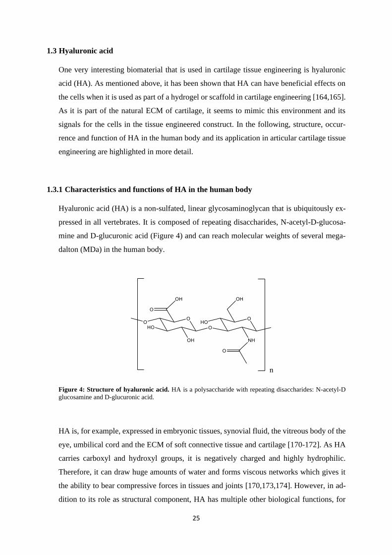
Hyaluronic acid (HA) is a non-sulfated, linear glycosaminoglycan that is ubiquitously ex-
pressed in all vertebrates. It is composed of repeating disaccharides, N-acetyl-D-glucosa-
mine and D-glucuronic acid (Figure 4) and can reach molecular weights of several mega-
dalton (MDa) in the human body.
Figure 4: Structure of hyaluronic acid. HA is a polysaccharide with repeating disaccharides: N-acetyl-D
glucosamine and D-glucuronic acid.
HA is, for example, expressed in embryonic tissues, synovial fluid, the vitreous body of the
eye, umbilical cord and the ECM of soft connective tissue and cartilage [170-172]. As HA
carries carboxyl and hydroxyl groups, it is negatively charged and highly hydrophilic.
Therefore, it can draw huge amounts of water and forms viscous networks which gives it
the ability to bear compressive forces in tissues and joints [170,173,174]. However, in ad-
dition to its role as structural component, HA has multiple other biological functions, for
26
example in cell and organ development, cell migration and proliferation, cancer, inflamma-
tion and tissue injury [174]. It is fascinating that this molecule with a relatively simple
structure is present in so many tissues and has so many different functions. However, the
biology of HA is much more complex than it was thought at first. Garantziotis and Savani
wrote in their review from 2019: “The HA matrix can be best viewed as a canvas that is
continually woven, unraveled, and decorated by dynamic patterns of hyaladherins which
help shape HA-specific effects.” [174]. This sentence describes the important balance be-
tween HA anabolism and catabolism and its many binding partners, the hyaladherins. Other
factors that additionally expand HA function are its molecular weight, chemical modifica-
tions, macromolecular structure, microenvironment and its downstream signalling [174].
HA synthesis and degradation is performed constantly in the body and disruption of this
delicate balance is associated with pathologies like inflammation or cancer [175,176]. HA
is synthesized by three different hyaluronic acid synthases (HAS) that are located in the cell
membrane. The three HAS are thought to produce HA of different length [170,177,178].
HA is degraded by hyaluronidases (HYAL) and reactive oxygen species (ROS) or internal-
ised and degraded by cells [171,174,175,179]. When HA is newly synthesized or degraded,
its molecular weight distribution changes in this specific area. The molecular weight (MW)
of HA is an important factor for the different functions of HA. However, high and low MW
HA are often defined differently in different studies.
For example, high MW HA has been found to act anti-inflammatory, while low MW frag-
ments and oligosaccharides increase the expression of proinflammatory chemokines
[174,180,181]. HA also can have opposing functions in cancer as low MW HA supports
cell migration, invasion and angiogenesis while very high MW HA has been linked to can-
cer resistance [170,182,183]. Accumulation of HA in various tumours has been associated
with poor prognosis [184,185], and HA metabolism has been suggested as a target for can-
cer therapies [176,186].
The versatile functions of HA are also controlled by HA binding partners, the hyaladherins.
Aggrecan, versican, link protein and TSG-6 (tumour necrosis factor-stimulated gene 6) are
examples for matrix hyaladherins, and CD44 (cluster of differentiation 44), CD168 (also
called RHAMM: Receptor for hyaluronan mediated motility), TLR2,4 (toll-like-receptor
2,4), HARE (hyaluronan receptor for endocytosis), and LYVE-1 (lymphatic vessel endo-
thelial hyaluronan receptor 1) are cell surface receptors for HA [187]. Aggrecan and link
protein, for example, bind to HA in cartilage ECM to form huge networks of proteoglycans
and glycosaminoglycans and thereby contribute greatly to the structure and biomechanical
27
functions of cartilage ECM. CD44 and CD168 are the main cell surface receptors of HA.
Through them, HA can act as signal transductor and activate diverse signalling pathways in
the cells. These signals are again dependent on the cell type, the identity, timing, amount
and location of receptors, the amount, structure and MW of the binding HA and other in-
fluencing factors [174]. CD44 has almost as variable functions as HA itself and is present
in almost all human cells [178,188]. In addition to HA, it can also bind to other ECM com-
ponents, like fibronectin, osteopontin and collagen [189] and has several different isoforms
as it is subject to alternative splicing [190]. The receptor is involved in cell adhesion, mo-
tility, growth, development and survival, tumorigenesis, inflammation and wound healing.
It tethers HA to the cell surface and is responsible for its internalisation [178,183,191]. Sig-
nalling pathways that are known to be activated by CD44 include PI3K/PDK1/Akt,
Ras/RAF1/MEK/ERK1/2, PLCε-Ca2+ signalling and SMAD signalling for BMP7 activa-
tion [178,192]. Ezrin, merlin and erbB1,2 have been described to form a complex with
Hsp90 and cytosolic CD44 domain [178,193].
CD168 is also subject to alternative splicing and has several isoforms that determine the
location of the HA-receptor. CD168 can be found on the cell membrane, in the cytoplasm,
in mitochondria and in the nucleus [178,194]. CD168 is involved in cell motility, wound
healing, cancer, inflammation and mitotic spindle formation [195]. It is not a transmem-
brane protein, and it can act as co-receptor for HA on the cell surface together with integral
membrane proteins [178]. It can, for example, associate with CD44 and protein tyrosine
kinase receptors like PDGFR (platelet-derived growth factor receptor) to regulate HA- and
growth factor-induced MAPK/ERK1,2 signalling that can lead to motility and invasion in
cancer cells [196,197]. Other signalling factors CD168 can influence are for example Ras
(Ras: short for rat sarcoma), FAK (focal adhesion kinase), PKC (protein kinase C), c-Src
(Src: short for sarcoma), NF-κB (nuclear factor kappa-light-chain-enhancer of activated B
cells) and PI3K (phosphatidylinositol kinase) [178].
Even though the known HA signalling and functions are already very versatile and complex,
research will probably discover many more in the future.
1.3.2 Applications of HA in articular cartilage tissue engineering
Hydrogels that mimic biological cues of the natural microenvironment of the used cells are
thought to direct the differentiation and development of the cells in specific directions. Hy-
aluronic acid, the multifunctional component of many different tissues, has been used for
28
different tissue engineering strategies, for example for the engineering of fat tissue, cancer
models, heart valves and cardiac repair, neural tissue and especially for cartilage tissue
[173,198,199]. As described before, HA is a main component of articular cartilage tissue,
and contributes greatly to the biomechanical functions of cartilage by forming highly hy-
drated networks with aggrecan. Additionally, it has many different functions as signalling
molecule [5,178]. HA is important in embryonic development of cartilage and bone. During
the early stage of limb bud development before condensation of cells, HA is thought to
separate cells from each other and to increase cell migration and division. However, in later
stages the HA amount decreases, probably because it is internalised through CD44 and de-
graded by the cells [200]. When cartilage matures postnatally, HA is again expressed in
higher amounts together with the other ECM molecules [4]. In mature cartilage, HA forms
huge networks and is attached to chondrocytes that regulate its degradation and synthesis
and receive extracellular signals via HA receptors [178]. HA has been implied in signalling
pathways that are important for chondrogenesis, for example TFGβ and BMP signalling
[201-204]. TGFβ and sonic hedgehog signalling can, for example, increase expression of
hyaluronan synthase 2 [205,206] which leads to increased expression of HA. Wnt signalling
that is involved in cartilage development and maintenance can also be influenced by HA
and its receptors [207,208]. These examples demonstrate the involvement and importance
of HA in native biological processes in cartilage even if the exact mechanisms of HA as
biomimetic material in tissue engineering have not been intensely studied so far. However,
there are several studies that have directly demonstrated the beneficial effects of HA-scaf-
folds on cells and developing cartilage ECM [164,165,209,210]. Additionally, blocking of
HA receptors with antibodies has been shown to decrease chondrogenesis in HA hydrogels
[211]. The disadvantage of HA is that is has poor mechanical properties and degrades rap-
idly in its natural form. This can be prevented through chemical modification and crosslink-
ing with other natural or synthetic polymers [212]. The possibility to easily modify HA, its
biocompatibility and biological activity are some of the reasons why it is so frequently used
in tissue engineering. There are a multitude of different combinations and modifications for
HA, and some of them were reviewed by Burdick et al. [198] and Highley et al. [213].
HA has also been used as bioink material for 3D bioprinting [164,169,214,215]. 3D bi-
oprinting of cells and hydrogel materials allows for the construction of complex tissue en-
gineering constructs [155]. A 3D bioprinter can deposit cell/hydrogel suspension in prede-
fined patterns and on top of each other, layer by layer. Therefore, personalized implants,
complex tissues with several different cells or materials or for example zonally layered
29
cartilage constructs can be produced by 3D bioprinting. However, not all materials have
suitable properties for the use as bioink material. As HA has versatile functions and can
easily be modified, it is a popular bioink material for 3D bioprinting approaches [150,216].
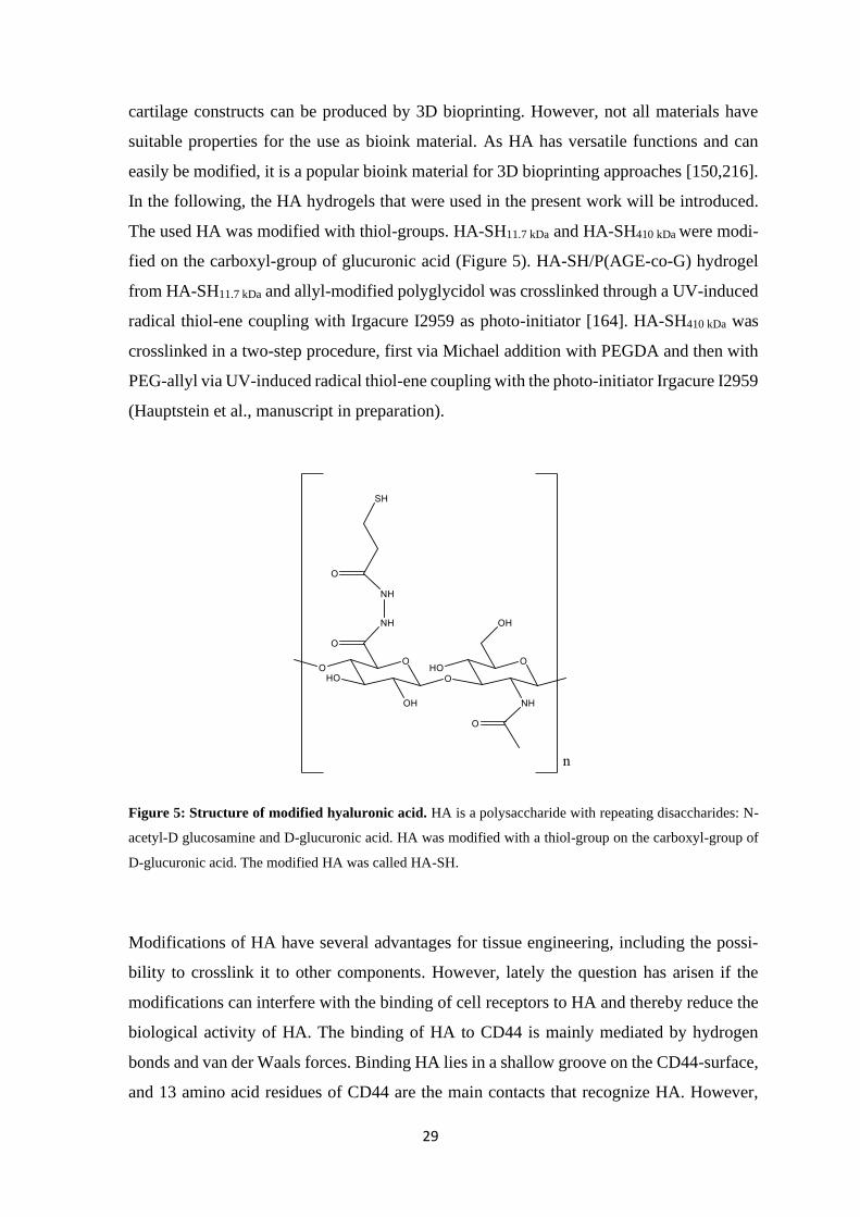
In the following, the HA hydrogels that were used in the present work will be introduced.
The used HA was modified with thiol-groups. HA-SH11.7 kDa and HA-SH410 kDa were modi-
fied on the carboxyl-group of glucuronic acid (Figure 5). HA-SH/P(AGE-co-G) hydrogel
from HA-SH11.7 kDa and allyl-modified polyglycidol was crosslinked through a UV-induced
radical thiol-ene coupling with Irgacure I2959 as photo-initiator [164]. HA-SH410 kDa was
crosslinked in a two-step procedure, first via Michael addition with PEGDA and then with
PEG-allyl via UV-induced radical thiol-ene coupling with the photo-initiator Irgacure I2959
(Hauptstein et al., manuscript in preparation).
Figure 5: Structure of modified hyaluronic acid. HA is a polysaccharide with repeating disaccharides: N-
acetyl-D glucosamine and D-glucuronic acid. HA was modified with a thiol-group on the carboxyl-group of
D-glucuronic acid. The modified HA was called HA-SH.
Modifications of HA have several advantages for tissue engineering, including the possi-
bility to crosslink it to other components. However, lately the question has arisen if the
modifications can interfere with the binding of cell receptors to HA and thereby reduce the
biological activity of HA. The binding of HA to CD44 is mainly mediated by hydrogen
bonds and van der Waals forces. Binding HA lies in a shallow groove on the CD44-surface,
and 13 amino acid residues of CD44 are the main contacts that recognize HA. However,
30
also the rest of the groove contributes to HA binding. Most important for HA recognition
are the carboxyl group of glucuronic acid and the N-acetyl-group of N-acetyl-glucosamine,
but also the hydroxyl groups on C6 of N-acetyl-glucosamine and on C3 and C2 of glucu-
ronic acid are involved [217]. These groups are also the sites where most modifications are
introduced to HA.
It has been shown that deacetylation and/or sulfation (on C6-OH) of N-acetyl-glucosamine
in HA lead to decreased binding of CD44 to HA [218]. Kwon et al. demonstrated that it is
important for CD44-HA interaction where a modification is introduced, how high the de-
gree of modification is and what kind of modification is attached to HA. They showed that
increased degree of modification (circa 40 %) generally decreases CD44-HA binding and
chondrogenesis in HA hydrogels. Adhesion of CD44 to a HA hydrogel was decreased es-
pecially with a bulky and hydrophobic modification (norbornene) on the carboxyl group
[219].
These studies show that it is important to consider the consequences on biological functions
when a hydrogel material is altered or modified. The possibilities for HA modifications and
applications are manifold but it is important to consider that changed materials can have
changed effects on the employed cells.
31
1.4 Goals of the thesis
Articular cartilage has limited capacity for self-healing and repair as it is a tissue without
blood supply [4,5]. Different clinical treatments are employed to repair damage to articular
cartilage, but those approaches mostly yield biomechanically inferior repair tissue. The
complete regeneration of native articular cartilage is still a challenge, but techniques like
tissue engineering and 3D-bioprinting are promising to improve the repair of cartilage de-
fects. However, more studies on different cell sources, biomaterials and cell signals for im-
proving cartilage tissue engineering are needed to enhance the quality of cartilage repair in
the future.
Therefore, one aim of this thesis was to evaluate a new promising cell source for articular
cartilage engineering, articular cartilage progenitor cells (ACPCs), in comparison to mes-
enchymal stromal cells (MSCs). Two different hydrogels were employed for this compari-
son, the widely used agarose and a hyaluronan (HA)-based hydrogel consisting of thiol-
modified HA (HA-SH) crosslinked with allyl-modified polyglycidol (HA-SH/P(AGE-co-
G)). Additionally, the effects of low oxygen tension and zonal layering were investigated.
HA can be used as scaffold for tissue engineering but has also been shown to transmit bio-
logical signals to different cells and to enhance chondrogenesis in MSCs
[164,165,173,212,220] Therefore, the second goal of this thesis was to evaluate the contri-
bution of the signalling molecule HA to the chondrogenic gene expression of MSCs. Gene
expression levels of HA cell surface receptors and important factors for differentiation were
investigated in response to HA in the culture medium and HA as part of a cell-encapsulating
hydrogel.
1.4.1 Comparison of chondrogenesis of ACPCs and MSCs in different hydrogels
ACPCs have been detected in the superficial layer of articular cartilage several years ago
[92,93]. It has been proposed that they could represent a promising cell source for articular
cartilage engineering. They do not lose their chondrogenic capacity after a few population
doublings like chondrocytes [94,101], and they do not tend to form hypertrophic cartilage
like MSCs [94,102,103]. However, the number of studies that compare ACPCs directly to
MSCs in hydrogels are limited [103,107], and studies in agarose, a widely used hydrogel
for articular chondrogenesis, are completely missing. Different oxygen tension and zonal
structure are two important factors when articular cartilage formation is considered that
have not extensively been investigated so far for ACPCs. A previous study [103] has shown
32
promising results for layered constructs with ACPCs in the upper layer and MSCs in the
lower layer of a hydrogel construct.
Therefore, ACPCs and MSCs were seeded into agarose hydrogel, and both monoculture
and layered co-culture constructs were produced. ACPCs were seeded into the upper layer
and MSCs into the lower layer. ACPCs and MSCs were chondrogenically differentiated for
28 days in chondrogenic medium containing TGFβ1 and under 21 % oxygen or 2 % oxygen.
Chondrogenic differentiation and formation of extracellular matrix (ECM) were analysed
with immunohistochemical stainings, biochemical assays and qRT-PCR (see section
4.1.1.).
As HA is an attractive material for cartilage engineering and it has been shown that cells
can perform differently in different hydrogels [157], the chondrogenesis of ACPCs and
MSCs was compared in a second, HA-based, hydrogel. This HA-SH/P(AGE-co-G) hydro-
gel was developed at the Department for Functional Materials in Medicine and Dentistry,
University of Würzburg, within the framework of the EU research project HydroZONES
(Bioactivated hierarchical hydrogels as zonal implants for articular cartilage regeneration)
and has previously been shown to support chondrogenesis of MSCs [164]. Zonal constructs
consisting of ACPCs in an upper layer and MSCs in a lower layer in this hydrogel were
used in the HydroZONES project for a long-term in vivo cartilage regeneration study in
horses. The evaluation of ECM production and chondrogenesis of ACPCs, MSCs and their
zonal constructs in the HA-SH/P(AGE-co-G) hydrogel that was performed in this thesis
served additionally as an in vitro evaluation for the in vivo animal study.
For this comparison, ACPCs and MSCs were encapsulated in HA-SH/P(AGE-co-G) hydro-
gel and monoculture and zonally layered co-culture constructs were produced. They were
chondrogenically differentiated for 28 days in chondrogenic medium with TGFβ1. Chon-
drogenic differentiation and formation of extracellular matrix (ECM) were analysed with
immunohistochemical stainings and biochemical assays (see section 4.1.2.). Additionally,
the differences in ECM production and chondrogenesis of the same cells in the two different
hydrogels agarose and HA-SH/P(AGE-co-G) were evaluated (see section 4.1. and espe-
cially 4.1.3.).

33
1.4.2 Evaluation of the contribution of hyaluronan to chondrogenic gene expression of
MSCs
It is known that HA has many different functions as signalling molecule [170,174,193,194].
HA is a natural component of articular cartilage tissue and has been shown to support chon-
drogenesis in tissue engineering approaches [164,165]. For the generation of hydrogels, HA
mostly has to be modified with functional groups that can be used to crosslink the polymer
to stable constructs [213]. However, such modifications can change the binding of HA to
the cells and its effect on them [218,219]. It is of great interest to evaluate how unmodified
HA and the modified and crosslinked HA in the hydrogels affect chondrogenic gene ex-
pression of MSCs. Additionally, it is interesting if and how the processing of HA hydrogels,
such as in 3D bioprinting, influences gene expression and HA signalling in HA-based hy-
drogels.
Therefore, the effect of unmodified HA on the gene expression of MSCs in 2D monolayer
or 3D pellet culture was investigated, and the gene expression of MSCs encapsulated in a
HA-based hydrogel was analysed in cast and 3D-bioprinted constructs for seven days. HA
cell surface receptors, transcription factors (chondrogenic, osteogenic and adipogenic), dif-
ferentiation markers and stemness markers were investigated by qRT-PCR (see section
4.2.).

34

35
Material

36
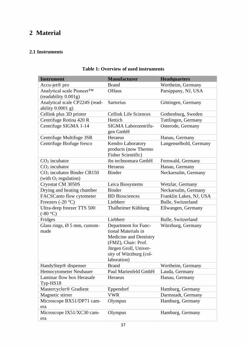
37
2 Material
2.1 Instruments
Table 1: Overview of used instruments
Instrument Manufacturer Headquarters
Accu-jet® pro Brand Wertheim, Germany
Analytical scale Pioneer™
(readability 0.001g)
OHaus Parsippany, NJ, USA
Analytical scale CP224S (read-
ability 0.0001 g)
Sartorius Göttingen, Germany
Cellink plus 3D printer Cellink Life Sciences Gothenburg, Sweden
Centrifuge Rotina 420 R Hettich Tuttlingen, Germany
Centrifuge SIGMA 1-14 SIGMA Laborzentrifu-
gen GmbH
Osterode, Germany
Centrifuge Multifuge 3SR Heraeus Hanau, Germany
Centrifuge Biofuge fresco Kendro Laboratory
products (now Thermo
Fisher Scientific)
Langenselbold, Germany
CO2 incubator ibs technomara GmbH Fernwald, Germany
CO2 incubator Heraeus Hanau, Germany
CO2 incubator Binder CB150
(with O2 regulation)
Binder Neckarsulm, Germany
Cryostat CM 3050S Leica Biosystems Wetzlar, Germany
Drying and heating chamber Binder Neckarsulm, Germany
FACSCanto flow cytometer BD Biosciences Franklin Lakes, NJ, USA
Freezers (-20 °C) Liebherr Bulle, Switzerland
Ultra-deep freezer TTS 500
(-80 °C)
Thalheimer Kühlung Ellwangen, Germany
Fridges Liebherr Bulle, Switzerland
Glass rings, Ø 5 mm, custom-
made
Department for Func-
tional Materials in
Medicine and Dentistry
(FMZ), Chair: Prof.
Jürgen Groll, Univer-
sity of Würzburg (col-
laboration)
Würzburg, Germany
HandyStep® dispenser Brand Wertheim, Germany
Hemocytometer Neubauer Paul Marienfeld GmbH Lauda, Germany
Laminar flow box Herasafe
Typ-HS18
Heraeus Hanau, Germany
Mastercycler® Gradient Eppendorf Hamburg, Germany
Magnetic stirrer VWR Darmstadt, Germany
Microscope BX51/DP71 cam-
era
Olympus Hamburg, Germany
Microscope IX51/XC30 cam-
era
Olympus Hamburg, Germany
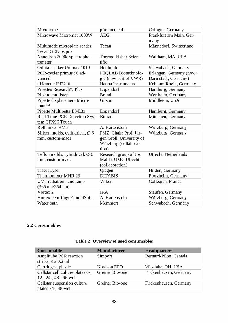
38
Microtome pfm medical Cologne, Germany
Microwave Micromat 1000W AEG Frankfurt am Main, Ger-
many
Multimode microplate reader
Tecan GENios pro
Tecan Männedorf, Switzerland
Nanodrop 2000c spectropho-
tometer
Thermo Fisher Scien-
tific
Waltham, MA, USA
Orbital shaker Unimax 1010 Heidolph Schwabach, Germany
PCR-cycler primus 96 ad-
vanced
PEQLAB Biotechnolo-
gie (now part of VWR)
Erlangen, Germany (now:
Darmstadt, Germany)
pH-meter HI2210 Hanna Instruments Kehl am Rhein, Germany
Pipettes Research® Plus Eppendorf Hamburg, Germany
Pipette multistep Brand Wertheim, Germany
Pipette displacement Micro-
man™
Gilson Middleton, USA
Pipette Multipette E3/E3x Eppendorf Hamburg, Germany
Real-Time PCR Detection Sys-
tem CFX96 Touch
Biorad München, Germany
Roll mixer RM5 A. Hartenstein Würzburg, Germany
Silicon molds, cylindrical, Ø 6
mm, custom-made
FMZ, Chair: Prof. Jür-
gen Groll, University of
Würzburg (collabora-
tion)
Würzburg, Germany
Teflon molds, cylindrical, Ø 6
mm, custom-made
Research group of Jos
Malda, UMC Utrecht
(collaboration)
Utrecht, Netherlands
TissueLyser Qiagen Hilden, Germany
Thermomixer MHR 23 DITABIS Pforzheim, Germany
UV irradiation hand lamp
(365 nm/254 nm)
Vilber Collégien, France
Vortex 2 IKA Staufen, Germany
Vortex-centrifuge CombiSpin A. Hartenstein Würzburg, Germany
Water bath Memmert Schwabach, Germany
2.2 Consumables
Table 2: Overview of used consumables
Consumable Manufacturer Headquarters
Amplitube PCR reaction
stripes 8 x 0.2 ml
Simport Bernard-Pilon, Canada
Cartridges, plastic Nordson EFD Westlake, OH, USA
Cellstar cell culture plates 6-,
12-, 24-, 48-, 96-well
Greiner Bio-one Frickenhausen, Germany
Cellstar suspension culture
plates 24-, 48-well
Greiner Bio-one Frickenhausen, Germany
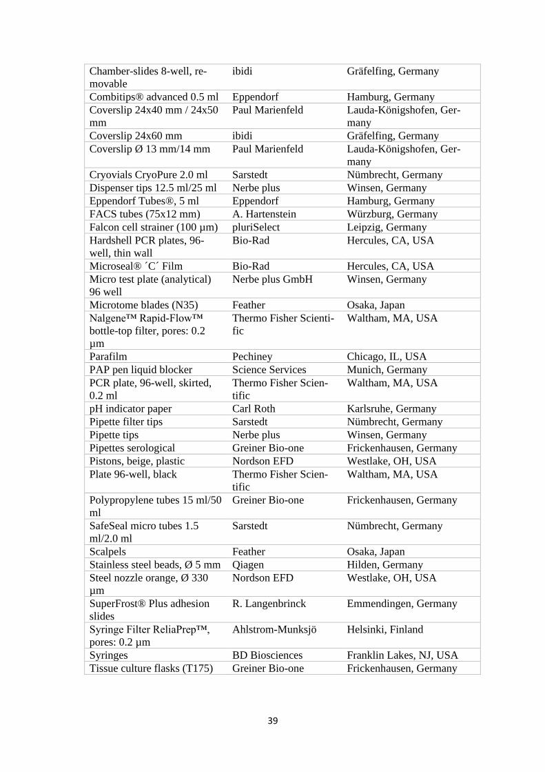
39
Chamber-slides 8-well, re-
movable
ibidi Gräfelfing, Germany
Combitips® advanced 0.5 ml Eppendorf Hamburg, Germany
Coverslip 24x40 mm / 24x50
mm
Paul Marienfeld Lauda-Königshofen, Ger-
many
Coverslip 24x60 mm ibidi Gräfelfing, Germany
Coverslip Ø 13 mm/14 mm Paul Marienfeld Lauda-Königshofen, Ger-
many
Cryovials CryoPure 2.0 ml Sarstedt Nümbrecht, Germany
Dispenser tips 12.5 ml/25 ml Nerbe plus Winsen, Germany
Eppendorf Tubes®, 5 ml Eppendorf Hamburg, Germany
FACS tubes (75x12 mm) A. Hartenstein Würzburg, Germany
Falcon cell strainer (100 µm) pluriSelect Leipzig, Germany
Hardshell PCR plates, 96-
well, thin wall
Bio-Rad Hercules, CA, USA
Microseal® ´C´ Film Bio-Rad Hercules, CA, USA
Micro test plate (analytical)
96 well
Nerbe plus GmbH Winsen, Germany
Microtome blades (N35) Feather Osaka, Japan
Nalgene™ Rapid-Flow™
bottle-top filter, pores: 0.2
µm
Thermo Fisher Scienti-
fic
Waltham, MA, USA
Parafilm Pechiney Chicago, IL, USA
PAP pen liquid blocker Science Services Munich, Germany
PCR plate, 96-well, skirted,
0.2 ml
Thermo Fisher Scien-
tific
Waltham, MA, USA
pH indicator paper Carl Roth Karlsruhe, Germany
Pipette filter tips Sarstedt Nümbrecht, Germany
Pipette tips Nerbe plus Winsen, Germany
Pipettes serological Greiner Bio-one Frickenhausen, Germany
Pistons, beige, plastic Nordson EFD Westlake, OH, USA
Plate 96-well, black Thermo Fisher Scien-
tific
Waltham, MA, USA
Polypropylene tubes 15 ml/50
ml
Greiner Bio-one Frickenhausen, Germany
SafeSeal micro tubes 1.5
ml/2.0 ml
Sarstedt Nümbrecht, Germany
Scalpels Feather Osaka, Japan
Stainless steel beads, Ø 5 mm Qiagen Hilden, Germany
Steel nozzle orange, Ø 330
µm
Nordson EFD Westlake, OH, USA
SuperFrost® Plus adhesion
slides
R. Langenbrinck Emmendingen, Germany
Syringe Filter ReliaPrep™,
pores: 0.2 µm
Ahlstrom-Munksjö Helsinki, Finland
Syringes BD Biosciences Franklin Lakes, NJ, USA
Tissue culture flasks (T175) Greiner Bio-one Frickenhausen, Germany
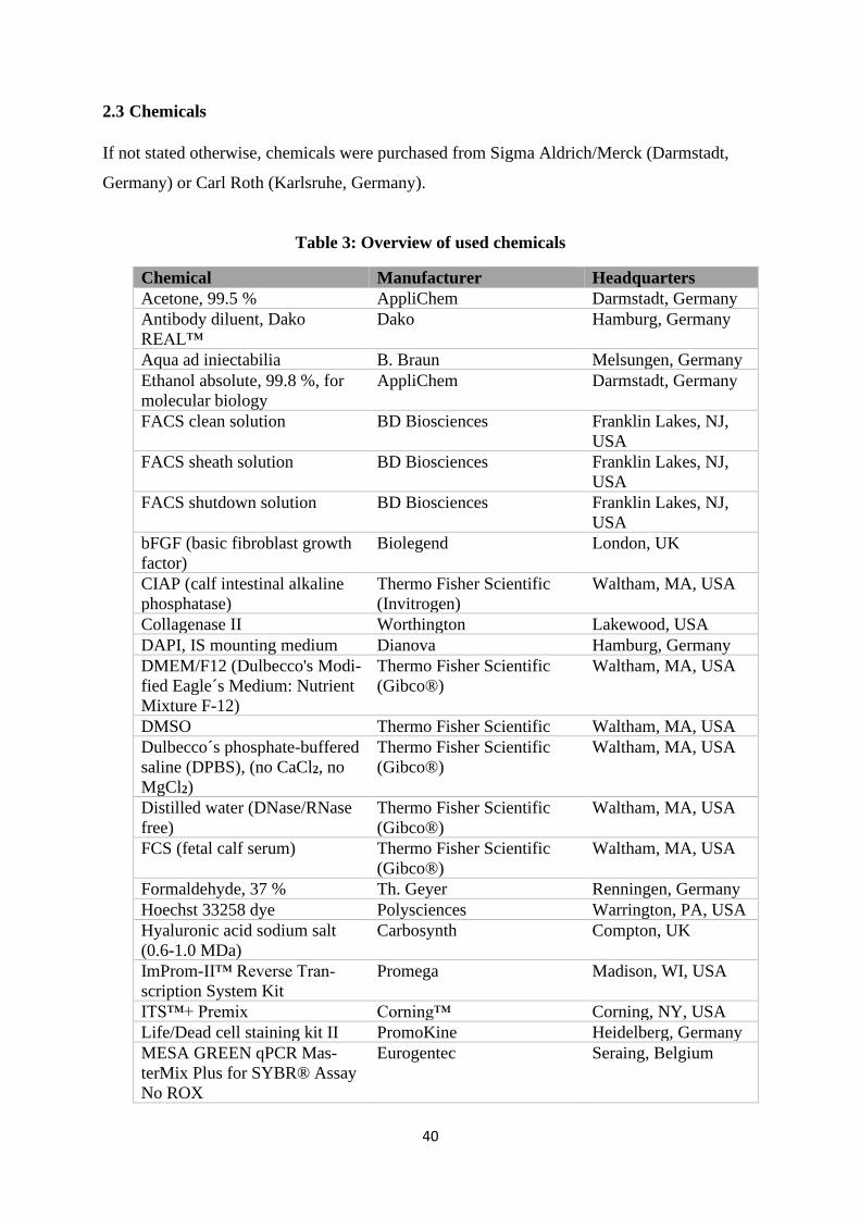
40
2.3 Chemicals
If not stated otherwise, chemicals were purchased from Sigma Aldrich/Merck (Darmstadt,
Germany) or Carl Roth (Karlsruhe, Germany).
Table 3: Overview of used chemicals
Chemical Manufacturer Headquarters
Acetone, 99.5 % AppliChem Darmstadt, Germany
Antibody diluent, Dako
REAL™
Dako Hamburg, Germany
Aqua ad iniectabilia B. Braun Melsungen, Germany
Ethanol absolute, 99.8 %, for
molecular biology
AppliChem Darmstadt, Germany
FACS clean solution BD Biosciences Franklin Lakes, NJ,
USA
FACS sheath solution BD Biosciences Franklin Lakes, NJ,
USA
FACS shutdown solution BD Biosciences Franklin Lakes, NJ,
USA
bFGF (basic fibroblast growth
factor)
Biolegend London, UK
CIAP (calf intestinal alkaline
phosphatase)
Thermo Fisher Scientific
(Invitrogen)
Waltham, MA, USA
Collagenase II Worthington Lakewood, USA
DAPI, IS mounting medium Dianova Hamburg, Germany
DMEM/F12 (Dulbecco's Modi-
fied Eagle´s Medium: Nutrient
Mixture F-12)
Thermo Fisher Scientific
(Gibco®)
Waltham, MA, USA
DMSO Thermo Fisher Scientific Waltham, MA, USA
Dulbecco´s phosphate-buffered
saline (DPBS), (no CaCl2, no
MgCl2)
Thermo Fisher Scientific
(Gibco®)
Waltham, MA, USA
Distilled water (DNase/RNase
free)
Thermo Fisher Scientific
(Gibco®)
Waltham, MA, USA
FCS (fetal calf serum) Thermo Fisher Scientific
(Gibco®)
Waltham, MA, USA
Formaldehyde, 37 % Th. Geyer Renningen, Germany
Hoechst 33258 dye Polysciences Warrington, PA, USA
Hyaluronic acid sodium salt
(0.6-1.0 MDa)
Carbosynth Compton, UK
ImProm-II™ Reverse Tran-
scription System Kit
Promega Madison, WI, USA
ITS™+ Premix Corning™ Corning, NY, USA
Life/Dead cell staining kit II PromoKine Heidelberg, Germany
MESA GREEN qPCR Mas-
terMix Plus for SYBR® Assay
No ROX
Eurogentec Seraing, Belgium
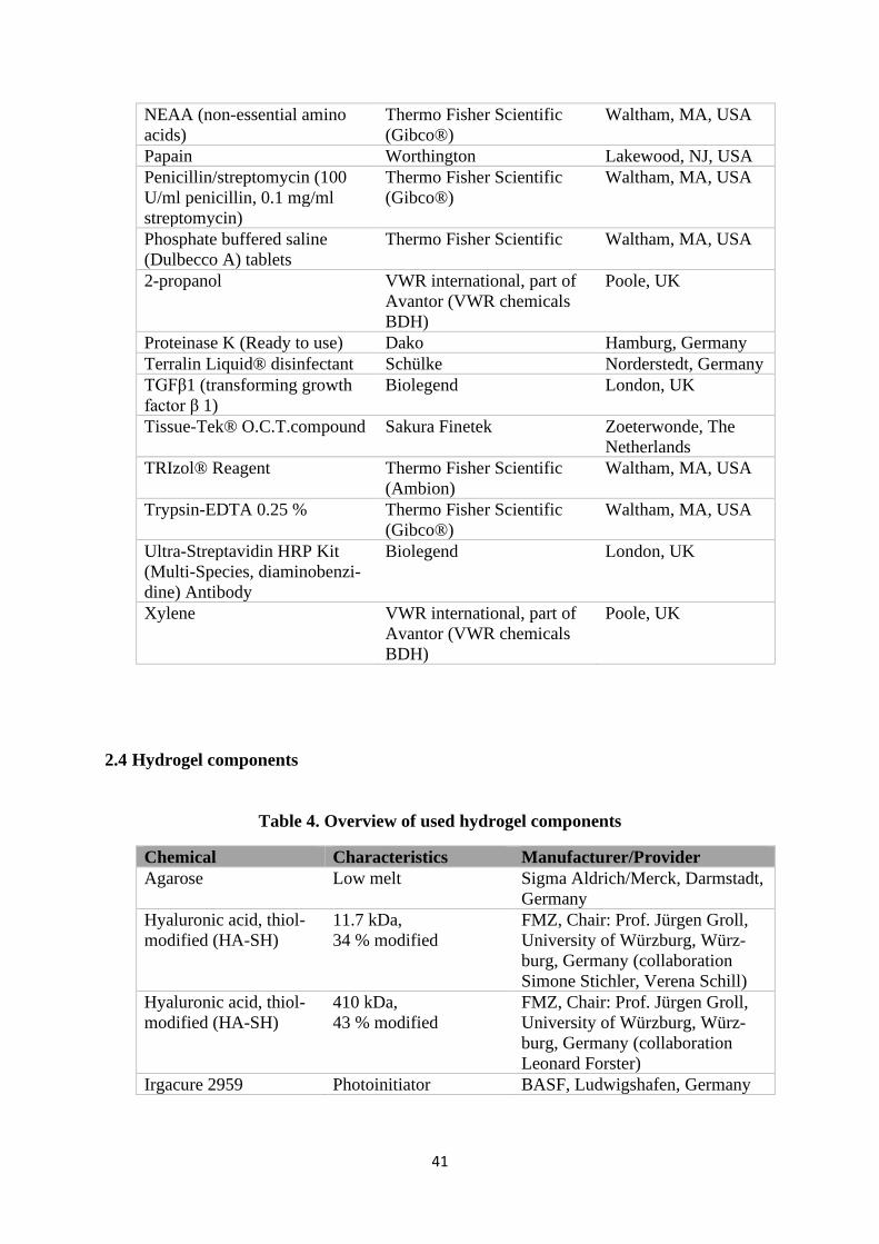
41
NEAA (non-essential amino
acids)
Thermo Fisher Scientific
(Gibco®)
Waltham, MA, USA
Papain Worthington Lakewood, NJ, USA
Penicillin/streptomycin (100
U/ml penicillin, 0.1 mg/ml
streptomycin)
Thermo Fisher Scientific
(Gibco®)
Waltham, MA, USA
Phosphate buffered saline
(Dulbecco A) tablets
Thermo Fisher Scientific Waltham, MA, USA
2-propanol VWR international, part of
Avantor (VWR chemicals
BDH)
Poole, UK
Proteinase K (Ready to use) Dako Hamburg, Germany
Terralin Liquid® disinfectant Schülke Norderstedt, Germany
TGFβ1 (transforming growth
factor β 1)
Biolegend London, UK
Tissue-Tek® O.C.T.compound Sakura Finetek Zoeterwonde, The
Netherlands
TRIzol® Reagent Thermo Fisher Scientific
(Ambion)
Waltham, MA, USA
Trypsin-EDTA 0.25 % Thermo Fisher Scientific
(Gibco®)
Waltham, MA, USA
Ultra-Streptavidin HRP Kit
(Multi-Species, diaminobenzi-
dine) Antibody
Biolegend London, UK
Xylene VWR international, part of
Avantor (VWR chemicals
BDH)
Poole, UK
2.4 Hydrogel components
Table 4. Overview of used hydrogel components
Chemical Characteristics Manufacturer/Provider
Agarose Low melt Sigma Aldrich/Merck, Darmstadt,
Germany
Hyaluronic acid, thiol-
modified (HA-SH)
11.7 kDa,
34 % modified
FMZ, Chair: Prof. Jürgen Groll,
University of Würzburg, Würz-
burg, Germany (collaboration
Simone Stichler, Verena Schill)
Hyaluronic acid, thiol-
modified (HA-SH)
410 kDa,
43 % modified
FMZ, Chair: Prof. Jürgen Groll,
University of Würzburg, Würz-
burg, Germany (collaboration
Leonard Forster)
Irgacure 2959 Photoinitiator BASF, Ludwigshafen, Germany
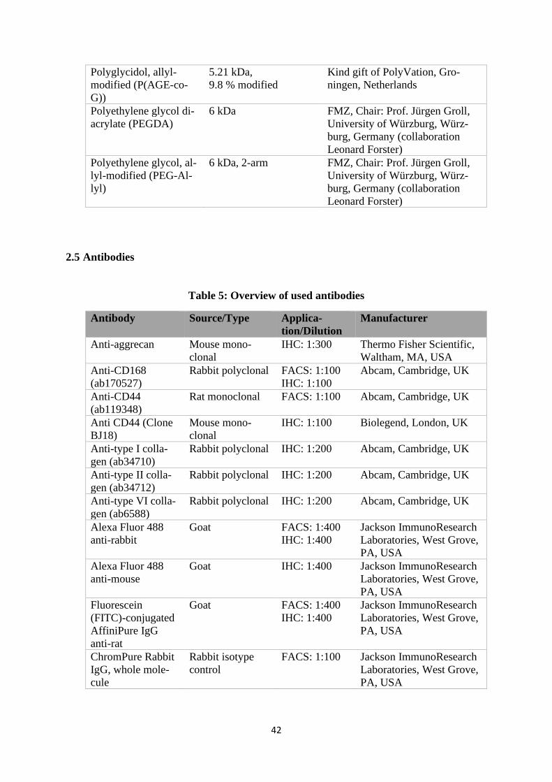
42
Polyglycidol, allyl-
modified (P(AGE-co-
G))
5.21 kDa,
9.8 % modified
Kind gift of PolyVation, Gro-
ningen, Netherlands
Polyethylene glycol di-
acrylate (PEGDA)
6 kDa FMZ, Chair: Prof. Jürgen Groll,
University of Würzburg, Würz-
burg, Germany (collaboration
Leonard Forster)
Polyethylene glycol, al-
lyl-modified (PEG-Al-
lyl)
6 kDa, 2-arm FMZ, Chair: Prof. Jürgen Groll,
University of Würzburg, Würz-
burg, Germany (collaboration
Leonard Forster)
2.5 Antibodies
Table 5: Overview of used antibodies
Antibody Source/Type Applica-
tion/Dilution
Manufacturer
Anti-aggrecan Mouse mono-
clonal
IHC: 1:300 Thermo Fisher Scientific,
Waltham, MA, USA
Anti-CD168
(ab170527)
Rabbit polyclonal FACS: 1:100
IHC: 1:100
Abcam, Cambridge, UK
Anti-CD44
(ab119348)
Rat monoclonal FACS: 1:100
Abcam, Cambridge, UK
Anti CD44 (Clone
BJ18)
Mouse mono-
clonal
IHC: 1:100 Biolegend, London, UK
Anti-type I colla-
gen (ab34710)
Rabbit polyclonal IHC: 1:200 Abcam, Cambridge, UK
Anti-type II colla-
gen (ab34712)
Rabbit polyclonal IHC: 1:200 Abcam, Cambridge, UK
Anti-type VI colla-
gen (ab6588)
Rabbit polyclonal IHC: 1:200 Abcam, Cambridge, UK
Alexa Fluor 488
anti-rabbit
Goat FACS: 1:400
IHC: 1:400
Jackson ImmunoResearch
Laboratories, West Grove,
PA, USA
Alexa Fluor 488
anti-mouse
Goat IHC: 1:400 Jackson ImmunoResearch
Laboratories, West Grove,
PA, USA
Fluorescein
(FITC)-conjugated
AffiniPure IgG
anti-rat
Goat FACS: 1:400
IHC: 1:400
Jackson ImmunoResearch
Laboratories, West Grove,
PA, USA
ChromPure Rabbit
IgG, whole mole-
cule
Rabbit isotype
control
FACS: 1:100 Jackson ImmunoResearch
Laboratories, West Grove,
PA, USA
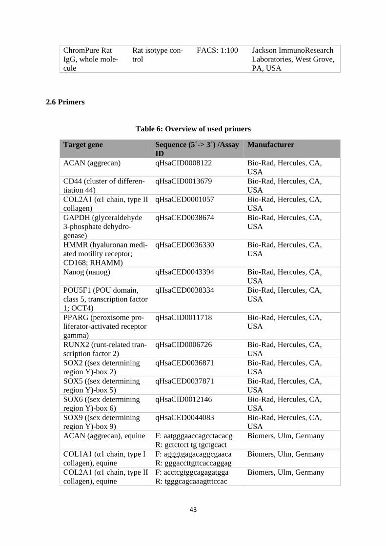
43
ChromPure Rat
IgG, whole mole-
cule
Rat isotype con-
trol
FACS: 1:100 Jackson ImmunoResearch
Laboratories, West Grove,
PA, USA
2.6 Primers
Table 6: Overview of used primers
Target gene Sequence (5´-> 3´) /Assay
ID
Manufacturer
ACAN (aggrecan) qHsaCID0008122 Bio-Rad, Hercules, CA,
USA
CD44 (cluster of differen-
tiation 44)
qHsaCID0013679 Bio-Rad, Hercules, CA,
USA
COL2A1 (α1 chain, type II
collagen)
qHsaCED0001057 Bio-Rad, Hercules, CA,
USA
GAPDH (glyceraldehyde
3-phosphate dehydro-
genase)
qHsaCED0038674 Bio-Rad, Hercules, CA,
USA
HMMR (hyaluronan medi-
ated motility receptor;
CD168; RHAMM)
qHsaCED0036330 Bio-Rad, Hercules, CA,
USA
Nanog (nanog) qHsaCED0043394 Bio-Rad, Hercules, CA,
USA
POU5F1 (POU domain,
class 5, transcription factor
1; OCT4)
qHsaCED0038334 Bio-Rad, Hercules, CA,
USA
PPARG (peroxisome pro-
liferator-activated receptor
gamma)
qHsaCID0011718 Bio-Rad, Hercules, CA,
USA
RUNX2 (runt-related tran-
scription factor 2)
qHsaCID0006726 Bio-Rad, Hercules, CA,
USA
SOX2 ((sex determining
region Y)-box 2)
qHsaCED0036871 Bio-Rad, Hercules, CA,
USA
SOX5 ((sex determining
region Y)-box 5)
qHsaCED0037871 Bio-Rad, Hercules, CA,
USA
SOX6 ((sex determining
region Y)-box 6)
qHsaCID0012146 Bio-Rad, Hercules, CA,
USA
SOX9 ((sex determining
region Y)-box 9)
qHsaCED0044083 Bio-Rad, Hercules, CA,
USA
ACAN (aggrecan), equine F: aatgggaaccagcctacacg
R: gctctcct tg tgctgcact
Biomers, Ulm, Germany
COL1A1 (α1 chain, type I
collagen), equine
F: agggtgagacaggcgaaca
R: gggaccttgttcaccaggag
Biomers, Ulm, Germany
COL2A1 (α1 chain, type II
collagen), equine
F: acctcgtggcagagatgga
R: tgggcagcaaagtttccac
Biomers, Ulm, Germany
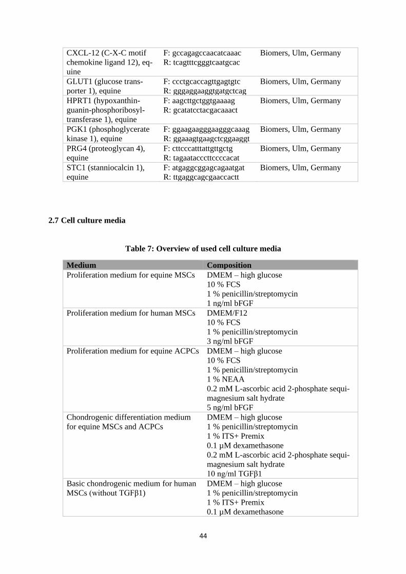
44
CXCL-12 (C-X-C motif
chemokine ligand 12), eq-
uine
F: gccagagccaacatcaaac
R: tcagtttcgggtcaatgcac
Biomers, Ulm, Germany
GLUT1 (glucose trans-
porter 1), equine
F: ccctgcaccagttgagtgtc
R: gggaggaaggtgatgctcag
Biomers, Ulm, Germany
HPRT1 (hypoxanthin-
guanin-phosphoribosyl-
transferase 1), equine
F: aagcttgctggtgaaaag
R: gcatatcctacgacaaact
Biomers, Ulm, Germany
PGK1 (phosphoglycerate
kinase 1), equine
F: ggaagaagggaagggcaaag
R: ggaaagtgaagctcggaaggt
Biomers, Ulm, Germany
PRG4 (proteoglycan 4),
equine
F: cttcccatttattgttgctg
R: tagaatacccttccccacat
Biomers, Ulm, Germany
STC1 (stanniocalcin 1),
equine
F: atgaggcggagcagaatgat
R: ttgaggcagcgaaccactt
Biomers, Ulm, Germany
2.7 Cell culture media
Table 7: Overview of used cell culture media
Medium Composition
Proliferation medium for equine MSCs DMEM – high glucose
10 % FCS
1 % penicillin/streptomycin
1 ng/ml bFGF
Proliferation medium for human MSCs DMEM/F12
10 % FCS
1 % penicillin/streptomycin
3 ng/ml bFGF
Proliferation medium for equine ACPCs DMEM – high glucose
10 % FCS
1 % penicillin/streptomycin
1 % NEAA
0.2 mM L-ascorbic acid 2-phosphate sequi-
magnesium salt hydrate
5 ng/ml bFGF
Chondrogenic differentiation medium
for equine MSCs and ACPCs
DMEM – high glucose
1 % penicillin/streptomycin
1 % ITS+ Premix
0.1 µM dexamethasone
0.2 mM L-ascorbic acid 2-phosphate sequi-
magnesium salt hydrate
10 ng/ml TGFβ1
Basic chondrogenic medium for human
MSCs (without TGFβ1)
DMEM – high glucose
1 % penicillin/streptomycin
1 % ITS+ Premix
0.1 µM dexamethasone
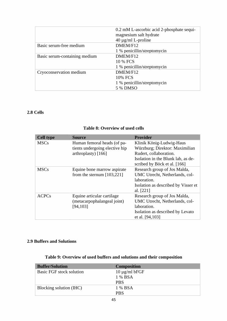
45
0.2 mM L-ascorbic acid 2-phosphate sequi-
magnesium salt hydrate
40 µg/ml L-proline
Basic serum-free medium DMEM/F12
1 % penicillin/streptomycin
Basic serum-containing medium DMEM/F12
10 % FCS
1 % penicillin/streptomycin
Cryoconservation medium DMEM/F12
10% FCS
1 % penicillin/streptomycin
5 % DMSO
2.8 Cells
Table 8: Overview of used cells
Cell type Source Provider
MSCs Human femoral heads (of pa-
tients undergoing elective hip
arthroplasty) [166]
Klinik König-Ludwig-Haus
Würzburg; Direktor: Maximilian
Rudert, collaboration.
Isolation in the Blunk lab, as de-
scribed by Böck et al. [166]
MSCs Equine bone marrow aspirate
from the sternum [103,221]
Research group of Jos Malda,
UMC Utrecht, Netherlands, col-
laboration.
Isolation as described by Visser et
al. [221]
ACPCs Equine articular cartilage
(metacarpophalangeal joint)
[94,103]
Research group of Jos Malda,
UMC Utrecht, Netherlands, col-
laboration.
Isolation as described by Levato
et al. [94,103]
2.9 Buffers and Solutions
Table 9: Overview of used buffers and solutions and their composition
Buffer/Solution Composition
Basic FGF stock solution 10 µg/ml bFGF
1 % BSA
PBS
Blocking solution (IHC) 1 % BSA
PBS
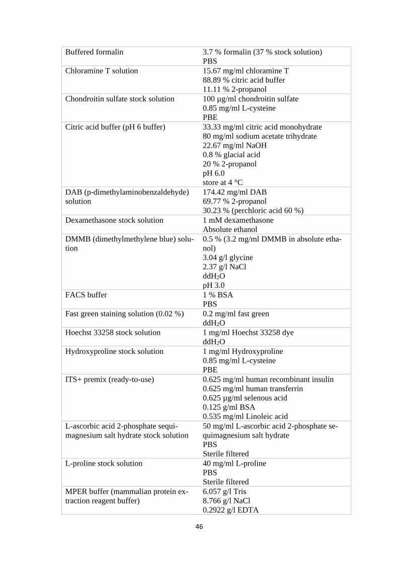
46
Buffered formalin 3.7 % formalin (37 % stock solution)
PBS
Chloramine T solution 15.67 mg/ml chloramine T
88.89 % citric acid buffer
11.11 % 2-propanol
Chondroitin sulfate stock solution 100 µg/ml chondroitin sulfate
0.85 mg/ml L-cysteine
PBE
Citric acid buffer (pH 6 buffer) 33.33 mg/ml citric acid monohydrate
80 mg/ml sodium acetate trihydrate
22.67 mg/ml NaOH
0.8 % glacial acid
20 % 2-propanol
pH 6.0
store at 4 °C
DAB (p-dimethylaminobenzaldehyde)
solution
174.42 mg/ml DAB
69.77 % 2-propanol
30.23 % (perchloric acid 60 %)
Dexamethasone stock solution 1 mM dexamethasone
Absolute ethanol
DMMB (dimethylmethylene blue) solu-
tion
0.5 % (3.2 mg/ml DMMB in absolute etha-
nol)
3.04 g/l glycine
2.37 g/l NaCl
ddH2O
pH 3.0
FACS buffer 1 % BSA
PBS
Fast green staining solution (0.02 %) 0.2 mg/ml fast green
ddH2O
Hoechst 33258 stock solution 1 mg/ml Hoechst 33258 dye
ddH2O
Hydroxyproline stock solution 1 mg/ml Hydroxyproline
0.85 mg/ml L-cysteine
PBE
ITS+ premix (ready-to-use) 0.625 mg/ml human recombinant insulin
0.625 mg/ml human transferrin
0.625 µg/ml selenous acid
0.125 g/ml BSA
0.535 mg/ml Linoleic acid
L-ascorbic acid 2-phosphate sequi-
magnesium salt hydrate stock solution
50 mg/ml L-ascorbic acid 2-phosphate se-
quimagnesium salt hydrate
PBS
Sterile filtered
L-proline stock solution 40 mg/ml L-proline
PBS
Sterile filtered
MPER buffer (mammalian protein ex-
traction reagent buffer)
6.057 g/l Tris
8.766 g/l NaCl
0.2922 g/l EDTA
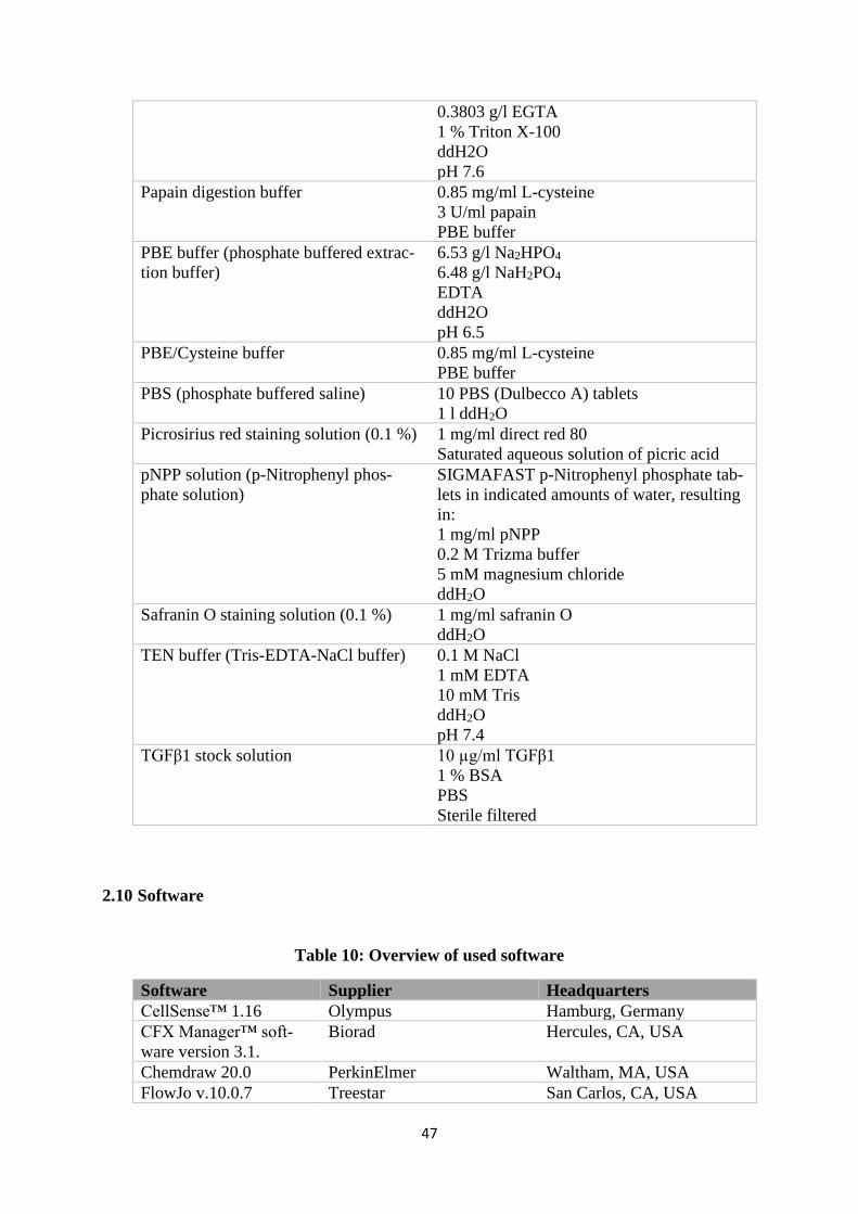
47
0.3803 g/l EGTA
1 % Triton X-100
ddH2O
pH 7.6
Papain digestion buffer 0.85 mg/ml L-cysteine
3 U/ml papain
PBE buffer
PBE buffer (phosphate buffered extrac-
tion buffer)
6.53 g/l Na2HPO4
6.48 g/l NaH2PO4
EDTA
ddH2O
pH 6.5
PBE/Cysteine buffer 0.85 mg/ml L-cysteine
PBE buffer
PBS (phosphate buffered saline) 10 PBS (Dulbecco A) tablets
1 l ddH2O
Picrosirius red staining solution (0.1 %) 1 mg/ml direct red 80
Saturated aqueous solution of picric acid
pNPP solution (p-Nitrophenyl phos-
phate solution)
SIGMAFAST p-Nitrophenyl phosphate tab-
lets in indicated amounts of water, resulting
in:
1 mg/ml pNPP
0.2 M Trizma buffer
5 mM magnesium chloride
ddH2O
Safranin O staining solution (0.1 %) 1 mg/ml safranin O
ddH2O
TEN buffer (Tris-EDTA-NaCl buffer) 0.1 M NaCl
1 mM EDTA
10 mM Tris
ddH2O
pH 7.4
TGFβ1 stock solution 10 µg/ml TGFβ1
1 % BSA
PBS
Sterile filtered
2.10 Software
Table 10: Overview of used software
Software Supplier Headquarters
CellSense™ 1.16 Olympus Hamburg, Germany
CFX Manager™ soft-
ware version 3.1.
Biorad Hercules, CA, USA
Chemdraw 20.0 PerkinElmer Waltham, MA, USA
FlowJo v.10.0.7 Treestar San Carlos, CA, USA

48
GraphPad Prism Ver-
sion 6.0
GraphPad Software LaJolla, CA, USA
Inkscape 0.92.4 The Inkscape Project -
Microsoft 365 Microsoft Redmond, WA, USA

49
Methods

50
51
3 Methods
3.1 Cell culture
3.1.1 Cell isolation
Bone marrows were recovered, after informed consent, from the explanted femoral heads
of patients undergoing elective hip arthroplasty. The procedure was approved by the local
Ethics Committee of the University of Würzburg (186/18). Human bone marrow-derived
mesenchymal stromal cells (MSCs) were isolated as previously described [166]. Briefly,
bone debris and marrow were washed in PBS several times and the resulting solution was
centrifuged to pellet the cells. They were seeded on tissue culture plastic and the non-ad-
herent cells were removed after two days [166].
Equine MSCs were isolated from equine bone marrow aspirate from the sternum as de-
scribed by Visser et al. [221]. The cells were kindly provided by the research group of Prof.
Jos Malda (UMC Utrecht) for a collaboration project and the present work. Briefly, a Ficoll-
Paque density gradient was used to isolate the mononuclear cell fraction and cells were
subsequently seeded on tissue culture plastic [103,221].
Equine ACPCs were isolated from equine articular cartilage from the metacarpophalangeal
joint as described by Levato et al. [103] and Williams et al. [94]. The cells were kindly
provided by the research group of Prof. Jos Malda (UMC Utrecht) for a collaboration pro-
ject and the present work. Cartilage tissue was harvested with a scalpel under sterile condi-
tions, minced and digested for 2 h in 0.2 % pronase solution (0.2 wt% pronase in DMEM
(with GlutaMAX™ and pyruvate), 10 µl/ml HEPES buffer, 1 µl/ml gentamicin) at 37 °C.
Then, pronase solution was removed and cartilage was incubated for 12 h in 0.075 % col-
lagenase II solution (0.075 wt% collagenase II in DMEM (with GlutaMAX™ and py-
ruvate), 10 µl/ml HEPES buffer, 1 µl/ml gentamicin, 50 µl/ml FCS) at 37 °C for further
digestion. After that, the digested tissue was sieved through a 70 µm cell strainer and the
resulting cell suspension was centrifuged for 5 min at 300 x g. The cells were counted,
resuspended in serum-free medium and plated on fibronectin coated 6-well plates (10 µg/ml
fibronectin in PBS+Ca+Mg for 1 h at 37 °C). The cell density was 500 cells/cm2. After 20
min, medium was removed, and plates were washed with PBS+Ca+Mg to remove non-
adherent cells. The adherent cells were cultured for 6 days (DMEM (high glucose, Gluta-
MAX™, pyruvate), 10 % FCS, 1 % PS, 0.2 mM ascorbic acid-2-phosphate, 5 ng/ml bFGF).
52
After 6 days, colonies with more than 32 cells were harvested individually, pooled and then
expanded further in 2D culture.
3.1.2 2D cell expansion
Circa 1*106 human MSCs were seeded per T175 flask in 25 ml of proliferation medium for
human MSCs (see Table 7, pp. 44-45). Equine ACPCs and MSCs were seeded at 4*105
cells per T175 flask in 22 ml of their respective proliferation medium (see Table 7, pp. 44-
45). Medium was exchanged every two to three days and cells were cultured to 80-90 %
confluency in an incubator (37 °C, 5 % CO2, 21 % O2, humidified). For passaging and
harvest of cells, a 0.25 % trypsin-EDTA solution was used.
3.1.3 Treatment of cells in 2D monolayer with HA
50000 human MSCs per well were seeded in 6-well plates in basic serum-containing me-
dium (see Table 7, pp. 44-45) to allow cell attachment to the plastic surface. 2 ml medium
per well were used. After one day cells were washed once with DPBS and medium was
exchanged with basic serum-free medium (see Table 7, pp. 44-45) to starve the cells for one
day. After one day, medium was exchanged to basic serum-free medium with or without
the supplementation of HA (1 mg/ml; 0.6-1.0 MDa). After two days, cells were harvested
for qRT-PCR analysis.
3.1.4 Treatment of cells in 3D pellet culture with HA
2*105 cells per well were seeded in a 96-well plate with V-shaped bottom. The 96-well plate
was centrifuged for 5 min at 500 x g to pellet the cells at the bottom of the wells. For 3D
pellet culture of human MSCs, pellets were cultured in basic serum-containing medium (see
Table 7, pp. 44-45) for one day to allow condensation and formation of pellets. After one
day, pellets were washed once with DPBS and then the medium was exchanged with basic
serum-free medium (see Table 7, pp. 44-45) for one day to starve the cells. Then, basic
serum-free medium with or without the supplementation of HA (2 mg/ml; 0.6-1.0 MDa)
was added to the pellets. Pellets were harvested for qRT-PCR analysis after one day with
or without HA supplementation and three pellets were pooled for one sample.
53
3.1.5 3D agarose hydrogel culture
40 mg per ml (4 %) low melt agarose in DPBS were heated up with a microwave until
agarose was dissolved completely. The solution was then slowly cooled down in a thermo-
mixer to 40 °C and mixed 1:1 with a cell solution at a concentration of 40*106 cells per ml.
This resulted in a 2 % agarose gel with 20*106 cells per ml. 55 µl of this gel were cast into
custom made silicone or Teflon molds (Ø 6 mm). The molds with the cast gel were shortly
put into a fridge to cool the gel down below 25 °C to solidify the hydrogel around the en-
capsulated cells. The solid cylindrical constructs were then collected with a small spoon and
put in 24- or 48-well plates (suspension culture). Equine MSCs and ACPCs in agarose were
cultured for one, 14 and 28 days in their respective chondrogenic differentiation medium
(see Table 7, pp. 44-45). Two to three ml medium per well were used. Cells were cultured
in either a normal incubator (37 °C, 5 % CO2, 21 % O2, humidified) or a hypoxia incubator
(37 °C, 5 % CO2, 2 % O2, humidified). Medium was exchanged every two to three days.
3.1.6 3D HA-SH hydrogel culture
3.1.6.1 HA-SH/P(AGE-co-G) hydrogels
5 wt% P(AGE-co-G) were dissolved in DPBS by vortexing and sterile filtered through a
0.2 µm syringe filter. The needed amount of HA-SH11.7 kDa (5 wt%) was irradiated with an
UV hand lamp at 254 nm for 10 min to sterilize the material. Then, 0.05 wt% photoinitiator
Irgacure2959 and 5 wt% HA-SH11.7 kDa were added and dissolved by vortexing. The mixture
was protected from light and pH was adjusted to 7.4. A cell pellet was resuspended in the
resulting 10 wt% hydrogel precursor solution so that the end-concentration was 20*106
cells/ml. 55 µl per construct of precursor solution with cells were cast into custom made
silicone or Teflon molds (Ø 6 mm). For crosslinking of the hydrogel precursor solution, it
was irradiated with an UV hand lamp at 365 nm for 10 min. Afterwards, hydrogel constructs
were put into 24- or 48-well plates (suspension culture) with two to three ml of chondro-
genic differentiation medium (see Table 7, pp. 44-45) per well and cultured in an incubator
(37 °C, 5 % CO2, 21 % O2, humidified). Medium was exchanged every two to three days.
Equine ACPCs and MSCs were cultured for one and 28 days.
54
3.1.6.2 HA-SH /PEGDA/PEG-allyl/Irgacure2959 hydrogels
To produce a hydrogel precursor solution of 0.5 wt% HA-SH, 0.5 wt% PEGDA, 1 wt%
PEG-allyl and 0.05 wt% Irgacure I2959 and PBS, stock solutions of the components were
prepared. 4 wt% PEGDA, 4 wt% PEG-allyl and 0.5 wt% Irgacure were dissolved in DPBS.
1 wt% HA-SH was dissolved in 154 mM HEPES buffer (pH 7.6) shortly before it was used
to resuspend the cell pellet of human MSCs. HA-SH410 kDa was used for the formation of
HA-SH/PEGDA/PEG-Allyl/Irgacure2959 hydrogels. Calculated amounts of PEGDA,
PEG-allyl and Irgacure stock solution and PBS were mixed and added to the HA-SH/cell
suspension so that final concentrations were 0.5 wt% HA-SH, 0.5 wt% PEGDA, 1 wt%
PEG-allyl, 0.05 wt% Irgacure and 20*106 cells/ml. The crosslinking reaction (Michael ad-
dition) was started the moment that HA-SH and PEGDA were put together. The hydrogel
precursor solution was incubated at 37 °C for 60 min (predetermined). The vial was mixed
gently every 10 min to avoid sedimentation of cells. After that time, the solution had become
viscous enough to keep its shape when it was pipetted with a displacement pipette or when
it was bioprinted. The now “printable” hydrogel precursor solution was then either cast into
glass rings (Ø 5 mm) or extruded through a 330 µm steel nozzle with 50 kPa by the bi-
oprinter Cellink plus (40 µl per construct). The printed hydrogel precursor was collected in
a tube and subsequently cast into glass rings (Ø 5 mm). Once the solution was in the glass
rings, it was irradiated with an UV hand lamp at 365 nm for 10 min. The resulting hydrogels
were cultured in 48-well plates in 1 ml chondrogenic differentiation medium for human
MSCs without TGFβ1 for one, four and seven days. Medium was exchanged on day two,
day four and day six.
3.1.7 Preparation of zonal hydrogels
Zonal hydrogels were prepared with either HA-SH/P(AGE-co-G) or with agarose hydro-
gels. For agarose hydrogels, the first/lower layer of the construct was cast into a mold (cus-
tom made silicone or Teflon molds (Ø 6 mm) and the agarose was cooled down shortly to
get a semi-solid hydrogel. When the second/upper layer was cast on the first layer, the first
layer was not disrupted but could also still attach to the second layer, as it was not com-
pletely solid yet. A similar mechanism was used for zonal HA-SH/P(AGE-co-G) hydrogels.
The first/lower layer was irradiated only for 5 min at 365 nm so that not all functional groups
were crosslinked, then the second/upper layer was cast on top and the complete hydrogel
was irradiated again for 5 min at 365 nm to complete the crosslinking. In both zonal agarose
55
and HA-SH/P(AGE-co-G) hydrogels the lower, MSC containing layer had a volume of 35
µl and the upper ACPC containing layer had a volume of 20 µl.
3.2 Staining of cells and tissue sections
3.2.1 Cell viability assay
To investigate viability of cells that were encapsulated in hydrogels, constructs were washed
once in PBS and then incubated in PBS with 0.1 % calcein acetoxymethyl ester (calcein-
AM) (green) and 0.05 % ethidium homodimer III (red) (Life/Dead cell staining kit II,
Promokine) for 30-45 min at RT. After incubation, constructs were washed once with PBS
and then images were taken with an inverse fluorescent microscope (Olympus IX51/XC30).
Cell viability was assessed on d1 after cell encapsulation and on d7.
3.2.2 Sectioning of 3D constructs
3.2.2.1 Cryo-sectioning
After harvesting of 3D hydrogel constructs, they were fixed with 3.7 % buffered formalin
overnight at 4 °C. They were then washed with PBS and embedded in Tissue-Tek®
O.C.T.compound overnight at RT in a wet chamber. On the next day, the constructs were
flash-frozen with liquid nitrogen in Tissue-Tek® O.C.T.compound and stored at -20 °C.
Cryo-sectioning was performed with a cryostat (Leica CM 1850) at -24 °C. Sections were
cut to be 6 µm thick, collected on SuperFrost® Plus adhesion glass slides and stored at -20
°C until they were stained.
3.2.2.2 Paraffin sectioning
3D hydrogel constructs were harvested and then fixed with 3.7 % buffered formalin over-
night at 4 °C. On the next day, they were incubated for 1 h in 70 % ethanol, then for 2 h in
two changes of 96 % ethanol. Next, they were incubated in 100 % ethanol for 1 h and then
in fresh 100 % ethanol overnight at RT. On the next day, samples were incubated in two
changes of xylene for 4 h. Afterwards, samples were put into hot liquid paraffin for 2-3 h,
were then embedded in paraffin, cooled down and solidified on a cold plate overnight. Sam-
ples were stored at RT until they were sectioned. Paraffin sectioning was performed with a
56
microtome and sections were cut to be 1 µm thick. They were put in a warm water bath and
collected on adhesion glass slides. Afterwards, sections on glass slides were incubated at 60
°C overnight and stored at room temperature (RT) until they were stained.
3.2.3 Safranin O staining
Paraffin sections needed to be deparaffinized before staining could begin. First, they were
incubated in two changes of xylene for 10 min. Then, they were incubated in two changes
of 100 % ethanol for 6 min. Afterwards, sections were incubated in 80 %, 70 % and 50 %
ethanol for 1 min each. After this, cryo-sections and paraffin sections were stained the same
way. Sections were incubated in ddH2O for 1 min, then in Weigert´s hematoxylin for 6 min.
Then they were shortly rinsed with ddH2O and 0.5 % hydrochloric acid in ethanol. Subse-
quently, sections were rinsed for 5 min under running tap water and then they were incu-
bated in 0.02 % Fast Green solution (see Table 9, pp. 45-47) for 4 min. Then, sections were
rinsed in 1 % acetic acid and were stained for 6 min in 0.1 % Safranin O solution (see Table
9, pp. 45-47). They were incubated in 100 % ethanol for 1 min, in 2-propanol for 2 min and
in xylene for 2 min before they were covered in entellan and coverslips. Samples were dried
under a fume hood for 2 days and images were taken with a microscope (Olympus
BX51/DP71).
3.2.4 Picrosirius red staining
Paraffin sections were deparaffinized as described for Safranin O staining (in 3.2.3.) Then,
deparaffinized sections and cryo-sections were stained the same way. They were incubated
in Weigert´s Hematoxylin for 8 min, rinsed under running tap water for 10 min and then
incubated in 0.1 % picrosirius red (see Table 9, pp. 45-47) for 60 min. Afterwards, sections
were incubated in two changes of 0.5 % acetic acid for 5 min, then in 100 % ethanol for 1
min, in 100 % 2-propanol for 2 min and in xylene for another 2 min. They were covered in
entellan and coverslips and were dried under a fume hood for 2 days. Images were taken
with a microscope (Olympus BX51/DP71).
3.2.5 Immunohistochemistry
Paraffin sections were deparaffinized as described in 3.2.3. Afterwards, they were washed
three times with PBS for 5 min and sections were circled with a PAP-pen. For antigen re-
trieval, sections were first incubated in a 0.1 % pronase solution in PBS for 30 min at 37
57
°C. Then, sections were washed three times for 5 min with PBS and incubated in a 1 %
hyaluronidase solution in PBS for 30 min at 37 °C. Subsequently, they were again washed
three times with PBS for 5 min and then incubated in blocking solution (1 % BSA in PBS)
for 30 min at RT. Sections were then incubated with a primary antibody overnight at RT in
a wet chamber. Dilutions used for the respective antibodies can be found in Table 5 (pp. 42-
43). For cryo-sections and formalin-fixed 2D cell culture, antigen retrieval was performed
by incubation with proteinase K for 10 min at 37 °C instead of pronase and hyaluronidase.
The other steps were the same for all samples. On the next day, sections/cells were washed
three times for 5 min with PBS and incubated with a secondary antibody (Table 5, pp. 42-
43) for 60 min at RT in the dark. Afterwards, sections were washed three times for 5 min
with PBS and mounted with DAPI mounting medium. Fluorescent images were taken with
a microscope (Olympus BX51/DP71).
For immunohistochemical staining with diaminobenzidine chromogen and peroxidase en-
zyme, Ultra-Streptavidin HRP Kit (Multi-Species, diaminobenzidine) Antibody from Bio-
legend was used according to the manufacturer’s instructions. Used primary antibodies and
dilutions can be found in Table 5 (pp. 42-43) and antigen retrieval was performed as de-
scribed above.
3.3 Biochemical assays
3.3.1 Papain digestion
Before biochemical assays could be performed with harvested 3D samples, they had to be
digested using papain. Wet weight of hydrogels was determined directly after harvesting.
Hydrogels or pellets were transferred to 2 ml SafeSeal micro tubes and a defined amount of
PBE/cysteine buffer (500µl or 200 µl) (see Table 9, pp. 45-47) and one stainless steel bead
per tube were added. Samples were homogenized using a TissueLyser at 25 Hz for 5 min.
Afterwards, the same amount of PBE/cysteine buffer as before was added to the homoge-
nized samples, containing papain. The end concentration of papain in the samples was 3
U/ml. Samples were shaken at 60 °C in a thermomixer for 16 -20 h and then stored at -20
°C until biochemical assays were performed.
58
3.3.2 DNA assay
To determine the amount of DNA present in the samples, a fluorescence assay with DNA
intercalating dye Hoechst 33258 was performed [222]. 200 µl dye solution (1 µl Hoechst
33258 stock solution per 10 ml TEN buffer) (see Table 9, pp. 45-47) were added to 10 µl
digested sample (pure or diluted in PBE/cysteine buffer). Quantification of DNA amount
was performed by a Tecan microplate reader (excitation: 360 nm, emission: 460 nm) and
salmon testis DNA was used as a standard.
3.3.3 Glycosaminoglycan assay
For determination of glycosaminoglycan (GAG) amount in the samples, a dimethyl-
methylenblue (DMMB) assay was performed [223]. 200 µl of DMMB (see Table 9, pp. 45-
47) were added to 50 µl of sample (mostly diluted between 1:5 to 1:50 in PBE/cysteine
buffer) and measured with a Tecan microplate reader at 525 nm. Bovine chondroitin sulfate
was used as a standard.
3.3.4 Hydroxyproline assay
Collagen content of samples was determined by a hydroxyproline assay. 10-100 µl sample
were added to 100 µl of 37 % hydrochloric acid (fuming hydrochloric acid). Samples were
heated to 105 °C in a thermomixer for 16-20 h. On the next day, SafeSeal micro tubes with
samples were opened and HCl fumes could evaporate under a fume hood for 2 h. The dried
samples were dissolved in 500 µl ddH2O and 100 µl of this solution were added to 50 µl of
chloramine T solution (see Table 9, pp. 45-47). Samples were incubated for 20 min at RT.
Then 50 µl of DAB solution (see Table 9, pp. 45-47) was added to each well and incubated
for 30 min at 65 °C in a drying chamber. The quantification of hydroxyproline was per-
formed by a Tecan microplate reader at 560 nm, with L-hydroxyproline as standard. The
amount of total collagen was calculated using a hydroxyproline to collagen ratio of 1:10
[224,225].
3.3.5 Alkaline phosphatase activity assay
For investigation of alkaline phosphatase (ALP) activity in hydrogel samples, no papain
digestion was performed. Constructs were harvested, wet weights were determined, and
samples were put directly in MPER buffer on ice to preserve enzyme activity of ALP. Then,
small steel beads were added, and samples were homogenized using a TissueLyser at 15 Hz
59
for 2 min. Homogenized samples were stored at -80 °C or were directly analysed. Whenever
possible, samples were stored on ice. ALP activity assay was performed using the substrate
pNPP (p-nitrophenyl phosphate). pNPP solution was prepared using SIGMAFAST™ p-
Nitrophenyl phosphate tablets, according to manufacturer’s instructions (see Table 9, pp.
45-47). 25 µl sample were added to 50 µl pNPP solution and a kinetic measurement was
performed by a Tecan microplate reader. Dephosphorylation of pNPP by ALP resulted in
the production of a yellow compound that was detected at 405 nm. Readings at 655 nm
were subtracted to correct for non-specific background values. The kinetic protocol run for
30 min with a reading every two minutes and shaking in between. Calf intestinal alkaline
phosphatase was used as standard [226].
3.4 Quantitative real-time PCR analysis
For quantitative real-time PCR analysis, one sample was one complete hydrogel, three pel-
lets or one 6-well full of 2D cultured cells. For pellets and hydrogels, samples were homog-
enized by a TissueLyser and stainless-steel beads at 25 Hz for 5 min before RNA extraction
with TRIzol® reagent. cDNA was transcribed from total RNA with ImProm-II™ Reverse
Transcription System Kit and MESA GREEN qPCR MasterMix Plus for SYBR® Assay
No ROX was used for qPCR (quantitative polymerase chain reaction). Analysis was per-
formed with a Real-Time PCR Detection System CFX96 Touch. Used primers (see Table
6, pp. 43-44) were obtained from two different sources: Ready to use primers from Bio-Rad
and self-designed primers from Biomers. Cycling protocol for Bio-Rad primers was: 5 min
at 95 °C for initial denaturation, then 40 cycles of 15 sec at 95 °C, 30 sec at 60 °C and 30
sec at 70 °C. Cycling protocol for Biomer primer was: 6 min at 95 °C for initial denaturation,
then 40 cycles of 15 sec at 95 °C, 30 sec at 60 °C and 30 sec at 72 °C. A melting curve
analysis for PCR product integrity was performed (0.5 °C increments from 65 °C to 95 °C).
Software CFX Manager™ (software version 3.1.) was used to process the raw data and the
relative expression levels were normalized to the housekeeper genes GAPDH or HPRT1
and determined using the 2–ΔΔCT method. Whenever useful, samples were additionally nor-
malized to a d1, d0 or untreated sample value.
60
3.5 Flow cytometry
Flow cytometry was used to analyse the existence and quantity of cell surface receptors
CD44 and CD168 on human MSCs. Therefore, cells were harvested with 0.25 % Trypsin-
EDTA from an 80-90 % confluent 2D culture and stained with respective antibodies. Cells
were transferred to FACS tubes and centrifuged for 7 min at 400 x g and 4 °C. Cells were
blocked with 5 % goat serum in FACS buffer for 20 min at 4 °C. For permeabilization of
cells, 0.3 % Triton X-100 was added to the blocking solution. Cells were washed two times
with FACS buffer, then they were incubated with the primary antibodies (see Table 5, pp.
42-43) for 25 min at 4 °C. Afterwards, cells were washed two times with FACS buffer and
incubated with the secondary (goat) antibody (see Table 5, pp. 42-43) for 20 min at 4 °C in
the dark. Subsequently, cells were washed twice with FACS buffer and kept on ice in FACS
buffer until measured. One tube of unstained cells was used to adjust the settings of the
FACSCanto flow cytometer. The resulting data from the measurement were processed us-
ing FlowJo v.10.0.7.
3.6 Statistical analysis
Graph Pad Prism 6.0 (GraphPad Software, La Jolla, CA, USA) was used for statistical anal-
ysis. Statistical significance was determined with Student´s t-test (Figures 16, 31 A-D, 32
A-F, 33 A-F, 34 A-D, 35 A-D), one-way ANOVA (Figures 9, 10, 11, 13, 14, 18, 19, 24, 28,
31 E-F, 32 G-I, 33 G-I, 34 E-F, 35 E-F) or two-way ANOVA (Figures 17, 21, 22, 25).
Tukey´s post-hoc test was used in conjunction with one-way and two-way ANOVA.

61
Results and Discussion

62
63
4 Results and discussion
4.1 Articular cartilage tissue engineering with ACPCs and MSCs
Tissue engineering is a promising approach in articular cartilage repair. Additionally, it al-
lows for versatile research on cartilage development, function and structure which is essen-
tial knowledge to develop successful treatments for cartilage defects. Chondrocytes har-
vested from cartilage or stem cells that can be differentiated into chondrocytes are used to
produce tissue with the aim that it will be as similar to native articular cartilage as possible.
If the right combination of cell type, scaffold and other stimuli is used, cartilage similar to
native tissue can be produced.
MSCs are considered as potential cell source for cartilage repair as they can differentiate
into chondrocytes and do not lose this chondrogenic potential when they are expanded in
2D culture in contrast to chondrocytes [35-37]. However, they also tend to produce fibro-
cartilage rather than articular cartilage and can undergo terminal differentiation and hyper-
trophy [35,70,71]. This can lead to endochondral ossification and decreased functionality
of cartilage tissue. ACPCs are a relatively new, promising cell source for cartilage repair
that have been found in the upper layer of articular cartilage [92,93]. They have the same
advantage over chondrocytes as MSCs because they can be expanded without losing their
ability to differentiate chondrogenically [101]. In previous studies, a high chondrogenic po-
tential and no tendency to differentiate terminally was detected [94,102]. Therefore, ACPCs
are considered as promising alternative cell source for cartilage engineering. Animal models
and even one pilot clinical study in humans have been used to test the suitability of ACPCs
for cartilage repair [94,98,104,105]. However, only two studies have compared the chon-
drogenesis of MSCs and ACPCs directly in hydrogels [103,107]. As the dependence of cell
performance on hydrogel composition has not been completely resolved yet, additional
studies in other hydrogels are needed to critically evaluate chondrogenesis of ACPCs in
comparison to MSCs.
One important factor for articular cartilage function is its zonal structure. At the moment, it
cannot be restored by clinical treatments and therefore, tissue engineering has developed
several strategies to produce tissue with zones similar to native cartilage. One approach uses
different cell types for different zonal layers. In previous studies, a combination of ACPCs
in the upper layer and MSCs in the lower layer showed promising results [103,107].
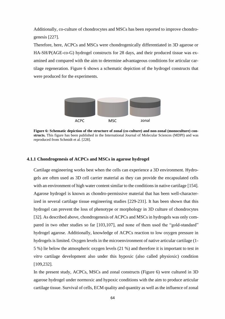
64
Additionally, co-culture of chondrocytes and MSCs has been reported to improve chondro-
genesis [227].
Therefore, here, ACPCs and MSCs were chondrogenically differentiated in 3D agarose or
HA-SH/P(AGE-co-G) hydrogel constructs for 28 days, and their produced tissue was ex-
amined and compared with the aim to determine advantageous conditions for articular car-
tilage regeneration. Figure 6 shows a schematic depiction of the hydrogel constructs that
were produced for the experiments.
Figure 6: Schematic depiction of the structure of zonal (co-culture) and non-zonal (monoculture) con-
structs. This figure has been published in the International Journal of Molecular Sciences (MDPI) and was
reproduced from Schmidt et al. [228].
4.1.1 Chondrogenesis of ACPCs and MSCs in agarose hydrogel
Cartilage engineering works best when the cells can experience a 3D environment. Hydro-
gels are often used as 3D cell carrier material as they can provide the encapsulated cells
with an environment of high water content similar to the conditions in native cartilage [154].
Agarose hydrogel is known as chondro-permissive material that has been well-character-
ized in several cartilage tissue engineering studies [229-231]. It has been shown that this
hydrogel can prevent the loss of phenotype or morphology in 3D culture of chondrocytes
[32]. As described above, chondrogenesis of ACPCs and MSCs in hydrogels was only com-
pared in two other studies so far [103,107], and none of them used the “gold-standard”
hydrogel agarose. Additionally, knowledge of ACPCs reaction to low oxygen pressure in
hydrogels is limited. Oxygen levels in the microenvironment of native articular cartilage (1-
5 %) lie below the atmospheric oxygen levels (21 %) and therefore it is important to test in
vitro cartilage development also under this hypoxic (also called physioxic) condition
[109,232].
In the present study, ACPCs, MSCs and zonal constructs (Figure 6) were cultured in 3D
agarose hydrogel under normoxic and hypoxic conditions with the aim to produce articular
cartilage tissue. Survival of cells, ECM quality and quantity as well as the influence of zonal
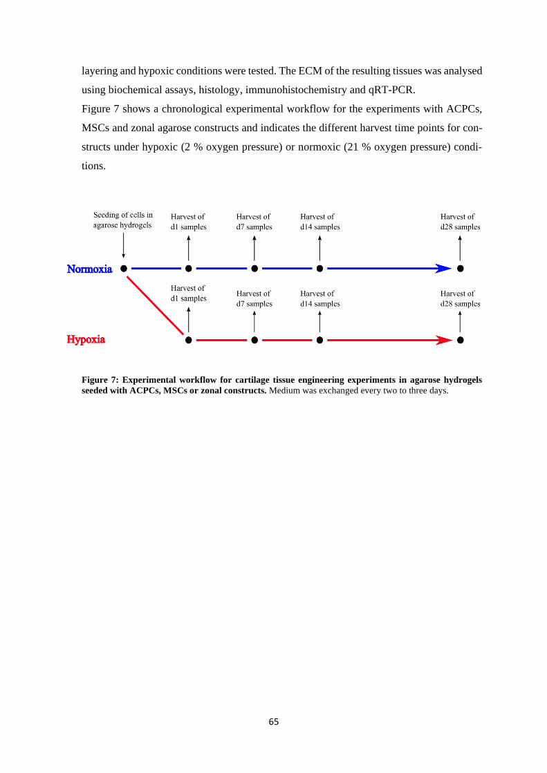
65
layering and hypoxic conditions were tested. The ECM of the resulting tissues was analysed
using biochemical assays, histology, immunohistochemistry and qRT-PCR.
Figure 7 shows a chronological experimental workflow for the experiments with ACPCs,
MSCs and zonal agarose constructs and indicates the different harvest time points for con-
structs under hypoxic (2 % oxygen pressure) or normoxic (21 % oxygen pressure) condi-
tions.
Figure 7: Experimental workflow for cartilage tissue engineering experiments in agarose hydrogels
seeded with ACPCs, MSCs or zonal constructs. Medium was exchanged every two to three days.
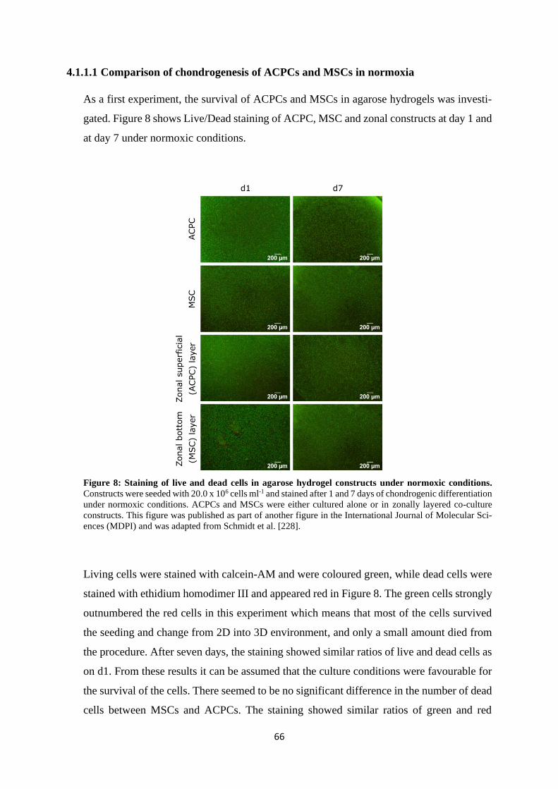
66
4.1.1.1 Comparison of chondrogenesis of ACPCs and MSCs in normoxia
As a first experiment, the survival of ACPCs and MSCs in agarose hydrogels was investi-
gated. Figure 8 shows Live/Dead staining of ACPC, MSC and zonal constructs at day 1 and
at day 7 under normoxic conditions.
Figure 8: Staining of live and dead cells in agarose hydrogel constructs under normoxic conditions.
Constructs were seeded with 20.0 x 10⁶ cells ml-1 and stained after 1 and 7 days of chondrogenic differentiation
under normoxic conditions. ACPCs and MSCs were either cultured alone or in zonally layered co-culture
constructs. This figure was published as part of another figure in the International Journal of Molecular Sci-
ences (MDPI) and was adapted from Schmidt et al. [228].
Living cells were stained with calcein-AM and were coloured green, while dead cells were
stained with ethidium homodimer III and appeared red in Figure 8. The green cells strongly
outnumbered the red cells in this experiment which means that most of the cells survived
the seeding and change from 2D into 3D environment, and only a small amount died from
the procedure. After seven days, the staining showed similar ratios of live and dead cells as
on d1. From these results it can be assumed that the culture conditions were favourable for
the survival of the cells. There seemed to be no significant difference in the number of dead
cells between MSCs and ACPCs. The staining showed similar ratios of green and red
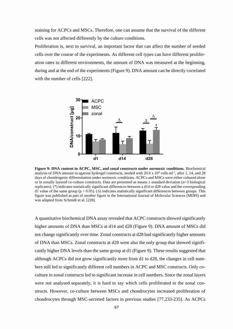
67
staining for ACPCs and MSCs. Therefore, one can assume that the survival of the different
cells was not affected differently by the culture conditions.
Proliferation is, next to survival, an important factor that can affect the number of seeded
cells over the course of the experiments. As different cell types can have different prolifer-
ation rates in different environments, the amount of DNA was measured at the beginning,
during and at the end of the experiments (Figure 9). DNA amount can be directly correlated
with the number of cells [222].
Figure 9: DNA content in ACPC, MSC, and zonal constructs under normoxic conditions. Biochemical
analysis of DNA amount in agarose hydrogel constructs, seeded with 20.0 x 10⁶ cells ml-1, after 1, 14, and 28
days of chondrogenic differentiation under normoxic conditions. ACPCs and MSCs were either cultured alone
or in zonally layered co-culture constructs. Data are presented as means ± standard deviation (n=3 biological
replicates). (*) indicates statistically significant differences between a d14 or d28 value and the corresponding
d1 value of the same group (p < 0.05). (Δ) indicates statistically significant differences between groups. This
figure was published as part of another figure in the International Journal of Molecular Sciences (MDPI) and
was adapted from Schmidt et al. [228].
A quantitative biochemical DNA assay revealed that ACPC constructs showed significantly
higher amounts of DNA than MSCs at d14 and d28 (Figure 9). DNA amount of MSCs did
not change significantly over time. Zonal constructs at d28 had significantly higher amounts
of DNA than MSCs. Zonal constructs at d28 were also the only group that showed signifi-
cantly higher DNA levels than the same group at d1 (Figure 9). These results suggested that
although ACPCs did not grow significantly more from d1 to d28, the changes in cell num-
bers still led to significantly different cell numbers in ACPC and MSC constructs. Only co-
culture in zonal constructs led to significant increase in cell numbers. Since the zonal layers
were not analysed separately, it is hard to say which cells proliferated in the zonal con-
structs. However, co-culture between MSCs and chondrocytes increased proliferation of
chondrocytes through MSC-secreted factors in previous studies [77,233-235]. As ACPCs
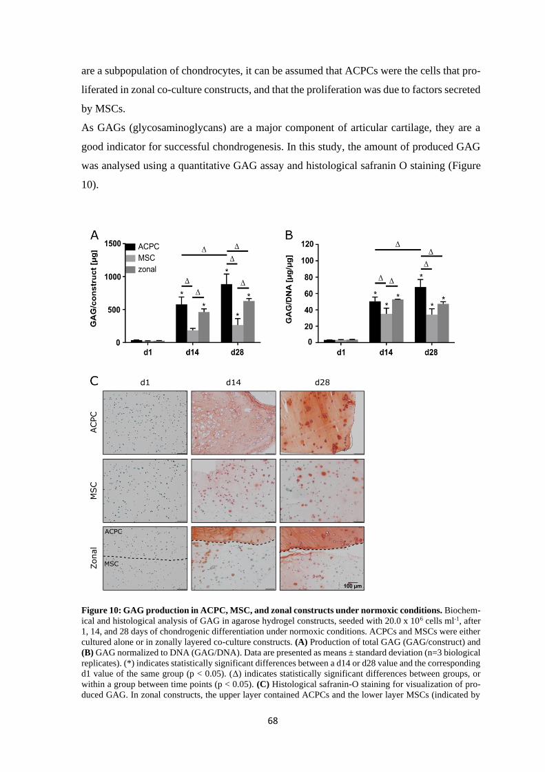
68
are a subpopulation of chondrocytes, it can be assumed that ACPCs were the cells that pro-
liferated in zonal co-culture constructs, and that the proliferation was due to factors secreted
by MSCs.
As GAGs (glycosaminoglycans) are a major component of articular cartilage, they are a
good indicator for successful chondrogenesis. In this study, the amount of produced GAG
was analysed using a quantitative GAG assay and histological safranin O staining (Figure
10).
Figure 10: GAG production in ACPC, MSC, and zonal constructs under normoxic conditions. Biochem-
ical and histological analysis of GAG in agarose hydrogel constructs, seeded with 20.0 x 10⁶ cells ml-1, after
1, 14, and 28 days of chondrogenic differentiation under normoxic conditions. ACPCs and MSCs were either
cultured alone or in zonally layered co-culture constructs. (A) Production of total GAG (GAG/construct) and
(B) GAG normalized to DNA (GAG/DNA). Data are presented as means ± standard deviation (n=3 biological
replicates). (*) indicates statistically significant differences between a d14 or d28 value and the corresponding
d1 value of the same group (p < 0.05). (Δ) indicates statistically significant differences between groups, or
within a group between time points (p < 0.05). (C) Histological safranin-O staining for visualization of pro-
duced GAG. In zonal constructs, the upper layer contained ACPCs and the lower layer MSCs (indicated by
69
the dashed line). This figure has been published in the International Journal of Molecular Sciences (MDPI)
and was reproduced from Schmidt et al. [228].
Figure 10 A shows the amount of GAG per one construct, and Figure 10 B shows the
amount of produced GAG per DNA. The normalisation “per DNA” includes the infor-
mation from Figure 9 (p. 67) and is used for better comparability between the constructs of
different cell types with different amounts of cells. Figures 10 A and B show that all three
constructs (ACPC, MSC, zonal) produced significant amounts of GAG over the time of the
experiment. However, ACPCs produced significantly more GAG than MSCs up to d14 and
d28. On d28, the amount of GAG in ACPC constructs was even significantly higher than
the amount in zonal constructs. GAG amount of ACPC constructs also increased signifi-
cantly from d14 to d28 contrary to MSC and zonal constructs. Zonal constructs contained
significantly more GAG than MSC constructs on d14 and d28 except for d28 in Figure 10
B. The difference in GAG production between ACPCs and MSCs was also clearly visible
in the histological safranin O staining (Figure 10 C). The signal for MSC constructs was
much weaker than the signal for ACPC constructs at d14 and d28. While ACPCs seemed to
spread their GAG evenly through the whole construct with a stronger staining around the
cells, MSCs mainly showed staining around the cells with only very faint staining in the
rest of the gel. These trends were reflected in zonal constructs where the two layers can be
distinguished easily by staining of the contained GAG. The upper layer with ACPCs was
stained more intensely than the lower MSC layer. The mixture of the ACPC layer that con-
tained high levels of GAG and the less GAG containing MSC layer in zonal constructs
could explain GAG levels of zonal constructs in Figure 10 A and B that lay between the
levels of ACPCs and MSCs. These results suggested that ACPCs produced more GAG in
agarose gel than MSCs. A direct effect of the co-culture of ACPCs and MSCs in zonal gels
on the production of GAG could not be detected.
Collagen is another important component of articular cartilage tissue. As chondrogenesis of
ACPCs and MSCs was compared in this study, the production of total collagen was ana-
lysed using a quantitative hydroxyproline assay and picrosirius red staining of the hydrogels
(Figure 11). The amount of total collagen per construct significantly increased in all three
constructs (ACPC, MSC, zonal) from d1 to d14 and from d14 to d28 (Figure 11 A). The
levels of total collagen per construct in the different constructs were similar at the different
time points (Figure 11 A). However, when collagen was normalised to the amount of DNA
in the constructs, MSCs showed significantly higher levels of collagen than ACPCs and
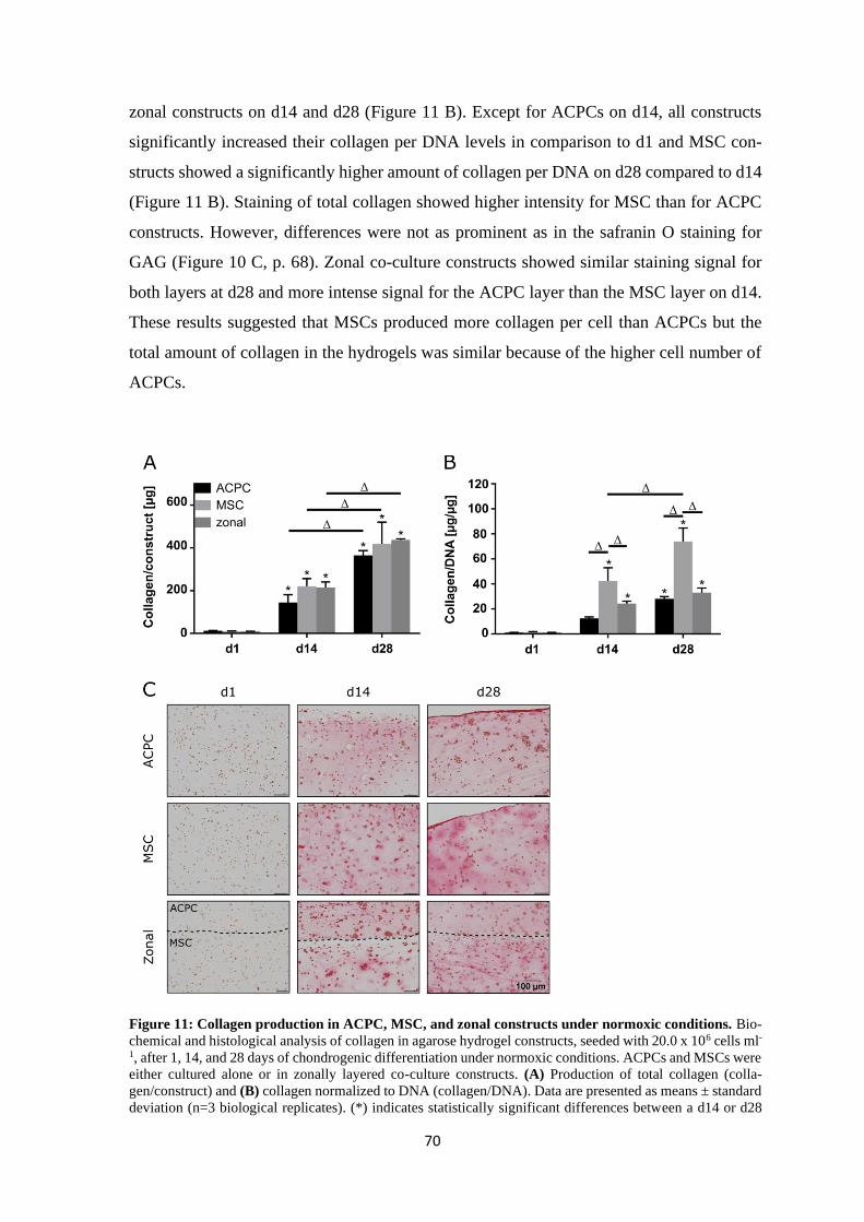
70
zonal constructs on d14 and d28 (Figure 11 B). Except for ACPCs on d14, all constructs
significantly increased their collagen per DNA levels in comparison to d1 and MSC con-
structs showed a significantly higher amount of collagen per DNA on d28 compared to d14
(Figure 11 B). Staining of total collagen showed higher intensity for MSC than for ACPC
constructs. However, differences were not as prominent as in the safranin O staining for
GAG (Figure 10 C, p. 68). Zonal co-culture constructs showed similar staining signal for
both layers at d28 and more intense signal for the ACPC layer than the MSC layer on d14.
These results suggested that MSCs produced more collagen per cell than ACPCs but the
total amount of collagen in the hydrogels was similar because of the higher cell number of
ACPCs.
Figure 11: Collagen production in ACPC, MSC, and zonal constructs under normoxic conditions. Bio-
chemical and histological analysis of collagen in agarose hydrogel constructs, seeded with 20.0 x 10⁶ cells ml-
1, after 1, 14, and 28 days of chondrogenic differentiation under normoxic conditions. ACPCs and MSCs were
either cultured alone or in zonally layered co-culture constructs. (A) Production of total collagen (colla-
gen/construct) and (B) collagen normalized to DNA (collagen/DNA). Data are presented as means ± standard
deviation (n=3 biological replicates). (*) indicates statistically significant differences between a d14 or d28
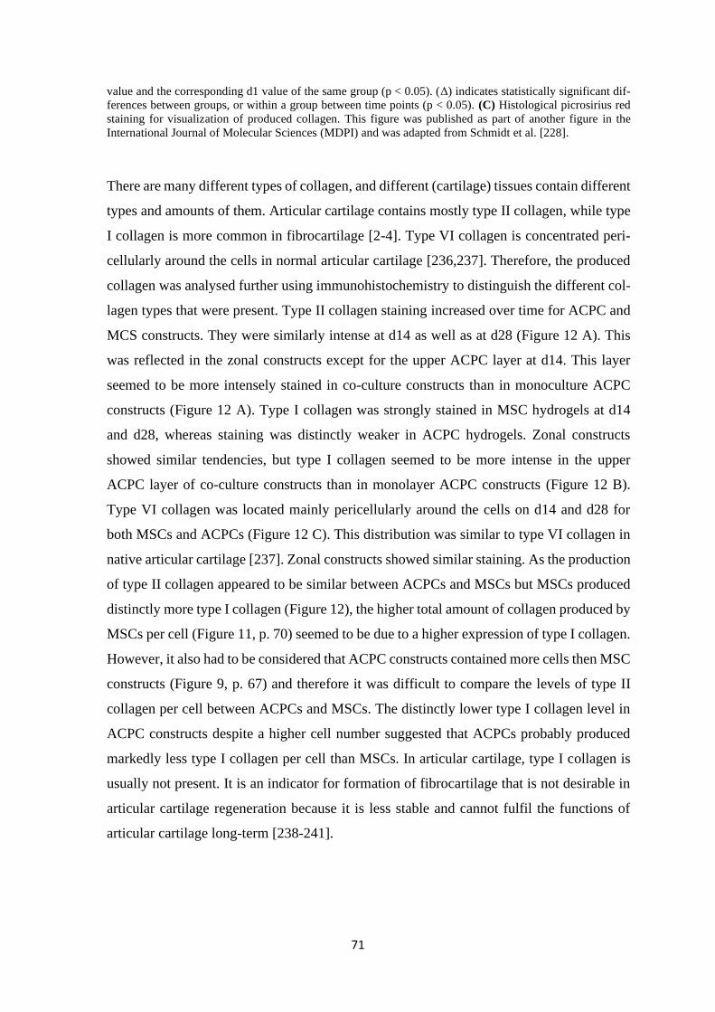
71
value and the corresponding d1 value of the same group (p < 0.05). (Δ) indicates statistically significant dif-
ferences between groups, or within a group between time points (p < 0.05). (C) Histological picrosirius red
staining for visualization of produced collagen. This figure was published as part of another figure in the
International Journal of Molecular Sciences (MDPI) and was adapted from Schmidt et al. [228].
There are many different types of collagen, and different (cartilage) tissues contain different
types and amounts of them. Articular cartilage contains mostly type II collagen, while type
I collagen is more common in fibrocartilage [2-4]. Type VI collagen is concentrated peri-
cellularly around the cells in normal articular cartilage [236,237]. Therefore, the produced
collagen was analysed further using immunohistochemistry to distinguish the different col-
lagen types that were present. Type II collagen staining increased over time for ACPC and
MCS constructs. They were similarly intense at d14 as well as at d28 (Figure 12 A). This
was reflected in the zonal constructs except for the upper ACPC layer at d14. This layer
seemed to be more intensely stained in co-culture constructs than in monoculture ACPC
constructs (Figure 12 A). Type I collagen was strongly stained in MSC hydrogels at d14
and d28, whereas staining was distinctly weaker in ACPC hydrogels. Zonal constructs
showed similar tendencies, but type I collagen seemed to be more intense in the upper
ACPC layer of co-culture constructs than in monolayer ACPC constructs (Figure 12 B).
Type VI collagen was located mainly pericellularly around the cells on d14 and d28 for
both MSCs and ACPCs (Figure 12 C). This distribution was similar to type VI collagen in
native articular cartilage [237]. Zonal constructs showed similar staining. As the production
of type II collagen appeared to be similar between ACPCs and MSCs but MSCs produced
distinctly more type I collagen (Figure 12), the higher total amount of collagen produced by
MSCs per cell (Figure 11, p. 70) seemed to be due to a higher expression of type I collagen.
However, it also had to be considered that ACPC constructs contained more cells then MSC
constructs (Figure 9, p. 67) and therefore it was difficult to compare the levels of type II
collagen per cell between ACPCs and MSCs. The distinctly lower type I collagen level in
ACPC constructs despite a higher cell number suggested that ACPCs probably produced
markedly less type I collagen per cell than MSCs. In articular cartilage, type I collagen is
usually not present. It is an indicator for formation of fibrocartilage that is not desirable in
articular cartilage regeneration because it is less stable and cannot fulfil the functions of
articular cartilage long-term [238-241].
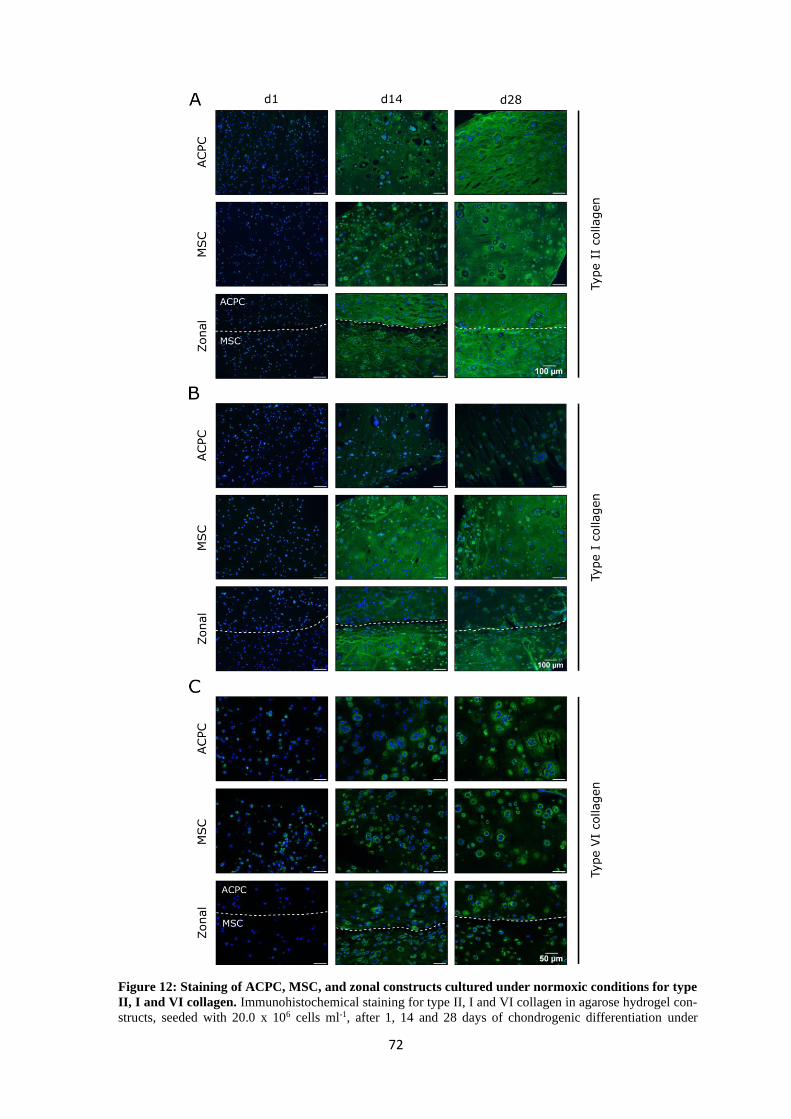
72
Figure 12: Staining of ACPC, MSC, and zonal constructs cultured under normoxic conditions for type
II, I and VI collagen. Immunohistochemical staining for type II, I and VI collagen in agarose hydrogel con-
structs, seeded with 20.0 x 10⁶ cells ml-1, after 1, 14 and 28 days of chondrogenic differentiation under
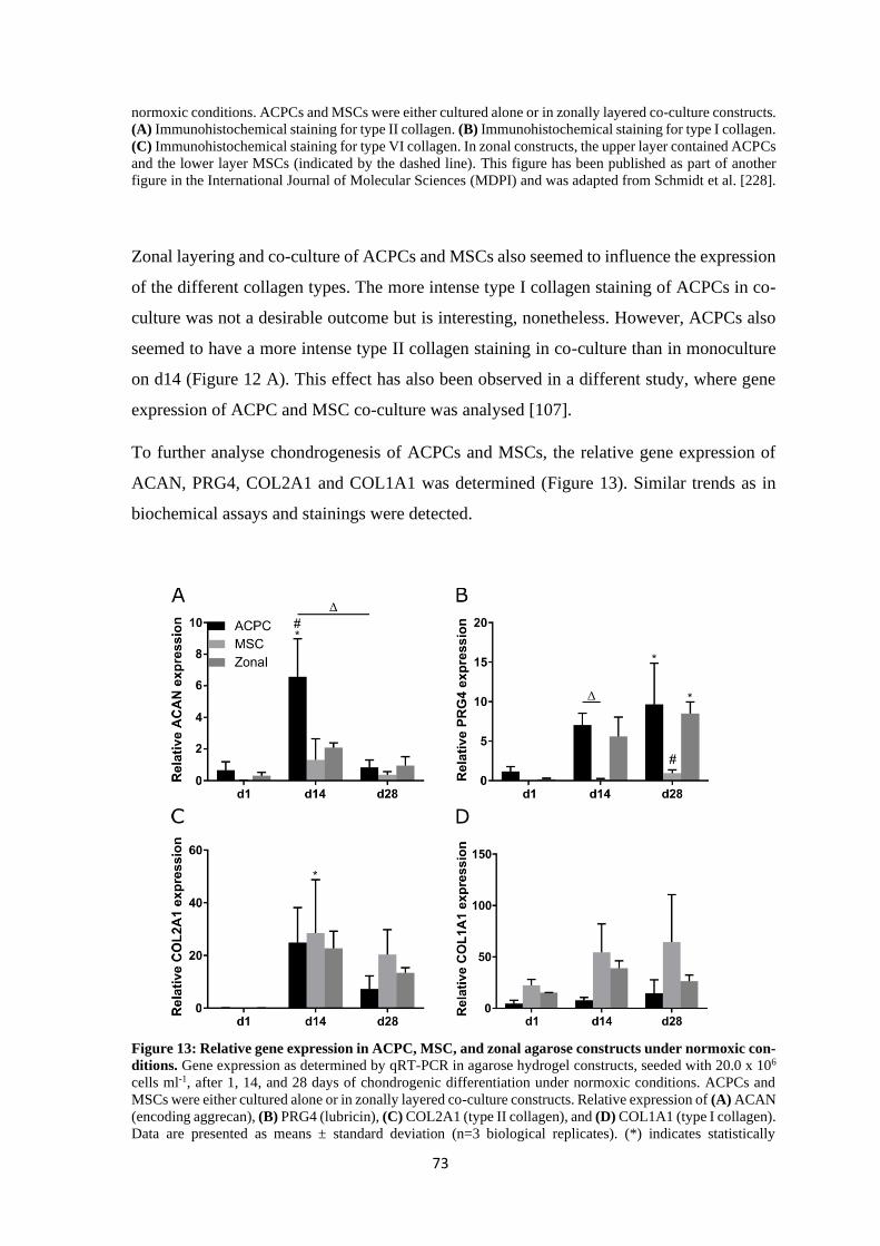
73
normoxic conditions. ACPCs and MSCs were either cultured alone or in zonally layered co-culture constructs.
(A) Immunohistochemical staining for type II collagen. (B) Immunohistochemical staining for type I collagen.
(C) Immunohistochemical staining for type VI collagen. In zonal constructs, the upper layer contained ACPCs
and the lower layer MSCs (indicated by the dashed line). This figure has been published as part of another
figure in the International Journal of Molecular Sciences (MDPI) and was adapted from Schmidt et al. [228].
Zonal layering and co-culture of ACPCs and MSCs also seemed to influence the expression
of the different collagen types. The more intense type I collagen staining of ACPCs in co-
culture was not a desirable outcome but is interesting, nonetheless. However, ACPCs also
seemed to have a more intense type II collagen staining in co-culture than in monoculture
on d14 (Figure 12 A). This effect has also been observed in a different study, where gene
expression of ACPC and MSC co-culture was analysed [107].
To further analyse chondrogenesis of ACPCs and MSCs, the relative gene expression of
ACAN, PRG4, COL2A1 and COL1A1 was determined (Figure 13). Similar trends as in
biochemical assays and stainings were detected.
Figure 13: Relative gene expression in ACPC, MSC, and zonal agarose constructs under normoxic con-
ditions. Gene expression as determined by qRT-PCR in agarose hydrogel constructs, seeded with 20.0 x 10⁶
cells ml-1, after 1, 14, and 28 days of chondrogenic differentiation under normoxic conditions. ACPCs and
MSCs were either cultured alone or in zonally layered co-culture constructs. Relative expression of (A) ACAN
(encoding aggrecan), (B) PRG4 (lubricin), (C) COL2A1 (type II collagen), and (D) COL1A1 (type I collagen).
Data are presented as means ± standard deviation (n=3 biological replicates). (*) indicates statistically
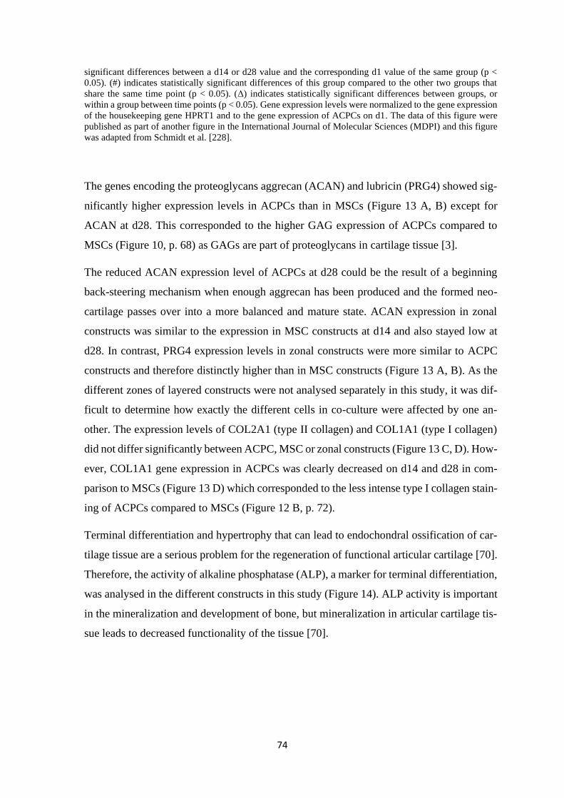
74
significant differences between a d14 or d28 value and the corresponding d1 value of the same group (p <
0.05). (#) indicates statistically significant differences of this group compared to the other two groups that
share the same time point (p < 0.05). (Δ) indicates statistically significant differences between groups, or
within a group between time points (p < 0.05). Gene expression levels were normalized to the gene expression
of the housekeeping gene HPRT1 and to the gene expression of ACPCs on d1. The data of this figure were
published as part of another figure in the International Journal of Molecular Sciences (MDPI) and this figure
was adapted from Schmidt et al. [228].
The genes encoding the proteoglycans aggrecan (ACAN) and lubricin (PRG4) showed sig-
nificantly higher expression levels in ACPCs than in MSCs (Figure 13 A, B) except for
ACAN at d28. This corresponded to the higher GAG expression of ACPCs compared to
MSCs (Figure 10, p. 68) as GAGs are part of proteoglycans in cartilage tissue [3].
The reduced ACAN expression level of ACPCs at d28 could be the result of a beginning
back-steering mechanism when enough aggrecan has been produced and the formed neo-
cartilage passes over into a more balanced and mature state. ACAN expression in zonal
constructs was similar to the expression in MSC constructs at d14 and also stayed low at
d28. In contrast, PRG4 expression levels in zonal constructs were more similar to ACPC
constructs and therefore distinctly higher than in MSC constructs (Figure 13 A, B). As the
different zones of layered constructs were not analysed separately in this study, it was dif-
ficult to determine how exactly the different cells in co-culture were affected by one an-
other. The expression levels of COL2A1 (type II collagen) and COL1A1 (type I collagen)
did not differ significantly between ACPC, MSC or zonal constructs (Figure 13 C, D). How-
ever, COL1A1 gene expression in ACPCs was clearly decreased on d14 and d28 in com-
parison to MSCs (Figure 13 D) which corresponded to the less intense type I collagen stain-
ing of ACPCs compared to MSCs (Figure 12 B, p. 72).
Terminal differentiation and hypertrophy that can lead to endochondral ossification of car-
tilage tissue are a serious problem for the regeneration of functional articular cartilage [70].
Therefore, the activity of alkaline phosphatase (ALP), a marker for terminal differentiation,
was analysed in the different constructs in this study (Figure 14). ALP activity is important
in the mineralization and development of bone, but mineralization in articular cartilage tis-
sue leads to decreased functionality of the tissue [70].
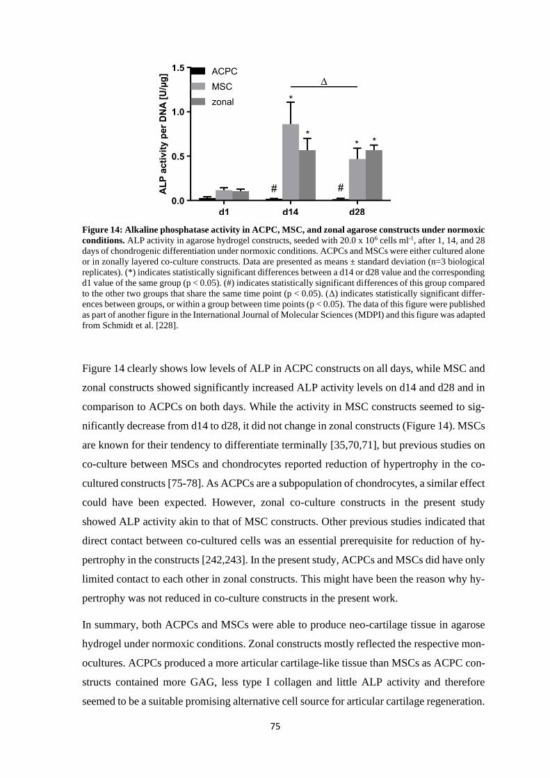
75
Figure 14: Alkaline phosphatase activity in ACPC, MSC, and zonal agarose constructs under normoxic
conditions. ALP activity in agarose hydrogel constructs, seeded with 20.0 x 10⁶ cells ml-1, after 1, 14, and 28
days of chondrogenic differentiation under normoxic conditions. ACPCs and MSCs were either cultured alone
or in zonally layered co-culture constructs. Data are presented as means ± standard deviation (n=3 biological
replicates). (*) indicates statistically significant differences between a d14 or d28 value and the corresponding
d1 value of the same group (p < 0.05). (#) indicates statistically significant differences of this group compared
to the other two groups that share the same time point (p < 0.05). (Δ) indicates statistically significant differ-
ences between groups, or within a group between time points (p < 0.05). The data of this figure were published
as part of another figure in the International Journal of Molecular Sciences (MDPI) and this figure was adapted
from Schmidt et al. [228].
Figure 14 clearly shows low levels of ALP in ACPC constructs on all days, while MSC and
zonal constructs showed significantly increased ALP activity levels on d14 and d28 and in
comparison to ACPCs on both days. While the activity in MSC constructs seemed to sig-
nificantly decrease from d14 to d28, it did not change in zonal constructs (Figure 14). MSCs
are known for their tendency to differentiate terminally [35,70,71], but previous studies on
co-culture between MSCs and chondrocytes reported reduction of hypertrophy in the co-
cultured constructs [75-78]. As ACPCs are a subpopulation of chondrocytes, a similar effect
could have been expected. However, zonal co-culture constructs in the present study
showed ALP activity akin to that of MSC constructs. Other previous studies indicated that
direct contact between co-cultured cells was an essential prerequisite for reduction of hy-
pertrophy in the constructs [242,243]. In the present study, ACPCs and MSCs did have only
limited contact to each other in zonal constructs. This might have been the reason why hy-
pertrophy was not reduced in co-culture constructs in the present work.
In summary, both ACPCs and MSCs were able to produce neo-cartilage tissue in agarose
hydrogel under normoxic conditions. Zonal constructs mostly reflected the respective mon-
ocultures. ACPCs produced a more articular cartilage-like tissue than MSCs as ACPC con-
structs contained more GAG, less type I collagen and little ALP activity and therefore
seemed to be a suitable promising alternative cell source for articular cartilage regeneration.
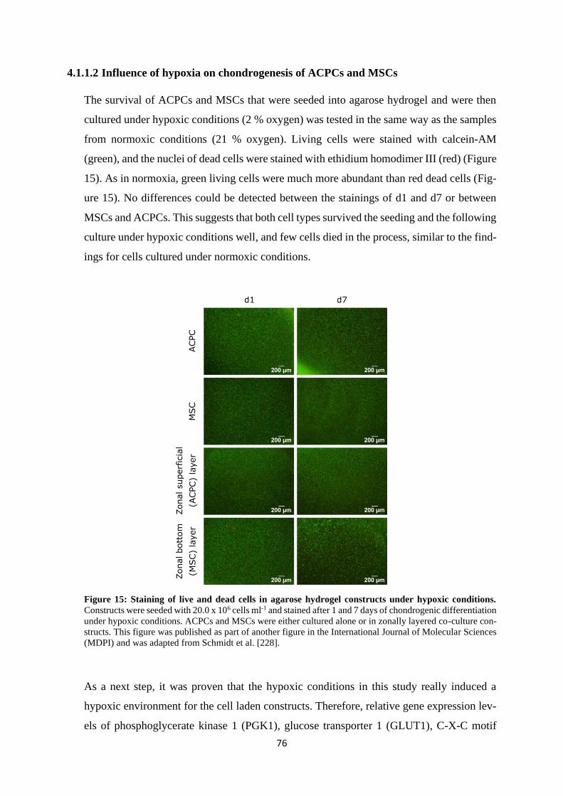
76
4.1.1.2 Influence of hypoxia on chondrogenesis of ACPCs and MSCs
The survival of ACPCs and MSCs that were seeded into agarose hydrogel and were then
cultured under hypoxic conditions (2 % oxygen) was tested in the same way as the samples
from normoxic conditions (21 % oxygen). Living cells were stained with calcein-AM
(green), and the nuclei of dead cells were stained with ethidium homodimer III (red) (Figure
15). As in normoxia, green living cells were much more abundant than red dead cells (Fig-
ure 15). No differences could be detected between the stainings of d1 and d7 or between
MSCs and ACPCs. This suggests that both cell types survived the seeding and the following
culture under hypoxic conditions well, and few cells died in the process, similar to the find-
ings for cells cultured under normoxic conditions.
Figure 15: Staining of live and dead cells in agarose hydrogel constructs under hypoxic conditions.
Constructs were seeded with 20.0 x 10⁶ cells ml-1 and stained after 1 and 7 days of chondrogenic differentiation
under hypoxic conditions. ACPCs and MSCs were either cultured alone or in zonally layered co-culture con-
structs. This figure was published as part of another figure in the International Journal of Molecular Sciences
(MDPI) and was adapted from Schmidt et al. [228].
As a next step, it was proven that the hypoxic conditions in this study really induced a
hypoxic environment for the cell laden constructs. Therefore, relative gene expression lev-
els of phosphoglycerate kinase 1 (PGK1), glucose transporter 1 (GLUT1), C-X-C motif
77
chemokine ligand 12 (CXCL12) and stanniocalcin 1 (STC1) were analysed. These are genes
that are known to be upregulated by hypoxia inducible factors (HIFs) as reaction to low
oxygen tension [244,245]. For all tested genes, relative expression was upregulated under
hypoxic conditions in comparison to normoxic conditions in both ACPCs and MSCs (Fig-
ure 16). STC1 gene expression was the only one that was not upregulated significantly but
with a strong upward trend. This demonstrated that the conditions used in this study were
able to provide a hypoxic environment for the cells and that they reacted to this environment
in the expected manner with upregulation of HIF target genes.
Figure 16: Relative gene expression of HIF target genes in ACPC and MSC agarose constructs. Gene
expression as determined by qRT-PCR in agarose hydrogel constructs, seeded with 20.0 x 10⁶ cells ml-1, after
7 days of chondrogenic differentiation under hypoxic (2 % O2) and normoxic (21 % O2) conditions. Relative
expression of PGK1 in (A) ACPC constructs and (B) MSC constructs. Relative expression of GLUT1 in (C)
ACPC constructs and (D) MSC constructs. Relative expression of CXCL12 in (E) ACPC constructs and (F)
MSC constructs. Relative expression of STC1 in (G) ACPC constructs and (H) MSC constructs. Data are
presented as means ± standard deviation (n=3 biological replicates). (*) indicates statistically significant dif-
ferences between two values (p < 0.05). Gene expression levels of HIF target genes were normalized to the
gene expression of the housekeeping gene HPRT1 and to the gene expression levels of the respective normoxia
values. This figure has been published in part in the International Journal of Molecular Sciences (MDPI) and
was adapted from Schmidt et al. [228].
The DNA amount of constructs cultured in normoxia or hypoxia was compared to identify
if cells grew differently in different oxygen environment. Figure 17 shows the different
DNA amounts of constructs at d1, d14 and d28 under hypoxic or normoxic conditions.
Normoxia samples are shown as striped columns because they were already discussed be-
fore (Figure 9, p. 67) and are shown only as comparison to hypoxic samples. It was
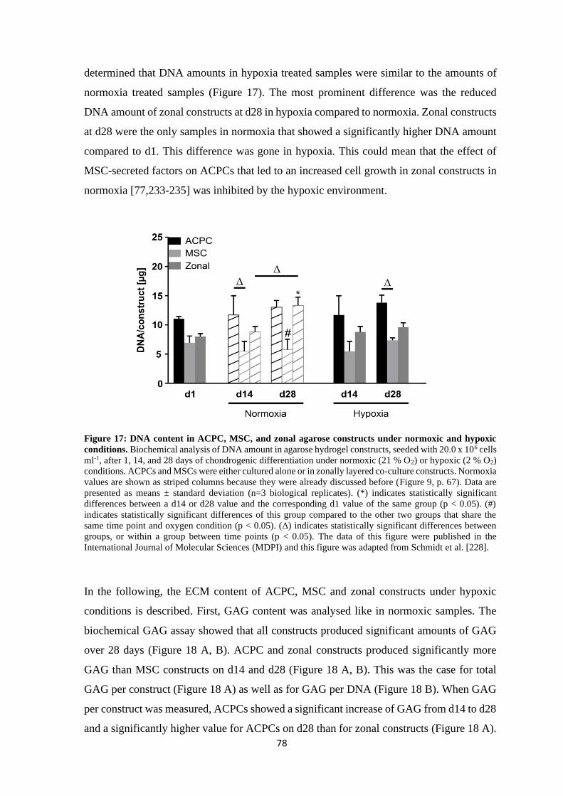
78
determined that DNA amounts in hypoxia treated samples were similar to the amounts of
normoxia treated samples (Figure 17). The most prominent difference was the reduced
DNA amount of zonal constructs at d28 in hypoxia compared to normoxia. Zonal constructs
at d28 were the only samples in normoxia that showed a significantly higher DNA amount
compared to d1. This difference was gone in hypoxia. This could mean that the effect of
MSC-secreted factors on ACPCs that led to an increased cell growth in zonal constructs in
normoxia [77,233-235] was inhibited by the hypoxic environment.
Figure 17: DNA content in ACPC, MSC, and zonal agarose constructs under normoxic and hypoxic
conditions. Biochemical analysis of DNA amount in agarose hydrogel constructs, seeded with 20.0 x 10⁶ cells
ml-1, after 1, 14, and 28 days of chondrogenic differentiation under normoxic (21 % O2) or hypoxic (2 % O2)
conditions. ACPCs and MSCs were either cultured alone or in zonally layered co-culture constructs. Normoxia
values are shown as striped columns because they were already discussed before (Figure 9, p. 67). Data are
presented as means ± standard deviation (n=3 biological replicates). (*) indicates statistically significant
differences between a d14 or d28 value and the corresponding d1 value of the same group (p < 0.05). (#)
indicates statistically significant differences of this group compared to the other two groups that share the
same time point and oxygen condition (p < 0.05). (Δ) indicates statistically significant differences between
groups, or within a group between time points (p < 0.05). The data of this figure were published in the
International Journal of Molecular Sciences (MDPI) and this figure was adapted from Schmidt et al. [228].
In the following, the ECM content of ACPC, MSC and zonal constructs under hypoxic
conditions is described. First, GAG content was analysed like in normoxic samples. The
biochemical GAG assay showed that all constructs produced significant amounts of GAG
over 28 days (Figure 18 A, B). ACPC and zonal constructs produced significantly more
GAG than MSC constructs on d14 and d28 (Figure 18 A, B). This was the case for total
GAG per construct (Figure 18 A) as well as for GAG per DNA (Figure 18 B). When GAG
per construct was measured, ACPCs showed a significant increase of GAG from d14 to d28
and a significantly higher value for ACPCs on d28 than for zonal constructs (Figure 18 A).
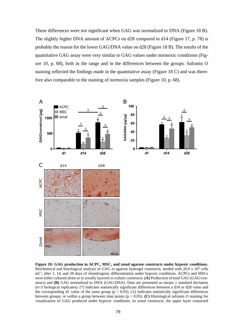
79
These differences were not significant when GAG was normalized to DNA (Figure 18 B).
The slightly higher DNA amount of ACPCs on d28 compared to d14 (Figure 17, p. 78) is
probably the reason for the lower GAG/DNA value on d28 (Figure 18 B). The results of the
quantitative GAG assay were very similar to GAG values under normoxic conditions (Fig-
ure 10, p. 68), both in the range and in the differences between the groups. Safranin O
staining reflected the findings made in the quantitative assay (Figure 18 C) and was there-
fore also comparable to the staining of normoxia samples (Figure 10, p. 68).
Figure 18: GAG production in ACPC, MSC, and zonal agarose constructs under hypoxic conditions.
Biochemical and histological analysis of GAG in agarose hydrogel constructs, seeded with 20.0 x 10⁶ cells
ml-1, after 1, 14, and 28 days of chondrogenic differentiation under hypoxic conditions. ACPCs and MSCs
were either cultured alone or in zonally layered co-culture constructs. (A) Production of total GAG (GAG/con-
struct) and (B) GAG normalized to DNA (GAG/DNA). Data are presented as means ± standard deviation
(n=3 biological replicates). (*) indicates statistically significant differences between a d14 or d28 value and
the corresponding d1 value of the same group (p < 0.05). (Δ) indicates statistically significant differences
between groups, or within a group between time points (p < 0.05). (C) Histological safranin O staining for
visualization of GAG produced under hypoxic conditions. In zonal constructs, the upper layer contained
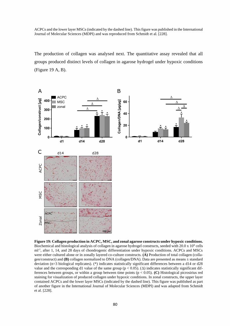
80
ACPCs and the lower layer MSCs (indicated by the dashed line). This figure was published in the International
Journal of Molecular Sciences (MDPI) and was reproduced from Schmidt et al. [228].
The production of collagen was analysed next. The quantitative assay revealed that all
groups produced distinct levels of collagen in agarose hydrogel under hypoxic conditions
(Figure 19 A, B).
Figure 19: Collagen production in ACPC, MSC, and zonal agarose constructs under hypoxic conditions.
Biochemical and histological analysis of collagen in agarose hydrogel constructs, seeded with 20.0 x 10⁶ cells
ml-1, after 1, 14, and 28 days of chondrogenic differentiation under hypoxic conditions. ACPCs and MSCs
were either cultured alone or in zonally layered co-culture constructs. (A) Production of total collagen (colla-
gen/construct) and (B) collagen normalized to DNA (collagen/DNA). Data are presented as means ± standard
deviation (n=3 biological replicates). (*) indicates statistically significant differences between a d14 or d28
value and the corresponding d1 value of the same group (p < 0.05). (Δ) indicates statistically significant dif-
ferences between groups, or within a group between time points (p < 0.05). (C) Histological picrosirius red
staining for visualization of produced collagen under hypoxic conditions. In zonal constructs, the upper layer
contained ACPCs and the lower layer MSCs (indicated by the dashed line). This figure was published as part
of another figure in the International Journal of Molecular Sciences (MDPI) and was adapted from Schmidt
et al. [228].
81
The amount of collagen per construct increased significantly from d1 to d14 and from d14
to d28 (Figure 19 A). The same was true for samples that were cultured under normoxic
conditions (Figure 11 A, p. 70), however, in hypoxia, all values were only approximately
half of the respective values under normoxic conditions (Figure 19 A). When collagen was
normalized to DNA (Figure 19 B), MSC and zonal constructs significantly increased their
collagen content from d1 to d14 and from d14 to d28. ACPC constructs on d28 were sig-
nificantly higher than on d1. MSCs produced significantly more collagen than ACPCs and
zonal constructs on d28. Collagen/DNA values were, like total collagen values, approxi-
mately half as high in hypoxia (Figure 19 B) as in normoxia (Figure 11 B, p. 70). This effect
was also visible in the picrosirius red stainings (Figure 19 C) that were weaker than
picrosirius red stainings under normoxic conditions (Figure 11 C, p. 70). Altogether, hy-
poxia reduced collagen production in all three groups in agarose hydrogel.
The production of different types of collagen (type I, II and VI) was analysed under hypoxic
conditions (Figure 20) to get an insight on how it was changed by low oxygen pressure and
which of the different collagens was affected by the decrease of total collagen in hypoxia
(Figure 19, p. 80). Type II collagen (Figure 20 A) was stained more intensely in ACPC
monoculture constructs compared to MSC constructs and zonal constructs showed a more
intense staining for ACPCs in the upper layer than in monoculture (Figure 20 A). ACPC
constructs displayed distinctly less intense staining for type I collagen (Figure 20 B) than
MSC constructs. However, type I collagen staining was considerably weaker in the zonal
lower layer with MSCs than in the MSC monoculture (Figure 20 B). MSCs and ACPCs
showed distinctly weaker staining for type II collagen in hypoxia (Figure 20 A) compared
to normoxia (Figure 12 A, p. 72). This decrease was more pronounced in MSCs and also
visible in the lower layer of zonal constructs (Figure 20 A). Type VI collagen (Figure 20 C)
staining was very similar to the staining of normoxia samples (Figure 12 C, p. 72) as it was
mostly pericellular around the cells for both ACPCs and MSC and monocultures as well as
zonal co-cultures. This type VI collagen localisation was similar to the distribution in native
articular cartilage [237].
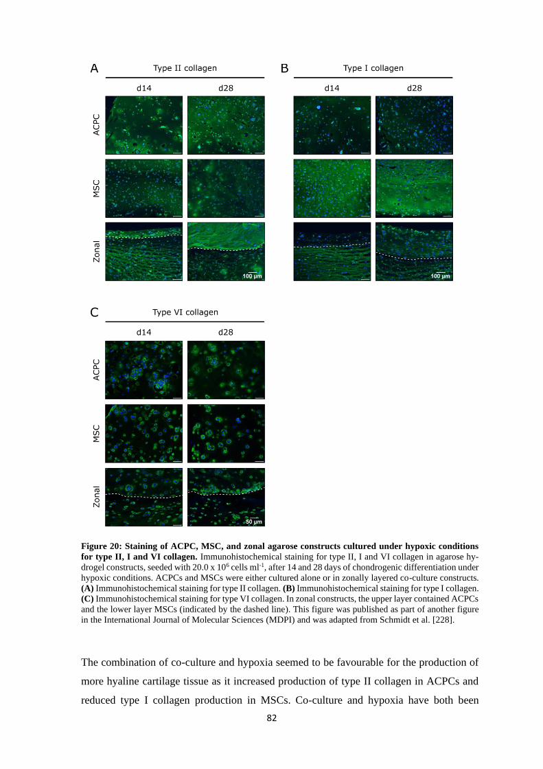
82
Figure 20: Staining of ACPC, MSC, and zonal agarose constructs cultured under hypoxic conditions
for type II, I and VI collagen. Immunohistochemical staining for type II, I and VI collagen in agarose hy-
drogel constructs, seeded with 20.0 x 10⁶ cells ml-1, after 14 and 28 days of chondrogenic differentiation under
hypoxic conditions. ACPCs and MSCs were either cultured alone or in zonally layered co-culture constructs.
(A) Immunohistochemical staining for type II collagen. (B) Immunohistochemical staining for type I collagen.
(C) Immunohistochemical staining for type VI collagen. In zonal constructs, the upper layer contained ACPCs
and the lower layer MSCs (indicated by the dashed line). This figure was published as part of another figure
in the International Journal of Molecular Sciences (MDPI) and was adapted from Schmidt et al. [228].
The combination of co-culture and hypoxia seemed to be favourable for the production of
more hyaline cartilage tissue as it increased production of type II collagen in ACPCs and
reduced type I collagen production in MSCs. Co-culture and hypoxia have both been
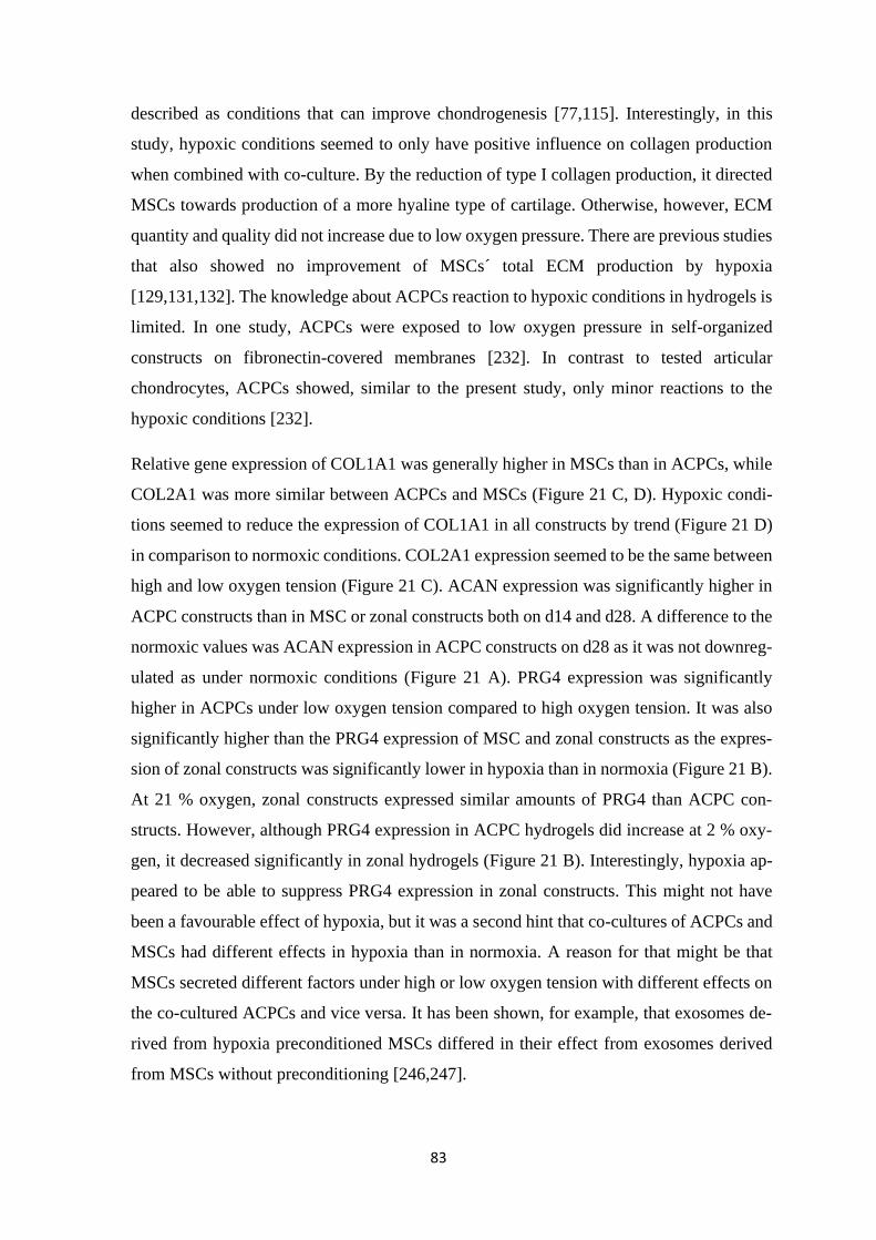
83
described as conditions that can improve chondrogenesis [77,115]. Interestingly, in this
study, hypoxic conditions seemed to only have positive influence on collagen production
when combined with co-culture. By the reduction of type I collagen production, it directed
MSCs towards production of a more hyaline type of cartilage. Otherwise, however, ECM
quantity and quality did not increase due to low oxygen pressure. There are previous studies
that also showed no improvement of MSCs´ total ECM production by hypoxia
[129,131,132]. The knowledge about ACPCs reaction to hypoxic conditions in hydrogels is
limited. In one study, ACPCs were exposed to low oxygen pressure in self-organized
constructs on fibronectin-covered membranes [232]. In contrast to tested articular
chondrocytes, ACPCs showed, similar to the present study, only minor reactions to the
hypoxic conditions [232].
Relative gene expression of COL1A1 was generally higher in MSCs than in ACPCs, while
COL2A1 was more similar between ACPCs and MSCs (Figure 21 C, D). Hypoxic condi-
tions seemed to reduce the expression of COL1A1 in all constructs by trend (Figure 21 D)
in comparison to normoxic conditions. COL2A1 expression seemed to be the same between
high and low oxygen tension (Figure 21 C). ACAN expression was significantly higher in
ACPC constructs than in MSC or zonal constructs both on d14 and d28. A difference to the
normoxic values was ACAN expression in ACPC constructs on d28 as it was not downreg-
ulated as under normoxic conditions (Figure 21 A). PRG4 expression was significantly
higher in ACPCs under low oxygen tension compared to high oxygen tension. It was also
significantly higher than the PRG4 expression of MSC and zonal constructs as the expres-
sion of zonal constructs was significantly lower in hypoxia than in normoxia (Figure 21 B).
At 21 % oxygen, zonal constructs expressed similar amounts of PRG4 than ACPC con-
structs. However, although PRG4 expression in ACPC hydrogels did increase at 2 % oxy-
gen, it decreased significantly in zonal hydrogels (Figure 21 B). Interestingly, hypoxia ap-
peared to be able to suppress PRG4 expression in zonal constructs. This might not have
been a favourable effect of hypoxia, but it was a second hint that co-cultures of ACPCs and
MSCs had different effects in hypoxia than in normoxia. A reason for that might be that
MSCs secreted different factors under high or low oxygen tension with different effects on
the co-cultured ACPCs and vice versa. It has been shown, for example, that exosomes de-
rived from hypoxia preconditioned MSCs differed in their effect from exosomes derived
from MSCs without preconditioning [246,247].
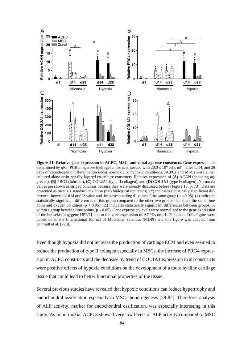
84
Figure 21: Relative gene expression in ACPC, MSC, and zonal agarose constructs. Gene expression as
determined by qRT-PCR in agarose hydrogel constructs, seeded with 20.0 x 10⁶ cells ml-1, after 1, 14, and 28
days of chondrogenic differentiation under normoxic or hypoxic conditions. ACPCs and MSCs were either
cultured alone or in zonally layered co-culture constructs. Relative expression of (A) ACAN (encoding ag-
grecan), (B) PRG4 (lubricin), (C) COL2A1 (type II collagen), and (D) COL1A1 (type I collagen). Normoxia
values are shown as striped columns because they were already discussed before (Figure 13, p. 73). Data are
presented as means ± standard deviation (n=3 biological replicates). (*) indicates statistically significant dif-
ferences between a d14 or d28 value and the corresponding d1 value of the same group (p < 0.05). (#) indicates
statistically significant differences of this group compared to the other two groups that share the same time
point and oxygen condition (p < 0.05). (Δ) indicates statistically significant differences between groups, or
within a group between time points (p < 0.05). Gene expression levels were normalized to the gene expression
of the housekeeping gene HPRT1 and to the gene expression of ACPCs on d1. The data of this figure were
published in the International Journal of Molecular Sciences (MDPI) and this figure was adapted from
Schmidt et al. [228].
Even though hypoxia did not increase the production of cartilage ECM and even seemed to
reduce the production of type II collagen especially in MSCs, the increase of PRG4 expres-
sion in ACPC constructs and the decrease by trend of COL1A1 expression in all constructs
were positive effects of hypoxic conditions on the development of a more hyaline cartilage
tissue that could lead to better functional properties of the tissue.
Several previous studies have revealed that hypoxic conditions can reduce hypertrophy and
endochondral ossification especially in MSC chondrogenesis [79-82]. Therefore, analysis
of ALP activity, marker for endochondral ossification, was especially interesting in this
study. As in normoxia, ACPCs showed very low levels of ALP activity compared to MSC
85
and zonal constructs. However, ALP activity of MSC and zonal constructs was significantly
reduced by hypoxia both on d14 and d28 (Figure 22). These findings were in concert with
reports from the literature [80,81,102]. Together with the downregulation of COL1A1 ex-
pression, the reduction of ALP activity in MSCs showed that hypoxia had positive effects
on the quality of cartilage tissue formed by MSCs. However, ACPC constructs displayed
exceptionally low ALP activity under all conditions, which appeared to make them, when
combined with agarose gels, more suitable for the formation of stable hyaline cartilage tis-
sue.
Figure 22: Alkaline phosphatase activity in ACPC, MSC, and zonal agarose constructs. ALP activity in
agarose hydrogel constructs, seeded with 20.0 x 10⁶ cells ml-1, after 1, 14, and 28 days of chondrogenic dif-
ferentiation under normoxic and hypoxic conditions. ACPCs and MSCs were either cultured alone or in zon-
ally layered co-culture constructs. Normoxia values are shown as striped columns because they were already
discussed before (Figure 14, p. 75). Data are presented as means ± standard deviation (n=3 biological repli-
cates). (*) indicates statistically significant differences between a d14 or d28 value and the corresponding d1
value of the same group (p < 0.05). (#) indicates statistically significant differences of this group compared to
the other two groups that share the same time point and oxygen condition (p < 0.05). (Δ) indicates statistically
significant differences between groups, or within a group between time points (p < 0.05). The data of this
figure were published in the International Journal of Molecular Sciences (MDPI) and this figure was adapted
from Schmidt et al. [228].
86
4.1.1.3 Summary of chondrogenesis of ACPCs and MSCs in agarose hydrogel
In the present study, three different constructs were tested: Agarose hydrogels laden with
ACPCs, MSCs or both, in zonal layers with ACPCs on top and MSCs in the lower layer.
They were compared in their ability to differentiate chondrogenically and produce cartilage
neo-tissue similar to hyaline articular cartilage under normoxic (21 % oxygen) and hypoxic
(2 % oxygen) conditions. All three constructs were able to produce distinct amounts of car-
tilage ECM in all conditions, however, quality and quantity of the formed tissue were dif-
ferent between the three groups. ACPCs produced in general more GAG and proteoglycans
than MSCs, while MSCs produced more total collagen. Values for zonal constructs mostly
lay in between those of ACPCs and MSCs. When collagen content was analysed further, it
was shown that ACPCs produced much less type I collagen than MSCs under both normoxic
and hypoxic condition. ACPCs also had very low levels of ALP activity compared to MSCs.
Hypoxia reduced production of collagen in general but especially in MSCs, and signifi-
cantly decreased ALP activity in MSC and zonal constructs. Gene expression of PRG4 in
ACPCs was increased by low oxygen pressure, and gene expression of type I collagen was
decreased by trend in all groups. Hypoxia in combination with co-culture seemed to increase
production of type II collagen in ACPCs and reduced type I collagen production in MSCs
while co-culture alone seemed to increase type I and type II collagen production in ACPCs.
Hypoxia also decreased relative PRG4 gene expression in zonal constructs. Taken together,
hypoxia appeared, despite the reduction of type II collagen production, to improve the car-
tilage tissue formed by MSCs by reducing type I collagen gene expression and ALP activity.
The resulting tissue was less fibrocartilaginous and less hypertroph than the tissue produced
by MSCs under normoxic conditions. Zonal co-cultures appeared to have influence on the
type II collagen/type I collagen ratio and were affected differently by hypoxia than mono-
cultures. It is possible that factors secreted by MSCs (or ACPCs) that are responsible for
the different effects in co-culture also differ between normoxia and hypoxia. This could
explain differences between mono- und co-culture in hypoxia and normoxia. It has been
shown, for example, that exosomes derived from hypoxia preconditioned MSCs differed in
their effect from exosomes derived from MSCs without preconditioning [246,247]. That
would be an interesting topic to explore in further experiments.
Taken together, in agarose hydrogels, ACPCs outperformed MSC as well as zonal con-
structs, as they produced more GAG and proteoglycans and similar levels of type II colla-
gen, while their type I collagen content was much lower. Additionally, ACPCs showed only
little tendency towards terminal differentiation (shown by ALP activity), regardless of

87
oxygen tension. ACPCs produced cartilage tissue that was more similar to hyaline cartilage
tissue in both normoxia and hypoxia. Therefore, in this study in agarose hydrogels, they
appeared to be a better choice for cartilage tissue engineering than MSCs.
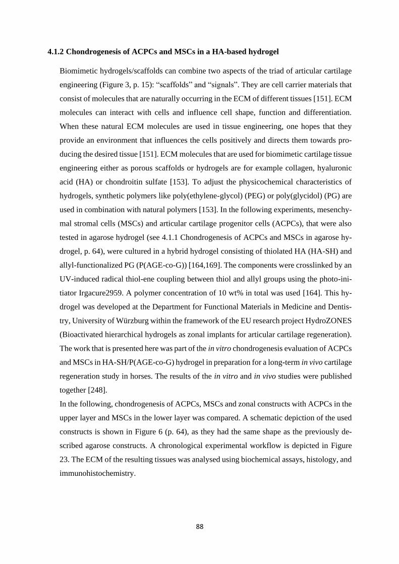
88
4.1.2 Chondrogenesis of ACPCs and MSCs in a HA-based hydrogel
Biomimetic hydrogels/scaffolds can combine two aspects of the triad of articular cartilage
engineering (Figure 3, p. 15): “scaffolds” and “signals”. They are cell carrier materials that
consist of molecules that are naturally occurring in the ECM of different tissues [151]. ECM
molecules can interact with cells and influence cell shape, function and differentiation.
When these natural ECM molecules are used in tissue engineering, one hopes that they
provide an environment that influences the cells positively and directs them towards pro-
ducing the desired tissue [151]. ECM molecules that are used for biomimetic cartilage tissue
engineering either as porous scaffolds or hydrogels are for example collagen, hyaluronic
acid (HA) or chondroitin sulfate [153]. To adjust the physicochemical characteristics of
hydrogels, synthetic polymers like poly(ethylene-glycol) (PEG) or poly(glycidol) (PG) are
used in combination with natural polymers [153]. In the following experiments, mesenchy-
mal stromal cells (MSCs) and articular cartilage progenitor cells (ACPCs), that were also
tested in agarose hydrogel (see 4.1.1 Chondrogenesis of ACPCs and MSCs in agarose hy-
drogel, p. 64), were cultured in a hybrid hydrogel consisting of thiolated HA (HA-SH) and
allyl-functionalized PG (P(AGE-co-G)) [164,169]. The components were crosslinked by an
UV-induced radical thiol-ene coupling between thiol and allyl groups using the photo-ini-
tiator Irgacure2959. A polymer concentration of 10 wt% in total was used [164]. This hy-
drogel was developed at the Department for Functional Materials in Medicine and Dentis-
try, University of Würzburg within the framework of the EU research project HydroZONES
(Bioactivated hierarchical hydrogels as zonal implants for articular cartilage regeneration).
The work that is presented here was part of the in vitro chondrogenesis evaluation of ACPCs
and MSCs in HA-SH/P(AGE-co-G) hydrogel in preparation for a long-term in vivo cartilage
regeneration study in horses. The results of the in vitro and in vivo studies were published
together [248].
In the following, chondrogenesis of ACPCs, MSCs and zonal constructs with ACPCs in the
upper layer and MSCs in the lower layer was compared. A schematic depiction of the used
constructs is shown in Figure 6 (p. 64), as they had the same shape as the previously de-
scribed agarose constructs. A chronological experimental workflow is depicted in Figure
23. The ECM of the resulting tissues was analysed using biochemical assays, histology, and
immunohistochemistry.
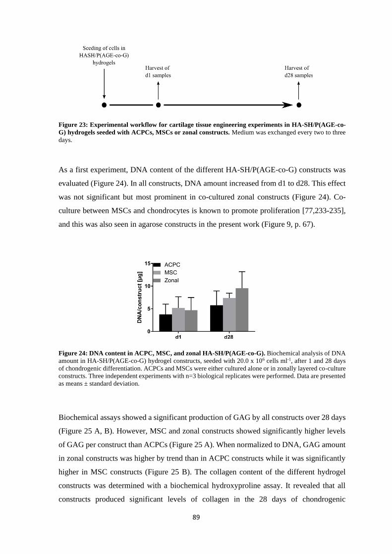
89
Figure 23: Experimental workflow for cartilage tissue engineering experiments in HA-SH/P(AGE-co-
G) hydrogels seeded with ACPCs, MSCs or zonal constructs. Medium was exchanged every two to three
days.
As a first experiment, DNA content of the different HA-SH/P(AGE-co-G) constructs was
evaluated (Figure 24). In all constructs, DNA amount increased from d1 to d28. This effect
was not significant but most prominent in co-cultured zonal constructs (Figure 24). Co-
culture between MSCs and chondrocytes is known to promote proliferation [77,233-235],
and this was also seen in agarose constructs in the present work (Figure 9, p. 67).
Figure 24: DNA content in ACPC, MSC, and zonal HA-SH/P(AGE-co-G). Biochemical analysis of DNA
amount in HA-SH/P(AGE-co-G) hydrogel constructs, seeded with 20.0 x 10⁶ cells ml-1, after 1 and 28 days
of chondrogenic differentiation. ACPCs and MSCs were either cultured alone or in zonally layered co-culture
constructs. Three independent experiments with n=3 biological replicates were performed. Data are presented
as means ± standard deviation.
Biochemical assays showed a significant production of GAG by all constructs over 28 days
(Figure 25 A, B). However, MSC and zonal constructs showed significantly higher levels
of GAG per construct than ACPCs (Figure 25 A). When normalized to DNA, GAG amount
in zonal constructs was higher by trend than in ACPC constructs while it was significantly
higher in MSC constructs (Figure 25 B). The collagen content of the different hydrogel
constructs was determined with a biochemical hydroxyproline assay. It revealed that all
constructs produced significant levels of collagen in the 28 days of chondrogenic
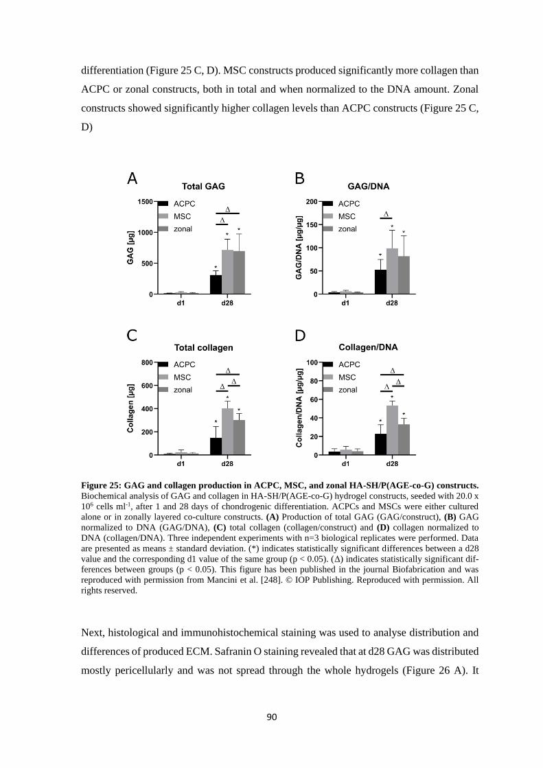
90
differentiation (Figure 25 C, D). MSC constructs produced significantly more collagen than
ACPC or zonal constructs, both in total and when normalized to the DNA amount. Zonal
constructs showed significantly higher collagen levels than ACPC constructs (Figure 25 C,
D)
Figure 25: GAG and collagen production in ACPC, MSC, and zonal HA-SH/P(AGE-co-G) constructs.
Biochemical analysis of GAG and collagen in HA-SH/P(AGE-co-G) hydrogel constructs, seeded with 20.0 x
106 cells ml-1, after 1 and 28 days of chondrogenic differentiation. ACPCs and MSCs were either cultured
alone or in zonally layered co-culture constructs. (A) Production of total GAG (GAG/construct), (B) GAG
normalized to DNA (GAG/DNA), (C) total collagen (collagen/construct) and (D) collagen normalized to
DNA (collagen/DNA). Three independent experiments with n=3 biological replicates were performed. Data
are presented as means ± standard deviation. (*) indicates statistically significant differences between a d28
value and the corresponding d1 value of the same group (p < 0.05). (Δ) indicates statistically significant dif-
ferences between groups (p < 0.05). This figure has been published in the journal Biofabrication and was
reproduced with permission from Mancini et al. [248]. © IOP Publishing. Reproduced with permission. All
rights reserved.
Next, histological and immunohistochemical staining was used to analyse distribution and
differences of produced ECM. Safranin O staining revealed that at d28 GAG was distributed
mostly pericellularly and was not spread through the whole hydrogels (Figure 26 A). It
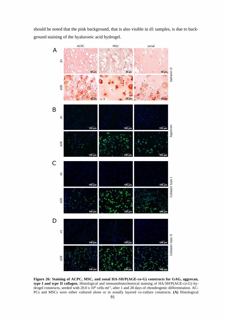
91
should be noted that the pink background, that is also visible in d1 samples, is due to back-
ground staining of the hyaluronic acid hydrogel.
Figure 26: Staining of ACPC, MSC, and zonal HA-SH/P(AGE-co-G) constructs for GAG, aggrecan,
type I and type II collagen. Histological and immunohistochemical staining of HA-SH/P(AGE-co-G) hy-
drogel constructs, seeded with 20.0 x 10⁶ cells ml-1, after 1 and 28 days of chondrogenic differentiation. AC-
PCs and MSCs were either cultured alone or in zonally layered co-culture constructs. (A) Histological
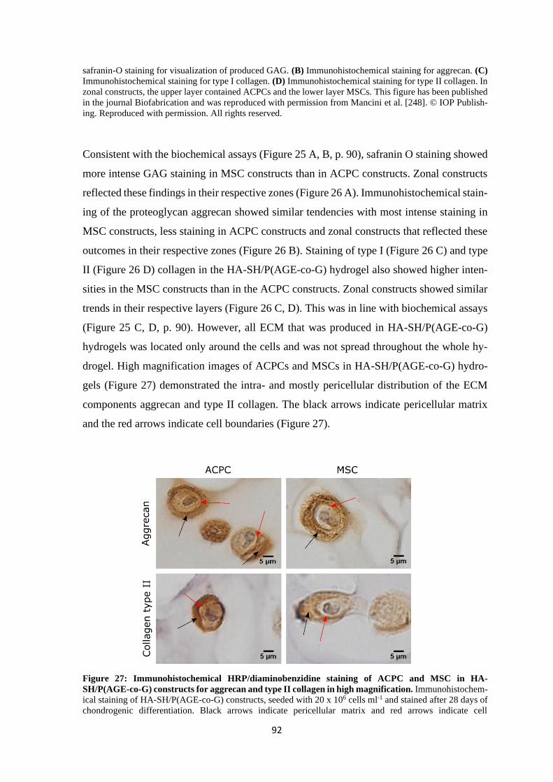
92
safranin-O staining for visualization of produced GAG. (B) Immunohistochemical staining for aggrecan. (C)
Immunohistochemical staining for type I collagen. (D) Immunohistochemical staining for type II collagen. In
zonal constructs, the upper layer contained ACPCs and the lower layer MSCs. This figure has been published
in the journal Biofabrication and was reproduced with permission from Mancini et al. [248]. © IOP Publish-
ing. Reproduced with permission. All rights reserved.
Consistent with the biochemical assays (Figure 25 A, B, p. 90), safranin O staining showed
more intense GAG staining in MSC constructs than in ACPC constructs. Zonal constructs
reflected these findings in their respective zones (Figure 26 A). Immunohistochemical stain-
ing of the proteoglycan aggrecan showed similar tendencies with most intense staining in
MSC constructs, less staining in ACPC constructs and zonal constructs that reflected these
outcomes in their respective zones (Figure 26 B). Staining of type I (Figure 26 C) and type
II (Figure 26 D) collagen in the HA-SH/P(AGE-co-G) hydrogel also showed higher inten-
sities in the MSC constructs than in the ACPC constructs. Zonal constructs showed similar
trends in their respective layers (Figure 26 C, D). This was in line with biochemical assays
(Figure 25 C, D, p. 90). However, all ECM that was produced in HA-SH/P(AGE-co-G)
hydrogels was located only around the cells and was not spread throughout the whole hy-
drogel. High magnification images of ACPCs and MSCs in HA-SH/P(AGE-co-G) hydro-
gels (Figure 27) demonstrated the intra- and mostly pericellular distribution of the ECM
components aggrecan and type II collagen. The black arrows indicate pericellular matrix
and the red arrows indicate cell boundaries (Figure 27).
Figure 27: Immunohistochemical HRP/diaminobenzidine staining of ACPC and MSC in HA-
SH/P(AGE-co-G) constructs for aggrecan and type II collagen in high magnification. Immunohistochem-
ical staining of HA-SH/P(AGE-co-G) constructs, seeded with 20 x 106 cells ml-1 and stained after 28 days of
chondrogenic differentiation. Black arrows indicate pericellular matrix and red arrows indicate cell
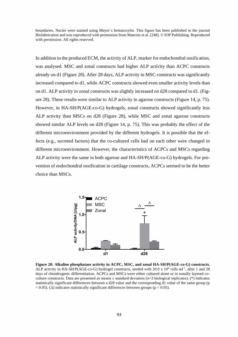
93
boundaries. Nuclei were stained using Mayer´s hematoxylin. This figure has been published in the journal
Biofabrication and was reproduced with permission from Mancini et al. [248]. © IOP Publishing. Reproduced
with permission. All rights reserved.
In addition to the produced ECM, the activity of ALP, marker for endochondral ossification,
was analysed. MSC and zonal constructs had higher ALP activity than ACPC constructs
already on d1 (Figure 28). After 28 days, ALP activity in MSC constructs was significantly
increased compared to d1, while ACPC constructs showed even smaller activity levels than
on d1. ALP activity in zonal constructs was slightly increased on d28 compared to d1. (Fig-
ure 28). These results were similar to ALP activity in agarose constructs (Figure 14, p. 75).
However, in HA-SH/P(AGE-co-G) hydrogels, zonal constructs showed significantly less
ALP activity than MSCs on d28 (Figure 28), while MSC and zonal agarose constructs
showed similar ALP levels on d28 (Figure 14, p. 75). This was probably the effect of the
different microenvironment provided by the different hydrogels. It is possible that the ef-
fects (e.g., secreted factors) that the co-cultured cells had on each other were changed in
different microenvironment. However, the characteristics of ACPCs and MSCs regarding
ALP activity were the same in both agarose and HA-SH/P(AGE-co-G) hydrogels. For pre-
vention of endochondral ossification in cartilage constructs, ACPCs seemed to be the better
choice than MSCs.
Figure 28: Alkaline phosphatase activity in ACPC, MSC, and zonal HA-SH/P(AGE-co-G) constructs.
ALP activity in HA-SH/P(AGE-co-G) hydrogel constructs, seeded with 20.0 x 10⁶ cells ml-1, after 1 and 28
days of chondrogenic differentiation. ACPCs and MSCs were either cultured alone or in zonally layered co-
culture constructs. Data are presented as means ± standard deviation (n=3 biological replicates). (*) indicates
statistically significant differences between a d28 value and the corresponding d1 value of the same group (p
< 0.05). (Δ) indicates statistically significant differences between groups (p < 0.05).
94
Summing up, ACPCs, MSCs and zonal constructs (consisting of an upper layer with ACPCs
and a lower layer with MSCs) were able to produce distinct amounts of cartilage ECM in
HA-SH/P(AGE-co-G) hydrogel. MSC constructs produced more GAG and collagen than
ACPC constructs, and zonal constructs mostly showed values that lay between those of
MSCs and ACPCs. While MSCs produced more ECM, ACPCs showed very small ALP
activity levels compared to MSC constructs. However, the pericellular ECM distribution in
all constructs was not optimal as it did not resemble natural ECM and did not lead to a
continuous, tissue-like construct. This effect has been seen before when high polymer con-
tent or high network density in hydrogels restricted homogenous ECM distribution of the
encapsulated cells [164,249-253]. Characteristics of crosslinked HA-SH/P(AGE-co-G) hy-
drogels also depend on the length of the used HA-SH and its degree of modification. In
general, the shorter the HA-SH, the more polymer is needed in total to generate a stable
hydrogel through crosslinking. If the polymer content is too small, no gel forms. The HA-
SH that was used here was relatively small and therefore, a high polymer content was
needed to form a gel with it. Additionally, the used hydrogel composition with a total pol-
ymer content of 10 wt% was originally chosen to produce a 3D-printable hydrogel. A high
concentration of polymers is often used for 3D-printing materials to improve stability and
shape fidelity during and after the printing process [150]. With 10 wt% polymer content
and additional unmodified high MW HA (1-2 MDa) to increase viscosity, it was possible
to double print the HA-SH/P(AGE-co-G) hydrogel together with polycaprolactone (PCL)
in a previous study [164]. As the hydrogel was not 3D-printed for the in vivo horse study
[248], the corresponding in vitro experiments in the present study were also performed with-
out 3D bioprinting. Another study with a very similar hydrogel system proved that a reduc-
tion of polymer content resulted in improved ECM distribution [169]. Therefore, the used
HA-SH/P(AGE-co-G) hydrogel should be developed further to find a way to combine the
needs of the used cells and, for example, the requirements for a stable hydrogel for bioprint-
ing with high shape fidelity.
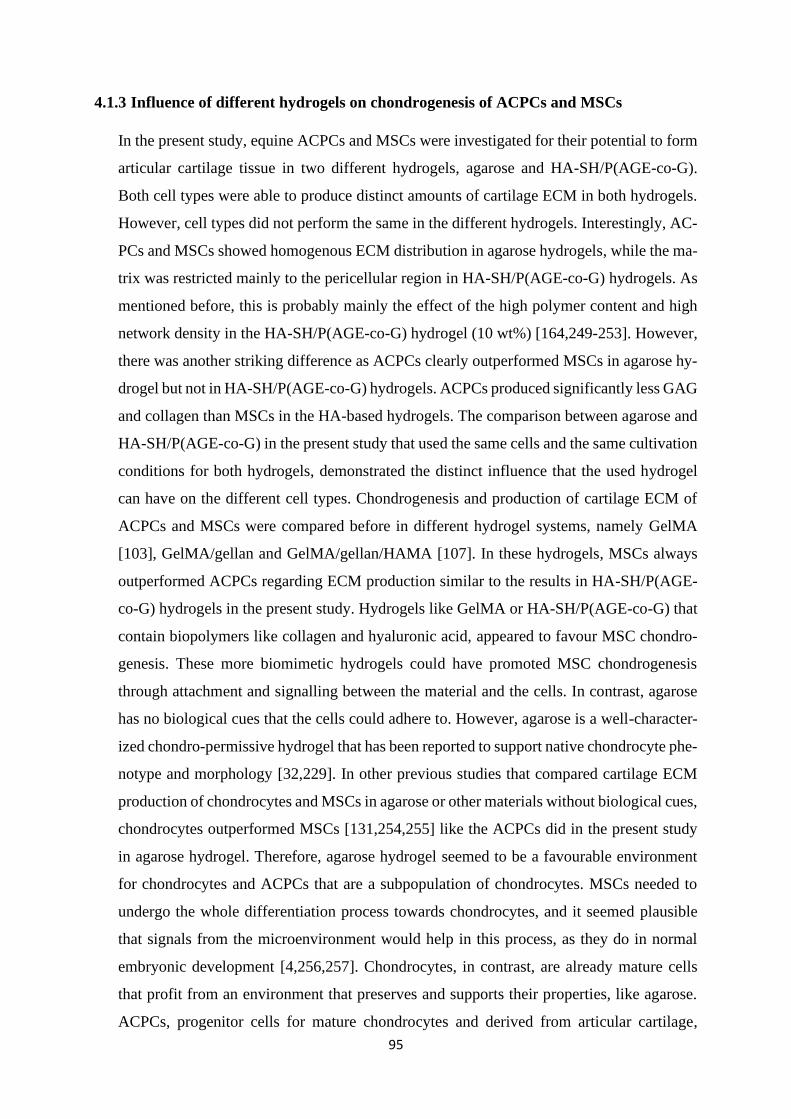
95
4.1.3 Influence of different hydrogels on chondrogenesis of ACPCs and MSCs
In the present study, equine ACPCs and MSCs were investigated for their potential to form
articular cartilage tissue in two different hydrogels, agarose and HA-SH/P(AGE-co-G).
Both cell types were able to produce distinct amounts of cartilage ECM in both hydrogels.
However, cell types did not perform the same in the different hydrogels. Interestingly, AC-
PCs and MSCs showed homogenous ECM distribution in agarose hydrogels, while the ma-
trix was restricted mainly to the pericellular region in HA-SH/P(AGE-co-G) hydrogels. As
mentioned before, this is probably mainly the effect of the high polymer content and high
network density in the HA-SH/P(AGE-co-G) hydrogel (10 wt%) [164,249-253]. However,
there was another striking difference as ACPCs clearly outperformed MSCs in agarose hy-
drogel but not in HA-SH/P(AGE-co-G) hydrogels. ACPCs produced significantly less GAG
and collagen than MSCs in the HA-based hydrogels. The comparison between agarose and
HA-SH/P(AGE-co-G) in the present study that used the same cells and the same cultivation
conditions for both hydrogels, demonstrated the distinct influence that the used hydrogel
can have on the different cell types. Chondrogenesis and production of cartilage ECM of
ACPCs and MSCs were compared before in different hydrogel systems, namely GelMA
[103], GelMA/gellan and GelMA/gellan/HAMA [107]. In these hydrogels, MSCs always
outperformed ACPCs regarding ECM production similar to the results in HA-SH/P(AGE-
co-G) hydrogels in the present study. Hydrogels like GelMA or HA-SH/P(AGE-co-G) that
contain biopolymers like collagen and hyaluronic acid, appeared to favour MSC chondro-
genesis. These more biomimetic hydrogels could have promoted MSC chondrogenesis
through attachment and signalling between the material and the cells. In contrast, agarose
has no biological cues that the cells could adhere to. However, agarose is a well-character-
ized chondro-permissive hydrogel that has been reported to support native chondrocyte phe-
notype and morphology [32,229]. In other previous studies that compared cartilage ECM
production of chondrocytes and MSCs in agarose or other materials without biological cues,
chondrocytes outperformed MSCs [131,254,255] like the ACPCs did in the present study
in agarose hydrogel. Therefore, agarose hydrogel seemed to be a favourable environment
for chondrocytes and ACPCs that are a subpopulation of chondrocytes. MSCs needed to
undergo the whole differentiation process towards chondrocytes, and it seemed plausible
that signals from the microenvironment would help in this process, as they do in normal
embryonic development [4,256,257]. Chondrocytes, in contrast, are already mature cells
that profit from an environment that preserves and supports their properties, like agarose.
ACPCs, progenitor cells for mature chondrocytes and derived from articular cartilage,

96
appeared to resemble chondrocytes in this regard. These results demonstrated clearly that it
is of paramount importance to choose the right microenvironment or hydrogel to achieve
the best performance of the different cell types.
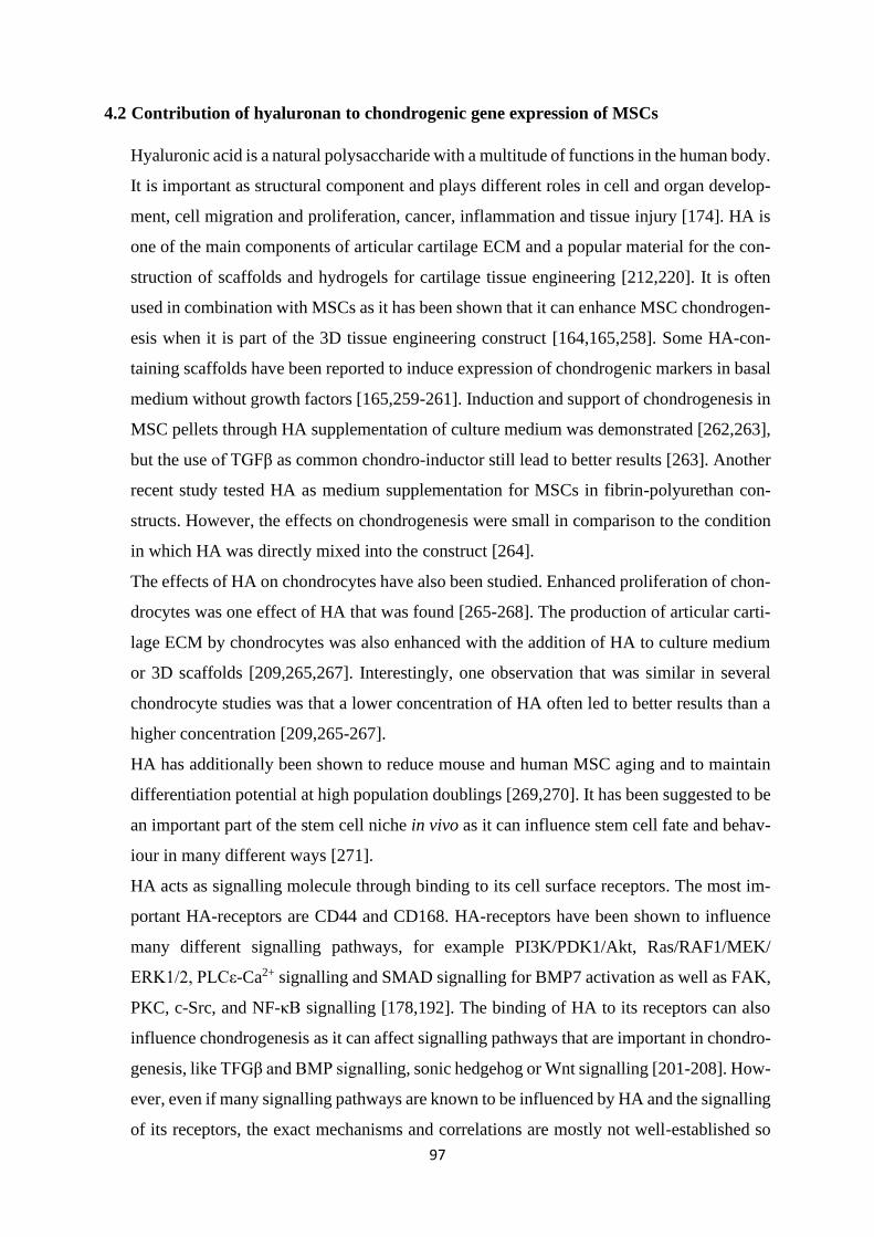
97
4.2 Contribution of hyaluronan to chondrogenic gene expression of MSCs
Hyaluronic acid is a natural polysaccharide with a multitude of functions in the human body.
It is important as structural component and plays different roles in cell and organ develop-
ment, cell migration and proliferation, cancer, inflammation and tissue injury [174]. HA is
one of the main components of articular cartilage ECM and a popular material for the con-
struction of scaffolds and hydrogels for cartilage tissue engineering [212,220]. It is often
used in combination with MSCs as it has been shown that it can enhance MSC chondrogen-
esis when it is part of the 3D tissue engineering construct [164,165,258]. Some HA-con-
taining scaffolds have been reported to induce expression of chondrogenic markers in basal
medium without growth factors [165,259-261]. Induction and support of chondrogenesis in
MSC pellets through HA supplementation of culture medium was demonstrated [262,263],
but the use of TGFβ as common chondro-inductor still lead to better results [263]. Another
recent study tested HA as medium supplementation for MSCs in fibrin-polyurethan con-
structs. However, the effects on chondrogenesis were small in comparison to the condition
in which HA was directly mixed into the construct [264].
The effects of HA on chondrocytes have also been studied. Enhanced proliferation of chon-
drocytes was one effect of HA that was found [265-268]. The production of articular carti-
lage ECM by chondrocytes was also enhanced with the addition of HA to culture medium
or 3D scaffolds [209,265,267]. Interestingly, one observation that was similar in several
chondrocyte studies was that a lower concentration of HA often led to better results than a
higher concentration [209,265-267].
HA has additionally been shown to reduce mouse and human MSC aging and to maintain
differentiation potential at high population doublings [269,270]. It has been suggested to be
an important part of the stem cell niche in vivo as it can influence stem cell fate and behav-
iour in many different ways [271].
HA acts as signalling molecule through binding to its cell surface receptors. The most im-
portant HA-receptors are CD44 and CD168. HA-receptors have been shown to influence
many different signalling pathways, for example PI3K/PDK1/Akt, Ras/RAF1/MEK/
ERK1/2, PLCε-Ca2+ signalling and SMAD signalling for BMP7 activation as well as FAK,
PKC, c-Src, and NF-κB signalling [178,192]. The binding of HA to its receptors can also
influence chondrogenesis as it can affect signalling pathways that are important in chondro-
genesis, like TFGβ and BMP signalling, sonic hedgehog or Wnt signalling [201-208]. How-
ever, even if many signalling pathways are known to be influenced by HA and the signalling
of its receptors, the exact mechanisms and correlations are mostly not well-established so
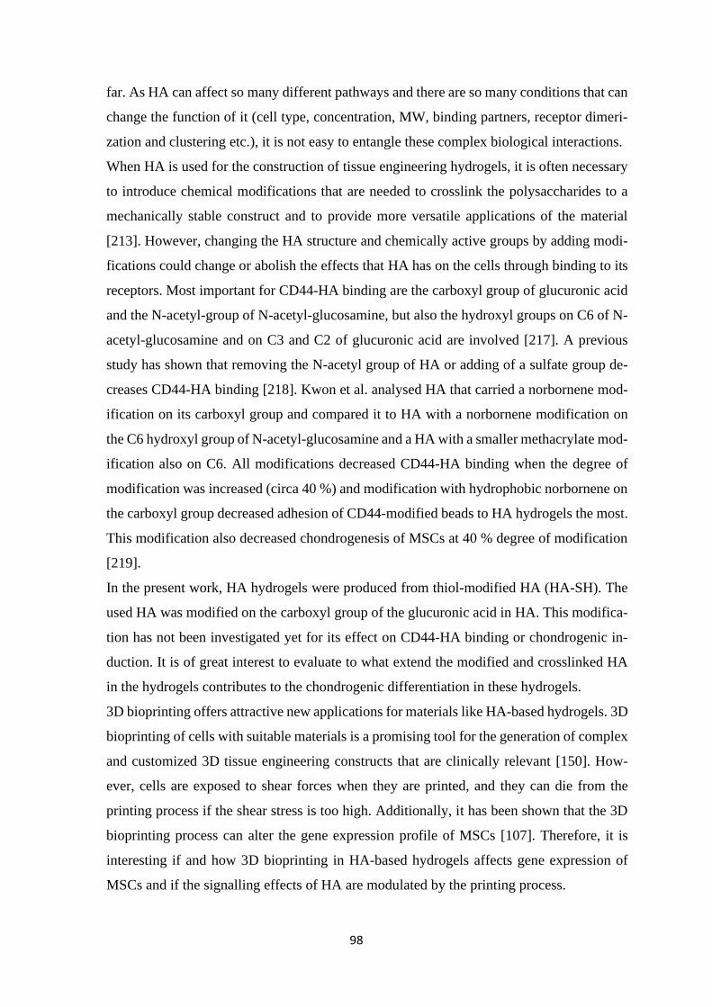
98
far. As HA can affect so many different pathways and there are so many conditions that can
change the function of it (cell type, concentration, MW, binding partners, receptor dimeri-
zation and clustering etc.), it is not easy to entangle these complex biological interactions.
When HA is used for the construction of tissue engineering hydrogels, it is often necessary
to introduce chemical modifications that are needed to crosslink the polysaccharides to a
mechanically stable construct and to provide more versatile applications of the material
[213]. However, changing the HA structure and chemically active groups by adding modi-
fications could change or abolish the effects that HA has on the cells through binding to its
receptors. Most important for CD44-HA binding are the carboxyl group of glucuronic acid
and the N-acetyl-group of N-acetyl-glucosamine, but also the hydroxyl groups on C6 of N-
acetyl-glucosamine and on C3 and C2 of glucuronic acid are involved [217]. A previous
study has shown that removing the N-acetyl group of HA or adding of a sulfate group de-
creases CD44-HA binding [218]. Kwon et al. analysed HA that carried a norbornene mod-
ification on its carboxyl group and compared it to HA with a norbornene modification on
the C6 hydroxyl group of N-acetyl-glucosamine and a HA with a smaller methacrylate mod-
ification also on C6. All modifications decreased CD44-HA binding when the degree of
modification was increased (circa 40 %) and modification with hydrophobic norbornene on
the carboxyl group decreased adhesion of CD44-modified beads to HA hydrogels the most.
This modification also decreased chondrogenesis of MSCs at 40 % degree of modification
[219].
In the present work, HA hydrogels were produced from thiol-modified HA (HA-SH). The
used HA was modified on the carboxyl group of the glucuronic acid in HA. This modifica-
tion has not been investigated yet for its effect on CD44-HA binding or chondrogenic in-
duction. It is of great interest to evaluate to what extend the modified and crosslinked HA
in the hydrogels contributes to the chondrogenic differentiation in these hydrogels.
3D bioprinting offers attractive new applications for materials like HA-based hydrogels. 3D
bioprinting of cells with suitable materials is a promising tool for the generation of complex
and customized 3D tissue engineering constructs that are clinically relevant [150]. How-
ever, cells are exposed to shear forces when they are printed, and they can die from the
printing process if the shear stress is too high. Additionally, it has been shown that the 3D
bioprinting process can alter the gene expression profile of MSCs [107]. Therefore, it is
interesting if and how 3D bioprinting in HA-based hydrogels affects gene expression of
MSCs and if the signalling effects of HA are modulated by the printing process.
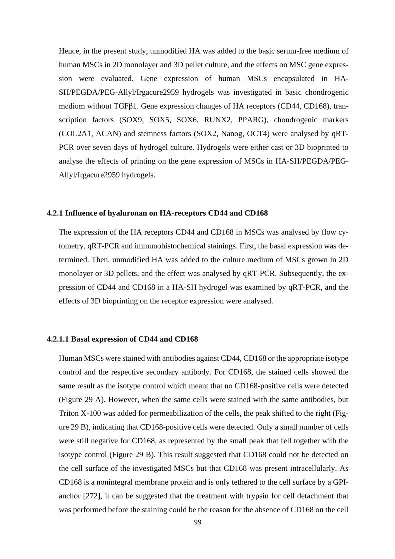
99
Hence, in the present study, unmodified HA was added to the basic serum-free medium of
human MSCs in 2D monolayer and 3D pellet culture, and the effects on MSC gene expres-
sion were evaluated. Gene expression of human MSCs encapsulated in HA-
SH/PEGDA/PEG-Allyl/Irgacure2959 hydrogels was investigated in basic chondrogenic
medium without TGFβ1. Gene expression changes of HA receptors (CD44, CD168), tran-
scription factors (SOX9, SOX5, SOX6, RUNX2, PPARG), chondrogenic markers
(COL2A1, ACAN) and stemness factors (SOX2, Nanog, OCT4) were analysed by qRT-
PCR over seven days of hydrogel culture. Hydrogels were either cast or 3D bioprinted to
analyse the effects of printing on the gene expression of MSCs in HA-SH/PEGDA/PEG-
Allyl/Irgacure2959 hydrogels.
4.2.1 Influence of hyaluronan on HA-receptors CD44 and CD168
The expression of the HA receptors CD44 and CD168 in MSCs was analysed by flow cy-
tometry, qRT-PCR and immunohistochemical stainings. First, the basal expression was de-
termined. Then, unmodified HA was added to the culture medium of MSCs grown in 2D
monolayer or 3D pellets, and the effect was analysed by qRT-PCR. Subsequently, the ex-
pression of CD44 and CD168 in a HA-SH hydrogel was examined by qRT-PCR, and the
effects of 3D bioprinting on the receptor expression were analysed.
4.2.1.1 Basal expression of CD44 and CD168
Human MSCs were stained with antibodies against CD44, CD168 or the appropriate isotype
control and the respective secondary antibody. For CD168, the stained cells showed the
same result as the isotype control which meant that no CD168-positive cells were detected
(Figure 29 A). However, when the same cells were stained with the same antibodies, but
Triton X-100 was added for permeabilization of the cells, the peak shifted to the right (Fig-
ure 29 B), indicating that CD168-positive cells were detected. Only a small number of cells
were still negative for CD168, as represented by the small peak that fell together with the
isotype control (Figure 29 B). This result suggested that CD168 could not be detected on
the cell surface of the investigated MSCs but that CD168 was present intracellularly. As
CD168 is a nonintegral membrane protein and is only tethered to the cell surface by a GPI-
anchor [272], it can be suggested that the treatment with trypsin for cell detachment that
was performed before the staining could be the reason for the absence of CD168 on the cell
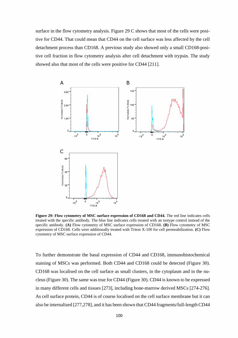
100
surface in the flow cytometry analysis. Figure 29 C shows that most of the cells were posi-
tive for CD44. That could mean that CD44 on the cell surface was less affected by the cell
detachment process than CD168. A previous study also showed only a small CD168-posi-
tive cell fraction in flow cytometry analysis after cell detachment with trypsin. The study
showed also that most of the cells were positive for CD44 [211].
Figure 29: Flow cytometry of MSC surface expression of CD168 and CD44. The red line indicates cells
treated with the specific antibody. The blue line indicates cells treated with an isotype control instead of the
specific antibody. (A) Flow cytometry of MSC surface expression of CD168. (B) Flow cytometry of MSC
expression of CD168. Cells were additionally treated with Triton X-100 for cell permeabilization. (C) Flow
cytometry of MSC surface expression of CD44.
To further demonstrate the basal expression of CD44 and CD168, immunohistochemical
staining of MSCs was performed. Both CD44 and CD168 could be detected (Figure 30).
CD168 was localised on the cell surface as small clusters, in the cytoplasm and in the nu-
cleus (Figure 30). The same was true for CD44 (Figure 30). CD44 is known to be expressed
in many different cells and tissues [273], including bone-marrow derived MSCs [274-276].
As cell surface protein, CD44 is of course localised on the cell surface membrane but it can
also be internalised [277,278], and it has been shown that CD44 fragments/full-length CD44
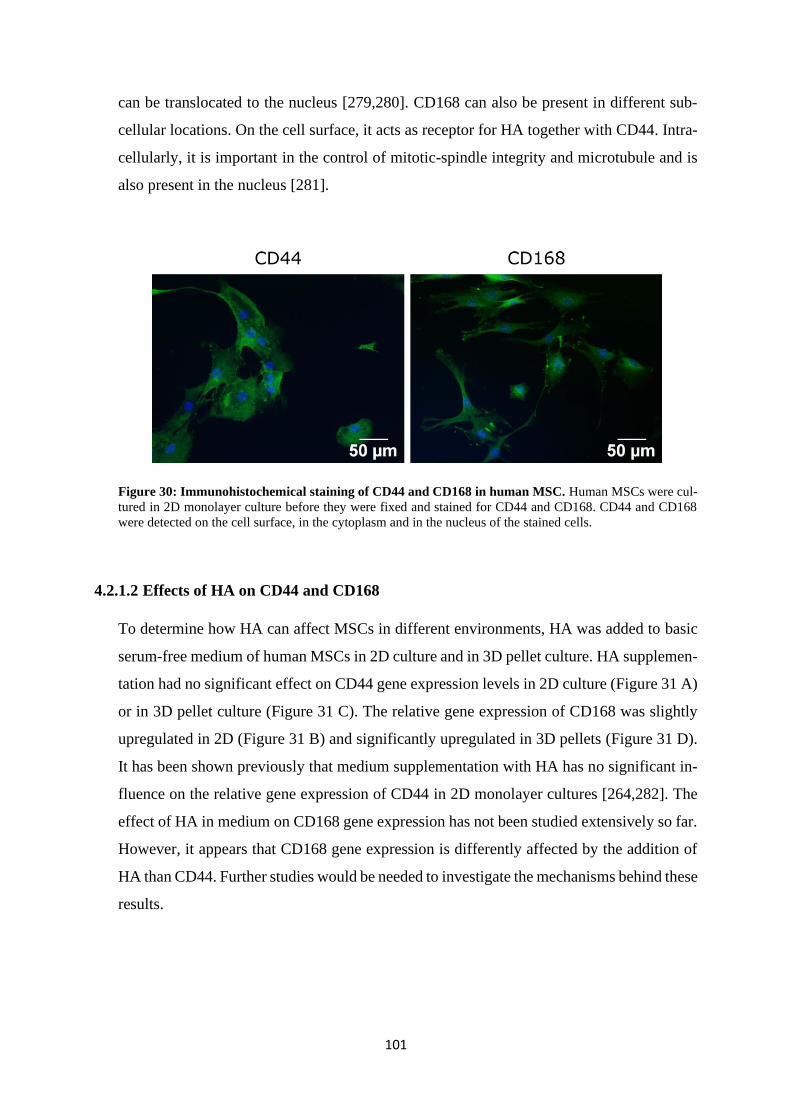
101
can be translocated to the nucleus [279,280]. CD168 can also be present in different sub-
cellular locations. On the cell surface, it acts as receptor for HA together with CD44. Intra-
cellularly, it is important in the control of mitotic-spindle integrity and microtubule and is
also present in the nucleus [281].
Figure 30: Immunohistochemical staining of CD44 and CD168 in human MSC. Human MSCs were cul-
tured in 2D monolayer culture before they were fixed and stained for CD44 and CD168. CD44 and CD168
were detected on the cell surface, in the cytoplasm and in the nucleus of the stained cells.
4.2.1.2 Effects of HA on CD44 and CD168
To determine how HA can affect MSCs in different environments, HA was added to basic
serum-free medium of human MSCs in 2D culture and in 3D pellet culture. HA supplemen-
tation had no significant effect on CD44 gene expression levels in 2D culture (Figure 31 A)
or in 3D pellet culture (Figure 31 C). The relative gene expression of CD168 was slightly
upregulated in 2D (Figure 31 B) and significantly upregulated in 3D pellets (Figure 31 D).
It has been shown previously that medium supplementation with HA has no significant in-
fluence on the relative gene expression of CD44 in 2D monolayer cultures [264,282]. The
effect of HA in medium on CD168 gene expression has not been studied extensively so far.
However, it appears that CD168 gene expression is differently affected by the addition of
HA than CD44. Further studies would be needed to investigate the mechanisms behind these
results.
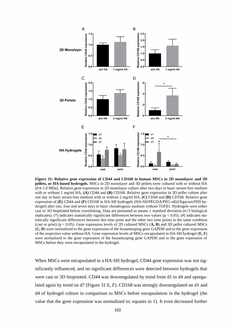
102
Figure 31: Relative gene expression of CD44 and CD168 in human MSCs in 2D monolayer and 3D
pellets, or HA-based hydrogels. MSCs in 2D monolayer and 3D pellets were cultured with or without HA
(0.6-1.0 MDa). Relative gene expression in 2D monolayer culture after two days in basic serum-free medium
with or without 1 mg/ml HA, (A) CD44 and (B) CD168. Relative gene expression in 3D pellet culture after
one day in basic serum-free medium with or without 2 mg/ml HA, (C) CD44 and (D) CD168. Relative gene
expression of (E) CD44 and (F) CD168 in HA-SH hydrogels (HA-SH/PEGDA/PEG-allyl/Irgacure2959 hy-
drogel) after one, four and seven days in basic chondrogenic medium without TGFβ1. Hydrogels were either
cast or 3D bioprinted before crosslinking. Data are presented as means ± standard deviation (n=3 biological
replicates). (*) indicates statistically significant differences between two values (p < 0.05). (#) indicates sta-
tistically significant differences between this time point and the other two time points in the same condition
(cast or print) (p < 0.05). Gene expression levels of 2D cultured MSCs (A, B) and 3D pellet cultured MSCs
(C, D) were normalized to the gene expression of the housekeeping gene GAPDH and to the gene expression
of the respective value without HA. Gene expression levels of MSCs encapsulated in HA-SH hydrogel (E, F)
were normalized to the gene expression of the housekeeping gene GAPDH and to the gene expression of
MSCs before they were encapsulated in the hydrogel.
When MSCs were encapsulated in a HA-SH hydrogel, CD44 gene expression was not sig-
nificantly influenced, and no significant differences were detected between hydrogels that
were cast or 3D bioprinted. CD44 was downregulated by trend from d1 to d4 and upregu-
lated again by trend on d7 (Figure 31 E, F). CD168 was strongly downregulated on d1 and
d4 of hydrogel culture in comparison to MSCs before encapsulation in the hydrogel (the
value that the gene expression was normalized to; equates to 1). It even decreased further
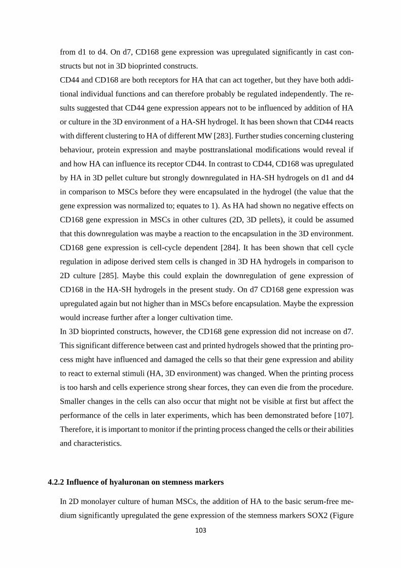
103
from d1 to d4. On d7, CD168 gene expression was upregulated significantly in cast con-
structs but not in 3D bioprinted constructs.
CD44 and CD168 are both receptors for HA that can act together, but they have both addi-
tional individual functions and can therefore probably be regulated independently. The re-
sults suggested that CD44 gene expression appears not to be influenced by addition of HA
or culture in the 3D environment of a HA-SH hydrogel. It has been shown that CD44 reacts
with different clustering to HA of different MW [283]. Further studies concerning clustering
behaviour, protein expression and maybe posttranslational modifications would reveal if
and how HA can influence its receptor CD44. In contrast to CD44, CD168 was upregulated
by HA in 3D pellet culture but strongly downregulated in HA-SH hydrogels on d1 and d4
in comparison to MSCs before they were encapsulated in the hydrogel (the value that the
gene expression was normalized to; equates to 1). As HA had shown no negative effects on
CD168 gene expression in MSCs in other cultures (2D, 3D pellets), it could be assumed
that this downregulation was maybe a reaction to the encapsulation in the 3D environment.
CD168 gene expression is cell-cycle dependent [284]. It has been shown that cell cycle
regulation in adipose derived stem cells is changed in 3D HA hydrogels in comparison to
2D culture [285]. Maybe this could explain the downregulation of gene expression of
CD168 in the HA-SH hydrogels in the present study. On d7 CD168 gene expression was
upregulated again but not higher than in MSCs before encapsulation. Maybe the expression
would increase further after a longer cultivation time.
In 3D bioprinted constructs, however, the CD168 gene expression did not increase on d7.
This significant difference between cast and printed hydrogels showed that the printing pro-
cess might have influenced and damaged the cells so that their gene expression and ability
to react to external stimuli (HA, 3D environment) was changed. When the printing process
is too harsh and cells experience strong shear forces, they can even die from the procedure.
Smaller changes in the cells can also occur that might not be visible at first but affect the
performance of the cells in later experiments, which has been demonstrated before [107].
Therefore, it is important to monitor if the printing process changed the cells or their abilities
and characteristics.
4.2.2 Influence of hyaluronan on stemness markers
In 2D monolayer culture of human MSCs, the addition of HA to the basic serum-free me-
dium significantly upregulated the gene expression of the stemness markers SOX2 (Figure
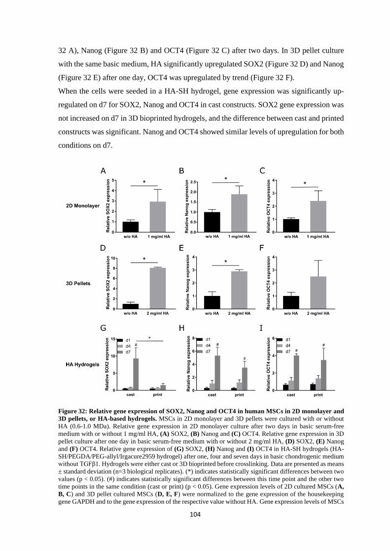
104
32 A), Nanog (Figure 32 B) and OCT4 (Figure 32 C) after two days. In 3D pellet culture
with the same basic medium, HA significantly upregulated SOX2 (Figure 32 D) and Nanog
(Figure 32 E) after one day, OCT4 was upregulated by trend (Figure 32 F).
When the cells were seeded in a HA-SH hydrogel, gene expression was significantly up-
regulated on d7 for SOX2, Nanog and OCT4 in cast constructs. SOX2 gene expression was
not increased on d7 in 3D bioprinted hydrogels, and the difference between cast and printed
constructs was significant. Nanog and OCT4 showed similar levels of upregulation for both
conditions on d7.
Figure 32: Relative gene expression of SOX2, Nanog and OCT4 in human MSCs in 2D monolayer and
3D pellets, or HA-based hydrogels. MSCs in 2D monolayer and 3D pellets were cultured with or without
HA (0.6-1.0 MDa). Relative gene expression in 2D monolayer culture after two days in basic serum-free
medium with or without 1 mg/ml HA, (A) SOX2, (B) Nanog and (C) OCT4. Relative gene expression in 3D
pellet culture after one day in basic serum-free medium with or without 2 mg/ml HA, (D) SOX2, (E) Nanog
and (F) OCT4. Relative gene expression of (G) SOX2, (H) Nanog and (I) OCT4 in HA-SH hydrogels (HA-
SH/PEGDA/PEG-allyl/Irgacure2959 hydrogel) after one, four and seven days in basic chondrogenic medium
without TGFβ1. Hydrogels were either cast or 3D bioprinted before crosslinking. Data are presented as means
± standard deviation (n=3 biological replicates). (*) indicates statistically significant differences between two
values (p < 0.05). (#) indicates statistically significant differences between this time point and the other two
time points in the same condition (cast or print) (p < 0.05). Gene expression levels of 2D cultured MSCs (A,
B, C) and 3D pellet cultured MSCs (D, E, F) were normalized to the gene expression of the housekeeping
gene GAPDH and to the gene expression of the respective value without HA. Gene expression levels of MSCs
105
encapsulated in HA-SH hydrogel (G, H, I) were normalized to the gene expression of the housekeeping gene
GAPDH and to the gene expression of MSCs before they were encapsulated in the hydrogel.
Maintenance of MSC stemness is an important topic for regenerative medicine because the
necessary step of in vitro expansion in 2D monolayer can reduce proliferation and differen-
tiation capacity in these cells, probably through aging processes [286-288]. MSCs are de-
fined by their ability to self-renew and to differentiate into chondrogenic, osteogenic and
adipogenic direction. Stemness is a prerequisite for these abilities [289]. If stemness is re-
duced in MSCs, so is the ability of the cells to differentiate.
SOX2, Nanog and OCT4 are stemness markers of embryonic stem cells (ESCs), and they
are also expressed in MSCs [290-292]. SOX2, OCT4 and Nanog have been shown to act
together as a core transcriptional network to maintain stemness [292]. When OCT4, SOX2
or Nanog were overexpressed in MSCs of different origin, proliferation and differentiation
capacity were enhanced [293-295]. In previous studies, HA has been found to maintain
stemness in MSCs [269,270] and to increase the expression of SOX2, Nanog and OCT4
[296,297]. The same was observed in the present study when HA was added to the basic
serum-free culture medium of 2D monolayer and 3D pellet cultures. MSCs in the HA-SH
hydrogel showed significant upregulation of the stemness markers on d7 which suggested
that maybe HA had similar biological functions when it was modified and crosslinked in
comparison to when it was added unmodified to the culture medium. The 3D microenvi-
ronment in a hydrogel could also contribute to the maintenance or upregulation over time
of stemness in MSCs. The relation between stemness, HA signalling and 3D microenviron-
ment would be an intriguing topic for further research.
On d7, cast hydrogels showed increased gene expression of MSCs for stemness markers but
3D bioprinted constructs had lower SOX2 gene expression levels in comparison. This sug-
gested that the 3D bioprinting process affected the MSCs in a way that gene regulation of
SOX2 was changed.
4.2.3 Influence of hyaluronan on different transcription factors
The impact of HA on differentiation of MSCs was investigated. Therefore, gene expression
of transcription factors of major differentiation directions of MSCs were analysed with HA
supplementation in basic serum-free medium and in HA-based hydrogels. The chondro-
genic transcription factors SOX9, SOX5 and SOX6, the osteogenic transcription factor
106
RUNX2 and the adipogenic transcription factor PPARG were examined in the present
study.
4.2.3.1 Influence on SOX9, SOX5 and SOX6
In 2D monolayer culture, the gene expression of SOX9 was significantly upregulated when
HA was added to the serum-free basic medium (Figure 33 A). SOX5 and SOX6 gene ex-
pression were not affected by HA in 2D monolayer (Figure 33 B, C). SOX9 gene expression
was also significantly upregulated in 3D pellet culture with HA (Figure 33 D), and SOX5
showed an increase by trend (Figure 33 E). SOX6 gene expression was not influenced by
HA in 3D pellet culture. (Figure 33 F). When MSCs were encapsulated in the HA-SH hy-
drogel, the gene expression of SOX9, SOX5 and SOX6 was significantly upregulated from
d1 to d7. SOX5 gene expression on d7 was slightly higher in printed constructs than in cast
constructs (Figure 33 H). SOX9, SOX5 and SOX6 are known to be transcription factors
that are essential for chondrogenesis [296-300]. They act cooperatively to activate transcrip-
tion of several chondrogenic genes like COL2A1 and ACAN through binding and activation
of enhancer sequences [301]. It has been shown previously that when SOX9 was lacking in
early chondrogenesis, no proper ECM could be produced because cells could not express
the respective genes [302]. When both SOX5 and SOX6 were lacking, expression of carti-
lage specific genes was severely hampered even though SOX9 was expressed normally
[303]. As the expression and cooperation of these three transcription factors is very im-
portant for early chondrogenesis, it was tested if HA can influence them. HA signalling has
been shown before to positively regulate chondrogenesis and, among other chondrogenesis-
associated factors, SOX9 gene expression [164,165,259,260,262,263,304,305]. Less is
known about the influence of HA on gene expression of SOX5 and SOX6. Here, HA did
not induce significant changes in SOX5 or SOX6 gene expression in 2D or 3D pellet culture
even if SOX5 was upregulated by trend in 3D pellet culture. HA might influence the differ-
ent SOX gene expressions differently. They act cooperatively, but they also have individual
functions in the formation of cartilage and in other tissues. SOX9 but not SOX5 and SOX6
were, for example, needed in pre-cartilaginous condensations [303]. On the other hand,
SOX5 and SOX6 did not need SOX9 for activation at the beginning of chondrogenesis
[306]. The results of the present study showed that HA influenced the gene expression of
SOX9 more than that of SOX5 and SOX6.
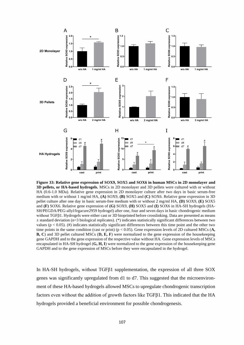
107
Figure 33: Relative gene expression of SOX9, SOX5 and SOX6 in human MSCs in 2D monolayer and
3D pellets, or HA-based hydrogels. MSCs in 2D monolayer and 3D pellets were cultured with or without
HA (0.6-1.0 MDa). Relative gene expression in 2D monolayer culture after two days in basic serum-free
medium with or without 1 mg/ml HA, (A) SOX9, (B) SOX5 and (C) SOX6. Relative gene expression in 3D
pellet culture after one day in basic serum-free medium with or without 2 mg/ml HA, (D) SOX9, (E) SOX5
and (F) SOX6. Relative gene expression of (G) SOX9, (H) SOX5 and (I) SOX6 in HA-SH hydrogels (HA-
SH/PEGDA/PEG-allyl/Irgacure2959 hydrogel) after one, four and seven days in basic chondrogenic medium
without TGFβ1. Hydrogels were either cast or 3D bioprinted before crosslinking. Data are presented as means
± standard deviation (n=3 biological replicates). (*) indicates statistically significant differences between two
values (p < 0.05). (#) indicates statistically significant differences between this time point and the other two
time points in the same condition (cast or print) (p < 0.05). Gene expression levels of 2D cultured MSCs (A,
B, C) and 3D pellet cultured MSCs (D, E, F) were normalized to the gene expression of the housekeeping
gene GAPDH and to the gene expression of the respective value without HA. Gene expression levels of MSCs
encapsulated in HA-SH hydrogel (G, H, I) were normalized to the gene expression of the housekeeping gene
GAPDH and to the gene expression of MSCs before they were encapsulated in the hydrogel.
In HA-SH hydrogels, without TGFβ1 supplementation, the expression of all three SOX
genes was significantly upregulated from d1 to d7. This suggested that the microenviron-
ment of these HA-based hydrogels allowed MSCs to upregulate chondrogenic transcription
factors even without the addition of growth factors like TGFβ1. This indicated that the HA
hydrogels provided a beneficial environment for possible chondrogenesis.
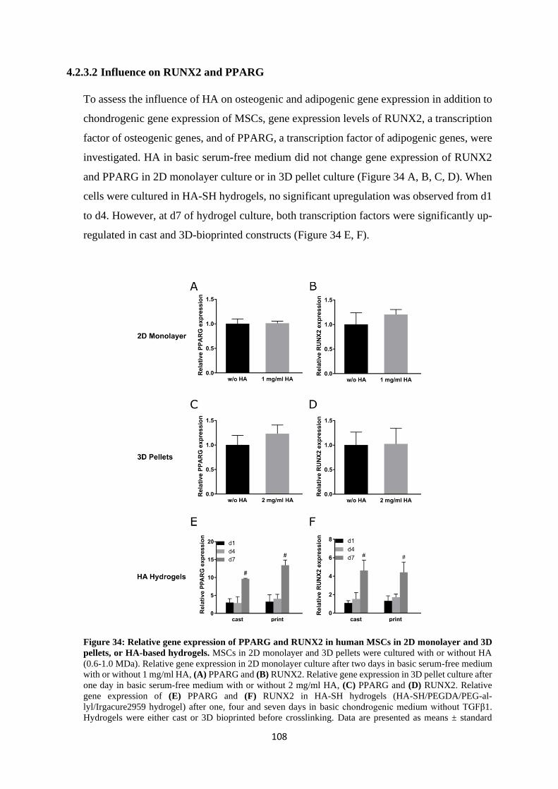
108
4.2.3.2 Influence on RUNX2 and PPARG
To assess the influence of HA on osteogenic and adipogenic gene expression in addition to
chondrogenic gene expression of MSCs, gene expression levels of RUNX2, a transcription
factor of osteogenic genes, and of PPARG, a transcription factor of adipogenic genes, were
investigated. HA in basic serum-free medium did not change gene expression of RUNX2
and PPARG in 2D monolayer culture or in 3D pellet culture (Figure 34 A, B, C, D). When
cells were cultured in HA-SH hydrogels, no significant upregulation was observed from d1
to d4. However, at d7 of hydrogel culture, both transcription factors were significantly up-
regulated in cast and 3D-bioprinted constructs (Figure 34 E, F).
Figure 34: Relative gene expression of PPARG and RUNX2 in human MSCs in 2D monolayer and 3D
pellets, or HA-based hydrogels. MSCs in 2D monolayer and 3D pellets were cultured with or without HA
(0.6-1.0 MDa). Relative gene expression in 2D monolayer culture after two days in basic serum-free medium
with or without 1 mg/ml HA, (A) PPARG and (B) RUNX2. Relative gene expression in 3D pellet culture after
one day in basic serum-free medium with or without 2 mg/ml HA, (C) PPARG and (D) RUNX2. Relative
gene expression of (E) PPARG and (F) RUNX2 in HA-SH hydrogels (HA-SH/PEGDA/PEG-al-
lyl/Irgacure2959 hydrogel) after one, four and seven days in basic chondrogenic medium without TGFβ1.
Hydrogels were either cast or 3D bioprinted before crosslinking. Data are presented as means ± standard
109
deviation (n=3 biological replicates). (*) indicates statistically significant differences between two values (p
< 0.05). (#) indicates statistically significant differences between this time point and the other two time points
in the same condition (cast or print) (p < 0.05). Gene expression levels of 2D cultured MSCs (A, B) and 3D
pellet cultured MSCs (C, D) were normalized to the gene expression of the housekeeping gene GAPDH and
to the gene expression of the respective value without HA. Gene expression levels of MSCs encapsulated in
HA-SH hydrogel (E, F) were normalized to the gene expression of the housekeeping gene GAPDH and to the
gene expression of MSCs before they were encapsulated in the hydrogel.
Unmodified HA did not influence the gene expression of the osteogenic transcription factor
RUNX2 or the adipogenic transcription factor PPARG, while it significantly upregulated
the chondrogenic transcription factor SOX9 in 2D and 3D pellet culture (Figure 33 A, D, p.
107). The reports about effects of HA on osteogenic or adipogenic differentiation are con-
troversial. Some studies found that HA increased osteogenesis marker or bone formation
[307-310], others did not see any effects or even downregulation [296,311]. These studies
also showed that MW and concentration of HA can have different effects on the different
factors of osteogenesis [308,310]. Some of the conflicting results are probably due to these
or other differences in the culture conditions, cell types or HA delivery methods. Zou et al.
demonstrated that 2D cultured MSCs in basic medium with addition of HA upregulated
SOX9 gene expression after seven days but downregulated the gene expression of the oste-
ogenic markers RUNX2, SP7 (osterix), ALP, COL1A1 and COL10A1 (type X collagen)
[312]. In the present study, gene expression was only examined after two days but the up-
regulation of SOX9 was already visible. RUNX2 gene expression was not changed by HA
after two days of 2D culture with HA. These results suggested that HA in basic medium
without serum supported the chondrogenic lineage of MSC differentiation rather than the
osteogenic lineage. The gene expression of RUNX2 in HA-SH hydrogels showed signifi-
cant upregulation in both cast and 3D bioprinted constructs from d1 to d7 and from d4 to
d7. This was not in line with the results from 2D and 3D pellet experiments. However, the
hydrogel experiment was longer than the others and RUNX2 expression only increased sig-
nificantly after 7 days. A longer experiment for 2D and 3D pellets could show how RUNX2
expression develops after a longer cultivation time under these conditions. However, even
if the exact effects of HA on osteogenesis are not completely clear yet, the application of
HA materials as cell and growth factor carriers in bone regeneration has generated promis-
ing results [313,314]. The upregulation of RUNX2 gene expression in the HA-SH hydrogel
in the present study might be the result of the specific 3D environment that the hydrogel
provided for the cells and/or of a biological effect of HA on the cells that has yet to be
elucidated.
110
The effect of HA on PPARG gene expression was very similar to the effect on RUNX2.
Studies about the effect of HA on adipogenesis had conflicting results, probably also due to
the inconsistencies in study design like different MW, concentrations, culture conditions or
cell types. Some works reported inhibitory effects of HA on adipogenesis [315-317], while
others showed that HA could promote adipogenesis [318,319]. However, a previous work
could demonstrate that CD44 and CD168 influenced adipogenesis very differently: While
CD44 was required for adipogenesis, CD168 was found to suppress adipogenesis [320].
These opposing functions of receptors that can both be influenced by HA could also be the
reason behind previous conflicting results. In the present study, PPARG gene expression
was not changed by HA in 2D monolayer or 3D pellet culture. Gene expression was only
analysed for one or two days in these experiments, and it is possible that PPARG expression
could change after a longer cultivation time. However, SOX9 was already significantly up-
regulated at these early time points and that suggested that HA promoted chondrogenic dif-
ferentiation stronger or earlier than it did adipogenic differentiation, similar to osteogenic
differentiation. PPARG gene expression was significantly upregulated in cells that were
cultivated for seven days in HA-SH hydrogels. HA-based gels and materials have been used
successfully for adipose tissue regeneration, even though the direct effects of HA on these
processes are not fully known. As the HA-SH hydrogel in the present study permitted up-
regulation of the adipogenic transcription factor PPARG, it may also be a suitable material
for adipogenic differentiation of MSCs.
4.2.4 Influence of hyaluronan on chondrogenic markers ACAN and COL2A1
After the result that HA influenced the chondrogenic transcription factor SOX9 more than
the osteogenic transcription factor RUNX2 and the adipogenic transcription factor PPARG,
the effect of HA on the chondrogenic markers type II collagen and aggrecan was evaluated.
The addition of HA to basic serum-free medium in 2D monolayer culture of MSCs signifi-
cantly upregulated the gene expression of COL2A1 (type II collagen) (Figure 35 A) and
ACAN (aggrecan) (Figure 35 B) after two days. In 3D pellet culture, gene expression of
COL2A1 (Figure 35 C) but not ACAN (Figure 35 D) was significantly upregulated after
one day of culture in basic serum-free medium with HA. When MSCs were encapsulated
in HA-SH hydrogels, COL2A1 gene expression was not changed significantly from d1 to
d4 or between cast and 3D bioprinted constructs. However, after seven days COL2A1 was
significantly upregulated in cast, but not in 3D bioprinted hydrogels (Figure 35 E). ACAN
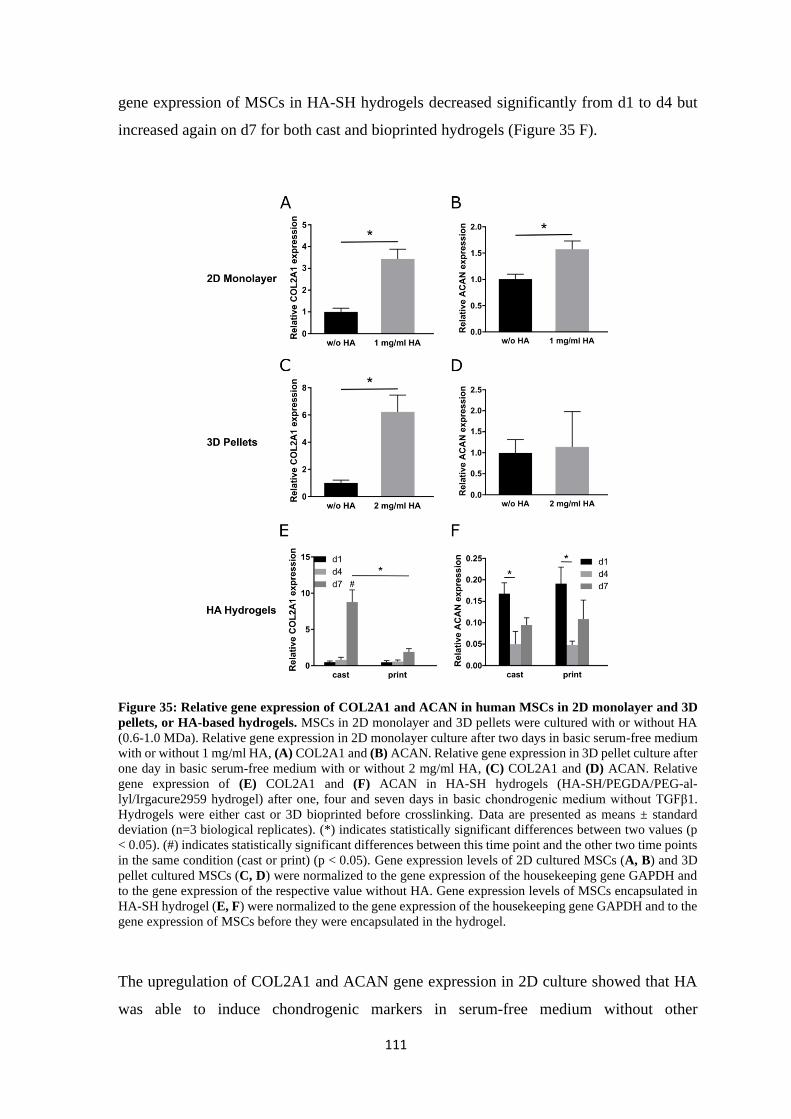
111
gene expression of MSCs in HA-SH hydrogels decreased significantly from d1 to d4 but
increased again on d7 for both cast and bioprinted hydrogels (Figure 35 F).
Figure 35: Relative gene expression of COL2A1 and ACAN in human MSCs in 2D monolayer and 3D
pellets, or HA-based hydrogels. MSCs in 2D monolayer and 3D pellets were cultured with or without HA
(0.6-1.0 MDa). Relative gene expression in 2D monolayer culture after two days in basic serum-free medium
with or without 1 mg/ml HA, (A) COL2A1 and (B) ACAN. Relative gene expression in 3D pellet culture after
one day in basic serum-free medium with or without 2 mg/ml HA, (C) COL2A1 and (D) ACAN. Relative
gene expression of (E) COL2A1 and (F) ACAN in HA-SH hydrogels (HA-SH/PEGDA/PEG-al-
lyl/Irgacure2959 hydrogel) after one, four and seven days in basic chondrogenic medium without TGFβ1.
Hydrogels were either cast or 3D bioprinted before crosslinking. Data are presented as means ± standard
deviation (n=3 biological replicates). (*) indicates statistically significant differences between two values (p
< 0.05). (#) indicates statistically significant differences between this time point and the other two time points
in the same condition (cast or print) (p < 0.05). Gene expression levels of 2D cultured MSCs (A, B) and 3D
pellet cultured MSCs (C, D) were normalized to the gene expression of the housekeeping gene GAPDH and
to the gene expression of the respective value without HA. Gene expression levels of MSCs encapsulated in
HA-SH hydrogel (E, F) were normalized to the gene expression of the housekeeping gene GAPDH and to the
gene expression of MSCs before they were encapsulated in the hydrogel.
The upregulation of COL2A1 and ACAN gene expression in 2D culture showed that HA
was able to induce chondrogenic markers in serum-free medium without other
112
chondrogenic inducers, probably via influencing SOX9 gene expression. The same was true
for COL2A1 in 3D pellet cultures. Cells in HA-SH hydrogels showed an upregulation of
COL2A1 gene expression on d7 but only in cast and not in printed constructs. The low
levels of COL2A1 gene expression on d7 in printed hydrogels in comparison to cast hydro-
gels indicated a negative effect of the printing procedure on the cells. A similar effect of 3D
bioprinting on MSC gene expression has also been shown in a previous study [107]. This
effect illustrates again that it is important to monitor the effects of 3D bioprinting procedures
on the cells.
ACAN gene expression was not influenced by the 3D printing process. However, it was
strongly downregulated on all time points in the HA-SH hydrogel in comparison to the
MSCs before they were encapsulated in the hydrogel (the value that the gene expression
was normalized to; equates to 1). Gene expression of ACAN even dropped from d1 to d4.
On d7 it was upregulated in comparison to d4 but still not as high as on d1. It is interesting
that gene expression of COL2A1 and ACAN that are both targets of SOX9 transcription
factor were so differently affected by the culture in HA-SH hydrogels. A previous study
showed that after 28 days of chondrogenic differentiation with TGFβ3 the gene expression
of ACAN was significantly reduced in HA hydrogels with increasing polymer content (1
%, 3 %, 5 %), while COL2A1 gene expression was not affected [253]. This shows one
factor, namely polymer content, that can affect the gene expression of COL2A1 and ACAN
differently. The HA-SH/PEGDA/PEG-Allyl/Irgacure2959 hydrogel in the present study
had a polymer content of 2 %, and it has been shown before that MSCs can produce abun-
dant aggrecan when they are chondrogenically differentiated with TGFβ1 for 21 days in
this hydrogel (Hauptstein et al., manuscript in preparation). However, in the present study,
MSCs were cultured without TGFβ1 and only for seven days. It appeared that the presence
of the cells in the 3D HA-based microenvironment and the upregulated SOX9 were not
enough to promote ACAN gene expression as they did for COL2A1. Maybe ACAN gene
expression is only upregulated when a stronger chondrogenic induction takes place, and
maybe the amount of polymer in the HA-SH/PEGDA/PEG-Allyl/Irgacure2959 hydrogel
affected ACAN gene expression negatively in this setting.
113
4.2.5 Summary of the effects of hyaluronan on MSCs
The effects of unmodified HA on the gene expression of 2D monolayer and 3D pellet cul-
tivated MSCs was investigated as well as the gene expression of MSCs encapsulated in cast
or 3D bioprinted HA-SH hydrogels. It was demonstrated that unmodified HA in the basic
serum-free culture medium of 2D cultured human MSCs upregulated the chondrogenic tran-
scription factor SOX9 (but not SOX5 and SOX6) rather than the osteogenic transcription
factor RUNX2 or the adipogenic transcription factor PPARG. Probably as a result from
SOX9 upregulation the chondrogenic markers COL2A1 and ACAN were also upregulated,
while the gene expression of the HA receptors CD44 and CD168 was not changed signifi-
cantly. In addition to the chondrogenic markers, the stemness markers SOX2, Nanog and
OCT4 were upregulated by HA supplementation as has been described in the literature
[165,262,263,296,297]. Unmodified HA alone cannot induce a complete chondrogenesis
with abundant ECM production and differentiation of MSCs to chondrocyte-like cells like
TGFβ1. In comparison to the upregulation of chondrogenic genes that happens under the
influence of TGFβ1 in HA-SH hydrogels until d7 (ACAN: up to 200-fold; COL2A1: up to
10000-fold [169], the upregulation under HA alone was much smaller for COL2A1 after
seven days (8-fold) and ACAN was even downregulated. HA supplementation rather caused
a tendency towards chondrogenic differentiation than a strong induction of chondrogenesis.
HA might be involved in the signalling pathways that direct chondrogenesis, but it is also
involved in the maintenance of stemness. Differentiation and maintenance of stemness are
two processes that are inevitably connected with one another. The maintenance of stemness
is in the end also the maintenance of the ability to differentiate [289]. HA has been suggested
to regulate the balance between differentiation and quiescence of stem cells especially in
the stem cell niche [271]. The MSCs under the influence of HA were most likely still stem
cells but with a tendency towards chondrogenic differentiation. Therefore, it was possible
that both stemness markers and chondrogenic markers were upregulated. The exact mech-
anism of HA function in this context should be further analysed, for example with investi-
gation of the involved proteins and signalling pathways in addition to gene expression.
The effects of HA on relative gene expression in MSCs that were cultured in 3D pellets
were mostly similar to the HA effects on gene expression of MSCs in 2D monolayer culture.
The small differences between 2D monolayer and 3D pellet culture that were seen in the
gene expression levels were most likely the results of the different cell microenvironments.
3D cell cultures like pellets usually represent the natural environment of cells better than
114
2D cultures. Several factors are different in 3D culture, for example cell shape, exposure to
medium, cell junctions, gene expression levels and response to stimuli [321].
The regulation of the relative gene expression of MSCs in cast HA-SH hydrogels (HA-
SH/PEGDA/PEG-allyl/Irgacure2959 hydrogel) was similar for most of the investigated
markers. The genes showed significant upregulation of expression from d1 to d7. This effect
of cultivation in HA-based hydrogels did not correspond entirely to the effects of HA that
were seen in 2D and 3D pellet cultures. For example, RUNX2 and PPARG were not af-
fected by HA in 2D or 3D pellet culture but were strongly upregulated in HA hydrogels. It
is possible that the upregulations in the hydrogel did not (only) originate from biological
effects of HA but also from the 3D environment as such that the hydrogel provided for the
cells. HA-based hydrogels are often used successfully for regeneration of adipose or bone
tissue [313,314,319,322,323]. Here, it was demonstrated that our HA-SH/PEGDA/PEG-
allyl/Irgacure2959 hydrogel that is usually used in chondrogenic differentiation experi-
ments of MSCs, might also be suitable for osteogenesis or adipogenesis as it did not inhibit
the upregulation of RUNX2 and PPARG. The HA-SH hydrogel also appeared to be suitable
for the maintenance of stemness in MSCs. As all of these genes were upregulated at the
same time in the same cells, it can be suggested that no exclusive differentiation direction
was induced in the HA-SH hydrogel without growth factors like TGFβ1 for chondrogenesis.
This could mean that either the biological activity of HA was reduced when it was modified
and crosslinked into a hydrogel or that the influence of the microenvironment that was pro-
vided by the hydrogel was so strong that other effects, like that of HA, were overruled. A
combination of both is also possible. Previous studies have shown that modification of HA
can reduce its binding to and effects on cells [218,219].
CD168, CD44 and ACAN gene expression levels were regulated differently from the rest
of the investigated genes in HA-SH hydrogels. These genes might probably be influenced
by additional other factors like cell cycle regulation for CD168, clustering for CD44 or
polymer content for ACAN. Further research would be needed to investigate the different
factors that are influencing gene expression in this context in more detail.
3D bioprinting of MSCs in HA-SH precursor solution mostly did not change relative gene
expression levels in comparison to MSCs in cast hydrogels. Interestingly, COL2A1, SOX2,
and CD168 showed significantly lower gene expression levels on d7 in printed than in cast
hydrogel constructs. This suggested that the printing process itself influenced the cells in a
way that gene expression of several factors was still affected after seven days of culture in
HA-SH hydrogels. A downregulation of COL2A1 in MSCs in printed hydrogel constructs
115
was also demonstrated in a previous work [107]. Further studies with different applied print-
ing pressure and viscosity of the hydrogel solution would be needed to elucidate how and
why gene expression is changed by the mechanical forces that act on the cells during print-
ing. Analysis of involved proteins and signalling pathways could add valuable information
to such investigations. If the involved mechanisms could be elucidated, it would not only
expand our knowledge about the effects of mechanical stress on MSCs and the role of the
3D environment of HA-SH hydrogels in MSC gene expression regulation, but it could also
enable us to make 3D bioprinting more cell-friendly.
The gene expression studies that were performed in this work can act as preliminary results
for a continued project on the signalling effects of HA in general and on the effects of HA
in 3D hydrogels in particular. For example, the effects of HA of different MW or concen-
tration could be further analysed. The experiments that were presented here were also
mostly of short duration, and HA effects could also be evaluated in the long-term. Addi-
tionally, the three culture conditions that were analysed in this study cannot completely be
compared to each other as several factors are different between them that could modulate
the effects of HA on the cells. For example, 2D and 3D pellet experiments could be per-
formed with modified HA to directly compare it to unmodified HA in the same culture
conditions. To evaluate the contribution of HA signalling to the different effects in HA
hydrogels, HA receptors like CD44 or CD168 could be blocked, or hydrogels with similar
characteristics but without HA could be used for comparison. Binding of unmodified HA
and modified HA to CD44 could also be directly analysed, for example with FACS studies.
Also, the underlying signalling pathways and regulations could be further studied. As only
gene expression was analysed so far, protein expression and activity would be very inter-
esting additional results. All this and more will probably be done in future studies. This
work on the effect of HA on cells can be seen as a preliminary study for a future project.

116

117
Conclusion

118
119
5 Conclusion
Articular cartilage is a tissue without blood supply and therefore limited capacity for self-re-
generation upon injury. As a result, cartilage injury can lead to degeneration of the tissue and
the development of osteoarthritis. Osteoarthritis is one of the leading causes of disability in the
elderly population due to chronic pain and reduced mobility. Current clinical treatments cannot
regenerate the damaged tissue to its previous native form and function. Tissue engineering and
3D bioprinting are promising to improve the results of cartilage repair tissue in the future. Even
if the regeneration of the native form of articular cartilage has not been achieved so far, the
potential of tissue engineering is still tremendous. New cell sources, new materials and signals
for the cells are constantly discovered, developed, improved and tested with the aim to enhance
the quality of the resulting tissue.
A relatively new, promising cell source, articular cartilage progenitor cells (ACPCs), was in-
vestigated in this work in comparison to mesenchymal stromal cells (MSCs). In agarose, a gold-
standard hydrogel for cartilage tissue engineering, ACPCs produced a more articular cartilage-
like tissue with more GAG, less type I collagen and low ALP activity in comparison to the neo-
cartilage tissue assembled by MSCs. Hypoxic conditions improved the quality of the neo-car-
tilage tissue produced by MSCs by making it less hypertrophic and less fibrocartilaginous. An
increase in ECM production was not achieved by hypoxic conditions or by zonal co-culturing
of ACPCs and MSCs. However, interactions between the two cell types in zonal agarose con-
structs were detected as, for example, the staining intensities of type II and type I collagen were
modulated in zonal constructs in comparison to non-zonal monoculture constructs under
normoxic and hypoxic conditions. In the HA-based hydrogel HA-SH/P(AGE-co-G), MSCs
produced more cartilage ECM than ACPCs and achieved thereby a zonal ECM distribution that
was similar to native articular cartilage with more ECM in the deep zone. However, for both
cell types, the formed ECM was located only pericellularly and was not spread throughout the
whole hydrogel like in agarose. These results clearly emphasised that the chondrogenic poten-
tial of ACPCs and MSCs is strongly dependent on the cell microenvironment. ACPCs can be a
promising cell source for articular cartilage engineering if they are cultivated in a suitable hy-
drogel environment like agarose. When the present thesis and previous studies
[103,107,131,254,255] were taken into consideration, it appeared that chondrogenesis of AC-
PCs was supported by hydrogels without biological attachment sites, such as agarose, and that
hydrogels containing biological cues like HA were beneficial for chondrogenic differentiation
of MSCs. Additionally, a microenvironment with a high polymer content like in HA-SH/
120
P(AGE-co-G) hydrogels can prevent homogenous ECM distribution by restricting the ECM to
the pericellular region. Therefore, it is of paramount importance to thoroughly evaluate com-
patibility between cells and their hydrogel microenvironment to achieve the best possible re-
sults in the engineering of articular cartilage tissue.
As HA appeared to have beneficial effects on the chondrogenesis of MSCs in the present work
and in previous studies [164,165,258], the contribution of HA to chondrogenic gene expression
was investigated in more detail. It was shown that HA supplementation supported chondrogenic
but not adipogenic or osteogenic gene expression in 2D and 3D pellet cultures. The investigated
HA-based hydrogel did not support a specific differentiation lineage in encapsulated MSCs but
appeared to be a suitable environment for several different applications like maintenance of
stemness and chondrogenic, osteogenic or adipogenic differentiation. The investigation of
MSCs´ gene expression under the influence of HA in this work showed promising results for
the use of HA in MSC-based tissue engineering. The effects of modified and crosslinked HA
in hydrogels on the cells is of special interest and should be elucidated further in future studies.
This might enable tissue engineering approaches that can use the full potential that the versatile,
bioactive molecule HA has to offer.
The investigation of the 3D bioprinting process of MSCs in HA-based hydrogels showed that
3D printing did not influence most of the examined gene expression levels in MSCs, but nev-
ertheless, three genes were downregulated in printed versus cast constructs. These results high-
lighted that 3D printing can change the behaviour of cells in the long-term, and that printed
cells should be monitored for these changes before other experiments are performed with them.
However, these results also offer the opportunity for more extensive research that could expand
our knowledge about the influence of mechanical stress and 3D microenvironment on MSCs
and lead to the development of more cell-friendly printing processes.
In total, it was shown that the chondrogenic potential of ACPCs and MSCs is strongly depend-
ent on the cell microenvironment, especially the used hydrogels. ACPCs can be a promising
cell source for articular cartilage engineering if they are cultivated in a suitable hydrogel like
agarose. The results of this thesis expand the knowledge in the area of articular cartilage engi-
neering with regard to the rational combination of cell types and hydrogel materials and open
up new possible approaches to the regeneration of articular cartilage tissue.

121
Bibliography

122
123
6 Bibliography
1. Naumann, A.; Dennis, J.E.; Awadallah, A.; Carrino, D.A.; Mansour, J.M.; Kastenbauer,
E.; Caplan, A.I. Immunochemical and mechanical characterization of cartilage subtypes
in rabbit. J Histochem Cytochem 2002, 50, 1049-1058,
doi:10.1177/002215540205000807.
2. Fithian, D.C.; Kelly, M.A.; Mow, V.C. Material properties and structure-function
relationships in the menisci. Clin Orthop Relat Res 1990, 19-31.
3. Carballo, C.B.; Nakagawa, Y.; Sekiya, I.; Rodeo, S.A. Basic Science of Articular
Cartilage. Clin Sports Med 2017, 36, 413-425, doi:10.1016/j.csm.2017.02.001.
4. Camarero-Espinosa, S.; Rothen-Rutishauser, B.; Foster, E.J.; Weder, C. Articular
cartilage: from formation to tissue engineering. Biomater Sci 2016, 4, 734-767,
doi:10.1039/c6bm00068a.
5. Fox, S.A.J.; Bedi, A.; Rodeo, S.A. The basic science of articular cartilage: structure,
composition, and function. Sports Health 2009, 1, 461-468,
doi:10.1177/1941738109350438.
6. Monaghan, N.G.; Wyss, J.F. Joint Pain. In Pain Management and Palliative Care: A
Comprehensive Guide, Sackheim, K.A., Ed. Springer New York: New York, NY, 2015;
doi:10.1007/978-1-4939-2462-2_19pp. 131-140.
7. Decker, R.S.; Koyama, E.; Pacifici, M. Articular Cartilage: Structural and
Developmental Intricacies and Questions. Curr Osteoporos Rep 2015, 13, 407-414,
doi:10.1007/s11914-015-0290-z.
8. Ondrésik, M.; Oliveira, J.M.; Reis, R.L. Knee Articular Cartilage. In Regenerative
Strategies for the Treatment of Knee Joint Disabilities, Oliveira, J.M., Reis, R.L., Eds.
Springer International Publishing: Cham, 2017; doi:10.1007/978-3-319-44785-8_1pp.
3-20.
9. Sanchez-Adams, J.; Leddy, H.A.; McNulty, A.L.; O'Conor, C.J.; Guilak, F. The
mechanobiology of articular cartilage: bearing the burden of osteoarthritis. Curr
Rheumatol Rep 2014, 16, 451, doi:10.1007/s11926-014-0451-6.
10. Houck, D.A.; Kraeutler, M.J.; Belk, J.W.; Frank, R.M.; McCarty, E.C.; Bravman, J.T.
Do Focal Chondral Defects of the Knee Increase the Risk for Progression to
Osteoarthritis? A Review of the Literature. Orthop J Sports Med 2018, 6,
2325967118801931, doi:10.1177/2325967118801931.
11. Sacitharan, P.K. Ageing and Osteoarthritis. Subcell Biochem 2019, 91, 123-159,
doi:10.1007/978-981-13-3681-2_6.
12. Glyn-Jones, S.; Palmer, A.J.; Agricola, R.; Price, A.J.; Vincent, T.L.; Weinans, H.; Carr,
A.J. Osteoarthritis. Lancet 2015, 386, 376-387, doi:10.1016/s0140-6736(14)60802-3.
13. Cross, M.; Smith, E.; Hoy, D.; Nolte, S.; Ackerman, I.; Fransen, M.; Bridgett, L.;
Williams, S.; Guillemin, F.; Hill, C.L.; Laslett, L.L.; Jones, G.; Cicuttini, F.; Osborne,
R.; Vos, T.; Buchbinder, R.; Woolf, A.; March, L. The global burden of hip and knee
osteoarthritis: estimates from the global burden of disease 2010 study. Ann Rheum Dis
2014, 73, 1323-1330, doi:10.1136/annrheumdis-2013-204763.
14. Schizas, N.; Savvidou, O.; Triantafyllopoulos, I.; Papadakis, S.; Dontas, I.;
Papagelopoulos, P. Adjuvant therapies for the enhancement of microfracture technique
in cartilage repair. Orthop Rev (Pavia) 2019, 11, 7950-7950, doi:10.4081/or.2019.7950.
15. Richter, D.L.; Schenck, R.C., Jr.; Wascher, D.C.; Treme, G. Knee Articular Cartilage
Repair and Restoration Techniques: A Review of the Literature. Sports Health 2016, 8,
153-160, doi:10.1177/1941738115611350.
124
16. Makris, E.A.; Gomoll, A.H.; Malizos, K.N.; Hu, J.C.; Athanasiou, K.A. Repair and
tissue engineering techniques for articular cartilage. Nat Rev Rheumatol 2015, 11, 21-
34, doi:10.1038/nrrheum.2014.157.
17. Hangody, L.; Füles, P. Autologous osteochondral mosaicplasty for the treatment of full-
thickness defects of weight-bearing joints: ten years of experimental and clinical
experience. J Bone Joint Surg Am 2003, 85-A Suppl 2, 25-32, doi:10.2106/00004623-
200300002-00004.
18. Levy, Y.D.; Görtz, S.; Pulido, P.A.; McCauley, J.C.; Bugbee, W.D. Do fresh
osteochondral allografts successfully treat femoral condyle lesions? Clin Orthop Relat
Res 2013, 471, 231-237, doi:10.1007/s11999-012-2556-4.
19. Jiang, S.; Guo, W.; Tian, G.; Luo, X.; Peng, L.; Liu, S.; Sui, X.; Guo, Q.; Li, X. Clinical
Application Status of Articular Cartilage Regeneration Techniques: Tissue-Engineered
Cartilage Brings New Hope. Stem Cells Int 2020, 2020, 5690252,
doi:10.1155/2020/5690252.
20. Minas, T.; Von Keudell, A.; Bryant, T.; Gomoll, A.H. The John Insall Award: A
minimum 10-year outcome study of autologous chondrocyte implantation. Clin Orthop
Relat Res 2014, 472, 41-51, doi:10.1007/s11999-013-3146-9.
21. Peterson, L.; Vasiliadis, H.S.; Brittberg, M.; Lindahl, A. Autologous chondrocyte
implantation: a long-term follow-up. Am J Sports Med 2010, 38, 1117-1124,
doi:10.1177/0363546509357915.
22. O'Brien, F.J. Biomaterials & scaffolds for tissue engineering. Mater Today 2011, 14,
88-95, doi:10.1016/S1369-7021(11)70058-X.
23. Huang, B.J.; Hu, J.C.; Athanasiou, K.A. Cell-based tissue engineering strategies used
in the clinical repair of articular cartilage. Biomaterials 2016, 98, 1-22,
doi:10.1016/j.biomaterials.2016.04.018.
24. Kwon, H.; Brown, W.E.; Lee, C.A.; Wang, D.; Paschos, N.; Hu, J.C.; Athanasiou, K.A.
Surgical and tissue engineering strategies for articular cartilage and meniscus repair.
Nat Rev Rheumatol 2019, 15, 550-570, doi:10.1038/s41584-019-0255-1.
25. Kon, E.; Filardo, G.; Di Martino, A.; Marcacci, M. ACI and MACI. J Knee Surg 2012,
25, 17-22, doi:10.1055/s-0031-1299651.
26. Schuette, H.B.; Kraeutler, M.J.; McCarty, E.C. Matrix-Assisted Autologous
Chondrocyte Transplantation in the Knee: A Systematic Review of Mid- to Long-Term
Clinical Outcomes. Orthop J Sports Med 2017, 5, 2325967117709250,
doi:10.1177/2325967117709250.
27. Marlovits, S.; Aldrian, S.; Wondrasch, B.; Zak, L.; Albrecht, C.; Welsch, G.; Trattnig,
S. Clinical and radiological outcomes 5 years after matrix-induced autologous
chondrocyte implantation in patients with symptomatic, traumatic chondral defects. Am
J Sports Med 2012, 40, 2273-2280, doi:10.1177/0363546512457008.
28. Kreuz, P.C.; Müller, S.; Freymann, U.; Erggelet, C.; Niemeyer, P.; Kaps, C.;
Hirschmüller, A. Repair of focal cartilage defects with scaffold-assisted autologous
chondrocyte grafts: clinical and biomechanical results 48 months after transplantation.
Am J Sports Med 2011, 39, 1697-1705, doi:10.1177/0363546511403279.
29. Schneider, U.; Rackwitz, L.; Andereya, S.; Siebenlist, S.; Fensky, F.; Reichert, J.; Löer,
I.; Barthel, T.; Rudert, M.; Nöth, U. A prospective multicenter study on the outcome of
type I collagen hydrogel-based autologous chondrocyte implantation (CaReS) for the
repair of articular cartilage defects in the knee. Am J Sports Med 2011, 39, 2558-2565,
doi:10.1177/0363546511423369.
30. Müller, P.E.; Gallik, D.; Hammerschmid, F.; Baur-Melnyk, A.; Pietschmann, M.F.;
Zhang, A.; Niethammer, T.R. Third-generation autologous chondrocyte implantation
after failed bone marrow stimulation leads to inferior clinical results. Knee Surg Sports
Traumatol Arthrosc 2020, 28, 470-477, doi:10.1007/s00167-019-05661-6.
125
31. Matricali, G.A.; Dereymaeker, G.P.; Luyten, F.P. Donor site morbidity after articular
cartilage repair procedures: a review. Acta Orthop Belg 2010, 76, 669-674.
32. Benya, P.D.; Shaffer, J.D. Dedifferentiated chondrocytes reexpress the differentiated
collagen phenotype when cultured in agarose gels. Cell 1982, 30, 215-224,
doi:10.1016/0092-8674(82)90027-7.
33. Schnabel, M.; Marlovits, S.; Eckhoff, G.; Fichtel, I.; Gotzen, L.; Vécsei, V.; Schlegel,
J. Dedifferentiation-associated changes in morphology and gene expression in primary
human articular chondrocytes in cell culture. Osteoarthr Cartil 2002, 10, 62-70,
doi:10.1053/joca.2001.0482.
34. Caron, M.M.; Emans, P.J.; Coolsen, M.M.; Voss, L.; Surtel, D.A.; Cremers, A.; van
Rhijn, L.W.; Welting, T.J. Redifferentiation of dedifferentiated human articular
chondrocytes: comparison of 2D and 3D cultures. Osteoarthr Cartil 2012, 20, 1170-
1178, doi:10.1016/j.joca.2012.06.016.
35. Johnstone, B.; Hering, T.M.; Caplan, A.I.; Goldberg, V.M.; Yoo, J.U. In vitro
chondrogenesis of bone marrow-derived mesenchymal progenitor cells. Exp Cell Res
1998, 238, 265-272, doi:10.1006/excr.1997.3858.
36. Johnstone, B.; Yoo, J.U. Autologous mesenchymal progenitor cells in articular cartilage
repair. Clin Orthop Relat Res 1999, 367 Suppl, S156-162, doi:10.1097/00003086-
199910001-00017.
37. Pittenger, M.F.; Mackay, A.M.; Beck, S.C.; Jaiswal, R.K.; Douglas, R.; Mosca, J.D.;
Moorman, M.A.; Simonetti, D.W.; Craig, S.; Marshak, D.R. Multilineage potential of
adult human mesenchymal stem cells. Science 1999, 284, 143-147,
doi:10.1126/science.284.5411.143.
38. Confalonieri, D.; Schwab, A.; Walles, H.; Ehlicke, F. Advanced Therapy Medicinal
Products: A Guide for Bone Marrow-derived MSC Application in Bone and Cartilage
Tissue Engineering. Tissue Eng Part B Rev 2018, 24, 155-169,
doi:10.1089/ten.TEB.2017.0305.
39. Marquez-Curtis, L.A.; Janowska-Wieczorek, A.; McGann, L.E.; Elliott, J.A.
Mesenchymal stromal cells derived from various tissues: Biological, clinical and
cryopreservation aspects. Cryobiology 2015, 71, 181-197,
doi:10.1016/j.cryobiol.2015.07.003.
40. Dominici, M.; Le Blanc, K.; Mueller, I.; Slaper-Cortenbach, I.; Marini, F.; Krause, D.;
Deans, R.; Keating, A.; Prockop, D.; Horwitz, E. Minimal criteria for defining
multipotent mesenchymal stromal cells. The International Society for Cellular Therapy
position statement. Cytotherapy 2006, 8, 315-317, doi:10.1080/14653240600855905.
41. Shetty, A.A.; Kim, S.J.; Ahmed, S.; Trattnig, S.; Kim, S.A.; Jang, H.J. A cost-effective
cell- and matrix-based minimally invasive single-stage chondroregenerative technique
developed with validated vertical translation methodology. Ann R Coll Surg Engl 2018,
100, 240-246, doi:10.1308/rcsann.2017.0223.
42. Gupta, P.K.; Chullikana, A.; Rengasamy, M.; Shetty, N.; Pandey, V.; Agarwal, V.;
Wagh, S.Y.; Vellotare, P.K.; Damodaran, D.; Viswanathan, P.; Thej, C.;
Balasubramanian, S.; Majumdar, A.S. Efficacy and safety of adult human bone marrow-
derived, cultured, pooled, allogeneic mesenchymal stromal cells (Stempeucel®):
preclinical and clinical trial in osteoarthritis of the knee joint. Arthritis Res Ther 2016,
18, 301, doi:10.1186/s13075-016-1195-7.
43. Shapiro, S.A.; Kazmerchak, S.E.; Heckman, M.G.; Zubair, A.C.; O'Connor, M.I. A
Prospective, Single-Blind, Placebo-Controlled Trial of Bone Marrow Aspirate
Concentrate for Knee Osteoarthritis. Am J Sports Med 2017, 45, 82-90,
doi:10.1177/0363546516662455.
44. Lamo-Espinosa, J.M.; Mora, G.; Blanco, J.F.; Granero-Moltó, F.; Nuñez-Córdoba,
J.M.; Sánchez-Echenique, C.; Bondía, J.M.; Aquerreta, J.D.; Andreu, E.J.; Ornilla, E.;
126
Villarón, E.M.; Valentí-Azcárate, A.; Sánchez-Guijo, F.; Del Cañizo, M.C.; Valentí-
Nin, J.R.; Prósper, F. Intra-articular injection of two different doses of autologous bone
marrow mesenchymal stem cells versus hyaluronic acid in the treatment of knee
osteoarthritis: multicenter randomized controlled clinical trial (phase I/II). J Transl Med
2016, 14, 246, doi:10.1186/s12967-016-0998-2.
45. Soler, R.; Orozco, L.; Munar, A.; Huguet, M.; López, R.; Vives, J.; Coll, R.; Codinach,
M.; Garcia-Lopez, J. Final results of a phase I-II trial using ex vivo expanded autologous
Mesenchymal Stromal Cells for the treatment of osteoarthritis of the knee confirming
safety and suggesting cartilage regeneration. Knee 2016, 23, 647-654,
doi:10.1016/j.knee.2015.08.013.
46. Davatchi, F.; Sadeghi Abdollahi, B.; Mohyeddin, M.; Nikbin, B. Mesenchymal stem
cell therapy for knee osteoarthritis: 5 years follow-up of three patients. Int J Rheum Dis
2016, 19, 219-225, doi:10.1111/1756-185x.12670.
47. Vega, A.; Martín-Ferrero, M.A.; Del Canto, F.; Alberca, M.; García, V.; Munar, A.;
Orozco, L.; Soler, R.; Fuertes, J.J.; Huguet, M.; Sánchez, A.; García-Sancho, J.
Treatment of Knee Osteoarthritis With Allogeneic Bone Marrow Mesenchymal Stem
Cells: A Randomized Controlled Trial. Transplantation 2015, 99, 1681-1690,
doi:10.1097/tp.0000000000000678.
48. Orozco, L.; Munar, A.; Soler, R.; Alberca, M.; Soler, F.; Huguet, M.; Sentís, J.; Sánchez,
A.; García-Sancho, J. Treatment of knee osteoarthritis with autologous mesenchymal
stem cells: a pilot study. Transplantation 2013, 95, 1535-1541,
doi:10.1097/TP.0b013e318291a2da.
49. Pers, Y.M.; Rackwitz, L.; Ferreira, R.; Pullig, O.; Delfour, C.; Barry, F.; Sensebe, L.;
Casteilla, L.; Fleury, S.; Bourin, P.; Noël, D.; Canovas, F.; Cyteval, C.; Lisignoli, G.;
Schrauth, J.; Haddad, D.; Domergue, S.; Noeth, U.; Jorgensen, C. Adipose
Mesenchymal Stromal Cell-Based Therapy for Severe Osteoarthritis of the Knee: A
Phase I Dose-Escalation Trial. Stem Cells Transl Med 2016, 5, 847-856,
doi:10.5966/sctm.2015-0245.
50. Fodor, P.B.; Paulseth, S.G. Adipose Derived Stromal Cell (ADSC) Injections for Pain
Management of Osteoarthritis in the Human Knee Joint. Aesthet Surg J 2016, 36, 229-
236, doi:10.1093/asj/sjv135.
51. Jo, C.H.; Lee, Y.G.; Shin, W.H.; Kim, H.; Chai, J.W.; Jeong, E.C.; Kim, J.E.; Shim, H.;
Shin, J.S.; Shin, I.S.; Ra, J.C.; Oh, S.; Yoon, K.S. Intra-articular injection of
mesenchymal stem cells for the treatment of osteoarthritis of the knee: a proof-of-
concept clinical trial. Stem Cells 2014, 32, 1254-1266, doi:10.1002/stem.1634.
52. Emadedin, M.; Aghdami, N.; Taghiyar, L.; Fazeli, R.; Moghadasali, R.; Jahangir, S.;
Farjad, R.; Baghaban Eslaminejad, M. Intra-articular injection of autologous
mesenchymal stem cells in six patients with knee osteoarthritis. Arch Iran Med 2012,
15, 422-428.
53. Colombini, A.; Perucca Orfei, C.; Kouroupis, D.; Ragni, E.; De Luca, P.; ViganÒ, M.;
Correa, D.; de Girolamo, L. Mesenchymal stem cells in the treatment of articular
cartilage degeneration: New biological insights for an old-timer cell. Cytotherapy 2019,
21, 1179-1197, doi:10.1016/j.jcyt.2019.10.004.
54. Ha, C.W.; Park, Y.B.; Kim, S.H.; Lee, H.J. Intra-articular Mesenchymal Stem Cells in
Osteoarthritis of the Knee: A Systematic Review of Clinical Outcomes and Evidence of
Cartilage Repair. Arthroscopy 2019, 35, 277-288.e272,
doi:10.1016/j.arthro.2018.07.028.
55. Im, G.I. Tissue Engineering in Osteoarthritis: Current Status and Prospect of
Mesenchymal Stem Cell Therapy. Biodrugs 2018, 32, 183-192, doi:10.1007/s40259-
018-0276-3.
127
56. Zhang, R.; Ma, J.; Han, J.; Zhang, W.; Ma, J. Mesenchymal stem cell related therapies
for cartilage lesions and osteoarthritis. Am J Transl Res 2019, 11, 6275-6289.
57. Arshi, A.; Petrigliano, F.A.; Williams, R.J.; Jones, K.J. Stem Cell Treatment for Knee
Articular Cartilage Defects and Osteoarthritis. Curr Rev Musculoskelet Med 2020, 13,
20-27, doi:10.1007/s12178-020-09598-z.
58. Zhang, S.; Chu, W.C.; Lai, R.C.; Lim, S.K.; Hui, J.H.; Toh, W.S. Exosomes derived
from human embryonic mesenchymal stem cells promote osteochondral regeneration.
Osteoarthr Cartil 2016, 24, 2135-2140, doi:10.1016/j.joca.2016.06.022.
59. Toh, W.S.; Lai, R.C.; Hui, J.H.P.; Lim, S.K. MSC exosome as a cell-free MSC therapy
for cartilage regeneration: Implications for osteoarthritis treatment. Semin Cell Dev Biol
2017, 67, 56-64, doi:10.1016/j.semcdb.2016.11.008.
60. Vonk, L.A.; van Dooremalen, S.F.J.; Liv, N.; Klumperman, J.; Coffer, P.J.; Saris,
D.B.F.; Lorenowicz, M.J. Mesenchymal Stromal/stem Cell-derived Extracellular
Vesicles Promote Human Cartilage Regeneration In Vitro. Theranostics 2018, 8, 906-
920, doi:10.7150/thno.20746.
61. Nejadnik, H.; Hui, J.H.; Feng Choong, E.P.; Tai, B.C.; Lee, E.H. Autologous bone
marrow-derived mesenchymal stem cells versus autologous chondrocyte implantation:
an observational cohort study. Am J Sports Med 2010, 38, 1110-1116,
doi:10.1177/0363546509359067.
62. Akgun, I.; Unlu, M.C.; Erdal, O.A.; Ogut, T.; Erturk, M.; Ovali, E.; Kantarci, F.;
Caliskan, G.; Akgun, Y. Matrix-induced autologous mesenchymal stem cell
implantation versus matrix-induced autologous chondrocyte implantation in the
treatment of chondral defects of the knee: a 2-year randomized study. Arch Orthop
Trauma Surg 2015, 135, 251-263, doi:10.1007/s00402-014-2136-z.
63. Park, Y.-B.; Ha, C.-W.; Lee, C.-H.; Yoon, Y.C.; Park, Y.-G. Cartilage Regeneration in
Osteoarthritic Patients by a Composite of Allogeneic Umbilical Cord Blood-Derived
Mesenchymal Stem Cells and Hyaluronate Hydrogel: Results from a Clinical Trial for
Safety and Proof-of-Concept with 7 Years of Extended Follow-Up. Stem Cells Transl
Med 2017, 6, 613-621, doi:10.5966/sctm.2016-0157.
64. Kuroda, R.; Ishida, K.; Matsumoto, T.; Akisue, T.; Fujioka, H.; Mizuno, K.; Ohgushi,
H.; Wakitani, S.; Kurosaka, M. Treatment of a full-thickness articular cartilage defect
in the femoral condyle of an athlete with autologous bone-marrow stromal cells.
Osteoarthr Cartil 2007, 15, 226-231, doi:10.1016/j.joca.2006.08.008.
65. Wakitani, S.; Nawata, M.; Tensho, K.; Okabe, T.; Machida, H.; Ohgushi, H. Repair of
articular cartilage defects in the patello-femoral joint with autologous bone marrow
mesenchymal cell transplantation: three case reports involving nine defects in five
knees. J Tissue Eng Regen Med 2007, 1, 74-79, doi:10.1002/term.8.
66. Haleem, A.M.; Singergy, A.A.; Sabry, D.; Atta, H.M.; Rashed, L.A.; Chu, C.R.; El
Shewy, M.T.; Azzam, A.; Abdel Aziz, M.T. The Clinical Use of Human Culture-
Expanded Autologous Bone Marrow Mesenchymal Stem Cells Transplanted on
Platelet-Rich Fibrin Glue in the Treatment of Articular Cartilage Defects: A Pilot Study
and Preliminary Results. Cartilage 2010, 1, 253-261, doi:10.1177/1947603510366027.
67. Wakitani, S.; Mitsuoka, T.; Nakamura, N.; Toritsuka, Y.; Nakamura, Y.; Horibe, S.
Autologous bone marrow stromal cell transplantation for repair of full-thickness
articular cartilage defects in human patellae: two case reports. Cell Transplant 2004, 13,
595-600, doi:10.3727/000000004783983747.
68. Wakitani, S.; Okabe, T.; Horibe, S.; Mitsuoka, T.; Saito, M.; Koyama, T.; Nawata, M.;
Tensho, K.; Kato, H.; Uematsu, K.; Kuroda, R.; Kurosaka, M.; Yoshiya, S.; Hattori, K.;
Ohgushi, H. Safety of autologous bone marrow-derived mesenchymal stem cell
transplantation for cartilage repair in 41 patients with 45 joints followed for up to 11
years and 5 months. J Tissue Eng Regen Med 2011, 5, 146-150, doi:10.1002/term.299.
128
69. Koh, Y.G.; Choi, Y.J.; Kwon, O.R.; Kim, Y.S. Second-Look Arthroscopic Evaluation
of Cartilage Lesions After Mesenchymal Stem Cell Implantation in Osteoarthritic
Knees. Am J Sports Med 2014, 42, 1628-1637, doi:10.1177/0363546514529641.
70. Armiento, A.R.; Alini, M.; Stoddart, M.J. Articular fibrocartilage - Why does hyaline
cartilage fail to repair? Adv Drug Deliv Rev 2019, 146, 289-305,
doi:10.1016/j.addr.2018.12.015.
71. Pelttari, K.; Winter, A.; Steck, E.; Goetzke, K.; Hennig, T.; Ochs, B.G.; Aigner, T.;
Richter, W. Premature induction of hypertrophy during in vitro chondrogenesis of
human mesenchymal stem cells correlates with calcification and vascular invasion after
ectopic transplantation in SCID mice. Arthritis Rheum 2006, 54, 3254-3266,
doi:10.1002/art.22136.
72. Somoza, R.A.; Welter, J.F.; Correa, D.; Caplan, A.I. Chondrogenic differentiation of
mesenchymal stem cells: challenges and unfulfilled expectations. Tissue Eng Part B
Rev 2014, 20, 596-608, doi:10.1089/ten.TEB.2013.0771.
73. Studer, D.; Millan, C.; Öztürk, E.; Maniura-Weber, K.; Zenobi-Wong, M. Molecular
and biophysical mechanisms regulating hypertrophic differentiation in chondrocytes
and mesenchymal stem cells. Eur Cell Mater 2012, 24, 118-135; discussion 135,
doi:10.22203/ecm.v024a09.
74. Mueller, M.B.; Tuan, R.S. Functional characterization of hypertrophy in
chondrogenesis of human mesenchymal stem cells. Arthritis Rheum 2008, 58, 1377-
1388, doi:10.1002/art.23370.
75. Fischer, J.; Dickhut, A.; Rickert, M.; Richter, W. Human articular chondrocytes secrete
parathyroid hormone-related protein and inhibit hypertrophy of mesenchymal stem cells
in coculture during chondrogenesis. Arthritis Rheum 2010, 62, 2696-2706,
doi:10.1002/art.27565.
76. Cooke, M.E.; Allon, A.A.; Cheng, T.; Kuo, A.C.; Kim, H.T.; Vail, T.P.; Marcucio, R.S.;
Schneider, R.A.; Lotz, J.C.; Alliston, T. Structured three-dimensional co-culture of
mesenchymal stem cells with chondrocytes promotes chondrogenic differentiation
without hypertrophy. Osteoarthr Cartil 2011, 19, 1210-1218,
doi:10.1016/j.joca.2011.07.005.
77. Acharya, C.; Adesida, A.; Zajac, P.; Mumme, M.; Riesle, J.; Martin, I.; Barbero, A.
Enhanced chondrocyte proliferation and mesenchymal stromal cells chondrogenesis in
coculture pellets mediate improved cartilage formation. J Cell Physiol 2012, 227, 88-
97, doi:10.1002/jcp.22706.
78. Bian, L.; Zhai, D.Y.; Mauck, R.L.; Burdick, J.A. Coculture of human mesenchymal
stem cells and articular chondrocytes reduces hypertrophy and enhances functional
properties of engineered cartilage. Tissue Eng Part A 2011, 17, 1137-1145,
doi:10.1089/ten.TEA.2010.0531.
79. Sheehy, E.J.; Buckley, C.T.; Kelly, D.J. Oxygen tension regulates the osteogenic,
chondrogenic and endochondral phenotype of bone marrow derived mesenchymal stem
cells. Biochem Biophys Res Commun 2012, 417, 305-310,
doi:10.1016/j.bbrc.2011.11.105.
80. Zhu, M.; Feng, Q.; Bian, L. Differential effect of hypoxia on human mesenchymal stem
cell chondrogenesis and hypertrophy in hyaluronic acid hydrogels. Acta Biomater 2014,
10, 1333-1340, doi:10.1016/j.actbio.2013.12.015.
81. Leijten, J.; Georgi, N.; Moreira Teixeira, L.; van Blitterswijk, C.A.; Post, J.N.;
Karperien, M. Metabolic programming of mesenchymal stromal cells by oxygen tension
directs chondrogenic cell fate. Proc Natl Acad Sci U S A 2014, 111, 13954-13959,
doi:10.1073/pnas.1410977111.
129
82. Gawlitta, D.; van Rijen, M.H.; Schrijver, E.J.; Alblas, J.; Dhert, W.J. Hypoxia impedes
hypertrophic chondrogenesis of human multipotent stromal cells. Tissue Eng Part A
2012, 18, 1957-1966, doi:10.1089/ten.TEA.2011.0657.
83. Choi, K.H.; Choi, B.H.; Park, S.R.; Kim, B.J.; Min, B.H. The chondrogenic
differentiation of mesenchymal stem cells on an extracellular matrix scaffold derived
from porcine chondrocytes. Biomaterials 2010, 31, 5355-5365,
doi:10.1016/j.biomaterials.2010.03.053.
84. Varghese, S.; Hwang, N.S.; Canver, A.C.; Theprungsirikul, P.; Lin, D.W.; Elisseeff, J.
Chondroitin sulfate based niches for chondrogenic differentiation of mesenchymal stem
cells. Matrix Biol 2008, 27, 12-21, doi:10.1016/j.matbio.2007.07.002.
85. Feng, Q.; Lin, S.; Zhang, K.; Dong, C.; Wu, T.; Huang, H.; Yan, X.; Zhang, L.; Li, G.;
Bian, L. Sulfated hyaluronic acid hydrogels with retarded degradation and enhanced
growth factor retention promote hMSC chondrogenesis and articular cartilage integrity
with reduced hypertrophy. Acta Biomater 2017, 53, 329-342,
doi:10.1016/j.actbio.2017.02.015.
86. Kim, Y.J.; Kim, H.J.; Im, G.I. PTHrP promotes chondrogenesis and suppresses
hypertrophy from both bone marrow-derived and adipose tissue-derived MSCs.
Biochem Biophys Res Commun 2008, 373, 104-108, doi:10.1016/j.bbrc.2008.05.183.
87. Kafienah, W.; Mistry, S.; Dickinson, S.C.; Sims, T.J.; Learmonth, I.; Hollander, A.P.
Three-dimensional cartilage tissue engineering using adult stem cells from osteoarthritis
patients. Arthritis Rheum 2007, 56, 177-187, doi:10.1002/art.22285.
88. Narcisi, R.; Cleary, M.A.; Brama, P.A.; Hoogduijn, M.J.; Tüysüz, N.; ten Berge, D.;
van Osch, G.J. Long-term expansion, enhanced chondrogenic potential, and suppression
of endochondral ossification of adult human MSCs via WNT signaling modulation.
Stem Cell Rep 2015, 4, 459-472, doi:10.1016/j.stemcr.2015.01.017.
89. Deng, Y.; Lei, G.; Lin, Z.; Yang, Y.; Lin, H.; Tuan, R.S. Engineering hyaline cartilage
from mesenchymal stem cells with low hypertrophy potential via modulation of culture
conditions and Wnt/β-catenin pathway. Biomaterials 2019, 192, 569-578,
doi:10.1016/j.biomaterials.2018.11.036.
90. Yang, Y.; Liu, Y.; Lin, Z.; Shen, H.; Lucas, C.; Kuang, B.; Tuan, R.S.; Lin, H.
Condensation-Driven Chondrogenesis of Human Mesenchymal Stem Cells within Their
Own Extracellular Matrix: Formation of Cartilage with Low Hypertrophy and
Physiologically Relevant Mechanical Properties. Adv Biosyst 2019, 3, e1900229,
doi:10.1002/adbi.201900229.
91. Castro-Viñuelas, R.; Sanjurjo-Rodríguez, C.; Piñeiro-Ramil, M.; Hermida-Gómez, T.;
Fuentes-Boquete, I.M.; de Toro-Santos, F.J.; Blanco-García, F.J.; Díaz-Prado, S.M.
Induced pluripotent stem cells for cartilage repair: current status and future perspectives.
Eur Cell Mater 2018, 36, 96-109, doi:10.22203/eCM.v036a08.
92. Alsalameh, S.; Amin, R.; Gemba, T.; Lotz, M. Identification of mesenchymal progenitor
cells in normal and osteoarthritic human articular cartilage. Arthritis Rheum 2004, 50,
1522-1532, doi:10.1002/art.20269.
93. Dowthwaite, G.P.; Bishop, J.C.; Redman, S.N.; Khan, I.M.; Rooney, P.; Evans, D.J.;
Haughton, L.; Bayram, Z.; Boyer, S.; Thomson, B.; Wolfe, M.S.; Archer, C.W. The
surface of articular cartilage contains a progenitor cell population. J Cell Sci 2004, 117,
889-897, doi:10.1242/jcs.00912.
94. Williams, R.; Khan, I.M.; Richardson, K.; Nelson, L.; McCarthy, H.E.; Analbelsi, T.;
Singhrao, S.K.; Dowthwaite, G.P.; Jones, R.E.; Baird, D.M.; Lewis, H.; Roberts, S.;
Shaw, H.M.; Dudhia, J.; Fairclough, J.; Briggs, T.; Archer, C.W. Identification and
clonal characterisation of a progenitor cell sub-population in normal human articular
cartilage. PLoS One 2010, 5, e13246, doi:10.1371/journal.pone.0013246.
130
95. Majumdar, M.K.; Thiede, M.A.; Mosca, J.D.; Moorman, M.; Gerson, S.L. Phenotypic
and functional comparison of cultures of marrow-derived mesenchymal stem cells
(MSCs) and stromal cells. J Cell Physiol 1998, 176, 57-66, doi:10.1002/(sici)1097-
4652(199807)176:1<57::Aid-jcp7>3.0.Co;2-7.
96. Koelling, S.; Kruegel, J.; Irmer, M.; Path, J.R.; Sadowski, B.; Miro, X.; Miosge, N.
Migratory chondrogenic progenitor cells from repair tissue during the later stages of
human osteoarthritis. Cell Stem Cell 2009, 4, 324-335, doi:10.1016/j.stem.2009.01.015.
97. Fickert, S.; Fiedler, J.; Brenner, R.E. Identification of subpopulations with
characteristics of mesenchymal progenitor cells from human osteoarthritic cartilage
using triple staining for cell surface markers. Arthritis Res Ther 2004, 6, R422-432,
doi:10.1186/ar1210.
98. Jiang, Y.; Cai, Y.; Zhang, W.; Yin, Z.; Hu, C.; Tong, T.; Lu, P.; Zhang, S.; Neculai, D.;
Tuan, R.S.; Ouyang, H.W. Human Cartilage-Derived Progenitor Cells From Committed
Chondrocytes for Efficient Cartilage Repair and Regeneration. Stem Cells Transl Med
2016, 5, 733-744, doi:10.5966/sctm.2015-0192.
99. Yu, Y.; Zheng, H.; Buckwalter, J.A.; Martin, J.A. Single cell sorting identifies
progenitor cell population from full thickness bovine articular cartilage. Osteoarthr
Cartil 2014, 22, 1318-1326, doi:10.1016/j.joca.2014.07.002.
100. Nelson, L.; McCarthy, H.E.; Fairclough, J.; Williams, R.; Archer, C.W. Evidence of a
Viable Pool of Stem Cells within Human Osteoarthritic Cartilage. Cartilage 2014, 5,
203-214, doi:10.1177/1947603514544953.
101. Khan, I.M.; Bishop, J.C.; Gilbert, S.; Archer, C.W. Clonal chondroprogenitors maintain
telomerase activity and Sox9 expression during extended monolayer culture and retain
chondrogenic potential. Osteoarthr Cartil 2009, 17, 518-528,
doi:10.1016/j.joca.2008.08.002.
102. McCarthy, H.E.; Bara, J.J.; Brakspear, K.; Singhrao, S.K.; Archer, C.W. The
comparison of equine articular cartilage progenitor cells and bone marrow-derived
stromal cells as potential cell sources for cartilage repair in the horse. Vet J 2012, 192,
345-351, doi:10.1016/j.tvjl.2011.08.036.
103. Levato, R.; Webb, W.R.; Otto, I.A.; Mensinga, A.; Zhang, Y.; van Rijen, M.; van
Weeren, R.; Khan, I.M.; Malda, J. The bio in the ink: cartilage regeneration with
bioprintable hydrogels and articular cartilage-derived progenitor cells. Acta Biomater
2017, 61, 41-53, doi:10.1016/j.actbio.2017.08.005.
104. Frisbie, D.D.; McCarthy, H.E.; Archer, C.W.; Barrett, M.F.; McIlwraith, C.W.
Evaluation of articular cartilage progenitor cells for the repair of articular defects in an
equine model. J Bone Joint Surg Am 2015, 97, 484-493, doi:10.2106/jbjs.N.00404.
105. Xue, K.; Zhang, X.; Gao, Z.; Xia, W.; Qi, L.; Liu, K. Cartilage progenitor cells
combined with PHBV in cartilage tissue engineering. J Transl Med 2019, 17, 104,
doi:10.1186/s12967-019-1855-x.
106. Vinod, E.; James, J.V.; Sabareeswaran, A.; Amirtham, S.M.; Thomas, G.;
Sathishkumar, S.; Ozbey, O.; Boopalan, P. Intraarticular injection of allogenic
chondroprogenitors for treatment of osteoarthritis in rabbit knee model. J Clin Orthop
Trauma 2019, 10, 16-23, doi:10.1016/j.jcot.2018.07.003.
107. Mouser, V.H.M.; Levato, R.; Mensinga, A.; Dhert, W.J.A.; Gawlitta, D.; Malda, J. Bio-
ink development for three-dimensional bioprinting of hetero-cellular cartilage
constructs. Connect Tissue Res 2020, 61, 137-151,
doi:10.1080/03008207.2018.1553960.
108. Ortiz-Prado, E.; Dunn, J.F.; Vasconez, J.; Castillo, D.; Viscor, G. Partial pressure of
oxygen in the human body: a general review. Am J Blood Res 2019, 9, 1-14.
131
109. Carreau, A.; El Hafny-Rahbi, B.; Matejuk, A.; Grillon, C.; Kieda, C. Why is the partial
oxygen pressure of human tissues a crucial parameter? Small molecules and hypoxia. J
Cell Mol Med 2011, 15, 1239-1253, doi:10.1111/j.1582-4934.2011.01258.x.
110. Lund-Olesen, K. Oxygen tension in synovial fluids. Arthritis Rheum 1970, 13, 769-776,
doi:10.1002/art.1780130606.
111. Milner, P.I.; Fairfax, T.P.; Browning, J.A.; Wilkins, R.J.; Gibson, J.S. The effect of O2
tension on pH homeostasis in equine articular chondrocytes. Arthritis Rheum 2006, 54,
3523-3532, doi:10.1002/art.22209.
112. Zhou, S.; Cui, Z.; Urban, J.P. Factors influencing the oxygen concentration gradient
from the synovial surface of articular cartilage to the cartilage-bone interface: a
modeling study. Arthritis Rheum 2004, 50, 3915-3924, doi:10.1002/art.20675.
113. Lafont, J.E. Lack of oxygen in articular cartilage: consequences for chondrocyte
biology. Int J Exp Pathol 2010, 91, 99-106, doi:10.1111/j.1365-2613.2010.00707.x.
114. Sun, X.; Wei, Y. The role of hypoxia-inducible factor in osteogenesis and
chondrogenesis. Cytotherapy 2009, 11, 261-267, doi:10.1080/14653240902824765.
115. Thoms, B.L.; Dudek, K.A.; Lafont, J.E.; Murphy, C.L. Hypoxia promotes the
production and inhibits the destruction of human articular cartilage. Arthritis Rheum
2013, 65, 1302-1312, doi:10.1002/art.37867.
116. Provot, S.; Schipani, E. Fetal growth plate: a developmental model of cellular adaptation
to hypoxia. Ann N Y Acad Sci 2007, 1117, 26-39, doi:10.1196/annals.1402.076.
117. Lafont, J.E.; Talma, S.; Hopfgarten, C.; Murphy, C.L. Hypoxia promotes the
differentiated human articular chondrocyte phenotype through SOX9-dependent and -
independent pathways. J Biol Chem 2008, 283, 4778-4786,
doi:10.1074/jbc.M707729200.
118. Lafont, J.E.; Talma, S.; Murphy, C.L. Hypoxia-inducible factor 2alpha is essential for
hypoxic induction of the human articular chondrocyte phenotype. Arthritis Rheum 2007,
56, 3297-3306, doi:10.1002/art.22878.
119. Murphy, C.L.; Sambanis, A. Effect of oxygen tension and alginate encapsulation on
restoration of the differentiated phenotype of passaged chondrocytes. Tissue Eng 2001,
7, 791-803, doi:10.1089/107632701753337735.
120. Ströbel, S.; Loparic, M.; Wendt, D.; Schenk, A.D.; Candrian, C.; Lindberg, R.L.;
Moldovan, F.; Barbero, A.; Martin, I. Anabolic and catabolic responses of human
articular chondrocytes to varying oxygen percentages. Arthritis Res Ther 2010, 12, R34,
doi:10.1186/ar2942.
121. Markway, B.D.; Cho, H.; Johnstone, B. Hypoxia promotes redifferentiation and
suppresses markers of hypertrophy and degeneration in both healthy and osteoarthritic
chondrocytes. Arthritis Res Ther 2013, 15, R92, doi:10.1186/ar4272.
122. Pattappa, G.; Johnstone, B.; Zellner, J.; Docheva, D.; Angele, P. The Importance of
Physioxia in Mesenchymal Stem Cell Chondrogenesis and the Mechanisms Controlling
Its Response. Int J Mol Sci 2019, 20, 484, doi:10.3390/ijms20030484.
123. Pattappa, G.; Schewior, R.; Hofmeister, I.; Seja, J.; Zellner, J.; Johnstone, B.; Docheva,
D.; Angele, P. Physioxia Has a Beneficial Effect on Cartilage Matrix Production in
Interleukin-1 Beta-Inhibited Mesenchymal Stem Cell Chondrogenesis. Cells 2019, 8,
936, doi:10.3390/cells8080936.
124. Bae, H.C.; Park, H.J.; Wang, S.Y.; Yang, H.R.; Lee, M.C.; Han, H.S. Hypoxic condition
enhances chondrogenesis in synovium-derived mesenchymal stem cells. Biomater Res
2018, 22, 28, doi:10.1186/s40824-018-0134-x.
125. Portron, S.; Merceron, C.; Gauthier, O.; Lesoeur, J.; Sourice, S.; Masson, M.; Fellah,
B.H.; Geffroy, O.; Lallemand, E.; Weiss, P.; Guicheux, J.; Vinatier, C. Effects of in
vitro low oxygen tension preconditioning of adipose stromal cells on their in vivo
132
chondrogenic potential: application in cartilage tissue repair. PLoS One 2013, 8,
e62368, doi:10.1371/journal.pone.0062368.
126. Khan, W.S.; Adesida, A.B.; Tew, S.R.; Lowe, E.T.; Hardingham, T.E. Bone marrow-
derived mesenchymal stem cells express the pericyte marker 3G5 in culture and show
enhanced chondrogenesis in hypoxic conditions. J Orthop Res 2010, 28, 834-840,
doi:10.1002/jor.21043.
127. Adesida, A.B.; Mulet-Sierra, A.; Jomha, N.M. Hypoxia mediated isolation and
expansion enhances the chondrogenic capacity of bone marrow mesenchymal stromal
cells. Stem Cell Res Ther 2012, 3, 9, doi:10.1186/scrt100.
128. Rodenas-Rochina, J.; Kelly, D.J.; Gómez Ribelles, J.L.; Lebourg, M. Influence of
oxygen levels on chondrogenesis of porcine mesenchymal stem cells cultured in
polycaprolactone scaffolds. J Biomed Mater Res A 2017, 105, 1684-1691,
doi:10.1002/jbm.a.36043.
129. Desancé, M.; Contentin, R.; Bertoni, L.; Gomez-Leduc, T.; Branly, T.; Jacquet, S.;
Betsch, J.M.; Batho, A.; Legendre, F.; Audigié, F.; Galéra, P.; Demoor, M.
Chondrogenic Differentiation of Defined Equine Mesenchymal Stem Cells Derived
from Umbilical Cord Blood for Use in Cartilage Repair Therapy. Int J Mol Sci 2018,
19, 537, doi:10.3390/ijms19020537.
130. Portron, S.; Hivernaud, V.; Merceron, C.; Lesoeur, J.; Masson, M.; Gauthier, O.;
Vinatier, C.; Beck, L.; Guicheux, J. Inverse regulation of early and late chondrogenic
differentiation by oxygen tension provides cues for stem cell-based cartilage tissue
engineering. Cell Physiol Biochem 2015, 35, 841-857, doi:10.1159/000369742.
131. Meretoja, V.V.; Dahlin, R.L.; Wright, S.; Kasper, F.K.; Mikos, A.G. The effect of
hypoxia on the chondrogenic differentiation of co-cultured articular chondrocytes and
mesenchymal stem cells in scaffolds. Biomaterials 2013, 34, 4266-4273,
doi:10.1016/j.biomaterials.2013.02.064.
132. Merceron, C.; Vinatier, C.; Portron, S.; Masson, M.; Amiaud, J.; Guigand, L.; Chérel,
Y.; Weiss, P.; Guicheux, J. Differential effects of hypoxia on osteochondrogenic
potential of human adipose-derived stem cells. Am J Physiol Cell Physiol 2010, 298,
C355-364, doi:10.1152/ajpcell.00398.2009.
133. Anderson, D.E.; Markway, B.D.; Bond, D.; McCarthy, H.E.; Johnstone, B. Responses
to altered oxygen tension are distinct between human stem cells of high and low
chondrogenic capacity. Stem Cell Res Ther 2016, 7, 154, doi:10.1186/s13287-016-
0419-8.
134. Munir, S.; Foldager, C.B.; Lind, M.; Zachar, V.; Søballe, K.; Koch, T.G. Hypoxia
enhances chondrogenic differentiation of human adipose tissue-derived stromal cells in
scaffold-free and scaffold systems. Cell Tissue Res 2014, 355, 89-102,
doi:10.1007/s00441-013-1732-5.
135. Quinn, T.M.; Häuselmann, H.J.; Shintani, N.; Hunziker, E.B. Cell and matrix
morphology in articular cartilage from adult human knee and ankle joints suggests
depth-associated adaptations to biomechanical and anatomical roles. Osteoarthr Cartil
2013, 21, 1904-1912, doi:10.1016/j.joca.2013.09.011.
136. Schuurman, W.; Klein, T.J.; Dhert, W.J.; van Weeren, P.R.; Hutmacher, D.W.; Malda,
J. Cartilage regeneration using zonal chondrocyte subpopulations: a promising approach
or an overcomplicated strategy? J Tissue Eng Regen Med 2015, 9, 669-678,
doi:10.1002/term.1638.
137. Klein, T.J.; Malda, J.; Sah, R.L.; Hutmacher, D.W. Tissue engineering of articular
cartilage with biomimetic zones. Tissue Eng Part B Rev 2009, 15, 143-157,
doi:10.1089/ten.TEB.2008.0563.
138. Bothe, F.; Deubel, A.K.; Hesse, E.; Lotz, B.; Groll, J.; Werner, C.; Richter, W.;
Hagmann, S. Treatment of Focal Cartilage Defects in Minipigs with Zonal
133
Chondrocyte/Mesenchymal Progenitor Cell Constructs. Int J Mol Sci 2019, 20, 653,
doi:10.3390/ijms20030653.
139. Klein, T.J.; Schumacher, B.L.; Schmidt, T.A.; Li, K.W.; Voegtline, M.S.; Masuda, K.;
Thonar, E.J.; Sah, R.L. Tissue engineering of stratified articular cartilage from
chondrocyte subpopulations. Osteoarthr Cartil 2003, 11, 595-602, doi:10.1016/s1063-
4584(03)00090-6.
140. Kim, T.K.; Sharma, B.; Williams, C.G.; Ruffner, M.A.; Malik, A.; McFarland, E.G.;
Elisseeff, J.H. Experimental model for cartilage tissue engineering to regenerate the
zonal organization of articular cartilage. Osteoarthr Cartil 2003, 11, 653-664,
doi:10.1016/s1063-4584(03)00120-1.
141. Sharma, B.; Williams, C.G.; Kim, T.K.; Sun, D.; Malik, A.; Khan, M.; Leong, K.;
Elisseeff, J.H. Designing zonal organization into tissue-engineered cartilage. Tissue Eng
2007, 13, 405-414, doi:10.1089/ten.2006.0068.
142. Ng, K.W.; Ateshian, G.A.; Hung, C.T. Zonal chondrocytes seeded in a layered agarose
hydrogel create engineered cartilage with depth-dependent cellular and mechanical
inhomogeneity. Tissue Eng Part A 2009, 15, 2315-2324,
doi:10.1089/ten.tea.2008.0391.
143. Singh, M.; Berkland, C.; Detamore, M.S. Strategies and applications for incorporating
physical and chemical signal gradients in tissue engineering. Tissue Eng Part B Rev
2008, 14, 341-366, doi:10.1089/ten.teb.2008.0304.
144. Singh, M.; Morris, C.P.; Ellis, R.J.; Detamore, M.S.; Berkland, C. Microsphere-based
seamless scaffolds containing macroscopic gradients of encapsulated factors for tissue
engineering. Tissue Eng Part C Methods 2008, 14, 299-309,
doi:10.1089/ten.tec.2008.0167.
145. Woodfield, T.B.; Van Blitterswijk, C.A.; De Wijn, J.; Sims, T.J.; Hollander, A.P.;
Riesle, J. Polymer scaffolds fabricated with pore-size gradients as a model for studying
the zonal organization within tissue-engineered cartilage constructs. Tissue Eng 2005,
11, 1297-1311, doi:10.1089/ten.2005.11.1297.
146. Owida, H.A.; Yang, R.; Cen, L.; Kuiper, N.J.; Yang, Y. Induction of zonal-specific
cellular morphology and matrix synthesis for biomimetic cartilage regeneration using
hybrid scaffolds. J R Soc Interface 2018, 15, 20180310, doi:10.1098/rsif.2018.0310.
147. Cohen, D.L.; Lipton, J.I.; Bonassar, L.J.; Lipson, H. Additive manufacturing for in situ
repair of osteochondral defects. Biofabrication 2010, 2, 035004, doi:10.1088/1758-
5082/2/3/035004.
148. Fedorovich, N.E.; Schuurman, W.; Wijnberg, H.M.; Prins, H.J.; van Weeren, P.R.;
Malda, J.; Alblas, J.; Dhert, W.J. Biofabrication of osteochondral tissue equivalents by
printing topologically defined, cell-laden hydrogel scaffolds. Tissue Eng Part C
Methods 2012, 18, 33-44, doi:10.1089/ten.TEC.2011.0060.
149. Schuurman, W.; Khristov, V.; Pot, M.W.; van Weeren, P.R.; Dhert, W.J.; Malda, J.
Bioprinting of hybrid tissue constructs with tailorable mechanical properties.
Biofabrication 2011, 3, 021001, doi:10.1088/1758-5082/3/2/021001.
150. Malda, J.; Visser, J.; Melchels, F.P.; Jüngst, T.; Hennink, W.E.; Dhert, W.J.; Groll, J.;
Hutmacher, D.W. 25th anniversary article: Engineering hydrogels for biofabrication.
Adv Mater 2013, 25, 5011-5028, doi:10.1002/adma.201302042.
151. Armiento, A.R.; Stoddart, M.J.; Alini, M.; Eglin, D. Biomaterials for articular cartilage
tissue engineering: Learning from biology. Acta Biomater 2018, 65, 1-20,
doi:10.1016/j.actbio.2017.11.021.
152. Zhang, L.; Hu, J.; Athanasiou, K.A. The role of tissue engineering in articular cartilage
repair and regeneration. Crit Rev Biomed Eng 2009, 37, 1-57,
doi:10.1615/critrevbiomedeng.v37.i1-2.10.
134
153. Wasyłeczko, M.; Sikorska, W.; Chwojnowski, A. Review of Synthetic and Hybrid
Scaffolds in Cartilage Tissue Engineering. Membranes (Basel) 2020, 10, 348,
doi:10.3390/membranes10110348.
154. Spiller, K.L.; Maher, S.A.; Lowman, A.M. Hydrogels for the repair of articular cartilage
defects. Tissue Eng Part B Rev 2011, 17, 281-299, doi:10.1089/ten.TEB.2011.0077.
155. Benwood, C.; Chrenek, J.; Kirsch, R.L.; Masri, N.Z.; Richards, H.; Teetzen, K.;
Willerth, S.M. Natural Biomaterials and Their Use as Bioinks for Printing Tissues.
Bioengineering (Basel) 2021, 8, 27, doi:10.3390/bioengineering8020027.
156. Lee, K.Y.; Mooney, D.J. Alginate: properties and biomedical applications. Prog Polym
Sci 2012, 37, 106-126, doi:10.1016/j.progpolymsci.2011.06.003.
157. Daly, A.C.; Critchley, S.E.; Rencsok, E.M.; Kelly, D.J. A comparison of different
bioinks for 3D bioprinting of fibrocartilage and hyaline cartilage. Biofabrication 2016,
8, 045002, doi:10.1088/1758-5090/8/4/045002.
158. Rowley, J.A.; Madlambayan, G.; Mooney, D.J. Alginate hydrogels as synthetic
extracellular matrix materials. Biomaterials 1999, 20, 45-53, doi:10.1016/s0142-
9612(98)00107-0.
159. Nettles, D.L.; Elder, S.H.; Gilbert, J.A. Potential use of chitosan as a cell scaffold
material for cartilage tissue engineering. Tissue Eng 2002, 8, 1009-1016,
doi:10.1089/107632702320934100.
160. Sorushanova, A.; Delgado, L.M.; Wu, Z.; Shologu, N.; Kshirsagar, A.; Raghunath, R.;
Mullen, A.M.; Bayon, Y.; Pandit, A.; Raghunath, M.; Zeugolis, D.I. The Collagen
Suprafamily: From Biosynthesis to Advanced Biomaterial Development. Adv Mater
2019, 31, e1801651, doi:10.1002/adma.201801651.
161. Hospodiuk, M.; Dey, M.; Sosnoski, D.; Ozbolat, I.T. The bioink: A comprehensive
review on bioprintable materials. Biotechnol Adv 2017, 35, 217-239,
doi:10.1016/j.biotechadv.2016.12.006.
162. Kleinman, H.K.; Klebe, R.J.; Martin, G.R. Role of collagenous matrices in the adhesion
and growth of cells. J Cell Biol 1981, 88, 473-485, doi:10.1083/jcb.88.3.473.
163. Zhou, F.; Zhang, X.; Cai, D.; Li, J.; Mu, Q.; Zhang, W.; Zhu, S.; Jiang, Y.; Shen, W.;
Zhang, S.; Ouyang, H.W. Silk fibroin-chondroitin sulfate scaffold with immuno-
inhibition property for articular cartilage repair. Acta Biomater 2017, 63, 64-75,
doi:10.1016/j.actbio.2017.09.005.
164. Stichler, S.; Böck, T.; Paxton, N.; Bertlein, S.; Levato, R.; Schill, V.; Smolan, W.;
Malda, J.; Teßmar, J.; Blunk, T.; Groll, J. Double printing of hyaluronic
acid/poly(glycidol) hybrid hydrogels with poly(ε-caprolactone) for MSC
chondrogenesis. Biofabrication 2017, 9, 044108, doi:10.1088/1758-5090/aa8cb7.
165. Chung, C.; Burdick, J.A. Influence of three-dimensional hyaluronic acid
microenvironments on mesenchymal stem cell chondrogenesis. Tissue Eng Part A 2009,
15, 243-254, doi:10.1089/ten.tea.2008.0067.
166. Böck, T.; Schill, V.; Krähnke, M.; Steinert, A.F.; Tessmar, J.; Blunk, T.; Groll, J. TGF-
β1-Modified Hyaluronic Acid/Poly(glycidol) Hydrogels for Chondrogenic
Differentiation of Human Mesenchymal Stromal Cells. Macromol Biosci 2018, 18,
e1700390, doi:10.1002/mabi.201700390.
167. Pina, S.; Ribeiro, V.P.; Marques, C.F.; Maia, F.R.; Silva, T.H.; Reis, R.L.; Oliveira,
J.M. Scaffolding Strategies for Tissue Engineering and Regenerative Medicine
Applications. Materials (Basel) 2019, 12, 1824, doi:10.3390/ma12111824.
168. McCall, J.D.; Luoma, J.E.; Anseth, K.S. Covalently tethered transforming growth factor
beta in PEG hydrogels promotes chondrogenic differentiation of encapsulated human
mesenchymal stem cells. Drug Deliv Transl Res 2012, 2, 305-312, doi:10.1007/s13346-
012-0090-2.
135
169. Hauptstein, J.; Böck, T.; Bartolf-Kopp, M.; Forster, L.; Stahlhut, P.; Nadernezhad, A.;
Blahetek, G.; Zernecke-Madsen, A.; Detsch, R.; Jüngst, T.; Groll, J.; Teßmar, J.; Blunk,
T. Hyaluronic Acid-Based Bioink Composition Enabling 3D Bioprinting and Improving
Quality of Deposited Cartilaginous Extracellular Matrix. Adv Healthc Mater 2020, 9,
e2000737, doi:10.1002/adhm.202000737.
170. Abatangelo, G.; Vindigni, V.; Avruscio, G.; Pandis, L.; Brun, P. Hyaluronic Acid:
Redefining Its Role. Cells 2020, 9, 1743, doi:10.3390/cells9071743.
171. Fraser, J.R.; Laurent, T.C.; Laurent, U.B. Hyaluronan: its nature, distribution, functions
and turnover. J Intern Med 1997, 242, 27-33, doi:10.1046/j.1365-2796.1997.00170.x.
172. Kogan, G.; Soltés, L.; Stern, R.; Gemeiner, P. Hyaluronic acid: a natural biopolymer
with a broad range of biomedical and industrial applications. Biotechnol Lett 2007, 29,
17-25, doi:10.1007/s10529-006-9219-z.
173. Allison, D.D.; Grande-Allen, K.J. Review. Hyaluronan: a powerful tissue engineering
tool. Tissue Eng 2006, 12, 2131-2140, doi:10.1089/ten.2006.12.2131.
174. Garantziotis, S.; Savani, R.C. Hyaluronan biology: A complex balancing act of
structure, function, location and context. Matrix Biol 2019, 78-79, 1-10,
doi:10.1016/j.matbio.2019.02.002.
175. Heldin, P.; Lin, C.Y.; Kolliopoulos, C.; Chen, Y.H.; Skandalis, S.S. Regulation of
hyaluronan biosynthesis and clinical impact of excessive hyaluronan production. Matrix
Biol 2019, 78-79, 100-117, doi:10.1016/j.matbio.2018.01.017.
176. Lokeshwar, V.B.; Selzer, M.G. Hyalurondiase: both a tumor promoter and suppressor.
Semin Cancer Biol 2008, 18, 281-287, doi:10.1016/j.semcancer.2008.03.008.
177. Itano, N.; Sawai, T.; Yoshida, M.; Lenas, P.; Yamada, Y.; Imagawa, M.; Shinomura,
T.; Hamaguchi, M.; Yoshida, Y.; Ohnuki, Y.; Miyauchi, S.; Spicer, A.P.; McDonald,
J.A.; Kimata, K. Three isoforms of mammalian hyaluronan synthases have distinct
enzymatic properties. J Biol Chem 1999, 274, 25085-25092,
doi:10.1074/jbc.274.35.25085.
178. Vigetti, D.; Karousou, E.; Viola, M.; Deleonibus, S.; De Luca, G.; Passi, A. Hyaluronan:
biosynthesis and signaling. Biochim Biophys Acta 2014, 1840, 2452-2459,
doi:10.1016/j.bbagen.2014.02.001.
179. McCourt, P.A.; Hansen, B.; Svistuonov, D.; Johansson, S.; Longati, P.; Schledzewski,
K.; Kzhyshkowska, J.; Goerdt, S.; Johansson, S.; Smedsrød, B. The liver sinusoidal
endothelial cell hyaluronan receptor and its homolog, stabilin-1 - Their roles (known
and unknown) in endocytosis. Comp Hepatol 2004, 3 Suppl 1, S24, doi:10.1186/1476-
5926-2-s1-s24.
180. Litwiniuk, M.; Krejner, A.; Speyrer, M.S.; Gauto, A.R.; Grzela, T. Hyaluronic Acid in
Inflammation and Tissue Regeneration. Wounds 2016, 28, 78-88.
181. McKee, C.M.; Penno, M.B.; Cowman, M.; Burdick, M.D.; Strieter, R.M.; Bao, C.;
Noble, P.W. Hyaluronan (HA) fragments induce chemokine gene expression in alveolar
macrophages. The role of HA size and CD44. J Clin Invest 1996, 98, 2403-2413,
doi:10.1172/jci119054.
182. Tian, X.; Azpurua, J.; Hine, C.; Vaidya, A.; Myakishev-Rempel, M.; Ablaeva, J.; Mao,
Z.; Nevo, E.; Gorbunova, V.; Seluanov, A. High-molecular-mass hyaluronan mediates
the cancer resistance of the naked mole rat. Nature 2013, 499, 346-349,
doi:10.1038/nature12234.
183. Cyphert, J.M.; Trempus, C.S.; Garantziotis, S. Size Matters: Molecular Weight
Specificity of Hyaluronan Effects in Cell Biology. Int J Cell Biol 2015, 2015, 563818,
doi:10.1155/2015/563818.
184. Auvinen, P.; Tammi, R.; Kosma, V.M.; Sironen, R.; Soini, Y.; Mannermaa, A.;
Tumelius, R.; Uljas, E.; Tammi, M. Increased hyaluronan content and stromal cell
136
CD44 associate with HER2 positivity and poor prognosis in human breast cancer. Int J
Cancer 2013, 132, 531-539, doi:10.1002/ijc.27707.
185. Pirinen, R.; Tammi, R.; Tammi, M.; Hirvikoski, P.; Parkkinen, J.J.; Johansson, R.;
Böhm, J.; Hollmén, S.; Kosma, V.M. Prognostic value of hyaluronan expression in non-
small-cell lung cancer: Increased stromal expression indicates unfavorable outcome in
patients with adenocarcinoma. Int J Cancer 2001, 95, 12-17, doi:10.1002/1097-
0215(20010120)95:1<12::aid-ijc1002>3.0.co;2-e.
186. Liu, M.; Tolg, C.; Turley, E. Dissecting the Dual Nature of Hyaluronan in the Tumor
Microenvironment. Front Immunol 2019, 10, 947, doi:10.3389/fimmu.2019.00947.
187. Knudson, C.B. Hyaluronan and CD44: strategic players for cell-matrix interactions
during chondrogenesis and matrix assembly. Birth Defects Res C Embryo Today 2003,
69, 174-196, doi:10.1002/bdrc.10013.
188. Sherman, L.; Sleeman, J.; Herrlich, P.; Ponta, H. Hyaluronate receptors: key players in
growth, differentiation, migration and tumor progression. Curr Opin Cell Biol 1994, 6,
726-733, doi:10.1016/0955-0674(94)90100-7.
189. Chen, C.; Zhao, S.; Karnad, A.; Freeman, J.W. The biology and role of CD44 in cancer
progression: therapeutic implications. J Hematol Oncol 2018, 11, 64,
doi:10.1186/s13045-018-0605-5.
190. Prochazka, L.; Tesarik, R.; Turanek, J. Regulation of alternative splicing of CD44 in
cancer. Cell Signal 2014, 26, 2234-2239, doi:10.1016/j.cellsig.2014.07.011.
191. Knudson, C.B.; Knudson, W. Hyaluronan and CD44: modulators of chondrocyte
metabolism. Clin Orthop Relat Res 2004, S152-162.
192. Peterson, R.S.; Andhare, R.A.; Rousche, K.T.; Knudson, W.; Wang, W.; Grossfield,
J.B.; Thomas, R.O.; Hollingsworth, R.E.; Knudson, C.B. CD44 modulates Smad1
activation in the BMP-7 signaling pathway. J Cell Biol 2004, 166, 1081-1091,
doi:10.1083/jcb.200402138.
193. Toole, B.P. Hyaluronan: from extracellular glue to pericellular cue. Nat Rev Cancer
2004, 4, 528-539, doi:10.1038/nrc1391.
194. Turley, E.A.; Noble, P.W.; Bourguignon, L.Y. Signaling properties of hyaluronan
receptors. J Biol Chem 2002, 277, 4589-4592, doi:10.1074/jbc.R100038200.
195. Telmer, P.G.; Tolg, C.; McCarthy, J.B.; Turley, E.A. How does a protein with dual
mitotic spindle and extracellular matrix receptor functions affect tumor susceptibility
and progression? Commun Integr Biol 2011, 4, 182-185, doi:10.4161/cib.4.2.14270.
196. Zhang, S.; Chang, M.C.; Zylka, D.; Turley, S.; Harrison, R.; Turley, E.A. The
hyaluronan receptor RHAMM regulates extracellular-regulated kinase. J Biol Chem
1998, 273, 11342-11348, doi:10.1074/jbc.273.18.11342.
197. Hamilton, S.R.; Fard, S.F.; Paiwand, F.F.; Tolg, C.; Veiseh, M.; Wang, C.; McCarthy,
J.B.; Bissell, M.J.; Koropatnick, J.; Turley, E.A. The hyaluronan receptors CD44 and
Rhamm (CD168) form complexes with ERK1,2 that sustain high basal motility in breast
cancer cells. J Biol Chem 2007, 282, 16667-16680, doi:10.1074/jbc.M702078200.
198. Burdick, J.A.; Prestwich, G.D. Hyaluronic acid hydrogels for biomedical applications.
Adv Mater 2011, 23, H41-56, doi:10.1002/adma.201003963.
199. Halbleib, M.; Skurk, T.; de Luca, C.; von Heimburg, D.; Hauner, H. Tissue engineering
of white adipose tissue using hyaluronic acid-based scaffolds. I: in vitro differentiation
of human adipocyte precursor cells on scaffolds. Biomaterials 2003, 24, 3125-3132,
doi:10.1016/s0142-9612(03)00156-x.
200. Prein, C.; Beier, F. ECM signaling in cartilage development and endochondral
ossification. Curr Top Dev Biol 2019, 133, 25-47, doi:10.1016/bs.ctdb.2018.11.003.
201. Andhare, R.A.; Takahashi, N.; Knudson, W.; Knudson, C.B. Hyaluronan promotes the
chondrocyte response to BMP-7. Osteoarthr Cartil 2009, 17, 906-916,
doi:10.1016/j.joca.2008.12.007.
137
202. Luo, N.; Knudson, W.; Askew, E.B.; Veluci, R.; Knudson, C.B. CD44 and hyaluronan
promote the bone morphogenetic protein 7 signaling response in murine chondrocytes.
Arthritis Rheumatol 2014, 66, 1547-1558, doi:10.1002/art.38388.
203. Li, J.; Gorski, D.J.; Anemaet, W.; Velasco, J.; Takeuchi, J.; Sandy, J.D.; Plaas, A.
Hyaluronan injection in murine osteoarthritis prevents TGFbeta 1-induced synovial
neovascularization and fibrosis and maintains articular cartilage integrity by a CD44-
dependent mechanism. Arthritis Res Ther 2012, 14, R151, doi:10.1186/ar3887.
204. Mariani, E.; Pulsatelli, L.; Facchini, A. Signaling pathways in cartilage repair. Int J Mol
Sci 2014, 15, 8667-8698, doi:10.3390/ijms15058667.
205. Liu, J.; Li, Q.; Kuehn, M.R.; Litingtung, Y.; Vokes, S.A.; Chiang, C. Sonic hedgehog
signaling directly targets Hyaluronic Acid Synthase 2, an essential regulator of
phalangeal joint patterning. Dev Biol 2013, 375, 160-171,
doi:10.1016/j.ydbio.2012.12.018.
206. Ongchai, S.; Somnoo, O.; Kongdang, P.; Peansukmanee, S.; Tangyuenyong, S. TGF-β1
upregulates the expression of hyaluronan synthase 2 and hyaluronan synthesis in culture
models of equine articular chondrocytes. J Vet Sci 2018, 19, 735-743,
doi:10.4142/jvs.2018.19.6.735.
207. Liu, R.-M.; Sun, R.-G.; Zhang, L.-T.; Zhang, Q.-F.; Chen, D.-X.; Zhong, J.-J.; Xiao, J.-
H. Hyaluronic acid enhances proliferation of human amniotic mesenchymal stem cells
through activation of Wnt/β-catenin signaling pathway. Exp Cell Res 2016, 345, 218-
229, doi:10.1016/j.yexcr.2016.05.019.
208. Schmitt, M.; Metzger, M.; Gradl, D.; Davidson, G.; Orian-Rousseau, V. CD44 functions
in Wnt signaling by regulating LRP6 localization and activation. Cell Death Differ
2015, 22, 677-689, doi:10.1038/cdd.2014.156.
209. Amann, E.; Wolff, P.; Breel, E.; van Griensven, M.; Balmayor, E.R. Hyaluronic acid
facilitates chondrogenesis and matrix deposition of human adipose derived
mesenchymal stem cells and human chondrocytes co-cultures. Acta Biomater 2017, 52,
130-144, doi:10.1016/j.actbio.2017.01.064.
210. Levett, P.A.; Hutmacher, D.W.; Malda, J.; Klein, T.J. Hyaluronic acid enhances the
mechanical properties of tissue-engineered cartilage constructs. PLoS One 2014, 9,
e113216, doi:10.1371/journal.pone.0113216.
211. Bian, L.; Guvendiren, M.; Mauck, R.L.; Burdick, J.A. Hydrogels that mimic
developmentally relevant matrix and N-cadherin interactions enhance MSC
chondrogenesis. Proc Natl Acad Sci U S A 2013, 110, 10117-10122,
doi:10.1073/pnas.1214100110.
212. Chircov, C.; Grumezescu, A.M.; Bejenaru, L.E. Hyaluronic acid-based scaffolds for
tissue engineering. Rom J Morphol Embryol 2018, 59, 71-76.
213. Highley, C.B.; Prestwich, G.D.; Burdick, J.A. Recent advances in hyaluronic acid
hydrogels for biomedical applications. Curr Opin Biotechnol 2016, 40, 35-40,
doi:10.1016/j.copbio.2016.02.008.
214. Noh, I.; Kim, N.; Tran, H.N.; Lee, J.; Lee, C. 3D printable hyaluronic acid-based
hydrogel for its potential application as a bioink in tissue engineering. Biomater Res
2019, 23, 3, doi:10.1186/s40824-018-0152-8.
215. Antich, C.; de Vicente, J.; Jiménez, G.; Chocarro, C.; Carrillo, E.; Montañez, E.;
Gálvez-Martín, P.; Marchal, J.A. Bio-inspired hydrogel composed of hyaluronic acid
and alginate as a potential bioink for 3D bioprinting of articular cartilage engineering
constructs. Acta Biomater 2020, 106, 114-123, doi:10.1016/j.actbio.2020.01.046.
216. Petta, D.; D'Amora, U.; Ambrosio, L.; Grijpma, D.W.; Eglin, D.; D'Este, M. Hyaluronic
acid as a bioink for extrusion-based 3D printing. Biofabrication 2020, 12, 032001,
doi:10.1088/1758-5090/ab8752.
138
217. Banerji, S.; Wright, A.J.; Noble, M.; Mahoney, D.J.; Campbell, I.D.; Day, A.J.; Jackson,
D.G. Structures of the Cd44-hyaluronan complex provide insight into a fundamental
carbohydrate-protein interaction. Nat Struct Mol Biol 2007, 14, 234-239,
doi:10.1038/nsmb1201.
218. Bhattacharya, D.; Svechkarev, D.; Souchek, J.J.; Hill, T.K.; Taylor, M.A.; Natarajan,
A.; Mohs, A.M. Impact of structurally modifying hyaluronic acid on CD44 interaction.
J Mater Chem B 2017, 5, 8183-8192, doi:10.1039/c7tb01895a.
219. Kwon, M.Y.; Wang, C.; Galarraga, J.H.; Puré, E.; Han, L.; Burdick, J.A. Influence of
hyaluronic acid modification on CD44 binding towards the design of hydrogel
biomaterials. Biomaterials 2019, 222, 119451,
doi:10.1016/j.biomaterials.2019.119451.
220. Laurent, T.C.; Laurent, U.B.; Fraser, J.R. The structure and function of hyaluronan: An
overview. Immunol Cell Biol 1996, 74, A1-7, doi:10.1038/icb.1996.32.
221. Visser, J.; Gawlitta, D.; Benders, K.E.; Toma, S.M.; Pouran, B.; van Weeren, P.R.;
Dhert, W.J.; Malda, J. Endochondral bone formation in gelatin methacrylamide
hydrogel with embedded cartilage-derived matrix particles. Biomaterials 2015, 37, 174-
182, doi:10.1016/j.biomaterials.2014.10.020.
222. Kim, Y.J.; Sah, R.L.; Doong, J.Y.; Grodzinsky, A.J. Fluorometric assay of DNA in
cartilage explants using Hoechst 33258. Anal Biochem 1988, 174, 168-176,
doi:10.1016/0003-2697(88)90532-5.
223. Farndale, R.W.; Buttle, D.J.; Barrett, A.J. Improved quantitation and discrimination of
sulphated glycosaminoglycans by use of dimethylmethylene blue. Biochim Biophys
Acta 1986, 883, 173-177, doi:10.1016/0304-4165(86)90306-5.
224. Woessner, J.F., Jr. The determination of hydroxyproline in tissue and protein samples
containing small proportions of this imino acid. Arch Biochem Biophys 1961, 93, 440-
447, doi:10.1016/0003-9861(61)90291-0.
225. Hollander, A.P.; Heathfield, T.F.; Webber, C.; Iwata, Y.; Bourne, R.; Rorabeck, C.;
Poole, A.R. Increased damage to type II collagen in osteoarthritic articular cartilage
detected by a new immunoassay. J Clin Invest 1994, 93, 1722-1732,
doi:10.1172/jci117156.
226. Bessey, O.A.; Lowry, O.H.; Brock, M.J. A method for the rapid determination of
alkaline phosphates with five cubic millimeters of serum. J Biol Chem 1946, 164, 321-
329.
227. Hubka, K.M.; Dahlin, R.L.; Meretoja, V.V.; Kasper, F.K.; Mikos, A.G. Enhancing
chondrogenic phenotype for cartilage tissue engineering: monoculture and coculture of
articular chondrocytes and mesenchymal stem cells. Tissue Eng Part B Rev 2014, 20,
641-654, doi:10.1089/ten.TEB.2014.0034.
228. Schmidt, S.; Abinzano, F.; Mensinga, A.; Teßmar, J.; Groll, J.; Malda, J.; Levato, R.;
Blunk, T. Differential Production of Cartilage ECM in 3D Agarose Constructs by
Equine Articular Cartilage Progenitor Cells and Mesenchymal Stromal Cells. Int J Mol
Sci 2020, 21, 7071, doi:10.3390/ijms21197071.
229. Lee, D.A.; Bader, D.L. The development and characterization of an in vitro system to
study strain-induced cell deformation in isolated chondrocytes. In Vitro Cell Dev Biol
Anim 1995, 31, 828-835, doi:10.1007/bf02634565.
230. Salati, M.A.; Khazai, J.; Tahmuri, A.M.; Samadi, A.; Taghizadeh, A.; Taghizadeh, M.;
Zarrintaj, P.; Ramsey, J.D.; Habibzadeh, S.; Seidi, F.; Saeb, M.R.; Mozafari, M.
Agarose-Based Biomaterials: Opportunities and Challenges in Cartilage Tissue
Engineering. Polymers (Basel) 2020, 12, 1150, doi:10.3390/polym12051150.
231. Cigan, A.D.; Roach, B.L.; Nims, R.J.; Tan, A.R.; Albro, M.B.; Stoker, A.M.; Cook,
J.L.; Vunjak-Novakovic, G.; Hung, C.T.; Ateshian, G.A. High seeding density of human
chondrocytes in agarose produces tissue-engineered cartilage approaching native
139
mechanical and biochemical properties. J Biomech 2016, 49, 1909-1917,
doi:10.1016/j.jbiomech.2016.04.039.
232. Anderson, D.E.; Markway, B.D.; Weekes, K.J.; McCarthy, H.E.; Johnstone, B.
Physioxia Promotes the Articular Chondrocyte-Like Phenotype in Human
Chondroprogenitor-Derived Self-Organized Tissue. Tissue Eng Part A 2018, 24, 264-
274, doi:10.1089/ten.TEA.2016.0510.
233. Wu, L.; Leijten, J.C.; Georgi, N.; Post, J.N.; van Blitterswijk, C.A.; Karperien, M.
Trophic effects of mesenchymal stem cells increase chondrocyte proliferation and
matrix formation. Tissue Eng Part A 2011, 17, 1425-1436,
doi:10.1089/ten.TEA.2010.0517.
234. Wu, L.; Leijten, J.; van Blitterswijk, C.A.; Karperien, M. Fibroblast growth factor-1 is
a mesenchymal stromal cell-secreted factor stimulating proliferation of osteoarthritic
chondrocytes in co-culture. Stem Cells Dev 2013, 22, 2356-2367,
doi:10.1089/scd.2013.0118.
235. Liu, Y.; Lin, L.; Zou, R.; Wen, C.; Wang, Z.; Lin, F. MSC-derived exosomes promote
proliferation and inhibit apoptosis of chondrocytes via lncRNA-KLF3-AS1/miR-
206/GIT1 axis in osteoarthritis. Cell Cycle 2018, 17, 2411-2422,
doi:10.1080/15384101.2018.1526603.
236. Luo, Y.; Sinkeviciute, D.; He, Y.; Karsdal, M.; Henrotin, Y.; Mobasheri, A.;
Önnerfjord, P.; Bay-Jensen, A. The minor collagens in articular cartilage. Protein Cell
2017, 8, 560-572, doi:10.1007/s13238-017-0377-7.
237. Poole, C.A.; Ayad, S.; Schofield, J.R. Chondrons from articular cartilage: I.
Immunolocalization of type VI collagen in the pericellular capsule of isolated canine
tibial chondrons. J Cell Sci 1988, 90 ( Pt 4), 635-643.
238. Guilak, F.; Butler, D.L.; Goldstein, S.A. Functional tissue engineering: the role of
biomechanics in articular cartilage repair. Clin Orthop Relat Res 2001, 391 Suppl, S295-
305.
239. Roberts, S.; Menage, J.; Sandell, L.J.; Evans, E.H.; Richardson, J.B.
Immunohistochemical study of collagen types I and II and procollagen IIA in human
cartilage repair tissue following autologous chondrocyte implantation. Knee 2009, 16,
398-404, doi:10.1016/j.knee.2009.02.004.
240. Hale, J.E.; James Rudert, M.; Brown, T.D. Indentation assessment of biphasic
mechanical property deficits in size-dependent osteochondral defect repair. J Biomech
1993, 26, 1319-1325, doi:10.1016/0021-9290(93)90355-i.
241. Nehrer, S.; Spector, M.; Minas, T. Histologic analysis of tissue after failed cartilage
repair procedures. Clin Orthop Relat Res 1999, 365, 149-162, doi:10.1097/00003086-
199908000-00020.
242. Kim, M.; Steinberg, D.R.; Burdick, J.A.; Mauck, R.L. Extracellular vesicles mediate
improved functional outcomes in engineered cartilage produced from
MSC/chondrocyte cocultures. Proc Natl Acad Sci U S A 2019, 116, 1569-1578,
doi:10.1073/pnas.1815447116.
243. Zuo, Q.; Cui, W.; Liu, F.; Wang, Q.; Chen, Z.; Fan, W. Co-cultivated mesenchymal
stem cells support chondrocytic differentiation of articular chondrocytes. Int Orthop
2013, 37, 747-752, doi:10.1007/s00264-013-1782-z.
244. Dengler, V.L.; Galbraith, M.; Espinosa, J.M. Transcriptional regulation by hypoxia
inducible factors. Crit Rev Biochem Mol Biol 2014, 49, 1-15,
doi:10.3109/10409238.2013.838205.
245. Yeung, H.Y.; Lai, K.P.; Chan, H.Y.; Mak, N.K.; Wagner, G.F.; Wong, C.K. Hypoxia-
inducible factor-1-mediated activation of stanniocalcin-1 in human cancer cells.
Endocrinology 2005, 146, 4951-4960, doi:10.1210/en.2005-0365.
140
246. Yuan, N.; Ge, Z.; Ji, W.; Li, J. Exosomes Secreted from Hypoxia-Preconditioned
Mesenchymal Stem Cells Prevent Steroid-Induced Osteonecrosis of the Femoral Head
by Promoting Angiogenesis in Rats. Biomed Res Int 2021, 2021, 6655225,
doi:10.1155/2021/6655225.
247. Liu, W.; Li, L.; Rong, Y.; Qian, D.; Chen, J.; Zhou, Z.; Luo, Y.; Jiang, D.; Cheng, L.;
Zhao, S.; Kong, F.; Wang, J.; Zhou, Z.; Xu, T.; Gong, F.; Huang, Y.; Gu, C.; Zhao, X.;
Bai, J.; Wang, F.; Zhao, W.; Zhang, L.; Li, X.; Yin, G.; Fan, J.; Cai, W. Hypoxic
mesenchymal stem cell-derived exosomes promote bone fracture healing by the transfer
of miR-126. Acta Biomater 2020, 103, 196-212, doi:10.1016/j.actbio.2019.12.020.
248. Mancini, I.A.D.; Schmidt, S.; Brommer, H.; Pouran, B.; Schäfer, S.; Tessmar, J.;
Mensinga, A.; van Rijen, M.H.P.; Groll, J.; Blunk, T.; Levato, R.; Malda, J.; van
Weeren, P.R. A composite hydrogel-3D printed thermoplast osteochondral anchor as
example for a zonal approach to cartilage repair: in vivo performance in a long-term
equine model. Biofabrication 2020, 12, 035028, doi:10.1088/1758-5090/ab94ce.
249. Vainieri, M.L.; Lolli, A.; Kops, N.; D'Atri, D.; Eglin, D.; Yayon, A.; Alini, M.; Grad,
S.; Sivasubramaniyan, K.; van Osch, G. Evaluation of biomimetic hyaluronic-based
hydrogels with enhanced endogenous cell recruitment and cartilage matrix formation.
Acta Biomater 2020, 101, 293-303, doi:10.1016/j.actbio.2019.11.015.
250. Richardson, B.M.; Wilcox, D.G.; Randolph, M.A.; Anseth, K.S. Hydrazone covalent
adaptable networks modulate extracellular matrix deposition for cartilage tissue
engineering. Acta Biomater 2019, 83, 71-82, doi:10.1016/j.actbio.2018.11.014.
251. Bryant, S.J.; Chowdhury, T.T.; Lee, D.A.; Bader, D.L.; Anseth, K.S. Crosslinking
density influences chondrocyte metabolism in dynamically loaded photocrosslinked
poly(ethylene glycol) hydrogels. Ann Biomed Eng 2004, 32, 407-417,
doi:10.1023/b:abme.0000017535.00602.ca.
252. Erickson, I.E.; Huang, A.H.; Sengupta, S.; Kestle, S.; Burdick, J.A.; Mauck, R.L.
Macromer density influences mesenchymal stem cell chondrogenesis and maturation in
photocrosslinked hyaluronic acid hydrogels. Osteoarthr Cartil 2009, 17, 1639-1648,
doi:10.1016/j.joca.2009.07.003.
253. Bian, L.; Hou, C.; Tous, E.; Rai, R.; Mauck, R.L.; Burdick, J.A. The influence of
hyaluronic acid hydrogel crosslinking density and macromolecular diffusivity on human
MSC chondrogenesis and hypertrophy. Biomaterials 2013, 34, 413-421,
doi:10.1016/j.biomaterials.2012.09.052.
254. Mauck, R.L.; Yuan, X.; Tuan, R.S. Chondrogenic differentiation and functional
maturation of bovine mesenchymal stem cells in long-term agarose culture. Osteoarthr
Cartil 2006, 14, 179-189, doi:10.1016/j.joca.2005.09.002.
255. Huang, X.; Hou, Y.; Zhong, L.; Huang, D.; Qian, H.; Karperien, M.; Chen, W. Promoted
Chondrogenesis of Cocultured Chondrocytes and Mesenchymal Stem Cells under
Hypoxia Using In-situ Forming Degradable Hydrogel Scaffolds. Biomacromolecules
2018, 19, 94-102, doi:10.1021/acs.biomac.7b01271.
256. Richardson, S.M.; Kalamegam, G.; Pushparaj, P.N.; Matta, C.; Memic, A.;
Khademhosseini, A.; Mobasheri, R.; Poletti, F.L.; Hoyland, J.A.; Mobasheri, A.
Mesenchymal stem cells in regenerative medicine: Focus on articular cartilage and
intervertebral disc regeneration. Methods 2016, 99, 69-80,
doi:10.1016/j.ymeth.2015.09.015.
257. Decker, R.S. Articular cartilage and joint development from embryogenesis to
adulthood. Semin Cell Dev Biol 2017, 62, 50-56, doi:10.1016/j.semcdb.2016.10.005.
258. Pfeifer, C.G.; Berner, A.; Koch, M.; Krutsch, W.; Kujat, R.; Angele, P.; Nerlich, M.;
Zellner, J. Higher Ratios of Hyaluronic Acid Enhance Chondrogenic Differentiation of
Human MSCs in a Hyaluronic Acid-Gelatin Composite Scaffold. Materials (Basel)
2016, 9, 381, doi:10.3390/ma9050381.
141
259. Meng, F.; He, A.; Zhang, Z.; Zhang, Z.; Lin, Z.; Yang, Z.; Long, Y.; Wu, G.; Kang, Y.;
Liao, W. Chondrogenic differentiation of ATDC5 and hMSCs could be induced by a
novel scaffold-tricalcium phosphate-collagen-hyaluronan without any exogenous
growth factors in vitro. J Biomed Mater Res A 2014, 102, 2725-2735,
doi:10.1002/jbm.a.34948.
260. Reppel, L.; Schiavi, J.; Charif, N.; Leger, L.; Yu, H.; Pinzano, A.; Henrionnet, C.; Stoltz,
J.-F.; Bensoussan, D.; Huselstein, C. Chondrogenic induction of mesenchymal
stromal/stem cells from Wharton's jelly embedded in alginate hydrogel and without
added growth factor: an alternative stem cell source for cartilage tissue engineering.
Stem Cell Res Ther 2015, 6, 260-260, doi:10.1186/s13287-015-0263-2.
261. Prè, E.D.; Conti, G.; Sbarbati, A. Hyaluronic Acid (HA) Scaffolds and Multipotent
Stromal Cells (MSCs) in Regenerative Medicine. Stem Cell Rev Rep 2016, 12, 664-681,
doi:10.1007/s12015-016-9684-2.
262. Erggelet, C.; Neumann, K.; Endres, M.; Haberstroh, K.; Sittinger, M.; Kaps, C.
Regeneration of ovine articular cartilage defects by cell-free polymer-based implants.
Biomaterials 2007, 28, 5570-5580, doi:10.1016/j.biomaterials.2007.09.005.
263. Hegewald, A.A.; Ringe, J.; Bartel, J.; Krüger, I.; Notter, M.; Barnewitz, D.; Kaps, C.;
Sittinger, M. Hyaluronic acid and autologous synovial fluid induce chondrogenic
differentiation of equine mesenchymal stem cells: a preliminary study. Tissue Cell 2004,
36, 431-438, doi:10.1016/j.tice.2004.07.003.
264. Monaco, G.; El Haj, A.J.; Alini, M.; Stoddart, M.J. Sodium Hyaluronate Supplemented
Culture Media as a New hMSC Chondrogenic Differentiation Media-Model for in
vitro/ex vivo Screening of Potential Cartilage Repair Therapies. Front Bioeng
Biotechnol 2020, 8, 243, doi:10.3389/fbioe.2020.00243.
265. Akmal, M.; Singh, A.; Anand, A.; Kesani, A.; Aslam, N.; Goodship, A.; Bentley, G.
The effects of hyaluronic acid on articular chondrocytes. J Bone Joint Surg Br 2005, 87,
1143-1149, doi:10.1302/0301-620x.87b8.15083.
266. Ehlers, E.M.; Behrens, P.; Wünsch, L.; Kühnel, W.; Russlies, M. Effects of hyaluronic
acid on the morphology and proliferation of human chondrocytes in primary cell culture.
Ann Anat 2001, 183, 13-17, doi:10.1016/s0940-9602(01)80007-8.
267. Kawasaki, K.; Ochi, M.; Uchio, Y.; Adachi, N.; Matsusaki, M. Hyaluronic acid
enhances proliferation and chondroitin sulfate synthesis in cultured chondrocytes
embedded in collagen gels. J Cell Physiol 1999, 179, 142-148, doi:10.1002/(sici)1097-
4652(199905)179:2<142::Aid-jcp4>3.0.Co;2-q.
268. Patti, A.M.; Gabriele, A.; Vulcano, A.; Ramieri, M.T.; Della Rocca, C. Effect of
hyaluronic acid on human chondrocyte cell lines from articular cartilage. Tissue Cell
2001, 33, 294-300, doi:10.1054/tice.2001.0178.
269. Chen, P.Y.; Huang, L.L.; Hsieh, H.J. Hyaluronan preserves the proliferation and
differentiation potentials of long-term cultured murine adipose-derived stromal cells.
Biochem Biophys Res Commun 2007, 360, 1-6, doi:10.1016/j.bbrc.2007.04.211.
270. Wong, T.Y.; Chang, C.H.; Yu, C.H.; Huang, L.L.H. Hyaluronan keeps mesenchymal
stem cells quiescent and maintains the differentiation potential over time. Aging Cell
2017, 16, 451-460, doi:10.1111/acel.12567.
271. Solis, M.A.; Chen, Y.H.; Wong, T.Y.; Bittencourt, V.Z.; Lin, Y.C.; Huang, L.L.
Hyaluronan regulates cell behavior: a potential niche matrix for stem cells. Biochem Res
Int 2012, 2012, 346972, doi:10.1155/2012/346972.
272. Hall, C.L.; Turley, E.A. Hyaluronan: RHAMM mediated cell locomotion and signaling
in tumorigenesis. J Neurooncol 1995, 26, 221-229, doi:10.1007/bf01052625.
273. Sneath, R.J.; Mangham, D.C. The normal structure and function of CD44 and its role in
neoplasia. Mol Pathol 1998, 51, 191-200, doi:10.1136/mp.51.4.191.
142
274. Amati, E.; Perbellini, O.; Rotta, G.; Bernardi, M.; Chieregato, K.; Sella, S.; Rodeghiero,
F.; Ruggeri, M.; Astori, G. High-throughput immunophenotypic characterization of
bone marrow- and cord blood-derived mesenchymal stromal cells reveals common and
differentially expressed markers: identification of angiotensin-converting enzyme
(CD143) as a marker differentially expressed between adult and perinatal tissue sources.
Stem Cell Res Ther 2018, 9, 10, doi:10.1186/s13287-017-0755-3.
275. Guest, D.J.; Ousey, J.C.; Smith, M.R. Defining the expression of marker genes in equine
mesenchymal stromal cells. Stem Cells Cloning: Adv Appl 2008, 1, 1-9,
doi:10.2147/sccaa.s3824.
276. Veréb, Z.; Mázló, A.; Szabó, A.; Póliska, S.; Kiss, A.; Litauszky, K.; Koncz, G.; Boda,
Z.; Rajnavölgyi, É.; Bácsi, A. Vessel Wall-Derived Mesenchymal Stromal Cells Share
Similar Differentiation Potential and Immunomodulatory Properties with Bone
Marrow-Derived Stromal Cells. Stem Cells Int 2020, 2020, 8847038,
doi:10.1155/2020/8847038.
277. Pályi-Krekk, Z.; Barok, M.; Kovács, T.; Saya, H.; Nagano, O.; Szöllosi, J.; Nagy, P.
EGFR and ErbB2 are functionally coupled to CD44 and regulate shedding,
internalization and motogenic effect of CD44. Cancer Lett 2008, 263, 231-242,
doi:10.1016/j.canlet.2008.01.014.
278. Tammi, R.; Rilla, K.; Pienimaki, J.P.; MacCallum, D.K.; Hogg, M.; Luukkonen, M.;
Hascall, V.C.; Tammi, M. Hyaluronan enters keratinocytes by a novel endocytic route
for catabolism. J Biol Chem 2001, 276, 35111-35122, doi:10.1074/jbc.M103481200.
279. Janiszewska, M.; De Vito, C.; Le Bitoux, M.A.; Fusco, C.; Stamenkovic, I. Transportin
regulates nuclear import of CD44. J Biol Chem 2010, 285, 30548-30557,
doi:10.1074/jbc.M109.075838.
280. Okamoto, I.; Kawano, Y.; Murakami, D.; Sasayama, T.; Araki, N.; Miki, T.; Wong,
A.J.; Saya, H. Proteolytic release of CD44 intracellular domain and its role in the CD44
signaling pathway. J Cell Biol 2001, 155, 755-762, doi:10.1083/jcb.200108159.
281. Maxwell, C.A.; McCarthy, J.; Turley, E. Cell-surface and mitotic-spindle RHAMM:
moonlighting or dual oncogenic functions? J Cell Sci 2008, 121, 925-932,
doi:10.1242/jcs.022038.
282. Gómez-Aristizábal, A.; Kim, K.P.; Viswanathan, S. A Systematic Study of the Effect
of Different Molecular Weights of Hyaluronic Acid on Mesenchymal Stromal Cell-
Mediated Immunomodulation. PLoS One 2016, 11, e0147868,
doi:10.1371/journal.pone.0147868.
283. Yang, C.; Cao, M.; Liu, H.; He, Y.; Xu, J.; Du, Y.; Liu, Y.; Wang, W.; Cui, L.; Hu, J.;
Gao, F. The high and low molecular weight forms of hyaluronan have distinct effects
on CD44 clustering. J Biol Chem 2012, 287, 43094-43107,
doi:10.1074/jbc.M112.349209.
284. Sohr, S.; Engeland, K. RHAMM is differentially expressed in the cell cycle and
downregulated by the tumor suppressor p53. Cell Cycle 2008, 7, 3448-3460,
doi:10.4161/cc.7.21.7014.
285. Lee, Y.; Arai, Y.; Ahn, J.; Kim, D.; Oh, S.; Kang, D.; Lee, H.; Moon, J.J.; Choi, B.;
Lee, S.-H. Three-dimensional microenvironmental priming of human mesenchymal
stem cells in hydrogels facilitates efficient and rapid retroviral gene transduction via
accelerated cell cycle synchronization. NPG Asia Materials 2019, 11, 27,
doi:10.1038/s41427-019-0127-9.
286. Bonab, M.M.; Alimoghaddam, K.; Talebian, F.; Ghaffari, S.H.; Ghavamzadeh, A.;
Nikbin, B. Aging of mesenchymal stem cell in vitro. BMC Cell Biol 2006, 7, 14,
doi:10.1186/1471-2121-7-14.
287. Kretlow, J.D.; Jin, Y.Q.; Liu, W.; Zhang, W.J.; Hong, T.H.; Zhou, G.; Baggett, L.S.;
Mikos, A.G.; Cao, Y. Donor age and cell passage affects differentiation potential of
143
murine bone marrow-derived stem cells. BMC Cell Biol 2008, 9, 60, doi:10.1186/1471-
2121-9-60.
288. Ksiazek, K. A comprehensive review on mesenchymal stem cell growth and
senescence. Rejuvenation Res 2009, 12, 105-116, doi:10.1089/rej.2009.0830.
289. Melton, D.A.; Cowen, C. “Stemness”: Definitions, Criteria, and Standards. In Essentials
of Stem Cell Biology (Second Edition), Lanza, R., Gearhart, J., Hogan, B., Melton, D.,
Pedersen, R., Thomas, E.D., Thomson, J., Wilmut, I., Eds. Academic Press: San Diego,
2009; doi:10.1016/B978-0-12-374729-7.00083-4pp. xxiii-xxix.
290. Novak, D.; Hüser, L.; Elton, J.J.; Umansky, V.; Altevogt, P.; Utikal, J. SOX2 in
development and cancer biology. Semin Cancer Biol 2020, 67, 74-82,
doi:10.1016/j.semcancer.2019.08.007.
291. Riekstina, U.; Cakstina, I.; Parfejevs, V.; Hoogduijn, M.; Jankovskis, G.; Muiznieks, I.;
Muceniece, R.; Ancans, J. Embryonic stem cell marker expression pattern in human
mesenchymal stem cells derived from bone marrow, adipose tissue, heart and dermis.
Stem Cell Rev Rep 2009, 5, 378-386, doi:10.1007/s12015-009-9094-9.
292. Boyer, L.A.; Lee, T.I.; Cole, M.F.; Johnstone, S.E.; Levine, S.S.; Zucker, J.P.; Guenther,
M.G.; Kumar, R.M.; Murray, H.L.; Jenner, R.G.; Gifford, D.K.; Melton, D.A.; Jaenisch,
R.; Young, R.A. Core transcriptional regulatory circuitry in human embryonic stem
cells. Cell 2005, 122, 947-956, doi:10.1016/j.cell.2005.08.020.
293. Go, M.J.; Takenaka, C.; Ohgushi, H. Forced expression of Sox2 or Nanog in human
bone marrow derived mesenchymal stem cells maintains their expansion and
differentiation capabilities. Exp Cell Res 2008, 314, 1147-1154,
doi:10.1016/j.yexcr.2007.11.021.
294. Han, S.-M.; Han, S.-H.; Coh, Y.-R.; Jang, G.; Chan Ra, J.; Kang, S.-K.; Lee, H.-W.;
Youn, H.-Y. Enhanced proliferation and differentiation of Oct4- and Sox2-
overexpressing human adipose tissue mesenchymal stem cells. Exp Mol Med 2014, 46,
e101-e101, doi:10.1038/emm.2014.28.
295. Liu, T.M.; Wu, Y.N.; Guo, X.M.; Hui, J.H.; Lee, E.H.; Lim, B. Effects of ectopic Nanog
and Oct4 overexpression on mesenchymal stem cells. Stem Cells Dev 2009, 18, 1013-
1022, doi:10.1089/scd.2008.0335.
296. Asparuhova, M.B.; Chappuis, V.; Stähli, A.; Buser, D.; Sculean, A. Role of hyaluronan
in regulating self-renewal and osteogenic differentiation of mesenchymal stromal cells
and pre-osteoblasts. Clin Oral Investig 2020, 24, 3923-3937, doi:10.1007/s00784-020-
03259-8.
297. Sakai, S.; Ohi, H.; Taya, M. Gelatin/Hyaluronic Acid Content in Hydrogels Obtained
through Blue Light-Induced Gelation Affects Hydrogel Properties and Adipose Stem
Cell Behaviors. Biomolecules 2019, 9, 342, doi:10.3390/biom9080342.
298. Kozhemyakina, E.; Lassar, A.B.; Zelzer, E. A pathway to bone: signaling molecules
and transcription factors involved in chondrocyte development and maturation.
Development 2015, 142, 817-831, doi:10.1242/dev.105536.
299. Lefebvre, V. Roles and regulation of SOX transcription factors in skeletogenesis. Curr
Top Dev Biol 2019, 133, 171-193, doi:10.1016/bs.ctdb.2019.01.007.
300. Lefebvre, V.; Dvir-Ginzberg, M. SOX9 and the many facets of its regulation in the
chondrocyte lineage. Connect Tissue Res 2017, 58, 2-14,
doi:10.1080/03008207.2016.1183667.
301. Lefebvre, V.; Li, P.; de Crombrugghe, B. A new long form of Sox5 (L-Sox5), Sox6 and
Sox9 are coexpressed in chondrogenesis and cooperatively activate the type II collagen
gene. EMBO J 1998, 17, 5718-5733, doi:10.1093/emboj/17.19.5718.
302. Akiyama, H.; Chaboissier, M.C.; Martin, J.F.; Schedl, A.; de Crombrugghe, B. The
transcription factor Sox9 has essential roles in successive steps of the chondrocyte
144
differentiation pathway and is required for expression of Sox5 and Sox6. Genes Dev
2002, 16, 2813-2828, doi:10.1101/gad.1017802.
303. Smits, P.; Li, P.; Mandel, J.; Zhang, Z.; Deng, J.M.; Behringer, R.R.; de Crombrugghe,
B.; Lefebvre, V. The transcription factors L-Sox5 and Sox6 are essential for cartilage
formation. Dev Cell 2001, 1, 277-290, doi:10.1016/s1534-5807(01)00003-x.
304. Ogawa, Y.; Takahashi, N.; Takemoto, T.; Nishiume, T.; Suzuki, M.; Ishiguro, N.;
Kojima, T. Hyaluronan promotes TRPV4-induced chondrogenesis in ATDC5 cells.
PLoS One 2019, 14, e0219492, doi:10.1371/journal.pone.0219492.
305. Wu, S.C.; Chen, C.H.; Wang, J.Y.; Lin, Y.S.; Chang, J.K.; Ho, M.L. Hyaluronan size
alters chondrogenesis of adipose-derived stem cells via the CD44/ERK/SOX-9
pathway. Acta Biomater 2018, 66, 224-237, doi:10.1016/j.actbio.2017.11.025.
306. Liu, C.F.; Angelozzi, M.; Haseeb, A.; Lefebvre, V. SOX9 is dispensable for the
initiation of epigenetic remodeling and the activation of marker genes at the onset of
chondrogenesis. Development 2018, 145, doi:10.1242/dev.164459.
307. Amorim, S.; Martins, A.; Neves, N.M.; Reis, R.L.; Pires, R.A. Hyaluronic acid/poly-l-
lysine bilayered silica nanoparticles enhance the osteogenic differentiation of human
mesenchymal stem cells. J Mater Chem B 2014, 2, 6939-6946,
doi:10.1039/C4TB01071J.
308. Huang, L.; Cheng, Y.Y.; Koo, P.L.; Lee, K.M.; Qin, L.; Cheng, J.C.; Kumta, S.M. The
effect of hyaluronan on osteoblast proliferation and differentiation in rat calvarial-
derived cell cultures. J Biomed Mater Res A 2003, 66, 880-884,
doi:10.1002/jbm.a.10535.
309. Kawano, M.; Ariyoshi, W.; Iwanaga, K.; Okinaga, T.; Habu, M.; Yoshioka, I.;
Tominaga, K.; Nishihara, T. Mechanism involved in enhancement of osteoblast
differentiation by hyaluronic acid. Biochemical and Biophysical Research
Communications 2011, 405, 575-580, doi:10.1016/j.bbrc.2011.01.071.
310. Zhao, N.; Wang, X.; Qin, L.; Guo, Z.; Li, D. Effect of molecular weight and
concentration of hyaluronan on cell proliferation and osteogenic differentiation in vitro.
Biochem Biophys Res Commun 2015, 465, 569-574, doi:10.1016/j.bbrc.2015.08.061.
311. Kaneko, K.; Higuchi, C.; Kunugiza, Y.; Yoshida, K.; Sakai, T.; Yoshikawa, H.; Nakata,
K. Hyaluronan inhibits BMP-induced osteoblast differentiation. FEBS Lett 2015, 589,
447-454, doi:10.1016/j.febslet.2014.12.031.
312. Zou, L.; Zou, X.; Chen, L.; Li, H.; Mygind, T.; Kassem, M.; Bünger, C. Effect of
hyaluronan on osteogenic differentiation of porcine bone marrow stromal cells in vitro.
J Orthop Res 2008, 26, 713-720, doi:10.1002/jor.20539.
313. Zhai, P.; Peng, X.; Li, B.; Liu, Y.; Sun, H.; Li, X. The application of hyaluronic acid in
bone regeneration. Int J Biol Macromol 2020, 151, 1224-1239,
doi:10.1016/j.ijbiomac.2019.10.169.
314. Zhao, N.; Wang, X.; Qin, L.; Zhai, M.; Yuan, J.; Chen, J.; Li, D. Effect of hyaluronic
acid in bone formation and its applications in dentistry. J Biomed Mater Res A 2016,
104, 1560-1569, doi:10.1002/jbm.a.35681.
315. Park, B.G.; Lee, C.W.; Park, J.W.; Cui, Y.; Park, Y.S.; Shin, W.S. Enzymatic fragments
of hyaluronan inhibit adipocyte differentiation in 3T3-L1 pre-adipocytes. Biochem
Biophys Res Commun 2015, 467, 623-628, doi:10.1016/j.bbrc.2015.10.104.
316. Park, B.G.; Park, Y.S.; Park, J.W.; Shin, E.; Shin, W.S. Anti-obesity potential of
enzymatic fragments of hyaluronan on high-fat diet-induced obesity in C57BL/6 mice.
Biochem Biophys Res Commun 2016, 473, 290-295, doi:10.1016/j.bbrc.2016.03.098.
317. Wilson, N.; Steadman, R.; Muller, I.; Draman, M.; Rees, D.A.; Taylor, P.; Dayan, C.M.;
Ludgate, M.; Zhang, L. Role of Hyaluronan in Human Adipogenesis: Evidence from in-
Vitro and in-Vivo Studies. Int J Mol Sci 2019, 20, 2675, doi:10.3390/ijms20112675.
145
318. Ji, E.; Jung, M.Y.; Park, J.H.; Kim, S.; Seo, C.R.; Park, K.W.; Lee, E.K.; Yeom, C.H.;
Lee, S. Inhibition of adipogenesis in 3T3-L1 cells and suppression of abdominal fat
accumulation in high-fat diet-feeding C57BL/6J mice after downregulation of
hyaluronic acid. Int J Obes (Lond) 2014, 38, 1035-1043, doi:10.1038/ijo.2013.202.
319. Zhu, Y.; Kruglikov, I.L.; Akgul, Y.; Scherer, P.E. Hyaluronan in adipogenesis, adipose
tissue physiology and systemic metabolism. Matrix Biol 2019, 78-79, 284-291,
doi:10.1016/j.matbio.2018.02.012.
320. Bahrami, S.B.; Tolg, C.; Peart, T.; Symonette, C.; Veiseh, M.; Umoh, J.U.; Holdsworth,
D.W.; McCarthy, J.B.; Luyt, L.G.; Bissell, M.J.; Yazdani, A.; Turley, E.A. Receptor for
hyaluronan mediated motility (RHAMM/HMMR) is a novel target for promoting
subcutaneous adipogenesis. Integr Biol (Camb) 2017, 9, 223-237,
doi:10.1039/c7ib00002b.
321. Jensen, C.; Teng, Y. Is It Time to Start Transitioning From 2D to 3D Cell Culture? Front
Mol Biosci 2020, 7, 33, doi:10.3389/fmolb.2020.00033.
322. Flynn, L.E.; Prestwich, G.D.; Semple, J.L.; Woodhouse, K.A. Proliferation and
differentiation of adipose-derived stem cells on naturally derived scaffolds.
Biomaterials 2008, 29, 1862-1871, doi:10.1016/j.biomaterials.2007.12.028.
323. Hemmrich, K.; Van de Sijpe, K.; Rhodes, N.P.; Hunt, J.A.; Di Bartolo, C.; Pallua, N.;
Blondeel, P.; von Heimburg, D. Autologous in vivo adipose tissue engineering in
hyaluronan-based gels--a pilot study. J Surg Res 2008, 144, 82-88,
doi:10.1016/j.jss.2007.03.017.

146

147
Annex

148
149
A.1 List of Figures
Figure 1: Synovial joint ...................................................................................................... 9
Figure 2: Schematic representation of articular cartilage and its zonal structure ............ 11
Figure 3: Triad of articular cartilage engineering ............................................................ 15
Figure 4: Structure of hyaluronic acid .............................................................................. 25
Figure 5: Structure of modified hyaluronic acid .............................................................. 29
Figure 6: Schematic depiction of the structure of zonal (co-culture) and non-zonal
(monoculture) constructs .................................................................................. 64
Figure 7: Experimental workflow for cartilage tissue engineering experiments in
agarose hydrogels seeded with ACPCs, MSCs or zonal constructs ................. 65
Figure 8: Staining of live and dead cells in agarose hydrogel constructs under
normoxic conditions ......................................................................................... 66
Figure 9: DNA content in ACPC, MSC, and zonal constructs under normoxic
conditions .......................................................................................................... 67
Figure 10: GAG production in ACPC, MSC, and zonal constructs under normoxic
conditions .......................................................................................................... 68
Figure 11: Collagen production in ACPC, MSC, and zonal constructs under normoxic
conditions .......................................................................................................... 70
Figure 12: Staining of ACPC, MSC, and zonal constructs cultured under normoxic
conditions for type II, I and VI collagen .......................................................... 72
Figure 13: Relative gene expression in ACPC, MSC, and zonal agarose constructs
under normoxic conditions ............................................................................... 73
Figure 14: Alkaline phosphatase activity in ACPC, MSC, and zonal agarose
constructs under normoxic conditions .............................................................. 75
Figure 15: Staining of live and dead cells in agarose hydrogel constructs under
hypoxic conditions ............................................................................................ 76
Figure 16: Relative gene expression of HIF target genes in ACPC and MSC agarose
constructs .......................................................................................................... 77
150
Figure 17: DNA content in ACPC, MSC, and zonal agarose constructs under
normoxic and hypoxic conditions ..................................................................... 78
Figure 18: GAG production in ACPC, MSC, and zonal agarose constructs under
hypoxic conditions ............................................................................................ 79
Figure 19: Collagen production in ACPC, MSC, and zonal agarose constructs under
hypoxic conditions ............................................................................................ 80
Figure 20: Staining of ACPC, MSC, and zonal agarose constructs cultured under
hypoxic conditions for type II, I and VI collagen ............................................. 82
Figure 21: Relative gene expression in ACPC, MSC, and zonal agarose constructs ........ 84
Figure 22: Alkaline phosphatase activity in ACPC, MSC, and zonal agarose
constructs .......................................................................................................... 85
Figure 23: Experimental workflow for cartilage tissue engineering experiments in
HA-SH/P(AGE-co-G) hydrogels seeded with ACPCs, MSCs or zonal
constructs .......................................................................................................... 89
Figure 24: DNA content in ACPC, MSC, and zonal HA-SH/P(AGE-co-G)
constructs .......................................................................................................... 89
Figure 25: GAG and collagen production in ACPC, MSC, and zonal
HA-SH/P(AGE-co-G) constructs ..................................................................... 90
Figure 26: Staining of ACPC, MSC, and zonal HA-SH/P(AGE-co-G) constructs for
GAG, aggrecan, type I and type II collagen ..................................................... 91
Figure 27: Immunohistochemical HRP/diaminobenzidine staining of ACPC and MSC
HA-SH/P(AGE-co-G) constructs for aggrecan and type II collagen in high
magnification .................................................................................................... 92
Figure 28: Alkaline phosphatase activity in ACPC, MSC, and zonal
HA-SH/P(AGE-co-G) constructs ..................................................................... 93
Figure 29: Flow cytometry of MSC surface expression of CD168 and CD44 ................ 100
Figure 30: Immunohistochemical staining of CD44 and CD168 in human MSC ........... 101
Figure 31: Relative gene expression of CD44 and CD168 in human MSCs in 2D
monolayer and 3D pellets, or HA-based hydrogels ........................................ 102
151
Figure 32: Relative gene expression of SOX2, Nanog and OCT4 in human MSCs in
2D monolayer and 3D pellets, or HA-based hydrogels .................................. 104
Figure 33: Relative gene expression of SOX9, SOX5 and SOX6 in human MSCs in
2D monolayer and 3D pellets, or HA-based hydrogels .................................. 107
Figure 34: Relative gene expression of PPARG and RUNX2 in human MSCs in 2D
monolayer and 3D pellets, or HA-based hydrogels ........................................ 108
Figure 35: Relative gene expression of COL2A1 and ACAN in human MSCs in 2D
monolayer and 3D pellets, or HA-based hydrogels ........................................ 111

152
153
A.2 List of Tables
Table 1: Overview of used instruments ............................................................................ 37
Table 2: Overview of used consumables .......................................................................... 38
Table 3: Overview of used chemicals .............................................................................. 40
Table 4: Overview of used hydrogel components ............................................................ 41
Table 5: Overview of used antibodies .............................................................................. 42
Table 6: Overview of used primers .................................................................................. 43
Table 7: Overview of used cell culture media .................................................................. 44
Table 8: Overview of used cells ....................................................................................... 45
Table 9: Overview of used buffers and solutions and their composition ......................... 45
Table 10: Overview of used software ................................................................................ 47

154
155
A.3 List of Abbreviations
°C Degree Celsius
2D Two-dimensional
3D Three-dimensional
ACAN Gene of aggrecan
ACI Autologous chondrocyte implantation
ACPC Articular cartilage progenitor cell
Akt Protein kinase B
ALP Alkaline phosphatase
ANOVA Analysis of variance
bFGF Basic fibroblast growth factor
BMP Bone morphogenic protein
BMSC Bone marrow-derived stem/stromal cells
BSA Bovine serum albumin
Ca2+ Calcium/calcium ion
CaCl2 Calcium chloride
Calcein-AM Calcein acetoxymethyl ester
CD Cluster of differentiation
CD44 Cluster of differentiation 44
CD168 Cluster of differentiation 168; see also HMMR or RHAMM
cDNA Complementary deoxyribonucleic acid
CIAP Calf intestinal alkaline phosphatase
cm Centimetre
CO2 Carbon dioxide
COL1A1 Gene of α1 chain of type I collagen
COL10A1 Gene of α1 chain of type X collagen
COL2A1 Gene of α1 chain of type II collagen
c-src Cellular sarcoma tyrosine protein kinase
d Day
dd double distilled
DAB Para-dimethylaminobenzaldehyde
DAPI 4′,6-diamidino-2-phenylindole
DMEM Dulbecco's Modified Eagle´s Medium
DMEM/F12 Dulbecco's Modified Eagle´s Medium: Nutrient Mixture F-12
DMMB Dimethylmethylene blue
DMSO Dimethyl sulfoxide
DNA Deoxyribonucleic acid
DPBS Dulbecco´s phosphate buffered saline
ECM Extracellular matrix
EDTA Ethylenediaminetetraacetic acid
ErbB1,2 Erythroblastic leukemia viral oncogene B1,2; ErbB1: Epidermal growth
factor receptor 1; ErbB2: Human epidermal growth factor receptor 2
ERK1/2 Extracellular signal-regulated kinase 1/2
ESC Embryonic stem cell
FACS Fluorescence activated cell sorting/scanning; flow cytometry
FAK Focal adhesion kinase
FCS Fetal calf serum
FITC Fluorescein isothiocyanate
FMZ Department for Functional Materials in Medicine and Dentistry
156
g Gram
GAG Glycosaminoglycan
GAPDH Glyceraldehyde 3-phosphate dehydrogenase
GelMA Gelatin methacryloyl
GLUT1 Glucose transporter 1
GPI Glycosylphosphatidylinositol
h Hour
HA Hyaluronic acid
HAMA Hyaluronic acid methacrylate
HARE Hyaluronan receptor for endocytosis
HAS Hyaluronan synthase
HA-SH Thiol-modified hyaluronic acid
HCl Hydrogen chloride
HEPES 4-(2-hydroxyethyl)-1-piperazineethanesulfonic acid
HIF Hypoxia inducible factor
HLA-DR Human leukocyte antigen-DR isotype
HMMR Hyaluronan mediated motility receptor; see also CD168 or RHAMM
HPRT1 Hypoxanthine-guanine phosphoribosyltransferase
HRE Hypoxia inducible factor responsive element
HRP Horseradish peroxidase
Hsp90 Heat shock protein 90
HYAL Hyaluronidase
Hz Hertz
IHC Immunohistochemistry
iPSC Induced pluripotent stem cell
ITS Insulin-Transferrin-Selenium
kDa Kilodalton
kPa Kilopascal
LYVE-1 Lymphatic vessel endothelial hyaluronan receptor 1
MACT Matrix assisted chondrocyte transplantation
MAPK Mitogen-activated protein kinase
MDa Megadalton
MEK Mitogen-activated protein kinase kinase; MAPKK
mg Milligram
MgCl2 Magnesium chloride
min Minute
ml Millilitre
mm Millimetre
mM Millimolar
MMP13 Matrix metalloproteinase 13
MPER Mammalian protein extraction reagent
mRNA Messenger ribonucleic acid
MSC Mesenchymal stromal cells
MW Molecular weight
µg Microgram
µm Micrometre
µM Micromolar
NaCl Sodium chloride
NEAA Non-essential amino acids
NF-κB Nuclear factor kappa-light-chain-enhancer of activated B cells
ng Nanogram
157
nm Nanometre
O2 Oxygen
OA Osteoarthritis
OAT Osteochondral autograft transfer
OCA Osteochondral allograft transfer
OCT4 Octamer binding transcription factor 4; see POU5F1
CXCL-12 C-X-C motif chemokine ligand 12
P Passage
p. Page
PACI Particulate articular cartilage implantation
P(AGE-co-G) Allyl-modified polyglycidol
PBE Phosphate buffered extraction
PBS Phosphate buffered saline
PCL Polycaprolactone
PCR Polymerase chain reaction
PDGFR Platelet-derived growth factor receptor
PDK1 3-phosphoinositide-dependent protein kinase 1
PEG Polyethylene glycol
PEGDA Polyethylene glycol diacrylate
PG Polyglycidol
PGK1 Phosphoglycerate kinase 1
pH Potential of hydrogen
PI3K Phosphatidylinositol 3-kinase
PKC Protein kinase C
PLA Polylactic acid
PLCε Phospholipase C-ε
pNPP p-nitrophenyl phosphate
POU5F1 POU class 5 homeobox 1; see OCT4
pp. Pages
PPARG Peroxisome proliferator-activated receptor gamma
PRG4 Proteoglycan 4
PS Penicillin/streptomycin
qRT-PCR Quantitative real-time polymerase chain reaction
Ras Rat sarcoma
RAF1 Rapidly accelerated fibrosarcoma 1
RHAMM Receptor for hyaluronan mediated motility; see also CD168 or HMMR
RNA Ribonucleic acid
ROS Reactive oxygen species
Rpm Rounds per minute
RT Room temperature
RUNX2 Runt-related transcription factor 2
sec Second
SOX2 Sex determining region Y-box 2
SOX5 Sex determining region Y-box 5
SOX6 Sex determining region Y-box 6
SOX9 Sex determining region Y-box 9
SP7 SP7 transcription factor; osterix
STC1 Stanniocalcin 1
TEN Tris-EDTA-NaCl
TGFβ Transforming growth factor β
TLR2,4 Toll-like-receptor 2,4
158
TSG-6 Tumor necrosis factor-stimulated gene 6
U Unit
UV Ultraviolet
wt% Weight percent
x g Times gravity
159
A.4 Statement on Copyright and Self-plagiarism
The data presented in this thesis have been partially published in the International Journal for
Molecular Sciences (IJMS) as an original article entitled “Differential Production of Cartilage
ECM in 3D Agarose Constructs by Equine Articular Cartilage Progenitor Cells and Mesenchy-
mal Stromal Cells” and in the journal Biofabrication as an original article entitled “A composite
hydrogel-3D printed thermoplast osteochondral anchor as example for a zonal approach to car-
tilage repair: in vivo performance in a long-term equine model”.
The original article “Differential Production of Cartilage ECM in 3D Agarose Constructs by
Equine Articular Cartilage Progenitor Cells and Mesenchymal Stromal Cells” was published
by MDPI (parent publisher of IJMS) under an open access Creative Commons CC BY 4.0 li-
cense and copyright was retained by the authors. Data and illustrations from this article were
used in identical or modified form. I, Stefanie Schmidt, was mainly responsible for the produc-
tion of all data and the preparation of all Figures that were reused from this original article.
In accordance with the regulations of the parent publisher of the journal Biofabrication, IOP
Publishing, several Figures from the original article “A composite hydrogel-3D printed ther-
moplast osteochondral anchor as example for a zonal approach to cartilage repair: in vivo per-
formance in a long-term equine model” were used in unmodified form. I, Stefanie Schmidt, was
mainly responsible for the production of all data and the preparation of all Figures that were
reused from this original article.
Schmidt, S.; Abinzano, F.; Mensinga, A.; Teßmar, J.; Groll, J.; Malda, J.; Levato, R.; Blunk, T.
Differential Production of Cartilage ECM in 3D Agarose Constructs by Equine Articular Car-
tilage Progenitor Cells and Mesenchymal Stromal Cells. Int J Mol Sci 2020, 21, 7071,
doi:10.3390/ijms21197071.
Mancini, I.A.D.; Schmidt, S.; Brommer, H.; Pouran, B.; Schäfer, S.; Tessmar, J.; Mensinga, A.;
van Rijen, M.H.P.; Groll, J.; Blunk, T.; Levato, R.; Malda, J.; van Weeren, P.R. A composite
hydrogel-3D printed thermoplast osteochondral anchor as example for a zonal approach to car-
tilage repair: in vivo performance in a long-term equine model. Biofabrication 2020, 12,
035028, doi:10.1088/1758-5090/ab94ce.

160
161
A.5 Affidavit
Affidavit
I hereby confirm that my thesis entitled “Cartilage Tissue Engineering – Comparison of Ar-
ticular Cartilage Progenitor Cells and Mesenchymal Stromal Cells in Agarose and Hyaluronic
Acid-Based Hydrogels” is the result of my own work. I did not receive any help or support
from commercial consultants. All sources and / or materials applied are listed and specified in
the thesis.
Furthermore, I confirm that this thesis has not yet been submitted as part of another examina-
tion process neither in identical nor in similar form.
Place, Date Signature
Eidesstattliche Erklärung
Hiermit erkläre ich an Eides statt, die Dissertation „Tissue Engineering von Knorpel – Ver-
gleich von Gelenkknorpel-Vorläuferzellen und mesenchymalen Stromazellen in Agarose- und
Hyaluronsäure-basierten Hydrogelen“ eigenständig, d.h. insbesondere selbständig und ohne
Hilfe eines kommerziellen Promotionsberaters, angefertigt und keine anderen als die von mir
angegebenen Quellen und Hilfsmittel verwendet zu haben.
Ich erkläre außerdem, dass die Dissertation weder in gleicher noch in ähnlicher Form bereits
in einem anderen Prüfungsverfahren vorgelegen hat.
Ort, Datum Unterschrift

162

163
A.6 Acknowledgement

164

165
A.7 Curriculum Vitae

166


