Embed Size (px)
Citation preview
J. Anat.
(2006)
209
, pp481–493 doi: 10.1111/j.1469-7580.2006.00624.x
© 2006 The Authors Journal compilation © 2006 Anatomical Society of Great Britain and Ireland
Blackwell Publishing Ltd
REVIEW
Enhancing tissue integration in cartilage repair procedures
Charles W. Archer, Samantha Redman, Ilyas Khan, Joanna Bishop and Kirsty Richardson
School of Biosciences and Cardiff Institute of Tissue Engineering and Repair, Cardiff University, UK
Abstract
Arguably, the gold standard of biological repair of articular cartilage lesions is autologous chondrocyte trans-
plantation. Although the clinical outcomes appear to range between good and excellent in most cases, there are,
nevertheless, both clinical and biological challenges that remain to improve rehabilitation and clinical outcome.
One of the major biological problems relates to tissue integration of the reparative tissue into the host tissue at a
predictable level. Often within a single lesion, varying degrees of integration can be observed from total integration
through to non-integration as one passes through the defect. Here we briefly review some of the literature relating
to this problem and include some of our own data drawn from questions we have posed about the biological
nature of cartilage/cartilage integration. The nature and status of the tissue that comprises the wound lesion edge
is central to tissue integration, and controlling aspects of trauma and free-radical-induced cell death together with
matrix synthesis are identified as two components that require further investigation. Interestingly, there appears
to be a limited ability of chondrocytes to be able to infiltrate existing cartilage matrices and even to occupy empty
chondrocyte lacunae. Proliferation as a result of blunt and sharp trauma shows differential responses. As expected,
blunt trauma induces a greater proliferative burst than sharp trauma and is more widespread from the lesion edge.
However, in the case of sharp trauma, the basal cells enter proliferation before surface zone chondrocytes, which
is not the case in blunt wounds. Regulation of these and associated processes will be necessary in order to devise
strategies that can predict successful integration in biological repair procedures.
Key words
cartilage; chondrocyte; osteoarthritis; repair; tissue integration, tissue.
Introduction
The poor reparative potential of articular cartilage
has led to many strategies, both scientific and surgical,
to augment this potential and provide solutions to the
treatment of localized defects that result from joint
trauma or disease such as osteochondritis dissecans.
The two main procedures that have evolved are osteo-
chondral mosaicplasty and autologous chondrocyte
implantation (ACI) (for a review see Hunziker, 2002).
In the former case, osteochondral plugs are removed
from areas of the joint that are least loaded and
implanted into holes made in the subchondral plate.
Because the plugs are slightly larger than the holes
and are sunk into place, they are held firm until bone
integration takes place. This technique is used for the
treatment of both small cartilage defects and medium
osteoarthritic lesions (Gross, 2003). ACI, by contrast,
relies on the harvesting of cartilage around the peri-
phery of the joint, releasing the resident chondrocytes,
and expanding them in monolayer cultures, which are
then transferred into the defect and held in place by
periosteal or a collagenous flap sutured or adhered over
the defect. Depending on the literature that one con-
sults, both techniques provide good clinical outcomes.
However, in the case of ACI, in which chondral defects
alone may be treated, there is an added issue of inte-
gration between the new tissue that is formed from
Correspondence
Professor Charles W. Archer, School of Biosciences and Cardiff Institute of Tissue Engineering and Repair, Cardiff University, Museum Avenue, Cardiff CF10 3US, UK. E: [email protected]
Accepted for publication
25 June 2006
Enhancing tissue integration in cartilage repair procedures, C. W. Archer et al.
© 2006 The AuthorsJournal compilation © 2006 Anatomical Society of Great Britain and Ireland
482
the implanted chondrocytes and the existing host
tissue. Furthermore, there is a lack of blinded trials to
evaluate the true efficacy of the treatments.
The question of whether two pieces of cartilage are
able to fuse with each other was addressed many years
ago in relation to the differing developmental origins of
cartilage in terms of mesodermal and neural-crest-derived
chondrocytes. Thus, when somtaopleurally derived
mesenchyme of stage 18 (Hamburger & Hamilton, 1951)
was mixed with stage 13 somatic mesoderm, the two
populations segregated from each other, and this also
occurred if the same populations were mixed both from
stage 18 embryos (Zwilling, 1968). However, if stage 22
chick limb mesenchyme was mixed with 12-day mouse
limb mesenchyme, no sorting took place, emphasizing
the importance of differentiative status over species
differences. More recently, Johnson et al. (2004) tested
the ability of articular, costal and auricular chondro-
cytes embedded within a fibrin glue and held between
two discs of articular cartilage to form an integrated
matrix between them. They reported that all combina-
tions formed similar matrices that had integrated with
the articular cartilage discs. However, if similar experi-
ments are carried out with the matrix intact and the
data are analysed in terms of ability to fuse rather than
segregate or sort, the result is quite different. Con-
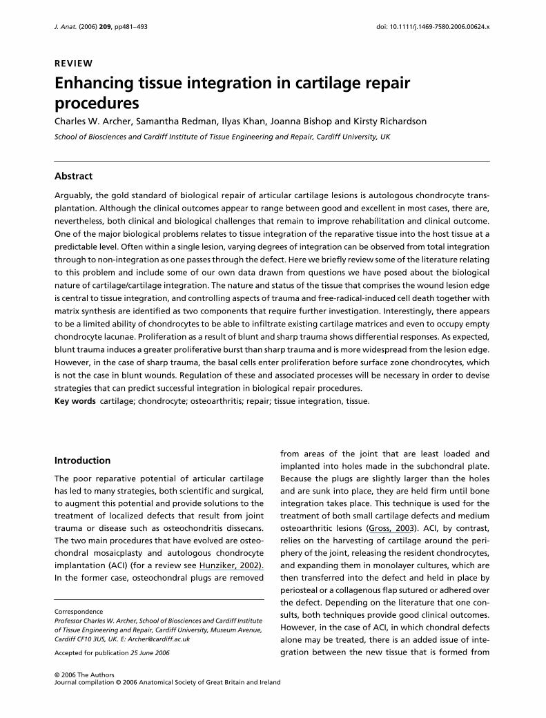
sequently, Chiakulus (1957) studied different cartilages
from the larval salamander
Ambystoma maculatum
(spotted salamander) and after transecting them,
transplanted them in apposition into the tail region.
Such combinations contained humerus/humerus (homo-
typic, somatopleural) and femur/Meckel’s cartilage
(heterotypic, somatopleural/neural crest). He found that
when the cartilage was from the same developmental
origin (i.e. somatopleural or neural crest) the cartilages
fused, but when the cartilages were from different
origins they did not fuse (Fig. 1). As Hall (2005) pointed
out, although Chiakulus explained his observations on
heterogeneity of developmental origin, other explana-
tions are also viable, such as the ability to hypertrophy
and even on intrinsic matrix differences. Corroborating
data were obtained by Fyfe & Hall (1979), who cultured
chick tibial cartilage with Meckel’s cartilage in opposi-
tion and again found that fusion occurred when two
cartilages were from the same origin but not when the
origins differed. It would be interesting to test whether
species differences also existed in addition to those dis-
cussed above. Further insights could also be gained by
testing whether a similar result is obtained if one of the
cartilages was devitalized, thus indicating whether cell
viability played a role in the fusion process.
These data are useful because they suggest caution
for applied tissue engineering with cartilage say from
the nose to repair cartilage of the knee. It is certainly
Fig. 1 Frontal sections through the tail fin of Ambystoma maculatum showing implanted cartilage elements that had been transected and placed in opposition to judge fusion ability between cartilages of differing developmental origin. (a) Femur (F) and femur, and (b) humerus (H) and humerus. Note complete fusion with evidence of neocartilage (NC) comprising the area between the original transects in these homotypic implants. (P), perichondrium. (c) Gill bar (G) and scapula (S). (d) Meckel’s cartilage (M) and femur (F) showing that in heterotypic implants (mesodermal and neural crest) no fusion takes place with cells occupying the boundaries between the opposed elements. (After Chiakulus, 1957.)
Enhancing tissue integration in cartilage repair procedures, C. W. Archer et al.
© 2006 The Authors Journal compilation © 2006 Anatomical Society of Great Britain and Ireland
483
worth considering repeating some of these experiments
with mammalian species, including humans.
Most recent studies on integrative repair in articular
cartilage have focused both on structural and on bio-
chemical aspects, with the former incorporating mechanical
testing of the integration. It has been appreciated for
some time that small proteoglycans are inhibitory to
integration of tissue generated within chondral defects
and that treatment of the defects with chondroitinase
ABC promotes integration (Quinn & Hunziker, 2002).
More recently, it has been reported that lubricin [sur-
face zone proteoglycan (SZP); proteoglycan 4 (PRG-4)],
which is present in synovial fluid, is also inhibitory to
integration of cartilage explants and constructs
in vitro
yet hyaluronan, a molecule occupying a large amount
of space, had no effect (Schaefer et al. 2004; Englert
et al. 2005). Clearly, it seems the responses are not solely
attributable to the stearic effects of the proteoglycans
and there appears to be some unknown specificity.
There have been many studies in a variety of model
systems both
in vitro
and
in vivo
where the strength
of the integration has been measured by a variety of
mechanical means. For example, in an equine model
of ACI, the tensile modulus of the repair tissue (0.6 MPa)
was much lower than that of intact controls (5.2 MPa).
Furthermore, the integration strength to failure was
less than half of that for intact controls (1.2 vs. 2.7 MPa
for intact tissue) and that the addition of insulin-like
growth factor (IGF) to the chondrocytes at implanta-
tion had no effects on these values (Gratz et al. 2006).
In a model where natural (agarose, fibrin) and syn-
thesized (polyglycolic acid) scaffolds were used as cell-
seeded constructs and implanted into annular cartilage
constructs that were cultured for up to 40 days, it was
found that significant differences existed between
the construct types. However, neither failure stress nor
energy to failure correlated with biochemical content
of the construct, and adhesion strength was always
significantly lower than native tissue as might be
expected. It has also been reported using another
in vitro
push-out model that pretreatment with low
dosages of hyaluronidase and collagenase improved
the integrative viability, matrix integrity and push-out
strength (1.32 MPa) as compared with non-treated controls
(0.84 MPa), thus lending support for the studies men-
tioned above (van de Breevaart Bravenboer et al. 2004).
Clearly, there is still much work to be done if we wish
to define the conditions that will ensure that any given
reparative procedure can predict good tissue integration.
Against the above work, we have asked a number of
questions regarding the nature of cartilage integration
and have carried out a number of experiments address-
ing these issues at a fundamental level.
Understanding the cartilage wound margin is essential to integration
Implicitly, in terms of whether transplanted cells or a
tissue construct will fuse functionally to the host tissue
is dependent on the tissue interface and whether the
host tissue is receptive to integration in terms of viabil-
ity, matrix metabolism and content as discussed above?
A matrix turning over slowly is likely to take a con-
siderable time to fuse. A viable tissue may fuse with a
devitalized matrix but in the absence of colonization of
that matrix, it will ultimately fail through mechanical
fatigue as a result of lack of turnover.
In order to investigate some of these aspects further,
we devised an
in vitro
model system that used articular
cartilage from both 7-day and 18-month bovine carti-
lage from the metcarpophalangeal joint. Briefly, a full-
depth 1-cm
2
(approx.) explant was removed by scalpel
and following excision, explants were incubated at
37
°
C in 5% CO
2
for 24 h to equilibrate the tissue prior
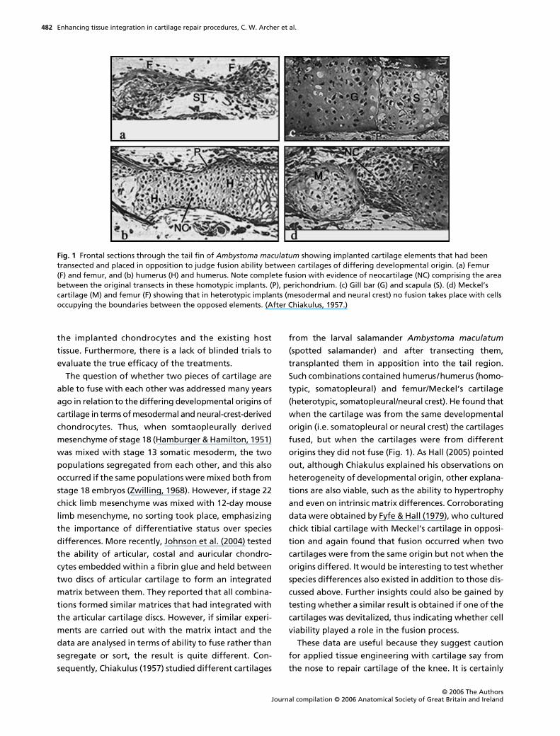
to wounding. The explants were wounded centrally
with a trephine of 1.7 mm internal diameter, from the
articular surface downwards removing a core of tissue.
A scalpel cut bisecting the trephine wound was then
made with a no. 23 scalpel blade to create a scalpel
wound on the same explant (Fig. 2). Wounded explants
and unwounded controls were incubated for up to
10 days with media changed every 48 h. Thus, the
trephine wound was considered blunt and representa-
tive of many orthopaedic instruments used in joint
procedures whilst the scalpel wound represented sharp
incision. Cell viability in response to wounding was
assessed using the live/dead cell kit (Molecular Probes,
Cambridge, UK) and matrix synthesis was assessed
both autoradiographically and quantitatively by
scintillation counting after labelling with
3
H-proline
(collagen) and
35
S (glycosaminoglycans) (Tew et al. 2000,
2001; Redman et al. 2004).
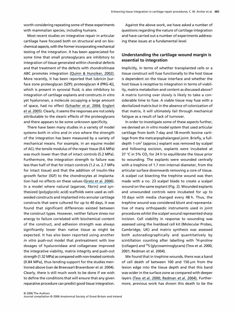
We found that in trephine wounds, there was a band
of cell death of between 100 and 150
µ
m from the
lesion edge into the tissue depth and that this band
was wider in the surface zone as compared with deeper
layers (Tew et al. 2000; Redman et al. 2004). Further-
more, previous work has shown this death to be the
Enhancing tissue integration in cartilage repair procedures, C. W. Archer et al.
© 2006 The AuthorsJournal compilation © 2006 Anatomical Society of Great Britain and Ireland
484
result of a combination of apoptosis and necrosis and
that the apoptosis was progressive for up to 10 days
(Tew et al. 2000). By contrast, in scalpel-made wounds,
there was little evidence of cell death and, of that
present, most was at the lesion edge (Fig. 3).
We then investigated the occurrence of proliferation
as a result of the two differing traumas by thymidine
incorporation. Wounded explants were pulse labelled
overnight and harvested on days 2, 5 and 10. Autoradio-
graphs were prepared and the labelled cells counted
as a function of lacunae. The explanation for this method
was that because of the progressive nature of the apop-
tosis, only late-stage apoptotic cells could be detected
in the histological sections, and we were not able to
detect those entering the apoptotic cycle that could
not divide. Our negative controls comprised explants
that did not contain a trephined or scalpel wound and
we counted the relevant area within the centre of the
Fig. 2 Diagrammatic representation of the trephine and scalpel wound created in articular cartilage full-depth explants. Serial cross-sections were taken for analysis through the scalpel wound then the trephine wound.
Fig. 3 Live (green)/dead (red) labelling of cross-sections, showing the wound edge in the surface zone and mid zone of articular cartilage 2 days after wounding with a trephine (a) and scalpel (b). Scale bar, 20 µm.
Enhancing tissue integration in cartilage repair procedures, C. W. Archer et al.
© 2006 The Authors Journal compilation © 2006 Anatomical Society of Great Britain and Ireland
485
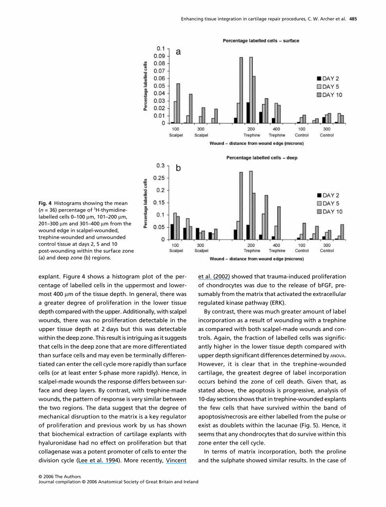
explant. Figure 4 shows a histogram plot of the per-
centage of labelled cells in the uppermost and lower-
most 400
µ
m of the tissue depth. In general, there was
a greater degree of proliferation in the lower tissue
depth compared with the upper. Additionally, with scalpel
wounds, there was no proliferation detectable in the
upper tissue depth at 2 days but this was detectable
within the deep zone. This result is intriguing as it suggests
that cells in the deep zone that are more differentiated
than surface cells and may even be terminally differen-
tiated can enter the cell cycle more rapidly than surface
cells (or at least enter S-phase more rapidly). Hence, in
scalpel-made wounds the response differs between sur-
face and deep layers. By contrast, with trephine-made
wounds, the pattern of response is very similar between
the two regions. The data suggest that the degree of
mechanical disruption to the matrix is a key regulator
of proliferation and previous work by us has shown
that biochemical extraction of cartilage explants with
hyaluronidase had no effect on proliferation but that
collagenase was a potent promoter of cells to enter the
division cycle (Lee et al. 1994). More recently, Vincent
et al. (2002) showed that trauma-induced proliferation
of chondrocytes was due to the release of bFGF, pre-
sumably from the matrix that activated the extracellular
regulated kinase pathway (ERK).
By contrast, there was much greater amount of label
incorporation as a result of wounding with a trephine
as compared with both scalpel-made wounds and con-
trols. Again, the fraction of labelled cells was signific-
antly higher in the lower tissue depth compared with
upper depth significant differences determined by
ANOVA
.
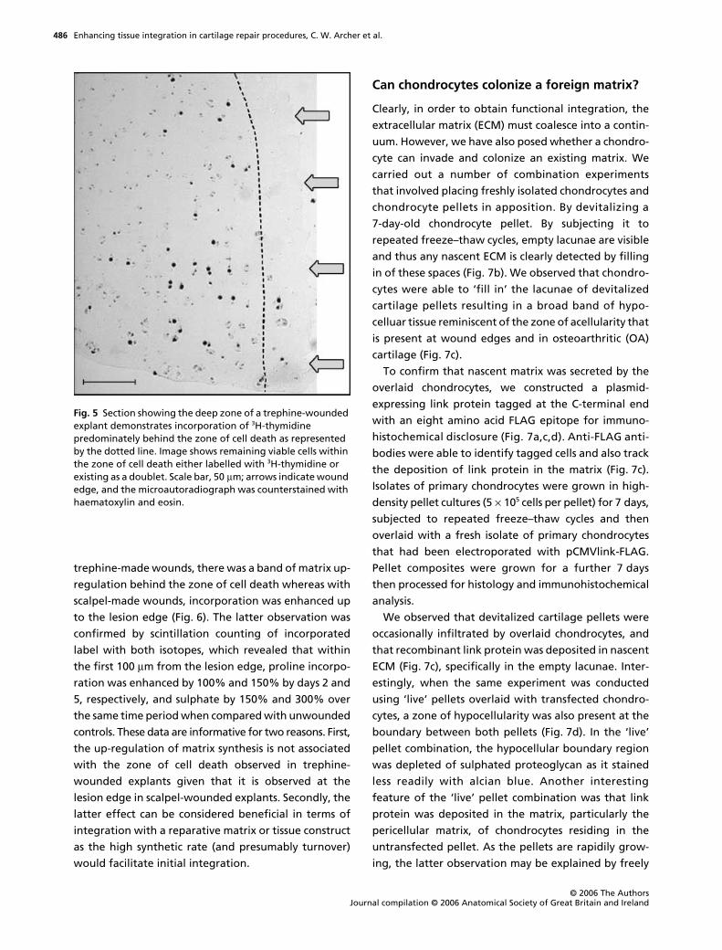
However, it is clear that in the trephine-wounded
cartilage, the greatest degree of label incorporation
occurs behind the zone of cell death. Given that, as
stated above, the apoptosis is progressive, analysis of
10-day sections shows that in trephine-wounded explants
the few cells that have survived within the band of
apoptosis/necrosis are either labelled from the pulse or
exist as doublets within the lacunae (Fig. 5). Hence, it
seems that any chondrocytes that do survive within this
zone enter the cell cycle.
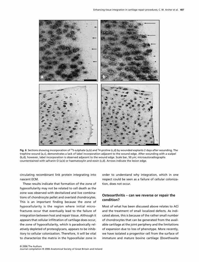
In terms of matrix incorporation, both the proline
and the sulphate showed similar results. In the case of
Fig. 4 Histograms showing the mean (n = 36) percentage of 3H-thymidine-labelled cells 0–100 µm, 101–200 µm, 201–300 µm and 301–400 µm from the wound edge in scalpel-wounded, trephine-wounded and unwounded control tissue at days 2, 5 and 10 post-wounding within the surface zone (a) and deep zone (b) regions.
Enhancing tissue integration in cartilage repair procedures, C. W. Archer et al.
© 2006 The AuthorsJournal compilation © 2006 Anatomical Society of Great Britain and Ireland
486
trephine-made wounds, there was a band of matrix up-
regulation behind the zone of cell death whereas with
scalpel-made wounds, incorporation was enhanced up
to the lesion edge (Fig. 6). The latter observation was
confirmed by scintillation counting of incorporated
label with both isotopes, which revealed that within
the first 100
µ
m from the lesion edge, proline incorpo-
ration was enhanced by 100% and 150% by days 2 and
5, respectively, and sulphate by 150% and 300% over
the same time period when compared with unwounded
controls. These data are informative for two reasons. First,
the up-regulation of matrix synthesis is not associated
with the zone of cell death observed in trephine-
wounded explants given that it is observed at the
lesion edge in scalpel-wounded explants. Secondly, the
latter effect can be considered beneficial in terms of
integration with a reparative matrix or tissue construct
as the high synthetic rate (and presumably turnover)
would facilitate initial integration.
Can chondrocytes colonize a foreign matrix?
Clearly, in order to obtain functional integration, the
extracellular matrix (ECM) must coalesce into a contin-
uum. However, we have also posed whether a chondro-
cyte can invade and colonize an existing matrix. We
carried out a number of combination experiments
that involved placing freshly isolated chondrocytes and
chondrocyte pellets in apposition. By devitalizing a
7-day-old chondrocyte pellet. By subjecting it to
repeated freeze–thaw cycles, empty lacunae are visible
and thus any nascent ECM is clearly detected by filling
in of these spaces (Fig. 7b). We observed that chondro-
cytes were able to ‘fill in’ the lacunae of devitalized
cartilage pellets resulting in a broad band of hypo-
celluar tissue reminiscent of the zone of acellularity that
is present at wound edges and in osteoarthritic (OA)
cartilage (Fig. 7c).
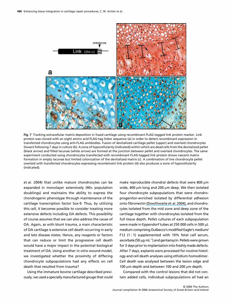
To confirm that nascent matrix was secreted by the
overlaid chondrocytes, we constructed a plasmid-
expressing link protein tagged at the C-terminal end
with an eight amino acid FLAG epitope for immuno-
histochemical disclosure (Fig. 7a,c,d). Anti-FLAG anti-
bodies were able to identify tagged cells and also track
the deposition of link protein in the matrix (Fig. 7c).
Isolates of primary chondrocytes were grown in high-
density pellet cultures (5
×
10
5
cells per pellet) for 7 days,
subjected to repeated freeze–thaw cycles and then
overlaid with a fresh isolate of primary chondrocytes
that had been electroporated with pCMVlink-FLAG.
Pellet composites were grown for a further 7 days
then processed for histology and immunohistochemical
analysis.
We observed that devitalized cartilage pellets were
occasionally infiltrated by overlaid chondrocytes, and
that recombinant link protein was deposited in nascent
ECM (Fig. 7c), specifically in the empty lacunae. Inter-
estingly, when the same experiment was conducted
using ‘live’ pellets overlaid with transfected chondro-
cytes, a zone of hypocellularity was also present at the
boundary between both pellets (Fig. 7d). In the ‘live’
pellet combination, the hypocellular boundary region
was depleted of sulphated proteoglycan as it stained
less readily with alcian blue. Another interesting
feature of the ‘live’ pellet combination was that link
protein was deposited in the matrix, particularly the
pericellular matrix, of chondrocytes residing in the
untransfected pellet. As the pellets are rapidily grow-
ing, the latter observation may be explained by freely
Fig. 5 Section showing the deep zone of a trephine-wounded explant demonstrates incorporation of 3H-thymidine predominately behind the zone of cell death as represented by the dotted line. Image shows remaining viable cells within the zone of cell death either labelled with 3H-thymidine or existing as a doublet. Scale bar, 50 µm; arrows indicate wound edge, and the microautoradiograph was counterstained with haematoxylin and eosin.
Enhancing tissue integration in cartilage repair procedures, C. W. Archer et al.
© 2006 The Authors Journal compilation © 2006 Anatomical Society of Great Britain and Ireland
487
circulating recombinant link protein integrating into
nascent ECM.
These results indicate that formation of the zone of
hypocellularity may not be related to cell death as the
zone was observed with devitalized and live combina-
tions of chondrocyte pellet and overlaid chondrocytes.
This is an important finding because the zone of
hypocellularity is the region where initial micro-
fractures occur that eventually lead to the failure of
integration between host and repair tissue. Although it
appears that cellular infiltration of cartilage does occur,
the zone of hypocellularity, which is paradoxically rel-
atively depleted of proteoglycans, appears to be inhib-
itory to cellular colonization. Therefore, it will be vital
to characterize the matrix in the hypocellular zone in
order to understand why integration, which in one
respect could be seen as a failure of cellular coloniza-
tion, does not occur.
Osteoarthritis – can we reverse or repair the condition?
Most of what has been discussed above relates to ACI
and the treatment of small localized defects. As indi-
cated above, this is because of the rather small number
of chondrocytes that can be generated from the avail-
able cartilage at the joint periphery and the limitations
of expansion due to loss of phenotype. More recently,
we have isolated a progenitor cell from the surface of
immature and mature bovine cartilage (Dowthwaite
Fig. 6 Sections showing incorporation of 35S-sulphate (a,b) and 3H-proline (c,d) by wounded explants 2 days after wounding. The trephine wound (a,c), demonstrates a lack of label incorporation adjacent to the wound edge. After wounding with a scalpel (b,d), however, label incorporation is observed adjacent to the wound edge. Scale bar, 50 µm; microautoradiographs counterstained with safranin O (a,b) or haematoxylin and eosin (c,d). Arrows indicate the lesion edge.
Enhancing tissue integration in cartilage repair procedures, C. W. Archer et al.
© 2006 The AuthorsJournal compilation © 2006 Anatomical Society of Great Britain and Ireland
488
et al. 2004) that unlike mature chondrocytes can be
expanded in monolayer extensively (40+ population
doublings) and maintains the ability to express the
chondrogenic phenotype through maintenance of the
cartilage transcription factor Sox-9. Thus, by utilizing
this cell, it becomes possible to consider treating more
extensive defects including OA defects. This possibility
of course assumes that we can also address the cause of
OA. Again, as with blunt trauma, a main characteristic
of OA cartilage is extensive cell death occurring in early
and late disease states. Hence, any reagents or factors
that can reduce or limit the progressive cell death
would have a major impact in the potential biological
treatment of OA. Using another
in vitro
wound model,
we investigated whether the proximity of differing
chondrocyte subpopulations had any effects on cell
death that resulted from trauma?
Using the immature bovine cartilage described previ-
ously, we used a specially manufactured gouge that could
make reproducible chondral defects that were 800
µ
m
wide, 400
µ
m long and 200
µ
m deep. We then isolated
four chondrocyte subpopulations that were chondro-
progenitor-enriched isolated by differential adhesion
onto fibronectin (Dowthwaite et al. 2004), and chondro-
cytes isolated from the mid zone and deep zone of the
cartilage together with chondrocytes isolated from the
full tissue depth. Pellet cultures of each subpopulation
were made in Eppendorf tubes at 250 000 cells in 500
µ
L
medium comprising Dulbecco’s modified Eagle’s medium/
F12 (1 : 1) supplemented with 10% fetal calf serum,
ascorbate (50
µ
g mL
−
1
) and gentamycin. Pellets were grown
for 3 days prior to implantation into freshly made defects.
After 7 days, explants were processed for routine histol-
ogy and cell death analyses using ethidium homodimer.
Cell death was analysed between the lesion edge and
100
µ
m depth and between 100 and 200
µ
m depth.
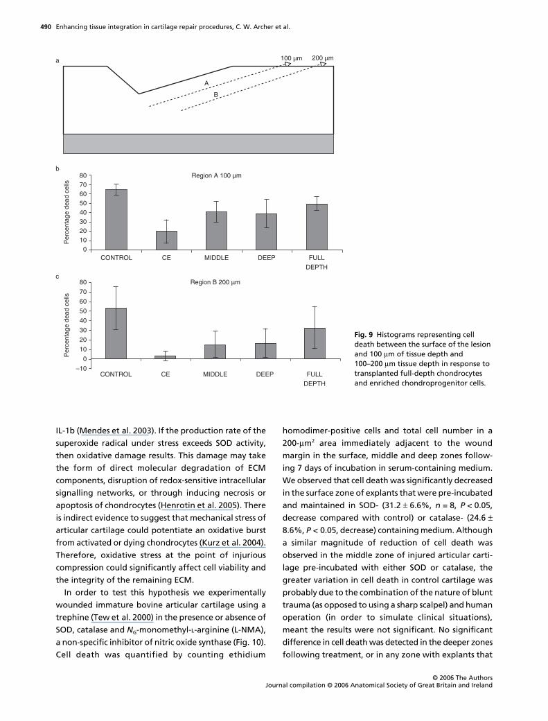
Compared with the control lesions that did not con-
tain added cells, individual subpopulations all had an
Fig. 7 Tracking extracellular matrix deposition in fused cartilage using recombinant FLAG-tagged link protein marker. Link protein was cloned with an eight amino acid FLAG-tag linker sequence (a) in order to detect recombinant expression in transfected chondrocytes using anti-FLAG antibodies. Fusion of devitalized cartilage pellet (upper) and overlaid chondrocytes (lower) following 7 days in culture (b). A zone of hypocellularity (indicated) within which are dead cells from the devitalized pellet (black arrow) and filled lacunae (white arrow) are formed at the junction between pellet and overlaid chondrocytes. The same experiment conducted using chondrocytes transfected with recombinant FLAG-tagged link protein shows nascent matrix formation in empty lacunae but limited colonization of the devitalized matrix (c). A combination of live chondrocyte pellet overlaid with transfected chondrocytes expressing recombinant link protein (d) also produces a zone of hypocellularity (indicated).
Enhancing tissue integration in cartilage repair procedures, C. W. Archer et al.
© 2006 The Authors Journal compilation © 2006 Anatomical Society of Great Britain and Ireland
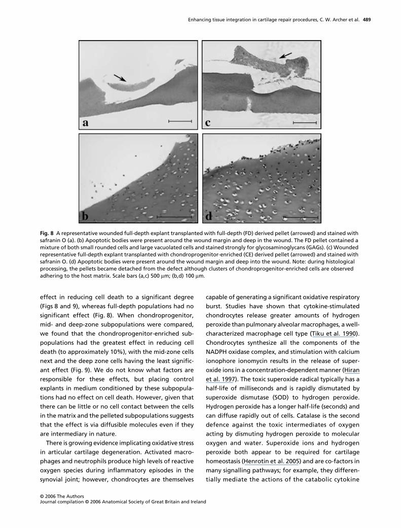
489
effect in reducing cell death to a significant degree
(Figs 8 and 9), whereas full-depth populations had no
significant effect (Fig. 8). When chondroprogenitor,
mid- and deep-zone subpopulations were compared,
we found that the chondroprogenitor-enriched sub-
populations had the greatest effect in reducing cell
death (to approximately 10%), with the mid-zone cells
next and the deep zone cells having the least signific-
ant effect (Fig. 9). We do not know what factors are
responsible for these effects, but placing control
explants in medium conditioned by these subpopula-
tions had no effect on cell death. However, given that
there can be little or no cell contact between the cells
in the matrix and the pelleted subpopulations suggests
that the effect is via diffusible molecules even if they
are intermediary in nature.
There is growing evidence implicating oxidative stress
in articular cartilage degeneration. Activated macro-
phages and neutrophils produce high levels of reactive
oxygen species during inflammatory episodes in the
synovial joint; however, chondrocytes are themselves
capable of generating a significant oxidative respiratory
burst. Studies have shown that cytokine-stimulated
chondrocytes release greater amounts of hydrogen
peroxide than pulmonary alveolar macrophages, a well-
characterized macrophage cell type (Tiku et al. 1990).
Chondrocytes synthesize all the components of the
NADPH oxidase complex, and stimulation with calcium
ionophore ionomycin results in the release of super-
oxide ions in a concentration-dependent manner (Hiran
et al. 1997). The toxic superoxide radical typically has a
half-life of milliseconds and is rapidly dismutated by
superoxide dismutase (SOD) to hydrogen peroxide.
Hydrogen peroxide has a longer half-life (seconds) and
can diffuse rapidly out of cells. Catalase is the second
defence against the toxic intermediates of oxygen
acting by dismuting hydrogen peroxide to molecular
oxygen and water. Superoxide ions and hydrogen
peroxide both appear to be required for cartilage
homeostasis (Henrotin et al. 2005) and are co-factors in
many signalling pathways; for example, they differen-
tially mediate the actions of the catabolic cytokine
Fig. 8 A representative wounded full-depth explant transplanted with full-depth (FD) derived pellet (arrowed) and stained with safranin O (a). (b) Apoptotic bodies were present around the wound margin and deep in the wound. The FD pellet contained a mixture of both small rounded cells and large vacuolated cells and stained strongly for glycosaminoglycans (GAGs). (c) Wounded representative full-depth explant transplanted with chondroprogenitor-enriched (CE) derived pellet (arrowed) and stained with safranin O. (d) Apoptotic bodies were present around the wound margin and deep into the wound. Note: during histological processing, the pellets became detached from the defect although clusters of chondroprogenitor-enriched cells are observed adhering to the host matrix. Scale bars (a,c) 500 µm; (b,d) 100 µm.
Enhancing tissue integration in cartilage repair procedures, C. W. Archer et al.
© 2006 The AuthorsJournal compilation © 2006 Anatomical Society of Great Britain and Ireland
490
IL-1b (Mendes et al. 2003). If the production rate of the
superoxide radical under stress exceeds SOD activity,
then oxidative damage results. This damage may take
the form of direct molecular degradation of ECM
components, disruption of redox-sensitive intracellular
signalling networks, or through inducing necrosis or
apoptosis of chondrocytes (Henrotin et al. 2005). There
is indirect evidence to suggest that mechanical stress of
articular cartilage could potentiate an oxidative burst
from activated or dying chondrocytes (Kurz et al. 2004).
Therefore, oxidative stress at the point of injurious
compression could significantly affect cell viability and
the integrity of the remaining ECM.
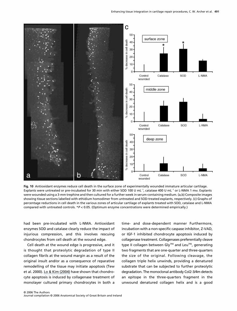
In order to test this hypothesis we experimentally
wounded immature bovine articular cartilage using a
trephine (Tew et al. 2000) in the presence or absence of
SOD, catalase and
N
G
-monomethyl-
L
-arginine (L-NMA),
a non-specific inhibitor of nitric oxide synthase (Fig. 10).
Cell death was quantified by counting ethidium
homodimer-positive cells and total cell number in a
200-
µ
m
2
area immediately adjacent to the wound
margin in the surface, middle and deep zones follow-
ing 7 days of incubation in serum-containing medium.
We observed that cell death was significantly decreased
in the surface zone of explants that were pre-incubated
and maintained in SOD- (31.2
±
6.6%,
n
= 8,
P
< 0.05,
decrease compared with control) or catalase- (24.6
±
8.6%,
P
< 0.05, decrease) containing medium. Although
a similar magnitude of reduction of cell death was
observed in the middle zone of injured articular carti-
lage pre-incubated with either SOD or catalase, the
greater variation in cell death in control cartilage was
probably due to the combination of the nature of blunt
trauma (as opposed to using a sharp scalpel) and human
operation (in order to simulate clinical situations),
meant the results were not significant. No significant
difference in cell death was detected in the deeper zones
following treatment, or in any zone with explants that
Fig. 9 Histograms representing cell death between the surface of the lesion and 100 µm of tissue depth and 100–200 µm tissue depth in response to transplanted full-depth chondrocytes and enriched chondroprogenitor cells.
Enhancing tissue integration in cartilage repair procedures, C. W. Archer et al.
© 2006 The Authors Journal compilation © 2006 Anatomical Society of Great Britain and Ireland
491
had been pre-incubated with L-NMA. Antioxidant
enzymes SOD and catalase clearly reduce the impact of
injurious compression, and this involves rescuing
chondrocytes from cell death at the wound edge.
Cell death at the wound edge is progressive, and it
is thought that proteolytic degradation of type II
collagen fibrils at the wound margin as a result of the
original insult and/or as a consequence of reparative
remodelling of the tissue may initiate apoptosis (Tew
et al. 2000). Lo & Kim (2004) have shown that chondro-
cyte apoptosis is induced by collagenase treatment of
monolayer cultured primary chondrocytes in both a
time- and dose-dependent manner Furthermore,
incubation with a non-specific caspase inhibitor, Z-VAD,
or IGF-1 inhibited chondrocyte apoptosis induced by
collagenase treatment. Collagenases preferentially cleave
type II collagen between Gly
794
and Leu
795
, generating
two fragments that are one-quarter and three-quarters
the size of the original. Following cleavage, the
collagen triple helix unwinds, providing a denatured
substrate that can be subjected to further proteolytic
degradation. The monoclonal antibody Col2-3/4m detects
an epitope in the three-quarters fragment in the
unwound denatured collagen helix and is a good
Fig. 10 Antioxidant enzymes reduce cell death in the surface zone of experimentally wounded immature articular cartilage. Explants were untreated or pre-incubated for 30 min with either SOD 100 U mL−1, catalase 400 U mL−1 or L-NMA 1 mM. Explants were wounded using a 3-mm trephine and then cultured for a further week in serum-containing medium. (a,b) Composite images showing tissue sections labelled with ethidium homodimer from untreated and SOD-treated explants, respectively. (c) Graphs of percentage reductions in cell death in the various zones of articular cartilage of explants treated with SOD, catalase and L-NMA compared with untreated controls. *P < 0.05. (Optimum enzyme concentrations were determined empirically.)
Enhancing tissue integration in cartilage repair procedures, C. W. Archer et al.
© 2006 The AuthorsJournal compilation © 2006 Anatomical Society of Great Britain and Ireland
492
marker of collagen degradation (Hollander et al.
1994).
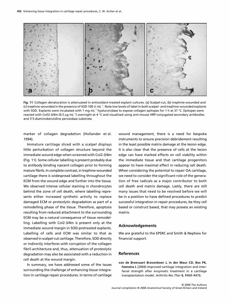
Immature cartilage sliced with a scalpel displays
little perturbation of collagen structure beyond the
immediate wound edge when screened with Col2-3/4m
(Fig. 11). Some cellular labelling is present probably due
to antibody binding nascent collagen prior to forming
mature fibrils. In complete contrast, in trephine-wounded
cartilage there is widespread labelling throughout the
ECM from the wound edge and further into the tissue.
We observed intense cellular staining in chondrocytes
behind the zone of cell death, where labelling repre-
sents either increased synthetic activity to replace
damaged ECM or proteolytic degradation as part of a
remodelling phase of the tissue. Therefore, apoptosis
resulting from reduced attachment to the surrounding
ECM may be a natural consequence of tissue remodel-
ling. Labelling with Col2-3/4m is present only at the
immediate wound margin in SOD-pretreated explants.
Labelling of cells and ECM was similar to that as
observed in scalpel-cut cartilage. Therefore, SOD directly
or indirectly interferes with corruption of the collagen
fibril architecture and, thus, attenuation of proteolytic
degradation may also be associated with a reduction in
cell death at the wound margin.
In summary, we have addressed some of the issues
surrounding the challenge of enhancing tissue integra-
tion in cartilage repair procedures. In terms of cartilage
wound management, there is a need for bespoke
instruments to ensure precision debridement resulting
in the least possible matrix damage at the lesion edge.
It is also clear that the presence of cells at the lesion
edge can have marked effects on cell viability within
the immediate tissue and that cartilage progenitors
appear to have maximal effect in reducing cell death.
When considering the potential to repair OA cartilage,
we need to consider the significant role of the genera-
tion of free radicals as a major contributor to both
cell death and matrix damage. Lastly, there are still
many issues that need to be resolved before we will
be in a position to have defined procedures to predict
successful integration in repair procedures, be they cell
based or construct based, that may possess an existing
matrix.
Acknowledgements
We are grateful to the EPSRC and Smith & Nephew for
financial support.
References
van de Breevaart Bravenboer J, In der Maur CD, Bos PK,Feenstra L
(2004) Improved cartilage integration and inter-facial strength after enzymatic treatment in a cartilagetransplantation model.
Arthritis Res Ther
6
, R469–R476.
Fig. 11 Collagen denaturation is attenuated in antioxidant-treated explant cultures. (a) Scalpel-cut, (b) trephine-wounded and (c) trephine-wounded in the presence of SOD 100 U mL−1. Note low levels of label in both scalpel- and trephine-wounded explants with SOD. Explants were incubated with 1 mg mL−1 hyaluronidase to expose collagen epitopes for 1 h at 37 °C. Epitopes were reacted with Coll2-3/4m (0.5 µg mL−1) overnight at 4 °C and visualized using anti-mouse HRP-conjugated secondary antibodies and 3′3-diaminobenzidine peroxidase substrate.
Enhancing tissue integration in cartilage repair procedures, C. W. Archer et al.
© 2006 The Authors Journal compilation © 2006 Anatomical Society of Great Britain and Ireland
493
Chiakulus JJ
(1957) The specificity and differential fusion ofcartilage derived mesoderm and mesectoderm.
J Exp Zool
136
, 287–300.
Dowthwaite GP, Bishop JC, Redman SN,
et al.
(2004) Thesurface of articular cartilage contains a progenitor cellpopulation.
J Cell Sci
117
, 889–897.
Englert C, McGowan KB, Klein TJ, Giurea A, Schumacher BL,Sah RL
(2005) Inhibition of integrative cartilage repair by pro-toglycan 4 in synovial fluid.
Arthritis Rheum
52
, 1091–1099.
Fyfe DM, Hall BK
(1979) Lack of association between avian car-tilages of different embryological origins when maintained
in vitro
.
Am J Anat
154
, 485–496.
Gratz KR, Wong VW, Chen AC, Fortier LA, Nixon AJ, Sah RL
(2006) Biomechanical assessment of tissue retrieved after invivo cartilage defect repair: tensile modlus of repair tissueand integration with host cartilage.
J Biomechanics
39
, 138–146.
Gross AE
(2003) Cartilage resurfacing: filling defects.
J Arthro-plasty
18
, 14–27.
Hall BK
(ed.) (2005)
Bones and Cartilage
. Amsterdam: Elsevier.
Hamburger V, Hamilton HL (1951) A series of normal stages inthe development of the chick embryo. J Morph 88, 49–92.
Henrotin Y, Kurz B, Aigner T (2005) Oxygen and reactiveoxygen species in cartilage degradation: friends or foes?Osteoarthritis Cartilage 13, 643–654.
Hiran TS, Moulton PJ, Hancock JT (1997) Detection of super-oxide and NADPH oxidase in porcine articular chondrocytes.Free Radic Biol Med 23, 736–743.
Hollander AP, Heathfield TF, Webber C, et al. (1994) Increaseddamage to type II collagen in osteoarthritic articular carti-lage detected by a new immunoassay. J Clin Invest 93, 1722–1732.
Hunziker EB (2002) Articular cartilage repair: basic science andclinical progress. A review of the current status and pros-pects. Osteoarthritis Cartilage 10, 432–463.
Johnson TS, Xu JW, Zaporojan VV, et al. (2004) Integrativerepair of cartilage with articular and nonarticular chondro-cytes. Tissue Engineering 10, 9–10.
Kurz B, Lemke A, Kehn M, et al. (2004) Influence of tissue
maturation and antioxidants on the apoptotic response ofarticular cartilage after injurious compression. ArthritisRheum 50, 123–130.
Lee D, Bentley G, Archer CW (1994) Proteoglycan depletionalone is not sufficient to stimulate proteoglycan synthesis incultured bovine cartilage explants. Osteoarthritis Cartilage2, 175–185.
Lo MY, Kim HT (2004) Chondrocyte apoptosis induced byhydrogen peroxide requires caspase activation but notmitochondrial pore transition. J Orthop Res 22, 1120–1125.
Mendes AF, Caramona MM, Carvalho AP, Lopes MC (2003)Differential roles of hydrogen peroxide and superoxide inmediating IL-1 induced NF-kappa B activation and iNOSexpression in bovine articular chondrocytes. J Cell Biochem88, 783–793.
Quinn TM, Hunziker EB (2002) Controlled enzymatic matrixdegradation for integrative cartilage repair: effects onviable cell density and proteoglycan depostion. Tissue Engi-neering 8, 799–806.
Redman SN, Dowthwaite GP, Thomson BM, Archer CW (2004)The cellular responses of articular cartilage to sharp andblunt trauma. Osteoarthritis Cartilage 12, 106–116.
Schaefer DB, Wendt D, Moretti M, et al. (2004) Lubricin reducescartilage–cartilage integration. Biorheology 41, 503–508.
Tew S, Kwan APL, Hann A, Thomson B, Archer CW (2000) Thereactions of articular cartilage to experimental wounding.Arthritis Rheumatism 43, 215–225.
Tew S, Redman S, Walker E, et al. (2001) Differences in repairresponses between immature and mature cartilage. ClinOrthopaedics Related Res 391S, 142–152.
Tiku ML, Liesch JB, Robertson FM (1990) Production of hydro-gen peroxide by rabbit articular chondrocytes. Enhance-ment by cytokines. J Immunol 145, 690–696.
Vincent T, Hermansson M, Bolton M, Wait R, Saklatvala J(2002) Basic FGF mediates an immediate response of articu-lar cartilage to mechanical injury. Proc Natl Acad Sci USA 99,8259–8264.
Zwilling E (1968) Morphogenetic phases in development.Devel Biol 2, S184–S207.


