Embed Size (px)
Citation preview
Stimuli-Responsive Liposome FusionMediated by Gold NanoparticlesDissaya Pornpattananangkul,†,‡ Sage Olson,†,‡ Santosh Aryal,†,‡ Marta Sartor,†,‡ Chun-Ming Huang,‡,§
Kenneth Vecchio,† and Liangfang Zhang†,‡,*†Department of Nanoengineering, ‡Moores Cancer Center, and §Division of Dermatology, University of California at San Diego, La Jolla, California 92093
Liposomes are spherical lipid vesicleswith a bilayered membrane structureconsisting of amphiphilic lipid mol-
ecules. They have been recognized as one
of the most widely used carriers for deliver-
ing a myriad of cosmeticeuticals, pharma-
ceuticals, and diagnostic and imaging
agents.1 Liposomes can carry both hydro-
philic and hydrophobic agents with high ef-
ficiency and protect them from undesired
effects of external conditions. Their surface
can be readily functionalized with specific
ligands that target liposomes and their pay-
loads to the sites of action. In addition, the
composition, size, surface charge, and other
formulation properties of liposomes can be
well-controlled to meet the needs of spe-
cific circumstances.1�4 However, the appli-
cations of liposomes are usually limited by
their instability. Liposomes, particularly with
sub-100 nm size, are prone to fuse with
one another to reduce their surface ten-
sion, leading to payload loss or undesired
mixing.5�8 Moreover, the resulting lipo-
somes with a size much larger than 100
nm are unlikely to transport through the
skin, therefore significantly diminishing
their use as a dermal drug delivery
vehicle.9,10
A few strategies have been employed
to overcome this problem aiming at im-
proving the use of liposomes as a potent
delivery nanocarrier.11�14 One extensively
used approach is to coat the liposome sur-
face with a “stealth” material such as poly-
ethylene glycol (PEG).15,16 The PEG layer not
only prevents liposomes from fusing with
one another but also enhances their in vivo
circulation lifetime by suppressing plasma
proteins from adsorbing onto the liposome
surface. The success of PEGylated liposomes
has led to a group of clinically approved
therapeutic products for systemic drugdelivery, including Doxil, AmBisome,DaunoXome, DepoCyt, and Visudyne.3,17 Al-though the polymer-coated liposomes haveshown great success for systemic drug de-livery, they are less frequently used for der-mal drug delivery, especially to treat bacte-rial infections. This is because the polymercoating will not only stabilize liposomesagainst fusion with one another but alsoprevent them from fusing with bacterialmembranes, to which the antimicrobialpayloads will be delivered. It is worth not-ing that bacteria usually interact with ve-sicular drug nanocarriers such as liposomesin a different manner from host cells or can-cerous cells. The cells can internalize the en-tire liposomes through endocytosis, whilethe bacteria preferentially go throughmembrane�membrane fusion.10,18 There-fore, it would be desirable to develop lipo-somes that are stabilized against fusionwith one another before they are placed atthe sites of action including the manufac-turing and storage periods, while their
*Address correspondence [email protected].
Received for review December 20, 2009and accepted March 08, 2010.
Published online March 17, 2010.10.1021/nn9018587
© 2010 American Chemical Society
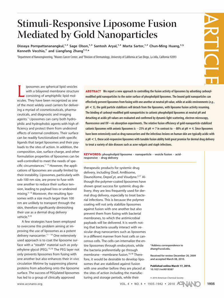
ABSTRACT We report a new approach to controlling the fusion activity of liposomes by adsorbing carboxyl-
modified gold nanoparticles to the outer surface of phospholipid liposomes. The bound gold nanoparticles can
effectively prevent liposomes from fusing with one another at neutral pH value, while at acidic environments (e.g.,
pH < 5), the gold particle stabilizers will detach from the liposomes, with liposome fusion activity resuming.
The binding of carboxyl-modified gold nanoparticles to cationic phospholipid liposomes at neutral pH and
detaching at acidic pH values are evaluated and confirmed by dynamic light scattering, electron microscopy,
fluorescence and UV�vis absorption experiments. The relative fusion efficiency of gold-nanoparticle-stabilized
cationic liposomes with anionic liposomes is �25% at pH � 7 in contrast to �80% at pH � 4. Since liposomes
have been extensively used as drug nanocarriers and the infectious lesions on human skin are typically acidic with
a pH < 5, these acid-responsive liposomes with tunable fusion ability hold great promise for dermal drug delivery
to treat a variety of skin diseases such as acne vulgaris and staph infections.
KEYWORDS: phospholipid liposome · nanoparticle · vesicle fusion · acid-responsive · drug delivery
ARTIC
LE
www.acsnano.org VOL. 4 ▪ NO. 4 ▪ 1935–1942 ▪ 2010 1935
fusion activity will be reinstalled once they are applied
onto the target skin sites.
Here we report a stimuli-responsive novel gold-
nanoparticle-stabilized liposome system in which small
gold nanoparticles (diameter �4 nm) bind to the sur-
face of liposomes (diameter � sub-100 nm) and thus
stabilize the liposomes at neutral pH. The bound gold
particle stabilizers detach from the liposomes when the
environment acidity increases to pH � 5, resulting inthe formation of bare liposomes that can actively fusewith various biological membranes. It has been well-documented that human skin is typically acidic (pH �
3.9�6.0),19 especially the infectious lesions on theskin.20 For example, the pH value is about 4.0 at theacne lesions21 and 4.5�6.3 at comedones.22 Thereforeacid-responsive liposomes with tunable fusion abilitywill be practically demanded for dermal drug delivery.Recently, Granick et al. have reported that binding smallpolystyrene particles (diameter �20 nm) to the surfaceof zwitterionic liposomes (diameter �200 nm) can sta-bilize liposomes against fusion.8,23,24 However, no study,to the best of our knowledge, has been reported to de-velop stimuli-responsive nanoparticle-stabilized lipo-somes for possible drug delivery applications.
The principle of this study, applying carboxyl-modified gold nanoparticles to mediate the fusion ac-
tivity of phospholipid liposomes, is illustrated in Figure
1. With a pKa � 5,25 the carboxylic group is deproto-
nated at pH � 7, resulting in negatively charged
Au�COO� nanoparticles, which can bind to cationic lip-
osomes through electrostatic attraction and thus stabi-
lize the liposomes. When the environment pH drops to
below 5, the carboxylic group will be protonated. The
resulting neutral Au�COOH nanoparticles will detach
from the liposome surface due to the lacking of bind-
ing forces, thereby freeing the liposomes. Gold nano-
particles are selected for this study because of their
fluorescence quenching properties that can be em-
ployed to indicate their binding and detaching process
and extent when a small fraction of fluorescent dyes is
doped into the liposome membranes. Moreover, gold
is a biocompatible noble metal26 with antimicrobial ac-
tivity against a wide variety of bacteria.27
RESULTS AND DISCUSSIONWe first prepared carboxyl-modified gold nanoparti-
cle (AuC)-stabilized liposomes (AuC-liposome). In the
study, cationic phospholipid liposomes consisting of 90
wt % hydrogenated L-�-phosphatidylcholine (EggPC)
and 10 wt % 1,2-di-(9Z-octadecenoyl)-3-trimethylammonium propane (DOTAP) were preparedthrough the well-known extrusion method.28 Dynamic
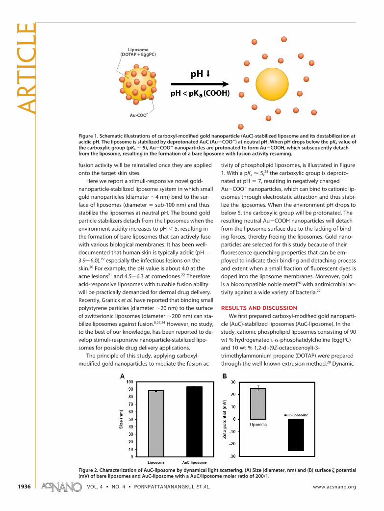
Figure 1. Schematic illustrations of carboxyl-modified gold nanoparticle (AuC)-stabilized liposome and its destabilization atacidic pH. The liposome is stabilized by deprotonated AuC (Au�COO�) at neutral pH. When pH drops below the pKa value ofthe carboxylic group (pKa � 5), Au�COO� nanoparticles are protonated to form Au�COOH, which subsequently detachfrom the liposome, resulting in the formation of a bare liposome with fusion activity resuming.
Figure 2. Characterization of AuC-liposome by dynamical light scattering. (A) Size (diameter, nm) and (B) surface � potential(mV) of bare liposomes and AuC-liposome with a AuC/liposome molar ratio of 200/1.
ART
ICLE
VOL. 4 ▪ NO. 4 ▪ PORNPATTANANANGKUL ET AL. www.acsnano.org1936
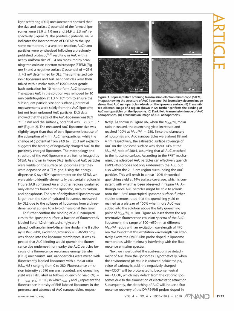
light scattering (DLS) measurements showed thatthe size and surface � potential of the formed lipo-somes were 88.0 � 1.0 nm and 24.9 � 2.3 mV, re-spectively (Figure 2). The positive � potential valueindicates the incorporation of DOTAP to the lipo-some membrane. In a separate reaction, AuC nano-particles were synthesized following a previouslypublished protocol,29,30 resulting in AuC with anearly uniform size of �4 nm measured by scan-ning transmission electron microscope (STEM) (Fig-ure 3) and a negative surface � potential of �25.6� 4.2 mV determined by DLS. The synthesized cat-ionic liposomes and AuC nanoparticles were thenmixed with a molar ratio of 1:200 under gentlebath sonication for 10 min to form AuC-liposome.The excess AuC in the solution was removed by 10min centrifugation at 1.3 104 rpm to ensure thesubsequent particle size and surface � potentialmeasurements were solely from the AuC-liposomebut not from unbound AuC particles. DLS datashowed that the size of the AuC-liposome was 92.9� 1.3 nm and the surface � potential was �25.3 � 0.7mV (Figure 2). The measured AuC-liposome size wasslightly larger than that of bare liposomes because ofthe adsorption of 4 nm AuC nanoparticles, while thechange of � potential from 24.9 to �25.3 mV explicitlysuggests the binding of negatively charged AuC to thepositively charged liposomes. The morphology andstructure of the AuC-liposome were further imaged bySTEM. As shown in Figure 3A,B, individual AuC particleswere visible on the surface of liposomes after theywere deposited on a TEM grid. Using the energy-dispersive X-ray (EDX) spectrometer on the STEM, wewere able to identify elementally that certain regions inFigure 3A,B contained Au and other regions containedonly elements found in the liposome, such as carbonand phosphorus. The size of dehydrated liposomes waslarger than the size of hydrated liposomes measuredby DLS due to the collapse of liposomes from a three-dimensional sphere to a two-dimensional thin layer.
To further confirm the binding of AuC nanoparti-cles to the liposome surface, a fraction of fluorescentlylabeled lipid, 1,2-dimyristoyl-sn-glycero-3-phosphoethanolamine-N-lissamine rhodamine B sulfo-nyl (DMPE-RhB, excitation/emission � 550/590 nm),was doped into the liposome membranes. It was ex-pected that AuC binding would quench the fluores-cence dye underneath or nearby the AuC particles be-cause of a fluorescence resonance energy transfer(FRET) mechanism. AuC nanoparticles were mixed withfluorescently labeled liposomes with a molar ratio(MAuC/ML) ranging from 0 to 280. Fluorescence emis-sion intensity at 590 nm was recorded, and quenchingyield was calculated as follows: quenching yield (%) �
(1 � IAuC�L/IL) 100, in which IAuC�L and IL represent thefluorescence intensity of RhB-labeled liposomes in thepresence and absence of AuC nanoparticles, respec-
tively. As shown in Figure 4A, when the MAuC/ML molarratio increased, the quenching yield increased andreached 100% at MAuC/ML � 280. Since the diametersof liposomes and AuC nanoparticles were about 88 and4 nm respectively, the estimated surface coverage ofAuC on the liposome surface was about 14% at theMAuC/ML ratio of 280:1, assuming that all AuC attachedto the liposome surface. According to the FRET mecha-nism, the adsorbed AuC particles can effectively quenchDMPE-RhB probes not only underneath the AuC butalso within the 2�5 nm region surrounding the AuCparticles. This will result in a near 100% theoreticalquenching yield at 14% surface coverage, which is con-sistent with what has been observed in Figure 4A. Al-though more AuC particles might be able to adsorbonto the �86% unoccupied liposome surface, furtherstudies demonstrated that the quenching yield re-mained as a plateau of 100% when more AuC wasadded into the solution above the fully quenchingpoint of MAuC/ML � 280. Figure 4A inset shows the rep-resentative fluorescence emission spectra of the AuC-liposome in the range of 500�650 nm at differentMAuC/ML ratios with an excitation wavelength of 470nm. We found that this excitation wavelength can effec-tively excite the DMPE-RhB probe doped in liposomemembranes while minimally interfering with the fluo-rescence emission spectra.
Next we investigated the acid-responsive detach-ment of AuC from the liposomes. Hypothetically, whenthe environment pH value is reduced below the pKa
value of carboxylic acid, the negatively chargedAu�COO� will be protonated to become neutralAu�COOH, which may detach from the cationic lipo-somes due to the elimination of electrostatic attraction.Subsequently, the detaching of AuC will induce a fluo-rescence recovery of the DMPE-RhB probes doped in
Figure 3. Representative scanning transmission electron microscope (STEM)images showing the structure of AuC-liposome. (A) Secondary electron imageshows that AuC nanoparticles adsorb on the liposome surface. (B) Transmit-ted electron image of a region shown in (A) further confirms the binding ofAuC nanoparticles on the liposome. (C) Dark field transmission image of AuCnanoparticles. (D) Transmission image of AuC nanoparticles.
ARTIC
LE
www.acsnano.org VOL. 4 ▪ NO. 4 ▪ 1935–1942 ▪ 2010 1937
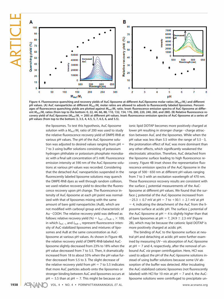
the liposomes. To test this hypothesis, AuC-liposomesolution with a MAuC/ML ratio of 200 was used to studythe relative fluorescence recovery yield of DMPE-RhB atvarious pH values. The pH of the AuC-liposome solu-tion was adjusted to desired values ranging from pH �
7 to 3 using buffer solutions consisting of potassiumhydrogen phthalate or potassium phosphate monoba-sic with a final salt concentration of 5 mM. Fluorescenceemission intensity at 590 nm of the AuC-liposome solu-tions at various pH values was recorded. Consideringthat the detached AuC nanoparticles suspended in thefluorescently labeled liposome solutions may quenchthe DMPE-RhB dyes as well through random collision,we used relative recovery yield to describe the fluores-cence recovery upon pH change. The fluorescence in-tensity of AuC-liposome at each pH point was normal-ized with that of liposomes mixing with the sameamount of bare gold nanoparticles (AuB), which arenot modified with carboxyl group and characteristic ofAu�COOH. The relative recovery yield was defined asfollows: relative recovery yield (%) � IAuC�L/IAuB�L 100,in which IAuC�L and IAuB�L represent fluorescence inten-sity of AuC-stabilized liposomes and mixtures of lipo-somes and AuB at the same concentration as AuC-liposome at various pH values. As shown in Figure 4B,the relative recovery yield of DMPE-RhB-labeled AuC-liposome slightly decreased from 23% to 18% when thepH value decreased from 7 to 5.5. Then, it dramaticallyincreased from 18 to about 55% when the pH value fur-ther decreased from 5.5 to 3. The slight decrease ofthe relative recovery yield from pH � 7 to 5.5 indicatesthat more AuC particles adsorb onto the liposomes orstronger binding between AuC and liposomes occurs atpH � 5.5 than at pH � 7. This might be because cat-
ionic lipid DOTAP becomes more positively charged atlower pH resulting in stronger charge�charge attrac-tion between AuC and the liposomes. While when thepH value was less than 5.5 within the range of 5.5�3,the protonation effect of AuC was more dominant thanany other effects, which significantly weakened theelectrostatic attraction. Therefore, AuC detached fromthe liposome surface leading to high fluorescence re-covery. Figure 4B inset shows the representative fluo-rescence emission spectra of the AuC-liposome in therange of 500�650 nm at different pH values rangingfrom 7 to 3 with an excitation wavelength of 470 nm.These fluorescence recovery results are consistent withthe surface � potential measurements of the AuC-liposome at different pH values. We found that the sur-face � potential of the AuC-liposome increased from�25.3 � 0.7 mV at pH � 7 to 30.1 � 2.1 mV at pH� 4, indicating the detachment of the AuC from the li-posome surface at acidic pH. The surface � potential ofthe AuC-liposome at pH � 4 is slightly higher than thatof bare liposomes at pH � 7, 24.9 � 2.3 mV (Figure2B), which may be because the cationic lipid DOTAP ismore positively charged at acidic pH.
The binding of AuC to the liposome surface at neu-tral pH and detaching at acidic pH were further exam-ined by measuring UV�vis absorption of AuC-liposomeat pH � 7 and 4, respectively, after the removal of un-bound AuC via proper centrifugation. Here, HCl wasused to adjust the pH of the AuC-liposome solutions in-stead of using buffer solutions because some UV ab-sorption of the buffer was detected. After incubatingthe AuC-stabilized cationic liposomes (not fluorescentlylabeled) with HCl for 10 min at pH � 7 and 4, the AuC-liposome solutions were centrifuged to precipitate un-
Figure 4. Fluorescence quenching and recovery yields of AuC-liposome at different AuC/liposome molar ratios (MAuC/ML) and differentpH values. (A) AuC nanoparticles at different MAuC/ML molar ratios are allowed to adsorb to fluorescently labeled liposomes. Percent-ages of fluorescence quenching yields are plotted against MAuC/ML ratio. Inset: fluorescence emission spectra of AuC-liposome at differ-ent MAuC/ML ratios (from top to the bottom: 0, 22, 44, 66, 88, 110, 132, 154, 176, 200, 220, 240, 260, and 280). (B) Relative fluorescence re-covery yield of AuC-liposome (MAuC/ML � 200) at different pH values. Inset: fluorescence emission spectra of AuC-liposome at a series ofpH values (from top to the bottom: 3, 3.5, 4, 4.5, 5, 7, 6.5, 6, and 5.5).
ART
ICLE
VOL. 4 ▪ NO. 4 ▪ PORNPATTANANANGKUL ET AL. www.acsnano.org1938
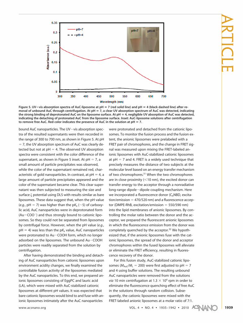
bound AuC nanoparticles. The UV�vis absorption spec-
tra of the resulted supernatants were then recorded in
the range of 300 to 700 nm, as shown in Figure 5. At pH
� 7, the UV absorption spectrum of AuC was clearly de-
tected but not at pH � 4. The observed UV absorption
spectra were consistent with the color difference of the
supernatant, as shown in Figure 5 inset. At pH � 7, a
small amount of particle precipitates was observed,
while the color of the supernatant remained red, char-
acteristic of gold nanoparticles. In contrast, at pH � 4, a
large amount of particle precipitates appeared and the
color of the supernatant became clear. This clear super-
natant was then subjected to measuring the size and
surface � potential using DLS with results similar as bare
liposomes. These data suggest that, when the pH value
(e.g., pH � 7) was higher than the pKa (�5) of carboxy-
lic acid, AuC nanoparticles were in deprotonated form
(Au�COO�) and thus strongly bound to cationic lipo-
somes. So they could not be separated from liposomes
by centrifugal force. However, when the pH value (e.g.,
pH � 4) was less than the pKa value, AuC nanoparticles
were protonated to Au�COOH form, which no longer
adsorbed on the liposomes. The unbound Au�COOH
particles were readily separated from the solution by
centrifugation.
After having demonstrated the binding and detach-
ing of AuC nanoparticles from cationic liposomes upon
environment acidity changes, we finally examined the
controllable fusion activity of the liposomes mediated
by the AuC nanoparticles. To this end, we prepared an-
ionic liposomes consisting of EggPC and lauric acid
(LA), which were mixed with AuC-stabilized cationic
liposomes at different pH values. It was expected that
bare cationic liposomes would bind to and fuse with an-
ionic liposomes intimately after the AuC nanoparticles
were protonated and detached from the cationic lipo-
somes. To monitor the fusion process and the fusion ex-
tent, the anionic liposomes were prelabeled with a
FRET pair of chromophores, and the change in FRET sig-
nal was measured upon mixing the FRET-labeled an-
ionic liposomes with AuC-stabilized cationic liposomes
at pH � 7 and 4. FRET is a widely used technique that
precisely measures the distance of two subjects at the
molecular level based on an energy transfer mechanism
of two chromophores.31 When the two chromophores
are in close proximity (�10 nm), the excited donor can
transfer energy to the acceptor through a nonradiative
long-range dipole�dipole coupling mechanism. Here
we incorporated a fluorescence donor (C6NBD, excita-
tion/emission � 470/520 nm) and a fluorescence accep-
tor (DMPE-RhB, excitation/emission � 550/590 nm)
into the lipid membranes of anionic liposomes. By con-
trolling the molar ratio between the donor and the ac-
ceptor, we prepared the fluorescent anionic liposomes
in which the fluorescence emission from the donor was
completely quenched by the acceptor.32 We hypoth-
esized that, if the anionic liposomes fuse with the cat-
ionic liposomes, the spread of the donor and acceptor
chromophores within the fused liposomes will alleviate
or eliminate the FRET efficiency, resulting in fluores-
cence recovery of the donor.
For this fusion study, AuC-stabilized cationic lipo-
somes (MAuC/ML � 200) were first adjusted to pH � 7
and 4 using buffer solutions. The resulting unbound
AuC nanoparticles were removed from the solutions
via 10 min centrifugation at 1.3 104 rpm in order to
eliminate the fluorescence quenching effect of free AuC
in the solutions through random collision. Subse-
quently, the cationic liposomes were mixed with the
FRET-labeled anionic liposomes at a molar ratio of 7:1.
Figure 5. UV�vis absorption spectra of AuC-liposome at pH � 7 (red solid line) and pH � 4 (black dashed line) after re-moval of unbound AuC through centrifugation. At pH � 7, a clear UV absorption spectrum of AuC was detected, indicatingthe strong binding of deprotonated AuC on the liposome surface. At pH � 4, negligible UV absorption of AuC was detected,indicating the detaching of protonated AuC from the liposome surface. Inset: AuC-liposome solutions after centrifugationto remove free AuC. Red color indicates the presence of AuC in the solution at pH � 7.
ARTIC
LE
www.acsnano.org VOL. 4 ▪ NO. 4 ▪ 1935–1942 ▪ 2010 1939
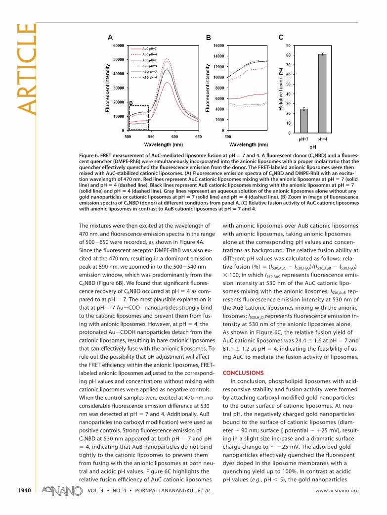
The mixtures were then excited at the wavelength of
470 nm, and fluorescence emission spectra in the range
of 500�650 were recorded, as shown in Figure 4A.
Since the fluorescent receptor DMPE-RhB was also ex-
cited at the 470 nm, resulting in a dominant emission
peak at 590 nm, we zoomed in to the 500�540 nm
emission window, which was predominantly from the
C6NBD (Figure 6B). We found that significant fluores-
cence recovery of C6NBD occurred at pH � 4 as com-
pared to at pH � 7. The most plausible explanation is
that at pH � 7 Au�COO� nanoparticles strongly bind
to the cationic liposomes and prevent them from fus-
ing with anionic liposomes. However, at pH � 4, the
protonated Au�COOH nanoparticles detach from the
cationic liposomes, resulting in bare cationic liposomes
that can effectively fuse with the anionic liposomes. To
rule out the possibility that pH adjustment will affect
the FRET efficiency within the anionic liposomes, FRET-
labeled anionic liposomes adjusted to the correspond-
ing pH values and concentrations without mixing with
cationic liposomes were applied as negative controls.
When the control samples were excited at 470 nm, no
considerable fluorescence emission difference at 530
nm was detected at pH � 7 and 4. Additionally, AuB
nanoparticles (no carboxyl modification) were used as
positive controls. Strong fluorescence emission of
C6NBD at 530 nm appeared at both pH � 7 and pH
� 4, indicating that AuB nanoparticles do not bind
tightly to the cationic liposomes to prevent them
from fusing with the anionic liposomes at both neu-
tral and acidic pH values. Figure 6C highlights the
relative fusion efficiency of AuC cationic liposomes
with anionic liposomes over AuB cationic liposomes
with anionic liposomes, taking anionic liposomes
alone at the corresponding pH values and concen-
trations as background. The relative fusion ability at
different pH values was calculated as follows: rela-
tive fusion (%) � (I530,AuC � I530,H2O)/(I530,AuB � I530,H2O)
100, in which I530,AuC represents fluorescence emis-
sion intensity at 530 nm of the AuC cationic lipo-
somes mixing with the anionic liosomes; I530,AuB rep-
resents fluorescence emission intensity at 530 nm of
the AuB cationic liposomes mixing with the anionic
liosomes; I530,H2O represents fluorescence emission in-
tensity at 530 nm of the anionic liposomes alone.
As shown in Figure 6C, the relative fusion yield of
AuC cationic liposomes was 24.4 � 1.6 at pH � 7 and
81.1 � 1.2 at pH � 4, indicating the feasibility of us-
ing AuC to mediate the fusion activity of liposomes.
CONCLUSIONSIn conclusion, phospholipid liposomes with acid-
responsive stability and fusion activity were formed
by attaching carboxyl-modified gold nanoparticles
to the outer surface of cationic liposomes. At neu-
tral pH, the negatively charged gold nanoparticles
bound to the surface of cationic liposomes (diam-
eter � 90 nm; surface � potential � 25 mV), result-
ing in a slight size increase and a dramatic surface
charge change to � �25 mV. The adsorbed gold
nanoparticles effectively quenched the fluorescent
dyes doped in the liposome membranes with a
quenching yield up to 100%. In contrast at acidic
pH values (e.g., pH � 5), the gold nanoparticles
Figure 6. FRET measurement of AuC-mediated liposome fusion at pH � 7 and 4. A fluorescent donor (C6NBD) and a fluores-cent quencher (DMPE-RhB) were simultaneously incorporated into the anionic liposomes with a proper molar ratio that thequencher effectively quenched the fluorescence emission from the donor. The FRET-labeled anionic liposomes were thenmixed with AuC-stabilized cationic liposomes. (A) Fluorescence emission spectra of C6NBD and DMPE-RhB with an excita-tion wavelength of 470 nm. Red lines represent AuC cationic liposomes mixing with the anionic liposomes at pH � 7 (solidline) and pH � 4 (dashed line). Black lines represent AuB cationic liposomes mixing with the anionic liposomes at pH � 7(solid line) and pH � 4 (dashed line). Gray lines represent an aqueous solution of the anionic liposomes alone without anygold nanoparticles or cationic liposomes at pH � 7 (solid line) and pH � 4 (dashed line). (B) Zoom in image of fluorescenceemission spectra of C6NBD (donor) at different conditions from panel A. (C) Relative fusion activity of AuC cationic liposomeswith anionic liposomes in contrast to AuB cationic liposomes at pH � 7 and 4.
ART
ICLE
VOL. 4 ▪ NO. 4 ▪ PORNPATTANANANGKUL ET AL. www.acsnano.org1940
detached from the liposome membranes at anextent depending on the environment acidity, re-sulting in fluorescence recovery of the dyes. Thebinding and detaching of gold nanoparticles fromthe liposomes were further confirmed by UV�vis ab-sorbance measurements. It was also demonstratedthat the adsorption of gold nanoparticles can freezethe liposomes from fusing against one another,while the fusion activity of liposomes resumes atacidic environments due to the detaching of goldparticle stabilizers. We speculate that similar strat-
egy can be generalized to anionic liposomes usingamine-modified gold nanoparticles, which will bestable at neutral condition but destabilized at basicenvironments in which the amine will be deproto-nated. Since the stability issues of liposomes haveimposed negative impacts on their medical and bio-logical applications as a drug delivery vehicle orfunctional nanocontainer, this work may provide anew paradigm of using liposomes as anenvironment-responsive nanocarrier with control-lable stability and fusion activity.
EXPERIMENTAL SECTIONMaterials. Hydrogenated L-�-phosphatidylcholine (EggPC),
1,2-di-(9Z-octadecenoyl)-3-trimethylammonium propane (DOTAP),Phytosphing, and 1,2-dimyristoyl-sn-glycero-3-phosphoethanolamine-N-lissamine rhodamine B sulfonyl (DMPE-RhB), and C6-NBD were purchased from Avanti Polar Lipids, Inc.Lauric acic (LA) was obtained from Sigma Aldrich (St Louis, MO). Inorder to prepare carboxyl-functionalized gold nanoparticles (AuC),the following chemicals were purchased: hydrogen tetrachloroau-rate (HAuCl4) (ACROS Organics), sodium borohydride (NaBH4)(ACROS Organics), and 3-mercaptopropionic acid (MPA) (Sigma Al-drich). Potassium hydrogen phthalate and potassium phosphatemonobasic were purchased from EMD and Sigma Aldrich, respec-tively, in order to prepare buffer solutions.
Preparation of Carboxyl-Modified Gold Nanoparticles (AuC). AuC nano-particles were prepared by a sodium borohydride reductionmethod described in full detail elsewhere.29,30 Briefly, aqueoussolution of HAuCl4 (10�4 M, 50 mL) was reduced by 0.005 g ofNaBH4 at ice cold temperature, resulting in the formation of baregold nanoparticles (AuB). AuB nanoparticles were functional-ized with carboxyl groups by overnight incubation with MPA (4 10�4 M). The resulting AuC nanoparticles were washed threetimes by an Amicon Ultra-4 centrifugal filter with a molecularweight cutoff of 10 kDa (Millipore, Billerica, MA) and suspendedin aqueous solution at pH � 6.8.
Preparation and Characterization of Liposomes and AuC-Liposome. Cat-ionic liposomes consisting of EggPC (zwitterionic phosphalipid)and DOTAP (cationic phospholipid) were prepared through thewell-known extrusion method.28 Briefly, 1.5 mg of EggPC andDOTAP mixture (weight ratio � 9:1) were dissolved in 1 mL ofchloroform. The solvent was evaporated by blowing argon gasover it for 15 min. Then, the dried lipid films were hydrated with3 mL of deionized water, followed by vortexing for 1 min andsonicating for 3 min in a bath sonicator (Fisher Scientific FS30D)to produce multilamellar vesicles (MLVs). A Ti probe (Branson 450sonifier) was used to sonicate the MLVs for 1�2 min at 20 W toproduce unilamellar vesicles. To form narrowly distributed smallunilamellar vesicels (SUVs), the solution was extruded through a100 nm pore-sized polycarbonate membrane 11 times. AuC-stabilized liposomes (AuC-liposome) were prepared by mixingliposomes and AuC nanoparticles at desired molar ratios undergentle bath sonication for 10 min.
The hydrodynamic size and surface � potential of the pre-pared liposomes and AuC-liposome were assessed by using theMalvern Zetasizer ZS (Malvern Instruments, UK). The mean diam-eter and � potential were determined through dynamic lightscattering (DLS) and electrophoretic mobility measurements, re-spectively. All characterization measurements were repeatedthree times at 25 °C. The morphology and structure of the AuC-liposome were characterized by a Hitachi HD2000 scanningtransmission electron microscope (STEM) equipped with a coldcathode field emission electron source and a turbo-pumpedmain chamber. Samples for STEM characterization were pre-pared by dispersing a solution containing the AuC-liposomeonto the surface of a carbon film coated Cu grid. The sampleswere air-dried and then coated with a thin amorphous carbonfilm by evaporation. All images were recorded in the STEM as
scanned beam images, using the secondary electron signal,which provides surface topology detail, the direct transmittedelectron beam (unscattered electrons) or the diffracted transmis-sion electrons collected on an annular dark field detector.
Fluorescence Quenching and Recovery Studies. DMPE-RhB-labeledliposomes were prepared by mixing 0.5 mol % of DMPE-RhBwith EggPC and DOTAP prior to liposome preparation. To moni-tor the quenching effect of AuC on the fluorescently labeled lip-osomes, AuC nanoparticles were mixed with the liposomes atdesired molar ratios (MAuC/ML) ranging from 0 to 280, followedby 10 min sonication. The fluorescence emission spectra ofDMPE-RhB in the range of 500�650 nm were measured by us-ing a fluorescent spectrophotometer (Infinite M200, TECAN,Switzerland) at an excitation wavelength of 470 nm. The emis-sion peak at 590 nm was selected to quantify the fluorescencequenching yield.
To study the fluorescence recovery yield of DMPE-RhB-labeled AuC-liposome at different pH values, the AuC-liposomesolution with a MAuC/ML � 200 was selected. The DMPE-RhB-labeled AuC-liposome were adjusted to desired pH values usingproper buffer solutions with target pH values (potassium hydro-gen phthalate buffer for pH � 3�5, and potassium phosphatemonobasic buffer for pH � 5.5�7). The actual pH value of eachAuC-liposome solution was measured by an Orion 3-star plusportable pH meter. The salt concentration of each AuC-liposomesolution after pH adjustment was 5 mM. The fluorescence emis-sion spectra of DMPE-RhB were measured as previously de-scribed. The mixtures of fluorescently labeled liposome andbare gold nanoparticles (AuB, no carboxyl modification) at thesame molar ratios were used as positive controls.
The UV�Vis Absorption Spectra of AuC-Liposomes at pH � 7 and 4. AuC-liposome were prepared following the protocol described above.To adjust the pH value of the AuC-liposome solution to pH � 4,0.1 M HCl was used because it did not induce any undesirable UVabsorption background. Unbound AuC nanoparticles were re-moved from the solution by centrifugation at 1.3 104 rpm for10 min. Absorption spectra in the range of 300�700 nm were re-corded by a spectrophotometer. To exclude possible UV absorp-tion from the cationic liposomes and background, free liposomes(without AuC addition) at the same concentration and pH valueas the AuC-liposome were measured, whose signal was sub-tracted from the measured AuC-liposome UV absorption spec-tra. All measurements were repeated three times.
AuC-Liposome Fusion Studies. To investigate the fusion activity ofAuC-liposome against other liposomes or target cells at differ-ent pH values, negatively charged liposomes consisting of EggPCand lauric acid (weight ratio � 9:1) were synthesized by an extru-sion method as described above to mimic negatively chargedcells. These anionic liposomes were labeled with a fluorescenceresonance energy transfer (FRET) pair of chromophores, a fluo-rescent donor (C6NBD, 0.1 mol %), and a fluorescent quencher(DMPE-RhB, 0.5 mol %). AuC cationic liposome (MAuC/ML � 200)solutions were prepared and adjusted to pH � 7 and 4. UnboundAuC nanoparticles were removed by centrifugation at 1.3 104
rpm for 10 min. The supernatants of the AuC cationic liposomeswere mixed with FRET-labeled anionic liposomes with a molar ra-tio of 7:1. Consequently, fluorescence emission spectra at the
ARTIC
LE
www.acsnano.org VOL. 4 ▪ NO. 4 ▪ 1935–1942 ▪ 2010 1941
range of 500�650 nm were obtained by exciting the samplesat 470 nm using a fluorescent spectrophotometer. AuB cationicliposome mixtures at the corresponding molar ratios and pH val-ues were used as positive controls. The FRET-labeled anionic lip-osomes alone (without the addition of cationic liposomes) atthe corresponding concentrations and pH values were used asnegative controls. All measurements were carried out at 25 °Cand repeated three times.
Acknowledgment. This work is supported by National Insti-tutes of Health Grant U54CA119335 and the University of Califor-nia San Diego.
REFERENCES AND NOTES1. Torchilin, V. P. Recent Advances with Liposomes as
Pharmaceutical Carriers. Nat. Rev. Drug Discovery 2005, 4,145–160.
2. Lian, T.; Ho, R. J. Y. Trends and Developments in LiposomeDrug Delivery Systems. J. Pharm. Sci. 2001, 90, 667–680.
3. Zhang, L.; Gu, F. X.; Chan, J. M.; Wang, A. Z.; Langer, R. S.;Farokhzad, O. C. Nanoparticles in Medicine: TherapeuticApplications and Developments. Clin. Pharmacol. Ther.2008, 83, 761–769.
4. Barenholz, Y. Liposome Application: Problems andProspects. Curr. Opin. Colloid Interface Sci. 2001, 6, 66–77.
5. Haluska, C. K.; Riske, K. A.; Marchi-Artzner, V.; Lehn, J.-M.;Lipowsky, R.; Dimova, R. Time Scales of Membrane FusionRevealed by Direct Imaging of Vesicle Fusion with HighTemporal Resolution. Proc. Natl. Acad. Sci. U.S.A. 2006, 103,15841–15846.
6. Lei, G.; MacDonald, R. C. Lipid Bilayer Vesicle Fusion:Intermediates Captured by High-Speed MicrofluorescenceSpectroscopy. Biophys. J. 2003, 85, 1585–1599.
7. Marrink, S.; Mark, A. E. The Mechanism of Vesicle Fusion asRevealed by Molecular Dynamics Simulations. J. Am. Chem.Soc. 2003, 125, 11144–11145.
8. Zhang, L.; Granick, S. How to Stabilize PhospholipidLiposome (Using Nanoparticles). Nano Lett. 2006, 6, 694–698.
9. Prausnitz, M. R.; Langer, R. Transdermal Drug Delivery. Nat.Biotechnol. 2008, 26, 1261–1268.
10. Sinico, C.; Fadda, A. M. Vesicular Carriers for Dermal DrugDelivery. Expert Opin. Drug Delivery 2009, 6, 813–825.
11. Kostarelos, K.; Tadros, T. F.; Luckham, P. F. PhysicalConjugation of (Tri-) Block Copolymers to Liposomestoward the Construction of Sterically Stabilized VesicleSystems. Langmuir 1999, 15, 369–376.
12. Ringsdorf, H.; Schlarb, B.; Venzmer, J. MolecularArchitecture and Function of Polymeric OrientedSystemsOModels for the Study of Organization, SurfaceRecognition, and Dynamics of Biomembranes. Angew.Chem., Int. Ed. Engl. 1988, 27, 113–158.
13. Semple, S. C.; Chonn, A.; Cullis, P. R. Influence ofCholesterol on the Association of Plasma Proteins withLiposomes. Biochemistry 1996, 35, 2521–2525.
14. Corma, A.; Dıaz, U.; Arrica, M.; Fernandez, E.; Ortega, I.Organic�Inorganic Nanospheres with ResponsiveMolecular Gates for Drug Storage and Release. Angew.Chem., Int. Ed. 2009, 48, 6247–6250.
15. Moghimi, S. M.; Szebeni, J. Stealth Liposomes and LongCirculating Nanoparticles: Critical Issues inPharmacokinetics, Opsonization and Protein-BindingProperties. Prog. Lipid Res. 2003, 42, 463–478.
16. Woodle, M. C. Controlling Liposome Blood Clearance bySurface-Grafted Polymers. Adv. Drug Delivery Rev. 1998, 32,139–152.
17. Davis, M. E.; Chen, Z. G.; Shin, D. M. NanoparticleTherapeutics: An Emerging Treatment Modality forCancer. Nat. Rev. Drug Discovery. 2008, 7, 771–782.
18. Castro, G. A.; Ferreira, L. A. Novel Vesicular and ParticulateDrug Delivery Systems for Topical Treatment of Acne.Expert Opin. Drug Delivery 2008, 5, 665–679.
19. Schafer-Korting, M.; Mehnert, W.; Korting, H. C. LipidNanoparticles for Improved Topical Application of Drugs
for Skin Diseases. Adv. Drug Delivery Rev. 2007, 59,427–443.
20. Schmid-Wendtner, M. H.; Korting, H. C. The pH of the SkinSurface and Its Impact on the Barrier Function. SkinPharmacol. Physiol. 2006, 19, 296–302.
21. Greenman, J. Follicular pH and the Development of Acne.Int. J. Dermatol. 1981, 20, 656–658.
22. Holland, D. B.; Cunliffe, W. J. Skin Surface and OpenComedone pH in Acne Patients. Acta Derm. Venereol.1983, 63, 155–158.
23. Wang, B.; Zhang, L.; Bae, S. C.; Granick, S. Nanoparticle-Induced Surface Reconstruction of PhospholipidMembranes. Proc. Natl. Acad. Sci. U.S.A. 2008, 105, 18171–18175.
24. Zhang, L.; Hong, L.; Yu, Y.; Bae, S. C.; Granick, S.Nanoparticle-Assisted Surface Immobilization ofPhospholipid Liposomes. J. Am. Chem. Soc. 2006, 128,9026–9027.
25. Liptak, M. D.; Shields, G. C. Accurate pK(a) Calculations forCarboxylic Acids Using Complete Basis Set and GaussianModels Combined with CPCM Continuum SolvationMethods. J. Am. Chem. Soc. 2001, 123, 7314–7319.
26. Shukla, R.; Bansal, V.; Chaudhary, M.; Basu, A.; Bhonde,R. R.; Sastry, M. Biocompatibility of Gold Nanoparticles andTheir Endocytotic Fate Inside the Cellular Compartment:A Microscopic Overview. Langmuir 2005, 21,10644–10654.
27. Boisselier, E.; Astruc, D. Gold Nanoparticles inNanomedicine: Preparations, Imaging, Diagnostics,Therapies and Toxicity. Chem. Soc. Rev. 2009, 38,1759–1782.
28. Mayer, L. D.; Hope, M. J.; Cullis, P. R. Vesicles of VariableSizes Produced by a Rapid Extrusion Procedure. Biochim.Biophys. Acta 1986, 858, 161–168.
29. Aryal, S.; Remant, B. K. C.; Dharmaraj, N.; Bhattarai, N.; Kim,C. H.; Kim, H. Y. Spectroscopic Identification of S�AuInteraction in Cysteine Capped Gold Nanoparticles.Spectrochim. Acta A 2006, 63, 160–163.
30. Patil, V.; Malvankar, R. B.; Sastry, M. Role of Particle Size inIndividual and Competitive Diffusion of Carboxylic AcidDerivatized Colloidal Gold Particles in ThermallyEvaporated Fatty Amine Films. Langmuir 1999, 15, 8197–8206.
31. Ha, T. Single-Molecule Fluorescence Resonance EnergyTransfer. Methods 2001, 25, 78–86.
32. Yang, D.; Pornpattananangkul, D.; Nakatsuji, T.; Chan, M.;Carson, D.; Huang, C. M.; Zhang, L. The AntimicrobialActivity of Liposomal Lauric Acids againstPropionibacterium acnes. Biomaterials 2009, 30,6035–6040.
ART
ICLE
VOL. 4 ▪ NO. 4 ▪ PORNPATTANANANGKUL ET AL. www.acsnano.org1942


