Embed Size (px)
Citation preview

International Journal of Pharmaceutics 235 (2002) 61–70
Development of novel topical tranexamic acid liposomeformulations
A. Manosroi *, K. Podjanasoonthon, J. ManosroiInstitute for Science and Technology Research and De�elopment,
Pharmaceutical-Cosmetic Raw Materials and Natural Products Research and De�elopment Center, Faculty of Pharmacy,Chiang Mai Uni�ersity, Chiang Mai 50200, Thailand
Received 4 June 2001; received in revised form 19 November 2001; accepted 30 November 2001
Abstract
The aims of this study were to develop novel liposome formulations for tranexamic acid (TA) from various lipidcompositions {neutral (hydrogenated soya phosphatidylcholine and cholesterol), positive (stearylamine) or negative(dicetyl phosphate) charged lipid}, and to investigate the effects of concentrations of TA (5 and 10% in DI water) andcharges on the physicochemical properties of liposomes. Liposomes were prepared by chloroform film method withsonication. The physical (appearance, pH, size, morphology) and chemical (drug encapsulation efficiency, transitiontemperature, enthalpy of transition) properties of liposomes were characterized. The TA contents were determinedspectrophotometrically at 415 nm, following derivatization with 2,4,6-trinitrobenzosulfonic acid. The chargedliposomes demonstrated better physical stability than the neutral liposomes. The percentages of TA entrapped in allliposome formulations varied between 13.2 and 15.6%, and were independent of TA concentrations and charges ofliposomes. Charges affected the physical stability, pH and size of liposomes. The particle sizes of negative blank andpositive liposomes (with and without the entrapped drug) were �10 times larger than the negative liposome with theentrapped TA. The multilamellar 7:2:1 molar ratio of hydrogenated soy phosphatidylcholine/cholesterol/dicetylphosphate entrapped with 10% TA liposome (10%TA, − ) was selected for further release study, due to its highphysical stability, small particle size and relatively high drug encapsulation efficiency. © 2002 Elsevier Science B.V.All rights reserved.
Keywords: Tranexamic acid; Topical liposomes; Hydrogenated soya phosphatidylcholine; Cholesterol; Dicetyl phosphate; Steary-lamine
www.elsevier.com/locate/ijpharm
1. Introduction
Liposomes have been widely investigated fortheir properties as model membranes and poten-tial drug delivery systems (Bangham et al., 1974;Gregoriadis, 1988; Lasic, 1992). They have be-come a valuable experimental and commercially
* Corresponding author. Tel.: +66-53-894806/944338/944342; fax: +66-53-894169/222741.
E-mail address: [email protected] (A. Manosroi).
0378-5173/02/$ - see front matter © 2002 Elsevier Science B.V. All rights reserved.
PII: S 0378 -5173 (01 )00980 -2
A. Manosroi et al. / International Journal of Pharmaceutics 235 (2002) 61–7062
important drug delivery system, owing to theirbiodegradability, biocompatibility, low toxicityand ability to entrap lipophilic and hydrophilicdrugs. Unfavorable pharmacokinetic profile of acertain drug can be altered by entrapping the drugin liposomes (Weissmann et al., 1977; Lopez-Bersetin et al., 1984). The tissue distribution ofdrug in liposome formulations can be controlled,by varying the particle size and composition oflipid, as well as modifying the surface charges ofliposomes. The application of liposomes has beenextended to drug efficacy and potency studies(Mayer et al., 1993; Mayer and St-Onge, 1995;Gabizon et al., 1996; Jin-Chul et al., 1997), reduc-tion of the toxicity of encapsulated drugs (Rah-man et al., 1990; Bally et al., 1994; Jin-Chul et al.,1997), targeting to specific tissue sites (Li et al.,1992; Kawakami et al., 2001), control the timingand the amount of drug released (Soltan et al.,2000), and the enhancement of penetration ofdrug into the skin with the slow release andmoisturizing effect (Mezei, 1991). Thus, liposomeis an excellent novel formulation as drug andcosmetic carriers.
Tranexamic acid (TA) is a hydrophilic drugused as an antifibrinolytic agent (Borea et al.,1993; Tibbelin et al., 1995; Waly, 1995; Mitsuhiroet al., 1997; MIMS Annual, 1998; Drug Facts andComparison, 1999). This drug has also beenclaimed to exhibit anti-inflammatory (Martindale,1996) and whitening effects for topical use(Maeda and Naganuma, 1998). The current com-mercially available preparations of TA are tabletsand injections (British Pharmacopoeia, 1998).However, there is none in liposome formulations.It has been demonstrated that TA may causeirritation and allergy (Martindale, 1996). TA en-trapped in multilamellar liposomes can potentiallyreduce the irritation and improve moisturizingeffect with prolonged action, resulting from thelipid in liposome formulations. TA which is awater soluble compound will be incorporated inthe aqueous layers of liposomal membrane. Thepresent study reports the development of multil-amellar liposome formulations for TA, preparedby a chloroform film method with sonication, andthe effects of various concentrations of TA (5 and10% solutions in deionized/DI water) entrapped
in liposomes and charges on the physicochemicalproperties of liposomes. The best formulation,evaluated from the appearance, pH, size, mor-phology, the percentages of entrapment of drug,transition temperature and enthalpy of transitionof liposomes, was selected for further releasestudy.
2. Materials and methods
2.1. Materials
TA was obtained from Asahi Chemical Indus-try Co., Ltd. (Japan). Hydrogenated soya phos-phatidylcholine (Emulmetik 950®) (HSC) was agift from JJ-Degussa (T) Ltd., Bangkok. Choles-terol (CHL), dicetyl phosphate (DCP), steary-lamine (SA), 2,4,6-trinitrobenzosulfonic acid andboric acid were obtained from Sigma ChemicalCompany (St. Louis, MO). Triton-X 100 waspurchased from BDH Ltd. (Poole, England).Chloroform and potassium dihydrogen or-thophosphate were of analytical reagent grade,and obtained from commercial sources.
2.2. Preparation of liposomes
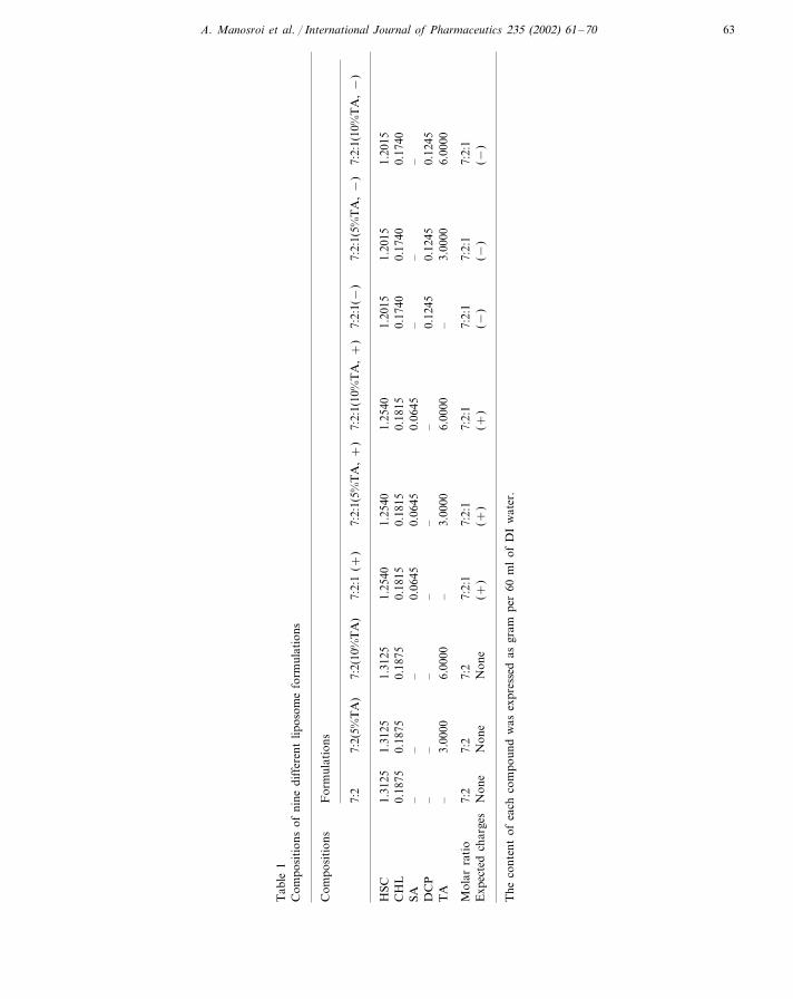
Liposome dispersion samples were prepared bya conventional chloroform film method with soni-cation, with the total lipid concentration of 25mg/ml. The lipids used were neutral (HSC andCHL) and charged lipids (SA for positivelycharged lipid and DCP for negatively chargedlipid). Liposome formulations composed of HSC/CHL=7:2; HSC/CHL/SA=7:2:1(+ ) and HSC/CHL/DCP=7:2:1(− ) at molar ratios, with andwithout the entrapped TA were prepared. Theconcentrations of TA incorporated into the lipo-some formulations were 5 and 10% (in DI water)(Table 1). For each lot, an amount of 60 ml ofeach liposome dispersion sample was prepared.
The lipid mixture was dissolved in 60 ml ofchloroform, and vacuum-desiccated (500 psi,65 °C) using a rotary evaporator (R-124 Buchi,Switzerland) for 90 min. A formed thin film layerwas flushed under a stream of nitrogen for 1 min.The thin film was re-suspended in either 60 ml of
A. Manosroi et al. / International Journal of Pharmaceutics 235 (2002) 61–70 63
Tab
le1
Com
posi
tion
sof
nine
diff
eren
tlip
osom
efo
rmul
atio
ns
Com
posi
tion
sF
orm
ulat
ions
7:2:
1(10
%T
A,+
)7:
2:1(
−)
7:2:
1(5%
TA
,−
)7:
2:1(
10%
TA
,−
)7:
27:
2(5%
TA
)7:
2(10
%T
A)
7:2:
1(+
)7:
2:1(
5%T
A,+
)
1.25
401.
2015
1.20
151.
2015
1.25
40H
SC1.
2540
1.31
251.
3125
1.31
250.
1875
0.18
150.
1815
0.17
400.
1740
0.17
400.
1875
0.18
75C
HL
0.18
150.
0645
–0.
0645
––
––
–0.
0645
SA–
0.12
450.
1245
0.12
45–
––
–D
CP
–3.
0000
–6.
0000
–3.
0000
6.00
003.
0000
6.00
00–
TA
Mol
arra
tio
7:2:
17:
27:
2:1
7:2:
17:
2:1
7:2
7:2
7:2:
17:
2:1
(+)
(−)
(−)
(−)
(+)
Non
eN
one
Exp
ecte
dch
arge
sN
one
(+)
The
cont
ent
ofea
chco
mpo
und
was
expr
esse
das
gram
per
60m
lof
DI
wat
er.
A. Manosroi et al. / International Journal of Pharmaceutics 235 (2002) 61–7064
5 or 10% TA solution in DI water (for liposomeswith the entrapped drug), or DI water alone (forliposomes without the entrapped drug). The dis-persion was weighed and swelled by swirling in awater bath (80 °C, 200 rpm) for 30 min. Smallmultilamellar vesicles were obtained following thesonication of large multilamellar vesicles for 10min, using a microtip probe sonicator (Vibracell,Sonics & Materials, Inc., Danbury, CT). The lipo-some dispersion samples were kept at 4 °C andprotected from light, prior to use.
Liposome dispersion sample (5 ml) was put intoa glass vial and stored at −80 °C for 24 h. Theglass vial was then put into a freeze dryer (ModelLioalfa 10, Telstar, Spain), with freezing conden-sor at −46 °C for 48 h, to obtain the freeze-driedliposome powder. The following pre-freezing con-ditions: at −25 °C for 90 min, −32 °C for 90min and −36 °C for 60 min, were employed inthis study.
2.3. Physical properties determination ofliposomes
The physical appearances of various liposomeformulations, DI water, 5 and 10% TA solutionsin DI water following storage at 4�1 °C for 24 hwere visually observed, and the pH of fresh lipo-some samples was measured by a pH meter(HORIBA, Ltd., Kyoto, Japan).
A small aliquot of liposome dispersion samplewas used to characterize the particle size and sizedistribution, using a Light Scattering Particle An-alyzer (Mastersizer S Long Bed Ver. 2.11,Malvern Instruments Ltd., Malvern, UK), 10days after sample preparation (kept at 4 °C). Theparticle size range was set between 0.05 to 800�m, with the beam length at 2.40 nm, dispersantrefractive index at 1.3300, and the polydispersemodel analysis was used. This study was per-formed in six replicates.
A drop of liposome dispersion sample was ap-plied on a 300-mesh formvar copper grid onparaffin and left for 10 min, to allow some of theliposome powder to adhere on the formvar. Theremaining dispersion was removed and a drop of2% aqueous solution of uranyl acetate was ap-plied for 5 min. The remaining solution was then
removed and the sample was air dried, prior tomeasurement with a Transmission Electron Mi-croscope (TEM 1200S JEOL, JEOL Ltd., Tokyo,Japan), to observe the morphology and lamellar-ity of liposomes.
An amount of 3–7 mg of HSC, CHL, SA,DCP, TA and the freeze-dried liposome powderwere placed on sample pans and then properlysealed. All samples were scanned at the rate of40 °C/min from 20 to 500 °C by a Thermogravi-metric Analyzer (Perkin–Elmer TGA 7, Perkin–Elmer Ltd., Norwalk, CONN), to determine theglass transition temperature.
The transition temperature (Tc) and enthalpy oftransition (�H) were determined from the ther-mogram, generated by a Differential ScanningCalorimeter (Perkin–Elmer DSC7). HSC, CHL,SA, DCP, TA and the freeze-dried liposome pow-der (3–7 mg) were put on sample pans and prop-erly sealed. An equal amount of DI water wasplaced on the reference pan. The temperatureranging from 20 to 200 °C with scan rate of5 °C/min was employed in this study. Indiumstandard and water were used to calibrate thecalorimeter. This experiment was performed in sixdeterminations.
2.4. Determination of drug encapsulationefficiency in liposomes
The encapsulation efficiencies of TA in the7:2:1(5%TA, + ), 7:2:1(10%TA, + ), 7:2:1(5%TA,− ) and 7:2:1(10%TA, − ) liposomes were deter-mined spectrophotometrically. Each liposome dis-persion sample (1.5 g) was mixed with 1.5 g of DIwater, and centrifuged at 150,000×g (4 °C) for90 min in a Centrikron T-1180 ultracentrifuge(Kontron Instruments, Milan, Italy). The super-natant was removed and diluted (100 times) withDI water, prior to the determination of theamount of the unentrapped TA. The pellet con-taining liposomes was re-suspended in 5 ml of10% Triton-X 100 solution and sonicated for 20min. This solution was further diluted (10 times)with Triton-X 100 solution, prior to the determi-nation of the amount of entrapped TA in lipo-somes. An amount of 0.1 ml of the above samples(unentrapped and entrapped TA) was withdrawn
A. Manosroi et al. / International Journal of Pharmaceutics 235 (2002) 61–70 65
and derivatized, following the procedures as de-scribed in the analytical method section, prior toassay by spectrophotometer. The loading of TAin liposomes was also calculated. Each samplewas prepared in two lots and the experimentswere performed in duplicates.
2.5. Analytical method for the determination ofTA content
The TA content in liposome formulations wasdetermined spectrophotometrically at 415 nm(Milton Roy Spectronic 1001 Plus, Rochester,NY), following derivatization with 2,4,6-trini-trobenzosulfonic acid (Atmaca, 1989). The colorreagent used was 1.68% (w/v) 2,4,6-trinitrobenzo-sulfonic acid solution in DI water, freshly pre-pared and protected from light, prior to use. Each0.1 ml of working standard solutions or sampleswas spiked with 0.25 ml of 0.025 M disodiumtetraborate solution (pH 10) and 0.25 ml of colorreagent, prior to standing at 25 °C for 30 min.The solution was then diluted to 5.0 ml with 0.1M potassium dihydrogen phosphate solution (pH4.5). The solution mixture in the absence of TAwas used as a blank. A calibration graph wasconstructed, by plotting the concentrations of TAagainst the absorbance values. The linearity ofassay was determined from five working standardsolutions of TA in DI water (concentrations: 4.0–20.0 �g/ml), prepared in triplicates. The correla-tion (r2), intercept and slope of the calibrationgraph were calculated. The absorbance values ofsamples were observed and compared with thoseobtained from the calibration graph, to determinethe amount of TA in samples. This experimentwas performed in duplicates of three lots ofsamples.
3. Results
3.1. Physical appearances and pH of liposomes
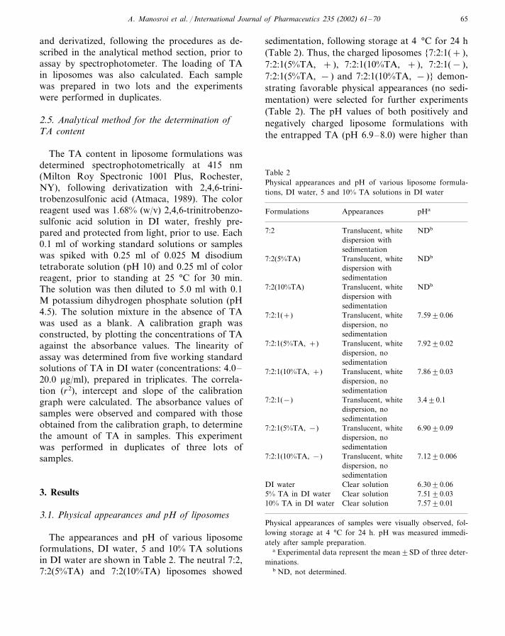
The appearances and pH of various liposomeformulations, DI water, 5 and 10% TA solutionsin DI water are shown in Table 2. The neutral 7:2,7:2(5%TA) and 7:2(10%TA) liposomes showed
sedimentation, following storage at 4 °C for 24 h(Table 2). Thus, the charged liposomes {7:2:1(+ ),7:2:1(5%TA, + ), 7:2:1(10%TA, + ), 7:2:1(− ),7:2:1(5%TA, − ) and 7:2:1(10%TA, − )} demon-strating favorable physical appearances (no sedi-mentation) were selected for further experiments(Table 2). The pH values of both positively andnegatively charged liposome formulations withthe entrapped TA (pH 6.9–8.0) were higher than
Table 2Physical appearances and pH of various liposome formula-tions, DI water, 5 and 10% TA solutions in DI water
Formulations Appearances pHa
Translucent, white7:2 NDb
dispersion withsedimentation
NDb7:2(5%TA) Translucent, whitedispersion withsedimentationTranslucent, white7:2(10%TA) NDb
dispersion withsedimentation
7.59�0.067:2:1(+) Translucent, whitedispersion, nosedimentation
7.92�0.02Translucent, white7:2:1(5%TA, +)dispersion, nosedimentationTranslucent, white 7.86�0.037:2:1(10%TA, +)dispersion, nosedimentationTranslucent, white7:2:1(−) 3.4�0.1dispersion, nosedimentation
7:2:1(5%TA, −) 6.90�0.09Translucent, whitedispersion, nosedimentation
7.12�0.0067:2:1(10%TA, −) Translucent, whitedispersion, nosedimentationClear solutionDI water 6.30�0.06Clear solution5% TA in DI water 7.51�0.03
10% TA in DI water Clear solution 7.57�0.01
Physical appearances of samples were visually observed, fol-lowing storage at 4 °C for 24 h. pH was measured immedi-ately after sample preparation.
a Experimental data represent the mean�SD of three deter-minations.
b ND, not determined.
A. Manosroi et al. / International Journal of Pharmaceutics 235 (2002) 61–7066
Table 3Particle sizes of six liposome formulations
Particle size (�m)aFormulations
7:2:1(+) 17.9�0.57:2:1(5%TA,+) 17.5�0.5
35.8�0.57:2:1(10%TA,+)7:2:1(−) 24.78�0.04
2.76�0.027:2:1(5%TA, −)2.05�0.037:2:1(10%TA, −)
Particle sizes of liposome formulations were measured 10 daysafter sample preparation (kept at 4 °C).
a Experimental data represent the mean�SD of six determi-nations.
samples were decomposed at high temperature(�200 °C), except SA which decomposed atlower than 200 °C. The Tc and �H values ofHSC, CHL, SA, DCP, TA, 7:2:1(+ ),7:2:1(5%TA, + ), 7:2:1(10%TA, + ), 7:2:1(− ),7:2:1(5%TA, − ) and 7:2:1(10%TA, − ) liposomeformulations are summarized in Table 5. Underthe experimental conditions, there was no peak ofTA observed in the DSC thermogram.
3.4. Tranexamic acid entrapped in liposomes
The calibration graph of TA solution in DIwater was shown to be linear (r2=0.9937), overthe concentration range 4.0–20.0 �g/ml. The re-gression equation was as follows: y=20.7903x−0.1942, where y is the concentration of TA(�g/ml) and x is the absorbance of the derivativeof TA formed with 2,4,6-trinitrobenzosulfonicacid (mAU*s). The percentages of TA recoveredfrom the liposome formulations with the en-trapped drug, using spectrophotometric assayvaried between 91.4 and 104.7%. The other com-ponents in liposome formulations neither reactedwith the color reagent nor demonstrated signifi-cant absorption at 415 nm. The percentages ofTA entrapped in liposomes, the free drug and theloading of TA in liposome formulations areshown in Table 6.
4. Discussion
The charged liposomes with and without theentrapped TA, showing no sedimentation follow-ing storage at 4 °C for 1 day, may indicate betterphysical stability than the neutral liposomes(Table 2). This may be associated with the effectsof charges on the surface of liposomes. TA is asynthetic amino acid, which has amino and car-boxylic groups. When TA dissolves in water, itwill ionize in equilibrium Eq. (1):
C8H15NO2+H2O � C8H16NO2++OH− (1)
The pH of samples increased when TA was incor-porated into the liposome systems (Table 2). Thismay be due to the effect of concentration of
that of DI water (pH 6.3), except the blank nega-tive liposome {7:2:1(− )} (pH 3.4, Table 2). Thus,the incorporation of TA in liposomes may in-crease pH of the system.
3.2. Liposome size, morphology and lamellarity ofliposomes
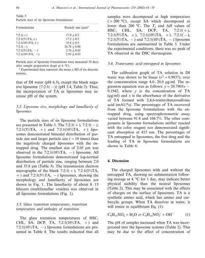
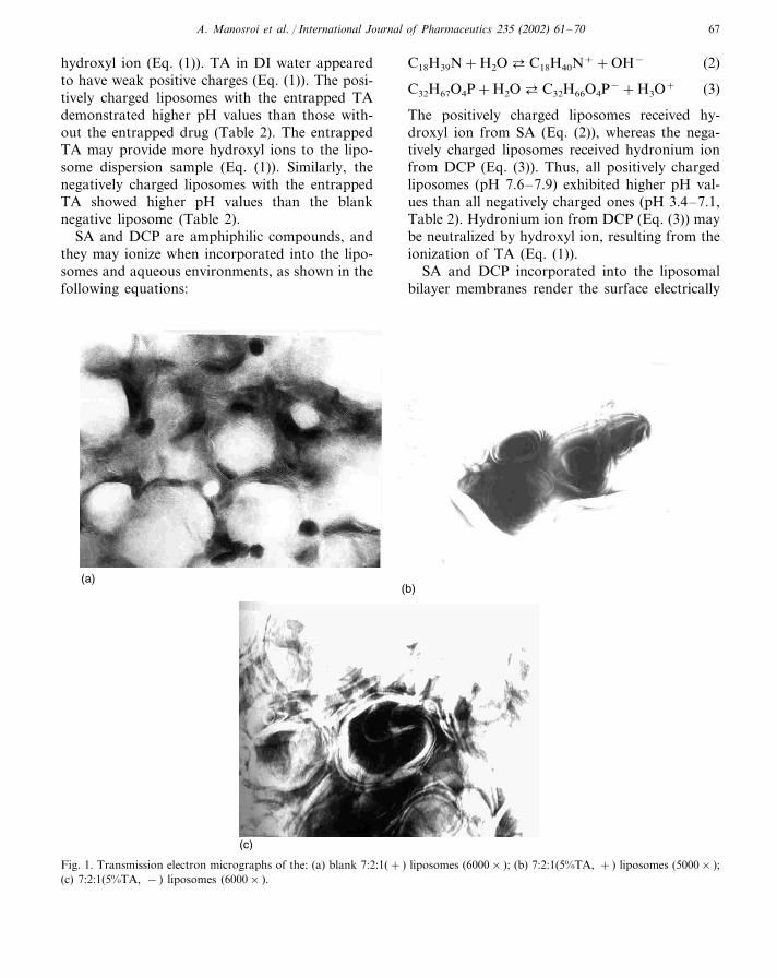
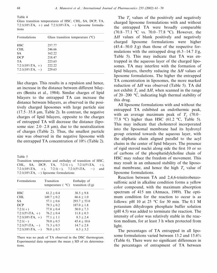
The particle sizes of six liposome formulationsare presented in Table 3. The 7:2:1(+ ), 7:2:1(− ),7:2:1(5%TA, + ) and 7:2:1(10%TA, + ) lipo-somes demonstrated bimodal distribution of par-ticle size and larger particle size (�10 times) thanthe negatively charged liposomes with the en-trapped drug. The smallest size of 2.05 �m wasobserved in the 7:2:1(10%TA, − ) liposome. Allliposome formulations demonstrated log-normaldistribution of particle size, ranging between 2.0and 35.8 �m (Table 3). The transmission electronmicrographs of the blank 7:2:1(+ ), 7:2:1(5%TA,+ ) and 7:2:1(5%TA, − ) liposomes, showing themorphology and lamellarity of liposomes areshown in Fig. 1. The lamellarity of about 8–15bilayers (multilamellar vesicles) was observed inall liposome formulations (Fig. 1).
3.3. Glass transition temperature, transitiontemperature and enthalpy of transition
The glass transition temperatures of HSC,CHL, SA, DCP, TA, 7:2:1(10%TA, + ) and7:2:1(10%TA, − ) liposome formulations are pre-sented in Table 4. The results indicated that all
A. Manosroi et al. / International Journal of Pharmaceutics 235 (2002) 61–70 67
hydroxyl ion (Eq. (1)). TA in DI water appearedto have weak positive charges (Eq. (1)). The posi-tively charged liposomes with the entrapped TAdemonstrated higher pH values than those with-out the entrapped drug (Table 2). The entrappedTA may provide more hydroxyl ions to the lipo-some dispersion sample (Eq. (1)). Similarly, thenegatively charged liposomes with the entrappedTA showed higher pH values than the blanknegative liposome (Table 2).
SA and DCP are amphiphilic compounds, andthey may ionize when incorporated into the lipo-somes and aqueous environments, as shown in thefollowing equations:
C18H39N+H2O � C18H40N++OH− (2)
C32H67O4P+H2O � C32H66O4P−+H3O+ (3)
The positively charged liposomes received hy-droxyl ion from SA (Eq. (2)), whereas the nega-tively charged liposomes received hydronium ionfrom DCP (Eq. (3)). Thus, all positively chargedliposomes (pH 7.6–7.9) exhibited higher pH val-ues than all negatively charged ones (pH 3.4–7.1,Table 2). Hydronium ion from DCP (Eq. (3)) maybe neutralized by hydroxyl ion, resulting from theionization of TA (Eq. (1)).
SA and DCP incorporated into the liposomalbilayer membranes render the surface electrically
Fig. 1. Transmission electron micrographs of the: (a) blank 7:2:1(+ ) liposomes (6000× ); (b) 7:2:1(5%TA, + ) liposomes (5000× );(c) 7:2:1(5%TA, − ) liposomes (6000× ).
A. Manosroi et al. / International Journal of Pharmaceutics 235 (2002) 61–7068
Table 4Glass transition temperatures of HSC, CHL, SA, DCP, TA,7:2:1(10%TA, +) and 7:2:1(10%TA, −) liposome formula-tions
Formulations Glass transition temperature (°C)
257.77HSC246.66CHL
SA 162.22218.47DCP223.63TA
7:2:1(10%TA, +) 222.22229.637:2:1(10%TA, −)
The Tc values of the positively and negativelycharged liposome formulations with and withoutthe entrapped TA were broadly comparable(70.8–77.1 °C vs. 70.0–77.8 °C). However, the�H values of blank positively and negativelycharged liposome formulations were higher(45.4–50.0 J/g) than those of the respective for-mulations with the entrapped drug (6.3–14.7 J/g,Table 5). This may indicate that TA was en-trapped in the aqueous layer of the charged lipo-somes. TA may interfere with the formation oflipid bilayers, thereby reducing the �H values ofliposome formulations. The higher the entrappedTA concentration in liposomes, the more markedreduction of �H was observed (Table 5). TA didnot exhibit Tc and �H, when scanned in the rangeof 20–200 °C, indicating high thermal stability ofthis drug.
All liposome formulations with and without theentrapped TA exhibited an endothermic peak,with an average maximum peak of Tc (70.0–77.8 °C) higher than HSC (61.2 °C, Table 5).This may indicate that cholesterol incorporatedinto the liposomal membrane had its hydroxylgroup oriented towards the aqueous layer, withthe aliphatic chain aligned parallel to the acylchains in the center of lipid bilayers. The presenceof rigid steroid nuclei along side the first 10 or soof carbons of the phosphatidylcholine chain ofHSC may reduce the freedom of movement. Thismay result in an enhanced stability of the liposo-mal membrane, and hence the high Tc value ofliposome formulations.
Reaction between TA and 2,4,6-trinitrobenzo-sulfonic acid in alkaline condition forms a yellowcolor compound, with the maximum absorptionspectrum of 415 nm (Atmaca, 1989). The opti-mum condition for the reaction to occur is asfollows: pH 10 at 25 °C for 30 min. The 0.1 Mpotassium dihydrogen phosphate buffer solution(pH 4.5) was added to terminate the reaction. Theintensity of color was relatively stable in the reac-tion medium, for at least 3 h when protected fromlight.
The percentages of TA entrapped in all lipo-some formulations varied between 13.2 and 15.6%(Table 6). There were no significant differences inthe percentages of entrapment of TA between
like charges. This results in a repulsion and hence,an increase in the distance between different bilay-ers (Benita et al., 1984). Similar charges of lipidbilayers to the entrapped TA can increase thedistance between bilayers, as observed in the posi-tively charged liposomes with large particle size(17.5–35.8 �m, Table 2). In contrast, the negativecharges of lipid bilayers, opposite to the chargesof entrapped TA will decrease the distance (lipo-some size: 2.0–2.8 �m), due to the neutralizationof charges (Table 2). Thus, the smallest particlesize was observed in the negative liposome withthe entrapped TA concentration of 10% (Table 2).
Table 5Transition temperatures and enthalpy of transition of HSC,CHL, SA, DCP, TA, 7:2:1(+), 7:2:1(5%TA, +),7:2:1(10%TA, +), 7:2:1(−), 7:2:1(5%TA, −) and7:2:1(10%TA, −) liposome formulations
Transition Enthalpy ofFormulationstemperature ( °C) transition (J/g)
61.2�0.4HSC 38.5�9.8149.7�0.2CHL 66.6�1.0
57.1�0.6SA 293.7�53.0187.0�1.8DCP 79.3�0.2
77.8�0.47:2:1(+) 50.0�7.37:2:1(5%TA, +) 11.8�0.376.2�0.4
77.1�1.1 8.3�2.47:2:1(10%TA, +)45.4�10.67:2:1(−) 70.0�0.5
7:2:1(5%TA, −) 14.7�2.871.3�0.570.8�0.57:2:1(10%TA, −) 6.3�3.2
There was no peak of TA observed in the DSC thermogram.Experimental data represent the mean�SD of six determina-tions.
A. Manosroi et al. / International Journal of Pharmaceutics 235 (2002) 61–70 69
Table 6Percentages of entrapment of TA, free TA, and the amount of TA per total lipid in liposome formulations
Formulations % EntrapmentForm of drug Loading of TA (�g/mg lipid)a
100.0Total drug –7:2:1(5%TA, +)14.7�0.6 275.2�13.9Entrapped drug85.3�0.2 –Free drug
Total drug7:2:1(10%TA, +) 100.0 –15.0�0.4Entrapped drug 627.0�10.885.0�0.9 –Free drug
100.07:2:1(5%TA, −) –Total drugEntrapped drug 15.6�0.7 290.4�5.7
84.5�2.6 –Free drug
7:2:1(10%TA, −) Total drug 100.0 –Entrapped drug 13.2�0.3 547.3�13.6
86.8�0.3 –Free drug
Experimental data represent the mean�SD of four determinations.a Total lipid in 1 ml of liposome dispersion sample was 25 mg.
different formulations (Table 6), as analyzed byANOVA (P�0.05). This may indicate that thecharges and TA concentrations did not affect thepercentages of TA entrapped in liposome formu-lations. The negatively charged liposome whichhad opposite charged to TA (weak positive) mayimprove the entrapment of drug in the liposomes.On the other hand, the positively charged lipo-somes with the entrapped drug demonstratedlarger particle size, and hence larger volume forthe entrapment of drug than the negative lipo-somes (Benita et al., 1984). These phenomena mayexplain why the percentages of entrapment of TAin the positive and negative liposomes werebroadly comparable (14.7–15.0% vs. 13.2–15.6%,Table 6).
The loading of TA in the charged liposomesincreased, as the initial concentration of drugincreased, from 275 �g/mg lipid {7:2:1(5%TA,+ )} to 627 �g/mg lipid {7:2:1(10%TA, + )}, andfrom 290 �g/mg lipid {7:2:1(5%TA, − )} to 547�g/mg lipid {7:2:1(10%TA, − )} (Table 6, Fold-vari et al., 1993).
In conclusion, the liposomes composed ofHSC/CHL/charged lipids at molar ratios of7:2:1(+ ) and 7:2:1(− ) with the entrapped TA (5and 10% solutions in DI water) were demon-strated as multilamellar vesicles. Thus, TA wasadvantageous when entrapped in liposome, since
this drug delivery system can potentitally reducethe skin irritation and improve moisturizing ef-fect. The particle sizes of all liposome formula-tions, with and without the entrapped TA were inthe range of 2.05–35.8 �m, with the smallest sizewas observed in the negative liposome with theentrapped TA concentration of 10%. The chargedliposomes with the entrapped TA demonstratedhigh physical stability. The concentrations of TAappeared to affect the pH, particle size and en-thalpy of transition of liposome formulations,whereas charges seemed to affect the physicalstability, pH and particle size of liposomes. Thebest formulation was concluded to be the nega-tively charged 7:2:1(10%TA, − ) liposome, whichexhibited high physical stability, small particle sizeand relatively high percentage of drug entrap-ment. This formulation will be further evaluatedfor release study. Overall, it has been demon-strated that TA which is a hydrophilic drug canbe favorably entrapped in liposomes.
Acknowledgements
The authors thank Dr Kuncoro Foe for hisassistance in the preparation of the manuscript.We also acknowledge JJ-Degussa (T) Ltd., Thai-land for the gifts of Emulmetik 950®, and Thistle
A. Manosroi et al. / International Journal of Pharmaceutics 235 (2002) 61–7070
Corp., Ltd. (Thailand) for the gifts of tranexamicacid. We thank the Research and Development ofNatural Products for Thai Traditional MedicinesResearch Unit, and Pharmaceutical-CosmeticsRaw Materials and Natural Products Researchand Development Center, Institute for Scienceand Technology Research and Development, Chi-ang Mai University, for financial support.
References
Atmaca, S., 1989. Spectrophotometric determination oftranexamic acid with 2,4,6-trinitrobenzosulfonic acid. ActaPharm. Turcica 31, 115–118.
Bally, M.B., Masin, D., Nayar, R., Cullis, P.R., Mayer, L.D.,1994. Transfer of liposomal drug carriers from the bloodto the peritoneal cavity of normal and ascitic tumor-bear-ing mice. Cancer Chemother. Pharmacol. 34, 137–146.
Bangham, A.D., Hill, M.W., Miller, G.A., 1974. French pres-sure cell liposome: preparation, properties, and potential.In: Gregoriadis, G. (Ed.), Liposome Technology, vol. 1.CRC Press, Boca Raton, pp. 37–49.
Benita, S., Plenecassagne, J.D., Caves, G., Drouin, D., Dong,P.L.H., Sincholle, D., 1984. Pilocarpine hydrochlorideliposomes: characterization in �itro and preliminary evalua-tion in �i�o in rabbit eye. J. Microencapsul. 1, 203–216.
Borea, G., Montebugnoli, L., Capuzzi, P., Magelli, C., 1993.Tranexamic acid as a mouthwash in anticoagulant-treatedpatients undergoing oral surgery. An alternative method todiscontinuing anticoagulant therapy. Oral Surg. Oral Med.Oral Pathol. 75, 29–31.
British Pharmacopoeia, 1998. Her Majesty’s Stationery Office,Norwich, pp. 1316–1317.
Drug Facts and Comparison, 1999. A Wolters Kluwer Com-pany, St. Louis, pp. 394–395.
Foldvari, M., Gesztes, A., Mezei, M., Cardinal, L., Kowal-czyk, I., Behl, M., 1993. Topical liposome local anesthetics:design, optimization and evaluation of formulations. DrugDev. Ind. Pharm. 19, 2499–2517.
Gabizon, A., Chemla, M., Tzemach, D., Horowitz, A.T.,Goren, D., 1996. Liposome longevity and stability circula-tion: effects on the in vivo delivery to tumors and thera-peutic efficacy of encapsulated anthracyclines. J. DrugTarget. 3, 391–398.
Gregoriadis, G., 1988. Liposome as Drug Carriers: RecentTrends and Progress. John Wiley and Sons, Chichester.
Jin-Chul, K., Eun-Ok, L., Jong-Yung, K., Soo-Kyoung, B.,Tae-Boo, C., Jong-Duk, K., 1997. Hemolytic and antifun-gal activity of liposome-entrapped amphotericin B pre-pared by the precipitation method. Pharm. Dev. Tech. 2,275–284.
Kawakami, S., Munakata, C., Fumoto, S., Yamashita, F.,
Hashida, M., 2001. Novel galactosylated liposomes forhepatocyte-selective targeting of lipophilic drugs. J. Pharm.Sci. 90, 105–113.
Lasic, D., 1992. Liposomes. Am. Sci. 80, 20–31.Li, L., Margolis, L.B., Lishko, L.V., 1992. Product-delivering
liposomes specifically target entrapped melanin to hairfollicles in histocultured intact skin, in vitro cell. Dev. Biol.28A, 679–681.
Lopez-Bersetin, G., Hopfer, R.L., Metha, K., Juliano, R.L.,1984. Liposome-encapsulated amphotericin B for treat-ment of disseminated candidiasis in neutropenic mice. J.Infect. Dis. 150, 278–283.
Maeda, K., Naganuma, M., 1998. Topical trans-4-aminomethylcyclohexane carboxylic acid prevents ultravio-let radiation-induced pigmentation. J. Photochem.Photobiol. B 47, 136–141.
Martindale XXXI, 1996. Royal Pharmaceutical Society ofGreat Britain, London, pp. 771–772.
Mayer, L.D., Nayar, R., Thies, R.L., Boman, N.L., Cullis,P.R., Bally, M.B., 1993. Identification of vesicle propertiesthat enhance the antitumor activity of liposomal vincristineagainst murine L1210 leukemia. Cancer Chemother. Phar-macol. 33, 17–24.
Mayer, L.D., St-Onge, G., 1995. Determination of free andliposome-associated doxorubicin and vincristine levels inplasma under equilibrium conditions employing ultrafiltra-tion techniques. Anal. Biochem. 232, 149–157.
Mezei, M., 1991. Delivering the good. Can. Pharm. J. 124,132–155.
MIMS Annual, 1998. Medimedia International Group, Singa-pore, pp. 958–960.
Mitsuhiro, D., Kenji, K., Peter, M.E., Kenneth, R., 1997.Trans-4-aminomethylcyclohexane carboxylic acid (T-AM-CHA), an anti-fibrinolytic agent, accelerates barrier recov-ery and prevents the epidermal hyperplasia induced byepidermal injury in hairless mice and humans. J. Invest.Dermatol. 109, 84–90.
Rahman, A., Treat, J., Roe, J.K., Potkul, L.A., Alvord, W.G.,1990. Phase I clinical trial and pharmacokinetic evaluationof liposomal encapsulated doxorubicin. J. Clin. Oncol. 8,1093–1100.
Soltan, M.A., Fadel, M.A., Medhat, W.I., 2000. Prolongedeffect of liposomes encapsulating pilocarpine HCl in nor-mal and glaucomatous rabbits. Int. J. Pharm. 198, 29–38.
Tibbelin, A., Aust, R., Bende, M., Holgersson, M., Petruson,B., Rundcrantz, H., Alander, U., 1995. Effect of localtranexamic acid gel in the treatment of epistaxis. J. Otorhi-nolaryngol. Relat. Spec. 57, 207–209.
Waly, N.G., 1995. Local anti-fibrinolytic treatment withtranexamic acid in hemophilic children undergoing dentalextractions. Egypt Dent. J. 41, 961–968.
Weissmann, G., Cohen, C., Hoffstein, S., 1977. Introductionof missing enzymes into the cytoplasm of cultured mam-malian cells by mean of fution-prone liposomes. Trans.Assoc. Am. Phys. 89, 171–183.


