Embed Size (px)
Citation preview
340 New J. Chem., 2012, 36, 340–349 This journal is c The Royal Society of Chemistry and the Centre National de la Recherche Scientifique 2012
Cite this: New J. Chem., 2012, 36, 340–349
Stimuli sensitive amphiphilic dendrimers
Rajasekhar R. Ramireddy, Krishna R. Raghupathi, Diego Amado Torres and
S. Thayumanavan*
Received (in Montpellier, France) 12th October 2011, Accepted 19th December 2011
DOI: 10.1039/c2nj20879b
In the past decade, there has been an increasing interest in supramolecular systems that can
undergo physical or chemical transformations upon encountering a specific stimulus. Micelle-
forming amphiphilic systems based on polymers and dendrimers are particularly preferred over
small molecule amphiphiles, due to their ability to sequester and release a vast library of
hydrophobic guest molecules at micromolar polymer or dendrimer concentrations. Here we
review a relatively underexplored, yet rapidly advancing, field of amphiphilic systems based on a
dendritic architecture that exhibit stimuli sensitive behaviour. In particular, we will be focusing on
stimuli such as temperature, pH, enzymatic and non-enzymatic proteins. These stimuli-responsive
systems offer a unique opportunity in the field of drug delivery and sensing.
Introduction
Amphiphilic molecules are primarily interesting due to the
self-assembled structures1–10 they exhibit both in solution
(micelles,11–15 vesicles16–19) and in thin films.20,21 The fundamental
driving force for the formation of these assemblies is to maintain a
favorable hydrophilic–lipophilic balance (HLB) between the
hydrophilic and the lipophilic functional group components of
the amphiphile. In solution, these molecules self-assemble only
above a certain concentration of the amphiphile, known as critical
aggregation concentration (CAC). Amphiphilic macromolecules
have garnered significant interest, because their CACs are sub-
stantially lower than their small molecule counterparts. For
example, assemblies achieved from macromolecules, such as
amphiphilic block copolymers, have CACs in the micro or
nanomolar range, compared to the millimolar CAC for small
molecule surfactants.22,23 Other polymeric systems, such as
amphiphilic homopolymers were also explored for their self-
assembling properties.24 Although polymers offer advantages over
their small molecule counterparts such as reduced CACs and
increased stability, the inherent non-uniform nature (polydispersity)
associated with them presents a challenge in reproducibility, when
studied for drug delivery and sensing applications.25 This calls for
molecules that possess salient features such as lower CACs and
higher stability, while being structurally uniform (monodisperse).
Dendrimers are particularly interesting among amphiphilic
macromolecules, because of their unique branched structures,
Department of Chemistry, University of Massachusetts,710 N. Pleasant Street, Amherst, Massachusetts 1003, USA.E-mail: [email protected]; Fax: +1 413 545 4490;Tel: +1 413 545 1313
Rajasekhar R. Ramireddy
Rajasekhar R. Ramireddyreceived his BSc degree fromSri Venkateshwara University(2006), Tirupathi, India, andMSc degree in Chemistry fromUniversity of Hyderabad (2008),Hyderabad, India. Since 2008 heis working as a PhD studentunder the guidance of ProfessorS. Thayumanavan at the Univer-sity of Massachusetts Amherst.His current research is focusedon the design and synthesis ofzwitterionic amphiphilic dendri-mers, polymers and their inter-actions with biomolecules such asproteins and enzymes.
Krishna R. Raghupathi
Krishna R. Raghupathi receivedhis B.Pharmacy degree fromOsmania University (2006),Hyderabad, India, and MS inChemistry from University ofSouth Dakota (2009) underthe guidance of Professor RanjitT. Koodali. Currently he is aPhD student in Chemistry at theUniversity of MassachusettsAmherst under the direction ofProfessor S. Thayumanavan.His research is focused on thedesign and synthesis of amphi-philic dendrimers, and polymersto study their self-assembly andstimuli-responsive properties.
NJC Dynamic Article Links
www.rsc.org/njc PERSPECTIVE
Publ
ishe
d on
16
Janu
ary
2012
. Dow
nloa
ded
by J
ohns
Hop
kins
Uni
vers
ity o
n 12
/07/
2015
17:
52:4
1.
View Article Online / Journal Homepage / Table of Contents for this issue
This journal is c The Royal Society of Chemistry and the Centre National de la Recherche Scientifique 2012 New J. Chem., 2012, 36, 340–349 341
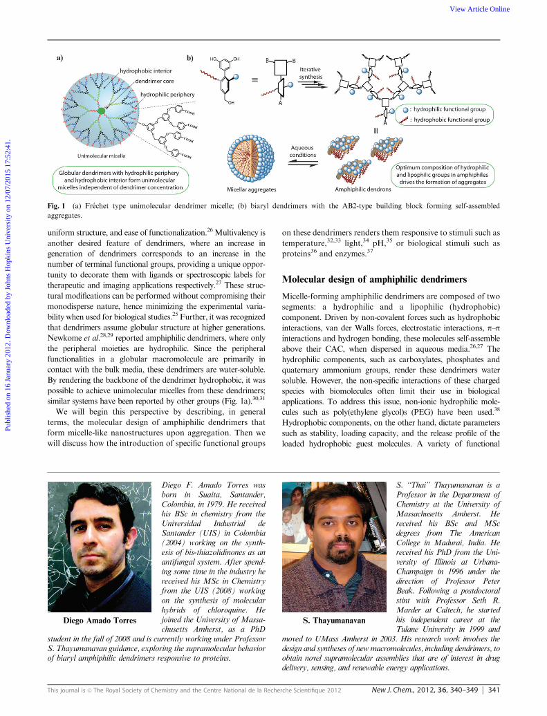
uniform structure, and ease of functionalization.26 Multivalency is
another desired feature of dendrimers, where an increase in
generation of dendrimers corresponds to an increase in the
number of terminal functional groups, providing a unique oppor-
tunity to decorate them with ligands or spectroscopic labels for
therapeutic and imaging applications respectively.27 These struc-
tural modifications can be performed without compromising their
monodisperse nature, hence minimizing the experimental varia-
bility when used for biological studies.25 Further, it was recognized
that dendrimers assume globular structure at higher generations.
Newkome et al.28,29 reported amphiphilic dendrimers, where only
the peripheral moieties are hydrophilic. Since the peripheral
functionalities in a globular macromolecule are primarily in
contact with the bulk media, these dendrimers are water-soluble.
By rendering the backbone of the dendrimer hydrophobic, it was
possible to achieve unimolecular micelles from these dendrimers;
similar systems have been reported by other groups (Fig. 1a).30,31
We will begin this perspective by describing, in general
terms, the molecular design of amphiphilic dendrimers that
form micelle-like nanostructures upon aggregation. Then we
will discuss how the introduction of specific functional groups
on these dendrimers renders them responsive to stimuli such as
temperature,32,33 light,34 pH,35 or biological stimuli such as
proteins36 and enzymes.37
Molecular design of amphiphilic dendrimers
Micelle-forming amphiphilic dendrimers are composed of two
segments: a hydrophilic and a lipophilic (hydrophobic)
component. Driven by non-covalent forces such as hydrophobic
interactions, van der Walls forces, electrostatic interactions, p–pinteractions and hydrogen bonding, these molecules self-assemble
above their CAC, when dispersed in aqueous media.26,27 The
hydrophilic components, such as carboxylates, phosphates and
quaternary ammonium groups, render these dendrimers water
soluble. However, the non-specific interactions of these charged
species with biomolecules often limit their use in biological
applications. To address this issue, non-ionic hydrophilic mole-
cules such as poly(ethylene glycol)s (PEG) have been used.38
Hydrophobic components, on the other hand, dictate parameters
such as stability, loading capacity, and the release profile of the
loaded hydrophobic guest molecules. A variety of functional
Fig. 1 (a) Frechet type unimolecular dendrimer micelle; (b) biaryl dendrimers with the AB2-type building block forming self-assembled
aggregates.
Diego Amado Torres
Diego F. Amado Torres wasborn in Suaita, Santander,Colombia, in 1979. He receivedhis BSc in chemistry from theUniversidad Industrial deSantander (UIS) in Colombia(2004) working on the synth-esis of bis-thiazolidinones as anantifungal system. After spend-ing some time in the industry hereceived his MSc in Chemistryfrom the UIS (2008) workingon the synthesis of molecularhybrids of chloroquine. Hejoined the University of Massa-chusetts Amherst, as a PhD
student in the fall of 2008 and is currently working under ProfessorS. Thayumanavan guidance, exploring the supramolecular behaviorof biaryl amphiphilic dendrimers responsive to proteins.
S. Thayumanavan
S. ‘‘Thai’’ Thayumanavan is aProfessor in the Department ofChemistry at the University ofMassachusetts Amherst. Hereceived his BSc and MScdegrees from The AmericanCollege in Madurai, India. Hereceived his PhD from the Uni-versity of Illinois at Urbana-Champaign in 1996 under thedirection of Professor PeterBeak. Following a postdoctoralstint with Professor Seth R.Marder at Caltech, he startedhis independent career at theTulane University in 1999 and
moved to UMass Amherst in 2003. His research work involves thedesign and syntheses of newmacromolecules, including dendrimers, toobtain novel supramolecular assemblies that are of interest in drugdelivery, sensing, and renewable energy applications.
Publ
ishe
d on
16
Janu
ary
2012
. Dow
nloa
ded
by J
ohns
Hop
kins
Uni
vers
ity o
n 12
/07/
2015
17:
52:4
1.
View Article Online
342 New J. Chem., 2012, 36, 340–349 This journal is c The Royal Society of Chemistry and the Centre National de la Recherche Scientifique 2012
groups, such as propylene glycol,39 trimethylene carbonate,40
e-caprolactone,41,42 and long alkyl chains, have been explored as
hydrophobic components.
Frechet and coworkers also reported on amphiphilic dendrimer
systems, which form unimolecular micelles in solution.30 The
amphiphilic components of these Frechet-type dendrimers
comprise hydrophilic periphery such as carboxylate or PEG
moieties, and a hydrophobic polyether core. Similarly, Newkome
et al.29 andMeijer et al.31 have also reported unimolecular micelles
based on saturated hydrocarbon and poly(propylene imine) (PPI)
cores respectively.
Our group has reported on a unique class of amphiphilic
dendrimers, based on a biaryl AB2 monomer (Fig. 1b).43 The
orthogonal placement of hydrophilic and hydrophobic compo-
nents, assisted by an inherent biaryl twist and solvophobic
interactions, renders these dendrimers facially amphiphilic.
In contrast to the well known unimolecular dendrimer micelles,
these biaryl dendrimers form micelle-like aggregates from several
amphiphilic dendrimers and hence have an associated CAC.44
Above the CAC, the equilibrium between the amphiphilic
dendrimers and the micellar aggregates opens up an opportunity
to incorporate stimuli-responsive characteristics as shown in Fig. 2.
Thermoresponsive dendrimers
Macromolecules that undergo temperature-dependent solubility
changes are highly attractive molecular systems for the develop-
ment of functional thermoresponsive materials. These materials
could be utilized for biomedical applications, such as programmed
drug delivery, because the temperature at the target site in the
body can be affected by ‘‘thermotherapy’’.45 Several polymers that
undergo phase transition through temperature change have
been developed. Among them poly(N-isopropylacrylamide)
(poly(NiPAM)) and PEG based systems have been extensively
studied.46
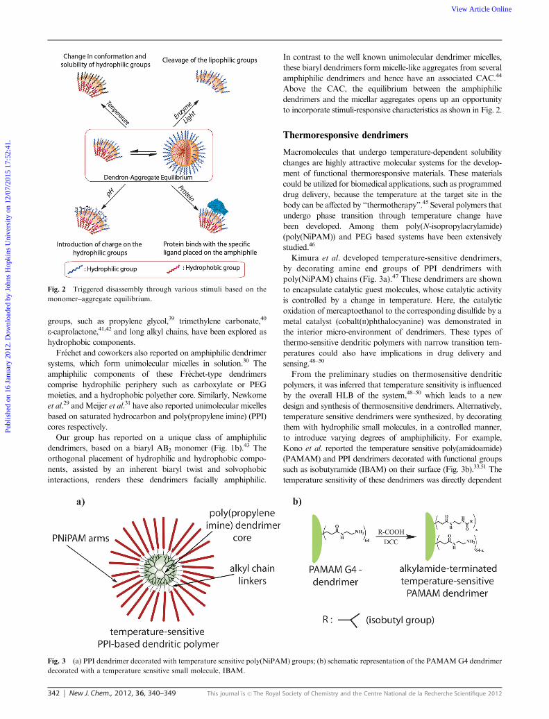
Kimura et al. developed temperature-sensitive dendrimers,
by decorating amine end groups of PPI dendrimers with
poly(NiPAM) chains (Fig. 3a).47 These dendrimers are shown
to encapsulate catalytic guest molecules, whose catalytic activity
is controlled by a change in temperature. Here, the catalytic
oxidation of mercaptoethanol to the corresponding disulfide by a
metal catalyst (cobalt(II)phthalocyanine) was demonstrated in
the interior micro-environment of dendrimers. These types of
thermo-sensitive dendritic polymers with narrow transition tem-
peratures could also have implications in drug delivery and
sensing.48–50
From the preliminary studies on thermosensitive dendritic
polymers, it was inferred that temperature sensitivity is influenced
by the overall HLB of the system,48–50 which leads to a new
design and synthesis of thermosensitive dendrimers. Alternatively,
temperature sensitive dendrimers were synthesized, by decorating
them with hydrophilic small molecules, in a controlled manner,
to introduce varying degrees of amphiphilicity. For example,
Kono et al. reported the temperature sensitive poly(amidoamide)
(PAMAM) and PPI dendrimers decorated with functional groups
such as isobutyramide (IBAM) on their surface (Fig. 3b).33,51 The
temperature sensitivity of these dendrimers was directly dependent
Fig. 2 Triggered disassembly through various stimuli based on the
monomer–aggregate equilibrium.
Fig. 3 (a) PPI dendrimer decorated with temperature sensitive poly(NiPAM) groups; (b) schematic representation of the PAMAMG4 dendrimer
decorated with a temperature sensitive small molecule, IBAM.
Publ
ishe
d on
16
Janu
ary
2012
. Dow
nloa
ded
by J
ohns
Hop
kins
Uni
vers
ity o
n 12
/07/
2015
17:
52:4
1.
View Article Online
This journal is c The Royal Society of Chemistry and the Centre National de la Recherche Scientifique 2012 New J. Chem., 2012, 36, 340–349 343
on their generation and change in pH, since these factors affect the
HLB of the system. Similarly, dendrimers decorated with various
amino acids and peptides were also developed to have a sharp
phase change or cloud point temperature.52
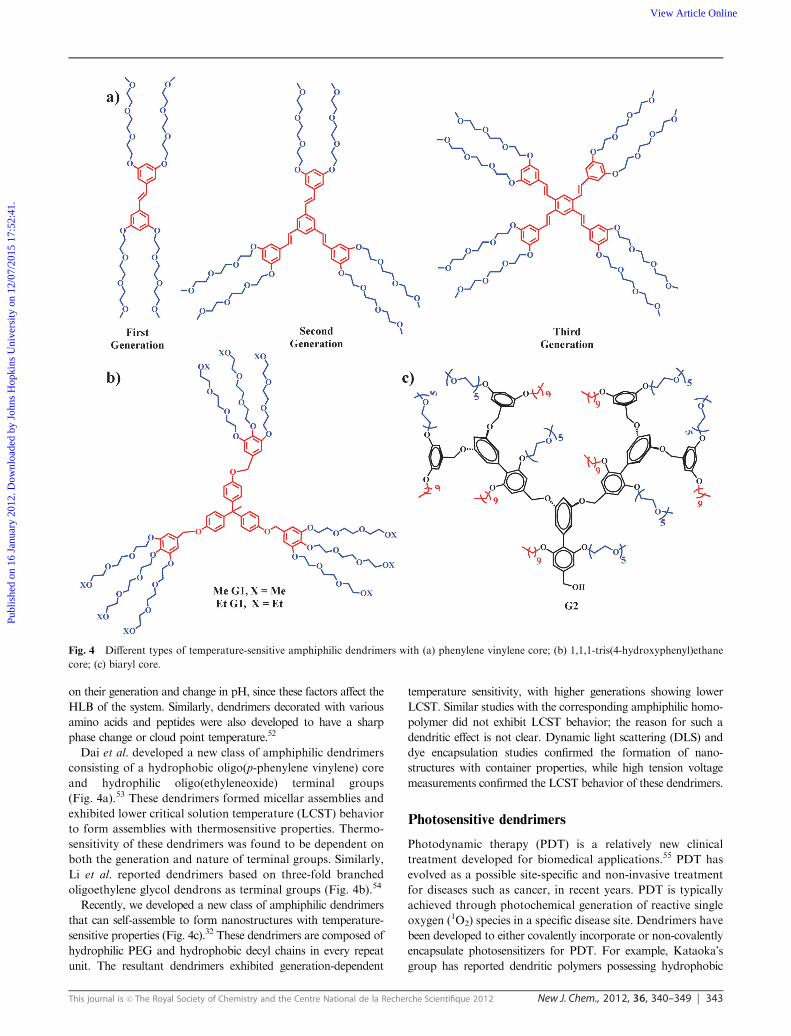
Dai et al. developed a new class of amphiphilic dendrimers
consisting of a hydrophobic oligo(p-phenylene vinylene) core
and hydrophilic oligo(ethyleneoxide) terminal groups
(Fig. 4a).53 These dendrimers formed micellar assemblies and
exhibited lower critical solution temperature (LCST) behavior
to form assemblies with thermosensitive properties. Thermo-
sensitivity of these dendrimers was found to be dependent on
both the generation and nature of terminal groups. Similarly,
Li et al. reported dendrimers based on three-fold branched
oligoethylene glycol dendrons as terminal groups (Fig. 4b).54
Recently, we developed a new class of amphiphilic dendrimers
that can self-assemble to form nanostructures with temperature-
sensitive properties (Fig. 4c).32 These dendrimers are composed of
hydrophilic PEG and hydrophobic decyl chains in every repeat
unit. The resultant dendrimers exhibited generation-dependent
temperature sensitivity, with higher generations showing lower
LCST. Similar studies with the corresponding amphiphilic homo-
polymer did not exhibit LCST behavior; the reason for such a
dendritic effect is not clear. Dynamic light scattering (DLS) and
dye encapsulation studies confirmed the formation of nano-
structures with container properties, while high tension voltage
measurements confirmed the LCST behavior of these dendrimers.
Photosensitive dendrimers
Photodynamic therapy (PDT) is a relatively new clinical
treatment developed for biomedical applications.55 PDT has
evolved as a possible site-specific and non-invasive treatment
for diseases such as cancer, in recent years. PDT is typically
achieved through photochemical generation of reactive single
oxygen (1O2) species in a specific disease site. Dendrimers have
been developed to either covalently incorporate or non-covalently
encapsulate photosensitizers for PDT. For example, Kataoka’s
group has reported dendritic polymers possessing hydrophobic
Fig. 4 Different types of temperature-sensitive amphiphilic dendrimers with (a) phenylene vinylene core; (b) 1,1,1-tris(4-hydroxyphenyl)ethane
core; (c) biaryl core.
Publ
ishe
d on
16
Janu
ary
2012
. Dow
nloa
ded
by J
ohns
Hop
kins
Uni
vers
ity o
n 12
/07/
2015
17:
52:4
1.
View Article Online
344 New J. Chem., 2012, 36, 340–349 This journal is c The Royal Society of Chemistry and the Centre National de la Recherche Scientifique 2012
photosensitizer core moieties and hydrophilic PEG groups with
efficient photosensitive properties both in vitro and in vivo.56,57 This
effect was achieved through the generation of 1O2 oxygen species
by the photosensitive protoporphyrin IX core. Similarly, Battah
and co-workers conjugated dendrimers with protoporphyrin IX
through ester bonds, and demonstrated the cytotoxicity of these
conjugates after photo-induced cleavage of ester bonds and hence
releasing the contents.58,59 On the other hand, Kojima et al.
demonstrated the photosensitive properties of PEG-decorated
PPI and PAMAM dendrimers, by non-covalent encapsulation of
photosensitizers.60a Oar et al. further improved this concept by
using a two-photon system to excite the photosensitizer.60b
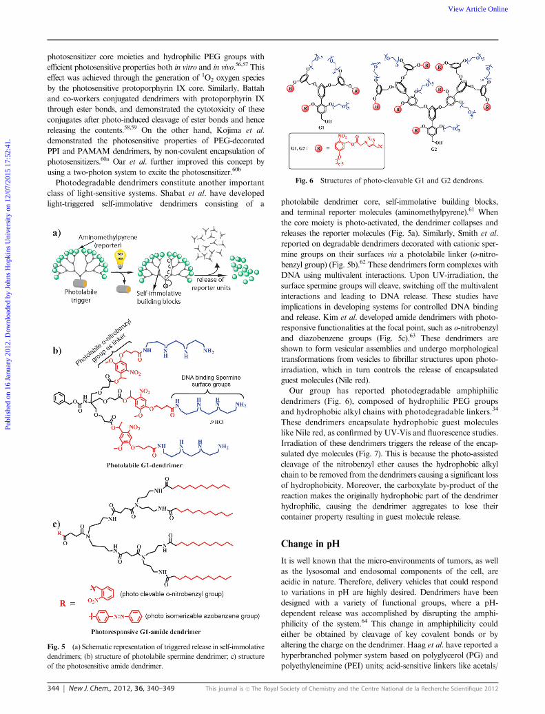
Photodegradable dendrimers constitute another important
class of light-sensitive systems. Shabat et al. have developed
light-triggered self-immolative dendrimers consisting of a photolabile dendrimer core, self-immolative building blocks,
and terminal reporter molecules (aminomethylpyrene).61 When
the core moiety is photo-activated, the dendrimer collapses and
releases the reporter molecules (Fig. 5a). Similarly, Smith et al.
reported on degradable dendrimers decorated with cationic sper-
mine groups on their surfaces via a photolabile linker (o-nitro-
benzyl group) (Fig. 5b).62 These dendrimers form complexes with
DNA using multivalent interactions. Upon UV-irradiation, the
surface spermine groups will cleave, switching off the multivalent
interactions and leading to DNA release. These studies have
implications in developing systems for controlled DNA binding
and release. Kim et al. developed amide dendrimers with photo-
responsive functionalities at the focal point, such as o-nitrobenzyl
and diazobenzene groups (Fig. 5c).63 These dendrimers are
shown to form vesicular assemblies and undergo morphological
transformations from vesicles to fibrillar structures upon photo-
irradiation, which in turn controls the release of encapsulated
guest molecules (Nile red).
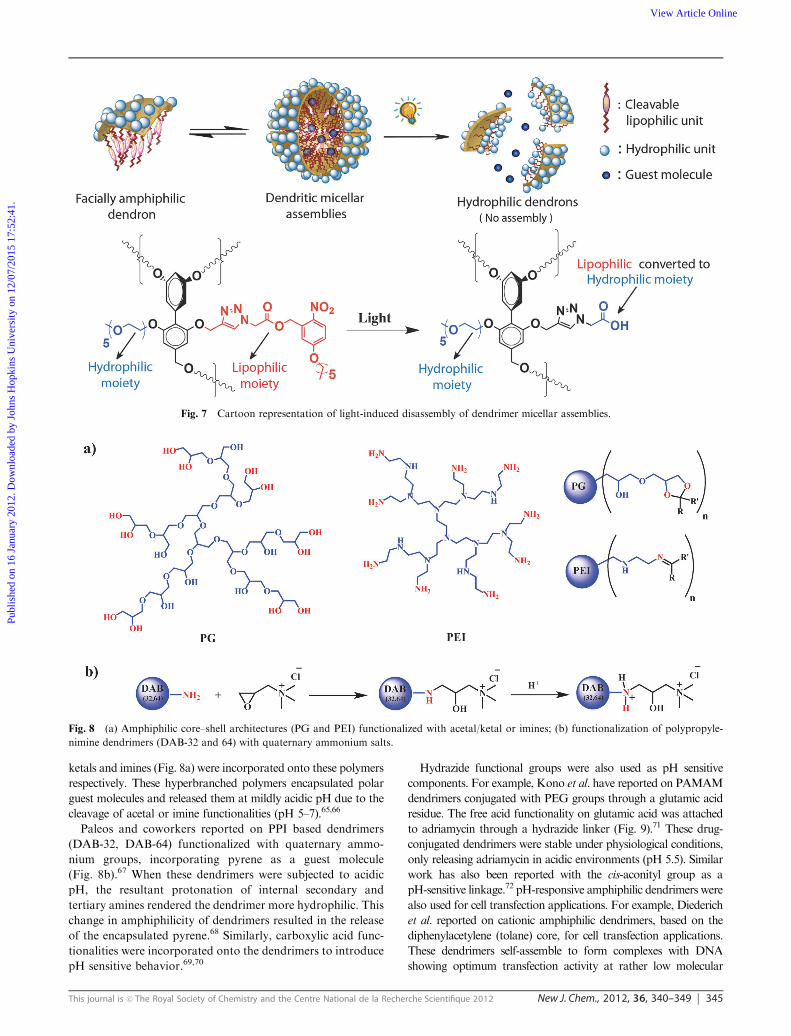
Our group has reported photodegradable amphiphilic
dendrimers (Fig. 6), composed of hydrophilic PEG groups
and hydrophobic alkyl chains with photodegradable linkers.34
These dendrimers encapsulate hydrophobic guest molecules
like Nile red, as confirmed by UV-Vis and fluorescence studies.
Irradiation of these dendrimers triggers the release of the encap-
sulated dye molecules (Fig. 7). This is because the photo-assisted
cleavage of the nitrobenzyl ether causes the hydrophobic alkyl
chain to be removed from the dendrimers causing a significant loss
of hydrophobicity. Moreover, the carboxylate by-product of the
reaction makes the originally hydrophobic part of the dendrimer
hydrophilic, causing the dendrimer aggregates to lose their
container property resulting in guest molecule release.
Change in pH
It is well known that the micro-environments of tumors, as well
as the lysosomal and endosomal components of the cell, are
acidic in nature. Therefore, delivery vehicles that could respond
to variations in pH are highly desired. Dendrimers have been
designed with a variety of functional groups, where a pH-
dependent release was accomplished by disrupting the amphi-
philicity of the system.64 This change in amphiphilicity could
either be obtained by cleavage of key covalent bonds or by
altering the charge on the dendrimer. Haag et al. have reported a
hyperbranched polymer system based on polyglycerol (PG) and
polyethyleneimine (PEI) units; acid-sensitive linkers like acetals/
Fig. 5 (a) Schematic representation of triggered release in self-immolative
dendrimers; (b) structure of photolabile spermine dendrimer; c) structure
of the photosensitive amide dendrimer.
Fig. 6 Structures of photo-cleavable G1 and G2 dendrons.
Publ
ishe
d on
16
Janu
ary
2012
. Dow
nloa
ded
by J
ohns
Hop
kins
Uni
vers
ity o
n 12
/07/
2015
17:
52:4
1.
View Article Online
This journal is c The Royal Society of Chemistry and the Centre National de la Recherche Scientifique 2012 New J. Chem., 2012, 36, 340–349 345
ketals and imines (Fig. 8a) were incorporated onto these polymers
respectively. These hyperbranched polymers encapsulated polar
guest molecules and released them at mildly acidic pH due to the
cleavage of acetal or imine functionalities (pH 5–7).65,66
Paleos and coworkers reported on PPI based dendrimers
(DAB-32, DAB-64) functionalized with quaternary ammo-
nium groups, incorporating pyrene as a guest molecule
(Fig. 8b).67 When these dendrimers were subjected to acidic
pH, the resultant protonation of internal secondary and
tertiary amines rendered the dendrimer more hydrophilic. This
change in amphiphilicity of dendrimers resulted in the release
of the encapsulated pyrene.68 Similarly, carboxylic acid func-
tionalities were incorporated onto the dendrimers to introduce
pH sensitive behavior.69,70
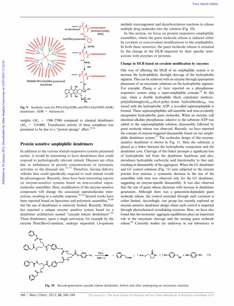
Hydrazide functional groups were also used as pH sensitive
components. For example, Kono et al. have reported on PAMAM
dendrimers conjugated with PEG groups through a glutamic acid
residue. The free acid functionality on glutamic acid was attached
to adriamycin through a hydrazide linker (Fig. 9).71 These drug-
conjugated dendrimers were stable under physiological conditions,
only releasing adriamycin in acidic environments (pH 5.5). Similar
work has also been reported with the cis-aconityl group as a
pH-sensitive linkage.72 pH-responsive amphiphilic dendrimers were
also used for cell transfection applications. For example, Diederich
et al. reported on cationic amphiphilic dendrimers, based on the
diphenylacetylene (tolane) core, for cell transfection applications.
These dendrimers self-assemble to form complexes with DNA
showing optimum transfection activity at rather low molecular
Fig. 7 Cartoon representation of light-induced disassembly of dendrimer micellar assemblies.
Fig. 8 (a) Amphiphilic core–shell architectures (PG and PEI) functionalized with acetal/ketal or imines; (b) functionalization of polypropyle-
nimine dendrimers (DAB-32 and 64) with quaternary ammonium salts.
Publ
ishe
d on
16
Janu
ary
2012
. Dow
nloa
ded
by J
ohns
Hop
kins
Uni
vers
ity o
n 12
/07/
2015
17:
52:4
1.
View Article Online
346 New J. Chem., 2012, 36, 340–349 This journal is c The Royal Society of Chemistry and the Centre National de la Recherche Scientifique 2012
weights (Mw = 1500–2700) compared to classical dendrimers
(Mw = 116000). Transfection activity of these complexes was
presumed to be due to a ‘‘proton sponge’’ effect.73,74
Protein sensitive amphiphilic dendrimers
In addition to the various stimuli-responsive systems presented
earlier, it would be interesting to have dendrimers that could
respond to pathologically relevant stimuli. Diseases are often
due to imbalances in protein concentrations or enzymatic
activities at the diseased site.75–77 Therefore, having delivery
vehicles that could specifically respond to such stimuli would
be advantageous. Recently, there have been interesting reports
on enzyme-sensitive systems based on non-covalent supra-
molecular assemblies. Here, modification of the enzyme sensitive
component will change the associated supramolecular inter-
actions, resulting in a suitable response.78–82 Several works have
been reported based on liposomes and polymeric assemblies,83,84
but the use of dendrimers is relatively limited. Recently, Shabat
has reported a unique enzyme sensitive system based on a
dendrimer architecture named ‘‘cascade release dendrimers’’.85
These dendrimers, upon a single activation, for example by the
enzyme Penicillin-G-amidase, undergo sequential 1,6-quinone
methide rearrangement and decarboxylation reactions to release
multiple drug molecules into the solution (Fig. 10).
In this section, we focus on protein responsive amphiphilic
assemblies, where the guest molecule release is induced either
by covalent or non-covalent modifications to the amphiphiles.
In both these scenarios, the guest molecule release is actuated
by the change in the HLB imparted by their specific inter-
actions with enzymes or proteins.
Change in HLB based on covalent modification by enzymes
One way of affecting the HLB of an amphiphilic system is to
increase the hydrophilicity through cleavage of the hydrophobic
segment. This can be achieved with an enzyme through appropriate
placement of an enzymatic substrate on the hydrophobic segment.
For example, Zhang et al. have reported on a phosphatase-
responsive system using a super-amphiphile concept.79 In this
case, when a double hydrophilic block copolymer methoxy-
poly(ethyleneglycol)114-block-poly(L-lysine hydrochloride)200 was
mixed with the hydrophobic ATP, a so-called superamphiphile is
formed. These superamphiphiles self-assemble and non-covalently
encapsulate hydrophobic guest molecules. When an enzyme, calf
intestinal alkaline phosphatase, selective to the substrate ATP was
added to the superamphiphile solution, disassembly followed by
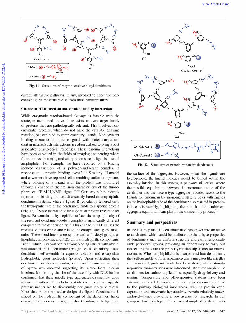
guest molecule release was observed. Recently, we have reported
the concept of enzyme-triggered disassembly based on our amphi-
philic dendrimer system.37 The molecular design of this enzyme-
sensitive dendrimer is shown in Fig. 11. Here the substrate is
placed as a linker between the hydrophobic component and the
dendrimer core. Cleavage of this linker prompts a significant loss
of hydrophobic tail from the dendrimer backbone and also,
introduces hydrophilic carboxylic acid functionality to that end,
resulting in disassembly of the aggregates. When the G1 dendrimer
and G1 control solutions (Fig. 11) were subjected to the enzyme
porcine liver esterase, a systematic decrease in the size of the
assemblies with time was observed only for the G1 dendrimer,
suggesting an enzyme-specific disassembly. It was also observed
that the rate of guest release decreases with increase in dendrimer
generation. Although there was a generation-dependent guest
molecule release, the control exercised through such variation is
rather limited. Accordingly, our group has recently explored an
enzyme sensitive dendrimer design where such control is imparted
through photochemical crosslinking reactions. Here, we have also
found that themonomer–aggregate equilibrium plays an important
role in the enzymatic cleavage and the ensuing guest molecule
release.86 Currently studies are underway in our laboratory to
Fig. 9 Synthetic route for PEG-Glu(ADR) and PEG-Glu(NHN-ADR)
dendrimers. ADR = Adriamycin.
Fig. 10 Second-generation cascade release dendrimer, before and after undergoing an enzymatic reaction.
Publ
ishe
d on
16
Janu
ary
2012
. Dow
nloa
ded
by J
ohns
Hop
kins
Uni
vers
ity o
n 12
/07/
2015
17:
52:4
1.
View Article Online
This journal is c The Royal Society of Chemistry and the Centre National de la Recherche Scientifique 2012 New J. Chem., 2012, 36, 340–349 347
discern alternative pathways, if any, involved to effect the non-
covalent guest molecule release from these nanocontainers.
Change in HLB based on non-covalent binding interactions
While enzymatic reaction-based cleavage is feasible with the
strategies mentioned above, there exists an even larger family
of proteins that are pathologically relevant. This involves non-
enzymatic proteins, which do not have the catalytic cleavage
reaction, but can bind to complementary ligands. Non-covalent
binding interactions of specific ligands with proteins are abun-
dant in nature. Such interactions are often utilized to bring about
associated physiological responses. These binding interactions
have been exploited in the fields of imaging and sensing where
fluorophores are conjugated with protein specific ligands in small
amphiphiles. For example, we have reported on a binding
induced disassembly of a polymer–surfactant complex in
response to a protein binding event.87,88 Similarly, Hamachi
and coworkers have reported self-assembling surfactant systems,
where binding of a ligand with the protein was monitored
through a change in the emission characteristics of the fluoro-
phore or 19F-MRI/NMR signal.89,90 Our group has recently
reported on binding induced disassembly based on amphiphilic
dendrimer systems, where a ligand R (covalently tethered onto
the hydrophilic face of the dendrimer) binds to a specific protein
(Fig. 12).36 Since the water-soluble globular protein (receptor for
ligand R) contains a hydrophilic surface, the amphiphilicity of
the resultant dendrimer–protein complex is significantly different
compared to the dendrimer itself. This change in HLB causes the
micelles to disassemble and release the encapsulated guest mole-
cules. These dendrimers were synthesized with decyl groups as
lipophilic components, and PEGunits as hydrophilic components.
Biotin, which is known for its strong binding affinity with avidin,
was attached to the dendrimer through ‘‘click’’ chemistry. These
dendrimers self-assemble in aqueous solution and encapsulate
hydrophobic guest molecules (pyrene). Upon subjecting these
dendrimeric solutions to avidin, a decrease in emission intensity
of pyrene was observed suggesting its release from micellar
interiors. Monitoring the size of the assembly with DLS further
confirmed that these micelle type aggregates disassemble upon
interaction with avidin. Selectivity studies with other non-specific
proteins neither led to disassembly nor guest molecule release.
Note that in this molecular design the ligand functionality is
placed on the hydrophilic component of the dendrimer, hence
disassembly can occur through the direct binding of the ligand on
the surface of the aggregate. However, when the ligands are
hydrophobic, the ligand moieties would be buried within the
assembly interior. In this system, a pathway still exists, where
the possible equilibrium between the monomeric state of the
dendrimer and the micelle-type aggregate provides access to the
ligands for binding in the monomeric state. Studies with ligands
on the hydrophobic side of the dendrimer also resulted in protein-
induced disassembly, highlighting the role that the dendrimer–
aggregate equilibrium can play in the disassembly process.91
Summary and perspectives
In the last 25 years, the dendrimer field has grown into an active
research area, which could be attributed to the unique properties
of dendrimers such as uniform structure and easily functionali-
zable peripheral groups, providing an opportunity to carry out
molecular-level structure–property relationship studies for macro-
molecules. When amphiphilicity is incorporated into dendrimers,
they self-assemble to form supramolecular aggregates like micelles
and vesicles. Significant work has been done, where stimuli-
responsive characteristics were introduced into these amphiphilic
dendrimers for various applications, especially drug delivery and
sensing. Temperature and pH-responsive systems have been
extensively studied. However, stimuli-sensitive systems responsive
to the primary biological imbalances, such as protein over-
expression and enzymatic hyperactivity, remain relatively under-
explored—hence providing a new avenue for research. In our
group we have developed a new class of amphiphilic dendrimers
Fig. 11 Structures of enzyme sensitive biaryl dendrimers.
Fig. 12 Structures of protein responsive dendrimers.
Publ
ishe
d on
16
Janu
ary
2012
. Dow
nloa
ded
by J
ohns
Hop
kins
Uni
vers
ity o
n 12
/07/
2015
17:
52:4
1.
View Article Online
348 New J. Chem., 2012, 36, 340–349 This journal is c The Royal Society of Chemistry and the Centre National de la Recherche Scientifique 2012
that are responsive to both enzymatic and non-enzymatic proteins.
These dendrimers serve as model systems for a novel category
of stimuli-sensitive drug delivery vehicles. Development of
reproducible methods for large scale syntheses of highly versatile
dendrimers, without compromising their fidelity for a targeted
application, is a remaining challenge in the field to make a large
scale impact in downstream biological applications.
Acknowledgements
We acknowledge NIGMS of the NIH (GM-065255) and Army
Research office (57858-CH) for financial support. We thank
Bhooshan Popere for critical comments.
References
1 T. M. Allen and P. R. Cullis, Science, 2004, 303, 1818–1822.2 G. M. Whitesides and M. Boncheva, Proc. Natl. Acad. Sci. U. S. A.,2002, 99, 4769–4774.
3 J. M. Zayed, N. Nouvel, U. Rauwald and O. A. Scherman, Chem.Soc. Rev., 2009, 39, 2806–2816.
4 J. M. J. Frechet, Proc. Natl. Acad. Sci. U. S. A., 2002, 99,4782–4787.
5 A. V. Davis, R. M. Yeh and K. N. Raymond, Proc. Natl. Acad.Sci. U. S. A., 2002, 99, 4793–4796.
6 J. M. Lehn, Rep. Prog. Phys., 2004, 67, 249–265.7 V. Percec, D. A. Wilson, P. Leowanawat, C. J. Wilson,A. D. Hughes, M. S. Kaucher, D. A. Hammer, D. H. Levine,A. J. Kim, F. S. Bates, K. P. Davis, T. P. Lodge, M. L. Klein,R. H. DeVane, E. Aqad, B. M. Rosen, A. O. Argintaru,M. J. Sienkowska, K. Rissanen, S. Nummelin and J. Ropponen,Science, 2010, 328, 1009–1014.
8 S. Hecht and J. M. J. Frechet, Angew. Chem., Int. Ed., 2001, 40,74–91.
9 S. H. Medina and M. E. H. El-Sayed, Chem. Rev., 2009, 109,3141–3157.
10 D. Peer, J. M. Karp, S. Hong, O. C. FaroKhzad, R. Margalit andR. Langer, Nat. Nanotechnol., 2007, 2, 751–760.
11 J. F. Gohy, H. Hofmeier, A. Alexeev and U. S. Schubert,Macromol.Chem. Phys., 2003, 204, 1524–1530.
12 Y. Bae and K. Kataoka, Adv. Drug Delivery Rev., 2009, 61,768–784.
13 K. T. Oh, T. K. Bronich and A. V. Kabanov, J. Controlled Release,2004, 94, 411–422.
14 A. V. Kabanov, I. R. Nazarova, I. V. Astafieva, E. V. Batrakova,V. Y. Alakhov, A. A. Yaroslavov and V. A. Kabanov,Macromolecules,1995, 28, 2303–2314.
15 J. Chayen, Cell Biochem. Funct., 1996, 14, 75.16 D. M. Vriezema, M. C. Aragones, J. A. A. W. Elemans,
J. J. L. M. Cornelissen, A. E. Rowan and R. J. M. Nolte, Chem.Rev., 2005, 105, 1445–1489.
17 M. S. Robinson, Trends Cell Biol., 2004, 14, 167–174.18 J.-H. Fuhrhop and T. Wang, Chem. Rev., 2004, 104, 2901–2937.19 K. Kita-Tokarczyk, J. Grumelard, T. Haefele and W. Meier,
Polymer, 2005, 46, 3540–3563.20 A. P. H. J. Schenning, C. Elissen-Roman, J.-W. Weener, M. W. P. L.
Baars, S. J. van der Gaast and E. W.Meijer, J. Am. Chem. Soc., 1998,120, 8199–8208.
21 Y. Chen, A. V. Ambade, D. R. Vutukuri and S. Thayumanavan,J. Am. Chem. Soc., 2006, 128, 14760–14761.
22 M. Malmsten and B. Lindman, Macromolecules, 1992, 25,5440–5445.
23 P. Munk, C. Ramireddy, M. Tian, S. E. Webber, K. Prochazkaand Z. Tuzar, Makromol. Chem., Macromol. Symp., 1992, 58,195–199.
24 S. Basu, D. R. Vutukuri, S. Shyamroy, B. S. Sandanaraj andS. Thayumanavan, J. Am. Chem. Soc., 2004, 126, 9890–9891.
25 C. C. Lee, J. A. MacKay, J. M. J. Frechet and F. C. Szoka, Nat.Biotechnol., 2005, 23, 1517–1526.
26 C. Park, J. Lee and C. Kim, Chem. Commun., 2011, 47,12042–12056.
27 D. Astruc, E. Boisselier and C. Ornelas, Chem. Rev., 2010, 110,1857–1959.
28 G. R. Newkome, Z. Q. Yao, G. R. Baker and V. K. Gupta, J. Org.Chem., 1985, 50, 2003–2004.
29 G. R. Newkome, C. N. Moorefield, G. R. Baker, M. J. Saundersand S. H. Grossman, Angew. Chem., Int. Ed. Engl., 1991, 30,1178–1180.
30 C. J. Hawker, K. L. Wooley and J. M. J. Frechet, J. Chem. Soc.,Perkin Trans. 1, 1993, 1287–1297.
31 J. F. G. A. Jansen, E. M. M. De Brabander-Van den Berg andE. W. Meijer, Science, 1994, 266, 1226–1229.
32 S. V. Aathimanikandan, E. N. Savariar and S. Thayumanavan,J. Am. Chem. Soc., 2005, 127, 14922–14929.
33 C. Kojima, S. Tsumura, A. Harada and K. Kono, J. Am. Chem.Soc., 2009, 131, 6052–6053.
34 V. Yesilyurt, R. Ramireddy and S. Thayumanavan, Angew. Chem.,Int. Ed., 2011, 50, 3038–3042.
35 A. Almutairi, S. J. Guillaudeu, M. Y. Berezin, S. Achilefu andJ. M. J. Frechet, J. Am. Chem. Soc., 2008, 130, 444–445.
36 M. A. Azagarsamy, V. Yesilyurt and S. Thayumanavan, J. Am.Chem. Soc., 2010, 132, 4550–4551.
37 M. A. Azagarsamy, P. Sokkalingam and S. Thayumanavan, J. Am.Chem. Soc., 2009, 131, 14184–14185.
38 K. Knop, R. Hoogenboom, D. Fischer and U. S. Schubert, Angew.Chem., Int. Ed., 2010, 49, 6288–6308.
39 A. V. Kabanov, E. V. Batrakova and V. Y. Alakhov, J. ControlledRelease, 2002, 82, 189–212.
40 Z. Zhang, D. W. Grijpma and J. Feijen, J. Controlled Release,2006, 112, 57–63.
41 C. Allen, J. Han, Y. Yu, D. Maysinger and A. Eisenberg,J. Controlled Release, 2000, 63, 275–286.
42 K. Letchford, J. Zastre, R. Liggins and H. Burt, Colloids Surf., B,2004, 35, 81–91.
43 P. Bharathi, H. D. Zhao and S. Thayumanavan, Org. Lett., 2001,3, 1961–1964.
44 D. R. Vutukuri, S. Basu and S. Thayumanavan, J. Am. Chem.Soc., 2004, 126, 15636–15637.
45 P. R. Stauffer and S. N. Goldberg, Int. J. Hyperthermia., 2004, 20,671–677.
46 M. Bikram and J. L. West, Expert. Opin. Drug Delivery, 2008, 5,1077–1091.
47 M. Kimura, M. Kato, T. Muto, K. Hanabusa and H. Shirai,Macromolecules, 2000, 33, 1117–1119.
48 Z. Yang, W. Zhang, J. Zou and W. Shi, Polymer, 2007, 48,931–938.
49 L. Zhu, G. Zhu, M. Li, E. Wang, R. Zhu and X. Qi, Eur. Polym. J.,2002, 38, 2503–2506.
50 Z. Yang, J. Xie, W. Zhou and W. Shi, J. Biomed. Mater. Res., PartA, 2009, 89A, 988–1000.
51 Y. Tono, C. Kojima, Y. Haba, T. Takahashi, A. Harada, S. Yagiand K. Kono, Langmuir, 2006, 22, 4920–4922.
52 G. A. Kinberger, W. Cai and M. Goodman, J. Am. Chem. Soc.,2002, 124, 15162–15163.
53 D. W. Chang and L. Dai, J. Mater. Chem., 2007, 17, 364–371.54 W. Li, A. Zhang, Y. Chen, K. Feldman, H. Wu and A. D. Schluter,
Chem. Commun., 2008, 5948–5950.55 D. E. J. G. J. Dolmans, D. Fukumura and R. K. Jain, Nat. Rev.
Cancer, 2003, 3, 380–387.56 N. Nishiyama, Y. Morimoto, W.-D. Jang and K. Kataoka, Adv.
Drug Delivery Rev., 2009, 61, 327–338.57 N. Nishiyama, H. R. Stapert, G.-D. Zhang, D. Takasu,
D.-L. Jiang, T. Nagano, T. Aida and K. Kataoka, BioconjugateChem., 2003, 14, 58–66.
58 S. H. Battah, C.-E. Chee, H. Nakanishi, S. Gerscher,A. J. MacRobert and C. Edwards, Bioconjugate Chem., 2001, 12,980–988.
59 S. Battah, S. Balaratnam, A. Casas, S. O’Neill, C. Edwards,A. Batlle, P. Dobbin and A. J. MacRobert, Mol. Cancer. Ther.,2007, 6, 876–885.
60 (a) C. Kojima, Y. Toi, A. Harada and K. Kono, Bioconjugatechem, 2007, 18, 663–670; (b) M. A. Oar, J. M. Serin, W. R. Dichtel,T. Y. Ohulchanskyy, P. N. Prasad and J. M. J. Frechet, Chem.Mater., 2005, 17, 2267–2275.
61 R. J. Amir, N. Pessah, M. Shamis and D. Shabat, Angew. Chem.,Int. Ed., 2003, 42, 4494–4499.
Publ
ishe
d on
16
Janu
ary
2012
. Dow
nloa
ded
by J
ohns
Hop
kins
Uni
vers
ity o
n 12
/07/
2015
17:
52:4
1.
View Article Online
This journal is c The Royal Society of Chemistry and the Centre National de la Recherche Scientifique 2012 New J. Chem., 2012, 36, 340–349 349
62 M. A. Kostiainen, D. K. Smith and O. Ikkala, Angew. Chem., Int.Ed., 2007, 46, 7600–7604.
63 C. Park, J. Lim, M. Yun and C. Kim, Angew. Chem., Int. Ed.,2008, 47, 2959–2963.
64 J. F. G. A. Jansen, E. W. Meijer and E. M. M. De Brabander-Vanden Berg, J. Am. Chem. Soc., 1995, 117, 4417–4418.
65 M. Kramer, J.-F. Stumbe, H. Turk, S. Krause, A. Komp,L. Delineau, S. Prokhorova, H. Kautz and R. Haag, Angew.Chem., Int. Ed., 2002, 41, 4252–4256.
66 S. Xu, M. Kramer and R. Haag, J. Drug Targeting, 2006, 14,367–374.
67 G. Pistolis, A. Malliaris, D. Tsiourvas and C. M. Paleos, Chem.–Eur.J., 1999, 5, 1440–1444.
68 Z. Sideratou, D. Tsiourvas and C. M. Paleos, Langmuir, 1999, 16,1766–1769.
69 D. Wan, H. Pu and X. Cai, Macromolecules, 2008, 41, 7787–7789.70 S. Y. Cho and H. R. Allcock, Macromolecules, 2007, 40,
3115–3121.71 K. Kono, C. Kojima, N. Hayashi, E. Nishisaka, K. Kiura,
S. Watarai and A. Harada, Biomaterials, 2008, 29, 1664–1675.72 S. Zhu, M. Hong, G. Tang, L. Qian, J. Lin, Y. Jiang and Y. Pei,
Biomaterials, 2010, 31, 1360–1371.73 D. Joester, M. Losson, R. Pugin, H. Heinzelmann, E. Walter,
H. P. Merkle and F. Diederich, Angew. Chem., Int. Ed., 2003, 42,1486–1490.
74 M. Guillot-Nieckowski, D. Joester, M. Stohr, M. Losson,M. Adrian, B. Wagner, M. Kansy, H. Heinzelmann, R. Pugin,F. Diederich and J.-L. Gallani, Langmuir, 2006, 23, 737–746.
75 S. Samantaray, R. Sharma, T. K. Chattopadhyaya, S. D. Guptaand R. Ralhan, J. Cancer Res. Clin. Oncol., 2004, 130, 37–44.
76 K. E. Wilson, S. P. Langdon, A. M. Lessells and W. R. Miller,Br. J. Cancer, 1996, 74, 999–1004.
77 H. L. Klein, DNA Repair, 2008, 7, 686–693.78 F. E. Alemdaroglu, J. Wang, M. Boersch, R. Berger and
A. Herrmann, Angew. Chem., Int. Ed., 2008, 47, 974–976.79 C. Wang, Q. Chen, Z. Wang and X. Zhang, Angew. Chem., Int.
Ed., 2011, 49, 8612–8615.80 R. V. Ulijn, J. Mater. Chem., 2006, 16, 2217–2225.81 P. D. Thornton, G. McConnell and R. V. Ulijn, Chem. Commun.,
2005, 47, 5913–5915.82 R. J. Amir, S. Zhong, D. J. Pochan and C. J. Hawker, J. Am.
Chem. Soc., 2009, 131, 13949–13951.83 P. Meers, Adv. Drug Delivery Rev., 2001, 53, 265–272.84 J. Davidsen, C. Vermehren, S. Frokjaer, O. G. Mouritsen and
K. Jorgensen, Int. J. Pharm., 2001, 214, 67–69.85 D. Shabat, J. Polym. Sci., Part A: Polym. Chem., 2006, 44,
1569–1578.86 K. R. Raghupathi, M. A. Azagarsamy and S. Thayumanavan,
Chem.–Eur. J., 2011, 17, 11752–11760.87 E. N. Savariar, S. Ghosh, D. C. Gonzalez and S. Thayumanavan,
J. Am. Chem. Soc., 2008, 130, 5416–5417.88 D. C. Gonzalez, E. N. Savariar and S. Thayumanavan, J. Am.
Chem. Soc., 2009, 131, 7708–7716.89 K. Mizusawa, Y. Ishida, Y. Takaoka, M. Miyagawa, S. Tsukiji
and I. Hamachi, J. Am. Chem. Soc., 2010, 132, 7291–7293.90 Y. Takaoka, T. Sakamoto, S. Tsukiji, M. Narazaki, T. Matsuda,
H. Tochio, M. Shirakawa and I. Hamachi, Nat. Chem., 2009, 1,557–561.
91 V. Yesilyurt, R. Ramireddy,M. A. Azagarsamy and S. Thayumanavan,Chem.–Eur. J., 2012, 18, 223–229.
Publ
ishe
d on
16
Janu
ary
2012
. Dow
nloa
ded
by J
ohns
Hop
kins
Uni
vers
ity o
n 12
/07/
2015
17:
52:4
1.
View Article Online


