Embed Size (px)
Citation preview

Cancers 2014, 6, 1579-1596; doi:10.3390/cancers6031579
cancers ISSN 2072-6694
www.mdpi.com/journal/cancers
Article
STAT3 Activities and Energy Metabolism: Dangerous Liaisons
Annalisa Camporeale 1,†,*, Marco Demaria 2,†, Emanuele Monteleone 1, Carlotta Giorgi 3,
Mariusz R. Wieckowski 4, Paolo Pinton 3 and Valeria Poli 1,*
1 Molecular Biotechnology Center and Department of Molecular Biotechnology and Life Sciences,
University of Turin, Via Nizza 52, Turin 10126, Italy; E-Mail: [email protected] 2 Buck Institute for Research on Aging, 8001 Redwood Blvd, Novato, CA 94945, USA;
E-Mail: [email protected] 3 Department of Experimental and Diagnostic Medicine, Section of General Pathology, Laboratory
for Technologies of Advances Therapies (LTTA), University of Ferrara, Via Fossato di Mortara 70,
Ferrara 44121, Italy; E-Mails: [email protected] (C.G.); [email protected] (P.P.) 4 Nencki Institute of Experimental Biology, Department of Biochemistry, Pasteur Str. 3, Warsaw 02-093,
Poland; E-Mail: [email protected]
† These authors equally contributed to this work.
* Authors to whom correspondence should be addressed; E-Mails: [email protected] (A.C.);
[email protected] (V.P.); Tel.: +39-11-670-6428 (A.C. & V.P.); Fax: +39-11-670-6432 (A.C. & V.P.).
Received: 13 May 2014; in revised form: 3 July 2014 / Accepted: 16 July 2014 /
Published: 31 July 2014
Abstract: STAT3 mediates cytokine and growth factor receptor signalling, becoming
transcriptionally active upon tyrosine 705 phosphorylation (Y-P). Constitutively Y-P
STAT3 is observed in many tumors that become addicted to its activity, and STAT3
transcriptional activation is required for tumor transformation downstream of several
oncogenes. We have recently demonstrated that constitutively active STAT3 drives a
metabolic switch towards aerobic glycolysis through the transcriptional induction of Hif-1α
and the down-regulation of mitochondrial activity, in both MEF cells expressing
constitutively active STAT3 (Stat3C/C) and STAT3-addicted tumor cells. This novel
metabolic function is likely involved in mediating pre-oncogenic features in the primary
Stat3C/C MEFs such as resistance to apoptosis and senescence and rapid proliferation.
Moreover, it strongly contributes to the ability of primary Stat3C/C MEFs to undergo
malignant transformation upon spontaneous immortalization, a feature that may explain the
well known causative link between STAT3 constitutive activity and tumor transformation
OPEN ACCESS

Cancers 2014, 6 1580
under chronic inflammatory conditions. Taken together with the recently uncovered role of
STAT3 in regulating energy metabolism from within the mitochondrion when phosphorylated
on Ser 727, these data place STAT3 at the center of a hub regulating energy metabolism
under different conditions, in most cases promoting cell survival, proliferation and
malignant transformation even though with distinct mechanisms.
Keywords: STAT3; metabolism; Warburg effect; aerobic glycolysis; cellular transformation;
mitochondrial activity
1. Introduction
The transcription factor Signal Transducer and Activator of Transcription 3 (STAT3) is activated
downstream of many cytokine and growth factor receptors, resulting in tyrosine phosphorylation,
dimerization via reciprocal phosphotyrosine-src homology (SH)-2 interactions and translocation to the
nucleus, where it binds to responsive elements on gene promoters. STAT3 can also be phosphorylated
on serine residue 727 (S-P) within its carboxyl-terminal Transcription Activation Domain [1,2], which
is thought to provide full trans-activating properties to the Y-P protein for optimal induction of a
subset of target genes [3].
STAT3 target genes and functions vary depending on the cellular system, but in most instances
correlate with cell survival and proliferation. Under physiological conditions, STAT3 activation is
transient and tightly regulated by negative feedback mechanisms mediated, among others, by
suppressor of cytokine signal (SOCS) and protein inhibitor of activated STAT (PIAS) proteins [4,5].
On the other hand, persistent STAT3 activation is commonly observed in tumors of different origin
and during chronic inflammation, downstream of continuous cytokine/growth factor stimulation or of
constitutive activity of non-receptor tyrosine kinases such as cSrc [6]. A direct role in oncogenesis was
demonstrated by the ability of the artificially mutated form Stat3C to lead to malignant transformation
when overexpressed in immortalized fibroblasts and epithelial cells [7]. STAT3 inhibition in many
tumor models/primary tumor cells leads to loss of survival and proliferation, suggesting addiction of
tumor cells to STAT3 activity [8,9]. Accordingly, STAT3 is required for malignant transformation by
many oncogenes, in primis vSrc, in a number of cell types [10].
In tumors, STAT3 is known to exert a number of well established functions correlating to
transcriptional activation of its target genes, including regulation of cell-cycle progression, apoptosis,
tumor angiogenesis, invasion, metastasis, and tumor cell evasion from the immune system, reflecting
the involvement of this factor in multiple steps of the oncogenic program [11]. Additionally, STAT3 is
considered as a key player in mediating inflammation-driven tumorigenesis, being constitutively
activated by chronically high levels of the pro-inflammatory cytokine IL-6 [12].
1.1. STAT3 Differentially Modified Forms and Cell Metabolism
Aberrant regulation of cell metabolism plays a central role in deterring the survival and growth
features of malignant cells. For example, most tumor cells display a metabolic switch towards aerobic

Cancers 2014, 6 1581
glycolysis, with increased glycolysis and decreased oxidative phosphorylation, even under conditions
of high oxygen tension [13–15]. This phenomenon is thought to favor the synthesis of essential cellular
components required for fast cell duplication. Recently, a number of observations have suggested that
STAT3 can act as a central regulator of cell metabolism at multiple levels, which may represent a core
function in promoting growth/survival of biologically distinct tumors [16]. Interestingly, specific and
often unconventional functions have been assigned to the differentially modified forms of this factor,
as summarized below.
First, STAT3 was shown to localize to mitochondria, where its S-P form enhances coupled
Complex I and II activity and reduces ROS production, while inducing aerobic glycolysis [17,18]. This
function enhances cell survival under stress conditions such as heart ischemia, and is required for cell
transformation downstream of Ras oncogenes, which elicit S, but not Y, phosphorylation of STAT3.
Additionally, STAT3 was shown to interact with cyclophilin F (best known as cyclophilin D) in the
mitochondrial matrix, thus inhibiting the opening of the mitochondrial permeability transition pore
(MPTP) and Calcium-induced apoptosis [19]. STAT3 S-P is known to enhance its interaction with the
complex I component GRIM-19, responsible for its mitochondrial translocation [20]. Although how
STAT3 phosphorylation can be regulated within the mitochondrion is not understood, the phosphatase
SHP2 was proposed as a potential player in dephosphorylating mitochondrial STAT3 [21].
Y-P STAT3 was also recently shown to play important roles in regulating energy metabolism.
Indeed, we have demonstrated that constitutively Y-P STAT3 can promote aerobic glycolysis and
down-regulate mitochondrial activity, partly acting via transcriptional induction of its well-recognized
transcriptional target Hif-1α [22]. The functional interaction between these two transcription factors is
further extended by the observation that STAT3 can specifically bind to HIF-1α target genes
promoters, allowing the formation of transcriptionally active HIF-1α/RNA Polymerase II complexes [23].
Both these activities can contribute to the reported ability of constitutively active STAT3 to act as a first
hit in oncogenic transformation [24]. Finally the pyruvate kinase M2 isoform (PKM2), an essential
regulator of aerobic glycolysis that can be induced by HIF-1α, was shown to be able to directly promote
chronic STAT3 Y-P. Thus STAT3, HIF-1α and PKM2 appear to take part in a positive feedback loop
responsible for supporting cell proliferation and survival [25,26].
Protein acetylation, a crucial post-translational modification that affects gene expression by
modulating both chromatin structure and the activity of many transcription factors, is a key factor in
regulating proliferation and energy metabolism in both normal and transformed cells [27]. STAT3 can
be acetylated (Ac-STAT3) by the CBP/p300 histone acetyltransferase in response to cytokines and
growth factors, favoring dimer stability, tyrosine phosphorylation and transcriptional activity [28].
Interestingly, STAT3 acetylation was shown to occur in the nucleus thanks to a complex with the
cancer stem cell marker CD44 and p300, leading to the binding to the cyclin D1 promoter, thus
triggering increased expression and cell proliferation [29]. Ac-STAT3 is also thought to favor
tumorigenesis by forming a complex with DNA methyltransferase 1 (DNMT1), thus leading to DNA
methylation and silencing of tumor suppressor genes [30]. Along with other histone deacetylases, the
NAD-dependent silent information regulator protein (SIRT)1 has been implicated in STAT3
deacetylation, in turn leading to reduced Y phosphorylation and transcriptional activity [31]. Recently,
SIRT1 was also shown to indirectly modulate STAT3 S-P and its mitochondrial functions, since Sirt1-null
MEF cells display high S-P STAT3 levels in mitochondria and increased mitochondrial activity [32].

Cancers 2014, 6 1582
Another SIRT family member, SIRT3, which localizes specifically to mitochondria, is implicated in
down-regulating mitochondrial respiration. Sirt3 genetic ablation leads to a metabolic switch towards
aerobic glycolysis due to increased ROS production and consequent HIF-1α stabilization [33]. No
direct interactions between SIRT3 and STAT3 have however been reported so far. Another player may
be the mTORC1 inhibitor REDD1, a known HIF-1α target that localizes to mitochondria. REDD1
inactivation results in increased ROS production, stabilization of HIF-1α and tumorigenesis [34].
These observations suggest that SIRT3 and REDD1 might cooperate in mitochondria to sustain
oxidative phosphorylation and ROS removal. Whether their activities may somehow regulate the
functions of mitochondrial STAT3 remains to be established. On the other hand, the nutrient-sensing
mTOR pathway has been implicated in regulating STAT3 activities by enhancing both its Y- and
S-phosphorylation [35–37].
Interestingly, STAT3 was recently shown to play a role in the regulation of the autophagic pathway,
another important metabolic process that has been implicated both positively and negatively in
tumorigenesis [38]. This occurs in the cytoplasm, where non-phosphorylated STAT3 can interact via
its SH2 domain with the autophagy inducer PKR/EIF2AK2 kinase, inhibiting its activity [39]. This
function can be inhibited, in addition to interfering with the SH2 domain by means of specific
compounds, by the activation of STAT3 Y-P, which decreases the pool of cytoplasmic STAT3
available for the interaction with PKR.
STAT3 appears to function as a hub to integrate different pro-survival and growth signals at the level
of the energy and respiratory metabolism, via nuclear, mitochondrial and cytoplasmic activities mediated
by its differentially modified forms [16]. Here, we discuss some of these results and how they may fit
together in a multidimensional vision of STAT3 functions under conditions of aberrant activation.
2. Results and Discussion
2.1. STAT3 Constitutive Activation Elicits Pre-Oncogenic Features in Stat3C/C MEFs.
Mice homozygous for the Stat3C k/in allele exhibited increased nuclear localization, prolonged Y-P
and enhanced transcriptional activity in response to cytokine stimuli in several tissues as well as in
mouse embryonic fibroblasts (MEFs) [40]. Compared to wild-type control cells, Stat3C/C cells displayed
enhanced proliferation and accelerated cell cycle, with a more rapid transition through the S-phase [22,24].
In addition, replicative senescence was strongly delayed and cells were highly resistant to apoptosis
induced by different stimuli such as UV treatment (23% of apoptotic cells after 24 h, compared to 45%
in the wild type controls, Figure 1A), H2O2 treatment and serum starvation ([22] and not shown).
Interestingly, Stat3C/C MEFs became more readily immortalized than their wild type counterparts when
subjected to the 3T3 spontaneous immortalization protocol (EF), and maintained significant resistance
to multiple apoptotic stimuli, similar to the primary cells (19% of dead cells upon UV light exposure
compared to 33% in wild type cells, Figure 1B) [24].
These features reflect the known activities of STAT3, promoting cell survival and proliferation, and
suggest that low levels of constitutively active STAT3 can promote pre-oncogenic features in primary
MEF cells.
Cancers 2014, 6 1583
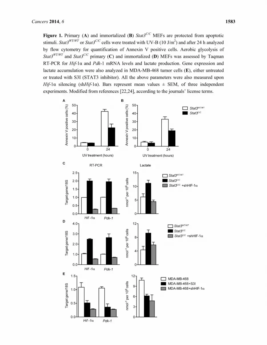
Figure 1. Primary (A) and immortalized (B) Stat3C/C MEFs are protected from apoptotic
stimuli. Stat3WT/WT or Stat3C/C cells were treated with UV-B (10 J/m2) and after 24 h analyzed
by flow cytometry for quantification of Annexin V positive cells. Aerobic glycolysis of
Stat3WT/WT and Stat3C/C primary (C) and immortalized (D) MEFs was assessed by Taqman
RT-PCR for Hif-1α and Pdk-1 mRNA levels and lactate production. Gene expression and
lactate accumulation were also analyzed in MDA-MB-468 tumor cells (E), either untreated
or treated with S3I (STAT3 inhibitor). All the above parameters were also measured upon
Hif-1α silencing (shHif-1α). Bars represent mean values ± SEM, of three independent
experiments. Modified from references [22,24], according to the journals’ license terms.
Cancers 2014, 6 1584
2.2. Constitutive STAT3 Activity Induces a Metabolic Switch to Aerobic Glycolysis in MEFs and
Tumor Cell Lines
A comparison of gene expression profiles in primary Stat3C/C and Stat3WT/WT MEFs performed via
microarray analysis showed more than one thousand differentially expressed genes [22]. Gene
Ontology (GO) analysis of up-regulated mRNAs revealed the presence of several genes involved in
aerobic glycolysis including hypoxia inducible factor (Hif)-1α (Figure 1C), which is a known STAT3
transcriptional target [41]. The up-regulation of several known HIF-1α target genes, including pyruvate
dehydrogenase kinase (Pdk)-1, the glucose transporter Glut1 and several enzymes involved in glycolysis
was also validated (Figure 1C and data not shown). These alterations correlated with enhanced
production of lactate and increased intake of glucose (Figure 1C and data not shown). Thus, Stat3C/C
MEFs appear to display a phenotype highly reminiscent of the well-known Warburg effect detected in
most cancer cells, with glucose metabolism occurring mainly via aerobic glycolysis. These features
were maintained in the immortalized Stat3C/C cells [24], which displayed STAT3-dependent increased
levels of Hif-1α mRNA and of several of its target genes as well as higher lactate production (Figure 1D).
These data demonstrated that low levels of aberrantly continuous STAT3 activation are sufficient to
drive a metabolic switch towards aerobic glycolysis, which in turn is known to promote the ability of
cancer cells to rapidly proliferate and to drive their plasticity to adapt and survive in environments of
limiting oxygen concentrations. This novel function may explain the addiction for STAT3 shown by so
many biologically different tumors. In order to verify these conclusions in a more physiological
context with non mutant STAT3, we analyzed the expression of Hif-1α, Pdk-1 and other glycolytic
genes in STAT3-dependent tumor cell lines, in the presence or absence of the S3I.201 STAT3
inhibitor. In agreement with the idea of STAT3 promoting aerobic glycolysis, all three cell lines tested,
i.e., MDA-MB-468, SKBR3 and DU145, displayed high expression of the Hif-1α and Pdk-1 mRNAs
and high lactate production, which could be significantly down-regulated by STAT3 inhibition
(Figure 1E and data not shown, [22]).
The relevance of this novel metabolic STAT3 function in supporting the viability of tumor cells was
also confirmed in vivo on xenograft tumors of MDA-MB468 cells. Since the levels of glucose uptake
are considered a direct indication of glycolytic metabolism, we assessed them by means of PET analysis
with the radioactive glucose-analogue 18F-FDG [22] (Figure 2). The tumors of control mice, not treated
with the S3I compound, displayed regular growth and progressively increasing ratio between glucose
uptake and tumor volume. Thus, glycolysis levels tended to increase even faster than tumor volume,
indicating progressively enhanced Warburg metabolism. In contrast, treatment with the S3I inhibitor
determined a growth arrest of the tumor, which correlated with a reduction in glucose uptake already
after 3 days. These observations suggest that the inhibition of STAT3 activity has prominent effects on
glucose metabolism also in vivo, and that this represents an important part of its pro-oncogenic activities.
Transcriptional induction is normally not believed sufficient to enhance HIF-1α expression at the
protein level, due to its continuous proteasome-mediated degradation occurring in the absence of
specific signals such as hypoxia or growth factors stimulation [42]. However, we could show that
Stat3C/C MEFs display higher HIF-1α protein expression than their wild type controls [22]. This
suggests that continuous, low level mRNA induction triggered by constitutively active STAT3 can
lead to a small (50%) increase in HIF-1α protein levels that is sufficient to enhance protein activity. In

Cancers 2014, 6 1585
turn, enhanced HIF-1α activity could explain the STAT3-dependent metabolic switch towards
glycolysis described above. Indeed, shRNA-mediated silencing of Hif-1α completely normalized
glycolysis levels in both primary and immortalized Stat3C/C MEFs, leading to down-regulation of many
genes involved in aerobic glycolysis including Pdk-1, and to reduction of lactate production, glucose
intake and sensitivity to glucose deprivation (Figure 1C,D and [22,24]). Hif-1α silencing could also
down-regulate Pdk-1 expression and lactate production in STAT3-addicted tumor cells, to an extent
similar to that obtained by STAT3 silencing (Figure 1E). Thus, STAT3-dependent Hif-1α induction
appears to represent the main mechanism responsible for the increased glycolysis and enhanced
glucose dependence observed in the Stat3C/C MEFs, strongly contributing to the in vivo growth of
MDA-MB-468 tumor xenografts. Of note, increased HIF-1α activity may enhance PKM2 expression
and initiate the positive feedback loop leading to continuous STAT3 Y-P [25].
Figure 2. In vivo STAT3-dependent glycolysis of MDA-MB-468 tumor cells, measured by
PET analysis as 18F-FDG uptake. Mice were inoculated with MDA-MB-468 cells and tumors
let grow up to 60 mm3 prior to S3I treatment (day 0). 18F-FDG was injected and images
acquired at days 0, 3 and 8 after the first S3I treatment. (A) Representative images (sagittal
and coronal sections) of one control and one S3I-treated mouse 8 days after treatment; (B)
Tumor volumes were measured with a caliper and indicated as percentage of growth relative
to day 0; (C) The lower graph represents the variation of glucose uptake normalized over
tumor volume at the indicated times after the first S3I treatment. Quantitative image analysis
of tracer uptake was evaluated by drawing region of interest (ROI) of tumor on the transaxial
images. Note decreased glucose uptake at day 3 (d3) and 8 (d8) upon S3I treatment, compared
to constant tumor volume. % of 18F-FDG uptake = (SUVd=n − SUVd=0) × 100/ SUVd=0.

Cancers 2014, 6 1586
Despite a well-accepted pro-tumorigenic role, STAT3 activity has also been correlated with good
prognosis in specific tumors [43,44] and mouse models of colorectal [45] and thyroid cancer [46]. In
particular in the latter, STAT3 Y-P negatively correlates with tumor size and aggressiveness in human
papillary thyroid carcinomas, and appears to paradoxically negatively regulate Hif-1α expression and
aerobic glycolysis under hypoxic conditions [46]. These observations suggest that the specific role of
STA3 can be strongly tissue- and context-dependent.
2.3. Reduced Mitochondrial Activity in Cells with a Constitutive Activation of STAT3
In addition to enhanced aerobic glycolysis, tumor cells that have undergone the Warburg effect also
display decreased oxidative respiration in mitochondria, which is partly the consequence of deviating
pyruvate towards glycolysis and may contribute to down-regulate ROS production and counteract
senescence [47]. Interestingly, in parallel to the up-regulation of genes involved in glycolysis, microarray
analysis of Stat3C/C and Stat3WT/WT MEFs showed significant down-regulation of genes belonging to
GO categories related to mitochondrial function [22]. In particular, the expression of nuclear-encoded
genes involved in mitochondrial function was significantly reduced, correlating with reduced protein
levels of representative components of the Electron Transport Chain (ETC). This down-regulation may
lead to reduced cellular respiration, similar to what observed in cancer cells displaying aerobic
glycolysis and the Warburg effect. Mitochondrial activity was assessed as the measure of Ca2+ uptake
upon ATP stimulation [48], which directly regulates oxidative phosphorylation [49–52]. Indeed,
mitochondrial Ca2+ uptake was significantly reduced as compared to controls in primary (Figure 3A) as
well as immortalized (Figure 3B) Stat3C/C MEFs. Accordingly, both mitochondrial ATP production and
basal respiratory chain activity were reduced [22]. Despite this observation, the ATP:ADP ratio was
increased in the Stat3C/C MEFs, confirming that these cells rely on energy production generated via
glycolysis [22]. Similar to MEFs, also the STAT3-addicted MDA-MB-468, SKBR3 and DU145
human tumor cells exhibited relatively low mitochondrial activity that was dependent on STAT3, since
it could be enhanced by treatment with the S3I inhibitor (Figure 3C and [22]). Interestingly, a reduced
mitochondrial Ca2+ uptake dramatically blunts the apoptotic response [53] preventing the mitochondrial
permeability transition pore opening [54], as observed in Stat3C/C cells [22].
Importantly, and in contrast to what observed for glycolysis, the reduced mitochondrial activity
observed in primary MEFs was independent from HIF activity, as shown by the failure of Hif-1α
silencing to increase Ca2+ uptake (Figure 3A and [22]). One possible explanation is the observed
STAT3-dependent down-regulation of nuclear genes encoding for mitochondrial proteins and the
consequent reduced levels of ETC components. Accordingly, Hif-1α silencing could not rescue the
expression of these genes [22]. Mitochondrial activity was independent of HIF-1α levels also in the
MDA-MB-468 cells (not shown).
In contrast to primary cells, in immortalized MEFs Hif-1α silencing could enhance Ca2+ uptake in
both wild type and Stat3C/C cells (Figure 3B), suggesting that its activity contributes to the control of
oxidative phosphorylation in immortalized fibroblasts regardless of their genotype [24]. However, the
levels of Ca2+ uptake remained significantly lower in Stat3C/C cells as compared to the wild type
controls even after Hif-1α silencing. Thus, the control of mitochondrial activity in these cells remains
at least partly HIF-independent also in immortalized cells.

Cancers 2014, 6 1587
Figure 3. Decreased mitochondrial activity of primary (A) or immortalized (B) Stat3C/C
MEFs, and of MDA-MB-468 tumor cells (C). MEFs of the indicated genotypes, either
silenced or not for Hif-1α, were transduced with a mitochondrial-targeted aequorin
(AEQ) [55]. Aequorin activity was measured upon challenging with 100 μM ATP as
indicated. Mitochondrial Ca2+ response was assessed in MDA-MB-468 cells treated or not
with S3I for 12 h (C). Data are representative of at least 10 traces, each from three
independent experiments. Modified from references [22,24], according to the journals’
license terms.
Taken together, these data suggest that constitutively active STAT3, observed downstream of
continuous stimulation by inflammatory cytokines such as IL-6 and oncogenes, promotes a Warburg-like
metabolic switch via two distinct nuclear mechanisms (Figure 4). First, the induction of Hif-1α expression,
which in turn mediates the up-regulation of genes involved in glycolysis. This increases glucose
consumption and promotes fast proliferation, leading to glucose dependence like in glycolytic cancer cells.
Second, the down-regulation of mitochondrial activity, which is totally or partly HIF-1α-independent and
may be caused by the reduced levels of ETC components, in turn caused by reduced expression of
nuclear genes encoding for mitochondrial proteins. Although at present we cannot determine whether
STAT3 can directly affect their transcription, we did not observe any enrichment in STAT3 responsive
elements in the promoters of the down-regulated genes [56]. Thus, we favor the idea of an indirect
Cancers 2014, 6 1588
regulation of a common repressor or of targeting microRNA(s). Impaired mitochondrial activity may
contribute to the reduced production of ROS observed in the Stat3C/C MEFs, which in turn is likely
correlated to their high resistance to apoptosis and senescence, two hallmarks of cellular
transformation. On the other hand, STAT3 may help regulating energy metabolism also via different
mechanisms. For example, it has been reported that human and murine hepatocellular carcinomas
show significantly reduced expression of gluconeogenic enzymes including PCG1α, mediated by the
up-regulation of miR-23a expression by an activated IL-6-STAT3 signalling pathway [57]. This leads
to increased glucose release, thus sustaining fast proliferation. On the other hand, PCG1α is known to
affect the levels and activity of numerous mitochondrial proteins, positively regulating mitochondrial
respiration [58,59], and to control ROS levels by regulating the expression of numerous ROS-detoxifying
enzymes [60]. Thus, STAT3-dependent down-regulation of PCG1α may also contribute to down-regulate
the levels of ETC components as well as mitochondrial activity.
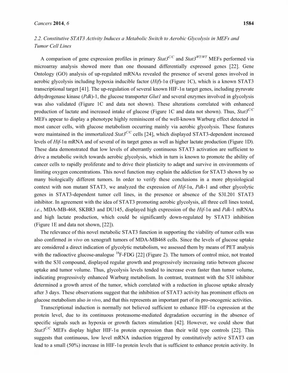
Figure 4. Constitutively active STAT3 regulates mRNA expression of metabolic genes.
Y-P STAT3 is the downstream effector of several oncogenic signals. Constitutively activated
STAT3 translocates into the nucleus where it induces aerobic glycolysis through (i) the
up-regulation of HIF-1α, which in turn mediates the induction of several mRNA involved
in glycolysis; and (ii) the down-regulation of mitochondrial genes and of ETC-complexes,
leading to reduced mitochondrial activity. The heatmaps display the mean log2 fold change
of signal intensity between Stat3C/C and wild type MEFs as assessed by microarray analysis.

Cancers 2014, 6 1589
Despite the decreased expression of about 500 mitochondrial genes and the normal mitochondrial
morphology [22], the mitochondrial mass was increased in the Stat3C/C MEFs [61], suggesting a
potential role of STAT3 in mitochondrial biogenesis. This might occur through the transcriptional
induction of c-myc, a bona fide STAT3 transcriptional target whose levels are increased in the Stat3C/C
MEFs and that is a well-known inducer of mitochondrial biogenesis [62].
2.4. STAT3C Triggers Tumorigenic Transformation in Immortalized MEFs
Similar to primary cells, spontaneously immortalized Stat3C/C MEFs proliferated much faster than their
wild type counterparts, and displayed loss of inhibition contact with a tendency to grow in multi-layers [24],
a feature typical of transformed cells, leading us to assess other transformation phenotypes such as focus
forming ability and anchorage-independent growth. In contrast to their wild type controls, immortalized
Stat3C/C MEFs were able to give rise to foci on plastic and to colonies in soft agar. HIF activity was
only partially responsible for these features, which could not be completely reverted by Hif-1α
silencing (Figure 5A and [24]). Finally, immortal Stat3C/C MEFs were able to give rise to tumors in
nude mice, confirming full malignant transformation. In vivo growth was totally STAT3-dependent
and only in part mediated by HIF-1α, as shown by silencing experiments (Figure 5B and [24]).
Figure 5. Immortalized Stat3C/C MEFs were silenced (white bars) or not (black bars) for
Hif-1α and their ability to form foci and colonies in soft agar was measured (A).
Immortalized MEFs of the indicated genotypes and treatments were tested for their ability
to grow in vivo in nude mice (B). Bars represent mean values ± SEM of three independent
experiments. Modified from reference [24], according to the journal’s license terms.
In contrast to primary MEFs, which require two oncogenic hits to become transformed, MEFs
spontaneously immortalized via the 3T3 protocol [63] are known to become competent for
transformation elicited by a single oncogene [64], suggesting that they have already undergone one
oncogenic hit. Our results show that low levels of constitutively active STAT3 are sufficient to transform
primary cells to full malignancy upon immortalization via a 3T3 protocol, suggesting therefore that
chronic STAT3 activity can act as a first hit in multistep carcinogenesis. This observation might
explain the key role of STAT3 in the causative link between chronic inflammation and cancer, since
IL-6-driven STAT3 constitutive activity is a hallmark of chronic inflammation [65]. According to our
data, cells exposed to chronic IL-6 signalling and displaying continuous STAT3 activation behave like

Cancers 2014, 6 1590
cells that have undergone a first oncogenic mutation, and will therefore be exquisitely sensitive to
mutagenic events occurring in the inflammatory microenvironment, rich in cytokines, growth factors
and reactive oxygen species.
Thus, there are many mechanism(s) through which constitutively active STAT3 sensitizes cells to
tumorigenic transformation, belonging to classical well described functions as well as to the novel
metabolic role that we have recently described. A prominent feature is no doubt played by the resistance to
programmed cell death in response to many different apoptotic stimuli. This is a well-known function of
STAT3, triggered also in our system by the transcriptional induction of anti-apoptotic genes as well as by
the reduced mitochondrial Ca2+ uptake [22,24]. The delayed senescence correlating with decreased
ROS production [22] and the increased proliferation here described likely correlate with the metabolic
switch towards aerobic glycolysis. Aerobic glycolysis is known to allow rapidly proliferating cells to
accumulate NADPH and carbon skeletons needed for anabolic reactions as a side-product of glucose
consumption and ATP production [15]. Additionally, decreased mitochondrial activity will help
switching the energy balance towards glycolysis allowing at the same time to reduce ROS production,
which will in turn help cell survival. Another important player in oncogenesis is c-myc, which
contributes to both increased proliferation and aerobic glycolysis and is a well known STAT3 target.
Indeed the levels of c-myc are elevated in the Stat3C/C MEFs [66]. Finally, Hif-1α induction may
initiate the already mentioned feed forward loop involving PKM2 and STAT3 that would contribute
maintaining high HIF-1α activity, high PKM2:PKM1 ratios and constitutive STAT3 Y-P [26].
3. Conclusions
STAT3 constitutive activation is featured by many types of tumors, and mainly occurs downstream
of the activation of several different oncogenic pathways [11,67]. Accordingly, STAT3 is required for
malignant transformation by many different oncogenes that trigger STAT3 Y-P, the prototype being
vSrc, in part by acting on glucose metabolism as described here. On the other hand, also oncogenes of
the RAS family have been shown to require STAT3 to transform cells, and this occurs via
phosphorylation on serine rather than on tyrosine [17,68]. Also S-P STAT3 mediates important
metabolic functions when localized to the mitochondrion, including preservation of mitochondrial
activity, induction of aerobic glycolysis and inhibition of the opening of the mitochondrial permeability
transition pore. Thus, STAT3 appears to be able to promote aerobic glycolysis both from within the
nucleus and the mitochondrion, while it plays opposite roles on mitochondrial activity, depending on
the oncogenic signal and the mode of activation (i.e., Y- versus S-P). To further complicate the picture,
also cytoplasmic, non-phosphorylated STAT3 is implicated in regulating cell survival and energy
metabolism through autophagy inhibition and, finally, STAT3 acetylation/deacetylation status, regulated
by oncogenes, transcriptional co-activators and different classes of HDAC enzymes, can affect the
activities of both Y-P and S-P STAT3. These observations contribute to explain the multiform and
sometimes contrasting activities described for STAT3, since its functions will vary according to cell
type-specific target genes, mode of activation and sub-cellular localization. In turn, any condition
altering the cellular concentration of one specific form of STAT3 is bound to affect the functions of all
other forms by directly or indirectly altering their abundance, as depicted in Figure 6. This consideration
needs to be kept in account when designing STAT3-inhibiting drugs.
Cancers 2014, 6 1591
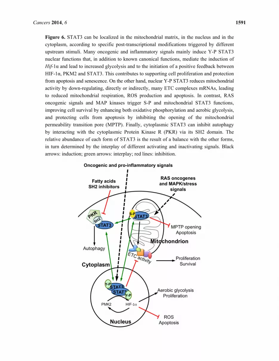
Figure 6. STAT3 can be localized in the mitochondrial matrix, in the nucleus and in the
cytoplasm, according to specific post-transcriptional modifications triggered by different
upstream stimuli. Many oncogenic and inflammatory signals mainly induce Y-P STAT3
nuclear functions that, in addition to known canonical functions, mediate the induction of
Hif-1α and lead to increased glycolysis and to the initiation of a positive feedback between
HIF-1α, PKM2 and STAT3. This contributes to supporting cell proliferation and protection
from apoptosis and senescence. On the other hand, nuclear Y-P STAT3 reduces mitochondrial
activity by down-regulating, directly or indirectly, many ETC complexes mRNAs, leading
to reduced mitochondrial respiration, ROS production and apoptosis. In contrast, RAS
oncogenic signals and MAP kinases trigger S-P and mitochondrial STAT3 functions,
improving cell survival by enhancing both oxidative phosphorylation and aerobic glycolysis,
and protecting cells from apoptosis by inhibiting the opening of the mitochondrial
permeability transition pore (MPTP). Finally, cytoplasmic STAT3 can inhibit autophagy
by interacting with the cytoplasmic Protein Kinase R (PKR) via its SH2 domain. The
relative abundance of each form of STAT3 is the result of a balance with the other forms,
in turn determined by the interplay of different activating and inactivating signals. Black
arrows: induction; green arrows: interplay; red lines: inhibition.

Cancers 2014, 6 1592
A deeper understanding of the interplay between the differentially phosphorylated forms of STAT3
and their relative sub-cellular distribution under specific pathological conditions and in different tumor
types may help designing function-specific inhibitors that may be tested as targeted therapeutic approaches.
Acknowledgments
Work in the authors’ laboratories is supported by grants from the Italian Cancer research
Association (AIRC), the American Italian Cancer Foundation, the Italian Ministry of Health, the Polish
Ministry of Science and Higher Education (W100/HFSC/2011), the Ateneo San Paolo Foundation and
the Truss and Gerrit van Riemsdijk Foundation, Liechtenstein.
Author Contributions
A.C. performed experiments and wrote the manuscript. M.D. was involved in designing the studies and
performed most experiments. C.G. and M.R.W. performed experiments. E.M. contributed to manuscript
writing. P.P. and M.R.W. were involved in the experimental design and discussion. V.P. conceived the
study and edited the text and figures.
Conflicts of Interest
The authors declare no conflict of interest.
References
1. Chung, J.; Uchida, E.; Grammer, T.C.; Blenis, J. STAT3 serine phosphorylation by ERK-dependent
and -independent pathways negatively modulates its tyrosine phosphorylation. Mol. Cell. Biol.
1997, 17, 6508–6516.
2. Yokogami, K.; Wakisaka, S.; Avruch, J.; Reeves, S.A. Serine phosphorylation and maximal activation
of STAT3 during CNTF signaling is mediated by the rapamycin target mTOR. Curr. Biol. 2000, 10,
47–50.
3. Aznar, S.; Valeron, P.F.; del Rincon, S.V.; Perez, L.F.; Perona, R.; Lacal, J.C. Simultaneous tyrosine
and serine phosphorylation of STAT3 transcription factor is involved in Rho A GTPase oncogenic
transformation. Mol. Biol. Cell 2001, 12, 3282–3294.
4. Kubo, M.; Hanada, T.; Yoshimura, A. Suppressors of cytokine signaling and immunity.
Nat. Immunol. 2003, 4, 1169–1176.
5. Shuai, K.; Liu, B. Regulation of gene-activation pathways by PIAS proteins in the immune system.
Nat. Rev. Immunol. 2005, 5, 593–605.
6. Haura, E.B.; Turkson, J.; Jove, R. Mechanisms of disease: Insights into the emerging role of
signal transducers and activators of transcription in cancer. Nat. Clin. Pract. Oncol. 2005, 2, 315–324.
7. Bromberg, J.F.; Wrzeszczynska, M.H.; Devgan, G.; Zhao, Y.; Pestell, R.G.; Albanese, C.;
Darnell, J.E., Jr. STAT3 as an oncogene. Cell 1999, 98, 295–303.
8. Alvarez, J.V.; Febbo, P.G.; Ramaswamy, S.; Loda, M.; Richardson, A.; Frank, D.A. Identification of a
genetic signature of activated signal transducer and activator of transcription 3 in human tumors.
Cancer Res. 2005, 65, 5054–5062.

Cancers 2014, 6 1593
9. Chiarle, R.; Simmons, W.J.; Cai, H.; Dhall, G.; Zamo, A.; Raz, R.; Karras, J.G.; Levy, D.E.;
Inghirami, G. STAT3 is required for ALK-mediated lymphomagenesis and provides a possible
therapeutic target. Nat. Med. 2005, 11, 623–629.
10. Weinstein, I.B. Cancer. Addiction to oncogenes—The Achilles heal of cancer. Science 2002, 297,
63–64.
11. Yu, H.; Jove, R. The STATs of cancer—New molecular targets come of age. Nat. Rev. Cancer
2004, 4, 97–105.
12. Bromberg, J.; Wang, T.C. Inflammation and cancer: IL-6 and STAT3 complete the link. Cancer Cell
2009, 15, 79–80.
13. Warburg, O. On respiratory impairment in cancer cells. Science 1956, 124, 269–270.
14. DeBerardinis, R.J.; Lum, J.J.; Hatzivassiliou, G.; Thompson, C.B. The biology of cancer:
Metabolic reprogramming fuels cell growth and proliferation. Cell Metab. 2008, 7, 11–20.
15. Vander Heiden, M.G.; Cantley, L.C.; Thompson, C.B. Understanding the Warburg effect: The
metabolic requirements of cell proliferation. Science 2009, 324, 1029–1033.
16. Demaria, M.; Camporeale, A.; Poli, V. STAT3 and metabolism: How many ways to use a single
molecule? Int. J. Cancer 2014, doi:10.1002/ijc.28767.
17. Gough, D.J.; Corlett, A.; Schlessinger, K.; Wegrzyn, J.; Larner, A.C.; Levy, D.E. Mitochondrial
STAT3 supports Ras-dependent oncogenic transformation. Science 2009, 324, 1713–1716.
18. Wegrzyn, J.; Potla, R.; Chwae, Y.J.; Sepuri, N.B.; Zhang, Q.; Koeck, T.; Derecka, M.;
Szczepanek, K.; Szelag, M.; Gornicka, A.; et al. Function of mitochondrial STAT3 in cellular
respiration. Science 2009, 323, 793–797.
19. Boengler, K.; Hilfiker-Kleiner, D.; Heusch, G.; Schulz, R. Inhibition of permeability transition
pore opening by mitochondrial STAT3 and its role in myocardial ischemia/reperfusion.
Basic Res. Cardiol. 2010, 105, 771–785.
20. Tammineni, P.; Anugula, C.; Mohammed, F.; Anjaneyulu, M.; Larner, A.C.; Sepuri, N.B. The
import of the transcription factor STAT3 into mitochondria depends on GRIM-19, a component
of the electron transport chain. J. Biol. Chem. 2013, 288, 4723–4732.
21. Zheng, H.; Li, S.; Hsu, P.; Qu, C.K. Induction of a tumor-associated activating mutation in protein
tyrosine phosphatase Ptpn11 (Shp2) enhances mitochondrial metabolism, leading to oxidative
stress and senescence. J. Biol. Chem. 2013, 288, 25727–25738.
22. Demaria, M.; Giorgi, C.; Lebiedzinska, M.; Esposito, G.; D’Angeli, L.; Bartoli, A.; Gough, D.J.;
Turkson, J.; Levy, D.E.; Watson, C.J.; et al. A STAT3-mediated metabolic switch is involved in
tumour transformation and STAT3 addiction. Aging (Albany NY) 2010, 2, 823–842.
23. Pawlus, M.R.; Wang, L.; Murakami, A.; Dai, G.; Hu, C.J. STAT3 or USF2 contributes to Hif
target gene specificity. PLoS One 2013, 8, e72358.
24. Demaria, M.; Misale, S.; Giorgi, C.; Miano, V.; Camporeale, A.; Campisi, J.; Pinton, P.; Poli, V.
STAT3 can serve as a hit in the process of malignant transformation of primary cells. Cell Death
Differ. 2012, 19, 1390–1397.
25. Gao, X.; Wang, H.; Yang, J.J.; Liu, X.; Liu, Z.R. Pyruvate kinase M2 regulates gene transcription
by acting as a protein kinase. Mol. Cell 2012, 45, 598–609.
26. Demaria, M.; Poli, V. PKM2, STAT3 and Hif-1alpha: The Warburg’s vicious circle. JAKSTAT
2012, 1, 194–196.

Cancers 2014, 6 1594
27. Zhao, S.; Xu, W.; Jiang, W.; Yu, W.; Lin, Y.; Zhang, T.; Yao, J.; Zhou, L.; Zeng, Y.; Li, H.; et al.
Regulation of cellular metabolism by protein lysine acetylation. Science 2010, 327, 1000–1004.
28. Yuan, Z.L.; Guan, Y.J.; Chatterjee, D.; Chin, Y.E. STAT3 dimerization regulated by reversible
acetylation of a single lysine residue. Science 2005, 307, 269–273.
29. Lee, J.L.; Wang, M.J.; Chen, J.Y. Acetylation and activation of STAT3 mediated by nuclear
translocation of CD44. J. Cell Biol. 2009, 185, 949–957.
30. Lee, H.; Zhang, P.; Herrmann, A.; Yang, C.; Xin, H.; Wang, Z.; Hoon, D.S.; Forman, S.J.; Jove, R.;
Riggs, A.D.; et al. Acetylated STAT3 is crucial for methylation of tumor-suppressor gene
promoters and inhibition by resveratrol results in demethylation. Proc. Natl. Acad. Sci. USA 2012,
109, 7765–7769.
31. Nie, Y.; Erion, D.M.; Yuan, Z.; Dietrich, M.; Shulman, G.I.; Horvath, T.L.; Gao, Q. STAT3 inhibition
of gluconeogenesis is downregulated by SirT1. Nat. Cell Biol. 2009, 11, 492–500.
32. Bernier, M.; Paul, R.K.; Martin-Montalvo, A.; Scheibye-Knudsen, M.; Song, S.; He, H.J.;
Armour, S.M.; Hubbard, B.P.; Bohr, V.A.; Wang, L.; et al. Negative regulation of STAT3
protein-mediated cellular respiration by SIRT1 protein. J. Biol. Chem. 2011, 286, 19270–19279.
33. Finley, L.W.; Carracedo, A.; Lee, J.; Souza, A.; Egia, A.; Zhang, J.; Teruya-Feldstein, J.; Moreira,
P.I.; Cardoso, S.M.; Clish, C.B.; et al. SIRT3 opposes reprogramming of cancer cell metabolism
through Hif 1alpha destabilization. Cancer Cell 2011, 19, 416–428.
34. Horak, P.; Crawford, A.R.; Vadysirisack, D.D.; Nash, Z.M.; deYoung, M.P.; Sgroi, D.; Ellisen, L.W.
Negative feedback control of Hif-1 through REDD1-regulated ROS suppresses tumorigenesis.
Proc. Natl. Acad. Sci. USA 2010, 107, 4675–4680.
35. Yang, F.; Zhang, W.; Li, D.; Zhan, Q. Gadd45a suppresses tumor angiogenesis via inhibition of
the mTOR/STAT3 protein pathway. J. Biol. Chem. 2013, 288, 6552–6560.
36. Goncharova, E.A.; Goncharov, D.A.; Damera, G.; Tliba, O.; Amrani, Y.; Panettieri, R.A., Jr.;
Krymskaya, V.P. Signal transducer and activator of transcription 3 is required for
abnormal proliferation and survival of TSC2-deficient cells: Relevance to pulmonary
lymphangioleiomyomatosis. Mol. Pharm. 2009, 76, 766–777.
37. Zhou, J.; Wulfkuhle, J.; Zhang, H.; Gu, P.; Yang, Y.; Deng, J.; Margolick, J.B.; Liotta, L.A.;
Petricoin, E., III; Zhang, Y. Activation of the PTEN/mTOR/STAT3 pathway in breast cancer
stem-like cells is required for viability and maintenance. Proc. Natl. Acad. Sci. USA 2007, 104,
16158–16163.
38. Roy, S.; Debnath, J. Autophagy and tumorigenesis. Semin. Immunopathol. 2010, 32, 383–396.
39. Shen, S.; Niso-Santano, M.; Adjemian, S.; Takehara, T.; Malik, S.A.; Minoux, H.; Souquere, S.;
Marino, G.; Lachkar, S.; Senovilla, L.; et al. Cytoplasmic STAT3 represses autophagy by
inhibiting PKR activity. Mol. Cell 2012, 48, 667–680.
40. Barbieri, I.; Pensa, S.; Pannellini, T.; Quaglino, E.; Maritano, D.; Demaria, M.; Voster, A.;
Turkson, J.; Cavallo, F.; Watson, C.J.; et al. Constitutively active STAT3 enhances neu-mediated
migration and metastasis in mammary tumors via upregulation of Cten. Cancer Res. 2010, 70,
2558–2567.

Cancers 2014, 6 1595
41. Niu, G.; Briggs, J.; Deng, J.; Ma, Y.; Lee, H.; Kortylewski, M.; Kujawski, M.; Kay, H.;
Cress, W.D.; Jove, R.; et al. Signal transducer and activator of transcription 3 is required for
hypoxia-inducible factor-1alpha RNA expression in both tumor cells and tumor-associated
myeloid cells. Mol. Cancer Res. 2008, 6, 1099–1105.
42. Salceda, S.; Caro, J. Hypoxia-inducible factor 1alpha (Hif-1alpha) protein is rapidly degraded by
the ubiquitin-proteasome system under normoxic conditions. Its stabilization by hypoxia depends
on redox-induced changes. J. Biol. Chem. 1997, 272, 22642–22647.
43. Dolled-Filhart, M.; Camp, R.L.; Kowalski, D.P.; Smith, B.L.; Rimm, D.L. Tissue microarray analysis
of signal transducers and activators of transcription 3 (STAT3) and phospho-STAT3 (Tyr705) in
node-negative breast cancer shows nuclear localization is associated with a better prognosis.
Clin. Cancer Res. 2003, 9, 594–600.
44. Pectasides, E.; Egloff, A.M.; Sasaki, C.; Kountourakis, P.; Burtness, B.; Fountzilas, G.; Dafni, U.;
Zaramboukas, T.; Rampias, T.; Rimm, D.; et al. Nuclear localization of signal transducer and
activator of transcription 3 in head and neck squamous cell carcinoma is associated with a better
prognosis. Clin. Cancer Res. 2010, 16, 2427–2434.
45. Musteanu, M.; Blaas, L.; Mair, M.; Schlederer, M.; Bilban, M.; Tauber, S.; Esterbauer, H.;
Mueller, M.; Casanova, E.; Kenner, L.; et al. STAT3 is a negative regulator of intestinal tumor
progression in Apc(Min) mice. Gastroenterology 2010, 138, 1003–1011.
46. Couto, J.P.; Daly, L.; Almeida, A.; Knauf, J.A.; Fagin, J.A.; Sobrinho-Simoes, M.; Lima, J.;
Maximo, V.; Soares, P.; Lyden, D.; et al. STAT3 negatively regulates thyroid tumorigenesis.
Proc. Natl. Acad. Sci. USA 2012, 109, E2361–E2370.
47. Bertout, J.A.; Patel, S.A.; Simon, M.C. The impact of O2 availability on human cancer.
Nat. Rev. Cancer 2008, 8, 967–975.
48. Marchi, S.; Pinton, P. The mitochondrial calcium uniporter complex: Molecular components,
structure and physiopathological implications. J. Physiol. 2014, 592 Pt 5, 829–839.
49. Clapham, D.E. Calcium signaling. Cell 2007, 131, 1047–1058.
50. Giorgi, C.; de Stefani, D.; Bononi, A.; Rizzuto, R.; Pinton, P. Structural and functional link
between the mitochondrial network and the endoplasmic reticulum. Int. J. Biochem. Cell Biol.
2009, 41, 1817–1827.
51. Rizzuto, R.; Pozzan, T. Microdomains of intracellular Ca2+: Molecular determinants and functional
consequences. Physiol. Rev. 2006, 86, 369–408.
52. Marchi, S.; Patergnani, S.; Pinton, P. The endoplasmic reticulum-mitochondria connection: One
touch, multiple functions. Biochim. Biophys. Acta 2014, 1837, 461–469.
53. Giorgi, C.; Baldassari, F.; Bononi, A.; Bonora, M.; de Marchi, E.; Marchi, S.; Missiroli, S.;
Patergnani, S.; Rimessi, A.; Suski, J.M.; et al. Mitochondrial Ca2+ and apoptosis. Cell Calcium
2012, 52, 36–43.
54. Bonora, M.; Bononi, A.; de Marchi, E.; Giorgi, C.; Lebiedzinska, M.; Marchi, S.; Patergnani, S.;
Rimessi, A.; Suski, J.M.; Wojtala, A.; et al. Role of the c subunit of the FO ATP synthase in
mitochondrial permeability transition. Cell Cycle 2013, 12, 674–683.
55. Bonora, M.; Giorgi, C.; Bononi, A.; Marchi, S.; Patergnani, S.; Rimessi, A.; Rizzuto, R.; Pinton, P.
Subcellular calcium measurements in mammalian cells using jellyfish photoprotein aequorin-based
probes. Nat. Protoc. 2013, 8, 2105–2118.

Cancers 2014, 6 1596
56. Monteleone, M.; Camporeale, A.; Poli, V. Molecular Biotechnology Center and Department of
Molecular Biotechnology and Life Sciences, University of Turin, Turin, Italy. Personal
observation, 2014.
57. Wang, B.; Hsu, S.H.; Frankel, W.; Ghoshal, K.; Jacob, S.T. STAT3-mediated activation of
microRNA-23a suppresses gluconeogenesis in hepatocellular carcinoma by down-regulating
glucose-6-phosphatase and peroxisome proliferator-activated receptor gamma, coactivator 1 alpha.
Hepatology 2012, 56, 186–197.
58. Austin, S.; Klimcakova, E.; St-Pierre, J. Impact of PGC-1alpha on the topology and rate of
superoxide production by the mitochondrial electron transport chain. Free Radic. Biol. Med. 2011,
51, 2243–2248.
59. Wenz, T.; Rossi, S.G.; Rotundo, R.L.; Spiegelman, B.M.; Moraes, C.T. Increased muscle PGC-1alpha
expression protects from sarcopenia and metabolic disease during aging. Proc. Natl. Acad. Sci. USA
2009, 106, 20405–20410.
60. St-Pierre, J.; Drori, S.; Uldry, M.; Silvaggi, J.M.; Rhee, J.; Jager, S.; Handschin, C.; Zheng, K.;
Lin, J.; Yang, W.; et al. Suppression of reactive oxygen species and neurodegeneration by the
PGC-1 transcriptional coactivators. Cell 2006, 127, 397–408.
61. Poli, V.; Wieckowski, M.R. Molecular Biotechnology Center and Department of Molecular
Biotechnology and Life Sciences, University of Turin, Turin, Italy (V.P.); Nencki Institute of
Experimental Biology, Department of Biochemistry, Warsaw, Poland (M.R.W.). Personal
observation, 2014.
62. Li, F.; Wang, Y.; Zeller, K.I.; Potter, J.J.; Wonsey, D.R.; O’Donnell, K.A.; Kim, J.W.; Yustein, J.T.;
Lee, L.A.; Dang, C.V. Myc stimulates nuclearly encoded mitochondrial genes and mitochondrial
biogenesis. Mol. Cell Biol. 2005, 25, 6225–6234.
63. Todaro, G.J.; Green, H. Quantitative studies of the growth of mouse embryo cells in culture and
their development into established lines. J. Cell Biol. 1963, 17, 299–313.
64. Krontiris, T.G.; Cooper, G.M. Transforming activity of human tumor DNAs. Proc. Natl. Acad.
Sci. USA 1981, 78, 1181–1184.
65. Hodge, D.R.; Hurt, E.M.; Farrar, W.L. The role of IL-6 and STAT3 in inflammation and cancer.
Eur. J. Cancer 2005, 41, 2502–2512.
66. Kiuchi, N.; Nakajima, K.; Ichiba, M.; Fukada, T.; Narimatsu, M.; Mizuno, K.; Hibi, M.; Hirano, T.
STAT3 is required for the gp130-mediated full activation of the c-myc gene. J. Exp. Med. 1999,
189, 63–73.
67. Avalle, L.; Pensa, S.; Regis, G.; Novelli, F.; Poli, V. STAT1 and STAT3 in tumorigenesis: A
matter of balance. JAKSTAT 2012, 1, 65–72.
68. Bromberg, J.F.; Horvath, C.M.; Besser, D.; Lathem, W.W.; Darnell, J.E., Jr. STAT3 activation is
required for cellular transformation by v-src. Mol. Cell Biol. 1998, 18, 2553–2558.
© 2014 by the authors; licensee MDPI, Basel, Switzerland. This article is an open access article
distributed under the terms and conditions of the Creative Commons Attribution license
(http://creativecommons.org/licenses/by/3.0/).