Embed Size (px)
Citation preview
Biochemical Pharmacology 79 (2010) 1398–1409
A novel small-molecule disrupts Stat3 SH2 domain–phosphotyrosine interactionsand Stat3-dependent tumor processes
Xiaolei Zhang a, Peibin Yue a, Steven Fletcher b, Wei Zhao a, Patrick T. Gunning b, James Turkson a,*a Burnett School of Biomedical Sciences, University of Central Florida College of Medicine, 6900 Lake Nona Boulevard, Orlando, FL 32827, USAb Department of Chemistry, University of Toronto at Mississauga, Mississauga, ON, L5L 1C6, Canada
A R T I C L E I N F O
Article history:
Received 12 October 2009
Accepted 4 January 2010
Keywords:
Stat3
S3I-201
S3I-201.1066
Small-molecule inhibitor
Antitumor effects
Antitumor cell effects
A B S T R A C T
The molecular modeling of the phosphotyrosine (pTyr)–SH2 domain interaction in the Stat3:Stat3
dimerization, combined with in silico structural analysis of the Stat3 dimerization disruptor, S3I-201, has
furnished a diverse set of analogs. We present evidence from in vitro biochemical and biophysical studies
that the structural analog, S3I-201.1066 directly interacts with Stat3 or the SH2 domain, with an affinity
(KD) of 2.74 mM, and disrupts the binding of Stat3 to the cognate pTyr–peptide, GpYLPQTV–NH2, with an
IC50 of 23 mM. Moreover, S3I-201.1066 selectively blocks the association of Stat3 with the epidermal
growth factor receptor (EGFR), and inhibits Stat3 tyrosine phosphorylation and nuclear translocation in
EGF-stimulated mouse fibroblasts. In cancer cells that harbor aberrant Stat3 activity, S3I-201.1066
inhibits constitutive Stat3 DNA-binding and transcriptional activities. By contrast, S3I-201.1066 has no
effect on Src activation or the EGFR-mediated activation of the Erk1/2MAPK pathway. S3I-201.1066
selectively suppresses the viability, survival, and malignant transformation of the human breast and
pancreatic cancer lines and the v-Src-transformed mouse fibroblasts harboring persistently active Stat3.
Treatment with S3I-201.1066 of malignant cells harboring aberrantly active Stat3 down-regulated the
expression of c-Myc, Bcl-xL, Survivin, the matrix metalloproteinase 9, and VEGF. The in vivo
administration of S3I-201.1066-induced significant antitumor response in mouse models of human
breast cancer, which correlates with the inhibition of constitutively active Stat3 and the suppression of
known Stat3-regulated genes. Our studies identify a novel small-molecule that binds with a high affinity
to Stat3, blocks Stat3 activation and function, and thereby induces antitumor response in human breast
tumor xenografts harboring persistently active Stat3.
� 2010 Elsevier Inc. All rights reserved.
Contents lists available at ScienceDirect
Biochemical Pharmacology
journa l homepage: www.e lsev ier .com/ locate /b iochempharm
1. Introduction
Signal transduction proteins have increased importance incarcinogenesis and tumor formation and represent attractivetargets for the development of novel anticancer therapeutics. Thesignal transducer and activator of transcription (STAT) family ofproteins are cytoplasmic transcription factors with important rolesin the responses to cytokines and growth factors, includingpromoting cell growth and differentiation, and inflammation andimmune responses [1,2]. Normal STAT’s activation is initiated bythe phosphorylation of a critical tyrosine residue upon the bindingof cytokines or growth factors to cognate receptors. STAT’sphosphorylation is induced by growth factor receptor tyrosinekinases, or cytoplasmic tyrosine kinases, such as Janus kinases(Jaks) and Src family kinases. While pre-existing STAT dimers havebeen detected [3,4], studies show that phosphorylation induces
* Corresponding author. Tel.: +1 407 266 7031; fax: +1 407 384 2062.
E-mail address: [email protected] (J. Turkson).
0006-2952/$ – see front matter � 2010 Elsevier Inc. All rights reserved.
doi:10.1016/j.bcp.2010.01.001
dimerization between two STAT monomers through a phospho-tyrosine interaction with the SH2 domain. In the nucleus, activeSTAT dimers bind to specific DNA-response elements in thepromoters of target genes and regulate gene expression. NormalSTAT activation is transient in accordance with physiologicalresponses. However, the persistent activation of certain STATfamily members, including Stat3 is frequently observed in manyhuman tumors. It is now well established that aberrant activationof Stat3 contributes to malignant transformation and tumorigene-sis. Evidence shows that persistently active Stat3 mediatesoncogenesis and tumor formation in part by the upregulation ofthe expression of critical genes, the dysregulation of cell growthand survival, the promotion of angiogenesis [2,5–11], and theinduction of tumor immune-tolerance [12,13]. Thus, the targetingof aberrant Stat3 signaling provides a novel strategy for treatingthe wide variety of human tumors that harbor abnormal Stat3activity.
The critical step of dimerization [14] between two monomerswithin the context of STAT activation presents an attractivestrategy to interfere with Stat3 signaling and functions and this
X. Zhang et al. / Biochemical Pharmacology 79 (2010) 1398–1409 1399
approach has been exploited in prior work [15–25]. Leadingagents from those earlier studies have been explored in therational design of optimized molecules, in conjunction withmolecular modeling of their binding to the Stat3 SH2 domain[18,19], per the X-ray crystal structure of the Stat3b homodimer[26]. One of those leads, S3I-201 [18] had previously beenshown to exert antitumor effects against human breast cancerxenografts via mechanisms that involve the inhibition ofaberrant Stat3 activity.
In the present study, key structural information from thecomputational modeling of S3I-201 bound to the Stat3 SH2 domainfacilitated the design of novel analogs of which S3I-201.1066shows an improved Stat3-inhibitory activity. S3I-210.1066 inhibitsStat3 DNA-binding activity with an IC50 value of 35 mM. Currentstudies provide evidence that S3I-201.1066 directly interacts withthe Stat3 protein in vitro, thereby disrupting Stat3 binding tocognate pTyr peptide motifs of receptors and inhibiting Stat3phosphorylation and activation, and Stat3 nuclear localization.Furthermore, evidence is provided that S3I-201.1066 selectivelyinduces antitumor cell effects in human breast and pancreaticcancer cells, and mouse transformed fibroblasts harboring aber-rant Stat3 activity, and inhibits growth of human breast tumors inxenografts.
2. Materials and methods
2.1. Cells and reagents
Normal mouse fibroblasts (NIH3T3) and counterparts trans-formed by v-Src (NIH3T3/v-Src), v-Ras (NIH3T3/v-Ras) or over-expressing the human epidermal growth factor (EGF) receptor(NIH3T3/hEGFR), and the human breast cancer (MDA-MB-231)and pancreatic cancer (Panc-1) cells have all been previouslyreported [15,27–29]. The normal human pancreatic duct epithelialcells (HPDEC) were a kind gift from Dr. Tsao (OCI, UHN-PMH,Toronto) [30], the Stat3 knockout mouse embryonic fibroblastsline was generously provided by Dr. Valerie Poli (University ofTurin) [31], and the ovarian cancer line, A2780S was a kind giftfrom Dr. Jin Q. Cheng (Moffitt Cancer Center and ResearchInstitute). The Stat3-dependent reporter, pLucTKS3 and theStat3-independent reporter, pLucSRE, and the v-Src transformedmouse fibroblasts that stably express pLucTKS3 (NIH3T3/v-Src/pLucTKS3) have all been previously reported [15,32,33]. Cells weregrown in Dulbecco’s modified Eagle’s medium (DMEM) containing10% heat-inactivated fetal bovine serum, or in the case of HPDEC,they were grown in keratinocyte-SFM (GIBCO, Invitrogen Corp.,Carlsbad, CA) supplemented with 0.2 ng EGF and 30 mg/ml bovinepituitary extract, and containing antimycol. Antibodies used areagainst Stat3, pY705Stat3, Src, pY416Src, Jak1, pJak1, Shc, pShc,Erk1/2, pErk1/2, and Survivin from Cell Signaling Technology(Danvers, MA), and anti-EGFR and anti-VEGF from Santa CruzBiotech (Santa Cruz, CA).
2.2. Cloning and protein expression
The coding regions for the murine Stat3 protein and the Stat3SH2 domain were amplified by PCR and cloned into vectors pET-44Ek/LIC (Novagen, EMD Chemicals, Gibbstown, NJ) and pET SUMO(Invitrogen), respectively. The primers used for amplificationwere: Stat3 Forward: GACGACGACAAGATGGCTCAGTGGAAC-CAGCTGC; Stat3 Reverse: GAGGAGAAGCCCGGTTATCACATGGGG-GAGGTAGCACACT; Stat3 SH2 Forward: ATGGGTTTCATCAGC-AAGGA; Stat3 SH2 Reverse: TCACCTACAGTACTTTCCAAATGC.Clones were sequenced to verify the correct sequences andorientation. His-tagged recombinant proteins were expressed inBL21 (DE3) cells and purified on Ni-ion sepharose column.
2.3. Nuclear extract preparation, gel shift assays, and densitometric
analysis
Nuclear extract preparations and electrophoretic mobility shiftassay (EMSA) were carried out as previously described [28,33]. The32P-labeled oligonucleotide probes used were hSIE (high affinitysis-inducible element from the c-fos gene, m67 variant, 50-AGCTTCATTTCCCGTAAATCCCTA) that binds Stat1 and Stat3 [34]and MGFe (mammary gland factor element from the bovine b-casein gene promoter, 50-AGATTTCTAGGAATTCAA) for Stat1 andStat5 binding [35,36]. Except where indicated, nuclear extractswere pre-incubated with compound for 30 min at room tempera-ture prior to incubation with the radiolabeled probe for 30 min at30 8C before subjecting to EMSA analysis. Bands corresponding toDNA-binding activities were scanned and quantified for eachconcentration of compound using ImageQuant and plotted aspercent of control (vehicle) against concentration of compound,from which the IC50 values were derived, as previously reported[37].
2.4. Immunoprecipitation, immunoblotting and densitometric
analyses
Immunoprecipitation from whole-cell lysates, and tumor tissuelysate preparation, and immunoblotting analysis were performedas previously described [17,18,33,38]. Primary antibodies usedwere anti-Stat3, pY705Stat3, pY416Src, Src, pErk1/2, Erk1/2, pJak1,Jak1, pShc, Shc, Grb 2, c-Myc, Bcl-xL, Survivin, MMP-9, and b-actin(Cell Signaling), and VEGF (Santa Cruz Biotech.).
2.5. Cell viability and proliferation assay
Cells in culture in 6-well or 96-well plates were treated with orwithout S3I-201.1066 for 24–144 h and subjected to CyQuant cellproliferation assay (Invitrogen Corp./Life Technologies Corp.), orharvested, and the viable cells counted by trypan blue exclusionwith phase-contrast microscopy.
2.6. Immunofluorescence imaging/confocal microscopy
NIH3T3/hEGFR cells were grown in multi-cell plates, serum-starved for 8 h and treated with or without S3I-201.1066 for30 min prior to stimulation by rhEGF (1 mg/ml) for 10 min. Cellswere fixed with ice-cold methanol for 15 min, washed 3 times inphosphate buffered saline (PBS), permeabilized with 0.2% Triton X-100 for 10 min, and further washed 3–4 times with PBS. Specimenswere then blocked in 1% bovine serum albumin (BSA) for 30 minand incubated with anti-EGFR (Santa Cruz) or anti-Stat3 (CellSignaling) antibody at 1:50 dilution at 4 8C overnight. Subsequent-ly, cells were rinsed 4–5 times in PBS, incubated with Alexa fluor546 rat antibody for EGFR detection and Alexa fluor 488 rabbitantibody for Stat3 detection (Invitrogen) for 1 h at roomtemperature in the dark. Specimens were then washed 5 timeswith PBS, covered with cover slides with VECTASHIELD mountingmedium containing DAPI (Vector Lab, Inc., Burlingame, CA), andexamined immediately under a Leica TCS SP5 confocal microscope(Germany) at the appropriate wavelengths. Images were capturedand processed using the Leica TCS SP 5 software.
2.7. Soft-agar colony formation assay
Colony formation assays were carried out in 6-well dishes, asdescribed previously [16,37]. Briefly, each well contained 1.5 ml of1% agarose in Dulbeco’s modified Eagle’s medium as the bottomlayer and 1.5 ml of 0.5% agarose in Dulbeco’s modified Eagle’smedium containing 4–6 � 103 NIH3T3/v-Src, NIH3T3/v-Ras,

X. Zhang et al. / Biochemical Pharmacology 79 (2010) 1398–14091400
A2780S, MDA-MB-231 or Panc-1 cells, as the top layer. Treatmentwith S3I-201.1066 was initiated 1 day after seeding cells by adding80 ml of medium with or without S3I-201.1066, and repeatingevery 2 or 3 days, until large colonies were evident. Colonies werequantified by staining with 20 ml of 1 mg/ml crystal violet(ThermoFisher, Waltham, MA), incubating at 37 8C overnight,and counting the next day under phase-contrast microscope.
2.8. Fluorescence polarization assay
Fluorescence polarization (FP) assay was conducted as previ-ously reported [23], with some modification using the phospho-peptide, 5-carboxyfluorescein-GpYLPQTV–NH2 (where pYrepresents phospho-Tyr) as probe and Stat3. A fixed concentrationof the fluorescently labeled peptide probe (10 nM) was incubatedwith an increasing concentration of the Stat3 protein for 30 min atroom temperature in the buffer, 50 mM NaCl, 10 mM HEPES, 1 mMEDTA, 0.1% Nonidet P-40, and the fluorescent polarizationmeasurements were determined using the POLARstar Omega(BMG LABTECH, Durham, NC), with the set gain adjustment at35 mP. The Z0 value was derived per the equationZ0 = 1 � (3SDbound + 3SDfree)/(mPbound �mPfree), where SD is thestandard deviation and mP is the average of fluorescencepolarization. In the ‘‘bound’’ state, 10 nM 5-carboxyfluorescein-GpYLPQTV–NH2 was incubated with 150 nM purified Stat3protein, while the ‘‘free’’ (unbound) state represents the samemixture, but incubated with an additional 10 mM unlabeled Ac-GpYLPQTV–NH2. For evaluating agents, Stat3 protein (150 nM)was incubated with serial concentrations of S3I-201.1066 at 30 8Cfor 60 min in the indicated assay buffer conditions. Prior to theaddition of the fluorescent probe, the protein:S3I-201.1066mixtures were allowed to equilibrate at room temperature for15 min. Probe was added at a final concentration of 10 nM andincubated for 30 min at room temperature following which the FPmeasurements were taken using the POLARstar Omega, with theset gain adjustment at 35 mP.
2.9. Surface plasmon resonance analysis
SensiQ and its analysis software Qdat (ICX Technologies,Oklahoma City, OK) were used to analyze the interaction betweenthe agent and the Stat3 protein and to determine the bindingaffinity. Purified Stat3 was immobilized on a HisCap Sensor Chip byinjecting 50 mg/ml of Stat3 onto the chip. Various concentrationsof S3I-201.1066 in running buffer (1� PBS, 0.5% DMSO) werepassed over the sensor chip to produce response signals. Theassociation and dissociation rate constants were calculated usingthe Qdat software. The ratio of the association and dissociation rateconstants was determined as the affinity (KD).
2.10. Colony survival assay
This was performed as previously reported [39]. Briefly, cellswere seeded as single-cell in 6-cm dishes (500 cells per well),treated once the next day with S3I-201.1066 for 48 h, and allowedto culture until large colonies were visible. Colonies were stainedwith crystal violet (ThermoFisher) for 4 h and counted underphase-contrast microscope.
2.11. Wound healing assay for migration
Wounds were made using pipette tips in monolayer cultures ofcells in 6-well plates. Cells were treated with or without increasingconcentrations of S3I-201.1066 and allowed to migrate into thedenuded area for 12–24 h. The migration of cells was visualized ata 10� magnification using an Axiovert 200 Inverted Fluorescence
Microscope (Zeiss, Gottingen, Germany), with pictures taken usinga mounted Canon Powershot A640 digital camera (Canon USA,Lake Success, NY). Cells that migrated into the denuded area werequantified.
2.12. Mice and in vivo tumor studies
Six-week-old female athymic nude mice were purchased fromHarlan and maintained in the institutional animal facilitiesapproved by the American Association for Accreditation ofLaboratory Animal Care. Athymic nude mice were injectedsubcutaneously in the left flank area with 5 � 106 human breastcancer MDA-MB-231 cells in 100 mL of PBS. After 5–10 days,tumors of a diameter of 3 mm were established. Animals weregrouped so that the mean tumor sizes in all groups were nearlyidentical, then given S3I-201.1066, i.v. at 3 mg kg�1 every 2 orevery 3 days for 17 days and monitored every 2 or 3 days, andtumor sizes were measured with calipers. Tumor volume, V, wascalculated according to the formula V = 0.52 � a2 � b, where a,smallest superficial diameter, b, largest superficial diameter. Foreach treatment group, the tumor volumes for each set ofmeasurements were statistically analyzed in comparison to thecontrol (non-treated) group. Upon completion of the study, tumorswere extracted and tumor tissue lysates were prepared forimmunoblotting and gel shit analyses.
2.13. Statistical analysis
Statistical analysis was performed on mean values using PrismGraphPad Software, Inc. (La Jolla, CA). The significance ofdifferences between groups was determined by the paired t-testat *p < 0.05, **p < 0.01, and ***p < 0.001.
3. Results
3.1. Computer-aided design of S3I-201 analogs as Stat3 inhibitors
Close structural analysis of the lowest genetic optimization forligand docking (GOLD) [40] conformation of the lead Stat3inhibitor, S3I-201 (green) (IC50 = 86 mM for inhibition of Stat3:-Stat3 [18]) (Fig. 1A and C) bound within the Stat3 SH2 domain(Fig. 1C), per the X-ray crystal structure of DNA-bound Stat3bhomodimer [26] showed significant complementary interactionsbetween the protein surface and the compound and identified keystructural requirements for tight binding. Docking studiespermitted in silico structural design of analogs of differing Stat3SH2 domain-binding characteristics in order to derive Stat3inhibitors of improved potency and selectivity. GOLD studiesshowed limited structural occupation of the Stat3 SH2 domain,identifying a potential means for improving inhibitor potency. TheSH2 domain is broadly composed of three sub-pockets, only two ofwhich are accessed by S3I-201 (Fig. 1C). Lead agent, S3I-201(Fig. 1A) has a glycolic acid scaffold with its carboxylic acidcondensed with hetero-trisubstituted aromatic species to furnishthe amide bond, and a hydroxyl moiety that has been tosylated.The ortho-hydroxybenzoic acid component is a known pTyrmimetic, and low energy GOLD studies consistently placed thisunit in the pTyr-binding site, making hydrogen bonds andelectrostatic interactions with Lys591, Ser611, Ser613 andArg609. Due to the strength of such interactions betweenoppositely charged ions, it is likely that a considerable portionof the binding between the SH2 domain and S3I-201 arises fromthe pTyr mimetic. The O-tosyl group binds in the mostlyhydrophobic pocket that is derived from the tetramethyleneportion of the side chain of Lys592 and the trimethylene portion ofthe side chain of Arg595, along with Ile597 and Ile634. Given the

Fig. 1. (A and B) Structures of (A) S3I-201, (B) S3I-201.1066; (C and D) GOLD of (C) S3I-201 (green), and (D) S3I-201 (green) and S3I-201.1066 (yellow) to the SH2 domain of
Stat3; arrow denotes potential binding sub-pocket accessed by S3I-201.1066, but not S3I-201.
X. Zhang et al. / Biochemical Pharmacology 79 (2010) 1398–1409 1401
potency of S3I-201 towards Stat3 inhibition, a rational syntheticprogram was undertaken to modify and optimize the core scaffoldto furnish more potent analogs. We additionally exploited keyhydrophobic interactions with Phe716, Ile659, Val637 and Trp623(Fig. 1C, see arrow) in generating compounds made of N-substituted (para-cyclohexyl)benzyl analogs [41], including S3I-201.1066 (Fig. 1B). Present study of the analog S3I-201.1066(Fig. 1B and D) was undertaken to derive biochemical andbiophysical evidence of binding to Stat3 and to define themechanisms of inhibition of Stat3 and its functions in the contextof Stat3-dependent malignant transformation and tumorigenesis.
3.2. Inhibition of Stat3 DNA-binding activity
S3I-201 analogs derived per in silico structural optimization andmolecular modeling of the binding to the Stat3 SH2 domain weresynthesized and evaluated in Stat3 DNA-binding assay in vitro, aspreviously done [18]. Nuclear extracts containing activated Stat3prepared from v-Src-transformed mouse fibroblasts (NIH3T3/v-Src)that harbor aberrantly active Stat3 were incubated for 30 min atroom temperature with or without increasing concentrations of theanalog, S3I-201.1066, prior to incubation with the radiolabeled hSIEprobe that binds to Stat3 and Stat1 and subjected to electrophoreticmobility shift assay (EMSA) analysis [18]. Stat3 DNA-bindingactivity was inhibited in a dose-dependent manner by S3I-201.1066 (Fig. 2A(i)), with average IC50 value of 35 � 09 mM. Thisvalue represents 2–3-fold improvement over the activity of the leadagent, S3I-201 (IC50 of 86 mM) [18]. For selectivity, nuclear extractscontaining activated Stat1, Stat3 and Stat5 prepared from EGF-stimulated NIH3T3/hEGFR (mouse fibroblasts over-expressing thehuman epidermal growth factor receptor) were pre-incubated at roomtemperature with or without increasing concentrations of S3I-201.1066 for 30 min, prior to incubation with the radiolabeledoligonucleotide probes and subjecting to EMSA analyses, as previously
done [18]. EMSA results of the binding studies using the hSIE probeshow the strongest complex of Stat3:Stat3 with the probe (Fig. 2A(ii)upper band, lanes 1 and 2), which is significantly disrupted at 50 mMS3I-201.1066 and completely disrupted at 100 mM S3I-201.1066(Fig. 2A(ii), upper band, lanes 2 and 3). EMSA analysis further showsa less intense Stat1:Stat3 complex (intermediate band), which issimilarly repressed at 50 mM and completely disrupted at 100 mM S3I-201.1066 (Fig. 2A(ii), lanes 2 and 3). By contrast, we observe nosignificant inhibition of the Stat1:Stat1 complex that is of the lowestintensity (lower band) at 50 mM S3I-201.1066, a moderate inhibition at100 mM S3I-201.1066, but a complete inhibition at 200 mM S3I-201.1066 (Fig. 2A(ii), lower band). Of importance, at the 100 mM S3I-201.1066 concentration at which only a moderate inhibition ofStat1:Stat1 complex occurred, the larger Stat3:Stat3 complex iscompletely dissociated (Fig. 2A(ii), lane 3). Moreover, EMSA analysisshowed no effect on Stat5:Stat5 complex with the MGFe probe, up to300 mM S3I-201.1066 (Fig. 2A(iii)). Thus, S3I-201.1066 preferentiallyinhibits DNA-binding activity of Stat3 over that of Stat1 or Stat5.
3.3. Inhibition of intracellular Stat3 activation
Stat3 is constitutively activated in a variety of malignant cells,including human breast and pancreatic cancer cells [9,10,20].Given the effect against Stat3 DNA-binding activity in vitro, weevaluated S3I-201.1066 in v-Src transformed mouse fibroblasts(NIH3T3/v-Src), human breast cancer (MDA-MB-231) and humanpancreatic cancer (Panc-1) lines that harbor aberrant Stat3 activity.Twenty-four hours after treatment, nuclear extracts were preparedfrom cells and subjected to Stat3 DNA-binding assay in vitro usingthe radiolabeled hSIE probe and analyzed by EMSA. Compared tothe control (0.05% DMSO-treated cells, lane 1), nuclear extractsfrom S3I-201.1066-treated NIH3T3/v-Src, Panc-1 and MDA-MB-231 cells showed dose-dependent decreases of constitutive Stat3activation, with significant inhibition at 50 mM S3I-201.1066

Fig. 2. Effects of S3I-201.1066 on the activities of STATs, Src, Shc, and Erks. (A) Nuclear extracts of equal total protein containing activated Stat1, Stat3, and/or Stat5 were pre-
incubated with or without S3I-201.1066 for 30 min at room temperature prior to the incubation with the radiolabeled (i) and (ii) hSIE probe that binds Stat1 and Stat3 or the
(iii) MGFe probe that binds Stat5 and subjected to EMSA analysis; (B) nuclear extracts of equal total protein prepared from the designated malignant cells following 24-h
treatment with or without S3I-201.1066 were subjected to in vitro DNA-binding assay using the radiolabeled hSIE probe and analyzed by EMSA; (C) cytosolic extracts of equal
total protein were prepared from 24 h, S3I-201.1066-treated or untreated NIH3T3/v-Src fibroblasts that stably express the Stat3-dependent luciferase reporter (pLucTKS3) or
from treated or untreated NIH3T3/v-Src fibroblasts, the human pancreatic (Panc-1) and breast (MDA-MB-231) carcinoma lines that are transiently transfected with pLucSRE
or pLucTKS3 and analyzed for luciferase activity using a luminometer; and (D) SDS-PAGE and Western blotting analysis of whole-cell lysates of equal total protein prepared
from S3I-201.1066-treated or untreated NIH3T3/v-Src, Panc-1 and MDA-MB-231 cells probing for pY705Stat3, Stat3, pY416Src, Src, pShc, Shc, pErk1/2 and Erk1/2. Positions of
STATs:DNA complexes or proteins in gel are labeled; control lanes (0) represent nuclear extracts treated with 0.05% DMSO, or nuclear extracts or whole-cell lysates prepared
from 0.05% DMSO-treated cells; luciferase activities were normalized to b-galactosidase activity. Data are representative of 3–4 independent determinations: **p < 0.05.
X. Zhang et al. / Biochemical Pharmacology 79 (2010) 1398–14091402
(Fig. 2B, compare lanes 2–5, 8–10, and 12–15 to their respectivecontrols (0)). Luciferase reporter studies were performed to furtherdetermine the effect of S3I-201.1066 on Stat3 transcriptionalactivity. Results show that the treatment with S3I-201.1066 of thev-Src transformed mouse fibroblasts (NIH3T3/v-Src) that stablyexpress the Stat3-dependent luciferase reporter (NIH3T3/v-Src/pLucTKS3) [15,32,33] significantly (**p < 0.01) repressed theinduction of the Stat3-dependent reporter (Fig. 2C, left panel,NIH3T3/v-Src/pLucTKS3). Similar results were obtained when thehuman pancreatic cancer, Panc-1 and breast cancer, MDA-MB-231cells harboring aberrant Stat3 activity were transiently transfectedwith the Stat3-dependent reporter, pLucTKS3 and treated with S3I-201.1066 (Fig. 2C, middle and right panels, pLucTKS3). By contrast,a similar treatment of malignant cells that are transientlytransfected with the Stat3-independent luciferase reporter,pLucSRE, which is driven by the serum response element (SRE)of the c-fos promoter, had no observable effect on the reporterinduction (Fig. 2C, pLucSRE). Moreover, immunoblotting analysisshowed a concentration-dependent reduction of pTyr705Stat3levels in NIH3T3/v-Src (Fig. 2D(i), top panel), Panc-1 cells(Fig. 2D(ii), top panel), and MDA-MB-231 (Fig. 2D(iii), top panel)
cells upon treatment with S3I-201.1066 for 24 h, presumablythrough the blockade of Stat3 binding to pTyr motifs of receptorsand the prevention of de novo phosphorylation by tyrosine kinases.By contrast, immunoblotting analysis showed no significant effectsof S3I-201.1066 on the phosphorylation of Src (pY416Src), Shc(pShc), and Erk1/2 (pErk1/2) under the same treatment conditions(Fig. 2D(i)–(iii), panels 2–4 from the top). In spite of the inhibitionof aberrant Stat3 activity, no observable change in total Stat3protein was made, consistent with previous reports [18,19]. Also,total Src, Shc and Erk1/2 protein levels remained unchanged. Weinfer that at the concentrations that inhibit Stat3 activity, S3I-201.1066 has minimal effect on Src, Shc and Erk1/2 activation.
3.4. In vitro evidence that S3I-201.1066 interacts with Stat3 (or SH2
domain) and selectively disrupts Stat3 binding to cognate pTyr peptide
motif of receptor
Given the computational modeling prediction that S3I-201.1066 interacts with the Stat3 SH2 domain, we deduce thatS3I-201.1066 blocks Stat3 DNA-binding activity by binding to theStat3 SH2 domain, thereby disrupting Stat3:Stat3 dimerization. To
X. Zhang et al. / Biochemical Pharmacology 79 (2010) 1398–1409 1403
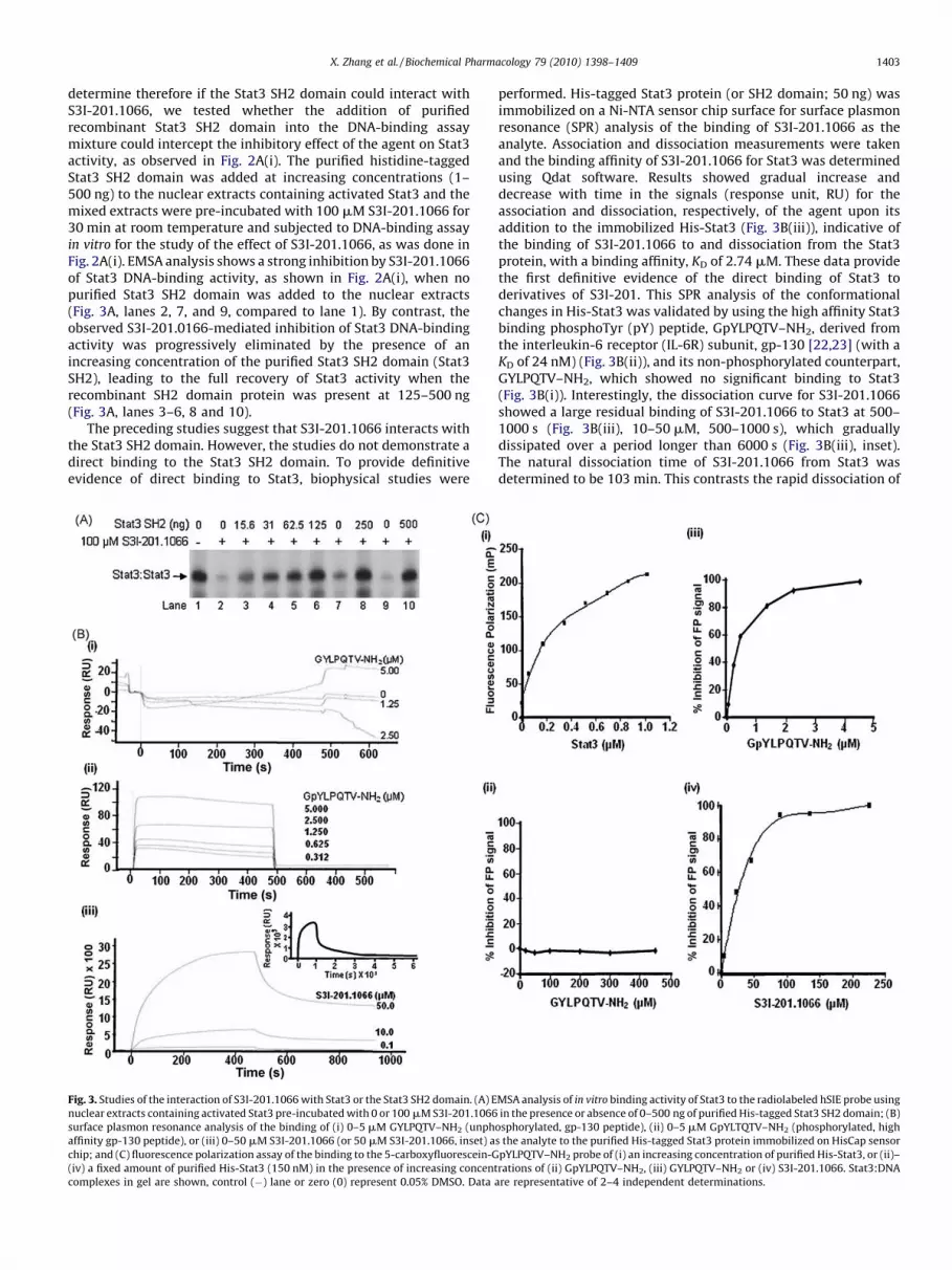
determine therefore if the Stat3 SH2 domain could interact withS3I-201.1066, we tested whether the addition of purifiedrecombinant Stat3 SH2 domain into the DNA-binding assaymixture could intercept the inhibitory effect of the agent on Stat3activity, as observed in Fig. 2A(i). The purified histidine-taggedStat3 SH2 domain was added at increasing concentrations (1–500 ng) to the nuclear extracts containing activated Stat3 and themixed extracts were pre-incubated with 100 mM S3I-201.1066 for30 min at room temperature and subjected to DNA-binding assayin vitro for the study of the effect of S3I-201.1066, as was done inFig. 2A(i). EMSA analysis shows a strong inhibition by S3I-201.1066of Stat3 DNA-binding activity, as shown in Fig. 2A(i), when nopurified Stat3 SH2 domain was added to the nuclear extracts(Fig. 3A, lanes 2, 7, and 9, compared to lane 1). By contrast, theobserved S3I-201.0166-mediated inhibition of Stat3 DNA-bindingactivity was progressively eliminated by the presence of anincreasing concentration of the purified Stat3 SH2 domain (Stat3SH2), leading to the full recovery of Stat3 activity when therecombinant SH2 domain protein was present at 125–500 ng(Fig. 3A, lanes 3–6, 8 and 10).
The preceding studies suggest that S3I-201.1066 interacts withthe Stat3 SH2 domain. However, the studies do not demonstrate adirect binding to the Stat3 SH2 domain. To provide definitiveevidence of direct binding to Stat3, biophysical studies were
Fig. 3. Studies of the interaction of S3I-201.1066 with Stat3 or the Stat3 SH2 domain. (A) E
nuclear extracts containing activated Stat3 pre-incubated with 0 or 100 mM S3I-201.1066
surface plasmon resonance analysis of the binding of (i) 0–5 mM GYLPQTV–NH2 (unph
affinity gp-130 peptide), or (iii) 0–50 mM S3I-201.1066 (or 50 mM S3I-201.1066, inset) a
chip; and (C) fluorescence polarization assay of the binding to the 5-carboxyfluorescein-G
(iv) a fixed amount of purified His-Stat3 (150 nM) in the presence of increasing concent
complexes in gel are shown, control (�) lane or zero (0) represent 0.05% DMSO. Data a
performed. His-tagged Stat3 protein (or SH2 domain; 50 ng) wasimmobilized on a Ni-NTA sensor chip surface for surface plasmonresonance (SPR) analysis of the binding of S3I-201.1066 as theanalyte. Association and dissociation measurements were takenand the binding affinity of S3I-201.1066 for Stat3 was determinedusing Qdat software. Results showed gradual increase anddecrease with time in the signals (response unit, RU) for theassociation and dissociation, respectively, of the agent upon itsaddition to the immobilized His-Stat3 (Fig. 3B(iii)), indicative ofthe binding of S3I-201.1066 to and dissociation from the Stat3protein, with a binding affinity, KD of 2.74 mM. These data providethe first definitive evidence of the direct binding of Stat3 toderivatives of S3I-201. This SPR analysis of the conformationalchanges in His-Stat3 was validated by using the high affinity Stat3binding phosphoTyr (pY) peptide, GpYLPQTV–NH2, derived fromthe interleukin-6 receptor (IL-6R) subunit, gp-130 [22,23] (with aKD of 24 nM) (Fig. 3B(ii)), and its non-phosphorylated counterpart,GYLPQTV–NH2, which showed no significant binding to Stat3(Fig. 3B(i)). Interestingly, the dissociation curve for S3I-201.1066showed a large residual binding of S3I-201.1066 to Stat3 at 500–1000 s (Fig. 3B(iii), 10–50 mM, 500–1000 s), which graduallydissipated over a period longer than 6000 s (Fig. 3B(iii), inset).The natural dissociation time of S3I-201.1066 from Stat3 wasdetermined to be 103 min. This contrasts the rapid dissociation of
MSA analysis of in vitro binding activity of Stat3 to the radiolabeled hSIE probe using
in the presence or absence of 0–500 ng of purified His-tagged Stat3 SH2 domain; (B)
osphorylated, gp-130 peptide), (ii) 0–5 mM GpYLTQTV–NH2 (phosphorylated, high
s the analyte to the purified His-tagged Stat3 protein immobilized on HisCap sensor
pYLPQTV–NH2 probe of (i) an increasing concentration of purified His-Stat3, or (ii)–
rations of (ii) GpYLPQTV–NH2, (iii) GYLPQTV–NH2 or (iv) S3I-201.1066. Stat3:DNA
re representative of 2–4 independent determinations.

X. Zhang et al. / Biochemical Pharmacology 79 (2010) 1398–14091404
the high affinity phosphopeptide, GpYLPQTV–NH2 from Stat3(Fig. 3B(ii)). The slower ‘‘off’’ rate for S3I-201.1066 could impact itsoverall functional effects, with implications for its in vivo
therapeutic application. Differences in the physicochemicalproperties would account for the different behaviors of theinteractions with the Stat3 protein.
The studies so far demonstrate that S3I-201.1066 interacts withStat3 or the Stat3 SH2 domain (data not shown). The interactionwith the Stat3 SH2 domain could block the binding of Stat3 tocognate pTyr peptide motifs of receptors. To verify that S3I-201.1066 disrupts pTyr–Stat3 SH2 domain interactions, henceStat3:Stat3 dimerization, we set up a fluorescence polarization (FP)study based on the binding of Stat3 to the high affinityphosphopeptide, GpYLPQTV–NH2 [22,23]. It has previously beenreported that Stat3 binds to GpYLPQTV–NH2 with a higher affinitythan to the Stat3-derived pTyr peptide, PpYLKTK. It is also reportedthat this high affinity peptide disrupted Stat3 DNA-binding activityin vitro with an IC50 value of 0.15 mM [22]. The FP assay utilizingthe 5-carboxyfluorescein-GpYLPQTV–NH2 as a probe showedincreasing fluorescence polarization signal (mP) with increasingconcentration (in mM) of purified His-Stat3 for a robust Z0 value of0.84 (Fig. 3C(i)), which closely matches the previously reportedvalue of 0.87 [23]. The test of the non-phosphorylated, unlabeledGYLPQTV–NH2 in the FP assay showed no evidence of inhibition(Fig. 3C(iii)), while as expected, the phosphorylated, unlabeledcounterpart, GpYLPQTV–NH2 induced a complete inhibition withan IC50 value of 0.3 mM (Fig. 3C(iii)), consistent with the previouslyreported value of 0.25 � 0.03 mM [23]. The FP assay was used tofurther test the ability of S3I-201.1066 to disrupt the Stat3 interactionwith cognate pTyr peptide (GpYLPQTV–NH2), which showed a
Fig. 4. Effect of S3I-201.1066 on the colocalization or association of Stat3 with EGF rece
microscopy of Stat3 colocalization with EGFR and Stat3 nuclear localization in EGF-stim
201.1066 for 30 min; or (B) (i) immunoblotting analysis of EGFR immunecomplex (uppe
MDA-MB-231 cells, or (ii) immunecomplexes of EGFR (upper panel) or Stat3 (lower pane
immunecomplexes of EGFR or Stat3 were probed for EGFR, Stat3, Shc, Grb 2, or Erk1/2
concentration-dependent inhibition of the fluorescent polarizationsignal (Fig. 3C(iv)). Inhibitory constant (IC50 value) was derived to be20 � 7.3 mM, which is within the range for the IC50 value (35 � 9 mM)determined for the inhibition of Stat3 DNA-binding activity(Fig. 2A(i)). These findings together demonstrate that S3I-201.1066binds to Stat3 or the Stat3 SH2 domain and disrupts the interaction ofStat3 with cognate pTyr peptide motifs. This mode of action underliesthe blocking Stat3 DNA-binding activity by S3I-201.1066.
To extend the studies to verify that S3I-201.1066 could disruptthe binding of Stat3 to receptors, mouse fibroblasts over-expressing the EGF receptor (NIH3T3/hEGFR) were treated withor without the compound prior to stimulation with EGF for 10 min.Cells were then subjected to immunofluorescence staining forEGFR (red) and Stat3 (green) and confocal microscopy for the EGF-induced colocalization of Stat3 and EGFR and the Stat3 nucleartranslocation. In the resting NIH3T3/hEGFR fibroblasts, EGFR (red)is widely localized at the plasma membrane, Stat3 (green) islocalized at both the plasma membrane and in the cytoplasm, withno visible presence in the nucleus (stained blue for DAPI), while thecolocalization of Stat3 with EGFR is minimal at the plasmamembrane (Fig. 4A, upper panels). The stimulation by EGF ofuntreated cells induced a strong nuclear presence of Stat3 (cyan formerged Stat3 (green) and DAPI (blue)), as well as the colocaliza-tions of EGFR and Stat3 (yellow for merged EGFR (red) and Stat3(green)) at the plasma membrane, cytoplasm, and peri-nuclearspace, and in the nucleus (Fig. 4A, bottom left). Both of the EGF-stimulated colocalization between EGFR and Stat3 and the Stat3nuclear localization events were strongly blocked when cells werepre-treated with S3I-201.1066 before stimulating with EGF(Fig. 4A, bottom right compared to non-treated, bottom left),
ptor and on Stat3 nuclear translocation. (A) Immunofluorescence imaging/confocal
ulated (1 mg/ml; 10 min) NIH3T3/hEGFR pre-treated with or without 50 mM S3I-
r panel) or whole-cell lysates (lower panel) from S3I-201.1066-treated Panc-1 and
l) were treated with the indicated concentrations of S3I-201.1066, and subsequentMAPK. Data are representative of 3 independent studies.

X. Zhang et al. / Biochemical Pharmacology 79 (2010) 1398–1409 1405
indicating that the compound disrupts Stat3 binding to EGFR. Weinfer that by blocking Stat3 binding to the receptor, S3I-201.1066attenuates Stat3 phosphorylation/activation and thereby preventsStat3 nuclear translocation. To investigate further the Stat3interaction with the EGFR receptor and the effect of S3I-201.1066, co-immunoprecipitation with immunoblotting studieswere performed in which EGFR immunecomplex prepared fromwhole-cell lysates of treated and untreated cancer cells wereblotted for Stat3, and for Shc and Grb 2 as negative control. Resultsshowed that the EGFR immunecomplex from the untreated Panc-1and MDA-MB-231 cells contained Stat3, Shc and Grb 2 (Fig. 4B(i),lanes 1 and 3, i.p. EGFR, blot Stat3, Shc, and Grb 2). By contrast,treatment of both cell lines with S3I-201.1066 significantlydiminished the level of Stat3 that associated with EGFRimmunecomplex of equal total protein, without affecting thelevels of Shc or Grb 2 (Fig. 4B(i), lanes 2 and 4, i.p. EGFR, blot Stat3,Shc and Grb 2). Western blotting of whole-cell lysates of equal totalprotein shows that the activated and total Erk1/2 levels areunaffected by the treatment of cells with S3I-201.1066 (Fig. 4B(i),input, blot pErk and Erk), and that the levels of Stat3 protein werethe same (Fig. 4B(i), input, blot Stat3). To further analyze the effectof S3I-201.1066 on Stat3 binding to EGFR, a sequential immune-complex precipitation study was performed in which EGFR andStat3 immunecomplexes were independently prepared fromwhole-cell lysates of untreated Panc-1 cells. Immunecomplexesof equal total protein were directly treated with 0, 30, 50, and100 mM S3I-201.1066 for 3 h, and then subjected to a second EGFRor Stat3 immunecomplex precipitation and immunoblottinganalysis. Compared to untreated samples (Fig. 4B(ii), lane 1),results show that the direct treatment with S3I-201.1066 of theEGFR immunecomplex dramatically diminished the level of Stat3protein that remained associated with EGFR in the complex(Fig. 4B(ii), i.p. EGFR, blot Stat3, lanes 2–4), but had no visible effecton the levels of Shc or Grb 2 (Fig. 4B(ii), i.p. EGFR, blot Shc or Grb 2).The EGFR levels in the immunecomplexes are the same (Fig. 4B(ii),upper band). Similarly, the Stat3 immunecomplex that is directlytreated with S3I-201.1066 and blotted for EGFR showed stronglyreduced EGFR levels, compared to the untreated Stat3 immune-complex of equal total protein (Fig. 4B(ii), i.p. Stat3, blot EGFR,compare lanes 2–4 to lanes 1). The Stat3 levels in theimmunecomplexes are the same (Fig. 4B(ii), i.p. Stat3, blot Stat3).Altogether, these findings strongly demonstrate that S3I-201.1066selectively disrupts the binding of Stat3 to cognate receptor motifs.By this mode of activity, S3I-201.1066 could block Stat3phosphorylation and hence, nuclear translocation.
3.5. S3I-201.1066 blocks growth, viability, malignant transformation,
and the migration of malignant cells harboring constitutively active
Stat3
Constitutively active Stat3 promotes malignant cell prolifera-tion, survival and malignant transformation [10,20,42]. We askedthe question whether S3I-201.1066 is able to selectively decreasethe viability and growth of malignant cells that harbor aberrantStat3 activity. The human breast (MDA-MB-231) and pancreatic(Panc-1) cancer lines and the v-Src-transformed mouse fibroblasts(NIH3T3/v-Src) that harbor constitutively active Stat3, and cellsthat do not harbor aberrant Stat3 activity (Stat3 knockout mouseembryonic fibroblasts (MEFs) (Stat3�/�) [31], normal humanpancreatic duct epithelial cells (HPDEC) [30], and the ovariancancer line, A2780S) in culture were treated with or without anincreasing concentration of S3I-201.1066 for up to 6 days andanalyzed for viable cell numbers by CyQuant cell proliferation/viability kit or trypan blue exclusion/phase-contrast microscopy.Compared to the control (DMSO-treated) cells, the mousefibroblasts transformed by v-Src (NIH3T3/v-Src), and the human
breast cancer, MDA-MB-231 and pancreatic cancer, Panc-1 linesshowed significantly reduced viable cell numbers (Fig. 5A) andwere growth inhibited (data not shown) following treatment withincreasing concentrations of S3I-201.1066 for 24–48 h. Bycontrast, the viability and growth of the Stat3-null MEFs(Stat3�/�), the ovarian cancer line, A2780S and the normal humanpancreatic duct epithelial cells (HPDEC) that do not harboraberrant Stat3 activity were not significantly altered by S3I-201.1066 treatment (Fig. 5A, and data not shown), with derivedIC50 values that are >100 mM, compared to values of 35, 48, and37 mM for the inhibition of NIH3T3/v-Src, Panc-1, and MDA-MB-231, respectively (Fig. 5A, lower panel). These findings indicatethat S3I-201.1066 exerts preferential biological effects on malig-nant cells that harbor constitutively active Stat3, with little effectson non-target cells at concentrations that inhibit Stat3 activity.
We extended these studies to examine the effect of S3I-201.1066 in colony survival assay performed as previouslyreported [39]. Cultured single-cells were untreated or treatedonce with S3I-201.1066 and allowed to grow until large colonieswere visible, which were stained and enumerated. Results showeda dose-dependent suppression of the number of colonies for the v-Src transformed mouse fibroblasts (NIH3T3/v-Src), and the humanpancreatic cancer, Panc-1 and breast cancer, MDA-MB-231 cells(Fig. 5B(iii)–(v)) (paired t-test was used to compare treatedsamples to their respective untreated controls). By contrast,minimal effect was observed on the colony numbers for mousefibroblasts transformed by v-Ras (NIH3T3/v-Ras) and the ovariancancer line, A2780S that do not harbor constitutively active Stat3(Fig. 5B(i) and (ii)). Furthermore, growth in soft-agar suspension ofNIH3T3/v-Src, MDA-MB-231 and Panc-1 cells treated with S3I-201.1066 was significantly inhibited (Fig. 5C(iii)–(v)), compared tothe minimal effect on the soft-agar growth of NIH3T3/v-Ras andthe ovarian cancer line, A2780S at concentrations that inhibit Stat3activity (Fig. 5C(i) and (ii)). Thus, S3I-201.1066 selectively blocksStat3-dependent malignant transformation.
Studies also demonstrate that Stat3 is important for tumorprogression [43,44]. To further investigate the biological effects ofS3I-201.1066 and to assess the ability to block Stat3-dependenttumor progression processes, a wound healing study wasperformed as a measure of the migration of malignant cells.Significantly reduced numbers of MDA-MB-231, Panc-1 andNIH3T3/v-Src cells migrating into the denuded area were observedfollowing 12–24 h treatment with S3I-201.1066 (Fig. 5D and datanot shown), with statistically significant lower numbers observedat 30 mM S3I-201.1066 treatment (data not shown). By contrast,the migration of NIH3T3/v-Ras fibroblasts was minimally affectedby the same treatment conditions (Fig. 5D). In the 12–24 htreatment duration, there was no evidence of apoptosis of thetreated cells (data not shown). These findings demonstrate thatS3I-201.1066 selectively suppresses the migration of malignantcells that harbor aberrant Stat3 activity.
3.6. S3I-201.1066 represses the expression of c-Myc, Bcl-xL, VEGF,
Survivin, and MMP-9
Known Stat3 target genes are critical to the dysregulatedbiological processes promoted by aberrantly active Stat3 [9,20,42].We sought to validate the inhibitory effect of S3I-201.1066 onaberrant Stat3 signaling and to define the underlying molecularmechanisms for the antitumor cell effects of the agent byinvestigating the changes in the induction of known Stat3-regulated genes. In the human breast carcinoma, MDA-MB-231and pancreatic cancer, Panc-1 lines, and the mouse fibroblaststransformed by v-Src, which harbor constitutively active Stat3,immunoblotting analysis of whole-cell lysates shows that treat-ment with 50 mM S3I-201.1066 for 24 h down-regulated the

Fig. 5. S3I-201.1066 suppresses viability, survival, malignant transformation and migration of malignant cells that harbor persistently active Stat3. (A and B) Human breast
(MDA-MB-231), pancreatic (Panc-1), and ovarian (A2780S) cancer cells, the v-Src transformed mouse fibroblasts (NIH3T3/v-Src) and their v-Ras-transformed counterparts
(NIH3T3/v-Ras), the Stat3-null mouse embryonic fibroblasts (Stat3�/�), and the normal human pancreatic duct epithelial cells (HPDEC) were treated once or untreated with
30–100 mM S3I-201.1066 for 24–48 h. (A and B) cells were (A) assayed for viability using CyQuant cell proliferation kit; IC50 values (bottom panel) were derived from
graphical representation, or (B) allowed to culture until large colonies were visible, which were stained with crystal violet and enumerated; (C) cells (NIH3T3/v-Src, NIH3T3/
v-Ras, A2780S, MDA-MB-231, and Panc-1) growing in soft-agar suspension were treated with or without 30–100 mM S3I-201.1066 every 2–3 days until large colonies were
visible, which were stained with crystal violet and enumerated; and (D) cells (MDA-MB-231, Panc-1, NIH3T3/v-Src and NIH3T3/v-Ras) in culture were wounded and treated
with or without 50 mM S3I-201.1066 for 12 or 24 h and allowed to migrate into the denuded area in a wound healing assay. Cultures were visualized at 10�magnification by
light microscopy and (i) cells that migrated into the denuded area counted and plotted against the concentration of S3I-201.1066 or (ii) cultures were photographed. Values
are the mean and SD of 3–4 independent determinations, data are representative of 4 independent studies. p-Values: *p < 0.05, and **p < 0.01.
X. Zhang et al. / Biochemical Pharmacology 79 (2010) 1398–14091406
expression of c-Myc, Bcl-xL, VEGF, Survivin, and MMP-9 proteins(Fig. 6A). Bands were quantified, normalized to b-actin, and thevalues corresponding to the band intensities for the samples fromtreated cells relative to the respective control (set at 1) are reportedin parenthesis. These data indicate that S3I-201.1066 sufficientlyrepresses the constitutive induction of Stat3-regulated genes. Weinfer that in doing so, S3I-201.1066 is able to thwart the ability ofaberrant Stat3 to promote the dysregulation of growth and survivalof malignant cells. These findings are in agreement with the resultsin Fig. 2C and together support the ability of S3I-201.1066 to blockStat3 transcriptional activity.
3.7. S3I-201.1066 inhibits growth of human breast tumor xenografts
Given Stat3’s importance in tumor growth and tumor progres-sion, we evaluated S3I-201.1066 in xenograft models of the humanbreast cancer (MDA-MB-231) cells that harbor aberrant Stat3activity. Compared to control (vehicle-treated) tumor-bearingmice, treatment (i.v. injection) with S3I-201.1066 at 3 mg/kg every
2 or 3 days for 17 days induced significant decrease in tumorgrowth (Fig. 6B). At the dosing schedule used, the drug was welltolerated and the animals showed no obvious signs of toxicity. Theunderlying premise of the antitumor effects is the ability of S3I-201.1066 to inhibit aberrant Stat3 activity. To determine whetherthe treatment with S3I-201.1066 modulated the in vivo activityand function of aberrant Stat3 in the human breast tumorxenografts, we evaluated the status of Stat3 activity and theexpression of known Stat3-regulated genes in vivo. Upon thecompletion of the study, control tumors and residual tumors fromtreated mice were harvested and tissue lysates were prepared andanalyzed by electrophoretic mobility shift assay using theradiolabeled hSIE probe that binds Stat3 (Fig. 6C(i)) or immuno-blotting (Fig. 6C(ii)). Representative data for one control, untreatedtumor and three treated tumor tissues showed both decreasedphosphorylation (pY705Stat3) (Fig. 6C(ii), upper band) and DNA-binding activity (Fig. 6C(i)) of Stat3 in tumors from treated mice(T1–T3, versus C). Furthermore, immunoblotting analysis showeddiminished expression of c-Myc, Bcl-xL, VEGF, and Survivin in the
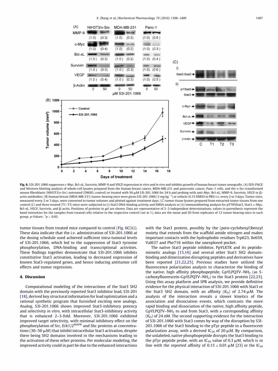
Fig. 6. S3I-201.1066 suppresses c-Myc, Bcl-xL, Survivin, MMP-9 and VEGF expression in vitro and in vivo and inhibits growth of human breast tumor xenografts. (A) SDS-PAGE
and Western blotting analysis of whole-cell lysates prepared from the human breast cancer, MDA-MB-231 and pancreatic cancer, Panc-1 cells, and the v-Src-transformed
mouse fibroblasts (NIH3T3/v-Src) untreated (DMSO, control) or treated with 50 mM S3I-201.1066 for 24 h and probing with anti-Myc, Bcl-xL, MMP-9, Survivin, VEGF or b-
actin antibodies; (B) human breast (MDA-MB-231) tumor-bearing mice were given S3I-201.1066 (3 mg kg�1) or vehicle (0.1% DMSO in PBS) i.v. every 2 or 3 days. Tumor sizes,
measured every 2 or 3 days, were converted to tumor volumes and plotted against treatment days; (C) tumor tissue lysates prepared from extracted tumor tissues from one
control (C) and three treated (T1–T3) mice were subjected to (i) Stat3 DNA-binding activity and EMSA analysis or (ii) immunoblotting analysis for pY705Stat3, Stat3, c-Myc,
Bcl-xL, VEGF, Survivin, and b-actin. Positions of proteins in gel are shown. Data are representative of 2–3 independent determinations, values in parenthesis represent the
band intensities for the samples from treated cells relative to the respective control (set at 1), data are the mean and SD from replicates of 12 tumor-bearing mice in each
group. p-Values: *p < 0.05.
X. Zhang et al. / Biochemical Pharmacology 79 (2010) 1398–1409 1407
tumor tissues from treated mice compared to control (Fig. 6C(ii)).These data indicate that the i.v. administration of S3I-201.1066 atthe dosing schedule used achieved sufficient intra-tumoral levelsof S3I-201.1066, which led to the suppression of Stat3 tyrosinephosphorylation, DNA-binding and transcriptional activities.These findings together demonstrate that S3I-201.1066 inhibitsconstitutive Stat3 activation, leading to decreased expression ofknown Stat3-regulated genes, and hence inducing antitumor celleffects and tumor regression.
4. Discussion
Computational modeling of the interactions of the Stat3 SH2domain with the previously reported Stat3 inhibitor lead, S3I-201[18], derived key structural information for lead optimization and arational synthetic program that furnished exciting new analogs.Analog, S3I-201.1066 shows improved Stat3-inhibitory potencyand selectivity in vitro, with intracellular Stat3-inhibitory activitythat is enhanced 2–3-fold. Moreover, S3I-201.1066 exhibitedimproved target selectivity, with minimal inhibitory effect on thephosphorylation of Src, Erk1/2MAPK and Shc proteins at concentra-tions (30–50 mM) that inhibit intracellular Stat3 activation, despitethere being SH2 domains involved in the mechanisms leading tothe activation of these other proteins. Per molecular modeling, theimproved activity could in part be due to the enhanced interactions
with the Stat3 protein, possibly by the (para-cyclohexyl)benzylmoiety that extends from the scaffold amide nitrogen and makesimportant contacts with the hydrophobic residues Trp623, Ile659,Val637 and Phe716 within the unexplored pocket.
The native Stat3 peptide inhibitor, PpYLKTK and its peptido-mimetic analogs [15,16] and several other Stat3 SH2 domain-binding and dimerization disrupting peptides and derivatives havebeen reported [21,22,25]. Previous studies have utilized thefluorescence polarization analysis to characterize the binding ofthe native, high affinity phosphopeptide, GpYLPQTV–NH2 (as 5-carboxyfluorescein-GpYLPQTV–NH2) to the Stat3 protein [22,23].Using this assay platform and SPR analysis, we provide definitiveevidence for the physical interaction of S3I-201.1066 with Stat3 orthe Stat3 SH2 domain, with an affinity (KD) of 2.74 mM. Theanalysis of the interaction reveals a slower kinetics of theassociation and dissociation events, which contrasts the morerapid binding and dissociation of the native, high affinity peptide,GpYLPQTV–NH2 to and from Stat3, with a corresponding affinity(KD) of 24 nM. The second supporting evidence for the interactionof S3I-201.1066 with Stat3 comes by way of the disruption by S3I-201.1066 of the Stat3 binding to the pTyr peptide in a fluorescentpolarization assay, with a derived IC50 of 20 mM. By comparison,the unlabeled, native phosphopeptide disrupts the Stat3 binding tothe pTyr peptide probe, with an IC50 value of 0.3 mM, which is inline with the reported affinity of 0.15 � 0.01 mM [23] or the IC50

X. Zhang et al. / Biochemical Pharmacology 79 (2010) 1398–14091408
value of 0.290 � 0.063 mM [21]. The higher affinity of the nativepeptide for the protein should be expected, given the more favorablephysicochemical properties that will facilitate a stronger binding tothe Stat3 protein. Nonetheless, data showing a slower dissociation ofS3I-201.1066 from Stat3 suggests this drug is likely to show a moreprolonged effect on the target and its function per a given dose.
Current study provides support for the binding of S3I-201.1066to Stat3 and for the disruption of the interaction between Stat3 andpTyr peptide. Given the disruption of the Stat3 binding to thecognate peptide, GpYLPQTV–NH2, we infer that inside cells, S3I-201.1066 could interfere with the ability of Stat3 (via SH2 domain)to bind to cognate pTyr motifs on receptors and thereby block de
novo phosphorylation by tyrosine kinases, as well as disrupt pre-existing Stat3:Stat3 dimers, particularly in malignant cells thatharbor aberrantly active Stat3. Accordingly, we present evidencethat both of the association of Stat3 with EGFR and the Stat3nuclear localization in ligand-stimulated cells are strongly blockedby the treatment of cells with S3I-201.1066. Although other Stat3dimerization disruptors have been previously identified throughmolecular modeling [19,45], the present study is the first toprovide biophysical evidence for a direct interaction of a small-molecule, dimerization disruptor with the Stat3 protein.
Substantive evidence demonstrates that aberrant Stat3 activitypromotes cancer cell growth and survival [15,16,29,46,47], andinduces tumor angiogenesis [48,49] and metastasis [43,49].Accordingly, inhibitors of Stat3 activation and signaling have beenshown to induce antitumor cell effects consistent with theabrogation of Stat3 function [15–19,37,50–52]. The present studyparallels those published reports in showing that a newly derivedagent, S3I-201.1066 induces the growth inhibition and the loss ofviability and survival of the human pancreatic cancer, Panc-1 andbreast cancer, MDA-MB-231 cells, and transformed mousefibroblasts (NIH3T3/v-Src) that harbor aberrant Stat3 activity,while having minimal effects on normal human pancreatic ductepithelial cells, the Stat3-null mouse embryonic fibroblasts [31],the ovarian cancer line, A2780S, and the viral Ras-transformedmouse fibroblasts that do not harbor aberrant Stat3 activity.Moreover, the S3I-201.1066-induced antitumor cell effects onmalignant cells harboring aberrant Stat3 activity occurred atsignificantly lower concentrations, 30–50 mM than the 100 mMcellular activity previously reported for the lead agent [18].Mechanistic insight into the biological effects of S3I-201.1066reveals the suppression of the constitutive expression of knownStat3-regulated genes, including c-Myc, Bcl-xL, VEGF, Survivin, andMMP-9, which control cell growth and apoptosis, promote tumorangiogenesis, or modulate tumor cell invasion [19,43,46,49,53,54].Furthermore, the effect of S3I-201.1066 on Stat3 oncogenicfunction is shown by the significant antitumor response inducedin human breast tumor xenografts following the in vivo
administration of this agent. Data also suggest that at the dosingschedule used, the i.v. administration of S3I-201.1066 achievedintra-tumoral levels sufficient to modulate activated Stat3 and itsfunction.
We report the application of computational modeling inconjunction with rational, structure-based virtual design approachfor the optimization of S3I-201. The new agent, S3I-201.1066 bindsto Stat3, disrupts Stat3 SH2 domain:pTyr interactions, and henceStat3:Stat3 dimerization and Stat3 binding to receptor, therebyinhibiting Stat3 phosphorylation, nuclear translocation andoncogenic functions, and inducing antitumor cell effects in vitro
and antitumor effects in vivo.
Acknowledgements
We thank all colleagues and members of our laboratory for thestimulating discussions. We also thank Vijay Shahani and Brent
D.G. Page at the University of Toronto for the GOLD moleculardocking studies and for the assistance with the chemical synthesis.This work was supported by the National Cancer Institute GrantsCA106439 (JT) and CA128865 (JT), and by the Leukemia andLymphoma Society of Canada (PTG) and the University of Toronto(PTG).
References
[1] Bromberg J. Signal transducers and activators of transcription as regulators ofgrowth, apoptosis and breast development. Breast Cancer Res 2000;2:86–90.
[2] Darnell Jr JE. Transcription factors as targets for cancer therapy. Nat Rev Cancer2002;2:740–9.
[3] Schroder M, Kroeger K, Volk HD, Eidne KA, Grutz G. Preassociation of nonacti-vated STAT3 molecules demonstrated in living cells using bioluminescenceresonance energy transfer: a new model of STAT activation? J Leukoc Biol2004;75:792–7.
[4] Sehgal PB. Paradigm shifts in the cell biology of STAT signaling. Semin Cell DevBiol 2008;19:329–40.
[5] Bromberg J, Darnell Jr JE. The role of STATs in transcriptional control and theirimpact on cellular function. Oncogene 2000;19:2468–73.
[6] Bowman T, Garcia R, Turkson J, Jove R. STATs in oncogenesis. Oncogene2000;19:2474–88.
[7] Turkson J, Jove R. STAT proteins: novel molecular targets for cancer drugdiscovery. Oncogene 2000;19:6613–26.
[8] Buettner R, Mora LB, Jove R. Activated STAT signaling in human tumorsprovides novel molecular targets for therapeutic intervention. Clin CancerRes 2002;8:945–54.
[9] Yu H, Jove R. The STATs of cancer-new molecular targets come of age. Nat RevCancer 2004;4:97–105.
[10] Turkson J. STAT proteins as novel targets for cancer drug discovery. ExpertOpin Ther Targets 2004;8:409–22.
[11] Darnell JE. Validating Stat3 in cancer therapy. Nat Med 2005;11:595–6.[12] Kortylewski M, Yu H. Stat3 as a potential target for cancer immunotherapy. J
Immunother 2007;30:131–9.[13] Kortylewski M, Yu H. Role of Stat3 in suppressing anti-tumor immunity. Curr
Opin Immunol 2008;20:228–33.[14] Shuai K, Horvath CM, Huang LH, Qureshi SA, Cowburn D, Darnell Jr JE.
Interferon activation of the transcription factor Stat91 involves dimerizationthrough SH2-phosphotyrosyl peptide interactions. Cell 1994;76:821–8.
[15] Turkson J, Ryan D, Kim JS, Zhang Y, Chen Z, Haura E, et al. Phosphotyrosylpeptides block Stat3-mediated DNA-binding activity, gene regulation and celltransformation. J Biol Chem 2001;276:45443–55.
[16] Turkson J, Kim JS, Zhang S, Yuan J, Huang M, Glenn M, et al. Novel peptido-mimetic inhibitors of signal transducer and activator of transcription 3dimerization and biological activity. Mol Cancer Ther 2004;3:261–9.
[17] Siddiquee K, Glenn M, Gunning P, Katt WP, Zhang S, Schroeck C, et al. Anoxazole-based small-molecule Stat3 inhibitor modulates Stat3 stability andprocessing and induces antitumor cell effects. ACS Chem Biol 2007;2:787–98.
[18] Siddiquee K, Zhang S, Guida WC, Blaskovich MA, Greedy B, Lawrence H, et al.Selective chemical probe inhibitor of Stat3, identified through structure-basedvirtual screening, induces antitumor activity. 1. Proc Natl Acad Sci U S A2007;104:7391–6.
[19] Song H, Wang R, Wang S, Lin J. A low-molecular-weight compound discoveredthrough virtual database screening inhibits Stat3 function in breast cancercells. Proc Natl Acad Sci U S A 2005;102:4700–5.
[20] Yue P, Turkson J. Targeting STAT3 in cancer: how successful are we? ExpertOpin Investig Drugs 2009;18:45–56.
[21] Coleman DRI, Ren Z, Mandal PK, Cameron AG, Dyer GA, Muranjan S, et al.Investigation of the binding determinants of phosphopeptides targeted to theSrc Homology. 2. Domain of the signal transducer and activator of transcrip-tion. 3. Development of a high-affinity peptide inhibitor. J Med Chem2005;48:6661–70.
[22] Ren Z, Cabell LA, Schaefer TS, McMurray JS. Identification of a high-affinityphosphopeptide inhibitor of stat3. Bioorg Med Chem Lett 2003;13:633–6.
[23] Schust J, Berg T. A high-throughput fluorescence polarization assay forsignal transducer and activator of transcription 3. Anal Biochem 2004;330:114–8.
[24] Gunning PT, Glenn MP, Siddiquee KA, Katt WP, Masson E, Sebti SM, et al.Targeting protein–protein interactions: suppression of Stat3 dimerizationwith rationally designed small-molecule, nonpeptidic SH2 domain binders.Chembiochem 2008;9:2800–3.
[25] Fletcher S, Turkson J, Gunning PT. Molecular approaches towards the inhibi-tion of the signal transducer and activator of transcription 3 (Stat3) protein.ChemMedChem 2008;3:1159–68.
[26] Becker S, Groner B, Muller CW. Three-dimensional structure of the Stat3betahomodimer bound to DNA. Nature 1998;394:145–51.
[27] Johnson PJ, Coussens PM, Danko AV, Shalloway D. Overexpressed pp60c-srccan induce focus formation without complete transformation of NIH 3T3 cells.Mol Cell Biol 1985;5:1073–83.
[28] Yu CL, Meyer DJ, Campbell GS, Larner AC, Carter-Su C, Schwartz J, et al.Enhanced DNA-binding activity of a Stat3-related protein in cells transformedby the Src oncoprotein. Science 1995;269:81–3.
X. Zhang et al. / Biochemical Pharmacology 79 (2010) 1398–1409 1409
[29] Garcia R, Bowman TL, Niu G, Yu H, Minton S, Muro-Cacho CA, et al. Constitutiveactivation of Stat3 by the Src and JAK tyrosine kinases participates in growthregulation of human breast carcinoma cells. Oncogene 2001;20:2499–513.
[30] Ouyang H, Mou LJ, Luk C, Liu N, Karaskova J, Squire J, et al. Immortal humanpancreatic duct epithelial cell lines with near normal genotype and phenotype.Am J Pathol 2000;157:1623–31.
[31] Maritano D, Sugrue ML, Tininini S, Dewilde S, Strobl B, Fu X, et al. The STAT3isoforms alpha and beta have unique and specific functions. Nat Immunol2004;5:401–9.
[32] Turkson J, Bowman T, Adnane J, Zhang Y, Djeu JY, et al. Requirement for Ras/Rac1-mediated p38 and c-Jun N-terminal kinase signaling in Stat3 transcrip-tional activity induced by the Src oncoprotein. Mol Cell Biol 1999;19:7519–28.
[33] Turkson J, Bowman T, Garcia R, Caldenhoven E, De Groot RP, Jove R. Stat3activation by Src induces specific gene regulation and is required for celltransformation. Mol Cell Biol 1998;18:2545–52.
[34] Wagner M, Kleeff J, Friess H, Buchler MW, Korc M. Enhanced expression of thetype II transforming growth factor-beta receptor is associated with decreasedsurvival in human pancreatic cancer. Pancreas 1999;19:370–6.
[35] Gouilleux F, Moritz D, Humar M, Moriggl R, Berchtold S, Groner B. Prolactinand interleukin-2 receptors in T lymphocytes signal through a MGF-STAT5-like transcription factor. Endocrinology 1995;136:5700–8.
[36] Seidel HM, Milocco LH, Lamb P, Darnell Jr JE, Stein RB, Rosen J. Spacing ofpalindromic half sites as a determinant of selective STAT (signal transducersand activators of transcription) DNA binding and transcriptional activity. ProcNatl Acad Sci U S A 1995;92:3041–5.
[37] Turkson J, Zhang S, Mora LB, Burns A, Sebti S, Jove R. A novel platinumcompound inhibits constitutive Stat3 signaling and induces cell cycle arrestand apoptosis of malignant cells. J Biol Chem 2005;280:32979–88.
[38] Zhang Y, Turkson J, Carter-Su C, Smithgall T, Levitzki A, Kraker A, et al.Activation of Stat3 in v-Src transformed fibroblasts requires cooperation ofJak1 kinase activity. J Biol Chem 2000;275:24935–44.
[39] Zhao S, Venkatasubbarao K, Lazor JW, Sperry J, Jin C, Cao L, et al. Inhibition ofSTAT3Tyr705 phosphorylation by Smad4 suppresses transforming growthfactor b–mediated invasion and metastasis in pancreatic cancer cells. CancerRes 2008;68:4221–8.
[40] Jones G, Willett P, Glen RC, Leach AR, Taylor R. Development and validation of agenetic algorithm for flexible docking. J Mol Biol 1997;267:727–48.
[41] Fletcher S, Jardeephi S, Zhang X, Yue P, Page BD, Sharmeen S, et al. Disruption oftranscriptionally active Stat3 dimers with non-phosphorylated. Salicylic acid-based small molecules: potent in vitro and tumour cell activities. ChemBio-Chem 2009;10:1959–64.
[42] Siddiquee KAZ. STAT3 as a target for inducing apoptosis in solid and hemato-logical tumors. Cell Res 2008;18:254–67.
[43] Xie TX, Wei D, Liu M, Gao AC, Ali-Osman F, Sawaya R, et al. Stat3 activationregulates the expression of matrix metalloproteinase-2 and tumor invasionand metastasis. Oncogene 2004;23:3550–60.
[44] Huang C, Cao J, Huang KJ, Zhang F, Jiang T, Zhu L, et al. Inhibition of STAT3activity with AG490 decreases the invasion of human pancreatic cancer cells invitro. Cancer Sci 2006;97:1417–23.
[45] Bhasin D, Cisek K, Pandharkar T, Regan N, Li C, Pandit B, et al. Design, synthesis,and studies of small molecule STAT3 inhibitors. Bioorg Med Chem Lett2008;18:391–5.
[46] Catlett-Falcone R, Landowski TH, Oshiro MM, Turkson J, Levitzki A, Savino R,et al. Constitutive activation of Stat3 signaling confers resistance to apoptosisin human U266 myeloma cells. Immunity 1999;10:105–15.
[47] Mora LB, Buettner R, Seigne J, Diaz J, Ahmad N, Garcia R, et al. Constitutiveactivation of Stat3 in human prostate tumors and cell lines: direct inhibition ofStat3 signaling induces apoptosis of prostate cancer cells. Cancer Res2002;62:6659–66.
[48] Niu G, Wright KL, Huang M, Song L, Haura E, Turkson J, et al. Constitutive Stat3activity up-regulates VEGF expression and tumor angiogenesis. Oncogene2002;21:2000–8.
[49] Wei D, Le X, Zheng L, Wang L, Frey JA, Gao AC, et al. Stat3 activation regulatesthe expression of vascular endothelial growth factor and human pancreaticcancer angiogenesis and metastasis. Oncogene 2003;22:319–29.
[50] Fuh B, Sobo M, Cen L, Josiah D, Hutzen B, Cisek K, et al. LLL-3 inhibits STAT3activity, suppresses glioblastoma cell growth and prolongs survival in a mouseglioblastoma model. Br J Cancer 2009;100:106–12.
[51] Blaskovich MA, Sun J, Cantor A, Turkson J, Jove R, Sebti SM. Discovery of JSI-124(cucurbitacin I), a selective Janus kinase/signal transducer and activator oftranscription 3 signaling pathway inhibitor with potent antitumor activityagainst human and murine cancer cells in mice. Cancer Res 2003;63:1270–9.
[52] Sun J, Blaskovich MA, Jove R, Livingston SK, Coppola D, Sebti SM. CucurbitacinQ: a selective STAT3 activation inhibitor with potent antitumor activity.Oncogene 2005;24:3236–45.
[53] Real PJ, Sierra A, De Juan A, Segovia JC, Lopez-Vega JM, Fernandez-Luna JL.Resistance to chemotherapy via Stat3-dependent overexpression of Bcl-2 inmetastatic breast cancer cells. Oncogene 2002;21:7611–8.
[54] Gritsko T, Williams A, Turkson J, Kaneko S, Bowman T, Huang M, et al.Persistent activation of stat3 signaling induces survivin gene expressionand confers resistance to apoptosis in human breast cancer cells. Clin CancerRes 2006;12:11–9.


