Embed Size (px)
Citation preview

Sonic Hedgehog Induces Angiogenesis via Rho Kinase-dependent Signaling in Endothelial Cells
Marie-Ange Renault, PhD1,2,3, Jérôme Roncalli, MD, PhD1, Jörn Tongers, MD1,4, TinaThorne, MS1, Ekaterina Klyachko, PhD1, Sol Misener1, Olga V. Volpert, PhD5, ShanuMehta, MS6, Aaron Burg, BS6, Corinne Luedemann, BS6, Gangjian Qin, MD1,7, RajKishore, PhD1,7, and Douglas W. Losordo, MD1,71 Feinberg Cardiovascular Research Institute, Northwestern University Feinberg School ofMedicine, Tarry 12-703, 303 East Chicago Ave., Chicago, IL 60611 USA2 INSERM U828, avenue du Haut-leveque, 33600 Pessac, France3 Université de Bordeaux Victor Ségalen, IFR4, 146 rue Leo Saignat, 33000 Bordeaux, France4 Department of Cardiology and Angiology, Hannover Medical School, Carl-Neuberg-Str. 1,30625 Hannover, Germany5 Department of Urology, Northwestern University Feinberg School of Medicine, Tarry 16, 303East Chicago Ave., Chicago, IL 60611 USA6 St Elizabeth Medical Center, 736 Cambridge St, Boston, MA, USA7 Division of Cardiology, Northwestern Memorial Hospital, Galter 11-240, 201 E. Huron St.,Chicago, IL 60611 USA
AbstractThe morphogen Sonic Hedgehog (Shh) promotes neovascularization in adults by inducing pro-angiogenic cytokine expression in fibroblasts; however, the direct effects of Shh on endothelialcell (EC) function during angiogenesis are unknown. Our findings indicate that Shh promotescapillary morphogenesis (tube length on Matrigel™ increased to 271±50% of the length inuntreated cells, p=0.00003), induces EC migration (modified Boyden chamber assay, 191±35% ofmigration in untreated cells, p=0.00009), and increases EC expression of matrix metalloproteinase9 (MMP-9) and osteopontin (OPN) mRNA (real-time RT-PCR), which are essential for Shh-induced angiogenesis both in vitro and in vivo. Shh activity in ECs is mediated by Rho, rather thanthrough the “classic” Shh signaling pathway, which involves the Gli transcription factors. The Rhodependence of Shh-induced EC angiogenic activity was documented both in vitro, with dominant-negative RhoA and Rho kinase (ROCK) constructs, and in vivo, with the ROCK inhibitor Y27632in the mouse corneal angiogenesis model. Finally, experiments performed in MMP-9- and OPN-knockout mice confirmed the roles of the ROCK downstream targets MMP-9 and OPN in Shh-induced angiogenesis. Collectively, our results identify a “non-classical” pathway by which Shhdirectly modulates EC phenotype and angiogenic activity.
Corresponding Author: Douglas W. Losordo, MD, Director, Feinberg Cardiovascular Research Institute, Northwestern UniversityFeinberg School of Medicine, 303 E. Chicago Ave., Tarry 14-725, Chicago, IL 60611, T: 312-695-0072, F: 312-695-0047, [email protected]'s Disclaimer: This is a PDF file of an unedited manuscript that has been accepted for publication. As a service to ourcustomers we are providing this early version of the manuscript. The manuscript will undergo copyediting, typesetting, and review ofthe resulting proof before it is published in its final citable form. Please note that during the production process errors may bediscovered which could affect the content, and all legal disclaimers that apply to the journal pertain.
NIH Public AccessAuthor ManuscriptJ Mol Cell Cardiol. Author manuscript; available in PMC 2011 September 1.
Published in final edited form as:J Mol Cell Cardiol. 2010 September ; 49(3): 490–498. doi:10.1016/j.yjmcc.2010.05.003.
NIH
-PA Author Manuscript
NIH
-PA Author Manuscript
NIH
-PA Author Manuscript

Keywordsangiogenesis; Sonic Hedgehog; endothelial cells; Gli transcription factors; ischemia
IntroductionHedgehog (Hh) proteins, which include Sonic hedgehog (Shh), Indian hedgehog, and Deserthedgehog, act as morphogens in multiple tissues during embryonic development [1–8]. Therole of Shh in vasculogenesis has been documented in a variety of contexts.Hypervascularization of the neuroectoderm has been observed after transgenicoverexpression of Shh in the dorsal neural tube of mouse embryos [9], Shh-deficientzebrafish exhibit disorganized endothelial precursors and are unable to form the dorsal aortaor axial vein [10], and the developing lungs of Shh-deficient mice lack propervascularization [5].
Our previous studies have shown that the embryonic Hh pathway is reactivated in adultanimal models of ischemic injury, including hind limb-ischemia [11] and myocardialinfarction [12], and that administration of Shh as a recombinant protein or via gene therapyenhances neovascularization of ischemic tissue by promoting both angiogenesis [11] and therecruitment of endothelial progenitor cells (EPCs) [12–13]. Shh induces fibroblasts andcardiomyocytes to overexpress several pro-angiogenic growth factors, including vascularendothelial growth factor (VEGF) and angiopoietin-1 (Ang1) [11–14], which suggests thatthe effect of Shh on vascular cells is indirect. Recently, we have shown that Shh influencesEPCs directly to promote EPC adhesion, migration, and proliferation [13].
In the classically described Hh-signaling cascade, interactions between Shh and the receptorPatched-1 (Ptch1) repress Ptch-1–mediated inhibition of Smoothened (Smo), whichsubsequently leads to activation of the Gli transcription factors, then activated Gli inducesexpression of Ptch1, Gli1, and other downstream target genes [15]. However, the nature andextent of Shh activity in mature endothelial cells (ECs) remain controversial. Cornealneovessel ECs and human umbilical vein endothelial cells (HUVECs) do not overexpressPtch1 in response to Shh stimulation [11]; nevertheless, Shh promotes capillarymorphogenesis in both HUVECs and murine brain capillary ECs [16]. Shh activates thephosphoinositide kinase 3 (PI3K)/Akt pathway in ECs [16], and other signal transductionpathways, such as the cyclic AMP/protein kinase A axis [17], can modulate the activity ofthe Gli transcription factors. Shh also induces expression of chicken ovalbumin upstreampromoter transcription factor II (COUP TFII) in mouse embryonic carcinoma cells [18] andextracellular signal-regulated kinases 1/2 (ERK 1/2) and protein kinase Cδ (PKCδ) infibroblasts [19]; Shh activates the Rho/Rho kinase (ROCK) pathway in neuronal cells [20].
The potential role of a nonclassical signaling pathway in Shh-mediated angiogenesis has notbeen fully explored. Accordingly, we performed a series of in vitro and in vivo experimentsto characterize the potential direct angiogenic effects of Shh. Our results reveal that Shhinfluences angiogenesis directly through the Rho/ROCK signaling pathway and by inducingexpression of downstream target genes such as matrix metalloproteinase 9 (MMP-9) andosteopontin (OPN). Thus, our investigation has identified a novel, nonclassical mechanismof Shh signaling that modulates neovascularization.
Materials and MethodsAll studies were approved by the Northwestern University Animal Care and Use Committee.
Renault et al. Page 2
J Mol Cell Cardiol. Author manuscript; available in PMC 2011 September 1.
NIH
-PA Author Manuscript
NIH
-PA Author Manuscript
NIH
-PA Author Manuscript
Cell linesExperiments were performed with bovine aortic endothelial cells (BAECs) (ATCC,Manassas, VA, USA), HUVECs and human coronary-artery endothelial cells (HCAECs)(Cambrex Corporation, East Rutherford, NJ, USA), NIH 3T3 embryonic fibroblasts (ATCC)and HeLa cells. Culture conditions are provided in the Supplemental Methods.
Tube formation assay25×103 serum-starved BAECs were seeded in the presence or absence of Shh protein in eachwell of a 48-well plate coated with 120 μL of growth-factor–reduced Matrigel™ (BDBiosciences, San Jose, CA, USA). Tube formation was examined by phase-contrastmicroscopy 6 hours later.
Migration assayCell migration was evaluated by using a modified Boyden’s chamber (Neuro Probe, Inc.,Bethesda, MD, USA) as detailed in the Supplemental Methods. Each condition was assayedin triplicate, and each experiment was performed at least three times.
In vitro proliferation assayCells were incubated with 200 μg/mL bromodeoxyuridine (BrdU) for 24 hours, fixed with100% methanol for 10 minutes at 4°C, then stained with immunofluorescent sheep anti-BrdU antibodies (Abcam Inc., Cambridge, MA, USA); nuclei were stained with DAPI. Eachexperiment was performed at least three times, and each condition in each experiment wasassayed in triplicate.
Quantitative real-time reverse transcriptase–polymerase chain reaction (RT-PCR)RNA was isolated from 3×10⁵ cells or homogenized tissue with RNA STAT-60 (TEL-TESTElectronics Labs Inc, Austin, TX, USA) according to the manufacturer’s instructions. TotalRNA was reverse transcribed with a Taqman cDNA Synthesis Kit (Applied Biosystems,Foster City, CA, USA) and amplification was performed with a Taqman 7500 (AppliedBiosystems). Primer and probe sequences are listed in the Supplemental Table. The relativeexpression of each mRNA was calculated by the comparative threshold cycle (CT) methodand normalized to 18S expression.
Plasmid and siRNA transfectionTransfection was performed as detailed in the Supplemental Methods; assays wereperformed 48 hours after transfection. The Gli-BS luciferase and mutGli luciferase plasmidswere kindly provided by Dr H. Sasaki [21]. The ROCK RB/PH (pEF-BOS-myc-Rho-kinaseRB/PH)- and RhoAN19 (pEF-BOS-HA-RhoA-N19)-expressing plasmids were kindlyprovided by Dr K. Kaibuchi [22]. The Ezrin dominant-negative plasmid (pCB-6-EzrinNTer) was kindly provided by Dr M. Arpin [23]. The sequences of the OPN- and MMP-9–inhibiting siRNAs and the nonsilencing, GFP siRNA (Dharmacon, Lafayette, CO, USA) areprovided in the Supplemental Methods.
Gene reporter assayBAECs and NIH3T3 fibroblasts were transfected in 190 mm2 wells by using TransFast™transfection reagent (Promega Corporation) according to the manufacturer’s instructions.Cells were co-transfected with 0.08 μg Gli-BS luciferase plasmid [21] or 0.08 μg mutGliluciferase plasmid [21] and 0.08 μg RSV-βgal plasmid. HUVECs were transfected by usingJetPEI™-HUVEC (Polyplus Transfection) according to the manufacturer’s instructions.Briefly, 2.5×10⁵ cells were seeded in 950 mm2 wells; 1 day later, the cells were co-
Renault et al. Page 3
J Mol Cell Cardiol. Author manuscript; available in PMC 2011 September 1.
NIH
-PA Author Manuscript
NIH
-PA Author Manuscript
NIH
-PA Author Manuscript

transfected with 0.65 μg Gli-BS luciferase or mutGli luciferase, and 0.65 μg pCMVnLacZ.Luciferase was assayed with a Luciferase assay system (Promega Corporation) and β-galactosidase activity was assayed as previously described [24]. For each sample, luciferaseactivity was normalized to β-galactosidase activity to compensate for differences intransfection efficiency. Each condition was assayed in triplicate, and each experiment wasperformed al least three times.
Rho pull down assay, Western blotRho activity was assayed by using a Rho activation kit (Upstate, Lake Placid, NY, USA)according to the manufacturer’s protocol. Briefly, Rho-GTP was precipitated with RotekinRBD agarose beads, then the amount of RhoA-GTP was evaluated by performing SDSPAGE with an anti-RhoA antibody (Cell signaling Technology, Inc., Danvers, MA, USA).
Phosphorylation of Akt, mitogen-activated protein kinase (MAPK) ERK 1/2 and p38 wasevaluated by performing SDS PAGE with anti-Akt1, anti-phospho-Akt (Santa CruzBiotechnology, Santa Cruz, CA, USA), anti-p42-p44, anti-phospho-p42-p44, anti-p38 andanti-phospho-p38 (Cell signaling) antibodies. VEGFA and fibroblast growth factor 2 (FGF2)proteins were detected with anti-VEGF (A20) and anti-FGF2 (147) antibodies (Santa CruzBiotechnology) respectively.
ImmunostainingCells were fixed with 100% methanol for 10 minutes at 4°C. Immunostaining wasperformed with goat anti-Actin antibody 1/50 (Santa Cruz Biotechnology) or with rabbitanti-Ezrin antibody 1/150 (Upstate). Normal goat IgG (Santa-Cruz Biotechnology) or rabbitIgG (Santa-Cruz Biotechnology) was used as controls, and secondary antibodies wereobtained from Jackson ImmunoResearch Laboratories (West Grove, PA, USA). Nuclei werestained with DAPI 1/5000.
Corneas were fixed and dissected in methanol, then embedded in paraffin. Sections werestained with rat anti-CD31 antibodies (BD Biosciences), goat anti-MMP-9 antibodies (SantaCruz Biotechnology), and goat anti-OPN antibodies (R&D Systems Inc., Minneapolis, MN,USA); primary antibodies were visualized with Alexa-Fluor–conjugated secondaryantibodies (Invitrogen). Nuclei were stained with DAPI (1/5000).
Mouse corneal angiogenesis assayC57BL/6J, C3HeB/FeJ, and FVB wild-type mice, MMP-9-null mice, and Gli3-haploinsufficient (Gli3XtJ) mice were obtained from The Jackson Laboratory, Bar Harbor,ME, USA. OPN-null mice were kindly provided by Dr. S. R Rittling [25], and Gli1-null(Gli1LacZ/LacZ) mice were kindly provided by Dr. A. Dlugosz [26]. Pellets were preparedand implanted in the corneas of 6- to 8-week-old mice as previously described [27]. Eightdays after pellet implantation, mice were injected with 50 μL fluorescien-BS1-Lectin I(Vector Laboratories Inc.) and sacrificed 15 minutes later, then eyes were harvested andfixed with 1% paraformaldehyde, and corneas were excised and prepared for fluorescentmicroscopy as detailed in the Supplemental Methods.
StatisticsResults are reported as mean±SD. Comparisons within groups were evaluated via analysis ofvariance and comparisons between individual groups were evaluated with the Student’s ttest.
Renault et al. Page 4
J Mol Cell Cardiol. Author manuscript; available in PMC 2011 September 1.
NIH
-PA Author Manuscript
NIH
-PA Author Manuscript
NIH
-PA Author Manuscript

ResultsShh promotes EC migration and capillary morphogenesis
The direct effects of Shh on EC angiogenic activity were investigated in vitro with twocultured EC lines: BAECs and HUVECs. In the modified Boyden chamber assay, BAECmigration increased at progressively higher Shh concentrations, reaching a maximum at 100ng/mL Shh (191±35% of migration in the absence of Shh, p=9×10⁻⁵) (Figure 1A);maximum HUVEC migration (280±82%, p=1×10⁻²⁵) was observed at 10 ng/mL Shh(Figure 1B). Pretreatment with cyclopamine, which interrupts Shh signaling by inhibitingSmo, abolished Shh-induced migration in both cell lines (Supplemental Figure 1), therebydemonstrating that the enhanced migration was an Shh-specific effect. Capillarymorphogenesis assays in Matrigel™ indicated that Shh concentrations as low as 10 ng/mLsignificantly promote the formation of tube-like structures (Figure 1C) by BAECs; total tubelength increased to 271±50% (p=3×10⁻⁵) of the length attained in the absence of Shh. Shhtreatment did not significantly alter the proliferation of BAECs (Figure 1D) or HUVECs(Figure 1E) when assessed via BrdU uptake.
Shh does not alter the expression of VEGF or Ang1 in ECs but increases MMP-9 and OPNexpression
Previously, we have shown that Shh increases the expression of several angiogenic factors,including VEGF and Ang1, in fibroblasts. Here, we determined whether Shh treatmentincreased VEGF and Ang1 expression in ECs by performing real-time RT-PCR analyses formRNA expression and Western blot analyses for protein expression. Neither VEGF norAng1 expression differed significantly in HUVECs treated with 0–1000 ng/mL Shh for 24or 48 hours (Figures 2A–D). However, exposure to 100 ng/mL Shh enhanced the expressionof MMP-9 and OPN to 488±68% (p=1.52×10⁻⁴) and 230±35% (p=2.04×10⁻⁵), respectively,of the levels measured in untreated cells (Figures 2E–F).
Shh-induced EC migration is dependent on MMP-9 and OPNThe role of MMP-9 and OPN in Shh-induced EC migration was evaluated by transfectingHUVECs with MMP-9 siRNA, OPN siRNA, or a control GFP siRNA; migration toward100 ng/mL Shh was assessed 48 hours later. When either MMP-9 or OPN mRNAexpression was inhibited (Figure 2G), Shh no longer induced HUVEC migration, indicatingthat MMP-9 and OPN are essential for Shh-induced EC migration.
Shh does not activate Gli expression or Gli-dependent transcription in ECsTo determine whether Shh increased Gli expression in ECs, Gli1, Gli2, and Gli3 mRNAexpression was measured via real-time RT-PCR in HUVECs, HCAECs, and NIH3T3fibroblasts; Gli mRNA expression was also measured in HeLa cells to serve as a positivecontrol for the human cell lines. HUVECs and HCAECs expressed substantial amounts ofGli3 mRNA, but very little Gli1 or Gli2 mRNA, and Gli expression did not changesignificantly after Shh treatment (Supplemental Figures 2A–C); however, Gli1 and Gli2expression were enhanced 200- and 2.5-fold, respectively, in NIH 3T3 fibroblasts treatedwith Shh.
The effect of Shh treatment on Gli-dependent transcription was evaluated in HUVECs,BAECs, and NIH3T3 fibroblasts transfected with a Gli-luciferase reporter plasmid; intransfected NIH3T3 fibroblasts, Shh treatment typically increases luciferase activity 7- to14-fold [19]. Shh treatment yielded no change in luciferase activity in HUVECs or BAECs(Figures 3A–B), but substantially increased luciferase activity in NIH3T3 fibroblasts (Figure3C). The lack of Shh-induced Gli transcriptional activity is also supported by the absence ofenhanced Ptch1 mRNA expression in Shh-treated HUVECs (Figure 3D).
Renault et al. Page 5
J Mol Cell Cardiol. Author manuscript; available in PMC 2011 September 1.
NIH
-PA Author Manuscript
NIH
-PA Author Manuscript
NIH
-PA Author Manuscript

Shh-induced EC migration, tube formation, and gene expression are mediated by the Rho/ROCK pathway
The phosphorylation states of several kinases were assayed by Western blot in ECs treatedwith 0–1000 ng/mL Shh for 30 minutes. Shh increased Akt phosphorylation slightly(Supplemental Figure 3A) in BAECs, which is consistent with the findings of Kanda et al.[16], but had no effect on the phosphorylation of p38 and ERK 1/2 MAPK or on COUP TFIImRNA expression in HUVECs (Supplemental Figure 3B). However, just 5 minutes of Shhtreatment increased phosphorylation of the G-protein Rho in BAECs and HUVECs (Figure4A).
Because the Rho proteins are involved in cytoskeletal rearrangement and phosphorylate theEzrin/Radixin/Moesin proteins [28], ECs were treated with or without Shh for 10 minutes,then the actin cytoskeleton and Ezrin localization were examined by immunostaining. Shhtreatment was associated with evidence of actin cytoskeletal rearrangement and with Ezrinrelocation to the membrane ruffles (Figures 4B–C); furthermore, Shh-induced Ezrinrelocalization was abolished by the presence of the ROCK inhibitor Y27632. Takentogether, these results indicate that Shh rapidly activated the Rho/ROCK pathway in ECs,which subsequently led to cytoskeletal changes that are associated with EC motility.
The role of RhoA and ROCK during Shh-induced EC activity was investigated by treatingECs with Y27632 and by transfecting BAECs with plasmids that expressed dominant-negative mutations for RhoA (RhoA N19), ROCK (ROCK RB/PH) [22], or Ezrin (EzNTer). Shh-induced EC migration was inhibited by both Y27632 (Figures 5A–B) and theexpression of RhoAN19 or ROCK RB/PH (Figure 5C), and RhoA N19 expression inhibitedShh-induced tube formation (Figure 5D). Shh-induced EC migration was also inhibited incells transfected with the Ez NTer plasmid (Figure 5E), indicating that Ezrin, a downstreamtarget of ROCK [28], is also involved in Shh-induced EC migration. The Shh-inducedexpression of both OPN and MMP-9 (Figures 5F–G) was inhibited in the presence ofY27632, suggesting that the Rho/ROCK pathway is essential for Shh-induced geneexpression in ECs.
Shh-induced corneal angiogenesis is dependent on the activity of Smo and the Rho/ROCKpathway and on MMP-9 and OPN expression, but not on Gli expression
The potential involvement of Smo, the Rho/ROCK pathway, MMP-9, OPN, and the Glitranscription factors in Shh-induced angiogenesis was assessed in vivo with the mousecorneal angiogenesis model; the pro-angiogenic effect of Shh in this model has beendemonstrated previously [11].
The involvement of Smo and the Rho/ROCK pathway were investigated in wild-type (WT)mice. Smo was evaluated by implanting pellets containing phosphate-buffered saline (PBS)or cyclopamine alone and in combination with Shh in the corneas of WT mice, thenmonitoring angiogenesis via in vivo fluorescein-BS-1 lectin perfusion. When paired withPBS, Shh dramatically increased angiogenesis, but this enhancement was abolished by thepresence of cyclopamine (Figure 6). To assess the contribution of the Rho/ROCK pathway,pellets containing PBS, Shh, Y27632, or Shh and Y27632 were implanted. Pelletscontaining Shh alone substantially increased angiogenesis, but no enhancement wasobserved after transplantation of pellets containing both Shh and Y27632. Collectively,these observations indicate that both Smo and the RhoA/ROCK pathway contribute to Shh-induced angiogenesis.
We confirmed that Shh increased MMP-9 and OPN expression in the corneas of WT miceby implanting Shh- or PBS-containing pellets and evaluating mRNA expression 7 days later.Corneal levels of both MMP-9 and OPN mRNA were higher after implantation with the Shh
Renault et al. Page 6
J Mol Cell Cardiol. Author manuscript; available in PMC 2011 September 1.
NIH
-PA Author Manuscript
NIH
-PA Author Manuscript
NIH
-PA Author Manuscript
pellet than after PBS-pellet implantation (Supplemental Figures 4A–B). Corneal sectionsdouble-stained for MMP-9 or OPN and the endothelial-cell marker CD31 (SupplementalFigure 4C) indicated that OPN, but not MMP-9, was expressed by ECs. To determinewhether MMP-9 and OPN contributed to Shh-induced angiogenesis, Shh- or PBS-containingpellets were implanted in the corneas of MMP-9- or OPN-null mice and their WTlittermates. Shh induced angiogenesis in the corneas of WT mice, but this enhancement wasimpaired in the corneas of MMP-9-null and OPN-null mice (Figure 6). These resultsdemonstrate that both MMP-9 and OPN are essential for in vivo Shh-induced angiogenesis,and that the contribution of OPN evolves, at least in part, from ECs, whereas MMP-9functions through different cell types.
Because Gli1 is the strongest transactivator among the Gli transcription factors [29], andGli3 is expressed in ECs (Supplemental Figure 2C), Gli activation during Shh-inducedangiogenesis was tested in Gli1-null mice and in Gli3-haploinsufficient mice. Shh-inducedangiogenesis was similar in WT and Gli1-null mice and in WT and Gli3-haploinsufficientmice (Figure 6), which suggests that the Gli transcription factors may not be involved inShh-induced angiogenesis in adult mice.
DiscussionPrevious investigations indicate that Shh is upregulated in ischemic tissue and enhancesrevascularization [11–12]. Shh is known to activate several transduction pathways, and mostinvolve the modulation of Gli activity. For example, Shh-induced activation of the Rho/ROCK pathway promoted Gli3 nuclear translocation [20] in neuronal cells, and the PKCδ,MAPK ERK 1/2, and PI3K/Akt pathways have been implicated in Shh-induced, Gli-dependent transcription in NIH 3T3 fibroblasts [19,30]. Shh induces expression of severalpro-angiogenic factors, such as VEGF and Ang1, in fibroblasts and cardiomyocytes [11–12].
The results reported here indicate that Shh-induced EC migration and capillarymorphogenesis does not occur through the classical Shh mechanism: Gli transcriptionalactivation. Instead, Shh activated the Rho/ROCK pathway, which is consistent with thefindings of a recent report [31]. Our results also demonstrate that Shh-induced ROCKactivation increases the expression of OPN and MMP-9, and that these two factors areessential for Shh-induced EC migration. OPN is an RGD-containing extracellular matrixprotein that induces angiogenesis, both in vivo and in vitro, by promoting EC survival andmigration [32], and matrix metalloproteinases such as MMP-9 are believed to promote cellmigration [33] by degrading type IV collagen in the basement membranes of vessels and theextracellular matrix [34].
ROCK regulates cell polarity and migration by inducing cellular contractions, protrusions,and focal adhesions. Like Shh, VEGF activates ROCK in ECs, which leads to thephosphorylation of focal adhesion kinase and subsequent EC migration [35], and VEGF-induced EC migration and tube formation are impeded by the ROCK inhibitor Y27632 [36].ROCK also regulates the actin-binding proteins Ezrin, Radixin, and Moesin. RhoAcolocalizes with Ezrin in the lamellipodia and ruffles of ECs [37], and phosphorylation ofMoesin by ROCK has been implicated in estradiol-induced EC migration [38].
The PI3K/Akt pathway is activated by Shh in ECs, but is not involved in Shh-induced Gli1nuclear translocation [16]. ROCK inhibits the PI3K/Akt pathway and the expression ofendothelial nitrous oxide synthase [39], which could lead to detrimental effects in thevascular system; however, little is known about the role of ROCK during angiogenesis invivo. Y27632 has been shown to attenuate hypoxia-induced angiogenesis in the lung [40],and fasudil, another ROCK inhibitor, inhibited VEGF-induced angiogenesis in an in vivo
Renault et al. Page 7
J Mol Cell Cardiol. Author manuscript; available in PMC 2011 September 1.
NIH
-PA Author Manuscript
NIH
-PA Author Manuscript
NIH
-PA Author Manuscript

Matrigel™ assay [41] and in the mouse corneal angiogenesis model [42]. Conditionalactivation of ROCK has been shown to promote tumor angiogenesis [43].
Gli1 is not essential for initial Shh signaling and ectopic activation of the Hh pathway duringdevelopment [26], and Bijlsma et al. have recently presented evidence indicating that Shh-induced fibroblast migration and cytoskeletal rearrangement may not require Gli activation[44]. Here, significant (14-fold) activation of Gli-dependent transcription was not apparentin NIH3T3 fibroblasts until 16 hours after Shh stimulation, whereas the Rho/ROCK-mediated modifications of Ezrin localization and cytoskeletal actin in ECs were observed 10minutes after Shh treatment, and increases in MMP-9 and OPN mRNA expression as well asEC migration were detected within 6 hours. Furthermore, Shh did not increase Gliexpression in ECs, and Shh-induced angiogenesis was intact in Gli1-null and Gli3-haploinsufficient mice. Nevertheless, we cannot exclude the potential involvement of Glitranscription factors in Shh-induced angiogenesis, because compensatory mechanisms couldalleviate the loss or downregulation of any single Gli transcription factor.
In conclusion, our current investigation identifies a novel mechanism for Shh-inducedneovascularization: the direct enhancement of EC migration and tube formation via ROCK-mediated increases in OPN and MMP-9 expression. Several of these effects (e.g., increasedmigration, MMP-9 expression, and OPN expression) were induced by a Shh concentrationof 10 ng/mL, which is likely similar to physiological Shh concentrations. It is reasonable toconsider whether these Shh-induced, ROCK-mediated effects support the function of othernearby cells, such as EPCs, smooth muscle cells [45–46], and muscle-derived progenitorcells [47], and whether Shh activation of the PI3K/Akt pathway, reported in ECs [16], EPCs[48], and fibroblasts [30] in vitro, may also induce favorable effects. These datacumulatively indicate that Shh has an important and complex role in neovascularization, andthe diverse nature of Shh activity could provide many promising approaches to therapeuticrevascularization.
Supplementary MaterialRefer to Web version on PubMed Central for supplementary material.
AcknowledgmentsThis work was supported by NIH Grants HL53354, HL57516, HL80137 and HL95874 (DWL). The authors reportno conflicts of interest.
References1. Bitgood MJ, Shen L, McMahon AP. Sertoli cell signaling by Desert hedgehog regulates the male
germline. Curr Biol 1996;6:298–304. [PubMed: 8805249]2. Chiang C, Litingtung Y, Lee E, Young KE, Corden JL, Westphal H, et al. Cyclopia and defective
axial patterning in mice lacking Sonic hedgehog gene function. Nature 1996;383:407–13. [PubMed:8837770]
3. Johnson RL, Tabin CJ. Molecular models for vertebrate limb development. Cell 1997;90:979–90.[PubMed: 9323126]
4. Parmantier E, Lynn B, Lawson D, Turmaine M, Namini SS, Chakrabarti L, et al. Schwann cell-derived Desert hedgehog controls the development of peripheral nerve sheaths. Neuron1999;23:713–24. [PubMed: 10482238]
5. Pepicelli CV, Lewis PM, McMahon AP. Sonic hedgehog regulates branching morphogenesis in themammalian lung. Curr Biol 1998;8:1083–6. [PubMed: 9768363]
6. Ramalho-Santos M, Melton DA, McMahon AP. Hedgehog signals regulate multiple aspects ofgastrointestinal development. Development 2000;127:2763–72. [PubMed: 10821773]
Renault et al. Page 8
J Mol Cell Cardiol. Author manuscript; available in PMC 2011 September 1.
NIH
-PA Author Manuscript
NIH
-PA Author Manuscript
NIH
-PA Author Manuscript

7. St-Jacques B, Dassule HR, Karavanova I, Botchkarev VA, Li J, Danielian PS, et al. Sonic hedgehogsignaling is essential for hair development. Curr Biol 1998;8:1058–68. [PubMed: 9768360]
8. St-Jacques B, Hammerschmidt M, McMahon AP. Indian hedgehog signaling regulates proliferationand differentiation of chondrocytes and is essential for bone formation. Genes Dev 1999;13:2072–86. [PubMed: 10465785]
9. Rowitch DH, S-Jacques B, Lee SM, Flax JD, Snyder EY, McMahon AP. Sonic hedgehog regulatesproliferation and inhibits differentiation of CNS precursor cells. J Neurosci 1999;19:8954–65.[PubMed: 10516314]
10. Brown LA, Rodaway AR, Schilling TF, Jowett T, Ingham PW, Patient RK, et al. Insights in toearly vasculogenesis revealed by expression of the ETS-domain transcription factor Fli-1 in typeand mutant zebrafish embryos. Mech Dev 2000;90:237–52. [PubMed: 10640707]
11. Pola R, Ling LE, Silver M, Corbley MJ, Kearney M, Blake Pepinsky R, et al. The morphogenSonic hedgehog is an indirect angiogenic agent upregulating two families of angiogenic growthfactors. Nat Med 2001;7:706–11. [PubMed: 11385508]
12. Kusano KF, Pola R, Murayama T, Curry C, Kawamoto A, Iwakura A, et al. Sonic hedgehogmyocardial gene therapy: tissue repair through transient reconstitution of embryonic signaling. NatMed 2005;11:1197–204. [PubMed: 16244652]
13. Asai J, Takenaka H, Kusano KF, Ii M, Luedemann C, Curry C, et al. Topical sonic hedgehog genetherapy accelerates wound healing in diabetes by enhancing endothelial progenitor cell-mediatedmicrovascular remodeling. Circulation 2006;113:2413–24. [PubMed: 16702471]
14. Pola R, Ling LE, Aprahamian TR, Barban E, Bosch-Marce M, Curry C, et al. Postnatalrecapitulation of embryonic hedgehog pathway in response to skeletal muscle ischemia.Circulation 2003;108:479–85. [PubMed: 12860919]
15. Villavicencio EH, Walterhouse DO, Iannaccone PM. The sonic hedgehog-patched-gli pathway inhuman development and disease. Am J Hum Genet 2000;67:1047–54. [PubMed: 11001584]
16. Kanda S, Mochizuki Y, Suematsu T, Miyata Y, Nomata K, Kanetake H. Sonic hedgehog inducescapillary morphogenesis by endothelial cells through phosphoinositide 3-kinase. J Biol Chem2003;278:8244–9. [PubMed: 12514186]
17. Sheng T, Chi S, Zhang X, Xie J. Regulation of Gli1 localization by the cAMP/protein kinase Asignaling axis through a site near the nuclear localization signal. J Biol Chem 2006;281:9–12.[PubMed: 16293631]
18. Krishnan V, Pereira FA, Qiu Y, Chen CH, Beachy PA, Tsai SY, et al. Mediation of Sonichedgehog-induced expression of COUP-TFII by a protein phosphatase. Science 1997;278(5345):1947–50. [PubMed: 9395397]
19. Riobo NA, Haines GM, Emerson CP Jr. Protein kinase C-delta and mitogen-activated protein/extracellular signal-regulated kinase-1 control GLI activation in hedgehog signaling. Cancer Res2006;66:839–45. [PubMed: 16424016]
20. Kasai K, Takahashi M, Osumi N, Sinnarajah S, Takeo T, Ikeda H, et al. The G12 family ofheterotrimeric G proteins and Rho GTPase mediate Sonic hedgehog signalling. Genes Cells2004;9:49–58. [PubMed: 14723707]
21. Sasaki H, Hui C, Nakafuku M, Kondoh H. A binding site for Gli proteins is essential forHNF-3beta floor plate enhancer activity in transgenics and can respond to Shh in vitro.Development 1997;124:1313–22. [PubMed: 9118802]
22. Amano M, Chihara K, Kimura K, Fukata Y, Nakamura N, Matsuura Y, et al. Formation of actinstress fibers and focal adhesions enhanced by Rho-kinase. Science 1997;275:1308–11. [PubMed:9036856]
23. Algrain M, Turunen O, Vaheri A, Louvard D, Arpin M. Ezrin contains cytoskeleton and membranebinding domains accounting for its proposed role as a membrane-cytoskeletal linker. J Cell Biol1993;120:129–39. [PubMed: 8416983]
24. Renault MA, Jalvy S, Belloc I, Pasquet S, Sena S, Olive M, et al. AP-1 is involved in UTP-inducedosteopontin expression in arterial smooth muscle cells. Circ Res 2003;93:674–81. [PubMed:12970113]
Renault et al. Page 9
J Mol Cell Cardiol. Author manuscript; available in PMC 2011 September 1.
NIH
-PA Author Manuscript
NIH
-PA Author Manuscript
NIH
-PA Author Manuscript

25. Rittling SR, Matsumoto HN, McKee MD, Nanci A, An XR, Novick KE, et al. Mice lackingosteopontin show normal development and bone structure but display altered osteoclast formationin vitro. J Bone Miner Res 1998;13:1101–11. [PubMed: 9661074]
26. Bai CB, Auerbach W, Lee JS, Stephen D, Joyner AL. Gli2, but not Gli1, is required for initial Shhsignaling and ectopic activation of the Shh pathway. Development 2002;129:4753–61. [PubMed:12361967]
27. Kenyon BM, Voest EE, Chen CC, Flynn E, Folkman J, D’Amato RJ. A model of angiogenesis inthe mouse cornea. Invest Ophthalmol Vis Sci 1996;37:1652–32.
28. Kishore R, Qin G, Luedemann C, Bord E, Hanley A, Silver M, et al. The cytoskeletal protein ezrinregulates EC proliferation and angiogenesis via TNF-alpha-induced transcriptional repression ofcyclin A. J Clin Invest 2005;115:1785–96. [PubMed: 15965500]
29. Sasaki H, Nishizaki Y, Hui C, Nakafuku M, Kondoh H. Regulation of Gli2 and Gli3 activities byan amino-terminal repression domain: implication of Gli2 and Gli3 as primary mediators of Shhsignaling. Development 1999;126:3915–24. [PubMed: 10433919]
30. Riobo NA, Lu K, Ai X, Haines GM, Emerson CP Jr. Phosphoinositide 3-kinase and Akt areessential for Sonic Hedgehog signaling. Proc Natl Acad Sci U S A 2006;103:4505–10. [PubMed:16537363]
31. Chinchilla P, Xiao L, Kazanietz MG, Riobo NA. Hedgehog proteins activate pro-angiogenicresponses in endothelial cells through non-canonical signaling pathways. Cell Cycle 2010:9.
32. Takagi H, Suzuma K, Otani A, Oh H, Koyama S, Ohashi H, et al. Role of vitronectin receptor-typeintegrins and osteopontin in ischemia-induced retinal neovascularization. Jpn J Ophthalmol2002;46:270–8. [PubMed: 12063036]
33. Van Den Steen PE, Wuyts A, Husson SJ, Proost P, Van Damme J, Opdenakker G. Gelatinase B/MMP-9 and neutrophil collagenase/MMP-8 process the chemokines human GCP-2/CXCL6,ENA-78/CXCL5 and mouse GCP-2/LIX and modulate their physiological activities. Eur JBiochem 2003;270:3739–49. [PubMed: 12950257]
34. Creemers EE, Cleutjens JP, Smits JF, Daemen MJ. Matrix metalloproteinase inhibition aftermyocardial infarction: a new approach to prevent heart failure? Circ Res 2001;89:201–10.[PubMed: 11485970]
35. Le Boeuf F, Houle F, Sussman M, Huot J. Phosphorylation of focal adhesion kinase (FAK) onSer732 is induced by rho-dependent kinase and is essential for proline-rich tyrosine kinase-2-mediated phosphorylation of FAK on Tyr407 in response to vascular endothelial growth factor.Mol Biol Cell 2006;17:3508–20. [PubMed: 16760434]
36. van Nieuw Amerongen GP, Koolwijk P, Versteilen A, van Hinsbergh VW. Involvement of RhoA/Rho kinase signaling in VEGF-induced endothelial cell migration and angiogenesis in vitro.Arterioscler Thromb Vasc Biol 2003;23:211–7. [PubMed: 12588761]
37. Menager C, Vassy J, Doliger C, Legrand Y, Karniguian A. Subcellular localization of RhoA andezrin at membrane ruffles of human endothelial cells: differential role of collagen and fibronectin.Exp Cell Res 1999;249:221–30. [PubMed: 10366421]
38. Simoncini T, Scorticati C, Mannella P, Fadiel A, Giretti MS, Fu XD, et al. Estrogen receptor alphainteracts with Galpha13 to drive actin remodeling and endothelial cell migration via the RhoA/Rhokinase/moesin pathway. Mol Endocrinol 2006;20:1756–71. [PubMed: 16601072]
39. Noma K, Oyama N, Liao JK. Physiological role of ROCKs in the cardiovascular system. Am JPhysiol Cell Physiol 2006;290:C661–8. [PubMed: 16469861]
40. Hyvelin JM, Howell K, Nichol A, Costello CM, Preston RJ, McLoughlin P. Inhibition of Rho-kinase attenuates hypoxia-induced angiogenesis in the pulmonary circulation. Circ Res2005;97:185–91. [PubMed: 15961717]
41. Yin L, Morishige K, Takahashi T, Hashimoto K, Ogata S, Tsutsumi S, et al. Fasudil inhibitsvascular endothelial growth factor-induced angiogenesis in vitro and in vivo. Mol Cancer Ther2007;6:1517–25. [PubMed: 17513600]
42. Hata Y, Miura M, Nakao S, Kawahara S, Kita T, Ishibashi T. Antiangiogenic properties of fasudil,a potent Rho-Kinase inhibitor. Jpn J Ophthalmol 2008;52:16–23. [PubMed: 18369695]
Renault et al. Page 10
J Mol Cell Cardiol. Author manuscript; available in PMC 2011 September 1.
NIH
-PA Author Manuscript
NIH
-PA Author Manuscript
NIH
-PA Author Manuscript

43. Croft DR, Sahai E, Mavria G, Li S, Tsai J, Lee WM, et al. Conditional ROCK activation in vivoinduces tumor cell dissemination and angiogenesis. Cancer Res 2004;64:8994–9001. [PubMed:15604264]
44. Bijlsma MF, Borensztajn KS, Roelink H, Peppelenbosch MP, Spek CA. Sonic hedgehog inducestranscription-independent cytoskeletal rearrangement and migration regulated by arachidonatemetabolites. Cell Signal 2007;19:2596–604. [PubMed: 17884337]
45. Sung HJ, Johnson CE, Lessner SM, Magid R, Drury DN, Galis ZS. Matrix metalloproteinase 9facilitates collagen remodeling and angiogenesis for vascular constructs. Tissue Eng 2005;11:267–76. [PubMed: 15738681]
46. Jalvy S, Renault MA, Leen LL, Belloc I, Bonnet J, Gadeau AP, et al. Autocrine expression ofosteopontin contributes to PDGF-mediated arterial smooth muscle cell migration. Cardiovasc Res2007;75:738–47. [PubMed: 17574222]
47. Ogata T, Ueyama T, Nomura T, Asada S, Tagawa M, Nakamura T, et al. Osteopontin is amyosphere-derived secretory molecule that promotes angiogenic progenitor cell proliferationthrough the phosphoinositide 3-kinase/Akt pathway. Biochem Biophys Res Commun2007;359:341–7. [PubMed: 17537408]
48. Fu JR, Liu WL, Zhou JF, Sun HY, Xu HZ, Luo L, et al. Sonic hedgehog protein promotes bonemarrow-derived endothelial progenitor cell proliferation, migration and VEGF production via PI3-kinase/Akt signaling pathways. Acta Pharmacol Sin 2006;27:685–93. [PubMed: 16723086]
Renault et al. Page 11
J Mol Cell Cardiol. Author manuscript; available in PMC 2011 September 1.
NIH
-PA Author Manuscript
NIH
-PA Author Manuscript
NIH
-PA Author Manuscript

Figure 1. Shh significantly induces EC migration and tube formation50×103 (A) serum-starved BAECs or (B) cultured HUVECs were seeded in the upperchamber of a modified Boyden chamber; the lower chamber contained 0–1000 ng/mL Shh.Migration was evaluated 6 hours later. (C) BAECs were suspended and cultured onMatrigel™ with or without 10 ng/mL Shh. Six hours later, tube formation was assessedunder a phase-contrast microscope. Tube formation was quantified as the total length oftubes per high-power field. Results are presented as a percentage of measurements obtainedin the absence of Shh. (D) Serum-starved BAECs and (E) HUVECs were treated with 0–1000 ng/mL Shh for 24 hours in the presence of 200 μM BrdU, then cells were stained forBrdU uptake with anti-BrdU antibodies (green); nuclei were stained with DAPI (blue).Proliferation was quantified as the number of BrdU+ cells. Three independent experiments,n=4 per condition for each experiment. ***p≤0.001 versus 0 ng/mL Shh, **p≤0.01 versus 0ng/mL Shh; NS indicates not significant (p>0.05).
Renault et al. Page 12
J Mol Cell Cardiol. Author manuscript; available in PMC 2011 September 1.
NIH
-PA Author Manuscript
NIH
-PA Author Manuscript
NIH
-PA Author Manuscript
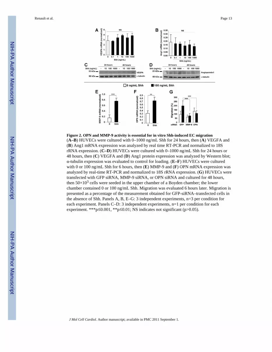
Figure 2. OPN and MMP-9 activity is essential for in vitro Shh-induced EC migration(A–B) HUVECs were cultured with 0–1000 ng/mL Shh for 24 hours, then (A) VEGFA and(B) Ang1 mRNA expression was analyzed by real time RT-PCR and normalized to 18SrRNA expression. (C–D) HUVECs were cultured with 0–1000 ng/mL Shh for 24 hours or48 hours, then (C) VEGFA and (D) Ang1 protein expression was analyzed by Western blot;α-tubulin expression was evaluated to control for loading. (E–F) HUVECs were culturedwith 0 or 100 ng/mL Shh for 6 hours, then (E) MMP-9 and (F) OPN mRNA expression wasanalyzed by real-time RT-PCR and normalized to 18S rRNA expression. (G) HUVECs weretransfected with GFP siRNA, MMP-9 siRNA, or OPN siRNA and cultured for 48 hours,then 50×103 cells were seeded in the upper chamber of a Boyden chamber; the lowerchamber contained 0 or 100 ng/mL Shh. Migration was evaluated 6 hours later. Migration ispresented as a percentage of the measurement obtained for GFP-siRNA–transfected cells inthe absence of Shh. Panels A, B, E–G: 3 independent experiments, n=3 per condition foreach experiment. Panels C–D: 3 independent experiments, n=1 per condition for eachexperiment. ***p≤0.001, **p≤0.01; NS indicates not significant (p>0.05).
Renault et al. Page 13
J Mol Cell Cardiol. Author manuscript; available in PMC 2011 September 1.
NIH
-PA Author Manuscript
NIH
-PA Author Manuscript
NIH
-PA Author Manuscript
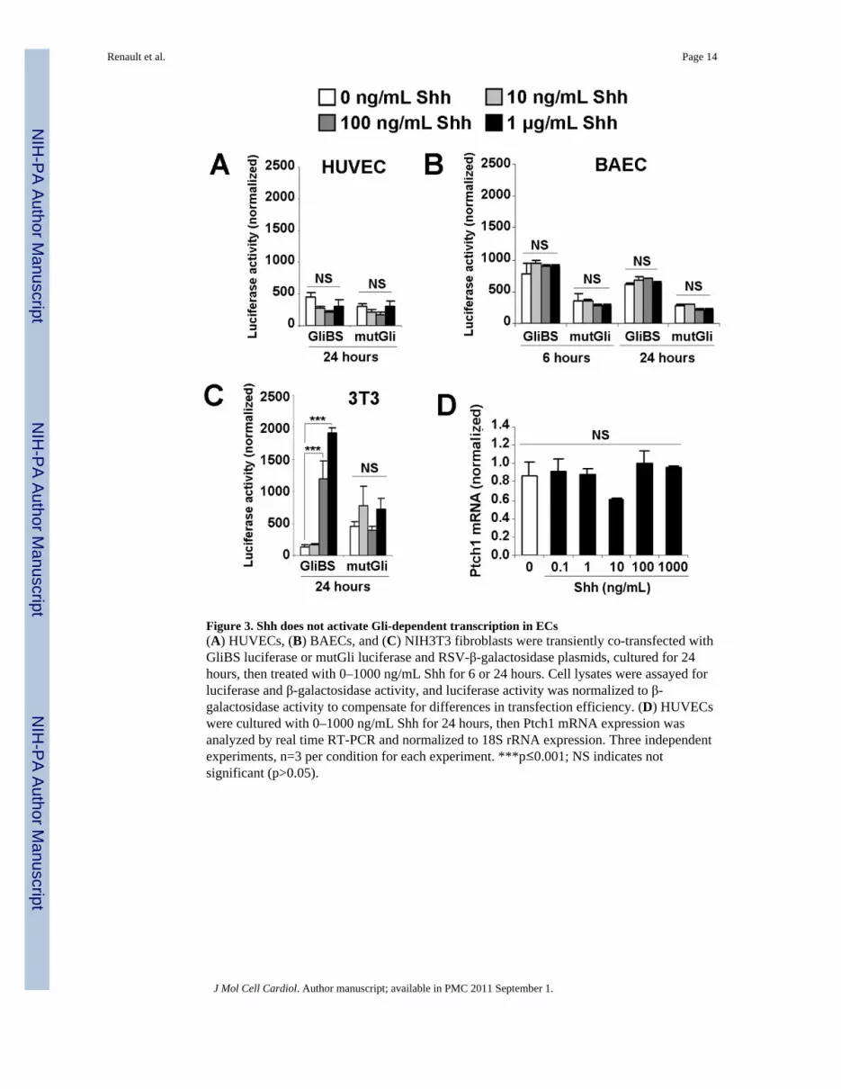
Figure 3. Shh does not activate Gli-dependent transcription in ECs(A) HUVECs, (B) BAECs, and (C) NIH3T3 fibroblasts were transiently co-transfected withGliBS luciferase or mutGli luciferase and RSV-β-galactosidase plasmids, cultured for 24hours, then treated with 0–1000 ng/mL Shh for 6 or 24 hours. Cell lysates were assayed forluciferase and β-galactosidase activity, and luciferase activity was normalized to β-galactosidase activity to compensate for differences in transfection efficiency. (D) HUVECswere cultured with 0–1000 ng/mL Shh for 24 hours, then Ptch1 mRNA expression wasanalyzed by real time RT-PCR and normalized to 18S rRNA expression. Three independentexperiments, n=3 per condition for each experiment. ***p≤0.001; NS indicates notsignificant (p>0.05).
Renault et al. Page 14
J Mol Cell Cardiol. Author manuscript; available in PMC 2011 September 1.
NIH
-PA Author Manuscript
NIH
-PA Author Manuscript
NIH
-PA Author Manuscript

Figure 4. Shh activates RhoA in ECs(A) Serum-starved BAECs and HUVECs were treated with 0 or 100 ng/mL Shh for 5minutes, then the level of RhoA-GTP was evaluated with the Rho pull down assay. (B)Serum-starved BAECs were treated with 0 or 100 ng/mL Shh for 10 minutes, then the actincytoskeleton was stained with an anti-actin antibody (green), and nuclei were stained withDAPI (blue). (C) HUVECs cultured in 1% FBS-containing medium were treated with 0 or100 ng/mL Shh in the presence or absence of 20 nM Y27632 for 10 minutes, then stainedfor Ezrin (red); nuclei were stained with DAPI (blue). Panel A: 3 independent experiments,n=1 per condition for each experiment; Panels B–C: 3 independent experiments, n=1 percondition for each experiment.
Renault et al. Page 15
J Mol Cell Cardiol. Author manuscript; available in PMC 2011 September 1.
NIH
-PA Author Manuscript
NIH
-PA Author Manuscript
NIH
-PA Author Manuscript

Figure 5. Shh-induced EC migration, tube formation, and gene expression are dependent onROCK(A) HUVECs and (B) BAECs were pretreated with 20 ng/mL Y27632, or control vehicle for30 minutes then seeded in the upper chamber of a modified Boyden chamber; the lowerchamber contained the indicated concentration of Shh. Migration was evaluated 6 hours laterand expressed as the fold-change from measurements obtained in the absence of Shh. (C–D)BAECs were transfected with plasmids containing an empty vector (pcDNA3) or plasmidsexpressing RhoA N19 or ROCK RB/PH, then starved for 24 hours in serum-free medium.(C) 50×103 transfected BAECs were seeded in the upper chamber of a modified Boydenchamber; the lower chamber contained 0 or 100 ng/mL Shh. Migration was evaluated 6hours later. (D) Transfected BAECs were suspended and cultured on Matrigel™ with orwithout 10 ng/mL Shh; tube formation was assessed 6 hours later, quantified as the totallength of tubes per high-power field. (E) BAECs were transfected with plasmids containingan empty vector (pCB6) or dominant-negative Ezrin (Ez Nter), starved for 24 hours inserum-free medium, then 50×103 cells were seeded in the upper chamber of a modifiedBoyden chamber; the lower chamber contained 0 or 100 ng/mL Shh. Migration wasevaluated 6 hours later. The results in panels C–E are presented as a percentage of themeasurements obtained for pcDNA3- or pCB6-transfected cells in the absence of Shh. (F–G) HUVECs were treated with 0 or 100 ng/mL Shh in the presence of 20 ng/mL Y27632, orcontrol vehicle for 6 hours. (F) OPN and (G) MMP-9 mRNA expression were analyzed byreal time RT-PCR and normalized to 18S rRNA expression. Three independent experiments,n=3 per condition for each experiment. ***p≤0.001, **p≤0.01, *p≤0.05.
Renault et al. Page 16
J Mol Cell Cardiol. Author manuscript; available in PMC 2011 September 1.
NIH
-PA Author Manuscript
NIH
-PA Author Manuscript
NIH
-PA Author Manuscript

Figure 6. Shh-induced corneal angiogenesis is dependent on the activity of Smo and the Rho/ROCK pathway and on MMP-9 and OPN expression, but not on Gli expressionPellets containing the indicated combinations of PBS, Shh, the Smo inhibitor cyclopamine(CYC), and the ROCK inhibitor Y27632 were implanted in the corneas of WT mice,MMP-9-knockout mice (MMP-9−/−) and their WT littermates, OPN-knockout mice(OPN−/−) and their WT littermates, Gli1-knockout (Gli1−/−) C57BL/6 mice, WT C57BL/6mice, Gli3-haploinsufficient (Gli3+/−) C3HeB/FeJ mice, and WT C3HeB/FeJ mice;angiogenesis was evaluated 8 days later via in vivo fluorecein-BS-1 lectin injection. n≥4mice (8 corneas) per group
Renault et al. Page 17
J Mol Cell Cardiol. Author manuscript; available in PMC 2011 September 1.
NIH
-PA Author Manuscript
NIH
-PA Author Manuscript
NIH
-PA Author Manuscript