Embed Size (px)
Citation preview

R E V I EW
A Review on Cutaneous and MusculoskeletalManifestations of CLOVES SyndromeEmel Öztürk Durmaz 1, Deniz Demircioğlu1, Pınar Yalınay Dikmen2, Yasemin Alanay3,Ahmet Alanay4, Cüyan Demirkesen5, Fatma Tokat5, Ercan Karaarslan6
1Department of Dermatology, Acıbadem Mehmet Ali Aydınlar University School of Medicine, İstanbul, Turkey; 2Department of Neurology, AcıbademMehmet Ali Aydınlar University School of Medicine, İstanbul, Turkey; 3Department of Pediatrics, Acıbadem Mehmet Ali Aydınlar University School ofMedicine, İstanbul, Turkey; 4Department of Orthopedics, Acıbadem Mehmet Ali Aydınlar University School of Medicine, İstanbul, Turkey;5Department of Pathology, Acıbadem Mehmet Ali Aydınlar University School of Medicine, İstanbul, Turkey; 6Department of Radiology, AcıbademMehmet Ali Aydınlar University School of Medicine, İstanbul, Turkey
Correspondence: Emel Öztürk Durmaz, Acıbadem Maslak Hospital, Büyükdere Caddesi 40, Maslak, Istanbul, 34457, Turkey, Tel +90 (212) 304 46 19,Fax +90 (212) 286 15 66, Email [email protected]
Abstract: CLOVES syndrome is a novel sporadic mosaic segmental overgrowth syndrome, currently categorized under the canopy ofPROS (PIK3CA-related overgrowth spectrum) disorders. All PROS disorders harbor heterozygous postzygotic activating somaticmutations involving the PIK3CA gene. As an upstream regulator of the PI3K/AKT/mTOR signal transduction pathway, activatingmutations of PIK3CA gene commence in uncontrolled growth of cutaneous, vascular (capillaries, veins, and lymphatics), adipose,neural, and musculoskeletal tissues. The excessive growth is segmental, patchy, asymmetric, and confined to body parts affected by themutation. The term ‘CLOVES’ is an acronym denoting congenital lipomatous overgrowth, vascular malformations, epidermal nevi andspinal (scoliosis) and/ or skeletal anomalies. The syndrome is characterized by an admixture of overgrown tissues, derived mainlyfrom mesoderm and neuroectoderm. Among PROS disorders, CLOVES syndrome represents the extreme end of the spectrum withmassive affection of almost the entire body. The syndrome might judiciously be treated with medications hampering with the PI3K/AKT/mTOR signal transduction pathway. This article aims at reviewing the cutaneous and musculoskeletal manifestations of CLOVESsyndrome, as the paradigm for PROS disorders. CLOVES syndrome and other PROS disorders are still misdiagnosed, underdiagnosed,underreported, and undertreated by the dermatology community.Keywords: CLOVES syndrome, PIK3CA-related overgrowth spectrum, cutaneous manifestations, port wine stains, lymphangiomas,lipomas, epidermal nevi
Definition and HistoryCLOVES syndrome (OMIM number 612918) is a recently described rare, sporadic (non-hereditary) complex mosaicovergrowth syndrome.1–3 It was initially described in 2007 by Sapp et al as a novel overgrowth syndrome and entitled as“CLOVE” syndrome.4 Although “CLOVE” syndrome had overlapping features with other overgrowth syndromes suchas Proteus syndrome, the affected patients could not fulfill the established diagnostic criteria for the other disorders.5,6
Upon recognition of skeletal and spinal abnormalities (scoliosis) as part of this new syndrome, the nomenclature has laterbeen revised by Alomari as ‘CLOVES’ syndrome.7 The term “CLOVES” is an acronym denoting congenital lipomatousovergrowth, vascular malformations, epidermal nevi and spinal (scoliosis) and/ or skeletal anomalies.1,2,8–13
EpidemiologyBecause of its heterogeneous nature and rarity, the number of published cases of CLOVES syndrome is under 200hitherto.11,14 The estimated incidence is less than 1:1,000,000.1,11,14,15 There is no gender predilection, and the syndromehas been reported in all races and ethnic groups.11,15 The disorder is congenital and has a prenatal onset; thus, it presentseither at birth or in early childhood.3,5,11,15–17 Segmental overgrowth is noticeable in most afflicted patients by one yearof age.18
Clinical, Cosmetic and Investigational Dermatology 2022:15 621–630 621© 2022 Öztürk Durmaz et al. This work is published and licensed by Dove Medical Press Limited. The full terms of this license are available at https://www.dovepress.com/terms.php and incorporate the Creative Commons Attribution – Non Commercial (unported, v3.0) License (http://creativecommons.org/licenses/by-nc/3.0/). By accessing
the work you hereby accept the Terms. Non-commercial uses of the work are permitted without any further permission from Dove Medical Press Limited, provided the work is properly attributed.For permission for commercial use of this work, please see paragraphs 4.2 and 5 of our Terms (https://www.dovepress.com/terms.php).
Clinical, Cosmetic and Investigational Dermatology Dovepressopen access to scientific and medical research
Open Access Full Text Article
Received: 29 November 2021Accepted: 17 March 2022Published: 13 April 2022
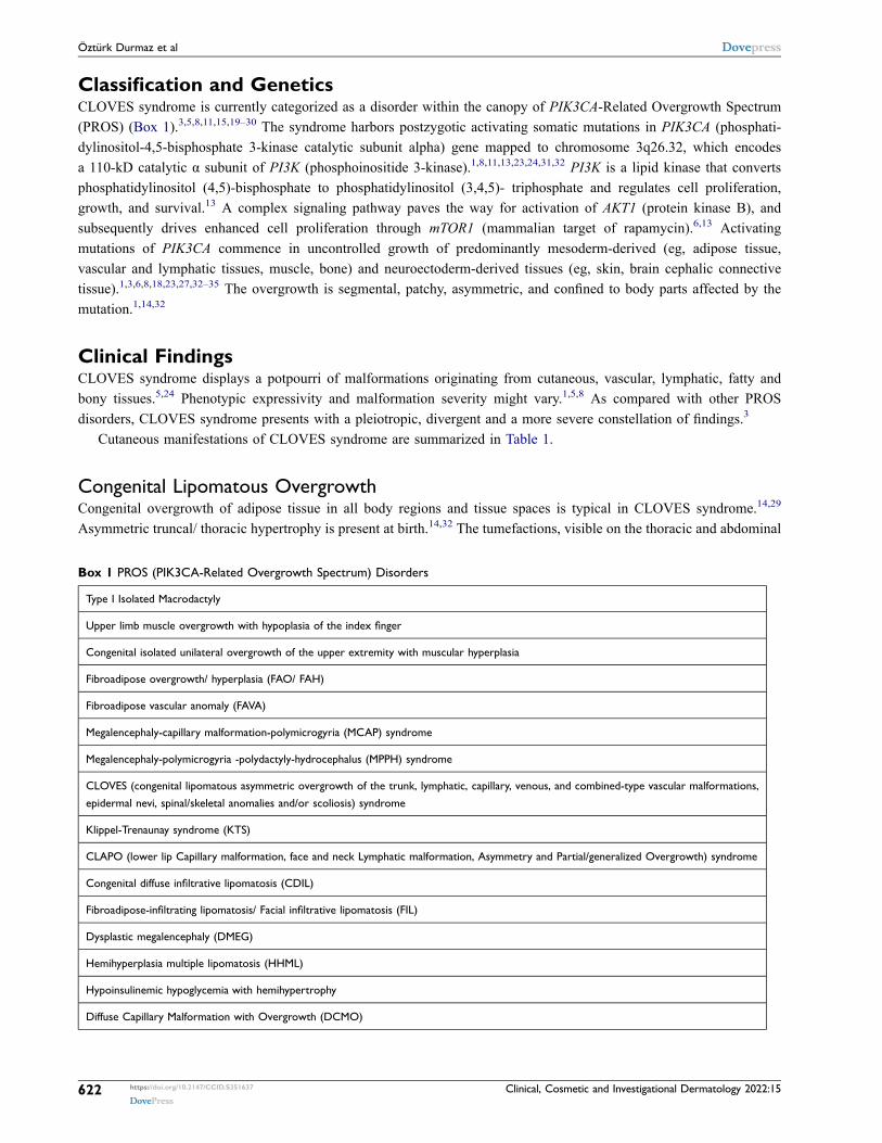
Classification and GeneticsCLOVES syndrome is currently categorized as a disorder within the canopy of PIK3CA-Related Overgrowth Spectrum(PROS) (Box 1).3,5,8,11,15,19–30 The syndrome harbors postzygotic activating somatic mutations in PIK3CA (phosphati-dylinositol-4,5-bisphosphate 3-kinase catalytic subunit alpha) gene mapped to chromosome 3q26.32, which encodesa 110-kD catalytic α subunit of PI3K (phosphoinositide 3-kinase).1,8,11,13,23,24,31,32 PI3K is a lipid kinase that convertsphosphatidylinositol (4,5)-bisphosphate to phosphatidylinositol (3,4,5)- triphosphate and regulates cell proliferation,growth, and survival.13 A complex signaling pathway paves the way for activation of AKT1 (protein kinase B), andsubsequently drives enhanced cell proliferation through mTOR1 (mammalian target of rapamycin).6,13 Activatingmutations of PIK3CA commence in uncontrolled growth of predominantly mesoderm-derived (eg, adipose tissue,vascular and lymphatic tissues, muscle, bone) and neuroectoderm-derived tissues (eg, skin, brain cephalic connectivetissue).1,3,6,8,18,23,27,32–35 The overgrowth is segmental, patchy, asymmetric, and confined to body parts affected by themutation.1,14,32
Clinical FindingsCLOVES syndrome displays a potpourri of malformations originating from cutaneous, vascular, lymphatic, fatty andbony tissues.5,24 Phenotypic expressivity and malformation severity might vary.1,5,8 As compared with other PROSdisorders, CLOVES syndrome presents with a pleiotropic, divergent and a more severe constellation of findings.3
Cutaneous manifestations of CLOVES syndrome are summarized in Table 1.
Congenital Lipomatous OvergrowthCongenital overgrowth of adipose tissue in all body regions and tissue spaces is typical in CLOVES syndrome.14,29
Asymmetric truncal/ thoracic hypertrophy is present at birth.14,32 The tumefactions, visible on the thoracic and abdominal
Box 1 PROS (PIK3CA-Related Overgrowth Spectrum) Disorders
Type I Isolated Macrodactyly
Upper limb muscle overgrowth with hypoplasia of the index finger
Congenital isolated unilateral overgrowth of the upper extremity with muscular hyperplasia
Fibroadipose overgrowth/ hyperplasia (FAO/ FAH)
Fibroadipose vascular anomaly (FAVA)
Megalencephaly-capillary malformation-polymicrogyria (MCAP) syndrome
Megalencephaly-polymicrogyria -polydactyly-hydrocephalus (MPPH) syndrome
CLOVES (congenital lipomatous asymmetric overgrowth of the trunk, lymphatic, capillary, venous, and combined-type vascular malformations,
epidermal nevi, spinal/skeletal anomalies and/or scoliosis) syndrome
Klippel-Trenaunay syndrome (KTS)
CLAPO (lower lip Capillary malformation, face and neck Lymphatic malformation, Asymmetry and Partial/generalized Overgrowth) syndrome
Congenital diffuse infiltrative lipomatosis (CDIL)
Fibroadipose-infiltrating lipomatosis/ Facial infiltrative lipomatosis (FIL)
Dysplastic megalencephaly (DMEG)
Hemihyperplasia multiple lipomatosis (HHML)
Hypoinsulinemic hypoglycemia with hemihypertrophy
Diffuse Capillary Malformation with Overgrowth (DCMO)
https://doi.org/10.2147/CCID.S351637
DovePress
Clinical, Cosmetic and Investigational Dermatology 2022:15622
Öztürk Durmaz et al Dovepress
Powered by TCPDF (www.tcpdf.org)
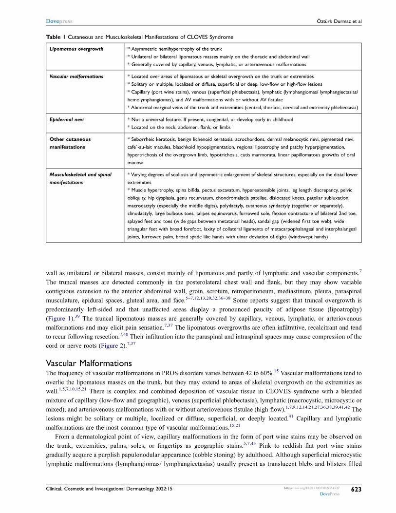
wall as unilateral or bilateral masses, consist mainly of lipomatous and partly of lymphatic and vascular components.7
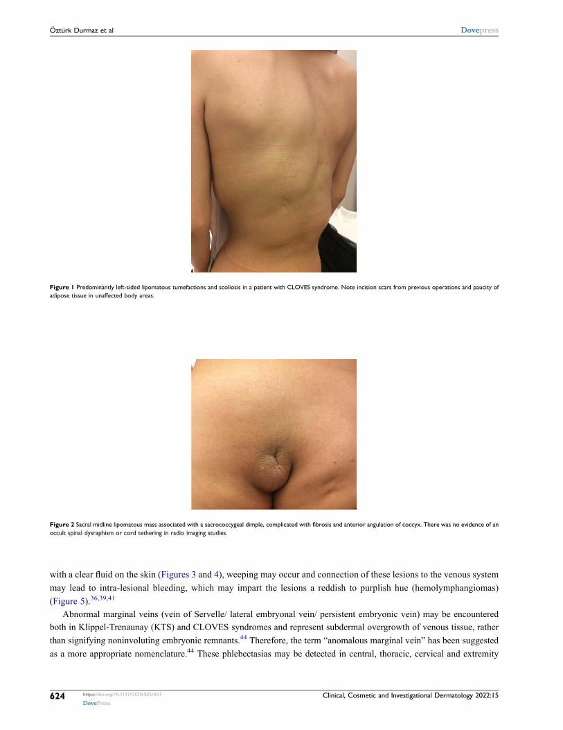
The truncal masses are detected commonly in the posterolateral chest wall and flank, but they may show variablecontiguous extension to the anterior abdominal wall, groin, scrotum, retroperitoneum, mediastinum, pleura, paraspinalmusculature, epidural spaces, gluteal area, and face.5–7,12,13,20,32,36–38 Some reports suggest that truncal overgrowth ispredominantly left-sided and that unaffected areas display a pronounced paucity of adipose tissue (lipoatrophy)(Figure 1).39 The truncal lipomatous masses are generally covered by capillary, venous, lymphatic, or arteriovenousmalformations and may elicit pain sensation.7,37 The lipomatous overgrowths are often infiltrative, recalcitrant and tendto recur following resection.7,40 Their infiltration into the paraspinal and intraspinal spaces may cause compression of thecord or nerve roots (Figure 2).7,37
Vascular MalformationsThe frequency of vascular malformations in PROS disorders varies between 42 to 60%.15 Vascular malformations tend tooverlie the lipomatous masses on the trunk, but they may extend to areas of skeletal overgrowth on the extremities aswell.1,5,7,10,15,21 There is complex and combined deposition of vascular tissue in CLOVES syndrome with a blendedmixture of capillary (low-flow and geographic), venous (superficial phlebectasia), lymphatic (macrocystic, microcystic ormixed), and arteriovenous malformations with or without arteriovenous fistulae (high-flow).1,7,9,12,14,21,27,36,38,39,41,42 Thelesions might be solitary or multiple, localized or diffuse, superficial, or deeply located.41 Capillary and lymphaticmalformations are the most common type of vascular malformations.15,21
From a dermatological point of view, capillary malformations in the form of port wine stains may be observed onthe trunk, extremities, palms, soles, or fingertips as geographic stains.5,7,43 Pink to reddish flat port wine stainsgradually acquire a purplish papulonodular appearance (cobble stoning) by adulthood. Although superficial microcysticlymphatic malformations (lymphangiomas/ lymphangiectasias) usually present as translucent blebs and blisters filled
Table 1 Cutaneous and Musculoskeletal Manifestations of CLOVES Syndrome
Lipomatous overgrowth * Asymmetric hemihypertrophy of the trunk
* Unilateral or bilateral lipomatous masses mainly on the thoracic and abdominal wall* Generally covered by capillary, venous, lymphatic, or arteriovenous malformations
Vascular malformations * Located over areas of lipomatous or skeletal overgrowth on the trunk or extremities* Solitary or multiple, localized or diffuse, superficial or deep, low-flow or high-flow lesions
* Capillary (port wine stains), venous (superficial phlebectasia), lymphatic (lymphangiomas/ lymphangiectasias/
hemolymphangiomas), and AV malformations with or without AV fistulae* Abnormal marginal veins of the trunk and extremities (central, thoracic, cervical and extremity phlebectasia)
Epidermal nevi * Not a universal feature. If present, congenital, or develop early in childhood* Located on the neck, abdomen, flank, or limbs
Other cutaneousmanifestations
* Seborrheic keratosis, benign lichenoid keratosis, acrochordons, dermal melanocytic nevi, pigmented nevi,cafe´-au-lait macules, blaschkoid hypopigmentation, regional lipoatrophy and patchy hyperpigmentation,
hypertrichosis of the overgrown limb, hypotrichosis, cutis marmorata, linear papillomatous growths of oral
mucosa
Musculoskeletal and spinalmanifestations
* Varying degrees of scoliosis and asymmetric enlargement of skeletal structures, especially on the distal lower
extremities* Muscle hypertrophy, spina bifida, pectus excavatum, hyperextensible joints, leg length discrepancy, pelvic
obliquity, hip dysplasia, genu recurvatum, chondromalacia patellae, dislocated knees, patellar subluxation,
macrodactyly (especially the middle digits), polydactyly, cutaneous syndactyly (together or separately),clinodactyly, large bulbous toes, talipes equinovarus, furrowed sole, flexion contracture of bilateral 2nd toe,
splayed feet and toes (wide gaps between metatarsal heads), sandal gap (widened first toe web), wide
triangular feet with broad forefoot, laxity of collateral ligaments of metacarpophalangeal and interphalangealjoints, furrowed palm, broad spade like hands with ulnar deviation of digits (windswept hands)
Clinical, Cosmetic and Investigational Dermatology 2022:15 https://doi.org/10.2147/CCID.S351637
DovePress623
Dovepress Öztürk Durmaz et al
Powered by TCPDF (www.tcpdf.org)
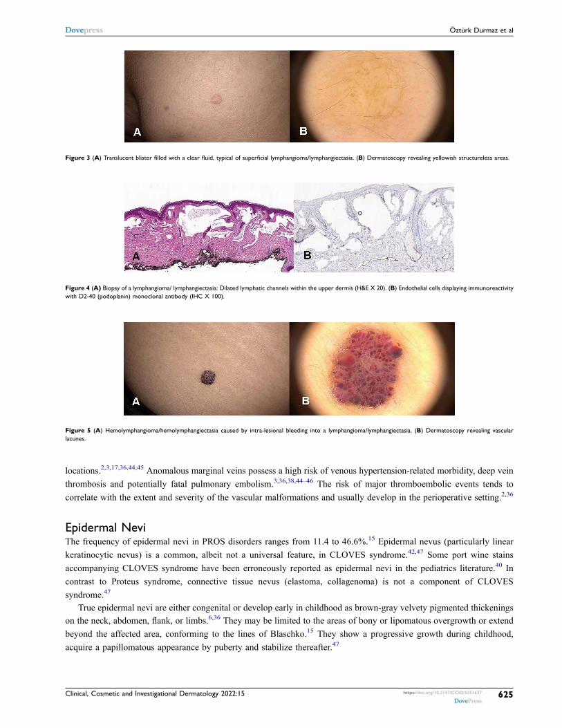
with a clear fluid on the skin (Figures 3 and 4), weeping may occur and connection of these lesions to the venous systemmay lead to intra-lesional bleeding, which may impart the lesions a reddish to purplish hue (hemolymphangiomas)(Figure 5).36,39,41
Abnormal marginal veins (vein of Servelle/ lateral embryonal vein/ persistent embryonic vein) may be encounteredboth in Klippel-Trenaunay (KTS) and CLOVES syndromes and represent subdermal overgrowth of venous tissue, ratherthan signifying noninvoluting embryonic remnants.44 Therefore, the term “anomalous marginal vein” has been suggestedas a more appropriate nomenclature.44 These phlebectasias may be detected in central, thoracic, cervical and extremity
Figure 2 Sacral midline lipomatous mass associated with a sacrococcygeal dimple, complicated with fibrosis and anterior angulation of coccyx. There was no evidence of anoccult spinal dysraphism or cord tethering in radio imaging studies.
Figure 1 Predominantly left-sided lipomatous tumefactions and scoliosis in a patient with CLOVES syndrome. Note incision scars from previous operations and paucity ofadipose tissue in unaffected body areas.
https://doi.org/10.2147/CCID.S351637
DovePress
Clinical, Cosmetic and Investigational Dermatology 2022:15624
Öztürk Durmaz et al Dovepress
Powered by TCPDF (www.tcpdf.org)
locations.2,3,17,36,44,45 Anomalous marginal veins possess a high risk of venous hypertension-related morbidity, deep veinthrombosis and potentially fatal pulmonary embolism.3,36,38,44–46 The risk of major thromboembolic events tends tocorrelate with the extent and severity of the vascular malformations and usually develop in the perioperative setting.2,36
Epidermal NeviThe frequency of epidermal nevi in PROS disorders ranges from 11.4 to 46.6%.15 Epidermal nevus (particularly linearkeratinocytic nevus) is a common, albeit not a universal feature, in CLOVES syndrome.42,47 Some port wine stainsaccompanying CLOVES syndrome have been erroneously reported as epidermal nevi in the pediatrics literature.40 Incontrast to Proteus syndrome, connective tissue nevus (elastoma, collagenoma) is not a component of CLOVESsyndrome.47
True epidermal nevi are either congenital or develop early in childhood as brown-gray velvety pigmented thickeningson the neck, abdomen, flank, or limbs.6,36 They may be limited to the areas of bony or lipomatous overgrowth or extendbeyond the affected area, conforming to the lines of Blaschko.15 They show a progressive growth during childhood,acquire a papillomatous appearance by puberty and stabilize thereafter.47
Figure 3 (A) Translucent blister filled with a clear fluid, typical of superficial lymphangioma/lymphangiectasia. (B) Dermatoscopy revealing yellowish structureless areas.
Figure 4 (A) Biopsy of a lymphangioma/ lymphangiectasia: Dilated lymphatic channels within the upper dermis (H&E X 20). (B) Endothelial cells displaying immunoreactivitywith D2-40 (podoplanin) monoclonal antibody (IHC X 100).
Figure 5 (A) Hemolymphangioma/hemolymphangiectasia caused by intra-lesional bleeding into a lymphangioma/lymphangiectasia. (B) Dermatoscopy revealing vascularlacunes.
Clinical, Cosmetic and Investigational Dermatology 2022:15 https://doi.org/10.2147/CCID.S351637
DovePress625
Dovepress Öztürk Durmaz et al
Powered by TCPDF (www.tcpdf.org)
Scoliosis and/or Skeletal AbnormalitiesSkeletal malformations may be severely deforming and consist of varying degrees of scoliosis and asymmetric enlarge-ment of skeletal structures on the extremities.6,7,13,37 Scoliosis may be congenital, or it may develop during childhood,sometimes because of lower limb asymmetry (Figure 1).36 Bony overgrowth most commonly affects the lowerextremities; distal segment is involved more frequently than the proximal segment.7,39 As the disease progresses bytime, proximal segment may also be involved.15,39 In contrast to Proteus syndrome, bone hypertrophy is not rapidlyprogressive or distorting in CLOVES syndrome.1
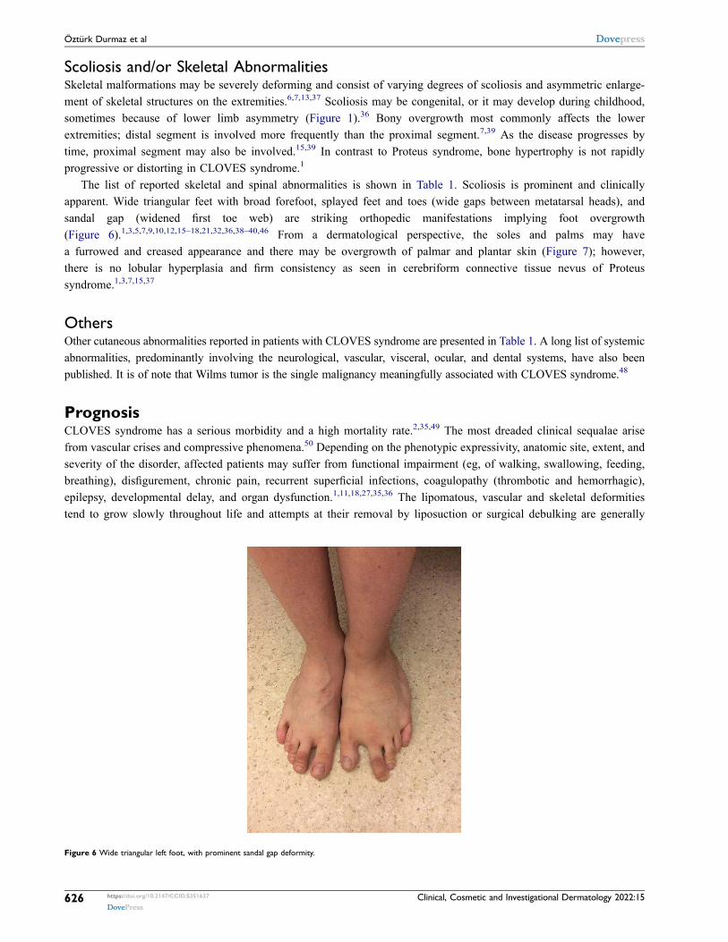
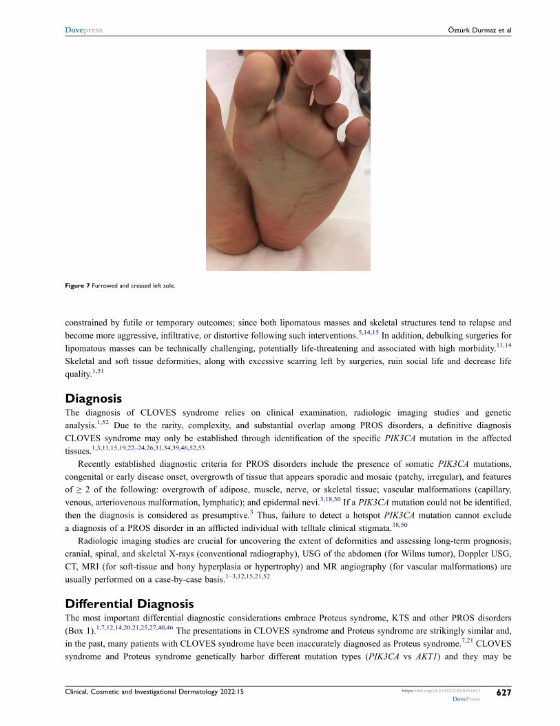
The list of reported skeletal and spinal abnormalities is shown in Table 1. Scoliosis is prominent and clinicallyapparent. Wide triangular feet with broad forefoot, splayed feet and toes (wide gaps between metatarsal heads), andsandal gap (widened first toe web) are striking orthopedic manifestations implying foot overgrowth(Figure 6).1,3,5,7,9,10,12,15–18,21,32,36,38–40,46 From a dermatological perspective, the soles and palms may havea furrowed and creased appearance and there may be overgrowth of palmar and plantar skin (Figure 7); however,there is no lobular hyperplasia and firm consistency as seen in cerebriform connective tissue nevus of Proteussyndrome.1,3,7,15,37
OthersOther cutaneous abnormalities reported in patients with CLOVES syndrome are presented in Table 1. A long list of systemicabnormalities, predominantly involving the neurological, vascular, visceral, ocular, and dental systems, have also beenpublished. It is of note that Wilms tumor is the single malignancy meaningfully associated with CLOVES syndrome.48
PrognosisCLOVES syndrome has a serious morbidity and a high mortality rate.2,35,49 The most dreaded clinical sequalae arisefrom vascular crises and compressive phenomena.50 Depending on the phenotypic expressivity, anatomic site, extent, andseverity of the disorder, affected patients may suffer from functional impairment (eg, of walking, swallowing, feeding,breathing), disfigurement, chronic pain, recurrent superficial infections, coagulopathy (thrombotic and hemorrhagic),epilepsy, developmental delay, and organ dysfunction.1,11,18,27,35,36 The lipomatous, vascular and skeletal deformitiestend to grow slowly throughout life and attempts at their removal by liposuction or surgical debulking are generally
Figure 6 Wide triangular left foot, with prominent sandal gap deformity.
https://doi.org/10.2147/CCID.S351637
DovePress
Clinical, Cosmetic and Investigational Dermatology 2022:15626
Öztürk Durmaz et al Dovepress
Powered by TCPDF (www.tcpdf.org)
constrained by futile or temporary outcomes; since both lipomatous masses and skeletal structures tend to relapse andbecome more aggressive, infiltrative, or distortive following such interventions.5,14,15 In addition, debulking surgeries forlipomatous masses can be technically challenging, potentially life-threatening and associated with high morbidity.11,14
Skeletal and soft tissue deformities, along with excessive scarring left by surgeries, ruin social life and decrease lifequality.1,51
DiagnosisThe diagnosis of CLOVES syndrome relies on clinical examination, radiologic imaging studies and geneticanalysis.1,52 Due to the rarity, complexity, and substantial overlap among PROS disorders, a definitive diagnosisCLOVES syndrome may only be established through identification of the specific PIK3CA mutation in the affectedtissues.1,3,11,15,19,22–24,26,31,34,39,46,52,53
Recently established diagnostic criteria for PROS disorders include the presence of somatic PIK3CA mutations,congenital or early disease onset, overgrowth of tissue that appears sporadic and mosaic (patchy, irregular), and featuresof ≥ 2 of the following: overgrowth of adipose, muscle, nerve, or skeletal tissue; vascular malformations (capillary,venous, arteriovenous malformation, lymphatic); and epidermal nevi.3,18,30 If a PIK3CA mutation could not be identified,then the diagnosis is considered as presumptive.3 Thus, failure to detect a hotspot PIK3CA mutation cannot excludea diagnosis of a PROS disorder in an afflicted individual with telltale clinical stigmata.38,50
Radiologic imaging studies are crucial for uncovering the extent of deformities and assessing long-term prognosis;cranial, spinal, and skeletal X-rays (conventional radiography), USG of the abdomen (for Wilms tumor), Doppler USG,CT, MRI (for soft-tissue and bony hyperplasia or hypertrophy) and MR angiography (for vascular malformations) areusually performed on a case-by-case basis.1–3,12,15,21,52
Differential DiagnosisThe most important differential diagnostic considerations embrace Proteus syndrome, KTS and other PROS disorders(Box 1).1,7,12,14,20,21,25,27,40,46 The presentations in CLOVES syndrome and Proteus syndrome are strikingly similar and,in the past, many patients with CLOVES syndrome have been inaccurately diagnosed as Proteus syndrome.7,21 CLOVESsyndrome and Proteus syndrome genetically harbor different mutation types (PIK3CA vs AKT1) and they may be
Figure 7 Furrowed and creased left sole.
Clinical, Cosmetic and Investigational Dermatology 2022:15 https://doi.org/10.2147/CCID.S351637
DovePress627
Dovepress Öztürk Durmaz et al
Powered by TCPDF (www.tcpdf.org)

clinically distinguished by the absence of cerebriform connective tissue nevi and visceral involvement in the former, anda postnatal onset of a severely distorting overgrowth and absence of truncal fatty-vascular overgrowth, paraspinal fast-flow lesions and acral abnormalities in the latter.5,7,12,21,31,38,39,52,54 Overgrowth in CLOVES syndrome is detailed as“ballooning” (gradual increase in the volume of the soft tissue mass), congenital, slowly progressive, mild, symmetric,and proportionate, while that in Proteus syndrome is acknowledged as distorted, postnatal, rapidly progressive, severe,asymmetric, and disproportionate (out of proportion to normal body growth).1,10,14,18,31,40,42,54 CLOVES syndrome andKTS may be distinguished by the absence of truncal involvement and preferential affection of the lower extremities in thelatter.14,15,21,46 High-flow AVM and spinal/ paraspinal AVM are features of CLOVES syndrome and Parkes-Webersyndrome, but not that of KTS.55
TreatmentThere is no specific cure for CLOVES syndrome.49 Until recently, patients with CLOVES syndrome were managedpalliatively by multidisciplinary, staged debulking surgeries for lipomatous and skeletal overgrowths (surgical excision,extremity or finger/ toe amputation, surgical correction of scoliosis), and vascular interventional techniques (sclerotherapyfor macrocystic lymphatic malformations, laser therapy for capillary malformations, coil embolization procedures for largevenous malformations, superior vena cava filters for central and thoracic phlebectasias).2,12,14,15,18,20,21,35,36,38,40,50 Patientswith low-flow malformations were prescribed anticoagulant medications for the prevention of thromboembolic disease.3,21,36
Identification of causative PIK3CA mutations empowered the use of older medications impeding the geneticpathway and the development of novel medications directly targeting the abnormal PIK3CA gene itself.20,34,49
Theoretically, such medications could halt the progressive overgrowth of tissues in PROS disorders.6 Sirolimus(rapamycin) and its congeners (everolimus, ridaforolimus, temsirolimus) are mTOR inhibitors, originally used asimmunosuppressant and anti-tumor medications.5,18,21,27,35 Oral sirolimus has been shown to exert anti-angiogenic,anti-lymphangiogenic and anti-proliferative effects in 85% of adults and children with PROS disorders.11,21,27,35,50,56
It is effective in low-flow vascular malformations (microcystic lymphatic malformations, and venous malformations)and it can reduce the volume of lipomatous overgrowths as well.11,21,27,52,55,57 Currently, several PI3K pathwayinhibitors, that have originally been developed for the treatment of cancers, are undergoing trials in PROSdisorders.20,50 Among these are alpelisib (BYL719, Novartis), taselisib (GDC032, Roche), pictilisib (GDC-0941),copanlisib (BAY 80–6946) and dactolisib (dual PI3K /mTOR inhibitor).5,37,50 Based on limited evidence, a reductionin the size and volume of lipomatous, vascular and skeletal overgrowths might be attained with low doses of PI3Kpathway inhibitors in PROS disorders.11,37,49,50,58,59 A pan AKT inhibitor miransertib (MK-7075, formerly ARQ092)is an emerging therapeutic option in the horizon.59–61 As the outcome of these treatments is suppressive, temporary,and partial (rather than curative, persistent, and complete), prolonged, and even life-long administration will berequired in affected patients.18,27,50 Thus, despite encouraging clinical efficacy, long-term safety issues of thesemedications will pose a therapeutic dilemma for the clinician27,37,46,56,57,62 Nevertheless, the future holds promise fortherapeutic optimism in CLOVES syndrome, KTS and other PROS disorders.
FundingThere is no funding to report.
DisclosureThe authors report no conflicts of interest in this work.
References1. Mahajan VK, Gupta M, Chauhan P, Mehta KS. Cloves syndrome: a rare disorder of overgrowth with unusual features - an uncommon phenotype?Indian Dermatol Online J. 2019;10(4):447–452. doi:10.4103/idoj.IDOJ_418_18
2. Alomari AI, Burrows PE, Lee EY, Hedequist DJ, Mulliken JB, Fishman SJ. CLOVES syndrome with thoracic and central phlebectasia: increasedrisk of pulmonary embolism. J Thorac Cardiovasc Surg. 2010;140(2):459–463. doi:10.1016/j.jtcvs.2010.04.023
3. Keppler-Noreuil KM, Rios JJ, Parker VE, et al. PIK3CA-related overgrowth spectrum (PROS): diagnostic and testing eligibility criteria, differentialdiagnosis, and evaluation. Am J Med Genet A. 2015;167A(2):287–295. doi:10.1002/ajmg.a.36836
https://doi.org/10.2147/CCID.S351637
DovePress
Clinical, Cosmetic and Investigational Dermatology 2022:15628
Öztürk Durmaz et al Dovepress
Powered by TCPDF (www.tcpdf.org)

4. Sapp JC, Turner JT, van de Kamp JM, van Dijk FS, Lowry RB, Biesecker LG. Newly delineated syndrome of congenital lipomatous overgrowth,vascular malformations, and epidermal nevi (CLOVE syndrome) in seven patients. Am J Med Genet A. 2007;143A(24):2944–2958. doi:10.1002/ajmg.a.32023
5. Akgumus G, Chang F, Li MM. Overgrowth syndromes caused by somatic variants in the phosphatidylinositol 3-Kinase/AKT/Mammalian target ofrapamycin pathway. J Mol Diagn. 2017;19(4):487–497. doi:10.1016/j.jmoldx.2017.04.001
6. Vahidnezhad H, Youssefian L, Uitto J. Molecular genetics of the PI3K-AKT-mTOR pathway in genodermatoses: diagnostic implications andtreatment opportunities. J Invest Dermatol. 2016;136(1):15–23. doi:10.1038/JID.2015.331
7. Alomari AI. Characterization of a distinct syndrome that associates complex truncal overgrowth, vascular, and acral anomalies: a descriptive studyof 18 cases of CLOVES syndrome. Clin Dysmorphol. 2009;18(1):1–7. doi:10.1097/MCD.0b013e328317a716
8. Hanafusa H, Morisada N, Nomura T, et al. A girl with CLOVES syndrome with a recurrent PIK3CA somatic mutation and pancreatic steatosis.Hum Genome Var. 2019;6:31. doi:10.1038/s41439-019-0063-9
9. Gopal B, Keshava SN, Selvaraj D. A rare newly described overgrowth syndrome with vascular malformations-Cloves syndrome. Indian J RadiolImaging. 2015;25(1):71–73. doi:10.4103/0971-3026.150166
10. Puvabanditsin S, Memon N, Chekmareva M, Di Stefano V, Mehta R. Cloves syndrome: a case report and perinatal diagnostic findings. GenetCouns. 2014;25(3):265–270.
11. Alomar S, Khedr RE, Alajlan S. CLOVES syndrome in a nine-month-old infant. Cureus. 2019;11(9):e5772. doi:10.7759/cureus.577212. Alomari AI, Chaudry G, Rodesch G, et al. Complex spinal-paraspinal fast-flow lesions in CLOVES syndrome: analysis of clinical and imaging
findings in 6 patients. AJNR Am J Neuroradiol. 2011;32(10):1812–1817. doi:10.3174/ajnr.A234913. Vahidnezhad H, Youssefian L, Baghdadi T, et al. Phenotypic heterogeneity in PIK3CA-related overgrowth spectrum. Br J Dermatol. 2016;175
(4):810–814. doi:10.1111/bjd.1461814. Quinn KE, Infante J, Thorson W, Thorson CM. Unique case of congenital lipomatous overgrowth with vascular malformations, epidermal nevi, and
skeletal/spinal anomalies syndrome in a pediatric patient. Cureus. 2020;12(9):e10737. doi:10.7759/cureus.1073715. Mathew L, George R, Sudhakar S, Keshava SN, Fouzia NA. Clinical profile of overgrowth syndromes consistent with PROS (PIK3CA-Related
Overgrowth Syndromes)-A case series. Indian Dermatol Online J. 2020;11(5):738–746. doi:10.4103/idoj.IDOJ_520_1916. Fernandez-Pineda I, Fajardo M, Chaudry G, Alomari AI. Perinatal clinical and imaging features of CLOVES syndrome. Pediatr Radiol. 2010;40
(8):1436–1439. doi:10.1007/s00247-010-1559-017. Damian L, Lebovici A, Pamfil C, Belizna C, Vulturar R. Rheumatoid arthritis and CLOVES syndrome: a tricky diagnosis. Diagnostics. 2020;10
(7):467. doi:10.3390/diagnostics1007046718. Keppler-Noreuil KM, Parker VE, Darling TN, Martinez-Agosto JA. Somatic overgrowth disorders of the PI3K/AKT/mTOR pathway & therapeutic
strategies. Am J Med Genet C Semin Med Genet. 2016;172(4):402–421. doi:10.1002/ajmg.c.3153119. Chang F, Liu L, Fang E, et al. Molecular diagnosis of mosaic overgrowth syndromes using a custom-designed next-generation sequencing panel.
J Mol Diagn. 2017;19(4):613–624. doi:10.1016/j.jmoldx.2017.04.00620. Loconte DC, Grossi V, Bozzao C, et al. Molecular and functional characterization of three different postzygotic mutations in PIK3CA-Related
Overgrowth Spectrum (PROS) patients: effects on PI3K/AKT/mTOR signaling and sensitivity to PIK3 inhibitors. PLoS One. 2015;10(4):e0123092.doi:10.1371/journal.pone.0123092
21. Bertino F, Braithwaite KA, Hawkins CM, et al. Congenital limb overgrowth syndromes associated with vascular anomalies. Radiographics.2019;39(2):491–515. doi:10.1148/rg.2019180136
22. Youssefian L, Vahidnezhad H, Baghdadi T, et al. Fibroadipose hyperplasia versus Proteus syndrome: segmental overgrowth with a mosaic mutationin the PIK3CA gene. J Invest Dermatol. 2015;135(5):1450–1453. doi:10.1038/jid.2015.15
23. Michel ME, Konczyk DJ, Yeung KS, et al. Causal somatic mutations in urine DNA from persons with the CLOVES subgroup of thePIK3CA-related overgrowth spectrum. Clin Genet. 2018;93(5):1075–1080. doi:10.1111/cge.13195
24. Kurek KC, Luks VL, Ayturk UM, et al. Somatic mosaic activating mutations in PIK3CA cause CLOVES syndrome. Am J Hum Genet. 2012;90(6):1108–1115. doi:10.1016/j.ajhg.2012.05.006
25. Rodriguez-Laguna L, Ibañez K, Gordo G, et al. CLAPO syndrome: identification of somatic activating PIK3CA mutations and delineation of thenatural history and phenotype. Genet Med. 2018;20(8):882–889. doi:10.1038/gim.2017.200
26. Kuentz P, St-Onge J, Duffourd Y, et al. Molecular diagnosis of PIK3CA-related overgrowth spectrum (PROS) in 162 patients and recommendationsfor genetic testing. Genet Med. 2017;19(9):989–997. doi:10.1038/gim.2016.220
27. de Grazia R, Giordano C, Cossio L, Downey C, Delucchi Á, Kramer D. CLOVES syndrome: treatment with oral rapamycin. Report of two cases.Rev Chil Pediatr. 2019;90(6):662–667. doi:10.32641/rchped.v90i6.1025
28. Alsaedi SA, Qurashi O, Bajunaid M, Altalhi AA, Shawli AM. One of the first cases with PIK3CA-related Overgrowth Spectrum (PROS) in SaudiArabia: a case report and literature review. Cureus. 2020;12(1):e6586. doi:10.7759/cureus.6586
29. Castiglioni C, Bertini E, Orellana P, et al. Activating PIK3CA somatic mutation in congenital unilateral isolated muscle overgrowth of the upperextremity. Am J Med Genet A. 2014;164A(9):2365–2369. doi:10.1002/ajmg.a.36651
30. Roth GM, Ferguson N, Wanat KA. Segmental epidermal nevus and mucosal neuromas associated with PIK3CA-related overgrowth spectrumdisorder. JAAD Case Rep. 2018;4(10):1080–1082. doi:10.1016/j.jdcr.2018.08.023
31. Vahidnezhad H, Youssefian L, Uitto J. Klippel-Trenaunay syndrome belongs to the PIK3CA-related overgrowth spectrum (PROS). Exp Dermatol.2016;25(1):17–19. doi:10.1111/exd.12826
32. Panteliades M, Silva CM, Gontijo B. What is your diagnosis? An Bras Dermatol. 2016;91(3):378–380. doi:10.1590/abd1806-4841.2016589733. Collins M, Krochmalnek E, Alsubhi S, Srour M. Teaching NeuroImages: CLOVES syndrome. Neurology. 2021;96(10):e1487–e1488. doi:10.1212/
WNL.000000000001085634. Lalonde E, Ebrahimzadeh J, Rafferty K, et al. Molecular diagnosis of somatic overgrowth conditions: a single-center experience. Mol Genet
Genomic Med. 2019;7(3):e536. doi:10.1002/mgg3.53635. Parker VER, Keppler-Noreuil KM, Faivre L, et al. Safety and efficacy of low-dose sirolimus in the PIK3CA-related overgrowth spectrum. Genet
Med. 2019;21(5):1189–1198. doi:10.1038/s41436-018-0297-936. Acosta S, Torres V, Paulos M, Cifuentes I. CLOVES syndrome: severe neonatal presentation. J Clin Diagn Res. 2017;11(4):TR01–TR03.
doi:10.7860/JCDR/2017/23801.9719
Clinical, Cosmetic and Investigational Dermatology 2022:15 https://doi.org/10.2147/CCID.S351637
DovePress629
Dovepress Öztürk Durmaz et al
Powered by TCPDF (www.tcpdf.org)
37. Manor J, Lalani SR. Overgrowth syndromes-evaluation, diagnosis, and management. Front Pediatr. 2020;8:574857. doi:10.3389/fped.2020.57485738. Mirzaa G, Conway R, Graham JM Jr, Dobyns WB. PIK3CA-related segmental overgrowth. In: Adam MP, Ardinger HH, Pagon RA, et al., editors.
GeneReviews®. Seattle: University of Washington, Seattle; 2013:1993–2020.39. Keppler-Noreuil KM, Sapp JC, Lindhurst MJ, et al. Clinical delineation and natural history of the PIK3CA-related overgrowth spectrum. Am J Med
Genet A. 2014;164A(7):1713–1733. doi:10.1002/ajmg.a.3655240. Sarici D, Akin MA, Kurtoglu S, Tubas F, Sarici SU. A neonate with CLOVES Syndrome. Case Rep Pediatr. 2014;2014:845074. doi:10.1155/2014/
84507441. Del Pozo J, Gómez-Tellado M, López-Gutiérrez JC. Vascular malformations in childhood. Actas Dermosifiliogr. 2012;103(8):661–678.
doi:10.1016/j.ad.2011.12.00642. Biesbroeck L, Brandling-Bennett HA. Update on epidermal nevi and associated syndromes. Curr Derm Rep. 2012;1:186–194. doi:10.1007/s13671-
012-0025-743. López-Gutiérrez JC, Redondo P, Ivars M. Fingertip capillary malformation and associated disorders: report of 9 cases. Pediatrics. 2017;140(1):
e20162967. doi:10.1542/peds.2016-296744. Lim Y, Fereydooni A, Brahmandam A, Dardik A, Choate K, Nassiri N. Mechanochemical and surgical ablation of an anomalous upper extremity
marginal vein in CLOVES syndrome identifies PIK3CA as the culprit gene mutation. J Vasc Surg Cases Innov Tech. 2020;6(3):438–442.doi:10.1016/j.jvscit.2020.05.013
45. Abu-Haniyeh A, Alkukhun L, Al-Natour M, Hassan M, Tonelli A. Arteriovenous malformation in CLOVES syndrome. Med Rep Case Stud.2017;2:135.
46. Pandita A, Panghal A, Gupta G, Naranje KM. Overgrowth syndrome in neonates: a rare case series with a review of the literature. BMJ Case Rep.2019;12(1):e225640. doi:10.1136/bcr-2018-225640
47. Garcias-Ladaria J, Cuadrado Rosón M, Pascual-López M. Epidermal nevi and related syndromes – part 1: keratinocytic nevi. Actas Dermosifiliogr.2018;109(8):677–686. doi:10.1016/j.ad.2018.05.005
48. Weinstein B, Henderson-Jackson E, Cruse CW, Brohl AS. PIK3CA-Related Overgrowth Syndrome (PROS) and angiosarcoma: a case report.Eplasty. 2020;20:ic6.
49. Venot Q, Blanc T, Rabia SH, et al. Targeted therapy in patients with PIK3CA-related overgrowth syndrome. Nature. 2018;558(7711):540–546.doi:10.1038/s41586-018-0217-9
50. Madsen RR, Vanhaesebroeck B, Semple RK. Cancer-Associated PIK3CA Mutations in Overgrowth Disorders. Trends Mol Med. 2018;24(10):856–870. doi:10.1016/j.molmed.2018.08.003
51. Steiner JE, Cottrell CE, Streicher JL, et al. Scarring in patients with PIK3CA-related overgrowth syndromes. JAMA Dermatol. 2018;154(4):452–455. doi:10.1001/jamadermatol.2017.6189
52. Bertino F, Chaudry G. Overgrowth syndromes associated with vascular anomalies. Semin Roentgenol. 2019;54(4):349–358. doi:10.1053/j.ro.2019.06.005
53. Mirzaa G, Timms AE, Conti V, et al. PIK3CA-associated developmental disorders exhibit distinct classes of mutations with variable expression andtissue distribution. JCI Insight. 2016;1(9):e87623. doi:10.1172/jci.insight.87623
54. Gucev ZS, Tasic V, Jancevska A, et al. Congenital lipomatous overgrowth, vascular malformations, and epidermal nevi (CLOVE) syndrome: CNSmalformations and seizures may be a component of this disorder. Am J Med Genet A. 2008;146A(20):2688–2690. doi:10.1002/ajmg.a.32515
55. Alomari AI, Orbach DB, Mulliken JB, et al. Klippel-trenaunay syndrome and spinal arteriovenous malformations: an erroneous association. AJNRAm J Neuroradiol. 2010;31(9):1608–1612. doi:10.3174/ajnr.A2167
56. Adams DM, Trenor CC, Hammill AM 3rd, Hammill AM, et al. Efficacy and safety of sirolimus in the treatment of complicated vascular anomalies.Pediatrics. 2016;137(2):e20153257. doi:10.1542/peds.2015-3257
57. Kinsler VA, Boccara O, Fraitag S, Torrelo A, Vabres P, Diociaiuti A. Mosaic abnormalities of the skin: review and guidelines from the EuropeanReference Network for rare skin diseases. Br J Dermatol. 2020;182(3):552–563. doi:10.1111/bjd.17924
58. Adashek JJ, Kato S, Lippman SM, Kurzrock R. The paradox of cancer genes in non-malignant conditions: implications for precision medicine.Genome Med. 2020;12(1):16. doi:10.1186/s13073-020-0714-y
59. Venot Q, Canaud G. PIK3CA-related overgrowth spectrum: animal model and drug discovery. C R Biol. 2021;344(2):189–201. doi:10.5802/crbiol.50
60. Yan W, Zhang B, Wang H, et al. Somatic frameshift mutation in PIK3CA causes CLOVES syndrome by provoking PI3K/AKT/mTOR pathway.Hereditas. 2021;158(1):18. doi:10.1186/s41065-021-00184-y
61. Canaud G, Hammill AM, Adams D, Vikkula M, Keppler-Noreuil KM. A review of mechanisms of disease across PIK3CA-related disorders withvascular manifestations. Orphanet J Rare Dis. 2021;16(1):306. doi:10.1186/s13023-021-01929-8
62. Luu M, Vabres P, Devilliers H, et al. Safety and efficacy of low-dose PI3K inhibitor taselisib in adult patients with CLOVES and Klippel-Trenaunaysyndrome (KTS): the TOTEM trial, a Phase 1/2 multicenter, open-label, single-arm study. Genet Med. 2021;23(12):2433–2442. doi:10.1038/s41436-021-01290-y
Clinical, Cosmetic and Investigational Dermatology Dovepress
Publish your work in this journalClinical, Cosmetic and Investigational Dermatology is an international, peer-reviewed, open access, online journal that focuses on the latestclinical and experimental research in all aspects of skin disease and cosmetic interventions. This journal is indexed on CAS. The manuscriptmanagement system is completely online and includes a very quick and fair peer-review system, which is all easy to use. Visit http://www.dovepress.com/testimonials.php to read real quotes from published authors.
Submit your manuscript here: https://www.dovepress.com/clinical-cosmetic-and-investigational-dermatology-journal
DovePress Clinical, Cosmetic and Investigational Dermatology 2022:15630
Öztürk Durmaz et al Dovepress
Powered by TCPDF (www.tcpdf.org)