Embed Size (px)
Citation preview
Ryanodine Receptor Luminal Ca2þ Regulation: Swapping Calsequestrinand Channel Isoforms
Jia Qin,† Giorgia Valle,‡ Alma Nani,† Haiyan Chen,† Josefina Ramos-Franco,† Alessandra Nori,‡ Pompeo Volpe,‡
and Michael Fill†*†Department of Molecular Biophysics and Physiology, Rush University Medical Center, Chicago, Illinois; and ‡Department of ExperimentalBiomedical Sciences, University of Padova, Padua, Italy
ABSTRACT Sarcoplasmic reticulum (SR) Ca2þ release in striated muscle is mediated by a multiprotein complex that includesthe ryanodine receptor (RyR) Ca2þ channel and the intra-SR Ca2þ buffering protein calsequestrin (CSQ). Besides its bufferingrole, CSQ is thought to regulate RyR channel function. Here, CSQ-dependent luminal Ca2þ regulation of skeletal (RyR1) andcardiac (RyR2) channels is explored. Skeletal (CSQ1) or cardiac (CSQ2) calsequestrin were systematically added to the luminalside of single RyR1 or RyR2 channels. The luminal Ca2þ dependence of open probability (Po) over the physiologically relevantrange (0.05–1 mM Ca2þ) was defined for each of the four RyR/CSQ isoform pairings. We found that the luminal Ca2þ sensitivityof single RyR2 channels was substantial when either CSQ isoform was present. In contrast, no significant luminal Ca2þ sensi-tivity of single RyR1 channels was detected in the presence of either CSQ isoform. We conclude that CSQ-dependent luminalCa2þ regulation of single RyR2 channels lacks CSQ isoform specificity, and that CSQ-dependent luminal Ca2þ regulation in skel-etal muscle likely plays a relatively minor (if any) role in regulating the RyR1 channel activity, indicating that the chief role of CSQ1in this tissue is as an intra-SR Ca2þ buffer.
Biophysical Journal Volume 97 October 2009 1961–1970 1961
INTRODUCTION
Fast changes in cytosolic free Ca2þ drive the contractile
machinery in cardiac and skeletal muscle (1). This Ca2þ is
released from the sarcoplasmic reticulum (SR) by specialized
SR Ca2þ release channels called ryanodine receptors (2). The
skeletal (RyR1) and cardiac (RyR2) channels are differen-
tially regulated, but both operate within a multiprotein
complex that mediates the process called excitation-contrac-
tion (EC) coupling (1,2). In both skeletal and cardiac muscle,
calsequestrin (CSQ) is associated with the intra-SR surface
of the RyR channel (3,4). The role of CSQ in skeletal and
cardiac EC coupling is currently a topic of debate.
One CSQ role is that of an intra-SR Ca2þ buffer. The need
for a Ca2þ buffer inside the SR of striated muscle is clear,
and CSQ likely provides much of the needed buffer power
(5). It can, because CSQ is a low-affinity and high-capacity
Ca2þ-binding protein (6–11). Many Ca2þ ions (~50 mol/
mole protein) can bind to a CSQ molecule with a KD of
~1–2 mM (10,12); see (14). Cardiac muscle contains one
CSQ isoform, CSQ2 (13,14). Fast-twitch skeletal muscle
fibers have another isoform (CSQ1), whereas slow-twitch
skeletal muscle fibers contain both isoforms (15). The two
CSQ isoforms are similar proteins, but the C-terminus of
CSQ2 contains additional acidic residues and consensus
phosphorylation sites (16).
Another CSQ role involves regulation of the RyR channel
(14,17–24). The general concept involves CSQ-dependent
luminal Ca2þ regulation where Ca2þ unbinding from CSQ
(as intra-SR Ca2þ levels fall during SR Ca2þ release) leads
Submitted March 18, 2009, and accepted for publication July 21, 2009.
*Correspondence: [email protected]
Editor: Toshinori Hoshi.
� 2009 by the Biophysical Society
0006-3495/09/10/1961/10 $2.00
to inhibition of the RyR channel (terminating Ca2þ release).
However, the cardiac and skeletal muscles of CSQ knockout
(KO) animals have nearly normal RyR regulation until those
muscles are functionally stressed or challenged (7,15,25).
The implication is that loss of CSQ is well compensated for
in the KO animals, and/or CSQ-dependent luminal Ca2þ
regulation is not essential to normal muscle function. This
does not diminish its potential importance during periods
of high activity or disease. In either case, CSQ-dependent
RyR regulation clearly warrants further investigation. Here,
CSQ-dependent regulation of single RyR channels is
explored in RyR and CSQ isoform swapping studies.
METHODS
Details about the chemicals/drugs used in this study, as well as the statistical
analysis applied, can be found in the Supporting Material.
Production of recombinant and isolationof native CSQ
The wild-type CSQ2 construct was generated as previously described
(12,26). Purification was done by phenyl-Sepharose purification either
in-column or in-batch. The CSQ2 protein was also isolated from adult rabbit
hearts using established procedures (27). The CSQ1 protein was isolated
from adult rabbit skeletal muscle using published procedures (28). Protein
was quantified according to standard procedures (29).
In vitro binding assay
Heavy SR microsomes were isolated from rabbit hearts and skeletal muscle
(30). Solubilized junctional SR vesicles (skeletal and cardiac) were diluted
10-fold in 20 mM Tris-HCl, 1 mM dithiothreitol fortified with protease
inhibitor, to reduce the high salt and detergent concentrations. The final
concentrations of detergents were 0.2% for TRITON and 0.3% for CHAPS,
doi: 10.1016/j.bpj.2009.07.030
1962 Qin et al.
respectively. The solubilization protocols used are those described previ-
ously for cardiac and skeletal muscle (31,32). Western blot analysis demon-
strated that measurable junctin and triadin protein levels were present in the
solubilized SR samples used and that these proteins were not degraded.
Solubilized membranes were centrifuged at 105,000 � g in a Beckman
(Fullerton, CA) Airfuge for 1 h. The supernatant was precleared with either
GST-affinity beads or T7-affinity beads for 2 h at 4�C to eliminate nonspe-
cific binding and then incubated with either GST-CSQ1 or T7-CSQ2 in the
suitable buffer for 20 h at 4�C in the presence of either 1 mM EGTA or
1 mM CaCl2. Bound proteins were eluted by boiling in the SDS sample
buffer, and subjected to SDS-PAGE (33) in 10% polyacrylamide gels. After
electrophoretic separation, proteins were either stained with Coomassie blue
or transferred onto nitrocellulose membranes. Western blots were probed
with the Sh33 antitriadin antibody or sc-3367 antijunctin antibodies.
Single RyR channel isolation
SR vesicles were prepared from adult rat ventricle and leg skeletal muscle
according to published methods (34), with minor modifications. Briefly,
the muscle was cut into pieces (10–30 g) and homogenized in a buffer solu-
tion containing NaCl 0.9%, and 10 mM Tris-maleate, pH 7.2. The homog-
enate was then centrifuged for 25 min (3000 � g). The supernatant was
centrifuged again at a higher speed for 25 min (20,000 � g). The resulting
supernatant was filtered through cheesecloth and centrifuged yet again for
1 h (100,000 � g). The pellet was then resuspended in a small amount of
the buffer solution containing 300 mM sucrose. Small samples were flash-
frozen for later use.
Single channel recording
Artificial lipid bilayers contained a 5:4:1 mixture (50 mg/ml in decane) of
phosphatidylethanolamine, phosphatidylserine, and phosphatidylcholine.
Bilayers were formed across a 100-mm hole in a 12-mm thick Teflon partition
that separated two baths. One bath (cis) was virtually grounded and initially
contained a HEPES-Tris solution (250 mM HEPES and 120 mM Tris,
pH 7.4). The other bath (trans) contained a 10 to 53 mM Ca-HEPES solu-
tion. After a stable bilayer was formed, 500 mM CsCl and 5–15 mg SR
vesicles were added to the cis bath while stirring. After channel activity
was observed, solutions in both compartments (volume 1 ml) were
exchanged at a rate of 4 ml/min (for 5 min) to establish the desired recording
conditions. In our hands, the cis bath always contained the cytosolic side of
the RyR channel (14,35).
Single RyR channels in the bilayer were stripped of endogenous CSQ
using a process applied by us previously (14). This process is analogous to
that applied by other groups (28,36). Briefly, CSQ was stripped (dissociated)
from single RyR channel using a high luminal Ca2þ (10–53 mM) wash
lasting at least 15 min. Note that the cytosolic side of the channel was never
subjected to the high-Ca2þ salt wash and thus this process did not ‘‘salt off’’
any cytosolic RyR-protein partners (e.g., FKBP) that may be present.
Biochemical confirmation of CSQ dissociation is impossible, since CSQ
stripping was done at the single-channel level. In some studies, 5 or 10 mg/ml
of CSQ was added to the luminal bath after the CSQ stripping process. This
yielded a CSQ concentration of ~100–200 nM and would have a negligible
effect on free Ca2þ levels. Such small amounts of CSQ have been success-
fully used in single RyR channel studies (14,28,36). To provide some
context, the intra-SR CSQ concentrations in cardiac and skeletal muscle cells
are thought to be ~100 and 600 mM, respectively (1,37).
The standard recording conditions were as follows (unless specified
differently). The cytosolic (cis) solution contained 0.75 mM free Ca2þ (buff-
ered using BAPTA and DiBromoBAPTA) and 250 mM HEPES-Tris
(pH 7.4). The required buffer mixture was calculated using the WinMAXC
2.05 program (Stanford University, Stanford, CA) and all Ca2þ buffered
solutions were verified by Ca2þ electrode. The luminal solution contained
0.01, 0.05, 0.25, 0.5, or 1 mM free Ca2þ, 250 mM HEPES-Tris (pH 7.4),
and 100 mM Csþ. The Csþ assured that ample charge carrier was always
present. The holding potential was 0 mV, so the net current was in the
Biophysical Journal 97(7) 1961–1970
lumen-to-cytosol direction and carried by a mixture of ions (Csþ and
Ca2þ). A published RyR permeation model (38) indicates that the Ca2þ
component of the net current was ~0.01 pA or ~0.2 pA with 0.05 or 1 mM
luminal Ca2þ present. Data acquisition and analysis was done using pClamp
software (Axon, Union City, CA). Single RyR channel recordings were
sampled at 10 kHz and filtered at 1 kHz. Dwell times, open probability
(Po), and burst properties were defined using the traditional methods. Burst
detection criteria required that a burst contain five events or more, and a Pois-
son Surprise (PS; see below) value of R18 (see the Supporting Material).
RESULTS
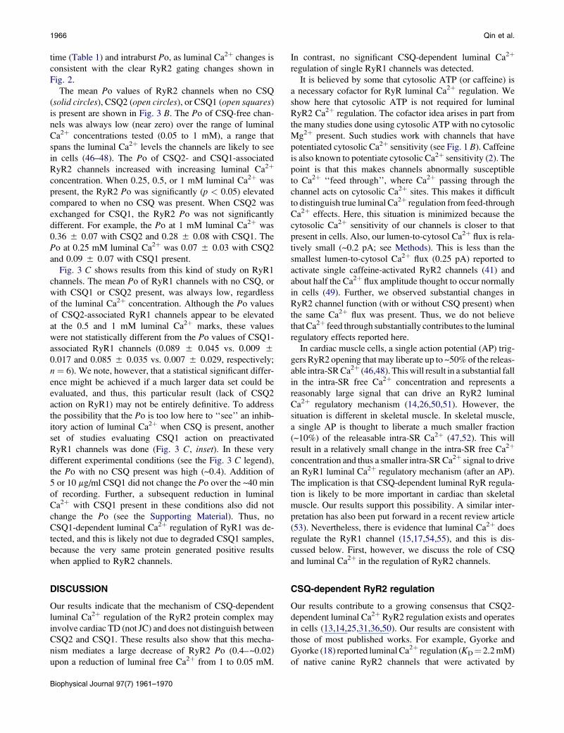
Binding studies
The RyR channel operates within a protein complex that
includes the proteins triadin (TD) and junctin (JC), and
CSQ-dependent RyR regulation is thought to involve the
TD and/or JC proteins (1). Affinity binding studies were
done to assess CSQ interaction with these proteins. Affinity
binding results between CSQ1 or CSQ2 and the cardiac and
skeletal isoforms of TD or JC are shown in Fig. 1 A. The blots
shown illustrate the three positive interactions found: cardiac
TD bound to CSQ2; and skeletal TD and JC bound to CSQ1.
No binding was observed for any other possible binding pairs
(see summary in Fig. 1 A, table). Note that cardiac TD and JC
did not bind CSQ1 and skeletal TD and JC did not bind CSQ2.
If a CSQ-JC-TD interaction is required for CSQ-dependent
RyR regulation, this predicts that the function of cardiac
and skeletal RyR-TD-JC complexes will depend on which
CSQ isoform is present.
Channel recording
A common single RyR channel recording condition was
utilized to facilitate cardiac and skeletal regulatory compar-
ison. Single RyR1 and RyR2 channel function were defined
with a single cytosolic agonist present (0.75 mM free Ca2þ).
Fig. 1 B illustrates the rationale for selecting this cytosolic
free Ca2þ level. Thin solid lines represent the cytosolic Ca2þ
sensitivities of single RyR channels reported by different
groups. The cytosolic Ca2þ sensitivity of the RyR2 and
RyR1 channels tested here is represented by thick solid lines
(10 mM luminal Ca2þ present). The arrowhead on the
abscissa marks the 0.75 mM cytosolic free Ca2þ point. Dashed
lines represent the published Ca2þ sensitivities of the channels
when cytosolic ATP (no Mg2þ) is present. The presence of
ATP results in channels that are exceptionally sensitive to
cytosolic Ca2þ. The dotted lines represent reported RyR2
Ca2þ sensitivities when a physiological level of cytosolic
MgATP is present. Note that physiological MgATP makes
channels less Ca2þ-sensitive compared to when ATP is
absent (solid lines). Thus, the common 0.75 mM Ca2þ only
recording condition used here 1), minimally activates both
channels (Po < 0.05); 2), does not make the channels excep-
tionally Ca2þ-sensitive; and 3), represents an activating Ca2þ
level likely encountered by these channels in cells. To avoid
having millimolar free Mg2þ present to act as a competing
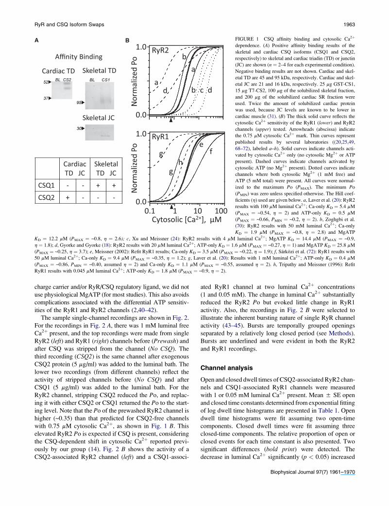
RyR and CSQ Isoform Swaps 1963
Affinity Binding bRyR21.0
Po
A B
Cardiac TD Skeletal TD
adb c
a
d
Nor
mal
ized
eRyR11.0
Skeletal JC 0.0
No
CardiacTD JC
SkeletalTD JC
efg
hg
rmal
ized
Po
CSQ1 - - + +
CSQ2 + - - -
Cytosolic [Ca2+] μM0.1 1 10 100
Nor
0.0
Cytosolic [Ca ], μM
FIGURE 1 CSQ affinity binding and cytosolic Ca2þ
dependence. (A) Positive affinity binding results of the
skeletal and cardiac CSQ isoforms (CSQ1 and CSQ2,
respectively) to skeletal and cardiac triadin (TD) or junctin
(JC) are shown (n ¼ 2–4 for each experimental condition).
Negative binding results are not shown. Cardiac and skel-
etal TD are 45 and 95 kDa, respectively. Cardiac and skel-
etal JC are 21 and 16 kDa, respectively. 25 mg GST-CS1,
15 mg T7-CS2, 100 mg of the solubilized skeletal fraction,
and 200 mg of the solubilized cardiac SR fraction were
used. Twice the amount of solubilized cardiac protein
was used, because JC levels are known to be lower in
cardiac muscle (31). (B) The thick solid curve reflects the
cytosolic Ca2þ sensitivity of the RyR1 (lower) and RyR2
channels (upper) tested. Arrowheads (abscissa) indicate
the 0.75 mM cytosolic Ca2þ mark. Thin curves represent
published results by several laboratories ((20,25,49,
68–72), labeled a–h). Solid curves indicate channels acti-
vated by cytosolic Ca2þ only (no cytosolic Mg2þ or ATP
present). Dashed curves indicate channels activated by
cytosolic ATP (no Mg2þ present). Dotted curves indicate
channels where both cytosolic Mg2þ (1 mM free) and
ATP (5 mM total) were present. All curves were normal-
ized to the maximum Po (PMAX). The minimum Po
(PMIN) was zero unless specified otherwise. The Hill coef-
ficients (h) used are given below. a, Laver et al. (20): RyR2
results with 100 mM luminal Ca2þ; Ca-only KD ¼ 5.4 mM
(PMAX ¼ ~0.54, h ¼ 2) and ATP-only KD ¼ 0.5 mM
(PMAX ¼ ~0.66, PMIN ¼ ~0.2, h ¼ 2). b, Zoghgbi et al.
(70): RyR2 results with 50 mM luminal Ca2þ; Ca-only
KD ¼ 1.9 mM (PMAX ¼ ~0.8, h ¼ 2.8) and MgATP
KD ¼ 12.2 mM (PMAX ¼ ~0.8, h ¼ 2.6); c, Xu and Meissner (24): RyR2 results with 4 mM luminal Ca2þ; MgATP KD ¼ 14.4 mM (PMAX ¼ ~0.9,
h ¼ 1.8); d, Gyorke and Gyorke (18): RyR2 results with 20 mM luminal Ca2þ; ATP-only KD ¼ 1.6 mM (PMAX ¼ ~0.27, h ¼ 1) and MgATP KD ¼ 25.8 mM
(PMAX ¼ ~0.25, h ¼ 3.7); e, Meissner (2002): Refit RyR1 results; Ca-only KD ¼ 3.5 mM (PMAX ¼ ~0.22, h ¼ 1.9); f, Sarkozi et al. (72): RyR1 results with
50 mM luminal Ca2þ; Ca-only KD ¼ 9.4 mM (PMAX ¼ ~0.35, h ¼ 1.2); g, Laver et al. (20): Results with 1 mM luminal Ca2þ; ATP-only KD ¼ 0.4 mM
(PMAX ¼ ~0.86, PMIN ¼ ~0.40, assumed h ¼ 2) and Ca-only KD ¼ 1.1 mM (PMAX ¼ ~0.55, assumed h ¼ 2). h, Tripathy and Meissner (1996): Refit
RyR1 results with 0.045 mM luminal Ca2þ; ATP-only KD ¼ 1.8 mM (PMAX ¼ ~0.9, h ¼ 2).
charge carrier and/or RyR/CSQ regulatory ligand, we did not
use physiological MgATP (for most studies). This also avoids
complications associated with the differential ATP sensitiv-
ities of the RyR1 and RyR2 channels (2,40–42).
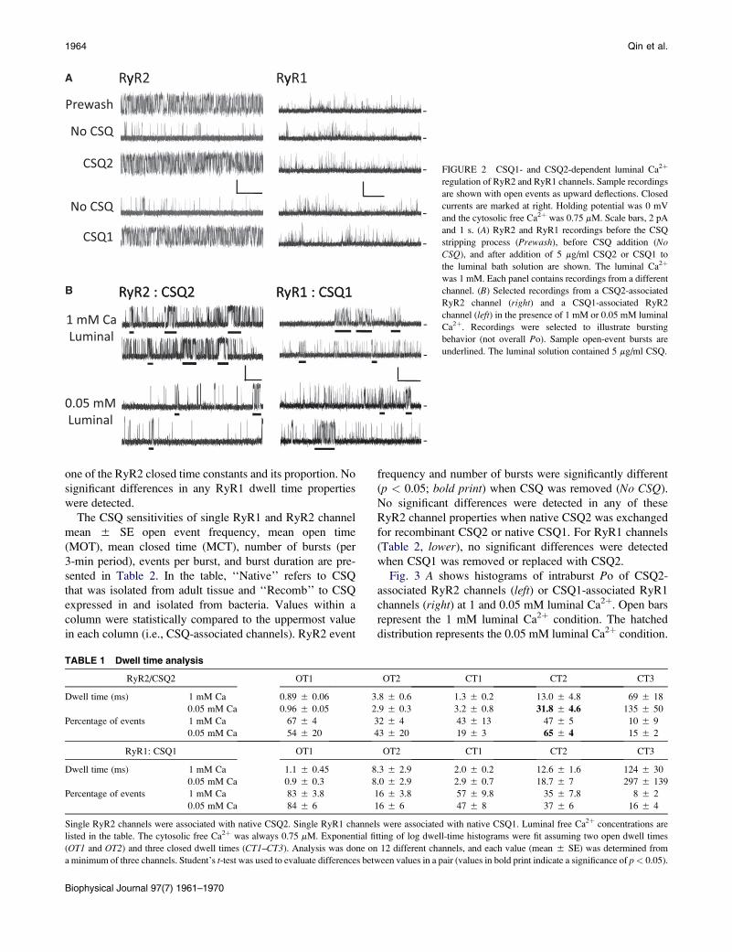
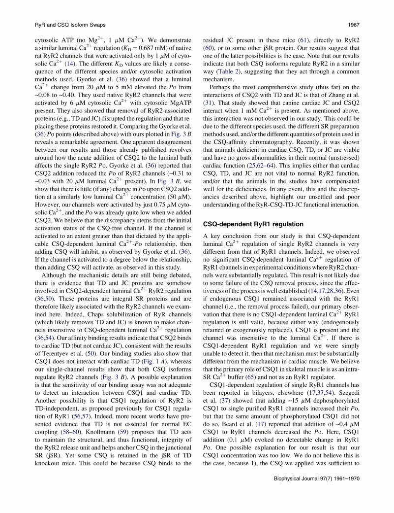
The sample single-channel recordings are shown in Fig. 2.
For the recordings in Fig. 2 A, there was 1 mM luminal free
Ca2þ present, and the top recordings were made from single
RyR2 (left) and RyR1 (right) channels before (Prewash) and
after CSQ was stripped from the channel (No CSQ). The
third recording (CSQ2) is the same channel after exogenous
CSQ2 protein (5 mg/ml) was added to the luminal bath. The
lower two recordings (from different channels) reflect the
activity of stripped channels before (No CSQ) and after
CSQ1 (5 mg/ml) was added to the luminal bath. For the
RyR2 channel, stripping CSQ2 reduced the Po, and replac-
ing it with either CSQ2 or CSQ1 returned the Po to the start-
ing level. Note that the Po of the prewashed RyR2 channel is
higher (~0.35) than that predicted for CSQ2-free channels
with 0.75 mM cytosolic Ca2þ, as shown in Fig. 1 B. This
elevated RyR2 Po is expected if CSQ is present, considering
the CSQ-dependent shift in cytosolic Ca2þ reported previ-
ously by our group (14). Fig. 2 B shows the activity of a
CSQ2-associated RyR2 channel (left) and a CSQ1-associ-
ated RyR1 channel at two luminal Ca2þ concentrations
(1 and 0.05 mM). The change in luminal Ca2þ substantially
reduced the RyR2 Po but evoked little change in RyR1
activity. Also, the recordings in Fig. 2 B were selected to
illustrate the inherent bursting nature of single RyR channel
activity (43–45). Bursts are temporally grouped openings
separated by a relatively long closed period (see Methods).
Bursts are underlined and were evident in both the RyR2
and RyR1 recordings.
Channel analysis
Open and closed dwell times of CSQ2-associated RyR2 chan-
nels and CSQ1-associated RyR1 channels were measured
with 1 or 0.05 mM luminal Ca2þ present. Mean 5 SE open
and closed time constants determined from exponential fitting
of log dwell time histograms are presented in Table 1. Open
dwell time histograms were fit assuming two open-time
components. Closed dwell times were fit assuming three
closed-time components. The relative proportion of open or
closed events for each time constant is also presented. Two
significant differences (bold print) were detected. The
decrease in luminal Ca2þ significantly (p < 0.05) increased
Biophysical Journal 97(7) 1961–1970
1964 Qin et al.
A RyR2 RyR1
-
-
Prewash
No CSQ
y y
-
-
CSQ2
No CSQ
-CSQ1
RyR2 : CSQ2 RyR1 : CSQ1
1 mM CaLuminal
B
-
-
RyR2 : CSQ2 RyR1 : CSQ1
0.05 mMLuminal
-
-
FIGURE 2 CSQ1- and CSQ2-dependent luminal Ca2þ
regulation of RyR2 and RyR1 channels. Sample recordings
are shown with open events as upward deflections. Closed
currents are marked at right. Holding potential was 0 mV
and the cytosolic free Ca2þ was 0.75 mM. Scale bars, 2 pA
and 1 s. (A) RyR2 and RyR1 recordings before the CSQ
stripping process (Prewash), before CSQ addition (NoCSQ), and after addition of 5 mg/ml CSQ2 or CSQ1 to
the luminal bath solution are shown. The luminal Ca2þ
was 1 mM. Each panel contains recordings from a different
channel. (B) Selected recordings from a CSQ2-associated
RyR2 channel (right) and a CSQ1-associated RyR2
channel (left) in the presence of 1 mM or 0.05 mM luminal
Ca2þ. Recordings were selected to illustrate bursting
behavior (not overall Po). Sample open-event bursts are
underlined. The luminal solution contained 5 mg/ml CSQ.
one of the RyR2 closed time constants and its proportion. No
significant differences in any RyR1 dwell time properties
were detected.
The CSQ sensitivities of single RyR1 and RyR2 channel
mean 5 SE open event frequency, mean open time
(MOT), mean closed time (MCT), number of bursts (per
3-min period), events per burst, and burst duration are pre-
sented in Table 2. In the table, ‘‘Native’’ refers to CSQ
that was isolated from adult tissue and ‘‘Recomb’’ to CSQ
expressed in and isolated from bacteria. Values within a
column were statistically compared to the uppermost value
in each column (i.e., CSQ-associated channels). RyR2 event
frequency and number of bursts were significantly different
(p < 0.05; bold print) when CSQ was removed (No CSQ).
No significant differences were detected in any of these
RyR2 channel properties when native CSQ2 was exchanged
for recombinant CSQ2 or native CSQ1. For RyR1 channels
(Table 2, lower), no significant differences were detected
when CSQ1 was removed or replaced with CSQ2.
Fig. 3 A shows histograms of intraburst Po of CSQ2-
associated RyR2 channels (left) or CSQ1-associated RyR1
channels (right) at 1 and 0.05 mM luminal Ca2þ. Open bars
represent the 1 mM luminal Ca2þ condition. The hatched
distribution represents the 0.05 mM luminal Ca2þ condition.
TABLE 1 Dwell time analysis
RyR2/CSQ2 OT1 OT2 CT1 CT2 CT3
Dwell time (ms) 1 mM Ca 0.89 5 0.06 3.8 5 0.6 1.3 5 0.2 13.0 5 4.8 69 5 18
0.05 mM Ca 0.96 5 0.05 2.9 5 0.3 3.2 5 0.8 31.8 5 4.6 135 5 50
Percentage of events 1 mM Ca 67 5 4 32 5 4 43 5 13 47 5 5 10 5 9
0.05 mM Ca 54 5 20 43 5 20 19 5 3 65 5 4 15 5 2
RyR1: CSQ1 OT1 OT2 CT1 CT2 CT3
Dwell time (ms) 1 mM Ca 1.1 5 0.45 8.3 5 2.9 2.0 5 0.2 12.6 5 1.6 124 5 30
0.05 mM Ca 0.9 5 0.3 8.0 5 2.9 2.9 5 0.7 18.7 5 7 297 5 139
Percentage of events 1 mM Ca 83 5 3.8 16 5 3.8 57 5 9.8 35 5 7.8 8 5 2
0.05 mM Ca 84 5 6 16 5 6 47 5 8 37 5 6 16 5 4
Single RyR2 channels were associated with native CSQ2. Single RyR1 channels were associated with native CSQ1. Luminal free Ca2þ concentrations are
listed in the table. The cytosolic free Ca2þ was always 0.75 mM. Exponential fitting of log dwell-time histograms were fit assuming two open dwell times
(OT1 and OT2) and three closed dwell times (CT1–CT3). Analysis was done on 12 different channels, and each value (mean 5 SE) was determined from
a minimum of three channels. Student’s t-test was used to evaluate differences between values in a pair (values in bold print indicate a significance of p< 0.05).
Biophysical Journal 97(7) 1961–1970
RyR and CSQ Isoform Swaps 1965
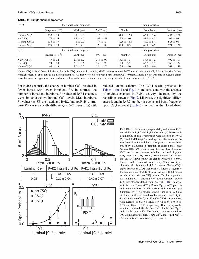
TABLE 2 Single channel properties
RyR2 Individual event properties Burst properties
Frequency (s�1) MOT (ms) MCT (ms) Number Event/burst Duration (ms)
Native CSQ2 133 5 19 17 5 9.0 15 5 10 41.7 5 12.8 43.7 5 3.6 492 5 101
No CSQ 71 5 16 2.5 5 1.5 103 5 57 9.4 5 3.0 33.9 5 4.8 502 5 93
Recomb CSQ2 138 5 17 11 5 7.7 18 5 6 52.5 5 15.6 47.9 5 5.4 565 5 96
Native CSQ1 129 5 19 12 5 4.9 23 5 8 42.4 5 8.3 40.1 5 4.9 571 5 121
RyR1 Individual event properties Burst properties
Frequency (s�1) MOT (ms) MCT (ms) Number Event/burst Duration (ms)
Native CSQ1 77 5 32 2.9 5 1.2 313 5 99 13.7 5 7.3 57.8 5 7.2 492 5 103
No CSQ 74 5 20 2.6 5 0.6 268 5 98 12.4 5 3.2 42.2 5 7.3 365 5 122
Native CSQ2 99 5 19 2.6 5 0.7 224 5 76 15.8 5 5.0 47.5 5 9.9 568 5 148
Native, CSQ isolated from adult tissue; Recomb, CSQ produced in bacteria; MOT, mean open time; MCT, mean closed time; PS, Poisson Surprise. Values
represent mean 5 SE of four to six different channels. All data were collected with 1 mM luminal Ca2þ present. Student’s t-test was used to evaluate differ-
ences between the uppermost value and other values within each column (values in bold print indicate a significance of p < 0.05).
For RyR2 channels, the change in luminal Ca2þ resulted in
fewer bursts with lower intraburst Po. In contrast, the
number of bursts and intraburst Po values of RyR1 channels
were similar at the two luminal Ca2þ levels. Mean intraburst
Po values (5 SE) are listed, and RyR2, but not RyR1, intra-
burst Po was statistically different (p< 0.01; bold print) with
reduced luminal calcium. The RyR1 results presented in
Tables 1 and 2 and Fig. 3 A are consistent with the absence
of obvious changes in RyR1 activity illustrated by the
recordings shown in Fig. 2. Likewise, the significant differ-
ences found in RyR2 number of events and burst frequency
upon CSQ removal (Table 2), as well as the closed dwell
RyR2 RyR130A RyR2 RyR1
Burs
ts
30
# o
f B
0
Luminal Ca2+ RyR2 Intra-Burst Po RyR1 Intra-Burst Po1 0 44 ± 0 05 0 36 ± 0 09
00 0.5 1
Intra-Burst Po0.5 1
Intra-Burst Po
1 0.44 ± 0.05 0.36 ± 0.090.05 0.21 ± 0.04 0.42 ± 0.07
B0.5
y no CSQRyR1RyR2
1P
C
n P
roba
bilit
y
CSQ2CSQ1
0 0 5 10μg/ml
Po
0.1 10.0
Ope
n
0.1 1
μg/ml
Luminal [Ca2+], mM Luminal [Ca2+], mM
1 mM0.05 mM
FIGURE 3 Intraburst open probability and luminal Ca2þ
sensitivity of RyR2 and RyR1 channels. (A) Bursts with
a minimum of five events/burst were detected in RyR2
(left) and RyR1 (right) recordings, and the intraburst Po
was determined for each burst. Histograms of the intraburst
Po, fit by a Gaussian distribution, at either 1 mM (openbars) or 0.05 mM (hatched area; bars not shown) luminal
Ca2þ are shown. Luminal solution contained 5 mg/ml
CSQ2 (left) and CSQ1 (right). Mean intraburst Po values
(5 SE) are shown below the graphs (bracket; p < 0.01;
t-test). Results generated from five RyR2 and five RyR1
channels. (B) Summary RyR2 Po results. Native CSQ2
(open circles) or CSQ1 (squares) was added (5 mg/ml) to
the luminal side of CSQ stripped channels. Solid circles
are the results with no CSQ present. The line represents
the luminal Ca2þ sensitivity of RyR2 channels before
CSQ was stripped (taken from Qin et al. (14)). The cyto-
solic free Ca2þ was 0.75 mM (no Mg or ATP present)
and points are mean 5 SE of six to eight channels. (C)
Summary RyR1 Po results. Symbols are as in B. Solid
circles are shifted to avoid symbol overlap. (Inset) RyR1
Po as a function of 0, 5, and 10 mg/ml CSQ1 concentration,
with average (5 SE) Po values of 0.42 5 0.10, 0.43 5
0.13, and 0.45 5 0.15, respectively. Here, the cytosolic
solution contained 20 mM free Ca2þ, 1 mM free Mg2þ,
and 5 mM total ATP. The luminal solution contained
100 Cs-methanesulfonate, 1 mM Ca2þ, and 1 mM Mg2þ.
These results are from four RyR1 channels.
Biophysical Journal 97(7) 1961–1970
1966 Qin et al.
time (Table 1) and intraburst Po, as luminal Ca2þ changes is
consistent with the clear RyR2 gating changes shown in
Fig. 2.
The mean Po values of RyR2 channels when no CSQ
(solid circles), CSQ2 (open circles), or CSQ1 (open squares)
is present are shown in Fig. 3 B. The Po of CSQ-free chan-
nels was always low (near zero) over the range of luminal
Ca2þ concentrations tested (0.05 to 1 mM), a range that
spans the luminal Ca2þ levels the channels are likely to see
in cells (46–48). The Po of CSQ2- and CSQ1-associated
RyR2 channels increased with increasing luminal Ca2þ
concentration. When 0.25, 0.5, or 1 mM luminal Ca2þ was
present, the RyR2 Po was significantly (p < 0.05) elevated
compared to when no CSQ was present. When CSQ2 was
exchanged for CSQ1, the RyR2 Po was not significantly
different. For example, the Po at 1 mM luminal Ca2þ was
0.36 5 0.07 with CSQ2 and 0.28 5 0.08 with CSQ1. The
Po at 0.25 mM luminal Ca2þ was 0.07 5 0.03 with CSQ2
and 0.09 5 0.07 with CSQ1 present.
Fig. 3 C shows results from this kind of study on RyR1
channels. The mean Po of RyR1 channels with no CSQ, or
with CSQ1 or CSQ2 present, was always low, regardless
of the luminal Ca2þ concentration. Although the Po values
of CSQ2-associated RyR1 channels appear to be elevated
at the 0.5 and 1 mM luminal Ca2þ marks, these values
were not statistically different from the Po values of CSQ1-
associated RyR1 channels (0.089 5 0.045 vs. 0.009 5
0.017 and 0.085 5 0.035 vs. 0.007 5 0.029, respectively;
n ¼ 6). We note, however, that a statistical significant differ-
ence might be achieved if a much larger data set could be
evaluated, and thus, this particular result (lack of CSQ2
action on RyR1) may not be entirely definitive. To address
the possibility that the Po is too low here to ‘‘see’’ an inhib-
itory action of luminal Ca2þ when CSQ is present, another
set of studies evaluating CSQ1 action on preactivated
RyR1 channels was done (Fig. 3 C, inset). In these very
different experimental conditions (see the Fig. 3 C legend),
the Po with no CSQ present was high (~0.4). Addition of
5 or 10 mg/ml CSQ1 did not change the Po over the ~40 min
of recording. Further, a subsequent reduction in luminal
Ca2þ with CSQ1 present in these conditions also did not
change the Po (see the Supporting Material). Thus, no
CSQ1-dependent luminal Ca2þ regulation of RyR1 was de-
tected, and this is likely not due to degraded CSQ1 samples,
because the very same protein generated positive results
when applied to RyR2 channels.
DISCUSSION
Our results indicate that the mechanism of CSQ-dependent
luminal Ca2þ regulation of the RyR2 protein complex may
involve cardiac TD (not JC) and does not distinguish between
CSQ2 and CSQ1. These results also show that this mecha-
nism mediates a large decrease of RyR2 Po (0.4–~0.02)
upon a reduction of luminal free Ca2þ from 1 to 0.05 mM.
Biophysical Journal 97(7) 1961–1970
In contrast, no significant CSQ-dependent luminal Ca2þ
regulation of single RyR1 channels was detected.
It is believed by some that cytosolic ATP (or caffeine) is
a necessary cofactor for RyR luminal Ca2þ regulation. We
show here that cytosolic ATP is not required for luminal
RyR2 Ca2þ regulation. The cofactor idea arises in part from
the many studies done using cytosolic ATP with no cytosolic
Mg2þ present. Such studies work with channels that have
potentiated cytosolic Ca2þ sensitivity (see Fig. 1 B). Caffeine
is also known to potentiate cytosolic Ca2þ sensitivity (2). The
point is that this makes channels abnormally susceptible
to Ca2þ ‘‘feed through’’, where Ca2þ passing through the
channel acts on cytosolic Ca2þ sites. This makes it difficult
to distinguish true luminal Ca2þ regulation from feed-through
Ca2þ effects. Here, this situation is minimized because the
cytosolic Ca2þ sensitivity of our channels is closer to that
present in cells. Also, our lumen-to-cytosol Ca2þ flux is rela-
tively small (~0.2 pA; see Methods). This is less than the
smallest lumen-to-cytosol Ca2þ flux (0.25 pA) reported to
activate single caffeine-activated RyR2 channels (41) and
about half the Ca2þ flux amplitude thought to occur normally
in cells (49). Further, we observed substantial changes in
RyR2 channel function (with or without CSQ present) when
the same Ca2þ flux was present. Thus, we do not believe
that Ca2þ feed through substantially contributes to the luminal
regulatory effects reported here.
In cardiac muscle cells, a single action potential (AP) trig-
gers RyR2 opening that may liberate up to ~50% of the releas-
able intra-SR Ca2þ (46,48). This will result in a substantial fall
in the intra-SR free Ca2þ concentration and represents a
reasonably large signal that can drive an RyR2 luminal
Ca2þ regulatory mechanism (14,26,50,51). However, the
situation is different in skeletal muscle. In skeletal muscle,
a single AP is thought to liberate a much smaller fraction
(~10%) of the releasable intra-SR Ca2þ (47,52). This will
result in a relatively small change in the intra-SR free Ca2þ
concentration and thus a smaller intra-SR Ca2þ signal to drive
an RyR1 luminal Ca2þ regulatory mechanism (after an AP).
The implication is that CSQ-dependent luminal RyR regula-
tion is likely to be more important in cardiac than skeletal
muscle. Our results support this possibility. A similar inter-
pretation has also been put forward in a recent review article
(53). Nevertheless, there is evidence that luminal Ca2þ does
regulate the RyR1 channel (15,17,54,55), and this is dis-
cussed below. First, however, we discuss the role of CSQ
and luminal Ca2þ in the regulation of RyR2 channels.
CSQ-dependent RyR2 regulation
Our results contribute to a growing consensus that CSQ2-
dependent luminal Ca2þ RyR2 regulation exists and operates
in cells (13,14,25,31,36,50). Our results are consistent with
those of most published works. For example, Gyorke and
Gyorke (18) reported luminal Ca2þ regulation (KD¼ 2.2 mM)
of native canine RyR2 channels that were activated by
RyR and CSQ Isoform Swaps 1967
cytosolic ATP (no Mg2þ, 1 mM Ca2þ). We demonstrate
a similar luminal Ca2þ regulation (KD¼ 0.687 mM) of native
rat RyR2 channels that were activated only by 1 mM of cyto-
solic Ca2þ (14). The different KD values are likely a conse-
quence of the different species and/or cytosolic activation
methods used. Gyorke et al. (36) showed that a luminal
Ca2þ change from 20 mM to 5 mM elevated the Po from
~0.08 to ~0.40. They used native RyR2 channels that were
activated by 6 mM cytosolic Ca2þ with cytosolic MgATP
present. They also showed that removal of RyR2-associated
proteins (e.g., TD and JC) disrupted the regulation and that re-
placing these proteins restored it. Comparing the Gyorke et al.
(36) Po points (described above) with ours plotted in Fig. 3 Breveals a remarkable agreement. One apparent disagreement
between our results and those already published revolves
around how the acute addition of CSQ2 to the luminal bath
affects the single RyR2 Po. Gyorke et al. (36) reported that
CSQ2 addition reduced the Po of RyR2 channels (~0.31 to
~0.03 with 20 mM luminal Ca2þ present). In Fig. 3 B, we
show that there is little (if any) change in Po upon CSQ2 addi-
tion at a similarly low luminal Ca2þ concentration (50 mM).
However, our channels were activated by just 0.75 mM cyto-
solic Ca2þ, and the Po was already quite low when we added
CSQ2. We believe that the discrepancy stems from the initial
activation status of the CSQ-free channel. If the channel is
activated to an extent greater than that dictated by the appli-
cable CSQ-dependent luminal Ca2þ-Po relationship, then
adding CSQ will inhibit, as observed by Gyorke et al. (36).
If the channel is activated to a degree below the relationship,
then adding CSQ will activate, as observed in this study.
Although the mechanistic details are still being debated,
there is evidence that TD and JC proteins are somehow
involved in CSQ2-dependent luminal Ca2þ RyR2 regulation
(36,50). These proteins are integral SR proteins and are
therefore likely associated with the RyR2 channels we exam-
ined here. Indeed, Chaps solubilization of RyR channels
(which likely removes TD and JC) is known to make chan-
nels insensitive to CSQ-dependent luminal Ca2þ regulation
(36,54). Our affinity binding results indicate that CSQ2 binds
to cardiac TD (but not cardiac JC), consistent with the results
of Terentyev et al. (50). Our binding studies also show that
CSQ1 does not interact with cardiac TD (Fig. 1 A), whereas
our single-channel results show that both CSQ isoforms
regulate RyR2 channels (Fig. 3 B). A possible explanation
is that the sensitivity of our binding assay was not adequate
to detect an interaction between CSQ1 and cardiac TD.
Another possibility is that CSQ1 regulation of RyR2 is
TD-independent, as proposed previously for CSQ1 regula-
tion of RyR1 (56,57). Indeed, more recent works have pre-
sented evidence that TD is not essential for normal EC
coupling (58–60). Knollmann (59) proposes that TD acts
to maintain the structural, and thus functional, integrity of
the RyR2 release unit and helps anchor CSQ in the junctional
SR (jSR). Yet some CSQ is retained in the jSR of TD
knockout mice. This could be because CSQ binds to the
residual JC present in these mice (61), directly to RyR2
(60), or to some other jSR protein. Our results suggest that
one of the latter possibilities is the case. Note that our results
indicate that both CSQ isoforms regulate RyR2 in a similar
way (Table 2), suggesting that they act through a common
mechanism.
Perhaps the most comprehensive study (thus far) on the
interactions of CSQ2 with TD and JC is that of Zhang et al.
(31). That study showed that canine cardiac JC and CSQ2
interact when 1 mM Ca2þ is present. As mentioned above,
this interaction was not observed in our study. This could be
due to the different species used, the different SR preparation
methods used, and/or the different quantities of protein used in
the CSQ-affinity chromatography. Recently, it was shown
that animals deficient in cardiac CSQ, TD, or JC are viable
and have no gross abnormalities in their normal (unstressed)
cardiac function (25,62–64). This implies either that cardiac
CSQ, TD, and JC are not vital to normal RyR2 function,
and/or that the animals in the studies have compensated
well for the deficiencies. In any event, this and the discrep-
ancies described above, highlight our unsettled and poor
understanding of the RyR-CSQ-TD-JC functional interaction.
CSQ-dependent RyR1 regulation
A key conclusion from our study is that CSQ-dependent
luminal Ca2þ regulation of single RyR2 channels is very
different from that of RyR1 channels. Indeed, we observed
no significant CSQ-dependent luminal Ca2þ regulation of
RyR1 channels in experimental conditions where RyR2 chan-
nels were substantially regulated. This result is not likely due
to some failure of the CSQ removal process, since the effec-
tiveness of the process is well established (14,17,28,36). Even
if endogenous CSQ1 remained associated with the RyR1
channel (i.e., the removal process failed), our primary obser-
vation that there is no CSQ1-dependent luminal Ca2þ RyR1
regulation is still valid, because either way (endogenously
retained or exogenously replaced), CSQ1 is present and the
channel was insensitive to the luminal Ca2þ. If there is
CSQ1-dependent RyR1 regulation and we were simply
unable to detect it, then that mechanism must be substantially
different from the mechanism in cardiac muscle. We believe
that the primary role of CSQ1 in skeletal muscle is as an intra-
SR Ca2þ buffer (65) and not as an RyR1 regulator.
CSQ1-dependent regulation of single RyR1 channels has
been reported in bilayers, elsewhere (17,37,54). Szegedi
et al. (37) showed that adding ~15 mM dephosphorylated
CSQ1 to single purified RyR1 channels increased their Po,
but that the same amount of phosphorylated CSQ1 did not
do so. Beard et al. (17) reported that addition of ~0.4 mM
CSQ1 to RyR1 channels decreased the Po. Here, CSQ1
addition (0.1 mM) evoked no detectable change in RyR1
Po. One possible explanation for our result is that our
CSQ1 concentration was too low. We do not believe this is
the case, because 1), the CSQ we applied was sufficient to
Biophysical Journal 97(7) 1961–1970
1968 Qin et al.
substantially alter single RyR2 function, 2), doubling CSQ
concentration generated no additional effect, and, 3) even
the 0.1 mM CSQ1 concentration applied here should provide
ample protein for the high-affinity CSQ1-RyR1 interaction
(56). We believe that the most likely explanation for the
disparate results lies in the different methods of RyR1
channel activation used in the different studies. For example,
RyR1 channels activated by ATP (no Mg2þ), as in the Beard
et al. (17) study, and those activated by just a low cytosolic
Ca2þ level, as done in this study, might be expected to
respond to regulatory challenges differently. The likely
reason for the discrepancy between our study and the Sze-
gedi et al. (37) results is that we used native RyR1 channels,
which are likely still associated with TD and JC, whereas
they used CHAPS-purified channels, which likely lack these
potentially important proteins. Wang et al. (54) also showed
that CSQ1 enhances RyR channel activity. They examined
CSQ1-dependent Ca2þ regulation of RyR1/RyR3 channels
isolated from C2C12 myotubes. The apparent disparity
between our results and theirs could be due to the presence
of RyR3 channels in their preparation, since in our study
only RyR1 channels were examined.
Fast-twitch skeletal muscle contains primarily CSQ1,
whereas slow-twitch muscle contains both CSQ1 and CSQ2
(15). Slow-twitch muscle also contains other cardiac isoforms
(e.g., SR ATPase, troponin C, etc.) (66). Here, we show that
there was no significant difference in single RyR1 channel
luminal Ca2þ regulation when CSQ1 or CSQ2 were present.
Thus, the CSQ2 is not likely to make RyR1 channels in slow-
twitch muscles operate differently than they do in fast-twitch
muscles. The CSQ2 could, however, change the intra-SR
Ca2þ buffer properties or it could be interacting with the
RyR3 channels that are present (67).
Finally, an analogous CSQ isoform swap study on ATP-
activated channels was recently published (60). The authors
report that the RyR2 Po is increased by both CSQ isoforms,
that CSQ2 dissociation reduces RyR2 Po, and that RyR1 Po
is modestly elevated by CSQ2. These findings are consistent
with our results. Wei et al. (60) also report that CSQ1 inhibits
RyR1 and a similar CSQ2-associated RyR2 Po at 100 nM
and 1 mM luminal Ca2þ concentration. This is contrary to
our results as well as to other published reports (e.g.,
(14,18,22)). We detected no CSQ1 action on RyR1 channels,
although previous studies have detected such action (e.g.,
(17,19,37)). This could be due to differences in species,
membrane potentials, and/or—of most importance—how the
channels are activated. Since several studies have reported
that CSQ action depends on the experimental conditions
used (e.g., (18,68,73)), we believe that this is the primary
cause of the apparent discrepancies outlined above.
SUPPORTING MATERIAL
Methods and a figure are available at http://www.biophysj.org/biophysj/
supplemental/S0006-3495(09)01293-4.
Biophysical Journal 97(7) 1961–1970
This work was supported by National Institutes of Health grants HL57832
and AR54098 (to M.F.) and HL71741 (to J.R.F), and by a Telethon grant
(GGP04066) to P.V.
REFERENCES
1. Bers, D. 2001. Excitation-Contraction Coupling and Cardiac Contrac-tile Force. Kluwer Academic,Dordrecht, The Netherlands.
2. Fill, M., and J. Copello. 2002. Ryanodine receptor calcium releasechannels. Physiol. Rev. 82:893–922.
3. Jorgensen, A. O., A. C. Shen, K. P. Campbell, and D. H. MacLennan.1983. Ultrastructural localization of calsequestrin in rat skeletal muscleby immunoferritin labeling of ultrathin frozen sections. J. Cell Biol.97:1573–1581.
4. Franzini-Armstrong, C., L. J. Kenney, and E. Varriano-Marston. 1987.The structure of calsequestrin in triads of vertebrate skeletal muscle:a deep-etch study. J. Cell Biol. 105:49–56.
5. MacLennan, D. H., and P. T. Wong. 1971. Isolation of a calcium-sequestering protein from sarcoplasmic reticulum. Proc. Natl. Acad.Sci. USA. 68:1231–1235.
6. Fliegel, L., M. Ohnishi, M. R. Carpenter, V. K. Khanna, R. A. Reithmeier,et al. 1987. Amino acid sequence of rabbit fast-twitch skeletal musclecalsequestrin deduced from cDNA and peptide sequencing. Proc. Natl.Acad. Sci. USA. 84:1167–1171.
7. Cho, J. H., Y. S. Oh, K. W. Park, J. Yu, K. Y. Choi, et al. 2000.Calsequestrin, a calcium sequestering protein localized at the sarco-plasmic reticulum, is not essential for body-wall muscle function inCaenorhabditis elegans. J. Cell Sci. 113:3947–3958.
8. Arai, M., K. Otsu, D. H. MacLennan, N. R. Alpert, and M. Periasamy.1991. Effect of thyroid hormone on the expression of mRNA encodingsarcoplasmic reticulum proteins. Circ. Res. 69:266–276.
9. Fujii, J., H. F. Willard, and D. H. MacLennan. 1990. Characterizationand localization to human chromosome 1 of human fast-twitch skeletalmuscle calsequestrin gene. Somat. Cell Mol. Genet. 16:185–189.
10. Scott, B., H. Simmerman, J. Collins, B. Nadal-Ginard, and L. Jones.1988. Complete amino acid sequence of canine cardiac calsequestrindeduced by cDNA cloning. J. Biol. Chem. 263:8958–8964.
11. Divet, A., S. Paesante, C. Grasso, D. Cavagna, C. Tiveron, et al. 2007.Increased Ca2þ storage capacity of the skeletal muscle sarcoplasmicreticulum of transgenic mice over-expressing membrane bound calciumbinding protein junctate. J. Cell. Physiol. 213:464–474.
12. di Barletta, M. R., S. Viatchenko-Karpinski, A. Nori, M. Memmi,D. Terentyev, et al. 2006. Clinical phenotype and functional character-ization of CASQ2 mutations associated with catecholaminergic poly-morphic ventricular tachycardia. Circulation. 114:1012–1019.
13. Lahat, H., E. Pras, T. Olender, N. Avidan, E. Ben-Asher, et al. 2001.A missense mutation in a highly conserved region of CASQ2 is associ-ated with autosomal recessive catecholamine-induced polymorphicventricular tachycardia in Bedouin families from Israel. Am. J. Hum.Genet. 69:1378–1384.
14. Qin, J., G. Valle, A. Nani, A. Nori, N. Rizzi, et al. 2008. Luminal Ca2þ
regulation of single cardiac ryanodine receptors: insights provided bycalsequestrin and its mutants. J. Gen. Physiol. 131:325–334.
15. Paolini, C., M. Quarta, A. Nori, S. Boncompagni, M. Canato, et al.2007. Reorganized stores and impaired calcium handling in skeletalmuscle of mice lacking calsequestrin-1. J. Physiol. 583:767–784.
16. Yano, K., and A. Zarain-Herzberg. 1994. Sarcoplasmic reticulum calse-questrins: structural and functional properties. Mol. Cell. Biochem.135:61–70.
17. Beard, N., M. Sakowska, A. Dulhunty, and D. Laver. 2002. Calseques-trin is an inhibitor of skeletal muscle ryanodine receptor calcium releasechannels. Biophys. J. 82:310–320.
18. Gyorke, I., and S. Gyorke. 1998. Regulation of the cardiac ryanodinereceptor channel by luminal Ca2þ involves luminal Ca2þ sensing sites.Biophys. J. 75:2801–2810.
RyR and CSQ Isoform Swaps 1969
19. Ikemoto, N., M. Ronjat, L. G. Meszaros, and M. Koshita. 1989. Postu-lated role of calsequestrin in the regulation of calcium release fromsarcoplasmic reticulum. Biochemistry. 28:6764–6771.
20. Laver, D. R., E. R. O’Neill, and G. D. Lamb. 2004. Luminal Ca2þ-regulated Mg2þ inhibition of skeletal RyRs reconstituted as isolatedchannels or coupled clusters. J. Gen. Physiol. 124:741–758.
21. Sitsapesan, R., and A. Williams. 1995. The gating of the sheep skeletalsarcoplasmic reticulum Ca2þ-release channel is regulated by luminalCa2þ. J. Membr. Biol. 146:133–144.
22. Terentyev, D., S. Viatchenko-Karpinski, H. Valdivia, A. Escobar, andS. Gyorke. 2002. Luminal Ca2þ controls termination and refractory behaviorof Ca2þ-induced Ca2þ release in cardiac myocytes. Circ. Res. 91:414–420.
23. Tripathy, A., and G. Meissner. 1996. Sarcoplasmic reticulum lumenalCa2þ has access to cytosolic activation and inactivation sites of skeletalmuscle Ca2þ release channel. Biophys. J. 70:2600–2615.
24. Xu, L., and G. Meissner. 1998. Regulation of cardiac muscle Ca2þ
release channel by sarcoplasmic reticulum lumenal Ca2þ. Biophys. J.75:2302–2312.
25. Knollmann, B. C., N. Chopra, T. Hlaing, B. Akin, T. Yang, et al. 2006.Casq2 deletion causes sarcoplasmic reticulum volume increase, prema-ture Ca2þ release, and catecholaminergic polymorphic ventriculartachycardia. J. Clin. Invest. 116:2510–2520.
26. Terentyev, D., A. Nori, M. Santoro, S. Viatchenko-Karpinski, Z. Kubalova,et al. 2006. Abnormal interactions of calsequestrin with the ryanodinereceptor calcium release channel complex linked to exercise-inducedsudden cardiac death. Circ. Res. 98:1151–1158.
27. Kobayashi, Y., B. Alseikhan, and L. Jones. 2000. Localization andcharacterization of the calsequestrin-binding domain of triadin 1.Evidence for a charged b-strand in mediating the protein-protein inter-action. J. Biol. Chem. 275:17639–17646.
28. Beard, N., M. Casarotto, L. Wei, M. Varsanyi, D. Laver, et al. 2005.Regulation of ryanodine receptors by calsequestrin: effect of highluminal Ca2þ and phosphorylation. Biophys. J. 88:3444–3454.
29. Lowery, O. H., N. J. Rosenbrough, A. L. Farr, and R. J. Randall. 1951.Protein measurement with the Folin phenol reagent. J. Biol. Chem.193:265–275.
30. Saito, A., S. Seiler, A. Chu, and S. Fleischer. 1984. Preparation andmorphology of sarcoplasmic reticulum terminal cisternae from rabbitskeletal muscle. J. Cell Biol. 99:875–885.
31. Zhang, L., J. Kelley, G. Schmeisser, Y. Kobayashi, and L. Jones. 1997.Complex formation between junctin, triadin, calsequestrin, and theryanodine receptor. Proteins of the cardiac junctional sarcoplasmicreticulum membrane. J. Biol. Chem. 272:23389–23397.
32. Shin, D. W., J. Ma, and D. H. Kim. 2000. The Asp-rich region at thecarboxyl-terminus of calsequestrin binds to Ca(2þ) and interacts withtriadin. FEBS Lett. 486:178–182.
33. Laemmli, U. K. 1970. Cleavage of structural proteins during theassembly of the head of bacteriophage T4. Nature. 227:680–685.
34. Sumida, M., T. Wang, F. Mandel, J. P. Froehlich, and A. Schwartz. 1978.Transient kinetics of Ca2þ transport of sarcoplasmic reticulum. A compar-ison of cardiac and skeletal muscle. J. Biol. Chem. 253:8772–8777.
35. Tu, Q., P. Velez, M. Cortes-Gutierrez, and M. Fill. 1994. Surface chargepotentiates conduction through the cardiac ryanodine receptor channel.J. Gen. Physiol. 103:853–867.
36. Gyorke, I., N. Hester, L. Jones, and S. Gyorke. 2004. The role of calse-questrin, triadin, and junctin in conferring cardiac ryanodine receptorresponsiveness to luminal calcium. Biophys. J. 86:2121–2128.
37. Szegedi, C., S. Sarkozi, A. Herzog, I. Jona, and M. Varsanyi. 1999.Calsequestrin: more than ‘‘only’’ a luminal Ca2þ buffer inside the sarco-plasmic reticulum. Biochem. J. 337:19–22.
38. Gillespie, D., L. Xu, Y. Wang, and G. Meissner. 2005. (De)constructingthe ryanodine receptor: modeling ion permeation and selectivity of thecalcium release channel. J. Phys. Chem. B. 109:15598–15610.
39. Reference deleted in proof.
40. Copello, J. A., S. Barg, A. Sonnleitner, M. Porta, P. Diaz-Sylvester,et al. 2002. Differential activation by Ca2þ, ATP and caffeine of cardiac
and skeletal muscle ryanodine receptors after block by Mg2þ. J. Membr.Biol. 187:51–64.
41. Xu, L., G. Mann, and G. Meissner. 1996. Regulation of cardiac Ca2þ
release channel (ryanodine receptor) by Ca2þ, Hþ, Mg2þ, and adenine
nucleotides under normal and simulated ischemic conditions. Circ. Res.79:1100–1109.
42. Sonnleitner, A., S. Fleischer, and H. Schindler. 1997. Gating of the skel-
etal calcium release channel by ATP is inhibited by protein phosphatase
1 but not by Mg2þ. Cell Calcium. 21:283–290.
43. Chopra, N., D. Laver, S. S. Davies, and B. C. Knollmann. 2009.
Amitriptyline activates cardiac ryanodine channels and causes sponta-
neous sarcoplasmic reticulum calcium release. Mol. Pharmacol.75:183–195.
44. Rosales, R. A., M. Fill, and A. L. Escobar. 2004. Calcium regulation of
single ryanodine receptor channel gating analyzed using HMM/MCMC
statistical methods. J. Gen. Physiol. 123:533–553.
45. Saftenku, E., A. J. Williams, and R. Sitsapesan. 2001. Markovian
models of low and high activity levels of cardiac ryanodine receptors.
Biophys. J. 80:2727–2741.
46. Bers, D. M. 2004. Macromolecular complexes regulating cardiac ryano-
dine receptor function. J. Mol. Cell. Cardiol. 37:417–429.
47. Launikonis, B. S., J. Zhou, L. Royer, T. R. Shannon, G. Brum, et al.
2006. Depletion ‘‘skraps’’ and dynamic buffering inside the cellular
calcium store. Proc. Natl. Acad. Sci. USA. 103:2982–2987.
48. Shannon, T., K. Ginsburg, and D. Bers. 2000. Potentiation of fractional
sarcoplasmic reticulum calcium release by total and free intra-sarco-
plasmic reticulum calcium concentration. Biophys. J. 78:334–343.
49. Kettlun, C., A. Gonzalez, E. Rios, and M. Fill. 2003. Unitary Ca2þ
current through mammalian cardiac and amphibian skeletal muscle
ryanodine receptor channels under near-physiological ionic conditions.
J. Gen. Physiol. 122:407–417.
50. Terentyev, D., S. Viatchenko-Karpinski, S. Vedamoorthyrao, S. Oduru,
I. Gyorke, et al. 2007. Protein protein interactions between triadin and
calsequestrin are involved in modulation of sarcoplasmic reticulum
calcium release in cardiac myocytes. J. Physiol. 583:71–80.
51. Chopra, N., P. J. Kannankeril, T. Yang, T. Hlaing, I. Holinstat, et al.
2007. Modest reductions of cardiac calsequestrin increase sarcoplasmic
reticulum Ca2þ leak independent of luminal Ca2þ and trigger ventric-
ular arrhythmias in mice. Circ. Res. 101:617–626.
52. Launikonis, B. S., J. Zhou, L. Royer, T. R. Shannon, G. Brum, et al.
2005. Confocal imaging of [Ca2þ] in cellular organelles by SEER,
shifted excitation and emission ratioing of fluorescence. J. Physiol.567:523–543.
53. Rıos, E., B. S. Launikonis, L. Royer, G. Brum, and J. Zhou. 2006. The
elusive role of store depletion in the control of intracellular calcium
release. J. Muscle Res. Cell Motil. 27:337–350.
54. Wang, Y., L. Xu, H. Duan, D. A. Pasek, J. P. Eu, et al. 2006. Knocking
down type 2 but not type 1 calsequestrin reduces calcium sequestration
and release in C2C12 skeletal muscle myotubes. J. Biol. Chem.281:15572–15581.
55. Wei, L., M. Varsanyi, A. F. Dulhunty, and N. A. Beard. 2006. The
conformation of calsequestrin determines its ability to regulate skeletal
ryanodine receptors. Biophys. J. 91:1288–1301.
56. Herzog, A., C. Szegedi, I. Jona, F. Herberg, and M. Varsanyi. 2000.
Surface plasmon resonance studies prove the interaction of skeletal
muscle sarcoplasmic reticular Ca(2þ) release channel/ryanodine receptor
with calsequestrin. FEBS Lett. 472:73–77.
57. Murray, B. E., and K. Ohlendieck. 1998. Complex formation between
calsequestrin and the ryanodine receptor in fast- and slow-twitch rabbit
skeletal muscle. FEBS Lett. 429:317–322.
58. Allen, P. D. 2009. Triadin, not essential, but useful. J. Physiol.587:3123–3124.
59. Knollmann, B. C. 2009. New roles of calsequestrin and triadin in
cardiac muscle. J. Physiol. 587:3081–3087.
Biophysical Journal 97(7) 1961–1970
1970 Qin et al.
60. Wei, L., A. D. Hanna, N. A. Beard, and A. F. Dulhunty. 2009. Uniqueisoform-specific properties of calsequestrin in the heart and skeletalmuscle. Cell Calcium. 45:474–484.
61. Chopra, N., T. Yang, P. Asghari, E. D. Moore, S. Huke, et al. 2009.Ablation of triadin causes loss of cardiac Ca2þ release units, impairedexcitation-contraction coupling, and cardiac arrhythmias. Proc. Natl.Acad. Sci. USA. 106:7636–7641.
62. Hong, C., S. Kwon, and D. H. Kim. 2007. Multiple functions of junctinand junctate, two distinct isoforms of aspartyl b-hydroxylase. Biochem.Biophys. Res. Commun. 362:1–4.
63. Shen, X., C. Franzini-Armstrong, J. R. Lopez, L. R. Jones, Y. M. Kobayashi,et al. 2007. Triadins modulate intracellular Ca(2þ) homeostasis but are notessential for excitation-contraction coupling in skeletal muscle. J. Biol.Chem. 282:37864–37874.
64. Yuan, Q., G. Fan, M. Dong, B. Altschafl, A. Diwan, et al. 2007.Sarcoplasmic reticulum calcium overloading in junctin deficiencyenhances cardiac contractility but increases ventricular automaticity.Circulation. 115:300–309.
65. Pape, P. C., K. Fenelon, C. R. H. Lamboley, and D. Stachura. 2007.Role of calsequestrin evaluated from changes in free and total calciumconcentrations in the sarcoplasmic reticulum of frog cut skeletal musclefibres. J. Physiol. 581:319–367.
66. Sukovich, D. A., J. Shabbeer, and M. Periasamy. 1993. Analysis of therabbit cardiac/slow twitch muscle sarcoplasmic reticulum calciumATPase (SERCA2) gene promoter. Nucleic Acids Res. 21:2723–2728.
Biophysical Journal 97(7) 1961–1970
67. Protasi, F., H. Takekura, Y. Wang, S. R. Chen, G. Meissner, et al. 2000.
RYR1 and RYR3 have different roles in the assembly of calcium release
units of skeletal muscle. Biophys. J. 79:2494–2508.
68. Laver, D. R. 2007. Ca2þ stores regulate ryanodine receptor Ca2þ release
channels via luminal and cytosolic Ca2þ sites. Clin. Exp. Pharmacol.
Physiol. 34:889–896.
69. Sitsapesan, R., and A. J. Williams. 1994. Regulation of the gating of the
sheep cardiac sarcoplasmic reticulum Ca(2þ)-release channel by luminal
Ca2þ. J. Membr. Biol. 137:215–226.
70. Zoghbi, M. E., J. A. Copello, C. A. Villalba-Galea, P. Velez, P. L. Diaz
Sylvester, et al. 2004. Differential Ca2þ and Sr2þ regulation of intracel-
lular divalent cations release in ventricular myocytes. Cell Calcium.
36:119–134.
71. Meissner, G. 2002. Regulation of mammalian ryanodine receptors.
Front. Biosci. 7:d2072–d2080.
72. Sarkozi, S., C. Szegedi, P. Szentesi, L. Csernoch, L. Kovacs, et al. 2000.
Regulation of the rat sarcoplasmic reticulum calcium release channel by
calcium. J. Muscle Res. Cell Motil. 21:131–138.
73. Laver, D. 2009. Luminal Ca(2þ) activation of cardiac ryanodine recep-
tors by luminal and cytoplasmic domains. Eur. Biophys. J. http://www.
ncbi.nlm.nih.gov/pubmed/19255753.


