Embed Size (px)
Citation preview

Biophysical Journal Volume 69 December 1995 2256-2267
Rotary DNA Motors
Charles Doering,* Bard Ermentrout,t and George Oster§*Center for Nonlinear Studies, Los Alamos National Laboratory, Los Alamos, New Mexico 87545; tDepartment of Mathematics, Universityof Pittsburgh, Pittsburgh, Pennsylvania 15260; and 5Department of Molecular and Cellular Biology, University of California,Berkeley, California 94720-3112 USA
ABSTRACT Many molecular motors move unidirectionally along a DNA strand powered by nucleotide hydrolysis. Thesemotors are multimeric ATPases with more than one hydrolysis site. We present here a model for how these motors generatethe requisite force to process along their DNA track. This novel mechanism for force generation is based on a fluctuatingelectrostatic field driven by nucleotide hydrolysis. We apply the principle to explain the motion of certain DNA helicases andthe portal protein, the motor that bacteriophages use to pump the genome into their capsids. The motor can reverse itsdirection without reversing the polarity of its electrostatic field, that is, without major structural modifications of the protein.We also show that the motor can be driven by an ion gradient; thus the mechanism may apply as well to the bacterial flagellarmotor and to ATP synthase.
INTRODUCTION
A variety of protein motors have the property that theyprocess unidirectionally along a polymer track, driven bynucleotide hydrolysis. Actin and tubulin provide thetracks for the most well-studied examples, myosin, kine-sin, and dynein. DNA is also traversed by a medley ofprotein motors that replicate, repair, and package thegenome. These protein machines differ from myosin andkinesin in several ways that suggest their mechanicalmechanism is different. First, certain DNA motors appearto be rotary, rather than reciprocating; i.e., they processnot by "walking" along their track, but by rotating aboutit, so that their procession is helical rather than linear.Moreover, they are multimeric and may hydrolyze nucle-otides at several catalytic sites (Patel and Hingorani,1994; Patel et al., 1994). Although it is clear that thesemotors are driven by nucleotide hydrolysis, the precisemechanism by which phosphate bond energy is trans-duced into a directed force remains mysterious. Here wepropose a novel mechanism for rotary molecular motorsthat we apply to a model for the portal protein and certainDNA helicases.
For many bacteriophages, the culmination of assemblyinvolves the insertion of several hundred kilobases ofgenomic DNA into the capsid head. This is an astoundingpackaging job, for the length of the DNA is -3500 timesas long as the capsid, yet the entire process is completedwithin 15-90 s, which suggests a velocity of more than 1,m/s. Although the packaging mechanism remainslargely mysterious, several known features provide someclues. First, one ATP is hydrolyzed in the packaging oftwo base pairs, or 9 X 10³ ATPs are needed to package
Receivedfor publication 17 March 1995 and infinalform 30 August 1995.Address reprint requests to Prof. George Oster, Department of ESPN,University of California, 201 Wellman Hall, Berkeley, CA 94720-3112.Tel.: 510-642-5277; Fax: 510-642-5277; E-mail: [email protected].© 1995 by the Biophysical Society0006-3495/95/12/2256/12 $2.00
the 1.8 X 10⁴ base pairs in the phage 4)29 (Guo et al.,1987). This suggests that DNA is driven into the capsidby some sort of ATPase motor. Second, DNA is insertedthrough a protein complex at the capsid base, the portalprotein, or "connector" (Black, 1988; Earnshaw andCasjens, 1980; Valpuesta and Carrascosa, 1994). Themolecular geometry of this structure is known for severalphage species (Carazo et al., 1986; Casjens et al., 1992;Donate and Carrascosa, 1991; Dube et al., 1993;Valpuesta et al., 1992), and its geometry and symmetriessuggest that DNA may be inserted in a rotary and/ortranslational motion through the central channel.DNA helicases are also nucleotide-driven motors that
can translocate along double- or single-stranded DNA atspeeds of up to 1000 nucleotides/s - 34 nm/s. Althoughrolling or walking mechanisms have been suggested forcertain helicases (Lohman, 1992; Wong and Lohman,1992), the structure of some helicases is similar to, butsimpler than, that of the portal proteins, suggesting arotary motion. For example, the T7 gene4 helicase is adoughnut-shaped hexameric ATPase with three hydroly-sis sites that translocates along ss-DNA (Egelman et al.,1995; Hingorani and Patel, 1993; Patel and Hingorani,1994; Patel et al., 1994). Fig. 1 shows the geometry of aportal protein from T3 bacteriophage and the helicasefrom T7 bacteriophage.
Here we shall present a model for the mechanochemistryof these two DNA motors, which we propose operate bysimilar mechanisms. For simplicity, we shall focus on theT7 helicase.
THE FLASHING FIELD MODEL
The model is based on the following central assumption (seeFig. 1): Binding of ATP to a hydrolysis site induces aconformational change that exposes a pair of negativeand positive charge regions near the inner surface of thechannel.
2256

Rotary DNA Motors
de-DNA
FIGURE 1 Geometry of the helicaseand portal proteins, showing their tubu-lar, multimeric structure. (Top) Sche-matic of the portal protein of the bac-teriophage T3 showing the DNAthreading through the portal orifice.(Bottom) Schematic cross-sectionalview of the T7 helicase showing a ss-DNA strand threading through the hole.(Bottom right) Top view showing thesix subunits assembled into a ring witha 2.5-3-nm hole. We have shown theATP binding sites located at the junc-tions between subunits, although theirexact location is not clear. As requiredby the model, the nucleotide bindingsites are contiguous to the charge-pairsites just beneath the surface of thehole. In both proteins, the 8-12-nmlength of the hole accommodates about1 /2-2 turns of the DNA helix. On thislength scale ds-DNA is quite stiff.
pairs
se-DNA
The charged regions are not of equal size, and they areoriented at an angle to the circumferential meridian. We willassume that there is one charge pair region per nucleotidehydrolysis site, and that the axes of the charge pair regions aretilted with respect to the axis of the hole. The helicase bindsthree nucleotides at a time, and so we infer it has three hydro-lysis sites (Patel et al., 1994; Patel and Hingorani, 1995);however, the portal protein has perhaps 12 or 13 hydrolysissites, located in two tiers (Casjens et al., 1992; Dube et al.,1993). As we will see, the pattern of hydrolysis affects theperformance of the motor (analogous to the firing sequence ofa car engine). Therefore, if each hydrolysis site controls theexpression of a single charge pair then, for more than a fewhydrolysis sites, the number of firing sequences becomes toolarge to compute.
Qualitatively, the model works like this. The negativelycharged phosphates spaced along the backbone of the DNAstrand interact sequentially with the field of the charge pair
regions as they "flash" on and off with the binding and hydro-lysis of ATP. Each charge pair field gives the closest phos-phate(s) an "electrostatic push" in the direction of the chargepair axis. The net effect of the charge pairs flashing on and offcreates a sustained torsional and axial thmst. It turns out thatthe order in which the fields flash is not too important; evenrandom flashing will drive the rotation. Note that the motor isan electrostatic machine that transduces strain fluctuations gen-erated by nucleotide hydrolysis into a fluctuating electric field.Biased diffusion plays no role in torque generation; Brownianmotion serves only as a "lubricant" to ensure the rotor does nothang up in local energy minima. Fig. 2 illustrates the modelgeometry and the operating principle.
APPROXIMATIONS AND PARAMETER VALUES
In deriving the model equations we make several simplify-ing approximations. None of these are essential to the mech-
Doering et al. 2257

Volume 69 December 1995
symmetry _ ho5phate5
..~~~~~~~~.........
-VV(O) ~ V(O
azr~ ~ ~ , ' I-'+ - -E X
I~ ~ IS. i
-l,-S -t----t-- --1 t
,~~~~ ~~ ~~ ~~~~~~~~ tur of th ropea *
(ta) (di)
FIGURE 2 (a) The potential (solid line) and the force (dashed line) of a single off-axis, screened dipole (Eq. 2) seen by a single negative phosphate chargealong the DNA backbone. The dashed line is the force on a negative charge. In the shaded regions the force on the phosphate is to the right. Although thereis a small region (white) where the force acts strongly to the left, the flashing, asymmetric potential produces a net force to the right. (b) Illustrating theflashing field principle. A pair of negative charges are rigidly connected and can execute overdamped motion along the x axis under the influence of thermalnoise and dipole 1, 2, or 3. The dipoles flash between configurations 1, 2, and 3 either sequentially or randomly, one being "on" at all times. The potentialsare each repeated periodically in the x direction. The flashing fields drive the negative charges to the right. (c) The helicase has been slit and unrolled toshow the inner surface of the hole. The diagonal line is the phosphate backbone of the DNA; the phosphates are shown as black dots (0). In this figure,the hole contains one turn of the helix. The three dipoles, labeled A, B, and C, are spaced along the top of the hole and inclined with respect to the axisof the hole. Assume that dipole A flashes on; the backbone charge closest to A feels the dipole field and is given both an axial and a circumferential push.This brings another charge into range of dipole B. If dipole B switches on after A switches off, this phosphate is given another push. Thus as the dipolesflash on they exert both an angular and a longitudinal push on the closest phosphates, bringing another phosphate within range of the next dipole's field.Because rotational diffusion is very rapid, "dead spots" do not hang up the DNA motion; i.e., Brownian motion acts as a "lubricant." The unidirectionalityis determined by the orientation of the dipole fields. The ability of the motor to reverse its direction of rotation requires an asymmetric field, which we modelas a multipole field consisting of a dipole plus an additional positive charge, as shown in d. (d) Potential and field lines for a charge pair tilted at 45°. Thepositive charge is twice the magnitude of the negative charge. The arrows depict the force experienced by a negative charge in the field. The potential profilein a corresponds to the section shown by the arrows.
anism, but they are necessary to formulating a tractablemathematical model.
1. The DNA is stiff. This is a good approximation be-cause the persistence length of DNA is about 50 nm,whereas the length of the DNA motors is ' 12 nm.
2. The DNA strand fits snugly into the hole in the motor.The diameter of a B-DNA strand is - 1.8 nm, whereas thediameter of the hole is 2.5 nm (helicase)-3.7 nm (portalprotein); thus there may be considerable wobble accompa-nying the motion. This greatly increases the complexity of
the computations without adding much to the principle ofoperation. Therefore, we have simplified the model byneglecting the wobble, e.g., the DNA threads through thehole snugly. In fact, the effective diameter of B-DNA,including hydration and charge, may be larger than thatmeasured diameter, and so the DNA strand may indeed fitsnugly into the central passage.
3. The sizes of the positive and negative charge regionsin the charge pairs are unknown, and so for simplicity wehave assumed a simple asymmetrical field consisting of a
2258 Biophysical Journal

Rotary DNA Motors
dipole plus a monopole. (The recently elucidated struc-ture of the Fl portion of ATP synthase shows a triplet ofequally spaced, unequal, charged regions lining the ori-fice facing the -y shaft. Cf. figure 5 of Abrahams et al.,1994.) Because the angle of the field is unknown, wehave assumed it is 45°.
4. The dielectric environment of the hole is unknown.Therefore, we have assumed a dielectric constant typical ofprotein interiors (E - 4-7). The central cavity in F1-ATPsynthase is hydrophobic (Abrahams et al., 1994), and wehave taken this as justification for this assumption.
5. The number of phosphates interacting with eachcharge pair depends on the fit of the DNA in the motorchannel and the interior dielectric environment. We haveassumed that only two phosphates sense the electrostaticfield at a time. Increasing the number of phosphate chargesthat sense the field will not alter the qualitative behavior ofthe model.
6. The net hydrolysis rate is chosen arbitrarily at100/s. The total flashing rate for 429 portal protein canbe obtained from the measured nucleotide hydrolysisrate, - 1500 s- l. However, the number of ATPase sites isunknown. For helicase, the net hydrolysis rate has not yetbeen determined. However, knowing that the velocity isabout 1000 nucleotides/s (-0.34 ,um/s) and assumingcompletely tight coupling (i.e., one hydrolysis per nucle-otide), an upper bound on helicase hydrolysis can beassigned.
Until the detailed structures are known, and the abovequantities are available, we can only demonstrate the prin-ciple of operation; however, the model points to definitemeasurable properties.
MODEL EQUATIONSThe equations describing the motion of the motor can bewritten in two equivalent ways: as stochastic ordinary dif-ferential (i.e., Langevin) equations describing the motion ofa single motor, or as diffusion (i.e., Fokker-Planck) equa-tions describing the motion of an ensemble of motors(Doering, 1990; Gardiner, 1985; Riskin, 1989). Theformer are generally easier to understand, simulate nu-merically, and compare with experiments, whereas thelatter are easier to obtain statistical properties of trajec-tories. We shall present the model as Langevin equations;the equivalent Fokker-Planck equations are presented inthe Appendix, along with the computational algorithms wehave employed.The equations describing the motor are as follows. Let 0
be the cyclical coordinate of the position of a dipole on theinner surface of the helicase. We put our coordinate systemon the DNA, so that it remains stationary while the helicasespins and translates. The circumferential (0) and transla-tional (z) components of the helicase motion are given by
the pair of Langevin equations:
dO _ dV(0, z, t);dt dO
Torque due Loadto potentials
242OkBT- 6(t),
Brownianmotion
0O 0<2rdz dV(z,+z, tk (
~dt - d= Tz 2 TzkBT' Z(t) I
Axial thrust Loaddue to
potentials
Brownianmotion
-L s z ' L.
Here the subscripts 0 and z refer to rotational and longitu-dinal quantities, respectively. 6,9,(t) are gaussian whitenoise processes satisfying (6(t)) = 0, (6(t) ((t - s))= 8(t - s). The motor is driven by the time variation of thepotentials' amplitudes, which are driven by the hydrolysiscycle. Although the magnitudes of the charge regions are
not equal, for simplicity we shall begin by modeling thespatial dependence by a periodic array of dipoles. Later weshall show the effect of the charge asymmetry.
RESULTS
The dynamics of the rotational motion as described by Eqs.la and lb reveals the stochastic character of the motion,including forward and backward fluctuations. Fig. 3 a
shows a stochastic simulation of the trajectory, (0(t), z(t)),for the helicase (three-dipole) situation showing the helicalprogression. The stochastic simulation method is describedin Appendix A and the diffusion equation method is de-scribed in Appendix B.The mechanical performance of the rotary motor is en-
capsulated in its load-velocity curve (Svoboda and Block,1994), which is best computed from the Fokker-Planckrepresentation of the dynamics. However, this computationis difficult if the axial and rotational motions are indepen-dent, and so we shall hereafter assume that the helicaseprocesses such that the rotational and translational motionsare coupled; that is, the protein winds along the DNA like a
nut on a screw. This reduces the degrees of freedom to one,
so that we can compute the rotary motion independently,then obtain the translational motion by trigonometry. Thestochastic simulation in Fig. 3 a shows that the motor workswithout this simplification.
For this calculation we use the potential due to an "off-axisscreened dipole potential" (i.e., the difference between twoequal but oppositely charged exponentially screened Coulomb
(la)
(lb)
Doering et al. 2259

Biophysical Journal
0 500 1000time
(a)
0.2 0.4 0.6 0.8 1 1.2 1.4
Load Torque [pN-nm]
z the radius along the inner surface. The potential due to each150 dipole interacting with the array of M charges on the DNA
is125
100
75(2b)mI
50The time-dependent potential V(x, t) acting in Eq. 1 is, at125 each instant of time, that due to one or another of the N
0 phase-shifted dipoles on the inner surface of the helicase.1500 2000 The configuration of dipoles switches in time between the N
phase-shifted configurations so that the full potential in thenth configuration can be written in terms of Varray(O) ac-
cording to
1.6 1.8
(b)
FIGURE 3 (a) Stochastic trajectory computed from Eqs. 1-3 for thehelicase with three flashing dipoles interacting with two phosphates at a
time. Dark line = 0(t) [radian]; gray line = z(t) [nm]. (b) Load-torque/angular velocity curve computed from the diffusion equations described inAppendix B. Some simulations were also carried out using a shootingmethod with the program xpp (available via WWW at http:Hinfo.pitt.edu/-phase/). Parameters: radius = 8 nm; well depth = 8 kBT; switchingrate = 100/s; inverse screening length = 50; off-axis = 0.5; Do = 7.5 X
104 nmrm/s.] (C is estimated from the expression for the frictional dragcoefficient of a cylinder of radius r spinning about its axis: ; = 2,q(wr2e),where 7q is the viscosity of the fluid (-0.01-0.1 poise) and (, r are thelength and radius, respectively. For helicase, r = 6.5 nm, e = 8 nm, so thatC = 6 (0.01 [poise])* (iT-* 6.52 - 8)[nm3] 6 X 10⁻⁶ pN-nm-s. Thus the
rotary diffusion coefficient DR = kBTI- 6 X 10⁵ s- I. The volume of the
portal protein is about twice that of the helicase (r 8 nm, h 10.5 nm),
so its rotary diffusion coefficient is about half that of helicase. Thisassumes that the viscosity retarding the portal protein's rotation is equiv-alent to water, which isprobably a low estimate.)
potentials located at ( = 0, R = r + b, = /4) (see Fig. 7):
Vdip(X) c2+ e-K +X[+ ] (2a)
where x = rO is the coordinate along the DNA charges (thediagonal in Fig. 2 c), K is the inverse screening length, andb is the distance off-axis (details of the electrostatic calcu-lations are given in the Appendix). Note that in this approx-
imation the force acting in the translational direction is zero.
The shape of Vdip is shown in Fig. 2 a, where the period is
Vn= Varray X+2lrN2 )
The conformations realized by V(O, t) switch among the Vnvalues either sequentially or randomly, as will be discussedbelow.
Fig. 3 b was computed using the method described inAppendix B. It shows a typical load torque-angular velocitycurve for the rotary motion, comparing the performancewhen the hydrolysis rates at the three sites are independent,and when the hydrolysis is correlated so that the flashingproceeds sequentially. There is good evidence that, at leastin kinesin, the hydrolysis rates at different sites are indeedcorrelated (Gilbert et al., 1995; Hackney, 1994; Peskin andOster, 1995), so it is quite possible that the sites alternate inthe rotary motors because of steric/elastic coupling. Fig. 3shows a comparison between the mechanical performancefor three "firing sequences": 1-*2->3->1, 3-*2--1->3, andrandom. Because of the asymmetry of the flashing potential,the 3->2->1 sequence outperforms the 1->2-->3 sequence,
with random firing intermediate between the two. (Note thatin these simulations precisely a single potential is on at a
time.)Because we have assumed that the angular and axial
velocities are not independent, the axial velocity along theDNA is obtained from the angular velocity times the radiusby simply multiplying by the sine of the tilt angle of thedipoles; because we have chosen the dipole angle as 450, thelinear and angular motions are equal. The simulations dem-onstrate that the observed linear velocities required forhelicase progression (_ 10-2 ,um/s) are easily achievablewith reasonable hydrolysis rates. The portal protein, how-ever, requires linear velocities of -1 ,um/s, which wouldrequire hydrolysis rates beyond those observed in otherATPases. However, because the portal protein is a muchlarger tredecameric structure, it may have as many as 12 (oreven more) hydrolysis sites. With this many flashing di-poles, the model can generate axial speeds in this range. Thedifficulty in simulating the portal protein is the large num-
ber of possible "firing sequences" with so many hydrolysissites. Given the disparity in performance in the three-sitehelicase model, a wide range of velocities are possible for
1600
1200
800AngularVelocity[1/O]
400
0
..
~~~~~~_.
.....I.........,
(3)
:.... .:.
-Ann '-qLx--
Volume 69 December 19952260

Rotary DNA Motors
the helicase, depending on the degree of coupling betweenthe hydrolysis sites. Thus, given our lack of knowledgeabout the number and correlation of hydrolysis sites in theportal protein, we must be satisfied with demonstrating theprinciple of operation and forgo quantitative predictionsuntil more kinetic and structural details are available.
Finally, measuring the statistics of the motion can givesome information about the motor's underlying mechanism.Samuel and Berg (1995) measured the total time for abacterial flagellar motor to rotate 10 revolutions under var-ious loads. Letting T1o be the time to rotate 10 revolutions,a plot of log var(T10) versus (velocity) = 20-i/(T1O) gave aslope of -2 (where () indicates averaging over sampletrajectories). That is, the relationship between the varianceof the sample trajectories and the mean velocity was thesame as a "Poisson stepper": at random times the motortakes a fixed size step. A constant torque motor that is freeto diffuse (i.e., rotational diffusion with a constant driftangular velocity) gives a slope of -3. Therefore, theyconcluded that the flagellar motor had internal barriers toreverse rotation, and so was more like a Poisson stepper. Onthe other hand, Svoboda et al. showed that the progressionof the kinesin motor was more correlated than a Poissonstepper because the trajectory variance grew more slowlythan the square of the time (Svoboda et al., 1994). Fig. 3 bshows that, in the low-load regime, the velocity of theflashing field motor is almost independent of the load, andso it operates nearly as a simple Poisson stepper. At highloads, however, the velocity decreases nearly linearly withload, and reverse motion becomes more important. A plot ofthe log variance versus average velocity over the entire loadrange yields a slope of about -2.6, intermediate betweenthe two extremes of a constant displacement and a constanttorque motor. This is expected because the flashing fieldsimpart to the rotor a constant impulse at random times, andso might be called a "Markov schtupper."
Proton-driven motors
Both the helicase and portal protein are driven by nucleotidehydrolysis. Our model posits that the elastic deformationsaccompanying nucleotide binding and hydrolysis are trans-mitted to the charge pair site. These fluctuating strainsmodulate the local electrostatic field acting on the phos-phate charges along the DNA. There are other proteinmotors whose mechanism for generating torque may besimilar, but which use as "fuel" not nucleotides, but iongradients, usually protons. Examples include ATP synthaseand the bacterial flagellar motor. Here we show how theflashing field model can be driven by ion gradients as wellas by nucleotide hydrolysis.
Fig. 4 a shows a schematic of how such a proton gradientcan produce an oscillating strain field. This strain field canbe transduced into a flashing electrostatic field, which wehave already shown can drive rotation. Proton channels aregenerally composed of one or more transmembrane a-heli-
E
1.
Ion QChannel X
0
0
*H+
~\i3 }., 05CILLATING FORCE8 ! Y W RAN5MIlTED TO DlPOLE5
y = 1
(a)
0.i
0.d
O..
6 :IX \/X \I1
2 bL/p tJ t /Lo 10
(b)
FIGURE 4 (a) Schematic of a charge-pair oscillator. Ions (e.g., protons)diffuse down their concentration gradient from z = 0 to z = L along thehydrophillic face of an a helix. Along the way they encounter a region ofnegative charge, q. Their rapid association and dissociation with this regionare affected by the proximity of a movable, positively charged region,depicted as the charge Q+ on a spring. The dynamics of the system isdescribed by Eq. 4. (b). The oscillatory dynamics computed from Eq. 4using the illustrative parameters: ; = 1, Q = 1, cl = 1.05, r = 0.1, k = 1,T = 20, k1 = 5, klo = 4, k20 = 300, k20 = 2; C is normalized to unity, andthe equilibrium position of Q+ is at y = 1. The fluctuating strains generatedby the proton flux are transmitted to the charge pair regions, whichgenerate the flashing electrostatic field driving the rotor.
ces (e.g., Monk et al., 1994) along (or between) which theprotons diffuse. We model this as a diffusion channel withperiodic minima (for example, representing the fixedcharges on the helical turns). Adjacent to the proton channelis a region of fixed negative charges. Nearby is an elasti-cally tethered region of positive charge, shown schemati-cally in Fig. 4 a as a spring (these charge regions areapparent in the structure of ATP synthase presented byWalker et al., 1994, their figure 5). Denote the movablecharge region by Q and the charge region abutting theproton channel by q. Protons diffusing upward in Fig. 4 areretarded by interacting with the local field near q, whichalters q's effective charge magnitude. As q decreases, thecoulombic force between q and Q decreases until the mov-able charge, Q, is pulled away by the elastic force. Asprotons dissociate from the fixed charge region the coulom-bic attraction between the fixed and movable charges re-gains its strength and the movable charge, Q, once again ispulled closer to the fixed charge. To demonstrate that thisqualitative picture indeed produces an oscillatory field, wedescribe the system mathematically as follows.
Doering et al. 2261
O.1
6

Volume 69 December 1995
The equations describing this interaction can be formu-lated in terms of the ion concentration at height z, C(z, t) andthe coordinate of the movable charge region, y(t). Becausethere are about five or six helical turns in traversing abilayer, we approximate the diffusion of the protons throughthe channel by a set of discrete compartments, in one ofwhich the interaction with the fixed charges takes place.Fig. 4 b shows that the proton flux causes the movablecharge to oscillate. The reason for this is most easily seen bytreating the protons as a single compartment adjacent to q.This yields a pair of equations similar to the van der Poloscillator:
dy; dt
Frictional forceon the
moveable charge
dC Idt 7
Protonconcentrationat the fixedcharge site
-Cy-Q(c C) 2 + k(l -y)
y2
Coulombicforce
-(kloe kY(I - C)T
Elastic
force
k2oek2y
Association Dissociation
Here T is the time constant for proton relaxation, k is theelastic constant of the movable charge, Q, r is a "hard-core"distance limiting the motion of Q, and (klo, kl, k20, k2) are
rate constants governing the binding and dissociation of theprotons to the fixed-charge region. In this approximation wehave neglected the interaction between the protons. Increas-ing the dimensionality of the proton diffusion channel tomany compartments, or to a continuum diffusion channel,does not change the basic dynamics: the proton flux inducesreciprocating oscillations of the movable charge Q thatpump the local electrostatic field; in turn, this drives themotion of the rotor as analyzed above.
Reversing the motor's direction
Whereas helicases are generally unidirectional (either5'-*3' or 3'->5'), the portal protein may reverse its direc-tion when injecting the phage DNA into the host. (Anotherpossibility is that the free energy accumulated in packagingthe DNA into the capsid is sufficient to drive injection intothe host simply by entropic expansion.) The bacterial flagel-lar rotor spontaneously reverses its direction of rotationstochastically every few seconds. This produces the "runs"and "tumbles" characteristic of bacterial swimming (Bergand Hippel, 1985). A surprising feature of the model is thatit can reverse its direction of rotation without changing thesymmetry of the charge pairs. Simply altering the shape of
the potentials, for example, by changing the spacing be-tween the charged regions, can reverse the direction ofrotation.
This effect is quite counter-intuitive; it arises because ofa) the asymmetry of the charge pairs (i.e., the size of thenegative and positive charge regions is not identical) andb) the mismatch between the number of stator charge pairsand the number of charges with which they interact (the"vernier" effect). From Eq. 2b we can compute the averagefield experienced by the rotor from all of the charge pairs byintegrating around the stator circumference. The asymmetryof the individual fields and the vernier effect conspire tocreate a net field seen by the assembly whose symmetry canswitch without switching the individual charge pair symme-tries. This is illustrated in Fig. 5 for the case of a stator withthree charge pairs acting on a rotor where two charges arewithin range of their fields. For illustrative purposes wehave shown the potential as a piecewise linear "wedge"whose left and right sides are of length Al and Ar, respec-tively (the electrostatic field works as well but is moredifficult to visualize).
Fig. 5 a is a bifurcation diagram showing how altering theshape of the potential, but not its symmetry, will reverse thedirection of rotation. Therefore, to reverse the motor, thecharge regions do not have to flip their polarity, but needonly change their separations or angles with respect to therotor. Fig. 5, b and c, shows this effect by illustrating thedifference in symmetry experienced by a single charge andby a pair of charges rotating in the composite field of threepotentials.
Because the spacing of the charges is influenced byBrownian motion, one can imagine that the operating pointin Fig. 5 a could stochastically drift between clockwise andcounterclockwise regions, leading to the stochastic switch-ing behavior of the bacterial flagellar motor. We will inves-tigate this model for the flagellar rotor more completely inanother publication.
DISCUSSION
We have proposed a model for the mechanochemical oper-ation of the rotary protein motors, DNA helicase andthe portal protein. We have taken our inspiration from therecent elucidations of the structures of several rotary proteinmotors. In our view, the common structural features arei) the existence of multiple nucleotide hydrolysis sites lo-cated circumferentially and equally spaced in apposition toa hydrophobic channel; ii) pairs of charged regions abuttingthe central channel. In at least one of these structures, theFl ATPase, the requisite structures have been identified(Abrahams et al., 1994). The model assumes that binding ofnucleotide to a circumferential site induces a mechanicalstrain in the protein lattice that is transmitted to the chargeregions. Changing the configuration of the charges withrespect to one another changes the local electrostatic field
2262 Biophysical Journal

Rotary DNA Motors
set up by the charge regions. Therefore, the negative phos-phate charges that constitute the DNA backbone see a
pattern of fluctuating electrostatic fields. These "flashingfields" produce a sustained electrostatic torque on the DNA.This torque does not require that the fields flash sequen-
vAZl + Ar i<
1.0
0.9
0.8
0.7
060.5
0.1 0.2 0.3 0.4 0.5 0.6 0.7 0.8 0.9 1.0
Ar(a)
1.0
ArV 0.V 0.5 Al-=0.1
v
0 w I I I I I I w .I
0 0.2 0.4 0.6 0.8 1.1x
1.0 *
0.5
0 0.2 0.4 0.6 0.8 1.1x
(b)
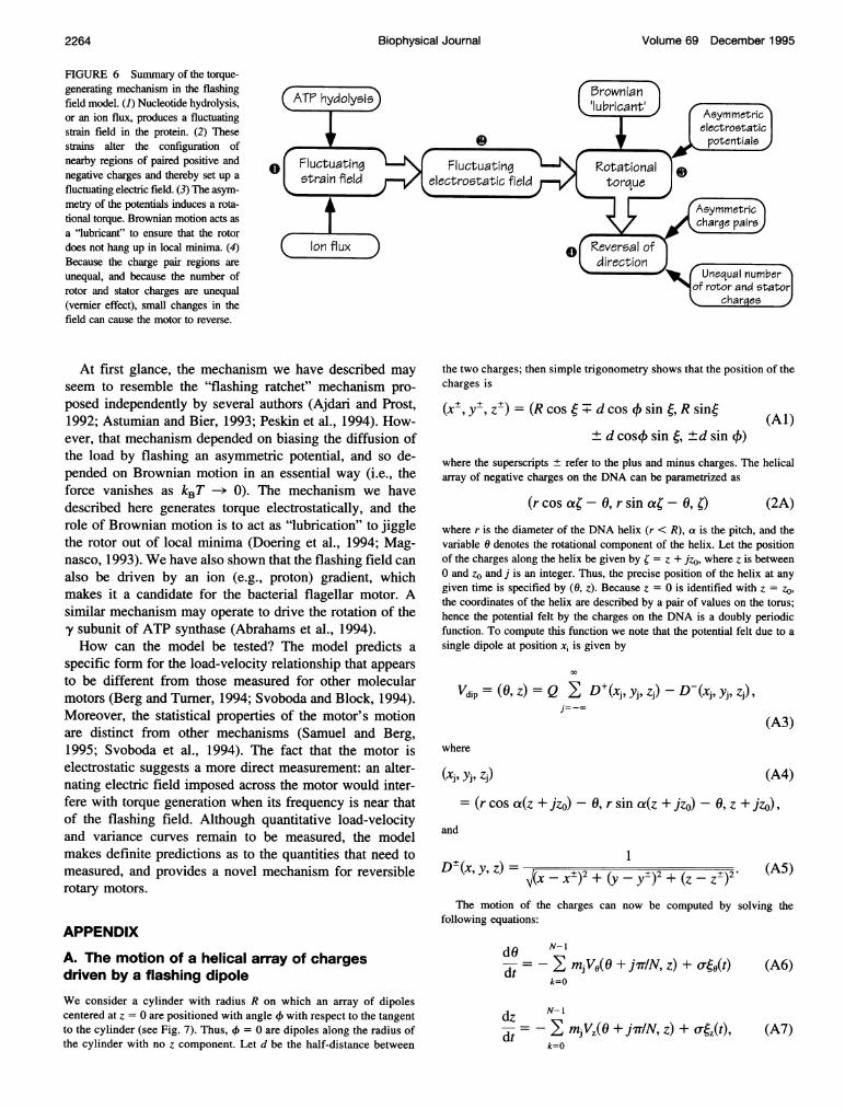
tially; it arises because each flashing field is asymmetrical.To our knowledge, this mechanism for torque generation isnovel. Fig. 6 summarizes the effects producing the rota-tional torque.
Surprisingly, the motor can reverse its direction in re-
sponse to relatively small alterations in the shape of thepotential field due to the charge pair regions. This does notrequire that the fields switch their polarity. Reversal resultsfrom the "vernier" effect that arises because the number offlashing fields is not the same as the number of entrainedcharges. This small shape modification could be controlled,for example, by phosphorylation/dephosphorylation (Gold-beter and Koshland, 1982). The bacterial flagellar rotorreverses stochastically every few seconds, and this reversalmechanism is an attractive candidate for this phenomenon.
Several authors have invoked the vernier effect in dis-cussing the portal protein. In 1978 Hendrix pointed outthat the pentagonal symmetry of the icosohedral capsidbase interfaces the hexagonal symmetry of the connector(Hendrix, 1978). This led him to suggest that this symmetrymismatch might be an essential element in a rotary motorthat screwed the DNA into the capsid. He reasoned that themismatch between potential minima on the rotor and statorwould produce a large number of shallow potentials thatwould offer little resistance to rotation. Although Hendrixdid not address the mechanism of torque generation, heimplied that the symmetry mismatch was somehow in-volved. However, later measurements showed that eachbase pair inserted consumed one-half ATP (Guo et al.,1987), and the 6-to-5 symmetry mismatch would consume
too much ATP per revolution (Dube et al., 1993). Recentmeasurements also reveal that the portal protein has a 12- or
13-fold symmetry, which led Dube et al. (1993) to suggestthat the rotary motor relies on the discrepancy between thetredecameric symmetry of the portal protein and the 10-foldhelical symmetry of the DNA strand (Dube et al., 1993).However, they postulated that insertion was not rotary, butresulted from an axial movement. In the mechanism of forcegeneration we have proposed, the relative motion of therotor and stator has both axial and rotary components. Theonly other model we are aware of that deals with the forcefor packaging is the osmotic pressure model of Serwer(1988).
1.0 Ar =0.5
Al =0.4
V 0.5
0 I I I -
0 0.2 0.4 0.6 0.8 1.0
x
1.2
01.1
0 0.2 0.4 0.6 0.8 1.0
(c)
FIGURE 5 Reversing the motor. (a) The parameter plane describing theshape of the potential. In (+) regions the motor runs clockwise; in (-)regions the motor runs counterclockwise. The shape of the potential ischaracterized by Al and Ar. (b and c) The shapes of the potential seen byeach charge on the rotor (top) and the shapes of the potentials seen by theassembly of two charges rotating in the field of three potentials (bottom).The bottom panels were computed by inserting the potential in the toppanel into Eq. 2b. In b the symmetry of the single and composite potentialsare the same, and the rotor moves clockwise. In c the symmetry of thesingle potentials is the same as in b, but the relative steepness of the sideshas changed. The rotor moves counterclockwise because the compositepotential seen by both charges has symmetry opposite that seen by a singlecharge.
2263Doering et al.
Volume 69 December 1995
FIGURE 6 Summary of the torque-generating mechanism in the flashingfield model. (1) Nucleotide hydrolysis,or an ion flux, produces a fluctuatingstrain field in the protein. (2) Thesestrains alter the configuration ofnearby regions of paired positive andnegative charges and thereby set up afluctuating electric field. (3) The asym-metry of the potentials induces a rota-tional torque. Brownian motion acts asa "lubricant" to ensure that the rotordoes not hang up in local minima. (4)Because the charge pair regions areunequal, and because the number ofrotor and stator charges are unequal(vernier effect), small changes in thefield can cause the motor to reverse.
At first glance, the mechanism we have described mayseem to resemble the "flashing ratchet" mechanism pro-posed independently by several authors (Ajdari and Prost,1992; Astumian and Bier, 1993; Peskin et al., 1994). How-ever, that mechanism depended on biasing the diffusion ofthe load by flashing an asymmetric potential, and so de-pended on Brownian motion in an essential way (i.e., theforce vanishes as kBT -> 0). The mechanism we havedescribed here generates torque electrostatically, and therole of Brownian motion is to act as "lubrication" to jigglethe rotor out of local minima (Doering et al., 1994; Mag-nasco, 1993). We have also shown that the flashing field canalso be driven by an ion (e.g., proton) gradient, whichmakes it a candidate for the bacterial flagellar motor. Asimilar mechanism may operate to drive the rotation of the'y subunit of ATP synthase (Abrahams et al., 1994).How can the model be tested? The model predicts a
specific form for the load-velocity relationship that appearsto be different from those measured for other molecularmotors (Berg and Turner, 1994; Svoboda and Block, 1994).Moreover, the statistical properties of the motor's motionare distinct from other mechanisms (Samuel and Berg,1995; Svoboda et al., 1994). The fact that the motor iselectrostatic suggests a more direct measurement: an alter-nating electric field imposed across the motor would inter-fere with torque generation when its frequency is near thatof the flashing field. Although quantitative load-velocityand variance curves remain to be measured, the modelmakes definite predictions as to the quantities that need tomeasured, and provides a novel mechanism for reversiblerotary motors.
APPENDIX
A. The motion of a helical array of chargesdriven by a flashing dipoleWe consider a cylinder with radius R on which an array of dipolescentered at z = 0 are positioned with angle 41 with respect to the tangentto the cylinder (see Fig. 7). Thus, 4 = 0 are dipoles along the radius ofthe cylinder with no z component. Let d be the half-distance between
the two charges; then simple trigonometry shows that the position of thecharges is
(x+, y+, zt) = (R cos + d cos 4 sin(, R sine
± d cos4 sin (, ±d sin 4)(Al)
where the superscripts ± refer to the plus and minus charges. The helicalarray of negative charges on the DNA can be parametrized as
(r cos a;-0, r sin ca'-0,O) (2A)
where r is the diameter of the DNA helix (r < R), a is the pitch, and thevariable 6 denotes the rotational component of the helix. Let the positionof the charges along the helix be given by ; = z + jzo, where z is betweenO and zo and j is an integer. Thus, the precise position of the helix at anygiven time is specified by (0, z). Because z = 0 is identified with z = zO,the coordinates of the helix are described by a pair of values on the torus;hence the potential felt by the charges on the DNA is a doubly periodicfunction. To compute this function we note that the potential felt due to asingle dipole at position xi is given by
Vdip = (0, z) = Q I D(xj, yj, zj) -D-(xj, yj, zj),j=-CO
(A3)
where
= (r cos a(z + jzo) -0, r sin a(z + jzo) -0, z + jzo),and
D-(x, y, z) = I-x+)2 + (y -V(X-x±)2+ ( ±)2 + (Z -Z±2 (A5)
The motion of the charges can now be computed by solving thefollowing equations:
dO N-I=->- m VO(O + j7TIN, z) + o-0(t)
k=O
dz N-I
d- - I m Vz(O + jTrIN, z) + o4(t),k=O
(A6)
(A7)
Biophysical Journal2264
(xi, yi, zj) (A4)

Rotary DNA Motors
At each instant of time the time-dependent potential, V(x, t), is due to oneor another of the N phase-shifted dipoles on the inner surface of thehelicase. The configuration of dipoles switches in time between the Nphase-shifted configurations so that the full potential in the. nth configu-ration is
v /arY(+~nn-)Y,=V,,Y +2- N (B2)
Because the number of combinations of charges and potentials is large,for simplicity in the following we consider M = 2 charges interacting withN = 3 possible dipole configurations.
Let X(t) be a realization of the random process indicating the position ofa phosphate along the inner radius of the helicase. The dynamics aredefined by the stochastic differential equation:
FIGURE 7 Geometry of the dipole locations with respect to the negativecharges on the DNA backbone.
where mj = 1 if the jth dipole is "on," otherwise it is zero. Here (j(t) areindependent white noise variables. To simplify things, we assume mj iszero except for exactly one value ofj, and that the rate of transition fromone state to the next is determined by r. In the simulation shown in Fig. 3,we assume no screening and take a = 2, b = 8, c = 0.2, r = 20, ar = 0.5,N = 6, Q = 5. The integration was carried out using a fixed step size of0.01 for 200,000 steps using the program xpp.
dt (B3)
where ; is the friction coefficient, V(x, t) is the time-dependent potential,which at any instant is either V1(x), V2(x), or V3(x), with switching rules tobe specified below, and x(t) is a Gaussian white noise process satisfying
(B4)
For notational simplicity we divide the stochastic differential equationthrough by C, identify the diffusion coefficient D = kBT/I, and rescale thepotentials by C, so the equation becomes
dXdt= -VIM t)+ AD(t). (B5)
The switching dynamics are modeled as random, Markovian transitionsbetween the different configurations. This means that the waiting time ineach configuration is a Poisson distributed random variable, independent ofall the other dynamics. The time scale of the switches is the inverse of theswitching rate, which will be denoted a; i.e., a is the rate of jumpingbetween subsequent configurations and a-' is the average time betweenswitches.
These stochastic dynamics are then fully described by a set of coupledFokker-Planck equations for the evolution of the joint probabilities P.(x, t)of finding the potential in configuration n and the charge array at positionx. For the case of random switching between the three configurations theequations have the form
at
PX, t))(B6)
B. Approximations to the flashing field equationsWe shall model the system as a periodic sequence of charges (DNAbackbone phosphates) moving through a periodic array of "off-axis-screened-dipole" potentials, each governed by Eq. 2. The phase of theunderlying potential is allowed to change because of ATP-generated con-
formational changes moving or "tuming on and off" the dipoles. Thepotential due to each dipole interacting with the array ofM charges on theDNA is
Fi-a 1/2a= /2a F2-a
1/2a 1/2a
1/2a \ PI(X, t)½/2a l P2(x, t) ,
F3- a/\P3(x, t)
where a is the rate of switching and the differential operator F,, is
a Iavn aI
ax ax axJ (B7)
V,arry = Vdip(x - 2'Tr M) (B1) (In this notation, an open derivative operator like a/ax operates on every-thing to its right.) For the case of sequential flashing in order 1--2--3--4
2265Doering et al.
(00) = 0, WOOS)) = S(t s).

Volume 69 December 1995
the coupled equations are
a P1(x, t)
a
a
0F2 - a
a
Pl(x, t)\0 PA(X, t) 1,
F3 - at P3(X, t)/
with the effective potential modified by addition of an extra term, -fx.Most importantly, even though the effective potential is then no longerperiodic on the interval [0, 2irr], the coupled Fokker-Planck equations arestill to be solved with periodic conditions in x.
Numerical solution is accomplished via spatial discretization of the(B8) problem wherein the continuous x variable is replaced by K discrete
positions xk = kAx, with k = 1, * *, K and Ax = 2lrr/KJ. The probabilitydensities Pn(x, t) are replaced by probability vectors
Pn,l (t)IPn,2(t)I
Pn(t) P
n,K(t)
whereas for the reverse sequence 1-->3--*2-i.l they are
a /Pl(x, t)- IP2(X, t)at \P3X, t)/
Fl-a a 0 \/Pl(x, t)F2- a a P2(X, t) ,
0 F3-a P3(x, t)
For either random or sequential flashing the x-space current J is defined bythe continuity equation for the marginal density P(x, t) = P1 + P2 + P3 ofthe position variable:
at axat ax, (B 10)
(B9) and the coupled Fokker-Planck equations are replaced by coupled masterequations of the form (e.g., for the random switching case)
a /Pi(t)\at \P2(t)/
F,- a ½/2a ½/2a \/p(t)1/2a F2-a ½/2a p2(t) ,½/2a ½/2a F3-aP- O
where the differential operators are now K X K matrices
(B 16)
-(Rn,l + 4,I)
Rn,lFn = °
Ln,l
Ln,2(-Rn,2 + Ln,2)
Rn,2
0
0 0 Rn,K
Ln,3 .... 0 0
-(Rn,3+ L,n3) ... 0 0 J.
0 Rn,K1 -(Rn,K + Ln,K)
Hence the current is
3 a Vn p PJ=-Pn-Dn
ax ax,n=l
(B 11)
To determine J, the full set of equations must be solved with periodicboundary conditions for x E [0, 2mnr] for each P,, and with the overallnormalization condition
2r21= P(x,t)dx. (B12)
0
The initial distributions at t = 0 must be nonnegative functions satisfying
the normalization condition. The average velocity of the particle is ex-
pressed in terms of the current as
2r2(v)= Jdx. (B13)
In the steady state, aPe/at = 0, and so J approaches a constant value as the
system evolves. In the steady state the average velocity is simply
(v) = JL. (B 14)
This formulation is for the "no-load" situation. If an external load torque,f, is applied to the DNA, then the equivalent particle dynamics is the same
The transition rates in these matrices are defined by
D fVn(Xk) - Vn(Xk+l)Rn,k =- exp, 2A
D fVn(Xk) - Vn(Xk-l)1Ln,k= X exp ~ 2A 9
(B 18)
where K + 1 is to be interpreted as 1, and 0 is to be interpreted as K.In the absence of switching, this discretization supports a true equilib-rium stationary state with vanishing currents and Boltzmann probabilitydistributions.
The numerical solutions to these equations were carried out usingMatLab on a Sun Sparcstation 20. The code is available via Internet ftpupon request.
We would like to thank Smita Patel and Manju Hingorani for sharingunpublished data on T7 helicase with us, and for invaluable conversationsand critical input. Thanks also to Richard Berry and Tim Elston for criticalreadings of the manuscript that enabled us to clarify several importantpoints.
The authors were supported by the following grants: NSF grantsPHY8958506 and PHY9214715 (CD), NSF grant DMS9303706 (BE), andNSF grant FD92-20719 (GO). CD and GO benefited from funding fromUniversity of California's INCOR program. CD was also supported by theDepartment of Energy and acknowledges the hospitality of the Mathemat-ical Sciences Research Institute at Berkeley, where part of this work was
performed.
(B15)
(B 17)
Biophysical Journal2266

Doering et al. Rotary DNA Motors 2267
REFERENCES
Abrahams, J., A. Leslie, R. Lutter, and J. Walker. 1994. Structure at 2.8 Aresolution of FI-ATPase from bovine heart mitochondria. Nature. 370:621-628.
Ajdari, A., and J. Prost. 1992. Mouvement induit par un potentiel peri-odique de basse symetrie: di6lectrophor6se puls6e. C. R. Acad. Sci.Paris. 315:1635-1639.
Astumian, R. D., and M. Bier. 1993. Fluctuation driven ratchets: molecularmotors. Phys. Rev. Lett. 72:1766-1769.
Berg, H., and L. Turner. 1994. Torque generated by the flagellar motor ofEscherichia coli. Biophys. J. 65:2201-2216.
Berg, O., and P. Hippel. 1985. Diffusion-controlled macromolecular inter-actions. Annu. Rev. Biophys. Biophys. Chem. 14:131-160.
Black, L. 1988. DNA packaging in dsDNA bacteriophages. In The Bac-teriophages. R. Calendar, editor. Plenum Press, New York. 320-373.
Carazo, J. M., L. E. Donate, L. Herranz, J. P. Secilla, and J. L. Carrascosa.1986. Three-dimensional reconstruction of the connector of bacterio-phage phi 29 at 1.8 nm resolution. J. Mol. Biol. 192:853-867.
Casjens, S., et al. 1992. Bacteriophage P22 portal protein is part of thegauge that regulates packing density of intravirion DNA. J. Mol. Biol.224:1055-1074.
Doering, C. 1990. Modeling complex systems: stochastic processes, sto-chastic differential equations, and Fokker-Planck equations. In 1990Lectures in Complex Systems. L. Nadel and D. Stein, editors. Addison-Wesley. Redwood City, CA. 3-51.
Doering, C., W. Horsthemke, and J. Riordan. 1994. Nonequilibrium fluc-tuation-induced transport. Phys. Rev. Lett. 72:2984-2987.
Donate, L. E., and J. L. Carrascosa. 1991. Characterization of a versatile invitro DNA-packaging system based on. Virology. 182:534-544.
Dube, P., P. Tavares, R. Lurz, and M. v. Heel. 1993. The portal protein ofbacteriophage SPP1: a DNA pump with 13-fold symmetry. EMBO J.12:1303-1309.
Earnshaw, W., and S. Casjens. 1980. DNA packaging by the double-stranded DNA bacteriophages. Cell. 21:319-31.
Egelman, H., X. Yu, R. Wild, M. Hingorani, and S. Patel. 1995. Bacterio-phage T7 helicase/primase proteins form rings around single-strandedDNA that suggest a general structure for hexameric helicases. Proc.Natl. Acad. Sci. USA. 92:3869-73.
Gardiner, C. 1985. Handbook of Stochastic Methods. Springer-Verlag,New York.
Gilbert, S., M. Webb, M. Brune, and K. Johnson. 1995. Pathway ofprocessive ATP hydrolysis. Nature. 373:671-676.
Goldbeter, A., and D. Koshland. 1982. Sensitivity amplification in bio-chemical systems. Q. Rev. Biophys. 15:555-591.
Guo, P., C. Peterson, and D. Anderson. 1987. Prohead and DNA-gp3-dependent ATPase activity of the DNA packaging protein gpl6 ofbacteriophage phi 29. J. Mol. Biol. 197:229-236.
Hackney, D. 1994. Evidence for alternating head catalysis by kinesinduring microtubule-stimulated ATP hydrolysis. Proc. Natl. Acad. Sci.USA. 91:6865-6869.
Hendrix, R. 1978. Symmetry mismatch and DNA packaging in largebacteriophages. Proc. Natl. Acad. Sci. USA. 75:4779-4783.
Hingorani, M., and S. Patel. 1993. Interactions of bacteriophage T7primase/helicase protein with single-stranded and double-strandedDNA. Biochemistry. 32:12478-12487.
Lohman, T. M. 1992. Escherichia coli DNA helicases: mechanisms ofDNA unwinding. Mol. Microbiol. 6:5-14.
Magnasco, M. 0. 1993. Forced thermal ratchets. Phys. Rev. Lett. 71:1477-1481.
Monk, B., et al. 1994. Modeling a conformationally sensitive region of themembrane sector of the fungal plasma membrane proton pump. J. Bioen-erg. Biomembr. 26:101-114.
Patel, S., and M. Hingorani. 1994. Nucleotide binding studies of bacterio-phage T7 DNA helicase-primase protein. In Molecular Motors: Struc-ture, Mechanics and Energy Transduction. Biophysical Society, Airlie,VA. 186s-189s.
Patel, S., and M. Hingorani. 1995. Nucleotide binding studies of bacterio-phage T7 DNA helicase-primase protein. Biophys. J. 68:186s-190s.
Patel, S., M. Hingorani, and W. Ng. 1994. The k318a mutant of bacterio-phage T7 dna primase-helicase protein is deficient in helicase but notprimase activity and inhibits primase-helicase protein wild-type activi-ties by heterooligomer formation. Biochemistry. 33:7857-7868.
Peskin, C., G. B. Ermentrout, and G. Oster. 1994. The correlation ratchet:a novel mechanism for generating directed motion by ATP hydrolysis. InCell Mechanics and Cellular Engineering. V. C. Mow, F. Guilak, R.Tran-Son-Tay, and R. Hochmuth, editors. Springer-Verlag, New York.479-489.
Peskin, C., and G. Oster. 1995. Coordinated ATPase activity explains themechanical performance of kinesin. Biophys. J. 68:202s-21 Is.
Riskin, H. 1989. The Fokker-Planck Equation. Springer-Verlag, NewYork.
Samuel, A., and H. Berg. 1995. Fluctuation analysis of rotational speed ofthe bacterial flagellar motor. Proc. Natl. Acad. Sci. USA. 92:3502-3506.
Serwer, P. 1988. The source of energy for bacteriophage DNA packaging:an osmotic pump explains the data. Biopolymers. 27:165-169.
Svoboda, K., and S. Block. 1994. Force and velocity measured for singlekinesin molecules. Cell. 77:773-784.
Svoboda, K., P. Mitra, and S. Block. 1994. Fluctuation analysis of motorprotein movement and single enzyme kinetics. Proc. Natl. Acad. Sci.USA. 91:11782-11786.
Valpuesta, J., and J. Carrascosa. 1994. Structure of viral connectors andtheir function in bacteriophage assembly and DNA packaging. Q. Rev.Biophys. 27:107-155.
Valpuesta, J. M., H. Fujisawa, S. Marco, J. M. Carazo, and J. L. Carras-cosa. 1992. Three-dimensional structure of T3 connector purified fromoverexpressing. J. Mol. Biol. 224:103-112.
Wong, I., and T. M. Lohman. 1992. Allosteric effects of nucleotidecofactactors on Escherichia coli Rep Helicase-DNA binding. Science.256:350-356.


