Embed Size (px)
Citation preview
TOXICOLOGICAL SCIENCES 118(2), 324–347 (2010)
doi:10.1093/toxsci/kfq178
Advance Access publication June 16, 2010
REVIEW
Nephrotoxicity of Uranium: Pathophysiological, Diagnostic andTherapeutic Perspectives
Laura Vicente-Vicente,*,† Yaremi Quiros,† Fernando Perez-Barriocanal,† Jose Miguel Lopez-Novoa,†Francisco Jose Lopez-Hernandez,†,‡ and Ana Isabel Morales*,†,1
*Unidad de Toxicologıa and †Departamento de Fisiologıa y Farmacologıa, Universidad de Salamanca, 37007 Salamanca, Spain; and ‡Unidad de
Investigacion, Hospital Universitario de Salamanca, 37007 Salamanca, Spain
1To whom correspondence should be addressed at Unidad de Toxicologıa, Universidad de Salamanca, Edificio Departamental, S-19, Campus Miguel de Unamuno,
37007 Salamanca, Spain. Fax: þ34-923-294-669. E-mail: [email protected].
Received April 12, 2010; accepted June 8, 2010
As in the case of other heavy metals, a considerable body of
evidence suggests that overexposure to uranium may cause
pathological alterations to the kidneys in both humans and
animals. In the present work, our aim was to analyze the available
data from a critical perspective that should provide a view of the
real danger of the nephrotoxicity of this metal for human beings.
A further aim was to elaborate a comparative compilation of the
renal pathophysiological data obtained in humans and experi-
mental animals with a view to gaining more insight into our
knowledge of the mechanisms of action and renal damage.
Finally, we address the existing perspectives for the improvement
of diagnostic methods and the treatment of intoxications by
uranium, performing an integrated analysis of all these aspects.
Key Words: uranium; nephrotoxicity; chronic; acute; diagnosis;
treatment.
Human beings are constantly exposed to a certain amount of
uranium because it is heterogeneously present in natural form
in food, the air, the soil, and water. The repercussions of this
natural exposure as regards human physiology and pathophys-
iology are not completely known. However, the evidence
gathered so far suggests that overexposure to uranium may
result in toxicity, which is derived from an excessive
accumulation of the element in the organism. This accumula-
tion, in turn, depends on the route of entry, the duration of
the exposure, the dose and the chemical compound of which
it forms part, and its absorption (Maynard and Hodge,
1949; Stokinger et al., 1953). Natural exposure, overexposure,
and intoxication can occur by ingestion, inhalation, or
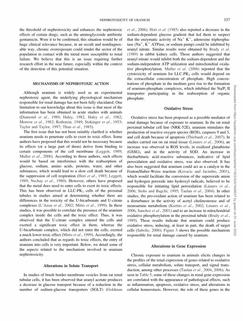
skin contact (Fig. 1). In any case, a small portion of the
uranium gains access to the circulation from which it
distributes throughout the body. Uranium accumulates mainly
in the bones (66%), kidneys (8%), and liver (16%) (ICRP,
1996), and it is eliminated with the urine, rapidly from the
blood and slowly from organ depots (ICRP, 1996; La Touche
et al., 1987).
Human beings may be subjected to pathological over-
exposure to the metal, both acutely and chronically, as
a consequence of (1) contamination of the usual sources of
normal exposure with high amounts of uranium arising from
the anisotropy of the distribution of the metal in the earth’s
crust, as in ground veins or water masses in contact with them,
or from human dumping and (2) direct contact with new
sources of exposure originated by human activity and enriched
in the element, such as in materiel and aeronautics or in the
fields of mining and industry. The main industrial use of
uranium is for fuel in nuclear reactors, which produce 17% of
the world’s electricity (Uranium Institute, 1996). Many
countries are being driven into nuclear energy generation as
a consequence of (1) the energy demand escalation, (2) the
limited reserve of fossil fuels, (3) the scarce development of
alternative sources, (4) the climate change, and (5) the
regulation imposed by the Kyoto treaty. In this sense, uranium
is one of the most useful fuels for nuclear energy production; it
is reasonably inexpensive and complies with the Kyoto
protocol. Other industrial uses of the element include the
manufacture of aircraft stabilizers, in satellites, and in naval
architecture (Wilkinson, 1962); in inertial orientation devices
and gyroscopes (ATSDR/CDC, 1990); in green or yellow
glasses (Peeks et al., 2002; Rossol, 1997); and in certain
luminous devices, in highly penetrating gun ammunitions
and in the production of high-energy x-rays (EPA, 1985).
For years, it was used in the manufacture of dental porcelain
(Thompson, 1976). The long half-life of the 238U isotope
(4.51 3 109 years) is a good proxy for estimating the age of
igneous rocks and in other types of radiometric dating
(ATSDR, 1999).
� The Author 2010. Published by Oxford University Press on behalf of the Society of Toxicology. All rights reserved.For permissions, please email: [email protected]
Dow
nloaded from https://academ
ic.oup.com/toxsci/article/118/2/324/1648537 by guest on 01 June 2022
The toxicity of the metal depends on several factors, such
as sex, age, the body mass index (Kurttio et al., 2006), and
species. Of all the mammals studied, humans seem to be the
least sensitive to uranium (Kathren and Burklin, 2008). An
interspecies order of sensitivity has been proposed: rabbit >rat > guinea pig > pig > mouse > dog > cat > human (Orcutt,
1949; Tannenbaum et al., 1951). Uranium is responsible for
both radiological and chemical toxicity. The radiological
toxicity has been theoretically associated with the production
of cancer. However, because the specific radioactivity of
natural uranium is low, there seems to be no evident danger of
cancer from radiological effects. The results of different
studies carried out on animals and humans are consistent with
this notion (Morris et al., 1990; Muller et al., 1967; Sanders,
1986; Stokinger et al., 1953). On the other hand, the chemical
toxicity has been associated with hepatic, lung, and renal
injury (UNSCEAR, 1988). It has also been suggested that the
net effects caused by this metal in the kidneys and lungs could
be because of cooperation between the chemical and
radiological properties through a complementary mechanism
of action, although this relationship has not been demon-
strated experimentally (Ballou et al., 1986; Filippova et al.,
1978; Spiegel, 1949; Spoor and Hursh, 1973; Stokinger et al.,1953).
No significant toxicity of uranium has been evidenced at
the cardiovascular (Boice et al., 2007; Dygert, 1949; Gilman
et al., 1998c), muscle-skeletal (Gilman et al., 1998c),
endocrine (Boice et al., 2007; Dygert, 1949; Gilman et al.,1998c; Maynard and Hodge, 1949; Stokinger et al., 1953),
gastrointestinal (Boice et al., 2007; Gilman et al., 1998c;
Maynard and Hodge, 1949), and skin (Boice et al., 2007;
Spiegel, 1949) levels. No effects on reproduction have been
reported in humans (Mays et al., 1985; McDiarmid et al.,2007). In contrast, in animal experimentation, relatively high
doses of uranium have been reported to elicit reproductive
abnormalities, manifested as a decrease in sperm counts
(Llobet et al., 1991), fetal toxicity (Domingo et al., 1989),
and testicular lesions (Maynard et al., 1953). In this article,
the available information on the nephrotoxicity of uranium
upon acute and chronic intoxication is thoroughly analyzed.
For this purpose, both data from human as well as animal
studies are critically compared. Human studies are scarcer and
less controlled than animal studies. As such, information from
experimental animals can, to a certain extent, be processed
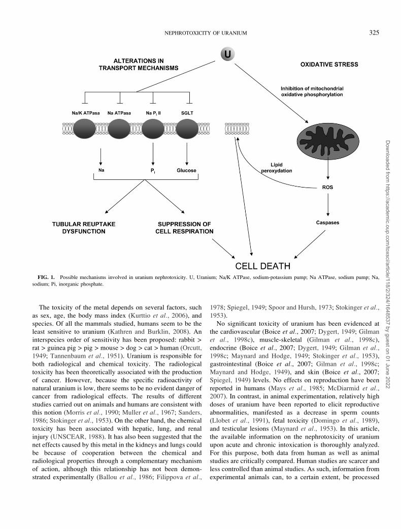
FIG. 1. Possible mechanisms involved in uranium nephrotoxicity. U, Uranium; Na/K ATPase, sodium-potassium pump; Na ATPase, sodium pump; Na,
sodium; Pi, inorganic phosphate.
NEPHROTOXICITY OF URANIUM 325
Dow
nloaded from https://academ
ic.oup.com/toxsci/article/118/2/324/1648537 by guest on 01 June 2022
and extrapolated into the framework delineated by data
obtained from human beings, in order to create a more
complete picture of the pathophysiological aspects of the
nephrotoxicity of uranium and the real risk posed by this
metal to the human being.
NEPHROTOXICITY BECAUSE OF ACUTE
OVEREXPOSURE
Acute Nephrotoxicity in Animal Models
Pathophysiological Studies
In animals, it has been possible to characterize the renal
damage caused by the element in detail, under predetermined
and controlled experimental conditions. Several studies have
reported decreases in creatinine clearance (Banday et al., 2008;
Haley, 1982; Sanchez et al., 2001; Shim et al., 2009), which is
indicative of a reduction in the glomerular filtration rate (GFR).
Congruently, with the decrease in the GFR, a significant
increase in the plasma concentration of creatinine and blood
ureic nitrogen (BUN) has been reported after uranium
administration (Banday et al., 2008; Fukuda et al., 2005b;
Sanchez et al., 2001; Shim et al., 2009; Taulan et al., 2006;
Yapar et al., 2010; Zimmerman et al., 2007). It is unknown
whether such a decrease in the GFR is because of (1) the
glomerular effects of uranium, (2) the tubuloglomerular
feedback brought about after tubular insult in order to prevent
an uncontrolled loss of water and electrolytes, or (3)
a combination of both. In this sense, in animals acutely
intoxicated with uranium, functional tubular alterations have
been observed. These are reflected in a significant increase in
electrolyte excretion (sodium, potassium, magnesium, calcium,
and inorganic phosphate) (Banday et al., 2008; Haley,
1982), proteins (Haley, 1982; Sanchez et al., 2001), b-2-
microglobulin (Fukuda et al., 2008), and glucose (Nomiyama
et al., 1974; Taulan et al., 2006). Increases in the urinary
activity of several enzymes indicating tissue lesion have also
been reported. At least in part, this could be explained by the
functional tubular alterations. Still, it is not possible to rule out
a direct effect of the metal on transport mechanisms or tubular
cell functions, independently of cell viability. Among the
increased urinary enzymes are N-acetyl glucosaminidase
(NAG) (Diamond et al., 1989; Fukuda et al., 2005b, 2008;
Sanchez et al., 2001) and alkaline phosphatase (ALP) (Banday
et al., 2008; Nomiyama et al., 1974). The increase in ALP in
urine has been linked to the loss of microvilli in the proximal
tubules, where this enzyme is mainly located (Yuile, 1973).
Increases in the activities of other enzymes in the urine, such as
gamma glutamyl transpeptidase (GGT), lactate dehydrogenase
(LDH), and acid phosphatase (Banday et al., 2008; Diamond
et al., 1989; Sanchez et al., 2001; Taulan et al., 2006), have
been described, pointing to the presence of tissue lesions. LDH
is a nonspecific marker of renal tissue lesion, but NAG, ALP,
and GGT are mainly markers of proximal tubule insult
(Emeigh Hart, 2005).
Other effects of uranium on the kidneys possibly related to
the function and viability of renal structures include (1)
alterations in the activities of several enzymes associated with
glycolysis (aldolase and phosphoglycerokinase), the tricarbox-
ylic acid cycle (isocitrate, succinate, and malate dehydro-
genases), and gluconeogenesis (FBPase and G6Pase) (Banday
et al., 2008); (2) disturbances in the oxidative balance
(Schramm et al., 2002), potentially related to increases in the
activity of superoxide dismutase (SOD) (Banday et al., 2008;
Schramm et al., 2002); and (3) increases in the plasma renin
levels, associated with increases in blood pressure (Kato et al.,1994; Mendelsohn and Smith, 1980).
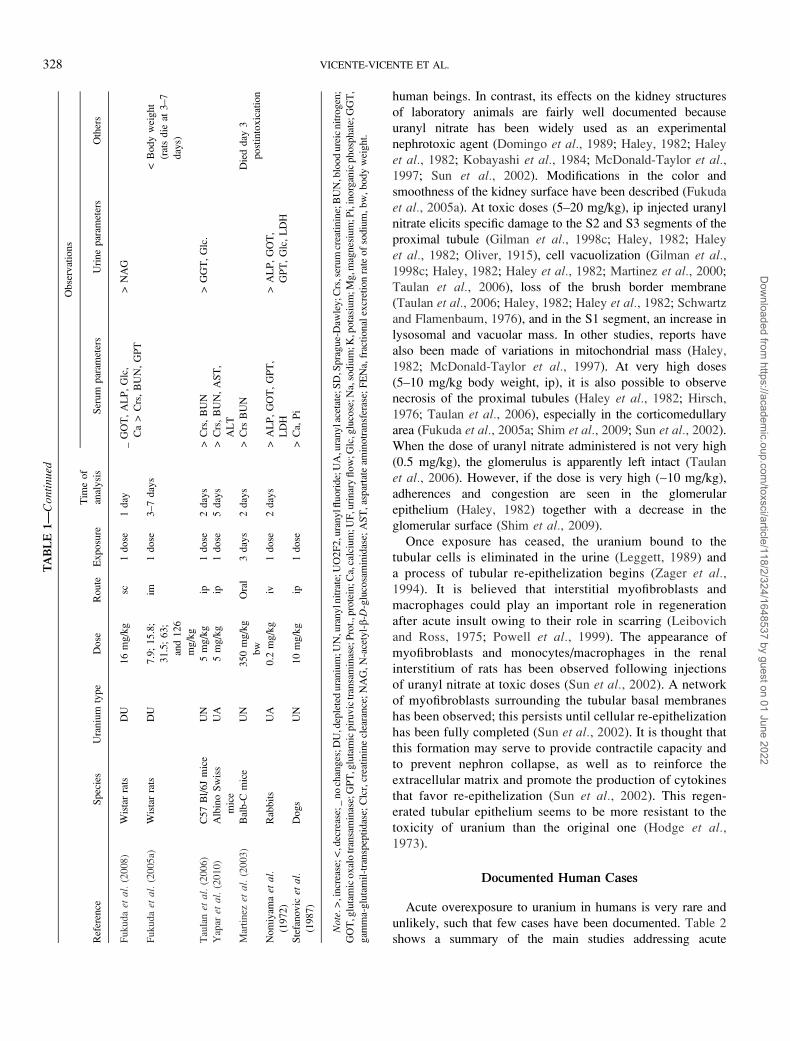
Table 1 summarizes the most relevant data obtained from the
acute intoxications with uranium carried out in experimental
animals, with regard to the alterations in renal function and
structure observed; the animal species; and the dose, route, and
duration of the exposure. The diversity of route of adminis-
tration, uranium type, and other factors introduces some
complexity when comparing the results from different studies.
Most of them have been done with rats, a species very sensitive
to uranium’s toxicity. Regardless of strain and route of
administration, single doses of a few (> 2) milligram per
kilogram are consistently and overtly nephrotoxic, as demon-
strated by alterations in classical parameters of renal function
(serum creatinine, BUN, etc.) and renal tissue status (e.g.,
urinary excretion of NAG), during the immediate days after
exposure. This is also true for other less sensitive species, such
as mice and dogs. There are fewer studies using lower doses,
which were all conducted in rats. However, it is evident that, at
least when uranium is administered ip, the toxic threshold for
single-dose exposures is set around 0.5 mg/kg. In the case of
the im administration, at least doses of or over 1 mg/kg induce
a renal injury that is still detectable 28 days after exposure
(Fukuda et al., 2005b). Because this is the only study
monitoring renal parameters so late after an acute exposure,
there is not enough evidence to know whether this is rather
specific of the im route or it can also be observed upon
intoxication by other routes.
All together, these data indicate that several species acutely
intoxicated with uranium undergo some degree of renal
damage in a dose-dependent and route-independent manner.
Doses of 5 mg/kg U (or higher) are overtly nephrotoxic for
rats, mice, and dogs. Doses higher than 0.5 mg/kg U are
nephrotoxic at least for rats. ‘‘Documented Human Cases’’
section presents the evidence on acute intoxication of humans
gathered in documented cases and compares the information
with that obtained in animals.
Morphological Renal Modifications
As far as we are aware, there are no histological studies
about the acute effects of uranium on kidney structure in
326 VICENTE-VICENTE ET AL.
Dow
nloaded from https://academ
ic.oup.com/toxsci/article/118/2/324/1648537 by guest on 01 June 2022
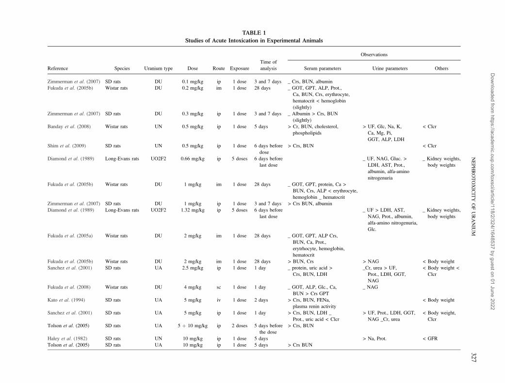
TABLE 1
Studies of Acute Intoxication in Experimental Animals
Reference Species Uranium type Dose Route Exposure
Time of
analysis
Observations
Serum parameters Urine parameters Others
Zimmerman et al. (2007) SD rats DU 0.1 mg/kg ip 1 dose 3 and 7 days _ Crs, BUN, albumin
Fukuda et al. (2005b) Wistar rats DU 0.2 mg/kg im 1 dose 28 days _ GOT, GPT, ALP, Prot.,
Ca, BUN, Crs, erythrocyte,
hematocrit < hemoglobin
(slightly)
Zimmerman et al. (2007) SD rats DU 0.3 mg/kg ip 1 dose 3 and 7 days _ Albumin > Crs, BUN
(slightly)
Banday et al. (2008) Wistar rats UN 0.5 mg/kg ip 1 dose 5 days > Cr, BUN, cholesterol,
phospholipids
> UF, Glc, Na, K,
Ca, Mg, Pi,
GGT, ALP, LDH
< Clcr
Shim et al. (2009) SD rats UN 0.5 mg/kg ip 1 dose 6 days before
dose
> Crs, BUN < Clcr
Diamond et al. (1989) Long-Evans rats UO2F2 0.66 mg/kg ip 5 doses 6 days before
last dose
_ UF, NAG, Gluc. >
LDH, AST, Prot.,
albumin, alfa-amino
nitrogenuria
_ Kidney weights,
body weights
Fukuda et al. (2005b) Wistar rats DU 1 mg/kg im 1 dose 28 days _ GOT, GPT, protein, Ca >
BUN, Crs, ALP < erythrocyte,
hemoglobin _ hematocrit
Zimmerman et al. (2007) SD rats DU 1 mg/kg ip 1 dose 3 and 7 days > Crs BUN, albumin
Diamond et al. (1989) Long-Evans rats UO2F2 1.32 mg/kg ip 5 doses 6 days before
last dose
_ UF > LDH, AST,
NAG, Prot., albumin,
alfa-amino nitrogenuria,
Glc.
_ Kidney weights,
body weights
Fukuda et al. (2005a) Wistar rats DU 2 mg/kg im 1 dose 28 days _ GOT, GPT, ALP Crs,
BUN, Ca, Prot.,
erytrhocyte, hemoglobin,
hematocrit
Fukuda et al. (2005b) Wistar rats DU 2 mg/kg im 1 dose 28 days > BUN, Crs > NAG < Body weight
Sanchez et al. (2001) SD rats UA 2.5 mg/kg ip 1 dose 1 day _ protein, uric acid >
Crs, BUN, LDH
_Cr, urea > UF,
Prot., LDH, GGT,
NAG
< Body weight <
Clcr
Fukuda et al. (2008) Wistar rats DU 4 mg/kg sc 1 dose 1 day _ GOT, ALP, Glc., Ca,
BUN > Crs GPT
_ NAG
Kato et al. (1994) SD rats UA 5 mg/kg iv 1 dose 2 days > Crs, BUN, FENa,
plasma renin activity
< Body weight
Sanchez et al. (2001) SD rats UA 5 mg/kg ip 1 dose 1 day > Crs, BUN, LDH _
Prot., uric acid < Clcr
> UF, Prot., LDH, GGT,
NAG _Cr, urea
< Body weight,
Clcr
Tolson et al. (2005) SD rats UA 5 þ 10 mg/kg ip 2 doses 5 days before
the dose
> Crs, BUN
Haley et al. (1982) SD rats UN 10 mg/kg ip 1 dose 5 days > Na, Prot. < GFR
Tolson et al. (2005) SD rats UA 10 mg/kg ip 1 dose 5 days > Crs BUN
NE
PH
RO
TO
XIC
ITY
OF
UR
AN
IUM
32
7
Dow
nloaded from https://academ
ic.oup.com/toxsci/article/118/2/324/1648537 by guest on 01 June 2022
human beings. In contrast, its effects on the kidney structures
of laboratory animals are fairly well documented because
uranyl nitrate has been widely used as an experimental
nephrotoxic agent (Domingo et al., 1989; Haley, 1982; Haley
et al., 1982; Kobayashi et al., 1984; McDonald-Taylor et al.,1997; Sun et al., 2002). Modifications in the color and
smoothness of the kidney surface have been described (Fukuda
et al., 2005a). At toxic doses (5–20 mg/kg), ip injected uranyl
nitrate elicits specific damage to the S2 and S3 segments of the
proximal tubule (Gilman et al., 1998c; Haley, 1982; Haley
et al., 1982; Oliver, 1915), cell vacuolization (Gilman et al.,1998c; Haley, 1982; Haley et al., 1982; Martinez et al., 2000;
Taulan et al., 2006), loss of the brush border membrane
(Taulan et al., 2006; Haley, 1982; Haley et al., 1982; Schwartz
and Flamenbaum, 1976), and in the S1 segment, an increase in
lysosomal and vacuolar mass. In other studies, reports have
also been made of variations in mitochondrial mass (Haley,
1982; McDonald-Taylor et al., 1997). At very high doses
(5–10 mg/kg body weight, ip), it is also possible to observe
necrosis of the proximal tubules (Haley et al., 1982; Hirsch,
1976; Taulan et al., 2006), especially in the corticomedullary
area (Fukuda et al., 2005a; Shim et al., 2009; Sun et al., 2002).
When the dose of uranyl nitrate administered is not very high
(0.5 mg/kg), the glomerulus is apparently left intact (Taulan
et al., 2006). However, if the dose is very high (~10 mg/kg),
adherences and congestion are seen in the glomerular
epithelium (Haley, 1982) together with a decrease in the
glomerular surface (Shim et al., 2009).
Once exposure has ceased, the uranium bound to the
tubular cells is eliminated in the urine (Leggett, 1989) and
a process of tubular re-epithelization begins (Zager et al.,1994). It is believed that interstitial myofibroblasts and
macrophages could play an important role in regeneration
after acute insult owing to their role in scarring (Leibovich
and Ross, 1975; Powell et al., 1999). The appearance of
myofibroblasts and monocytes/macrophages in the renal
interstitium of rats has been observed following injections
of uranyl nitrate at toxic doses (Sun et al., 2002). A network
of myofibroblasts surrounding the tubular basal membranes
has been observed; this persists until cellular re-epithelization
has been fully completed (Sun et al., 2002). It is thought that
this formation may serve to provide contractile capacity and
to prevent nephron collapse, as well as to reinforce the
extracellular matrix and promote the production of cytokines
that favor re-epithelization (Sun et al., 2002). This regen-
erated tubular epithelium seems to be more resistant to the
toxicity of uranium than the original one (Hodge et al.,1973).
Documented Human Cases
Acute overexposure to uranium in humans is very rare and
unlikely, such that few cases have been documented. Table 2
shows a summary of the main studies addressing acute
TA
BL
E1
—Con
tinu
ed
Ref
eren
ceS
pec
ies
Ura
niu
mty
pe
Do
seR
ou
teE
xp
osu
re
Tim
eo
f
anal
ysi
s
Ob
serv
atio
ns
Ser
um
par
amet
ers
Uri
ne
par
amet
ers
Oth
ers
Fu
ku
daet
al.
(20
08
)W
ista
rra
tsD
U1
6m
g/k
gsc
1d
ose
1d
ay_
GO
T,
AL
P,
Glc
,
Ca>
Crs
,B
UN
,G
PT
>N
AG
Fu
ku
daet
al.
(20
05
a)W
ista
rra
tsD
U7
.9;
15
.8;
31
.5;
63
;
and
12
6
mg
/kg
im1
do
se3
–7
day
s<
Bo
dy
wei
ght
(rat
sd
ieat
3–
7
day
s)
Tau
lanet
al.
(20
06
)C
57
Bl/
6J
mic
eU
N5
mg
/kg
ip1
do
se2
day
s>
Crs
,B
UN
>G
GT
,G
lc.
Yap
aret
al.
(20
10
)A
lbin
oS
wis
s
mic
e
UA
5m
g/k
gip
1d
ose
5d
ays
>C
rs,
BU
N,
AS
T,
AL
T
Mar
tin
ezet
al.
(20
03
)B
alb
-Cm
ice
UN
35
0m
g/k
g
bw
Ora
l3
day
s2
day
s>
Crs
BU
ND
ied
day
3
po
stin
tox
icat
ion
No
miy
amaet
al.
(19
72
)
Rab
bit
sU
A0
.2m
g/k
giv
1d
ose
2d
ays
>A
LP
,G
OT
,G
PT
,
LD
H
>A
LP
,G
OT
,
GP
T,
Glc
,L
DH
Ste
fan
ov
icet
al.
(19
87
)
Do
gs
UN
10
mg
/kg
ip1
do
se>
Ca,
Pi
Note.>
,in
crea
se;<
,dec
reas
e;_
no
chan
ges
;DU
,dep
lete
du
ran
ium
;UN
,ura
ny
lnit
rate
;UO
2F
2,u
ran
ylfl
uori
de;
UA
,ura
ny
lace
tate
;SD
,Sp
rag
ue-
Daw
ley
;Crs
,ser
um
crea
tin
ine;
BU
N,b
loo
du
reic
nit
rog
en;
GO
T,g
luta
mic
ox
alo
tran
sam
inas
e;G
PT
,glu
tam
icp
iru
vic
tran
sam
inas
e;P
rot.
,pro
tein
;Ca,
calc
ium
;UF
,uri
nar
yfl
ow
;Glc
,glu
cose
;Na,
sod
ium
;K,p
ota
sium
;Mg,m
agnes
ium
;Pi,
inorg
anic
phosp
hat
e;G
GT
,
gam
ma-
glu
tam
il-t
ransp
epti
das
e;C
lcr,
crea
tinin
ecl
eara
nce
;N
AG
,N
-ace
tyl-b-D
-glu
cosa
min
idas
e;A
ST
,as
par
tate
amin
otr
ansf
eras
e;F
EN
a,fr
acti
onal
excr
etio
nra
teof
sodiu
m,
bw
,body
wei
ght.
328 VICENTE-VICENTE ET AL.
Dow
nloaded from https://academ
ic.oup.com/toxsci/article/118/2/324/1648537 by guest on 01 June 2022
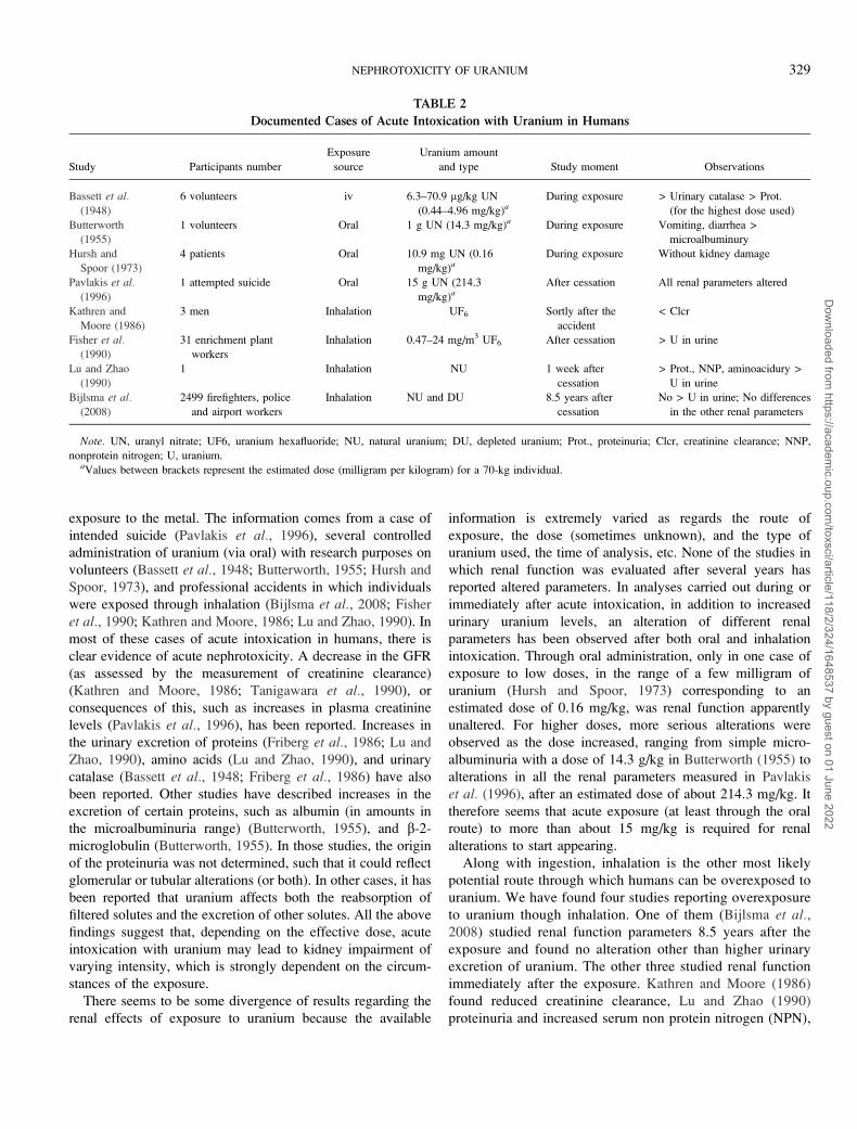
exposure to the metal. The information comes from a case of
intended suicide (Pavlakis et al., 1996), several controlled
administration of uranium (via oral) with research purposes on
volunteers (Bassett et al., 1948; Butterworth, 1955; Hursh and
Spoor, 1973), and professional accidents in which individuals
were exposed through inhalation (Bijlsma et al., 2008; Fisher
et al., 1990; Kathren and Moore, 1986; Lu and Zhao, 1990). In
most of these cases of acute intoxication in humans, there is
clear evidence of acute nephrotoxicity. A decrease in the GFR
(as assessed by the measurement of creatinine clearance)
(Kathren and Moore, 1986; Tanigawara et al., 1990), or
consequences of this, such as increases in plasma creatinine
levels (Pavlakis et al., 1996), has been reported. Increases in
the urinary excretion of proteins (Friberg et al., 1986; Lu and
Zhao, 1990), amino acids (Lu and Zhao, 1990), and urinary
catalase (Bassett et al., 1948; Friberg et al., 1986) have also
been reported. Other studies have described increases in the
excretion of certain proteins, such as albumin (in amounts in
the microalbuminuria range) (Butterworth, 1955), and b-2-
microglobulin (Butterworth, 1955). In those studies, the origin
of the proteinuria was not determined, such that it could reflect
glomerular or tubular alterations (or both). In other cases, it has
been reported that uranium affects both the reabsorption of
filtered solutes and the excretion of other solutes. All the above
findings suggest that, depending on the effective dose, acute
intoxication with uranium may lead to kidney impairment of
varying intensity, which is strongly dependent on the circum-
stances of the exposure.
There seems to be some divergence of results regarding the
renal effects of exposure to uranium because the available
information is extremely varied as regards the route of
exposure, the dose (sometimes unknown), and the type of
uranium used, the time of analysis, etc. None of the studies in
which renal function was evaluated after several years has
reported altered parameters. In analyses carried out during or
immediately after acute intoxication, in addition to increased
urinary uranium levels, an alteration of different renal
parameters has been observed after both oral and inhalation
intoxication. Through oral administration, only in one case of
exposure to low doses, in the range of a few milligram of
uranium (Hursh and Spoor, 1973) corresponding to an
estimated dose of 0.16 mg/kg, was renal function apparently
unaltered. For higher doses, more serious alterations were
observed as the dose increased, ranging from simple micro-
albuminuria with a dose of 14.3 g/kg in Butterworth (1955) to
alterations in all the renal parameters measured in Pavlakis
et al. (1996), after an estimated dose of about 214.3 mg/kg. It
therefore seems that acute exposure (at least through the oral
route) to more than about 15 mg/kg is required for renal
alterations to start appearing.
Along with ingestion, inhalation is the other most likely
potential route through which humans can be overexposed to
uranium. We have found four studies reporting overexposure
to uranium though inhalation. One of them (Bijlsma et al.,2008) studied renal function parameters 8.5 years after the
exposure and found no alteration other than higher urinary
excretion of uranium. The other three studied renal function
immediately after the exposure. Kathren and Moore (1986)
found reduced creatinine clearance, Lu and Zhao (1990)
proteinuria and increased serum non protein nitrogen (NPN),
TABLE 2
Documented Cases of Acute Intoxication with Uranium in Humans
Study Participants number
Exposure
source
Uranium amount
and type Study moment Observations
Bassett et al.
(1948)
6 volunteers iv 6.3–70.9 lg/kg UN
(0.44–4.96 mg/kg)aDuring exposure > Urinary catalase > Prot.
(for the highest dose used)
Butterworth
(1955)
1 volunteers Oral 1 g UN (14.3 mg/kg)a During exposure Vomiting, diarrhea >
microalbuminury
Hursh and
Spoor (1973)
4 patients Oral 10.9 mg UN (0.16
mg/kg)aDuring exposure Without kidney damage
Pavlakis et al.
(1996)
1 attempted suicide Oral 15 g UN (214.3
mg/kg)aAfter cessation All renal parameters altered
Kathren and
Moore (1986)
3 men Inhalation UF6 Sortly after the
accident
< Clcr
Fisher et al.
(1990)
31 enrichment plant
workers
Inhalation 0.47–24 mg/m3 UF6 After cessation > U in urine
Lu and Zhao
(1990)
1 Inhalation NU 1 week after
cessation
> Prot., NNP, aminoacidury >
U in urine
Bijlsma et al.
(2008)
2499 firefighters, police
and airport workers
Inhalation NU and DU 8.5 years after
cessation
No > U in urine; No differences
in the other renal parameters
Note. UN, uranyl nitrate; UF6, uranium hexafluoride; NU, natural uranium; DU, depleted uranium; Prot., proteinuria; Clcr, creatinine clearance; NNP,
nonprotein nitrogen; U, uranium.aValues between brackets represent the estimated dose (milligram per kilogram) for a 70-kg individual.
NEPHROTOXICITY OF URANIUM 329
Dow
nloaded from https://academ
ic.oup.com/toxsci/article/118/2/324/1648537 by guest on 01 June 2022
and Fisher et al. (1990) only higher excretion of uranium.
Except in Fisher et al. (1990), the concentration of uranium in
the air is not known (see Table 2). Accordingly, it is
impossible to draw conclusions on toxic dosage or sensitivity
through this route and much less to make comparisons with
other exposure routes. However, it is clear that feasible
accidents or sporadic circumstances can overexpose the
human being to uranium through inhalation resulting in some
degree of nephrotoxicity.
In conclusion, animal studies diverge from human studies in
the exposure route, which is a key determinant of uranium
bioavailability, and blood and tissue levels. However, it is clear
that acute intoxication with uranium leads to nephrotoxicity in
both animals and humans, in a dose-dependent manner. Yet
one study in rats (Kato et al., 1994) and another one in humans
(Bassett et al., 1948) of acute intoxication via iv with
similar doses yield interesting information. Humans exposed
to ~5 mg/kg U showed proteinuria as the most significant renal
alteration, whereas both serum creatinine and BUN increased
in rats subject to the same dose. This indicates that humans
underwent some degree of renal alteration (probably in tubular
reuptake) that did not end up in the renal dysfunction seen in
rats. This highlights the lower sensitivity to U toxicity of
humans when compared with rats. Through the iv route,
uranium surpasses interspecies differences posed by different
absorption and different influence of other physical barriers
working upon exposure through other routes.
Finally, it should be noted that it is not known whether acute
intoxication with uranium is able to trigger chronic renal
lesions that will progress irreversibly and autonomously
regardless of the presence of the metal, consistent with the
well-known fact that the concurrence of several acute renal
insults may drive the kidneys to enter an autonomous chronic
degenerative process (Basile, 2008). The few data available
concerning acute intoxications in human beings, together with
the lack of this type of study in animals, make it impossible to
draw conclusions. As a possible indication, the work of
Bijlsma et al. (2008) (see Table 2), in which renal function was
assessed years after the acute exposure, suggests that this
would not be the case, although the intensity of those
exposures is not known.
NEPHROTOXICITY BECAUSE OF CHRONIC
OVEREXPOSURE
Under certain circumstances, humans are chronically
overexposed to uranium. It remains largely unknown whether
such exposure may elicit kidney damage and neither are the
determinants of the possible nephrotoxicity known. It is also
unknown whether the possible nephrotoxicity is triggered
(1) as a subacute effect when a certain level of tissue
accumulation after a more or less long exposure time has been
surpassed or in contrast (2) as chronic renal damage that
gradually develops into irreversible degeneration that is even
independent of the presence of uranium, as occurs with most
of the causes of chronic renal impairment (CRI) (Remuzzi
et al., 2006). In this section, we attempt to shed some light on
these aspects.
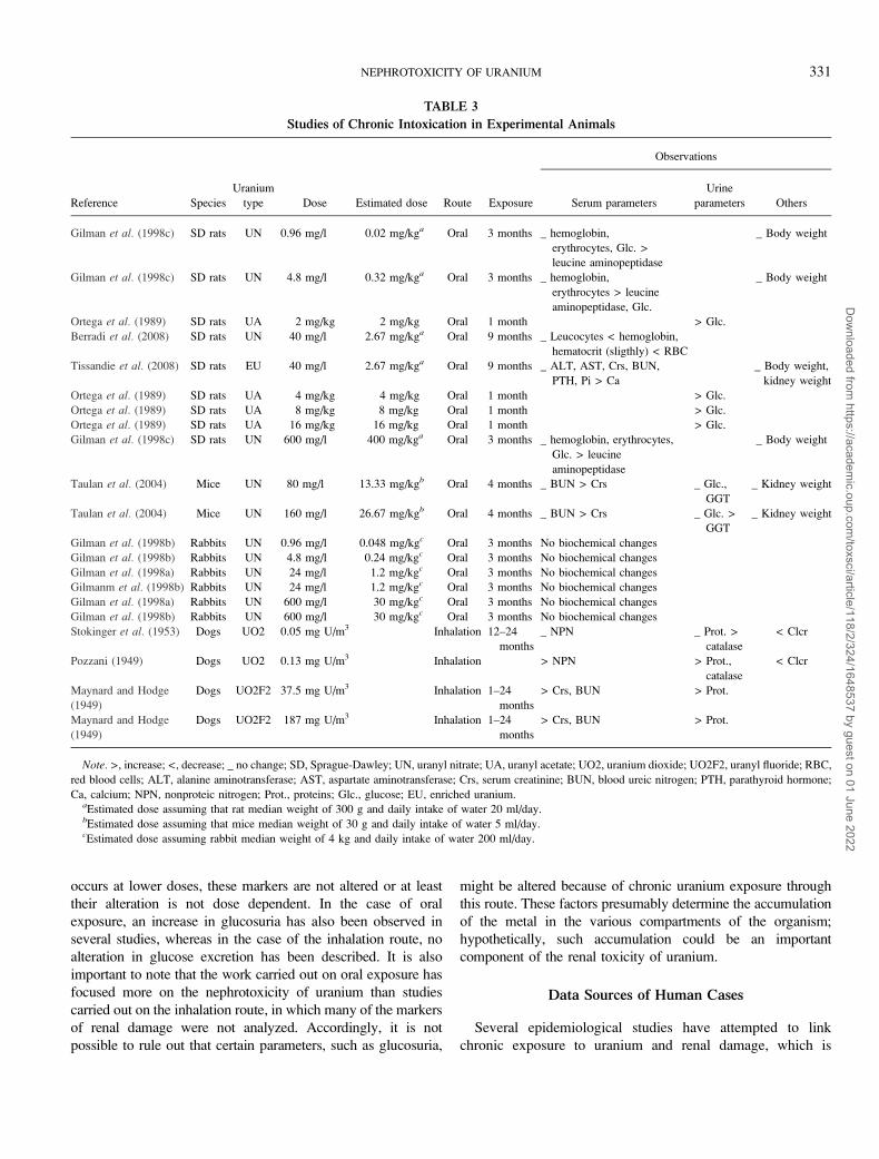
Pathophysiological Studies with Animal Models
In general, studies carried out in laboratory animals have
used higher doses of uranium than those found in human
exposure, although the time of exposure was much shorter
(months instead of years). Table 3 summarizes the most
relevant data obtained from the chronic exposure to uranium
in experimental animals, with regard to the alterations in renal
function, the animal species, uranium type, the estimated dose
(milligram per kilogram), and the route and duration of the
exposure. As commented above, the toxicity of uranium
depends on sex, age, the body mass index, and species. Data
from Table 3 are not in agreement with the interspecies
sensitivity classically reported (Orcutt, 1949; Tannenbaum
et al., 1951). Indeed, after chronic oral exposure, the rat
seems to be more sensitive than the rabbit. Rabbits showed
no biochemical changes in the estimated dose range 0.048–
30 mg/kg during 3 months of exposure. However, rats subject
to even milder conditions (exposure of 1 month and a lower
dosage range [0.02–16 mg/kg]) showed renal alterations,
including glucosuria and increased leucine aminopeptidase
activity. Comparison with the mouse is more controversial
because exposure time was longer (4 months), which may
have induced a higher accumulation resulting in greater
kidney damage. The importance of exposure time has been
evidenced by the studies of Berradi et al. (2008) and
Tissandie et al. (2008), which show major renal alterations
owing to longer time of exposure.
Regarding inhalation exposure to uranium, renal damage has
been reported in studies from 1 to 24 months mainly in dogs.
Stokinger et al. (1953), in a study conducted at low doses
(0.05 mg U/m3 air), observed that NPN levels in plasma were
normal, and there were no differences in the excretion of
urinary proteins, whereas a decrease in creatinine clearance and
an increase in urinary catalase were observed. At higher doses
(0.13 mg U/m3), an increase in protein excretion and NPN
(Pozzani, 1949) was observed. In other studies conducted
during a similar exposure time but with higher dose, changes in
the usual markers of renal function (serum creatinine, BUN,
and urinary proteins) were observed. Importantly, similar
results were obtained in a wide range of doses (37.5–187 mg
U/m3) (Maynard and Hodge, 1949).
It is difficult to compare the results obtained in oral and
inhalation exposure because, among many other factors, the
absorption process in each route is different. Generally, when
exposure occurs with high doses of uranium, the markers of
renal damage, such as plasma creatinine and nonprotein
nitrogen in plasma, are found to be altered, but when exposure
330 VICENTE-VICENTE ET AL.
Dow
nloaded from https://academ
ic.oup.com/toxsci/article/118/2/324/1648537 by guest on 01 June 2022
occurs at lower doses, these markers are not altered or at least
their alteration is not dose dependent. In the case of oral
exposure, an increase in glucosuria has also been observed in
several studies, whereas in the case of the inhalation route, no
alteration in glucose excretion has been described. It is also
important to note that the work carried out on oral exposure has
focused more on the nephrotoxicity of uranium than studies
carried out on the inhalation route, in which many of the markers
of renal damage were not analyzed. Accordingly, it is not
possible to rule out that certain parameters, such as glucosuria,
might be altered because of chronic uranium exposure through
this route. These factors presumably determine the accumulation
of the metal in the various compartments of the organism;
hypothetically, such accumulation could be an important
component of the renal toxicity of uranium.
Data Sources of Human Cases
Several epidemiological studies have attempted to link
chronic exposure to uranium and renal damage, which is
TABLE 3
Studies of Chronic Intoxication in Experimental Animals
Reference Species
Uranium
type Dose Estimated dose Route Exposure
Observations
Serum parameters
Urine
parameters Others
Gilman et al. (1998c) SD rats UN 0.96 mg/l 0.02 mg/kga Oral 3 months _ hemoglobin,
erythrocytes, Glc. >
leucine aminopeptidase
_ Body weight
Gilman et al. (1998c) SD rats UN 4.8 mg/l 0.32 mg/kga Oral 3 months _ hemoglobin,
erythrocytes > leucine
aminopeptidase, Glc.
_ Body weight
Ortega et al. (1989) SD rats UA 2 mg/kg 2 mg/kg Oral 1 month > Glc.
Berradi et al. (2008) SD rats UN 40 mg/l 2.67 mg/kga Oral 9 months _ Leucocytes < hemoglobin,
hematocrit (sligthly) < RBC
Tissandie et al. (2008) SD rats EU 40 mg/l 2.67 mg/kga Oral 9 months _ ALT, AST, Crs, BUN,
PTH, Pi > Ca
_ Body weight,
kidney weight
Ortega et al. (1989) SD rats UA 4 mg/kg 4 mg/kg Oral 1 month > Glc.
Ortega et al. (1989) SD rats UA 8 mg/kg 8 mg/kg Oral 1 month > Glc.
Ortega et al. (1989) SD rats UA 16 mg/kg 16 mg/kg Oral 1 month > Glc.
Gilman et al. (1998c) SD rats UN 600 mg/l 400 mg/kga Oral 3 months _ hemoglobin, erythrocytes,
Glc. > leucine
aminopeptidase
_ Body weight
Taulan et al. (2004) Mice UN 80 mg/l 13.33 mg/kgb Oral 4 months _ BUN > Crs _ Glc.,
GGT
_ Kidney weight
Taulan et al. (2004) Mice UN 160 mg/l 26.67 mg/kgb Oral 4 months _ BUN > Crs _ Glc. >
GGT
_ Kidney weight
Gilman et al. (1998b) Rabbits UN 0.96 mg/l 0.048 mg/kgc Oral 3 months No biochemical changes
Gilman et al. (1998b) Rabbits UN 4.8 mg/l 0.24 mg/kgc Oral 3 months No biochemical changes
Gilman et al. (1998a) Rabbits UN 24 mg/l 1.2 mg/kgc Oral 3 months No biochemical changes
Gilmanm et al. (1998b) Rabbits UN 24 mg/l 1.2 mg/kgc Oral 3 months No biochemical changes
Gilman et al. (1998a) Rabbits UN 600 mg/l 30 mg/kgc Oral 3 months No biochemical changes
Gilman et al. (1998b) Rabbits UN 600 mg/l 30 mg/kgc Oral 3 months No biochemical changes
Stokinger et al. (1953) Dogs UO2 0.05 mg U/m3 Inhalation 12–24
months
_ NPN _ Prot. >
catalase
< Clcr
Pozzani (1949) Dogs UO2 0.13 mg U/m3 Inhalation > NPN > Prot.,
catalase
< Clcr
Maynard and Hodge
(1949)
Dogs UO2F2 37.5 mg U/m3 Inhalation 1–24
months
> Crs, BUN > Prot.
Maynard and Hodge
(1949)
Dogs UO2F2 187 mg U/m3 Inhalation 1–24
months
> Crs, BUN > Prot.
Note. >, increase; <, decrease; _ no change; SD, Sprague-Dawley; UN, uranyl nitrate; UA, uranyl acetate; UO2, uranium dioxide; UO2F2, uranyl fluoride; RBC,
red blood cells; ALT, alanine aminotransferase; AST, aspartate aminotransferase; Crs, serum creatinine; BUN, blood ureic nitrogen; PTH, parathyroid hormone;
Ca, calcium; NPN, nonproteic nitrogen; Prot., proteins; Glc., glucose; EU, enriched uranium.aEstimated dose assuming that rat median weight of 300 g and daily intake of water 20 ml/day.bEstimated dose assuming that mice median weight of 30 g and daily intake of water 5 ml/day.cEstimated dose assuming rabbit median weight of 4 kg and daily intake of water 200 ml/day.
NEPHROTOXICITY OF URANIUM 331
Dow
nloaded from https://academ
ic.oup.com/toxsci/article/118/2/324/1648537 by guest on 01 June 2022
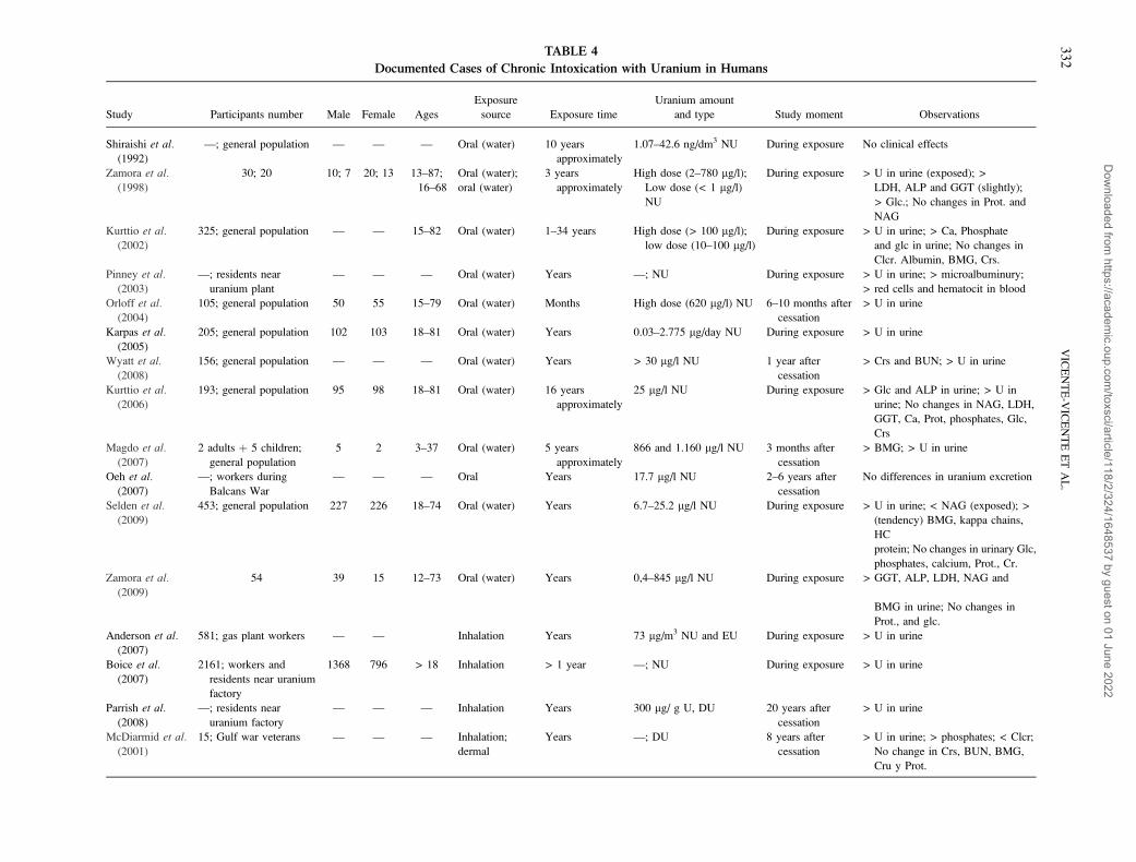
TABLE 4
Documented Cases of Chronic Intoxication with Uranium in Humans
Study Participants number Male Female Ages
Exposure
source Exposure time
Uranium amount
and type Study moment Observations
Shiraishi et al.(1992)
—; general population — — — Oral (water) 10 years
approximately
1.07–42.6 ng/dm3 NU During exposure No clinical effects
Zamora et al.
(1998)
30; 20 10; 7 20; 13 13–87;
16–68
Oral (water);
oral (water)
3 years
approximately
High dose (2–780 lg/l);
Low dose (< 1 lg/l)
NU
During exposure > U in urine (exposed); >
LDH, ALP and GGT (slightly);
> Glc.; No changes in Prot. and
NAG
Kurttio et al.
(2002)
325; general population — — 15–82 Oral (water) 1–34 years High dose (> 100 lg/l);
low dose (10–100 lg/l)
During exposure > U in urine; > Ca, Phosphate
and glc in urine; No changes in
Clcr. Albumin, BMG, Crs.
Pinney et al.
(2003)
—; residents near
uranium plant
— — — Oral (water) Years —; NU During exposure > U in urine; > microalbuminury;
> red cells and hematocit in blood
Orloff et al.(2004)
105; general population 50 55 15–79 Oral (water) Months High dose (620 lg/l) NU 6–10 months after
cessation
> U in urine
Karpas et al.
(2005)
205; general population 102 103 18–81 Oral (water) Years 0.03–2.775 lg/day NU During exposure > U in urine
Wyatt et al.
(2008)
156; general population — — — Oral (water) Years > 30 lg/l NU 1 year after
cessation
> Crs and BUN; > U in urine
Kurttio et al.
(2006)
193; general population 95 98 18–81 Oral (water) 16 years
approximately
25 lg/l NU During exposure > Glc and ALP in urine; > U in
urine; No changes in NAG, LDH,
GGT, Ca, Prot, phosphates, Glc,
Crs
Magdo et al.
(2007)
2 adults þ 5 children;
general population
5 2 3–37 Oral (water) 5 years
approximately
866 and 1.160 lg/l NU 3 months after
cessation
> BMG; > U in urine
Oeh et al.
(2007)
—; workers during
Balcans War
— — — Oral Years 17.7 lg/l NU 2–6 years after
cessation
No differences in uranium excretion
Selden et al.
(2009)
453; general population 227 226 18–74 Oral (water) Years 6.7–25.2 lg/l NU During exposure > U in urine; < NAG (exposed); >
(tendency) BMG, kappa chains,
HC
protein; No changes in urinary Glc,
phosphates, calcium, Prot., Cr.
Zamora et al.
(2009)
54 39 15 12–73 Oral (water) Years 0,4–845 lg/l NU During exposure > GGT, ALP, LDH, NAG and
BMG in urine; No changes in
Prot., and glc.
Anderson et al.
(2007)
581; gas plant workers — — Inhalation Years 73 lg/m3 NU and EU During exposure > U in urine
Boice et al.
(2007)
2161; workers and
residents near uranium
factory
1368 796 > 18 Inhalation > 1 year —; NU During exposure > U in urine
Parrish et al.
(2008)
—; residents near
uranium factory
— — — Inhalation Years 300 lg/ g U, DU 20 years after
cessation
> U in urine
McDiarmid et al.
(2001)
15; Gulf war veterans — — — Inhalation;
dermal
Years —; DU 8 years after
cessation
> U in urine; > phosphates; < Clcr;
No change in Crs, BUN, BMG,
Cru y Prot.
33
2V
ICE
NT
E-V
ICE
NT
EE
TA
L.
Dow
nloaded from https://academ
ic.oup.com/toxsci/article/118/2/324/1648537 by guest on 01 June 2022
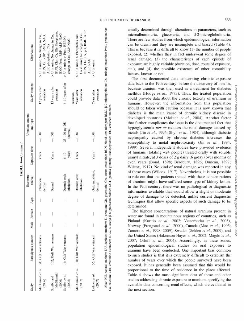
usually determined through alterations in parameters, such as
microalbuminuria, glucosuria, and b-2-microglobulinuria.
There are few studies from which epidemiological information
can be drawn and they are incomplete and biased (Table 4).
This is because it is difficult to know (1) the number of people
exposed, (2) whether they in fact underwent some degree of
renal damage, (3) the characteristics of each episode of
exposure are highly variable (duration, dose, route of exposure,
etc.), and (4) the possible existence of other comorbility
factors, known or not.
The first documented data concerning chronic exposure
date back to the 19th century, before the discovery of insulin,
because uranium was then used as a treatment for diabetes
mellitus (Hodge et al., 1973). Thus, the treated population
could provide data about the chronic toxicity of uranium in
humans. However, the information from this population
should be taken with caution because it is now known that
diabetes is the main cause of chronic kidney disease in
developed countries (Molitch et al., 2004). Another factor
that further complicates the issue is the documented fact that
hyperglycaemia per se reduces the renal damage caused by
metals (Jin et al., 1996; Shyh et al., 1984), although diabetic
nephropathy caused by chronic diabetes increases the
susceptibility to metal nephrotoxicity (Jin et al., 1994,
1999). Several independent studies have provided evidence
of humans (totaling ~24 people) treated orally with soluble
uranyl nitrate, at 3 doses of 2 g daily (6 g/day) over months or
even years (Bond, 1898; Bradbury, 1896; Duncan, 1897;
Wilcox, 1917). No kind of renal damage was reported in any
of these cases (Wilcox, 1917). Nevertheless, it is not possible
to rule out that the patients treated with these concentrations
of uranium might have suffered some type of kidney lesion.
In the 19th century, there was no pathological or diagnostic
information available that would allow a slight or moderate
degree of damage to be detected, unlike current diagnostic
techniques that allow specific aspects of such damage to be
determined.
The highest concentrations of natural uranium present in
water are found in mountainous regions of countries, such as
Finland (Kurttio et al., 2002; Vesterbacka et al., 2005),
Norway (Frengstad et al., 2000), Canada (Mao et al., 1995;
Zamora et al., 1998, 2009), Sweden (Selden et al., 2009), and
the United States (Hakonson-Hayes et al., 2002; Magdo et al.,2007; Orloff et al., 2004). Accordingly, in these zones,
population epidemiological studies on oral exposure to
uranium have been conducted. One important bias common
to such studies is that it is extremely difficult to establish the
number of years over which the people surveyed have been
exposed. It has generally been assumed that this would be
proportional to the time of residence in the place affected.
Table 4 shows the most significant data of these and other
studies addressing chronic exposure to uranium, specifying the
available data concerning renal effects, which are evaluated in
the next section.
TA
BL
E4
—Con
tinu
ed
Stu
dy
Par
tici
pan
tsnum
ber
Mal
eF
emal
eA
ges
Ex
po
sure
sou
rce
Ex
po
sure
tim
e
Ura
niu
mam
ou
nt
and
typ
eS
tud
ym
om
ent
Ob
serv
atio
ns
McD
iarm
idet
al.
(20
06
)
31
;G
ulf
War
vet
eran
s—
——
Inh
alat
ion
;
der
mal
Yea
rs—
;D
U1
2y
ears
afte
r
cess
atio
n
>U
inu
rin
e;N
och
ang
ein
Crs
,
BU
N,
Ca,
RB
P,
BM
G,
NA
G,
AL
P,
ph
osp
hat
es,
Clc
r
Sq
uib
ban
d
McD
iarm
id
(20
06
)
10
2;
Gu
lfW
arv
eter
ans
——
—D
erm
alY
ears
—;
DU
15
yea
rsaf
ter
cess
atio
n
>U
inu
rin
e;N
och
ang
ein
Crs
,
BU
N,
Clc
r,C
a,G
lcP
ho
sph
ates
,
BM
G,
Pro
t.,
RB
P,
AL
P,
NA
G
Sq
uib
bet
al.
(20
05
)
16
;G
ulf
War
vet
eran
s—
——
Der
mal
;o
ral;
inhal
atio
n
6–
10
yea
rs
25
–19
0l
gD
U
(der
mal
)
6–
10
yea
rsaf
ter
cess
atio
n
>U
inu
rin
e;>
Pro
t.,
RB
P;N
o
chan
ge
inC
rs,
AL
P,
NA
G
McD
iarm
idet
al.
(20
07
)
10
8;
Gu
lfW
arv
eter
ans
——
—D
erm
al;
ora
l;
inhal
atio
n
Yea
rs—
;D
UY
ears
afte
r
cess
atio
n
>U
inu
rin
e;>
Ph
osp
hat
esan
d
Ca
inu
rine;
No
chan
ge
inC
rs,
BU
N,
Clc
r,C
a,p
ot,
BM
G,
RB
P,
AL
P,
NA
G
Hel
mer
etal.
(20
07
)
56;
Gulf
War
vet
eran
s—
——
Ora
l;in
hal
atio
n;
der
mal
Yea
rs—
;D
UY
ears
afte
r
cess
atio
n
>U
inu
rin
e
Note.
NU
,n
atura
lu
ran
ium
;D
U,
dep
lete
du
ran
ium
;U
,u
ran
ium
;G
lc,
gu
cosu
ria;
Crs
,se
rum
crea
tin
ine;
BU
N,
blo
od
ure
an
itro
gen
;B
MG
,b-
2-m
icro
glo
buli
n;
Cru
,uri
nar
ycr
eati
nin
e;P
rot.
,pro
tein
uri
a;
Ca,
calc
ium
;C
lcr,
crea
tinin
ecl
eara
nce
;N
AG
,N
-ace
tyl-b-D
-glu
cosa
min
idas
e;G
GT
,gam
ma-
glu
tam
yl-
tran
spep
tidas
e;E
U,
enri
ched
ura
niu
m.
NEPHROTOXICITY OF URANIUM 333
Dow
nloaded from https://academ
ic.oup.com/toxsci/article/118/2/324/1648537 by guest on 01 June 2022

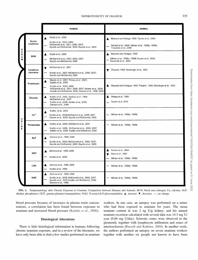
Pathophysiological Picture from Human Studies
The documented cases of chronic intoxication in humans
indicate that there are few situations in which uranium produces
symptoms of a reduction in glomerular filtration and azotemia,
such as a decrease in the GFR (McDiarmid et al., 2001), and an
increase in plasma creatinine and urea concentrations (Wyatt
et al., 2008). In most studies, no reports have been made of
alterations in these parameters, although a few authors have
described alterations in parameters related to the function and
integrity of the kidney structures, especially the tubular
compartment, although inconsistently among the different
studies. However, it is necessary to take into account that such
inconsistency could be because of strong biases among them as
regards the dose, duration and route of exposure, the time of
diagnosis, sex, age, and other determinant factors. Nonetheless,
as discussed below and reflected in Figure 2, it is interesting to
note that the same changes have also been observed in
laboratory animals exposed chronically to the metal (‘‘Patho-
logical Studies with Animal Models’’ section) and that the
pattern of damage is similar to that produced by acute
overexposure (with much higher doses—‘‘Nephrotoxicity
because of Acute Overexposure’’ section).
In some studies, reports have been made of proteinuria or the
excretion of certain specific proteins, such as albumin (retinol-
binding protein [RBP] and b-2-microglobulin, Magdo et al.,2007; Pinney et al., 2003), after uranium exposure. These
findings are inconsistent with those obtained by other authors
(Kurttio et al., 2006; McDiarmid et al., 2001, 2006, 2007;
Zamora et al., 1998). In part, this may be also because of
differences in sampling, the analytical method used, the
statistical analyses applied, or the level of exposure to uranium.
It is not known whether the observed general or selective
proteinuria is of glomerular or tubular origin.
As in the case of acute nephrotoxicity, studies have also
measured a series of kidney enzymes used as markers of tissue
damage, including ALP, GGT, LDH, and NAG (Kurttio et al.,2002, 2006; McDiarmid et al., 2006; Zamora et al., 1998). In
these studies, it was not possible to relate alterations in these
enzymes to the ingestion of uranium. Nevertheless, in rats,
a decrease in the activity of GGT in urine has been observed
following chronic ingestion of the metal (Niwa et al., 1993),
possibly because of the fact that the activity of this enzyme is
inhibited by uranium (Nechay et al., 1980). ALP activity seems
to be related to the chronic ingestion of uranium because
a tendency for it to increase in urine has been observed (Kurttio
et al., 2006; Zamora et al., 1998, 2009), although in other
studies, it seems to be unaltered (McDiarmid et al.,2006; Squibb and McDiarmid, 2006; Squibb et al., 2005).
A tendency for LDH activity to increase after chronic exposure
to uranium in humans has also been reported (Zamora et al.,1998, 2009). Some studies have reported alterations of other
tubular functions, such as increases in the excretion of calcium
(Kurttio et al., 2002; Squibb and McDiarmid, 2006), glucose
(Kurttio et al., 2002, 2006; Zamora et al., 1998), and phosphate
(Kurttio et al., 2002; McDiarmid et al., 2001). However, no
increases in calcium or phosphate levels have been observed in
other studies (McDiarmid et al., 2006; Selden et al., 2009).
However, no clear relation between exposure level, duration of
exposure, and observed renal effects can be drawn from the
available studies in humans (Table 3). Just as an example,
whereas Wyatt et al. (2008) found increased serum creatinine
in 156 people 1 year after the cessation of a chronic
overexposure of years to drinking water containing over
30 lg U/l, Kurttio et al. (2002) found no alterations in this
parameter in 325 people during an overexposure of years to
water containing over 100 lg U/l. Together with the results
obtained in animal models (‘‘Pathological Studies with Animal
Models’’ section), the available information indicates that
chronic exposure to uranium may lead to a variable degree of
renal damage, which in general terms ranges from no
detectable alterations to a mild injury mostly of tubular origin.
However, undetermined comorbidity factors seem to play an
important role at determining the final effect of uranium in the
kidneys.
Recent studies suggest that chronic exposure to uranium
would be associated with an increase in plasma renin
concentrations, which would result in an elevation of blood
pressure and hence a predisposition to hypertension in subjects
exposed to uranium (Kurttio et al., 2006). As commented
above (‘‘Nephrotoxicity because of Chronic Overdose’’
section), this effect has also been reported in acute over-
exposure to uranium. A new hypothesis has associated the
kidney damage produced by chronic ingestion of uranium with
the induction of renal anemia (anemia because of renal
disease), which has been described as an early symptom in
the progression of chronic renal disease (Berradi et al., 2008).
This conclusion was reached after the discovery, in laboratory
animals, of low red blood cell levels following chronic
exposure to the metal (Berradi et al., 2008). However, this
has not been corroborated in uranium workers (Shawky et al.,2002), although it has been observed in people living close to
nuclear power plants (Pinney et al., 2003) and in soldiers
exposed to uranium (Squibb and McDiarmid, 2006).
Finally, studies have been carried out to determine whether
age influences renal damage. This aspect is of special
relevance because children may be at a greater risk of
developing renal damage after uranium exposure because
they drink more water and food per kilogram of body weight
than adults (Ershow and Cantor, 1989). In one study, it was
found that chronic ingestion of uranium by a 3-year-old boy
caused a much greater increase in the urinary excretion of
b-2-microglobulin than the other individuals exposed to the
same amount of the metal (Magdo et al., 2007). Moreover,
adults with impaired renal function may also be at greater
risk. In these, a decrease in creatinine clearance together with
an increase in serum cystatin C levels has been reported
(Kurttio et al., 2006). Regarding the possible increase in
334 VICENTE-VICENTE ET AL.
Dow
nloaded from https://academ
ic.oup.com/toxsci/article/118/2/324/1648537 by guest on 01 June 2022
blood pressure because of increases in plasma renin concen-
trations, a correlation has been found between exposure to
uranium and increased blood pressure (Kurttio et al., 2006).
Histological Alterations
There is little histological information in humans following
chronic uranium exposure, and in a review of the literature, we
have only been able to find a few studies performed on uranium
workers. In one case, an autopsy was performed on a miner
who had been exposed to uranium for years. The mean
uranium content in was 2 ng U/g kidney, and his annual
uranium excretion calculated with several data was 14.3 mg U/
year (0.04 mg U/day). Sclerotic zones were observed in the
glomeruli, together with lymphocyte infiltration and zones of
arteriosclerosis (Russell and Kathren, 2004). In another work,
the authors performed an autopsy on seven uranium workers
together with another six people not known to have been
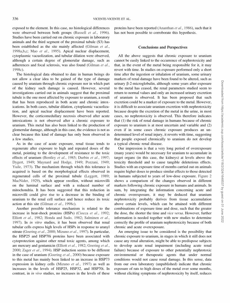
FIG. 2. Simptomatology after Chronic Exposure to Uranium. Comparison between Humans and Animals. BUN, blood urea nitrogen; Ca, calcium; ALP,
alkaline phosphatase; GGT, gamma-glutamyl-transpeptidase; NAG, N-actetyl-b-D-glucosaminidase, :, increase; ;, decrease; —, no change.
NEPHROTOXICITY OF URANIUM 335
Dow
nloaded from https://academ
ic.oup.com/toxsci/article/118/2/324/1648537 by guest on 01 June 2022
exposed to the element. In this case, no histological differences
were observed between both groups (Russell et al., 1996).
Studies have been carried out on chronic exposure in laboratory
animals and the third segment of the proximal tubule (S3) has
been established as the site mainly affected (Gilman et al.,1998a,b,c; Mao et al., 1995). Apical nuclear displacement,
cytoplasmic vacuolization, and tubular dilation were observed,
although a certain degree of glomerular damage, such as
adherences and focal sclerosis, was also found (Gilman et al.,1998c).
The histological data obtained to date in human beings do
not allow a clear idea to be gained of the type of damage
caused by uranium through chronic exposure nor in which part
of the kidney such damage is caused. However, several
investigations carried out in animals suggest that the proximal
tubule is the one most affected by exposure to uranium, a result
that has been reproduced in both acute and chronic intox-
ications. In both cases, tubular dilation, cytoplasmic vacuoliza-
tion, and apical nuclear displacement have been reported.
However, the corticomedullary necrosis observed after acute
intoxications is not observed after a chronic exposure to
uranium. This metal has also been linked to the production of
glomerular damage, although in this case, the evidence is not as
clear because this kind of damage has only been observed in
a few studies.
As in the case of acute exposure, renal tissue tends to
regenerate after exposure to high and repeated doses of the
metal, pointing to the development of resistance to the toxic
effects of uranium (Bentley et al., 1985; Durbin et al., 1997;
Dygert, 1949; Maynard and Hodge, 1949; Pozzani, 1949;
Yuile, 1973). The mechanism through which this tolerance is
acquired is based on the morphological effects observed in
regenerated cells of the proximal tubule (Leggett, 1989;
MacNider, 1929), which appear swollen, without microvilli
on the luminal surface and with a reduced number of
mitochondria. It has been suggested that this reduction in
microvilli could give rise to a decrease in the binding of
uranium to the renal cell surface and hence reduce its toxic
action at this site (Gilman et al., 1998c).
Another possible tolerance mechanism is related to the
increase in heat-shock proteins (HSPs) (Ciocca et al., 1992;
Elliott et al., 1982; Honda and Sudo, 1982; Salminen et al.,1997). In in vitro studies, it has been observed that renal
tubular cells express high levels of HSPs in response to uranyl
nitrate (Goering et al., 2000; Mizuno et al., 1997). In particular,
the HSP25 and HSP70i proteins have been associated with
cytoprotection against other renal toxic agents, among which
are mercury and gentamicin (Elliott et al., 1982; Goering et al.,2000; Zager et al., 1994). HSP induction seems to be different
in the case of uranium (Goering et al., 2000) because exposure
to this metal has mainly been linked to an increase in HSP73
expression in kidney cells (Mizuno et al., 1997) as well as
increases in the levels of HSP25, HSP32, and HSP70i. In
contrast, in in vivo studies, no increases in the levels of these
proteins have been reported (Ananthan et al., 1986), such that it
has not been possible to corroborate this hypothesis.
Conclusions and Perspectives
All the above suggests that chronic exposure to uranium
cannot be easily linked to the occurrence of nephrotoxicity and
that, in the event of the metal being responsible for it, it may
revert with time. In studies on exposure performed only a short
time after the ingestion or inhalation of uranium, some urinary
markers of renal damage have been found to be altered, such as
urinary b-2-microglobulin, although some years after exposure
to the metal has ceased, the renal parameters studied seem to
return to normal values and only an increased urinary excretion
of uranium is observed. It has been proposed that such
excretion could be a marker of exposure to the metal. However,
it is difficult to associate uranium excretion with nephrotoxicity
because despite the excretion of the metal in the urine, in most
cases, no nephrotoxicity is observed. This therefore indicates
that (1) the risk of renal damage in humans because of chronic
exposure to uranium is at most uncertain and variable and (2)
even if in some cases chronic exposure produces an un-
determined level of renal injury, it reverts with time, suggesting
that people exposed chronically to uranium do not develop
a typical chronic renal disease.
Our impression is that a very long period of overexposure
(many years) would be necessary for uranium to accumulate in
target organs (in this case, the kidneys) at levels above the
toxicity threshold and to cause tangible deleterious effects.
Studies with an exposure time of months carried out on animals
require higher doses to produce similar effects to those detected
in humans subjected to years of low-dose exposure. Figure 2
shows a comparison of the changes observed in different
markers following chronic exposure in humans and animals. In
sum, by integrating the information concerning acute and
chronic overexposure, it may be deduced that uranium
nephrotoxicity probably derives from tissue accumulation
above certain levels, which can be attained with different
combinations of exposure time and dose, such that the greater
the dose, the shorter the time and vice versa. However, further
information is needed together with new studies to determine
correctly the profile of uranium nephrotoxicity because of both
chronic and acute overexposure.
An emerging issue to be considered is the possibility that
chronic exposure to uranium, in stages in which it still does not
cause any renal alteration, might be able to predispose subjects
to develop acute renal impairment (including acute renal
failure) because of exposure to other potentially nephrotoxic
environmental or therapeutic agents that under normal
conditions would not cause renal damage. In this sense, data
from our own laboratory (unpublished) indicate that chronic
exposure of rats to high doses of the metal over some months,
without eliciting symptoms of nephrotoxicity by itself, reduces
336 VICENTE-VICENTE ET AL.
Dow
nloaded from https://academ
ic.oup.com/toxsci/article/118/2/324/1648537 by guest on 01 June 2022
the threshold of nephrotoxicity and enhances the nephrotoxic
effects of certain drugs, such as the aminoglycoside antibiotic
gentamicin. Were it to be confirmed, this situation would be of
huge clinical relevance because, in an occult and nondiagnos-
able way, chronic overexposure could render the sector of the
population in contact with the metal more susceptible to renal
failure. We believe that this is an issue requiring further
research effort in the near future, especially within the context
of the detection of this potential situation.
MECHANISMS OF NEPHROTOXIC ACTION
Although uranium is widely used as an experimental
nephrotoxic agent, the underlying physiological mechanism
responsible for renal damage has not been fully elucidated. One
limitation to our knowledge about this issue is that most of the
information has been obtained in acute studies with animals
(Diamond et al., 1989; Haley, 1982, Haley et al., 1982;
Morrow et al., 1982; Rothstein, 1949; Stokinger et al., 1953;
Taylor and Taylor, 1997; Thun et al., 1985).
The first issue that has not been suitably clarified is whether
uranium needs to penetrate cells to exert its toxic effect. Some
authors have proposed that this would not be necessary because
its effects (or a large part of them) derive from binding to
certain components of the cell membrane (Leggett, 1989;
Muller et al., 2006). According to those authors, such effects
would be based on interference with the reabsorption of
glucose, sodium, amino acids, proteins, water, and other
substances, which would lead to a slow cell death because of
the suppression of cell respiration (Hori et al., 1985; Leggett,
1989; Nechay et al., 1980). However, others have proposed
that the metal does need to enter cells to exert its toxic effects.
This has been observed in LLC-PK1 cells of the proximal
tubules in studies aimed at determining whether there are
differences in the toxicity of the U-bicarbonate and U-citrate
complexes (L’Azou et al., 2002; Mirto et al., 1999). In these
studies, it was possible to correlate the presence of the uranium
complex inside the cells and the toxic effect. Thus, it was
observed that the U-citrate complex entered the cells and
exerted a significant toxic effect in them, whereas the
U-bicarbonate complex, which did not enter the cells, exerted
a much lower toxic effect (Mirto et al., 1999). Accordingly, the
authors concluded that as regards its toxic effects, the entry of
uranium into cells is very important. Below, we detail some of
the aspects related to the mechanism involved in uranium
nephrotoxicity.
Alterations in Solute Transport
In studies of brush border membrane vesicles from rat renal
tubular cells, it has been observed that uranyl acetate produces
a decrease in glucose transport because of a reduction in the
number of sodium-glucose transporters (SGLT) (Goldman
et al., 2006). Hori et al. (1985) also reported a decrease in the
sodium-dependent glucose gradient that led them to suspect
that the enzymatic activity of Naþ Kþ, adenosine triphospha-
tase (Naþ, Kþ ATPase, or sodium pump) could be inhibited by
uranyl nitrate. Similar results were obtained by Brady et al.(1989) in rabbit kidney cells. Those authors suggested that
uranyl nitrate would inhibit both the sodium-dependent and the
sodium-independent ATP utilization and mitochondrial oxida-
tive phosphorylation. Muller et al. (2006) reported that the
cytotoxicity of uranium for LLC-PK1 cells would depend on
the extracellular concentration of phosphate. High concen-
trations of phosphate in the medium gave rise to the formation
of uranium-phosphate complexes, which inhibited the Na/Pi II
transporter participating in the reabsorption of organic
phosphate.
Oxidative Stress
Oxidative stress has been proposed as a possible mediator of
renal damage because of exposure to uranium. In the rat renal
proximal tubular cell line (NRK-52E), uranium stimulates the
production of reactive oxygen species (ROS), caspases 9 and 3,
and cell death because of apoptosis (Thiebault et al., 2007). In
studies carried out on rat renal tissue (Linares et al., 2006), an
increase was observed in ROS levels, in oxidized glutathione
(GSSG), and in the activity of SOD. An increase in
thiobarbituric acid–reactive substances, indicative of lipid
peroxidation and oxidative stress, was also observed. It has
also been suggested that uranium could act as a catalyst in the
Fenton/Haber-Weiss reaction (Kovacic and Jacintho, 2001),
which would facilitate the conversion of the superoxide anion
and hydrogen peroxide into hydroxyl radicals, believed to be
responsible for initiating lipid peroxidation (Linares et al.,2006; Stohs and Bagchi, 1995; Taulan et al., 2004). In other
studies, the pro-oxidant action of uranium has been related to
a disturbance in the activity of acetyl cholinesterase and of
monoamine metabolism (Kurttio et al., 2002; Linares et al.,2006; Sanchez et al., 2001) and to an increase in mitochondrial
oxidative phosphorylation in the proximal tubule (Brady et al.,1989). These results indicate that uranium could produce
oxidative stress, inducing, at least in part, the death of target
cells (Sabolic, 2006). Figure 3 shows the possible mechanism
responsible for renal damage caused by uranium.
Alterations in Gene Expression
Chronic exposure to uranium in animals elicits changes in
the profiles of the renal expression of genes related to oxidative
stress, cellular metabolism, solute transport, and signal trans-
duction, among other processes (Taulan et al., 2004, 2006). As
seen in Table 5, some of these changes in renal gene expression
are correlated with the appearance of pathological effects, such
as inflammation, apoptosis, oxidative stress, and alterations in
cellular homeostasis. However, the role of these genes in the
NEPHROTOXICITY OF URANIUM 337
Dow
nloaded from https://academ
ic.oup.com/toxsci/article/118/2/324/1648537 by guest on 01 June 2022
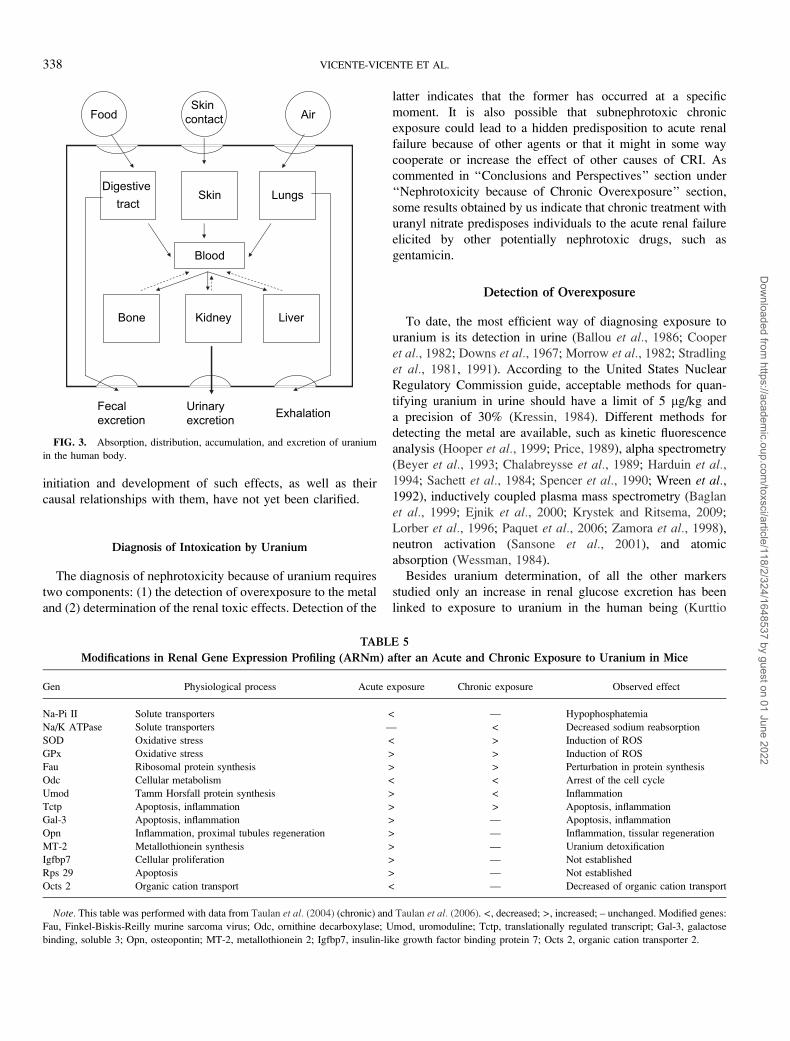
initiation and development of such effects, as well as their
causal relationships with them, have not yet been clarified.
Diagnosis of Intoxication by Uranium
The diagnosis of nephrotoxicity because of uranium requires
two components: (1) the detection of overexposure to the metal
and (2) determination of the renal toxic effects. Detection of the
latter indicates that the former has occurred at a specific
moment. It is also possible that subnephrotoxic chronic
exposure could lead to a hidden predisposition to acute renal
failure because of other agents or that it might in some way
cooperate or increase the effect of other causes of CRI. As
commented in ‘‘Conclusions and Perspectives’’ section under
‘‘Nephrotoxicity because of Chronic Overexposure’’ section,
some results obtained by us indicate that chronic treatment with
uranyl nitrate predisposes individuals to the acute renal failure
elicited by other potentially nephrotoxic drugs, such as
gentamicin.
Detection of Overexposure
To date, the most efficient way of diagnosing exposure to
uranium is its detection in urine (Ballou et al., 1986; Cooper
et al., 1982; Downs et al., 1967; Morrow et al., 1982; Stradling
et al., 1981, 1991). According to the United States Nuclear
Regulatory Commission guide, acceptable methods for quan-
tifying uranium in urine should have a limit of 5 lg/kg and
a precision of 30% (Kressin, 1984). Different methods for
detecting the metal are available, such as kinetic fluorescence
analysis (Hooper et al., 1999; Price, 1989), alpha spectrometry
(Beyer et al., 1993; Chalabreysse et al., 1989; Harduin et al.,1994; Sachett et al., 1984; Spencer et al., 1990; Wreen et al.,1992), inductively coupled plasma mass spectrometry (Baglan
et al., 1999; Ejnik et al., 2000; Krystek and Ritsema, 2009;
Lorber et al., 1996; Paquet et al., 2006; Zamora et al., 1998),
neutron activation (Sansone et al., 2001), and atomic
absorption (Wessman, 1984).
Besides uranium determination, of all the other markers
studied only an increase in renal glucose excretion has been
linked to exposure to uranium in the human being (Kurttio
FoodSkin
contact Air
Digestive
tractSkin Lungs
Blood
Bone Kidney Liver
Fecal excretion
Urinaryexcretion
Exhalation
FIG. 3. Absorption, distribution, accumulation, and excretion of uranium
in the human body.
TABLE 5
Modifications in Renal Gene Expression Profiling (ARNm) after an Acute and Chronic Exposure to Uranium in Mice
Gen Physiological process Acute exposure Chronic exposure Observed effect
Na-Pi II Solute transporters < — Hypophosphatemia
Na/K ATPase Solute transporters — < Decreased sodium reabsorption
SOD Oxidative stress < > Induction of ROS
GPx Oxidative stress > > Induction of ROS
Fau Ribosomal protein synthesis > > Perturbation in protein synthesis
Odc Cellular metabolism < < Arrest of the cell cycle
Umod Tamm Horsfall protein synthesis > < Inflammation
Tctp Apoptosis, inflammation > > Apoptosis, inflammation
Gal-3 Apoptosis, inflammation > — Apoptosis, inflammation
Opn Inflammation, proximal tubules regeneration > — Inflammation, tissular regeneration
MT-2 Metallothionein synthesis > — Uranium detoxification
Igfbp7 Cellular proliferation > — Not established
Rps 29 Apoptosis > — Not established
Octs 2 Organic cation transport < — Decreased of organic cation transport
Note. This table was performed with data from Taulan et al. (2004) (chronic) and Taulan et al. (2006). <, decreased; >, increased; – unchanged. Modified genes:
Fau, Finkel-Biskis-Reilly murine sarcoma virus; Odc, ornithine decarboxylase; Umod, uromoduline; Tctp, translationally regulated transcript; Gal-3, galactose
binding, soluble 3; Opn, osteopontin; MT-2, metallothionein 2; Igfbp7, insulin-like growth factor binding protein 7; Octs 2, organic cation transporter 2.
338 VICENTE-VICENTE ET AL.
Dow
nloaded from https://academ
ic.oup.com/toxsci/article/118/2/324/1648537 by guest on 01 June 2022
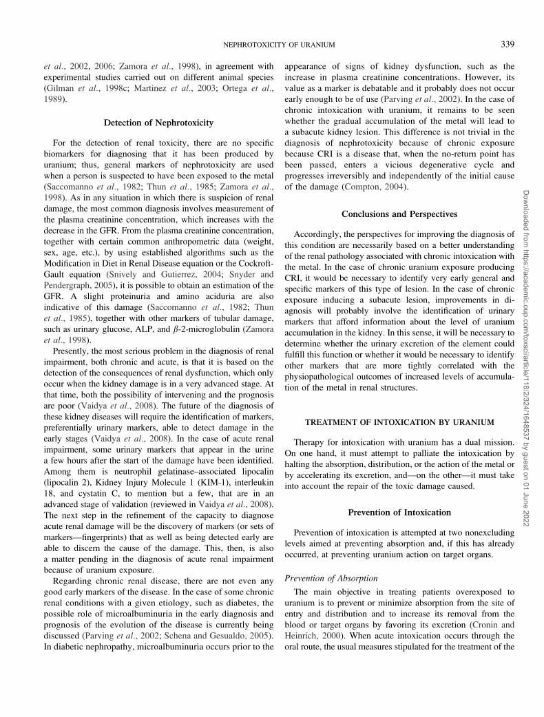
et al., 2002, 2006; Zamora et al., 1998), in agreement with
experimental studies carried out on different animal species
(Gilman et al., 1998c; Martinez et al., 2003; Ortega et al.,1989).
Detection of Nephrotoxicity
For the detection of renal toxicity, there are no specific
biomarkers for diagnosing that it has been produced by
uranium; thus, general markers of nephrotoxicity are used
when a person is suspected to have been exposed to the metal
(Saccomanno et al., 1982; Thun et al., 1985; Zamora et al.,1998). As in any situation in which there is suspicion of renal
damage, the most common diagnosis involves measurement of
the plasma creatinine concentration, which increases with the
decrease in the GFR. From the plasma creatinine concentration,
together with certain common anthropometric data (weight,
sex, age, etc.), by using established algorithms such as the
Modification in Diet in Renal Disease equation or the Cockroft-
Gault equation (Snively and Gutierrez, 2004; Snyder and
Pendergraph, 2005), it is possible to obtain an estimation of the
GFR. A slight proteinuria and amino aciduria are also
indicative of this damage (Saccomanno et al., 1982; Thun
et al., 1985), together with other markers of tubular damage,
such as urinary glucose, ALP, and b-2-microglobulin (Zamora
et al., 1998).
Presently, the most serious problem in the diagnosis of renal
impairment, both chronic and acute, is that it is based on the
detection of the consequences of renal dysfunction, which only
occur when the kidney damage is in a very advanced stage. At
that time, both the possibility of intervening and the prognosis
are poor (Vaidya et al., 2008). The future of the diagnosis of
these kidney diseases will require the identification of markers,
preferentially urinary markers, able to detect damage in the
early stages (Vaidya et al., 2008). In the case of acute renal
impairment, some urinary markers that appear in the urine
a few hours after the start of the damage have been identified.
Among them is neutrophil gelatinase–associated lipocalin
(lipocalin 2), Kidney Injury Molecule 1 (KIM-1), interleukin
18, and cystatin C, to mention but a few, that are in an
advanced stage of validation (reviewed in Vaidya et al., 2008).
The next step in the refinement of the capacity to diagnose
acute renal damage will be the discovery of markers (or sets of
markers—fingerprints) that as well as being detected early are
able to discern the cause of the damage. This, then, is also
a matter pending in the diagnosis of acute renal impairment
because of uranium exposure.
Regarding chronic renal disease, there are not even any
good early markers of the disease. In the case of some chronic
renal conditions with a given etiology, such as diabetes, the
possible role of microalbuminuria in the early diagnosis and
prognosis of the evolution of the disease is currently being
discussed (Parving et al., 2002; Schena and Gesualdo, 2005).
In diabetic nephropathy, microalbuminuria occurs prior to the
appearance of signs of kidney dysfunction, such as the
increase in plasma creatinine concentrations. However, its
value as a marker is debatable and it probably does not occur
early enough to be of use (Parving et al., 2002). In the case of
chronic intoxication with uranium, it remains to be seen
whether the gradual accumulation of the metal will lead to
a subacute kidney lesion. This difference is not trivial in the
diagnosis of nephrotoxicity because of chronic exposure
because CRI is a disease that, when the no-return point has
been passed, enters a vicious degenerative cycle and
progresses irreversibly and independently of the initial cause
of the damage (Compton, 2004).
Conclusions and Perspectives
Accordingly, the perspectives for improving the diagnosis of
this condition are necessarily based on a better understanding
of the renal pathology associated with chronic intoxication with
the metal. In the case of chronic uranium exposure producing
CRI, it would be necessary to identify very early general and
specific markers of this type of lesion. In the case of chronic
exposure inducing a subacute lesion, improvements in di-
agnosis will probably involve the identification of urinary
markers that afford information about the level of uranium
accumulation in the kidney. In this sense, it will be necessary to
determine whether the urinary excretion of the element could
fulfill this function or whether it would be necessary to identify
other markers that are more tightly correlated with the
physiopathological outcomes of increased levels of accumula-
tion of the metal in renal structures.
TREATMENT OF INTOXICATION BY URANIUM
Therapy for intoxication with uranium has a dual mission.
On one hand, it must attempt to palliate the intoxication by
halting the absorption, distribution, or the action of the metal or
by accelerating its excretion, and—on the other—it must take
into account the repair of the toxic damage caused.
Prevention of Intoxication
Prevention of intoxication is attempted at two nonexcluding
levels aimed at preventing absorption and, if this has already
occurred, at preventing uranium action on target organs.
Prevention of Absorption
The main objective in treating patients overexposed to
uranium is to prevent or minimize absorption from the site of
entry and distribution and to increase its removal from the
blood or target organs by favoring its excretion (Cronin and
Heinrich, 2000). When acute intoxication occurs through the
oral route, the usual measures stipulated for the treatment of the
NEPHROTOXICITY OF URANIUM 339
Dow
nloaded from https://academ
ic.oup.com/toxsci/article/118/2/324/1648537 by guest on 01 June 2022
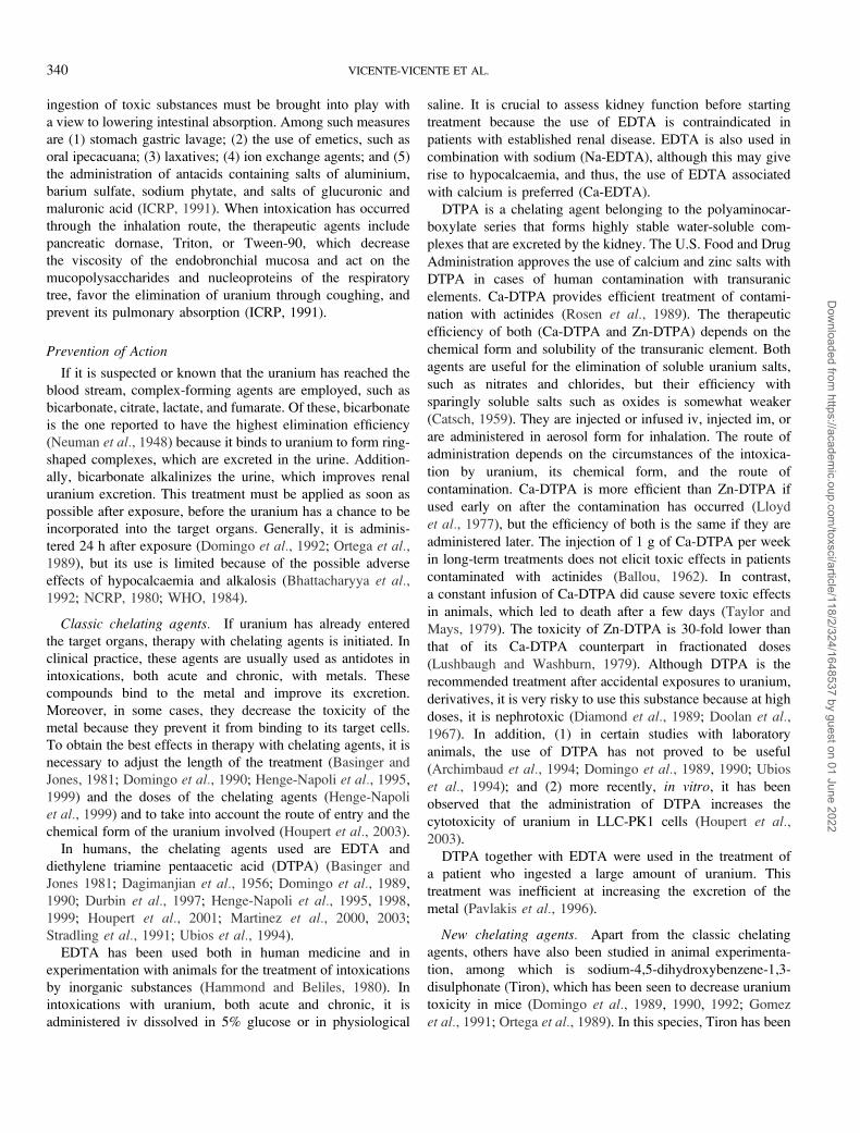
ingestion of toxic substances must be brought into play with
a view to lowering intestinal absorption. Among such measures
are (1) stomach gastric lavage; (2) the use of emetics, such as
oral ipecacuana; (3) laxatives; (4) ion exchange agents; and (5)
the administration of antacids containing salts of aluminium,
barium sulfate, sodium phytate, and salts of glucuronic and
maluronic acid (ICRP, 1991). When intoxication has occurred
through the inhalation route, the therapeutic agents include
pancreatic dornase, Triton, or Tween-90, which decrease
the viscosity of the endobronchial mucosa and act on the
mucopolysaccharides and nucleoproteins of the respiratory
tree, favor the elimination of uranium through coughing, and
prevent its pulmonary absorption (ICRP, 1991).
Prevention of Action
If it is suspected or known that the uranium has reached the
blood stream, complex-forming agents are employed, such as
bicarbonate, citrate, lactate, and fumarate. Of these, bicarbonate
is the one reported to have the highest elimination efficiency
(Neuman et al., 1948) because it binds to uranium to form ring-
shaped complexes, which are excreted in the urine. Addition-
ally, bicarbonate alkalinizes the urine, which improves renal
uranium excretion. This treatment must be applied as soon as
possible after exposure, before the uranium has a chance to be
incorporated into the target organs. Generally, it is adminis-
tered 24 h after exposure (Domingo et al., 1992; Ortega et al.,1989), but its use is limited because of the possible adverse
effects of hypocalcaemia and alkalosis (Bhattacharyya et al.,1992; NCRP, 1980; WHO, 1984).
Classic chelating agents. If uranium has already entered
the target organs, therapy with chelating agents is initiated. In
clinical practice, these agents are usually used as antidotes in
intoxications, both acute and chronic, with metals. These
compounds bind to the metal and improve its excretion.
Moreover, in some cases, they decrease the toxicity of the
metal because they prevent it from binding to its target cells.
To obtain the best effects in therapy with chelating agents, it is
necessary to adjust the length of the treatment (Basinger and
Jones, 1981; Domingo et al., 1990; Henge-Napoli et al., 1995,
1999) and the doses of the chelating agents (Henge-Napoli
et al., 1999) and to take into account the route of entry and the
chemical form of the uranium involved (Houpert et al., 2003).
In humans, the chelating agents used are EDTA and
diethylene triamine pentaacetic acid (DTPA) (Basinger and
Jones 1981; Dagimanjian et al., 1956; Domingo et al., 1989,
1990; Durbin et al., 1997; Henge-Napoli et al., 1995, 1998,
1999; Houpert et al., 2001; Martinez et al., 2000, 2003;
Stradling et al., 1991; Ubios et al., 1994).
EDTA has been used both in human medicine and in
experimentation with animals for the treatment of intoxications
by inorganic substances (Hammond and Beliles, 1980). In
intoxications with uranium, both acute and chronic, it is
administered iv dissolved in 5% glucose or in physiological
saline. It is crucial to assess kidney function before starting
treatment because the use of EDTA is contraindicated in
patients with established renal disease. EDTA is also used in
combination with sodium (Na-EDTA), although this may give
rise to hypocalcaemia, and thus, the use of EDTA associated
with calcium is preferred (Ca-EDTA).
DTPA is a chelating agent belonging to the polyaminocar-
boxylate series that forms highly stable water-soluble com-
plexes that are excreted by the kidney. The U.S. Food and Drug
Administration approves the use of calcium and zinc salts with
DTPA in cases of human contamination with transuranic
elements. Ca-DTPA provides efficient treatment of contami-
nation with actinides (Rosen et al., 1989). The therapeutic
efficiency of both (Ca-DTPA and Zn-DTPA) depends on the
chemical form and solubility of the transuranic element. Both
agents are useful for the elimination of soluble uranium salts,
such as nitrates and chlorides, but their efficiency with
sparingly soluble salts such as oxides is somewhat weaker
(Catsch, 1959). They are injected or infused iv, injected im, or
are administered in aerosol form for inhalation. The route of
administration depends on the circumstances of the intoxica-
tion by uranium, its chemical form, and the route of
contamination. Ca-DTPA is more efficient than Zn-DTPA if
used early on after the contamination has occurred (Lloyd
et al., 1977), but the efficiency of both is the same if they are
administered later. The injection of 1 g of Ca-DTPA per week
in long-term treatments does not elicit toxic effects in patients
contaminated with actinides (Ballou, 1962). In contrast,
a constant infusion of Ca-DTPA did cause severe toxic effects
in animals, which led to death after a few days (Taylor and
Mays, 1979). The toxicity of Zn-DTPA is 30-fold lower than
that of its Ca-DTPA counterpart in fractionated doses
(Lushbaugh and Washburn, 1979). Although DTPA is the
recommended treatment after accidental exposures to uranium,
derivatives, it is very risky to use this substance because at high
doses, it is nephrotoxic (Diamond et al., 1989; Doolan et al.,1967). In addition, (1) in certain studies with laboratory
animals, the use of DTPA has not proved to be useful
(Archimbaud et al., 1994; Domingo et al., 1989, 1990; Ubios
et al., 1994); and (2) more recently, in vitro, it has been
observed that the administration of DTPA increases the
cytotoxicity of uranium in LLC-PK1 cells (Houpert et al.,2003).
DTPA together with EDTA were used in the treatment of
a patient who ingested a large amount of uranium. This
treatment was inefficient at increasing the excretion of the
metal (Pavlakis et al., 1996).
New chelating agents. Apart from the classic chelating
agents, others have also been studied in animal experimenta-
tion, among which is sodium-4,5-dihydroxybenzene-1,3-
disulphonate (Tiron), which has been seen to decrease uranium
toxicity in mice (Domingo et al., 1989, 1990, 1992; Gomez
et al., 1991; Ortega et al., 1989). In this species, Tiron has been
340 VICENTE-VICENTE ET AL.
Dow
nloaded from https://academ
ic.oup.com/toxsci/article/118/2/324/1648537 by guest on 01 June 2022
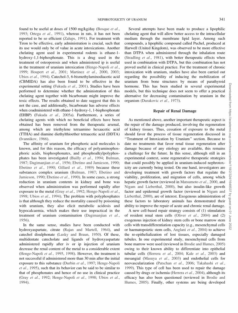
found to be useful at doses of 1500 mg/kg/day (Bosque et al.,1993; Ortega et al., 1991), whereas in rats, it has not been
reported to be so efficient (Zalups, 1991). For treatment with
Tiron to be effective, early administration is crucial, such that
its use would only be of value in acute intoxications. Another
chelating agent used in experimental rodents is ethane-1-
hydroxy-1,1-biphosphonate. This is a drug used in the
treatment of osteoporosis and when administered ip is useful
in the treatment of uranium intoxication (Henge-Napoli et al.,1999; Houpert et al., 2001; Martinez et al., 2000, 2003;
Ubios et al., 1994). Catechol-3, 6-bismethylaminodiacetic acid
(CBMIDA) has also been found to be effective in the
experimental setting (Fukuda et al., 2001). Studies have been
performed to determine whether the administration of this
chelating agent together with bicarbonate might improve the
toxic effects. The results obtained to date suggest that this is
not the case, and additionally, bicarbonate has adverse effects
when coadministered with ethane-1-hydroxy-1, 1-bisphosphonate
(EHBP) (Fukuda et al., 2005a). Furthermore, a series of
chelating agents with which no beneficial effects have been
obtained has been removed from the therapeutic arsenal,
among which are triethylene tetraamino hexaacetic acid
(TTHA) and diamine diethylthioether tetraacetic acid (DDTA)
(Ivannikov, 1966).
The affinity of uranium for phosphoric acid molecules is
known, and for this reason, the efficacy of polyaminophos-
phoric acids, bisphosphonates, and phosphoalkylpolyphos-
phates has been investigated (Bailly et al., 1994; Bulman,
1987; Dagimanjian et al., 1956; Ebetino and Jamieson, 1990;
Ebetino et al., 1990; Gillard et al., 1989) because these
substances complex uranium (Bulman, 1987; Ebetino and
Jamieson, 1990; Ebetino et al., 1990). In some cases, a strong
reduction in uranium contents in kidney and bone was
observed when administration was performed rapidly after
exposure to the metal (Gray et al., 1992; Henge-Napoli et al.,1998; Ubios et al., 1994). The problem with polyphosphates
is that although they reduce the mortality caused by poisoning
with uranium, they also elicit metabolic acidosis and
hypocalcaemia, which makes their use impractical in the
treatment of uranium contamination (Dagimanjian et al.,1956).
In the same sense, studies have been conducted with
hydroxyaspartate, citrate (Rajan and Martell, 1964), and
catechol disulphonate (Lusky and Braun, 1950). Of these,
multidentate catecholate and ligands of hydroxyaspartate
administered rapidly after iv or ip injection of uranium
decrease the renal content of the metal to a considerable extent
(Henge-Napoli et al., 1995, 1998). However, the treatment is
not successful if administered more than 30 min after the initial
exposure to this substance (Durbin et al., 1997; Henge-Napoli
et al., 1995), such that its behavior can be said to be similar to
that of phosphonates and hence of no use in clinical practice
(Gray et al., 1992; Henge-Napoli et al., 1998; Ubios et al.,1994).
Several attempts have been made to produce a lipophilic
chelating agent that will allow better access to the intracellular
medium through the membrane lipid layer. Among such
compounds, a lipophilic compound called Puchel, produced at
Harwell (United Kingdom), was observed to be more effective
than DTPA when administered through the inhalation route
(Stradling et al., 1981), with better therapeutic effects when
used in combination with DTPA, but this combination has not
proved useful in clinical practice. For the treatment of chronic
intoxication with uranium, studies have also been carried out
regarding the possibility of inducing the mobilization of
uranium from bone structures by means of parathyroid
hormone. This has been studied in several experimental
models, but this technique does not seem to offer a practical
alternative for decreasing contamination by uranium in the
organism (Durakovic et al., 1973).
Repair of Renal Damage
As mentioned above, another important therapeutic aspect is
the repair of the damage produced, involving the regeneration
of kidney tissues. Thus, cessation of exposure to the metal
should favor the process of tissue regeneration discussed in
‘‘Treatment of Intoxication by Uranium’’ section. Because to
date no treatments that favor renal tissue regeneration after
damage because of any etiology are available, this remains
a challenge for the future. In this sense, although still in an
experimental context, some regenerative therapeutic strategies
that could possibly be applied in uranium-induced nephrotox-
icity are currently being tested. On one hand, investigators are
developing treatment with growth factors that regulate the
viability, proliferation, and migration of cells, among which
hepatic growth factor (reviewed in Matsumoto et al., 2000, and
Nigam and Lieberthal, 2000), but also insulin-like growth
factor and epidermal growth factor (reviewed in Nigam and
Lieberthal, 2000), are of interest. Exogenous administration of
these factors to laboratory animals has demonstrated their
ability to improve the repair of acute and chronic renal damage.
A new cell-based repair strategy consists of (1) stimulation
of resident renal stem cells (Oliver et al., 2004) and (2)
exogenous injection of kidney stem cells or bone marrow stem
cells with transdifferentiation capacity (e.g., mesenchymal cells
or haematopoietic stem cells, Anglani et al., 2004) to achieve
the re-epithelialization of lost tissues, especially damaged
tubules. In one experimental study, mesenchymal cells from
bone marrow were used (reviewed in Brodie and Humes, 2005)
owing to their known ability to differentiate into epithelial
tubular cells (Herrera et al., 2004; Kale et al., 2003) and
mesangial (Masuya et al., 2003) and endothelial cells for
neovascularization (Patschan et al., 2006; Takahashi et al.,1999). This type of cell has been used to repair the damage
caused by drugs or ischemia (Herrera et al., 2004), although its
efficacy has also been questioned (reviewed in Brodie and
Humes, 2005). Finally, other systems are being developed
NEPHROTOXICITY OF URANIUM 341
Dow
nloaded from https://academ
ic.oup.com/toxsci/article/118/2/324/1648537 by guest on 01 June 2022
including (1) a bioartificial kidney consisting of a dialysis
system formed by a tubular structure containing ~1 billion
tubular cells (Humes et al., 2004) and (2) implants of
encapsulated cells with a view to removing uraemic toxins
and for drug administration (see Brodie and Humes, 2005).
ACKNOWLEDGMENTS
We thank Nicholas S. Skinner from the Servicio de Idiomas,
Universidad de Salamanca (Spain), for his assistance on the
English version of the manuscript.
REFERENCES
Agency for Toxic Substances and Disease Registry, Centers for Disease
Control and Prevention (ATSDR/CDC). (1990). Agency for Toxic
Substances and Disease Registry, Centers for Disease Control andPrevention. Subcommittee Report on Biological Indicators of Organ
Damage, Atlanta, GA. Available at: http://www.atsdr.cdc.gov/. Updated
August 28, 2008. Accessed July 7, 2010.
Agency for Toxic Substances and Disease Research (ATSDR). (1999). Agencyfor Toxic Substances and Disease Registry. Toxicological Profile for
Uranium. Department of Health and Human Services, Public Health Service,
Atlanta, GA.
Ananthan, J., Goldberg, A. L., and Voellmy, R. (1986). Abnormal proteins
serve as eukaryotic stress signals and trigger the activation of heat shock
genes. Science 232, 522–524.
Anderson, J. L., Spitz, H. B., and Yiin, J. H. (2007). Estimating active bone
marrow dose from occupational exposure to uranium at a former gaseous
diffusion plant. Health. Phys. 93, 113–119.
Anglani, F., Forino, M., Del Prete, D., Tosetto, E., Torregrossa, R., and
D’Angelo, A. (2004). In search of adult renal stem cells. J. Cell. Mol. Med. 8,
474–487.
Archimbaud, M., Henge-Napoli, M. H., Lilienbaum, D., Desloges, M., and
Montagne, C. (1994). Application of calixarenes for the decorporation of
uranium: present limitations and further trends. Radiat. Prot. Dosimetry. 53,
327–330.
Baglan, N., Cossonnet, C., Trompier, F., Ritt, J., and Berard, P. (1999).
Implementation of ICP-MS protocols for uranium urinary measurements in
worker monitoring. Health Phys. 77, 455–461.
Bailly, T., Burgada, R., Stradling, G. N., and Gray, S. A. (1994). In Synthesis of
new ligands for uranium decorporation. (J. R. Maisin, Ed.), Vol. 75, p. 37.
European Late Effects Project Group (EULEP) Newsletter, Mol, Belgium.
Ballou, J. E. (1962). Preliminary evaluation of several chelating agents for
plutonium removal. Health Phys. 8, 731–734.
Ballou, J. E., Gies, R. A., Case, A. C., Haggard, D. L., Buschbom, R. L., and
Ryan, J. L. (1986). Deposition and early disposition of inhaled
233UO2(NO3)2 and 232UO2(NO3)2 in the rat. Health Phys. 51, 755–771.
Banday, A. A., Priyamvada, S., Farooq, N., Yusufi, A. N., and Khan, F. (2008).
Effect of uranyl nitrate on enzymes of carbohydrate metabolism and brush
border membrane in different kidney tissues. Food Chem. Toxicol. 46,
2080–2088.
Basile, C. (2008). The long-term prognosis of acute kidney injury: acute renal
failure as a cause of chronic kidney disease. J. Nephrol. 21, 657–662.
Basinger, M. A., and Jones, M. M. (1981). Tiron (sodium 4,5-dihydrox-
ybenzene-1,3-disulfonate) as an antidote for acute uranium intoxication in
mice. Res. Commun. Chem. Pathol. Pharmacol. 34, 351–358.
Bassett, S. H., Frenkel, A., Cedars, N., Van Alstine, H., Waterhouse, C., and
Cusson, K. (1948). The Excretion of Hexavalent Uranium Following
Intravenous Administration. II. Studies on Human Subjects. U.S. Rochester:
The University of Rochester, URB37, Washington, DC.
Bentley, K. W., Stockwell, D. R., Britt, K. A., and Kerr, C. B. (1985). Transient
proteinuria and aminoaciduria in rodents following uranium intoxication.
Bull. Environ. Contam. Toxicol. 34, 407–416.
Berradi, H., Bertho, J. M., Dudoignon, N., Mazur, A., Grandcolas, L.,
Baudelin, C., Grison, S., Voisin, P., Gourmelon, P., and Dublineau, I.
(2008). Renal anemia induced by chronic ingestion of depleted uranium in
rats. Toxicol. Sci. 103, 397–408.
Beyer, D., Biehl, R., and Pilwat, G. (1993). Normal concentration of uranium in
urine. Health Phys. 64, 321.
Bhattacharyya, M. H., Breitenstein, B. D., Metivier, H., Muggenburg, B. A.,
Stradling, G. N., and Volf, V. (1992). Guidebook for the treatment of
accidental internal contamination of workers. Radiat. Prot. Dosimetry. 41, 1.
Bijlsma, J. A., Slottje, P., Huizink, A. C., Twisk, J. W., van der Voet, G. B.,
de Wolff, F. A., Vanhaecke, F., Moens, L., and Smid, T. (2008). Urinary
uranium and kidney function parameters in professional assistance workers
in the Epidemiological Study Air Disaster in Amsterdam (ESADA).
Nephrol. Dial. Transplant. 23, 249–255.
Boice, J. D., Jr., Cohen, S. S., Mumma, M. T., Chadda, B., and Blot, W. J.
(2007). Mortality among residents of Uravan, Colorado who lived near
a uranium mill, 1936–84. J. Radiol. Prot. 27, 299–319.
Bond, C. H. (1898). Remarks upon the value of uranium nitrate in the control of
glycosuria. Practitioner 8, 257–264.
Bosque, M. A., Domingo, J. L., Llobet, J. M., and Corbella, J. (1993).
Effectiveness of sodium 4,5-dihydroxybenzene-1,3-disulfonate (Tiron) in
protecting against uranium-induced developmental toxicity in mice.
Toxicology 79, 149–156.
Bradbury, J. (1896). Case of diabetes mellitus treated by uranium nitrate. Brit.
Med. J. 2, 847.
Brady, H. R., Kone, B. C., Brenner, R. M., and Gullans, S. R. (1989). Early
effects of uranyl nitrate on respiration and Kþ transport in rabbit proximal
tubule. Kidney Int. 36, 27–34.
Brodie, J. C., and Humes, H. D. (2005). Stem cell approaches for the treatment
of renal failure. Pharmacol. Rev. 57, 299–313.
Bulman, R. (1987). The chemistry of chelating agents in medical sciences.
Struct. Bonding 67, 91–141.
Butterworth, A. (1955). The significance and value of uranium in urine
analysis. Assoc. Ind. Med. Offrs. 5, 30–43.
Catsch, A. (1959). Die wirkung einiger chelatbildner auf die akute toxcitat von
uranilonitrat. Klin. Wochenschrift. 37, 657–660.
Chalabreysse, J., Beau, P., Chevalier, C., Jeanmaire, L., Bataller, G., and
Berard, P. (1989). French experience with uranium compounds. Radiat. Prot.
Dosimetry. 26, 49–56.
Ciocca, D. R., Fuqua, S. A., Lock-Lim, S., Toft, D. O., Welch, W. J., and
McGuire, W. L. (1992). Response of human breast cancer cells to heat shock
and chemotherapeutic drugs. Cancer Res. 52, 3648–3654.
Compton, A. (2004). Chronic kidney disease. Avoiding the point of no return.
Adv. Nurse Pract. 12, 75–78.
Cooper, J. R., Stradling, G. N., Smith, H., and Ham, S. E. (1982). The
behaviour of uranium-233 oxide and uranyl-233 nitrate in rats. Int. J. Radiat.
Biol. Relat. Stud. Phys. Chem. Med. 41, 421–433.
Cronin, R., and Heinrich, W. (2000). Toxic nephropaties. In The Kidney, 6th
ed. (B. M. Brenner, Ed.), pp. 1563–1596. W.B. Saunders Company,
Philadelphia.
Dagimanjian, R., Maynard, E., and Hodge, H. C. (1956). The effect of calcium
disodium ethylene diamine tetraacetate on uranium poisoning in rats.
J. Pharmacol. Exp. Ther. 117, 20–28.
342 VICENTE-VICENTE ET AL.
Dow
nloaded from https://academ
ic.oup.com/toxsci/article/118/2/324/1648537 by guest on 01 June 2022
Diamond, G. L., Morrow, P. E., Panner, B. J., Gelein, R. M., and Baggs, R. B.
(1989). Reversible uranyl fluoride nephrotoxicity in the Long Evans rat.
Fundam. Appl. Toxicol. 13, 65–78.
Domingo, J. L., Colomina, M. T., Llobet, J. M., Jones, M. M., Singh, P. K., and
Campbell, R. A. (1992). The action of chelating agents in experimental
uranium intoxication in mice: variations with structure and time of
administration. Fundam. Appl. Toxicol. 19, 350–357.
Domingo, J. L., Ortega, A., Llobet, J. M., and Corbella, J. (1990). Effectiveness
of chelation therapy with time after acute uranium intoxication. Fundam.
Appl. Toxicol. 14, 88–95.
Domingo, J. L., Ortega, A., Llobet, J. M., Paternain, J. L., and Corbella, J.
(1989). The effects of repeated parenteral administration of chelating agents
on the distribution and excretion of uranium. Res. Commun. Chem. Pathol.
Pharmacol. 64, 161–164.
Doolan, P. D., Schwartz, S. L., Hayes, J. R., Mullen, J. C., and
Cummings, N. B. (1967). An evaluation of the nephrotoxicity of
ethylenediaminetetraacetate and diethylenetriaminepentaacetate in the rat.
Toxicol. Appl. Pharmacol. 10, 481–500.
Downs, W. L., Wilson, H. B., Sylvester, G. E., Leach, L. J., and
Maynard, E. A. (1967). Excretion of uranium by rats following inhalation
of uranium dioxide. Health Phys. 13, 445–453.
Duncan, E. (1897). The treatment of diabetes mellitus by nitrate of uranium.
Brit. Med. J. 2, 1044–1047.
Durakovic, A. B., Hollins, J. G., and Storr, M. C. (1973). The influence of age
and sex on the metabolism of americium by rats. Health Phys. 24, 541–546.
Durbin, P. W., Kullgren, B., Xu, J., and Raymond, K. N. (1997). New agents for in
vivo chelation of uranium(VI): efficacy and toxicity in mice of multidentate
catecholate and hydroxypyridinonate ligands. Health Phys. 72, 865–879.
Dygert, H. (1949). Concepts in inhalation toxicology. In Pharmacology and
Toxicology of Uranium Compounds, 1st ed. (H. Hodge and C. Voegtlin,
Eds.), pp. 603–700. McGraw-Hill, New York.
Ebetino, F., Degenhardt, C., Jamieson, L. A., and Burdsall, D. (1990). Recent
work on the synthesis of phosphonatecontaining bone active heterocycles.
Heterocycles 30, 855–862.
Ebetino, F., and Jamieson, L. (1990). The design and synthesis of bone-active
phosphinic acid analogues 1. The pyridylaminomethanephosphonoalkyl-
phosphinates. Phosphorus Sulfur Silicon Relat. Elem. 51, 23–26.
Ejnik, J. W., Carmichael, A. J., Hamilton, M. M., McDiarmid, M., Squibb, K.,
Boyd, P., and Tardiff, W. (2000). Determination of the isotopic composition
of uranium in urine by inductively coupled plasma mass spectrometry.
Health Phys. 78, 143–146.
Elliott, W. C., Houghton, D. C., Gilbert, D. N., Baines-Hunter, J., and
Bennett, W. M. (1982). Gentamicin nephrotoxicity. II. Definition of
conditions necessary to induce acquired insensitivity. J. Lab. Clin. Med.
100, 513– 525. 1.
Emeigh Hart, S. G. (2005). Assessment of renal injury in vivo. J. Pharmacol.
Toxicol. Methods. 52, 30–45.
Environmental Protection Agency (EPA). (1985). Environmental Protection
Agency. Code of Federal Regulation Low-Level Radioactive Waste Policy
Amendments Act. Public Law 96–573, as amended, 42 USC 2021b et seq.
Environmental Protection Agency, Washington, DC. Available at: http://
www.epa.gov/rpdweb00/laws/laws_sum.html. Updated September 19, 1985.
Accessed July 7, 2010.
Ershow, A. B., and Cantor, K. P. (1989). Total Water and Tapwater Intake
in the United States: Population-based Estimates of Quantities and Sources.
Research Office, Federation of American Societies for Experimental
Biology, Rockville, WA.
Filippova, L. G., Nifativ, A., and Lyubchanskii, E. R. (1978). Some of the long-
term sequelae of giving rats enriched uranium. Radiobiology 18, 400–405.
Fisher, D. R., Swint, M. J., and Kathren, R. L. (1990). Evaluation of Health
Effects in Sequoyah Fuels Corporation Workers from Accidental Exposure
to Uranium Hexafluoride. Nuclear Regulatory Commission, Washington,
DC.
Frengstad, B., Skrede, A. K., Banks, D., Krog, J. R., and Siewers, U. (2000).
The chemistry of Norwegian groundwaters: III. The distribution of trace
elements in 476 crystalline bedrock groundwaters, as analysed by ICP-MS
techniques. Sci. Total Environ. 246, 21–40.
Friberg, L., Nordberg, G. F., and Vouk, V. B. (1986). General aspects. In
Handbook on the Toxicology of Metals (G.F.B. N, M. Nordberg, and
L. Friberg, Eds.), Vol. 1, pp. 623–637. Elsevier Science Publishing,
Amsterdam.
Fukuda, S., Iida, H., Ikeda, M., Yan, X., and Xie, Y. (2005a). Toxicity of
uranium and the removal effects of CBMIDA and EHBP in simulated
wounds of rats. Health Phys. 89, 81–88.
Fukuda, S., Iida, H., Yan, X., and Xie, X. (2001). Effects of CBMIDA on
removal of uranium in rats. Biomark Environ 4, 35–37.
Fukuda, S., Ikeda, M., Chiba, M., and Kaneko, K. (2005b). Clinical diagnostic
indicator of renal and bone damage in rats intramuscularly injected with
depleted uranium. Radiat. Prot. Dosimetry. 118, 307–314.
Fukuda, S., Ikeda, M., Nakamura, M., Katoh, A., Yan, X., Xie, Y., and
Kontoghiorghes, G. J. (2008). The effects of bicarbonate and its combination
with chelating agents used for the removal of depleted uranium in rats.
Hemoglobin 32, 191–198.
Gillard, R. D., Newman, P. D., and Collins, J. D. (1989). Speciation in aqueous
solutions of diethylenetriaminepentamethylenephosphonic acid and some
metal complexes. Polyhedron 8, 2077–2086.
Gilman, A. P., Moss, M. A., Villeneuve, D. C., Secours, V. E.,
Yagminas, A. P., Tracy, B. L., Quinn, J. M., Long, G., and Valli, V. E.
(1998a). Uranyl nitrate: 91-day exposure and recovery studies in the male
New Zealand white rabbit. Toxicol. Sci. 41, 138–151.
Gilman, A. P., Villeneuve, D. C., Secours, V. E., Yagminas, A. P.,
Tracy, B. L., Quinn, J. M., Valli, V. E., and Moss, M. A. (1998b).
Uranyl nitrate: 91-day toxicity studies in the New Zealand white rabbit.
Toxicol. Sci. 41, 129–137.
Gilman, A. P., Villeneuve, D. C., Secours, V. E., Yagminas, A. P., Tracy, B. L.,
Quinn, J. M., Valli, V. E., Willes, R. J., and Moss, M. A. (1998c). Uranyl
nitrate: 28-day and 91-day toxicity studies in the Sprague-Dawley rat.
Toxicol. Sci. 41, 117–128.
Goering, P. L., Fisher, B. R., Noren, B. T., Papaconstantinou, A., Rojko, J. L.,
and Marler, R. J. (2000). Mercury induces regional and cell-specific stress
protein expression in rat kidney. Toxicol. Sci. 53, 447–457.
Goldman, M., Yaari, A., Doshnitzki, Z., Cohen-Luria, R., and Moran, A.
(2006). Nephrotoxicity of uranyl acetate: effect on rat kidney brush border
membrane vesicles. Arch. Toxicol. 80, 387–393.
Gomez, M., Domingo, J. L., Llobet, J. M., and Corbella, J. (1991). Evaluation
of the efficacy of various chelating agents on urinary excretion and tissue
distribution of vanadium in rats. Toxicol. Lett. 57, 227–234.
Gray, S., Stradling, G., Pearce, M., Moody, J., and Ebetino, F. (1992). Efficacy
of Some Phosphonic Acid Derivatives for Enhancing the Excretion of
Uranium from the Rat. NRPB-M-339 National Radiological Protection
Board, Chilton, WI.
Hakonson-Hayes, A. C., Fresquez, P. R., and Whicker, F. W. (2002). Assesing
potential risks from exposure to nautal uranium in well water. J. Environ.
Radioact. 59, 29–40.
Haley, D. P. (1982). Morphologic changes in uranyl nitrate-induced acute renal
failure in saline- and water-drinking rats. Lab. Invest. 46, 196–208.
Haley, D. P., Bulger, R. E., and Dobyan, D. C. (1982). The long-term effects of
uranyl nitrate on the structure and function of the rat kidney. Virchows Arch.
B Cell Pathol. Incl. Mol. Pathol. 41, 181–192.
Hammond, P. B., and Beliles, R. P. (1980). Metals. In Toxicology. The Basic
Science of Poisons, 2nd ed. (U. J, and L. J. Casarett, Eds.), pp. 409–467.
MacMillan Publishing Company, New York.
NEPHROTOXICITY OF URANIUM 343
Dow
nloaded from https://academ
ic.oup.com/toxsci/article/118/2/324/1648537 by guest on 01 June 2022
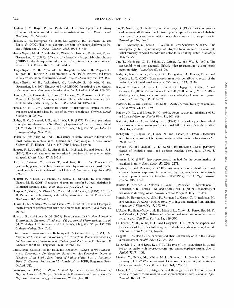
Harduin, J. C., Royer, P., and Piechowski, J. (1994). Uptake and urinary
excretion of uranium after oral administration in man. Radiat. Prot.
Dosimetry. 53, 245–248.
Helmer, D. A., Rossignol, M., Blatt, M., Agarwal, R., Teichman, R., and
Lange, G. (2007). Health and exposure concerns of veterans deployed to Iraq
and Afghanistan. J. Occup. Environ. Med. 49, 475–480.
Henge-Napoli, M. H., Ansoborlo, E., Chazel, V., Houpert, P., Paquet, F., and
Gourmelon, P. (1999). Efficacy of ethane-1-hydroxy-1,1-bisphosphonate
(EHBP) for the decorporation of uranium after intramuscular contamination
in rats. Int. J. Radiat. Biol. 75, 1473–1477.
Henge-Napoli, M. H., Ansoborlo, E., Houpert, P., Mirto, H., Paquest, F.,
Burgada, R., Hodgson, S., and Stradling, G. N. (1998). Progress and trends
in in vivo chelation of uranium. Radiat. Protect. Dosimetry. 79, 449–452.
Henge-Napoli, M. H., Archimbaud, M., Ansoborlo, E., Metivier, H., and
Gourmelon, P. (1995). Efficacy of 3,4,3-LIHOPO for reducing the retention
of uranium in rat after acute administration. Int. J. Radiat. Biol. 68, 389–393.
Herrera, M. B., Bussolati, B., Bruno, S., Fonsato, V., Romanazzi, G. M., and
Camussi, G. (2004). Mesenchymal stem cells contribute to the renal repair of
acute tubular epithelial injury. Int. J. Mol. Med. 14, 1035–1041.
Hirsch, G. H. (1976). Differential effects of nephrotoxic agents on renal
transport and metabolism by use of in vitro techniques. Environ. Health
Perspect. 15, 89–99.
Hodge, H. C., Stannard, J. N., and Hursh, J. B. (1973). Uranium, plutonium,
transplutonic elements. In Handbook of Experimental Pharmacology, 1st ed.
(H. C. Hodge, J. N. Stannard, and J. B. Hursh, Eds.), Vol. 36, pp. 165–195.
Springer-Verlag, New York.
Honda, N., and Sudo, M. (1982). Resistance to uranyl acetate-induced acute
renal failure in rabbits: renal function and morphology. In Acute Renal
Failure (H. E. Eliahou, Ed.), p. 105. John Libbey, London.
Hooper, F. J., Squibb, K. S., Siegel, E. L., McPhaul, K., and Keogh, J. P.
(1999). Elevated urine uranium excretion by soldiers with retained uranium
shrapnel. Health Phys. 77, 512–519.
Hori, R., Takano, M., Okano, T., and Inui, K. (1985). Transport of
p-aminohippurate, tetraethylammonium and D-glucose in renal brush border
membranes from rats with acute renal failure. J. Pharmacol. Exp. Ther. 233,
776–781.
Houpert, P., Chazel, V., Paquet, F., Bailly, T., Burgada, R., and Henge-
Napoli, M. H. (2001). Reduction of uranium transfer by local chelation in
simulated wounds in rats. Hum. Exp. Toxicol. 20, 237–241.
Houpert, P., Muller, D., Chazel, V., Claraz, M., and Paquet, F. (2003). Effect of
DTPA on the nephrotoxicity induced by uranium in the rat. Radiat. Prot.
Dosimetry. 105, 517–520.
Humes, H. D., Weitzel, W. F., and Fissell, W. H. (2004). Renal cell therapy in
the treatment of patients with acute and chronic renal failure. Blood Purif. 22,
60–72.
Hursh, J. B., and Spoor, N. H. (1973). Data on man. In Uranium Plutonium
Transplutonic Elements. Handbook of Experimental Pharmacology, 1st ed.
(H. C. Hodge, J. N. Stannard, and J. B. Hursh, Eds.), Vol. 36, pp. 197–239.
Springer-Verlag, New York.
International Commission on Radiological Protection (ICRP). (1991). In-
ternational Commission on Radiological Protection. Recommendations of
the International Commission on Radiological Protection. Publication 60,
Annals of the ICRP, Pergamon Press, Oxford, UK.
International Commission for Radiation Protection (ICRP). (1996). Interna-
tional Commission for Radiation Protection. Age-Dependent Doses to
Members of the Public from Intake of Radionuclides: Part 4, Inhalation
Dose Coefficients. Publication 72, Annals of the ICRP, Pergamon Press,
Oxford, UK.
Ivannikov, A. (1966). In Physiochemical Approaches to the Selection of
Organic Compounds Designed to Eliminate Radioactive Substances from the
Organism. Atomic Energy Commission, Washington, DC.
Jin, T., Nordberg, G., Sehlin, J., and Vesterberg, O. (1996). Protection against
cadmium-metallothionein nephrotoxicity in streptozotocin-induced diabetic
rats: role of increased metallothionein synthesis induced by streptozotocin.
Toxicology 106, 55–63.
Jin, T., Nordberg, G., Sehlin, J., Wallin, H., and Sandberg, S. (1999). The
susceptibility to nephrotoxicity of streptozotocin-induced diabetic rats
subchronically exposed to cadmium chloride in drinking water. Toxicology
142, 69–75.
Jin, T., Nordberg, G. F., Sehlin, J., Leffler, P., and Wu, J. (1994). The
susceptibility of spontaneously diabetic mice to cadmium-metallothionein
nephrotoxicity. Toxicology 89, 81–90.
Kale, S., Karihaloo, A., Clark, P. R., Kashgarian, M., Krause, D. S., and
Cantley, L. G. (2003). Bone marrow stem cells contribute to repair of the
ischemically injured renal tubule. J. Clin. Invest. 112, 42–49.
Karpas, Z., Lorber, A., Sela, H., Paz-Tal, O., Hagag, Y., Kurttio, P., and
Salonen, L. (2005). Measurement of the 234U/238U ratio by MC-ICPMS in
drinking water, hair, nails, and urine as an indicator of uranium exposure
source. Health. Phys. 89, 315–321.
Kathren, R. L., and Burklin, R. K. (2008). Acute chemical toxicity of uranium.
Health Phys. 94, 170–179.
Kathren, R. L., and Moore, R. H. (1986). Acute accidental inhalation of U:
a 38-year follow-up. Health Phys. 51, 609–619.
Kato, A., Hishida, A., and Nakajima, T. (1994). Effects of oxygen free radical
scavengers on uranium-induced acute renal failure in rats. Free Radic. Biol.
Med. 16, 855–859.
Kobayashi, S., Nagase, M., Honda, N., and Hishida, A. (1984). Glomerular
alterations in uranyl acetate-induced acute renal failure in rabbits. Kidney Int.
26, 808–815.
Kovacic, P., and Jacintho, J. D. (2001). Reproductive toxins: pervasive
theme of oxidative stress and electron transfer. Curr. Med. Chem. 8,
863–892.
Kressin, I. K. (1984). Spectrophotometric method for the determination of
uranium in urine. Anal. Chem. 56, 2269–2271.
Krystek, P., and Ritsema, R. (2009). An incident study about acute and
chronic human exposure to uranium by high-resolution inductively
coupled plasma mass spectrometry (HR-ICPMS). Int. J. Hyg. Environ.
Health. 212, 76–81.
Kurttio, P., Auvinen, A., Salonen, L., Saha, H., Pekkanen, J., Makelainen, I.,
Vaisanen, S. B., Penttila, I. M., and Komulainen, H. (2002). Renal effects of
uranium in drinking water. Environ. Health Perspect. 110, 337–342.
Kurttio, P., Harmoinen, A., Saha, H., Salonen, L., Karpas, Z., Komulainen, H.,
and Auvinen, A. (2006). Kidney toxicity of ingested uranium from drinking
water. Am J Kidney Dis 47, 972–982.
L’Azou, B., Henge-Napoli, M. H., Minaro, L., Mirto, H., Barrouillet, M. P.,
and Cambar, J. (2002). Effects of cadmium and uranium on some in vitro
renal targets. Cell Biol. Toxicol. 18, 329–340.
La Touche, Y. D., Willis, D. L., and Dawydiak, O. I. (1987). Absorption and
biokinetics of U in rats following an oral administration of uranyl nitrate
solution. Health Phys. 53, 147–162.
Leggett, R. W. (1989). The behavior and chemical toxicity of U in the kidney:
a reassessment. Health Phys. 57, 365–383.
Leibovich, S. J., and Ross, R. (1975). The role of the macrophage in wound
repair. A study with hydrocortisone and antimacrophage serum. Am. J.
Pathol. 78, 71–100.
Linares, V., Belles, M., Albina, M. L., Sirvent, J. J., Sanchez, D. J., and
Domingo, J. L. (2006). Assessment of the pro-oxidant activity of uranium in
kidney and testis of rats. Toxicol. Lett. 167, 152–161.
Llobet, J. M., Sirvent, J. J., Ortega, A., and Domingo, J. L. (1991). Influence of
chronic exposure to uranium on male reproduction in mice. Fundam. Appl.
Toxicol. 16, 821–829.
344 VICENTE-VICENTE ET AL.
Dow
nloaded from https://academ
ic.oup.com/toxsci/article/118/2/324/1648537 by guest on 01 June 2022
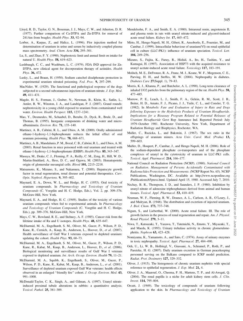
Lloyd, R. D., Taylor, G. N., Boseman, J. J., Mays, C. W., and Atherton, D. R.
(1977). Further comparison of Ca-DTPA and Zn-DTPA for removal of
241Am from beagles. Health Phys. 33, 92–94.
Lorber, A., Karpas, Z., and Halicz, L. (1996). Flor injection method for
determination of uranium in urine and serum by inductively coupled plasma
mass spectrometry. Anal. Chem. Acta 334, 295–301.
Lu, S., and Zhao, F. Y. (1990). Nephrotoxic limit and annual limit on intake for
natural U. Health Phys. 58, 619–623.
Lushbaugh, C. C., and Washburn, L. C. (1979). FDA IND approval for Zn-
DTPA, new clinical agent for decorporation therapy of actinides. Health
Phys. 36, 472.
Lusky, L., and Braun, H. (1950). Sodium catechol disulphonate protection in
experimental uranium nitrated poisoning. Fed. Proc. 9, 297–299.
MacNider, W. (1929). The functional and pathological response of the dogs
subjected to a second subcutaneous injection of uranium nitrate. J. Exp. Med.
49, 411–431.
Magdo, H. S., Forman, J., Graber, N., Newman, B., Klein, K., Satlin, L.,
Amler, R. W., Winston, J. A., and Landrigan, P. J. (2007). Grand rounds:
nephrotoxicity in a young child exposed to uranium from contaminated well
water. Environ. Health Perspect. 115, 1237–1241.
Mao, Y., Desmeules, M., Schaubel, D., Berube, D., Dyck, R., Brule, D., and
Thomas, B. (1995). Inorganic components of drinking water and micro-
albuminuria. Environ. Res. 71, 135–140.
Martinez, A. B., Cabrini, R. L., and Ubios, A. M. (2000). Orally administered
ethane-1-hydroxy-1,1-biphosphonate reduces the lethal effect of oral
uranium poisoning. Health Phys. 78, 668–671.
Martinez, A. B., Mandalunis, P. M., Bozal, C. B., Cabrini, R. L., and Ubios, A. M.
(2003). Renal function in mice poisoned with oral uranium and treated with
ethane-1-hydroxy-1,1-bisphosphonate (EHBP). Health Phys. 85, 343–347.
Masuya, M., Drake, C. J., Fleming, P. A., Reilly, C. M., Zeng, H., Hill, W. D.,
Martin-Studdard, A., Hess, D. C., and Ogawa, M. (2003). Hematopoietic
origin of glomerular mesangial cells. Blood 101, 2215–2218.
Matsumoto, K., Mizuno, S., and Nakamura, T. (2000). Hepatocyte growth
factor in renal regeneration, renal disease and potential therapeutics. Curr.
Opin. Nephrol. Hypertens. 9, 395–402.
Maynard, E. A., Down, W. L., and Hodge, H. C. (1953). Oral toxicity of
uranium compounds. In Pharmacology and Toxicology of Uranium
Compounds (C. Voegtlin and H. C. Hodge, Eds.), Vol. 2, pp. 309–376.
McGraw-Hill, New York.
Maynard, E. A., and Hodge, H. C. (1949). Studies of the toxicity of various
uranium compounds when fed to experimental animals. In Pharmacology
and Toxicology of Uranium Compounds (C. Voegtlin and H. C. Hodge,
Eds.), pp. 309–376. McGraw-Hill, New York.
Mays, C. W., Rowland, R. E., and Stehney, A. F. (1985). Cancer risk from the
lifetime intake of Ra and U isotopes. Health Phys. 48, 635–647.
McDiarmid, M. A., Engelhardt, S. M., Oliver, M., Gucer, P., Wilson, P. D.,
Kane, R., Cernich, A., Kaup, B., Anderson, L., Hoover, D., et al. (2007).
Health surveillance of Gulf War I veterans exposed to depleted uranium:
updating the cohort. Health Phys. 93, 60–73.
McDiarmid, M. A., Engelhardt, S. M., Oliver, M., Gucer, P., Wilson, P. D.,
Kane, R., Kabat, M., Kaup, B., Anderson, L., Hoover, D., et al. (2006).
Biological monitoring and surveillance results of Gulf War I veterans
exposed to depleted uranium. Int. Arch. Occup. Environ. Health 79, 11–21.
McDiarmid, M. A., Squibb, K., Engelhardt, S., Oliver, M., Gucer, P.,
Wilson, P. D., Kane, R., Kabat, M., Kaup, B., Anderson, L., et al. (2001).
Surveillance of depleted uranium exposed Gulf War veterans: health effects
observed in an enlarged ‘‘friendly fire’’ cohort. J. Occup. Environ. Med. 43,
991–1000.
McDonald-Taylor, C. K., Singh, A., and Gilman, A. (1997). Uranyl nitrate-
induced proximal tubule alterations in rabbits: a quantitative analysis.
Toxicol. Pathol. 25, 381–389.
Mendelsohn, F. A., and Smith, E. A. (1980). Intrarenal renin, angiotensin II,
and plasma renin in rats with uranyl nitrate-induced and glycerol-induced
acute renal failure. Kidney Int. 17, 465–472.
Mirto, H., Henge-Napoli, M. H., Gibert, R., Ansoborlo, E., Fournier, M., and
Cambar, J. (1999). Intracellular behaviour of uranium(VI) on renal epithelial
cell in culture (LLC-PK1): influence of uranium speciation. Toxicol. Lett.
104, 249–256.
Mizuno, S., Fujita, K., Furuy, R., Hishid, A., Ito, H., Tashim, Y., and
Kumagai, H. (1997). Association of HSP73 with the acquired resistance to
uranyl acetate-induced acute renal failure. Toxicology 117, 183–191.
Molitch, M. E., DeFronzo, R. A., Franz, M. J., Keane, W. F., Mogensen, C. E.,
Parving, H. H., and Steffes, M. W. (2004). Nephropathy in diabetes.
Diabetes Care 27(Suppl. 1), 79–83.
Morris, K. J., Khanna, P., and Batchelor, A. L. (1990). Long-term clearance of
inhaled UO2 particles from the pulmonary region of the rat. Health Phys. 58,
477–485.
Morrow, P. E., Leach, L. J., Smith, F. A., Gelein, R. M., Scott, J. B.,
Beiter, H. D., Amato, F. J., Picano, J. J., Yuile, C. L., and Consler, T. G.
(1982). In Metabolic Fate and Evaluation of Injury in Rats and Dogs
following Exposure to the Hydrolysis Products of Uranium Hexafluoride.
Implications for a Bioassay Program Related to Potential Releases of
Uranium Hexafluoride Govt Rep Announce Ind. Reported Period: July
1979–October 1981. Rochester University, New York, Department of
Radiation Biology and Biophysics, Rochester, WA.
Muller, C., Ruzicka, L., and Bakstein, J. (1967). The sex ratio in the
offsprings of uranium miners. Acta Univ. Carol. Med. (Praha) 13,
599–603.
Muller, D., Houpert, P., Cambar, J., and Henge-Napoli, M. H. (2006). Role of
the sodium-dependent phosphate co-transporters and of the phosphate
complexes of uranyl in the cytotoxicity of uranium in LLC-PK1 cells.
Toxicol. Appl. Pharmacol. 214, 166–177.
National Council on Radiation Protection (NCRP). (1980). National Council
on Radiation. Management of Persons Accidentally Contaminated with
Radionuclides Protection and Measurements. (NCRP Report No. 65). NCRP
Publications, Washington, DC. Available at: http://www.ncrponline.org
/Publications/65press.html. Updated April 15, 1980. Accessed July 7, 2010.
Nechay, B. R., Thompson, J. D., and Saunders, J. P. (1980). Inhibition by
uranyl nitrate of adenosine triphosphatases derived from animal and human
tissues. Toxicol. Appl. Pharmacol. 53, 410–419.
Neuman, W. F., Fleming, R. W., Dounce, A. L., Carlson, A. B., O’Leary, J.,
and Mulryan, B. (1948). The distribution and excretion of injected uranium.
J. Biol. Chem. 173, 737–748.
Nigam, S., and Lieberthal, W. (2000). Acute renal failure. III. The role of
growth factors in the process of renal regeneration and repair. Am. J. Physiol.
Renal Physiol. 279, 3–11.
Niwa, T., Katsuzaki, T., Yazawa, T., Tatemichi, N., Emoto, Y., Miyazaki, T.,
and Maeda, K. (1993). Urinary trehalase activity in chronic glomerulone-
phritis. Nephron 63, 423–428.
Nomiyama, K., Yamamoto, A., and Sato, C. (1974). Assay of urinary enzymes
in toxic nephropathy. Toxicol. Appl. Pharmacol. 27, 484–490.
Oeh, U., Li, W. B., Hollriegl, V., Giussani, A., Schramel, P., Roth, P., and
Paretzke, H. G. (2007). Daily uranium excretion in German peacekeeping
personnel serving on the Balkans compared to ICRP model prediction.
Radiat. Prot. Dosimetry 127, 329–332.
Oliver, J. (1915). The histogenesis of chronic uranium nephritis with special
reference to epithelial regeneration. J. Exp. Med. 21, 4.
Oliver, J. A., Maarouf, O., Cheema, F. H., Martens, T. P., and Al-Awqati, Q.
(2004). The renal papilla is a niche for adult kidney stem cells. J. Clin.
Invest. 114, 795–804.
Orcutt, J. (1949). The toxicology of compounds of uranium following
application to the skin. In Pharmacology and Toxicology of Uranium
NEPHROTOXICITY OF URANIUM 345
Dow
nloaded from https://academ
ic.oup.com/toxsci/article/118/2/324/1648537 by guest on 01 June 2022

Compounds, 1st ed. (H. Hodge and C. Voegtlin, Eds.), Vol. 3, pp. 377–414.
McGraw Hill, New York.
Orloff, K. G., Mistry, K., Charp, P., Metcalf, S., Marino, R., Shelly, T.,
Melaro, E., Donohoe, A. M., and Jones, R. L. (2004). Human exposure to
uranium in groundwater. Environ. Res. 94, 319–326.
Ortega, A., Domingo, J. L., Llobet, J. M., Tomas, J. M., and Paternain, J. L.
(1989). Evaluation of the oral toxicity of uranium in a 4-week drinking-water
study in rats. Bull. Environ. Contam. Toxicol. 42, 935–941.
Ortega, A., Sanchez, D. J., Domingo, J. L., Llobet, J. M., and Corbella, J.
(1991). Developmental toxicity evaluation of tiron (sodium 4,5-
dihydroxybenzene-1,3-disulfonate) in mice. Res Commun. Chem.
Pathol. Pharmacol. 73, 97–106.
Paquet, F., Houpert, P., Blanchardon, E., Delissen, O., Maubert, C., Dhieux, B.,
Moreels, A. M., Frelon, S., and Gourmelon, P. (2006). Accumulation and
distribution of uranium in rats after chronic exposure by ingestion. Health
Phys. 90, 139–147.
Parrish, R. R., Horstwood, M., Arnason, J. G., Chenery, S., Brewer, T.,
Lloyd, N. S., and Carpenter, D. O. (2008). Depleted uranium contamination
by inhalation exposure and its detection after approximately 20 years:
implications for human health assessment. Sci. Total Environ. 390, 58–68.
Parving, H. H., Chaturvedi, N., Viberti, G., and Mogensen, C. E. (2002). Does
microalbuminuria predict diabetic nephropathy? Diabetes Care 5, 406–407.
Patschan, D., Plotkin, M., and Goligorsky, M. S. (2006). Therapeutic use of
stem and endothelial progenitor cells in acute renal injury: Ca ira. Curr.
Opin. Pharmacol. 6, 176–183.
Pavlakis, N., Pollock, C. A., McLean, G., and Bartrop, R. (1996). Deliberate
overdose of uranium: toxicity and treatment. Nephron 72, 313–317.
Peeks, M., Walter, T., and Walter, S. (2002). Uranium, uranium alloys, and
uranium compounds. In Ullmann’s Encyclopedia of Industrial Chemistry,
6th ed. (W.-V. Verlag. Gmbh. and Co, Eds.), pp. 270–281. Wiley-VCH,
Weinheim, Germany.
Pinney, S. M., Freyberg, R. W., Levine, G. E., Brannen, D. E., Mark, L. S.,
Nasuta, J. M., Tebbe, C. D., Buckholz, J. M., and Wones, R. (2003). Health
effects in community residents near a uranium plant at Fernald, Ohio, USA.
Int. J. Occup. Med. Environ. Health 16, 139–153.
Powell, D. W., Mifflin, R. C., Valentich, J. D., Crowe, S. E., Saada, J. I., and
West, A. B. (1999). Myofibroblasts. I. Paracrine cells important in health and
disease. Am. J. Physiol. 277, 1–9.
Pozzani, U. C. (1949). Toxicity following inhalation. In Pharmacology and
Toxicology of Uranium Compounds (H. Hodge and C. Voegtlin, Eds.), Vol.
2, pp. 622–635. McGraw-Hill, New York.
Price, A. (1989). Review of methods for the assessment of intake of uranium by
workers at BNGL springfields. Radiat. Prot. Dosimetry. 26, 35–42.
Rajan, K., and Martell, A. (1964). Equilibrium studies of uranilo complexes.
Interaction of uranilo ion with citric acid. Inorg. Chem. 26, 1927–1944.
Remuzzi, G., Benigni, A., and Remuzzi, A. (2006). Mechanisms of progression
and regression of renal lesions of chronic nephropathies and diabetes. J. Clin.
Invest. 116, 288–296.
Rosen, L. C., Gur, D., Pan, S. I., and Wals, N. (1989). Long term removal of
Am-241 using Ca-DTPA. Health Phys. 39, 101–106.
Rossol, M. (1997). Ceramic Glazes, Frit Metal Enamels, and Glass; Personal
Communication and Supporting Materials. Arts, Crafts and Theater Safety,
New York.
Rothstein, A. (1949). Toxicity following inhalation: uranium trioxide. In
Pharmacology and Toxicology of Uranium Compounds, 1st ed. (H. Hodge
and C. Voegtlin, Eds.), Vol. 2, pp. 635–648. McGraw-Hill, New York.
Russell, J. J., and Kathren, R. L. (2004). Uranium deposition and retention in
a USTUR whole body case. Health Phys. 86, 273–284.
Russell, J. J., Kathren, R. L., and Dietert, S. E. (1996). A histological kidney
study of uranium and non-uranium workers. Health Phys. 70, 466–472.
Sabolic, I. (2006). Common mechanisms in nephropathy induced by toxic
metals. Nephron Physiol. 104, 107–114.
Saccomanno, G., Thun, M. J., Baker, D. B., Steenland, K., Smith, A. B.,
Halperin, W., and Berl, T. (1982). The contribution of uranium miners to
lung cancer histogenesis renal toxicity in uranium mill workers. Cancer Res.
82, 43–52.
Sachett, I. A., Nobrega, A. W., and Lauria, D. C. (1984). Determination of
uranium isotopes by chemical stripping and alpha-spectrometry. Health
Phys. 46, 133–139.
Salminen, W. F., Jr., Voellmy, R., and Roberts, S. M. (1997). Protection
against hepatotoxicity by a single dose of amphetamine: the potential role of
heat shock protein induction. Toxicol. Appl. Pharmacol. 147, 247–258.
Sanchez, D. J., Belles, M., Albina, M. L., Sirvent, J. J., and Domingo, J. L.
(2001). Nephrotoxicity of simultaneous exposure to mercury and uranium in
comparison to individual effects of these metals in rats. Biol. Trace Elem.
Res. 84, 139–154.
Sanders, C. L. (1986). Radiological Health. Toxicological Aspects of Energy
Production. Battelle Press, Columbus OH, Canada.
Sansone, U., Stellato, L., Jia, G., Rosamilia, S., Gaudino, S., Barbizzi, S., and
Belli, M. (2001). Levels of depleted uranium in Kosovo soils. Radiat. Prot.
Dosimetry. 97, 317–320.
Schena, F. P., and Gesualdo, L. (2005). Pathogenetic mechanisms of diabetic
nephropathy. J. Am. Soc. Nephrol. 16(Suppl. 1), 30–33.
Schramm, L., La, M., Heidbreder, E., Hecker, M., Beckman, J. S., Lopau, K.,
Zimmermann, J., Rendl, J., Reiners, C., Winderl, S., et al. (2002). L-arginine
deficiency and supplementation in experimental acute renal failure and in
human kidney transplantation. Kidney Int. 61, 1423–1432.
Schwartz, J. H., and Flamenbaum, W. (1976). Uranyl nitrate and HgCl2-
induced alterations in ion transport. Kidney Int. 6(Suppl.), 123–127.
Selden, A. I., Lundholm, C., Edlund, B., Hogdahl, C., Ek, B. M.,
Bergstrom, B. E., and Ohlson, C. G. (2009). Nephrotoxicity of uranium in
drinking water from private drilled wells. Environ. Res. 109, 486–494.
Shawky, S., Amer, H. A., Hussein, M. I., el-Mahdy, Z., and Mustafa, M. (2002).
Uranium bioassay and radioactive dust measurements at some uranium
processing sites in Egypt—health effects. J. Environ. Monit. 4, 588–591.
Shim, W. S., Park, J. H., Ahn, S. J., Han, L., Jin, Q. R., Li, H., Choi, M. K.,
Kim, D. D., Chung, S. J., and Shim, C. K. (2009). Testosterone-independent
down-regulation of Oct2 in the kidney medulla from a uranyl nitrate-induced
rat model of acute renal failure: effects on distribution of a model organic
cation, tetraethylammonium. J. Pharm. Sci. 98, 739–747.
Shiraishi, K., Igarashi, Y., Takaku, Y., Masuda, K., Yoshimizu, K.,
Nishimura, Y., Hongo, S., and Yamaguchi, H. (1992). Daily intakes of
232Th and 238U in Japanese males. Health. Phys. 63, 187–191.
Shyh, T. P., Beyer, M. M., and Friedman, E. A. (1984). Induced hyperglycemia
protects against mercury nephrotoxicity in the rat. Trans. Am. Soc. Artif.
Intern. Organs 30, 260–263.
Snively, C. S., and Gutierrez, C. (2004). Chronic kidney disease: prevention
and treatment of common complications. Am. Fam. Physician 70,
1921–1928.
Snyder, S., and Pendergraph, B. (2005). Detection and evaluation of chronic
kidney disease. Am. Fam. Physician 72, 1723–1732.
Spencer, H., Osis, D., Fisenne, I. M., Perry, P. M., and Harley, N. H. (1990).
Measured intake and excretion patterns of naturally occurring 234U, 238U,
and calcium in humans. Radiat. Res. 124, 90–95.
Spiegel, C. (1949). Uranium hexafluoride. In Pharmacology and Toxicology
of Uranium Compounds, 1st ed. (H. Hodge and C. Voegtlin, Eds.), Vol. 2,
pp. 532–547. McGraw-Hill, New York.
Spoor, N., and Hursh, J. (1973). Protection criteria. In Handbook
of Experimental Pharmacology (J. N. Stannard and J. B. Hursh, Eds.),
pp. 241–270. Springer- Verlag, Berlin.
346 VICENTE-VICENTE ET AL.
Dow
nloaded from https://academ
ic.oup.com/toxsci/article/118/2/324/1648537 by guest on 01 June 2022
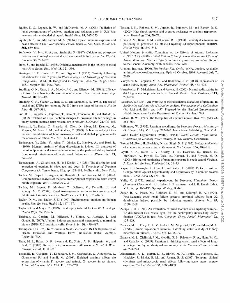
Squibb, K. S., Leggett, R. W., and McDiarmid, M. A. (2005). Prediction of
renal concentrations of depleted uranium and radiation dose in Gulf War
veterans with embedded shrapnel. Health Phys. 89, 267–273.
Squibb, K. S., and McDiarmid, M. A. (2006). Depleted uranium exposure and
health effects in Gulf War veterans. Philos. Trans. R. Soc. Lond. B Biol. Sci.361, 639–648.
Stefanovic, V., Ivic, M. A., and Strahinjic, S. (1987). Calcium and phosphate
metabolism in uranyl nitrate-induced acute renal failure. Arch. Int. Physiol.
Biochim. 95, 223–228.
Stohs, S., and Bagchi, D. (1995). Oxidative mechanisms in the toxicity of metal
ions. Free Radic. Biol. Med. 18, 321–336.
Stokinger, H. E., Baxter, R. C., and Dygent, H. (1953). Toxicity following
inhalation for 1 and 2 years. In Pharmacology and Toxicology of Uranium
Compounds, 1st ed. (H. Hodge and C. Voegtlin, Eds.), Vol. 2, pp. 1522–
1553. Mcgraw-Hill, New York.
Stradling, G. N., Gray, S. A., Moody, J. C., and Ellender, M. (1991). Efficacy
of tiron for enhancing the excretion of uranium from the rat. Hum. Exp.
Toxicol. 10, 195–198.
Stradling, G. N., Stather, J., Ham, S. E., and Sumner, S. A. (1981). The use of
puchel and DTPA for removing Pu-239 from the lungs of hamsters. Health
Phys. 41, 387–391.
Sun, D. F., Fujigaki, Y., Fujimoto, T., Goto, T., Yonemura, K., and Hishida, A.
(2002). Relation of distal nephron changes to proximal tubular damage in
uranyl acetate-induced acute renal failure in rats. Am. J. Nephrol. 22, 405–416.
Takahashi, T., Kalka, C., Masuda, H., Chen, D., Silver, M., Kearney, M.,
Magner, M., Isner, J. M., and Asahara, T. (1999). Ischemia- and cytokine-
induced mobilization of bone marrow-derived endothelial progenitor cells
for neovascularization. Nat. Med. 5, 434–438.
Tanigawara, Y., Saito, Y., Aiba, T., Ohoka, K., Kamiya, A., and Hori, R.
(1990). Moment analysis of drug disposition in kidney. III: transport of
p-aminohippurate and tetraethylammonium in the perfused kidney isolated
from uranyl nitrate-induced acute renal failure rats. J. Pharm. Sci. 79,
249–256.
Tannenbaum, A., Silverstone, H., and Koziol, J. (1951). The distribution and
excretion of uranium in mice, rats and dogs. In Toxicology of Uranium
Compounds (A. Tannenbaum, Ed.), pp. 128–181. McGraw-Hill, New York.
Taulan, M., Paquet, F., Argiles, A., Demaille, J., and Romey, M. C. (2006).
Comprehensive analysis of the renal transcriptional response to acute uranyl
nitrate exposure. BMC Genomics 7, 2.
Taulan, M., Paquet, F., Maubert, C., Delissen, O., Demaille, J., and
Romey, M. C. (2004). Renal toxicogenomic response to chronic uranyl
nitrate insult in mice. Environ. Health Perspect. 112, 1628–1635.
Taylor, D. M., and Taylor, S. K. (1997). Environmental uranium and human
health. Rev. Environ. Health 12, 147–157.
Taylor, G., and Mays, C. (1979). Fatal injury induced by Ca-DTPA in dogs.
Health Phys. 35, 858–860.
Thiebault, C., Carriere, M., Milgram, S., Simon, A., Avoscan, L., and
Gouget, B. (2007). Uranium induces apoptosis and is genotoxic to normal rat
kidney (NRK-52E) proximal cells. Toxicol. Sci. 98, 479–487.
Thompson, D. (1976). In Uranium in Dental Porcelain. IN US Department of
Health, Education and Welfare, HEW Publication (FDA) 76-8061,
Rockville, WA.
Thun, M. J., Baker, D. B., Steenland, K., Smith, A. B., Halperin, W., and
Berl, T. (1985). Renal toxicity in uranium mill workers. Scand. J. Work
Environ. Health 11, 83–90.
Tissandie, E., Gueguen, Y., Lobaccaro, J. M., Grandcolas, L., Aigueperse, J.,
Gourmelon, P., and Souidi, M. (2008). Enriched uranium affects the
expression of vitamin D receptor and retinoid X receptor in rat kidney.
J. Steroid Biochem. Mol. Biol. 110, 263–268.
Tolson, J. K., Roberts, S. M., Jortner, B., Pomeroy, M., and Barber, D. S.
(2005). Heat shock proteins and acquired resistance to uranium nephrotox-
icity. Toxicology 206, 59–73.
Ubios, A. M., Braun, E. M., and Cabrini, R. L. (1994). Lethality due to uranium
poisoning is prevented by ethane-1-hydroxy-1,1-biphosphonate (EHBP).
Health Phys. 66, 540–544.
United Nations Scientific Committee on the Effects of Atomic Radiation
(UNSCEAR). (1988). United Nations Scientific Committee on the Effects of
Atomic Radiation. Sources, Effects and Risks of Ionizing Radiation. Report
to the General Assembly, with annexes, New York.
Uranium Institute. (1996). The Nuclear Fuel Cycle. WNA, London. Available
at: http://www.world-nuclear.org. Updated October, 1996. Accessed July 7,
2010.
Vaidya, V. S., Ferguson, M. A., and Bonventre, J. V. (2008). Biomarkers of
acute kidney injury. Annu. Rev. Pharmacol. Toxicol. 48, 463–493.
Vesterbacka, P., Makelainen, I., and Arvela, H. (2005). Natural radioactivity in
drinking water in private wells in Finland. Radiat. Prot. Dosimetry 113,
223–232.
Wessman, R. (1984). An overview of the radiochemical analysis of uranium. In
Biokinetics and Analysis of Uranium in Man. Proceedings of a Colloquium
(W. Richland, Ed.), pp. 1–57. Prepared by the Hanford Environmental
Health Foundation for the Department of Energy, Richland, WA.
Wilcox, R. W. (1917). The theraputics of uranium nitrate. Med. Rec. (NY) 92,361–364.
Wilkinson, W. (1962). Uranium metallurgy. In Uranium Process Metallurgy
(R. Harper, Ed.), Vol. 1, pp. 722–745. Interscience Publishing, New York.
World Health Organization (WHO). (1984). World Health Organization.
Guidelines for Drinking Water Quality, WHO, Geneva, Switzerland.
Wrenn, M., Ruth, H., Burleigh, D., and Singh, N. P. (1992). Background levels
of uranium in human urine. J. Radioanal. Nucl. Chem. 156, 407–412.
Wyatt, S. A., Reitz, L. V., Croley, T. R., Hawkins, D., Barrett, E.,
McKeown, A., Powell, N., West, A., Hamner, T., and Royster, M. O.
(2008). Biological monitoring of uranium exposure in south central Virginia.
J. Expo. Sci. Environ. Epidemiol. 18, 59–75.
Yapar, K., Cavusoglu, K., Oruc, E., and Yalcin, E. (2010). Protective role of
Ginkgo biloba against hepatotoxicity and nephrotoxicity in uranium-treated
mice. J. Med. Food 13, 179–188.
Yuile, C. (1973). Animal experiments. In Uranium, Plutonium, Trans-plutonium Elements (H. C. Hodge, J. N. Stannard, and J. B. Hursh, Eds.),
Vol. 36, pp. 165–196. Springer-Verlag, Berlin.
Zager, R. A., Iwata, M., Burkhart, K. M., and Schimpf, B. A. (1994).
Post-ischemic acute renal failure protects proximal tubules from O2
deprivation injury, possibly by inducing uremia. Kidney Int. 45,
1760–1768.
Zalups, R. K. (1991). An evaluation of Tiron (sodium 4,5-dihydroxybenzene-
1,3-disulfonate) as a rescue agent for the nephropathy induced by uranyl
fluoride (UO2F2) in rats. Res. Commun. Chem. Pathol. Pharmacol. 72,
125–128.
Zamora, M. L., Tracy, B. L., Zielinski, J. M., Meyerhof, D. P., and Moss, M. A.
(1998). Chronic ingestion of uranium in drinking water: a study of kidney
bioeffects in humans. Toxicol. Sci. 43, 68–77.
Zamora, M. L., Zielinski, J. M., Moodie, G. B., Falcomer, R. A., Hunt, W. C.,
and Capello, K. (2009). Uranium in drinking water: renal effects of long-
term ingestion by an aboriginal community. Arch. Environ. Occup. Health
64, 228–241.
Zimmerman, K. L., Barber, D. S., Ehrich, M. F., Tobias, L., Hancock, S.,
Hinckley, J., Binder, E. M., and Jortner, B. S. (2007). Temporal clinical
chemistry and microscopic renal effects following acute uranyl acetate
exposure. Toxicol. Pathol. 35, 1000–1009.
NEPHROTOXICITY OF URANIUM 347
Dow
nloaded from https://academ
ic.oup.com/toxsci/article/118/2/324/1648537 by guest on 01 June 2022