Embed Size (px)
Citation preview
Jpn. J. Cancer Res. 92, 328–336, March 2001
328
Cisplatin-incorporated Polymeric Micelles Eliminate Nephrotoxicity, While Maintaining Antitumor Activity
Yasuo Mizumura,1 Yasuhiro Matsumura,1, 7 Tetsuya Hamaguchi,1 Nobuhiro Nishiyama,2
Kazunori Kataoka,2 Takanori Kawaguchi,3 William J. M. Hrushesky,4 Fuminori Moriyasu5 andTadao Kakizoe6
1Department of Medicine, 6The Director, National Cancer Center Hospital, 5-1-1 Tsukiji, Chuo-ku, Tokyo104-0045, 2Department of Materials Science, Graduate School of Engineering, The University of Tokyo,7-3-1 Hongo, Bunkyo-ku, Tokyo 113-8656, 32nd Department of Pathology, Fukushima Medical College, 1Hikarigaoka, Fukushima 960-1247, 4Stratton DVAMC/DVA Network 2, Albany Medical College ofUnion University, Albany, New York 12208 and 5Fourth Department of Medicine, Tokyo Medical Univer-sity, 6-7-1 Nishishinjyuku, Shinjyuku-ku, Tokyo 160-0023
cis-Diamminedichloroplatinum (II) (cisplatin, CDDP), a potent anticancer agent, was bound to theaspartic acid residues of poly(ethylene glycol)-poly(aspartic acid) (PEG-P(ASP)) block copolymerby ligand substitution reaction at the platinum atom of CDDP. The polymeric drug thus obtainedwas observed to form a micelle structure in aqueous medium, showing excellent water solubility. Inthe present study, in vitro and in vivo antitumor activity against several human tumor cell lines,toxicity and pharmacokinetic characteristics in rodents of CDDP-incorporated polymeric micelles(CDDP/m) were evaluated in comparison with those of CDDP. In vitro, CDDP/m exhibited 10–17% of the cytotoxicity of CDDP against human tumor cell lines. CDDP/m given by intravenous(i.v.) injection yielded higher and more sustained serum levels than CDDP. In vivo CDDP/m treat-ment resulted in higher and more sustained levels in tumor tissue than CDDP, and showed similarantitumor activity to CDDP against MKN 45 human gastric cancer xenograft. CDDP/m treatmentcaused much less renal damage than CDDP. These results indicate that CDDP/m treatment canreduce CDDP-induced nephrotoxicity without compromising the anticancer cytotoxicity of CDDP.
Key words: Polymeric micelles — Cisplatin — Nephrotoxicity — EPR effect — DDS
cis-Diamminedichloroplatinum (II) (cisplatin, CDDP),the most commonly used anticancer agent, consists of acentral platinum atom surrounded by four ligands, twoammonias and two chlorides.1) A high antitumor activityresults when the two chloride ligands in CDDP arebi-aquated in aqueous physiological environments; CDDPcan then interact directly with DNA and display cytotoxicactivity.2, 3) The clinical utility of CDDP is limited by sig-nificant general organ toxicity including myelosuppres-sion,4) ototoxicity,5) gastrointestinal disturbance,5, 6) andespecially acute nephrotoxicity.7) CDDP, a low-molecular-weight compound, is distributed readily into almost alltissues and intracellular compartments. CDDP traversesplasma membranes rapidly via passive diffusion or activetransport, and is also rapidly cleared from blood by glomer-ular excretion, limiting its therapeutic availability. Injec-tion of the maximum permissible amount of this low-molecular-weight drug to raise its therapeutic concentra-tion and AUC (area under the curve) results in severetoxicity without significantly greater antitumor efficacy.Therefore, several novel forms of controlled release drugdelivery have been designed to improve distribution and to
prolong the exposure of the tumor to an effective drugconcentration.
It is known that solid tumors generally possess the fol-lowing pathophysiological characteristics: hypervascular-ity, incomplete vascular architecture, secretion of vascularpermeability factors, and also the absence of effectivelymphatic drainage, preventing the efficient clearanceof accumulated macromolecules. These characteristics,unique to solid tumors, are believed to be the basis of theso-called EPR effect (enhanced permeability and retentioneffect).8, 9) Moreover, macromolecules have relatively pro-longed plasma half-lives because they are too large to passthrough the normal vessel walls, unless they are trappedby the reticuloendothelial system.10–12) Therefore it is rela-tively easy for macromolecules to extravasate into andaccumulate within tumor tissue on the basis of the EPReffect.
To make use of the EPR effect, several techniques havebeen developed. Yokoyama et al. reported that adriamycin(ADR)-containing polymeric micelles show dramaticallyhigher antitumor activity in vivo than free ADR, becauseof highly selective delivery to solid tumor tissue throughthe EPR effect.13, 14) We have concentrated upon construct-ing polymeric micelles composed of poly(ethylene glycol)-poly(aspartic acid) (PEG-P(ASP)) block copolymer.15, 16)
7 To whom correspondence should be addressed.E-mail: [email protected]
Cisplatin-incorporated Polymeric Micelles
329
This copolymer possesses aspartic acid residues capableof avidly binding at the platinum atom of CDDP by aligand substitution reaction. Furthermore, PEG-P(ASP)block copolymer conjugating platinum spontaneouslydevelops a micellar structure in aqueous solution. Thisstructure possesses high structural stability in distilledwater. In physiological saline, however, the micellesstart to dissociate slowly with an induction period ofapproximately 10 h, synchronized with the sustainedrelease of the platinum complex from the core. We believethat the sustained release profile of CDDP from themicelles would be advantageous for obtaining an enhancedantitumor effect in vivo.17, 18)
In the present study, we examine the potential utility ofCDDP-incorporated polymeric micelle (CDDP/m) as adrug delivery system for targeting therapy with CDDP bymeans of in vitro and in vivo experiments.
MATERIALS AND METHODS
Chemicals CDDP was purchased from Aldrich ChemicalCo., Inc. β-Benzyl L-aspartate and bis(trischloromethyl)-carbonate (triphosgene) were purchased from the PeptideInstitute, Inc., Osaka, and Tokyo Kasei Kogyo Co., Ltd.,Tokyo, respectively. These chemicals were used withoutfurther purification. Other chemicals were of reagent gradeand were used as purchased.Synthesis of CDDP/m, micelle-forming (PEG-P(ASP))block copolymer conjugating CDDP PEG-P(ASP) wassynthesized by a previously reported procedure.16, 18) Theprocedure for preparing CDDP/m and the method ofincorporating CDDP into polymeric micelles are brieflydisplayed in Fig. 1.
The N-carboxy anhydride of β-benzyl L-aspartate(BLA-NCA) was synthesized by the Fuchs-Farthingmethod using triphosgene. Poly(ethylene glycol)-poly(β-benzyl L-aspartate) block copolymer (PEG-PBLA) wasthen synthesized by polymerization of BLA-NCA indichloromethane (CH2Cl2) initiated by the terminal aminogroup of α-methyl-ω-aminopoly(ethylene glycol) (CH3O-PEG-NH2; Mw=5000). The degree of polymerization ofBLA in the block copolymer was determined to be 40.PEG-P(ASP) block copolymer was prepared from PEG-PBLA by removal of the benzyl groups in 0.1 N NaOH.PEG-P(ASP) block copolymer and CDDP were dissolvedin distilled water with a 1:1 molar ratio of CDDP to Aspresidue. The mixture was shaken at 37°C for 72 h toobtain micelle-forming (PEG-P(ASP)) block copolymerconjugating CDDP. Purification of CDDP-incorporatedmicelles was carried out by ultrafiltration and confirmedby gel-permeation chromatography. Finally, polymericmicelles containing CDDP with narrow size distributionwere obtained. The weight-average diameter of the parti-cles was approximately 20 nm. Pt content was determined
by using an atomic absorption spectrophotometer (Z-8000polarized Zeeman atomic absorption spectrophotometer(Hitachi Instruments, Inc., Tokyo)). The weight ratio ofCDDP in the micelles was 41.2±2.3 wt%.In vitro cytotoxicity Five colonic cancer cell lines(COLO201, COLO320, DLD-1, HT-29, LOVO), 5 gastriccancer cell lines (MKN-28, MKN-45, MKN-72, TMK-1,KATOIII), 4 breast cancer cell lines (MCF-7, 4A4, SK-
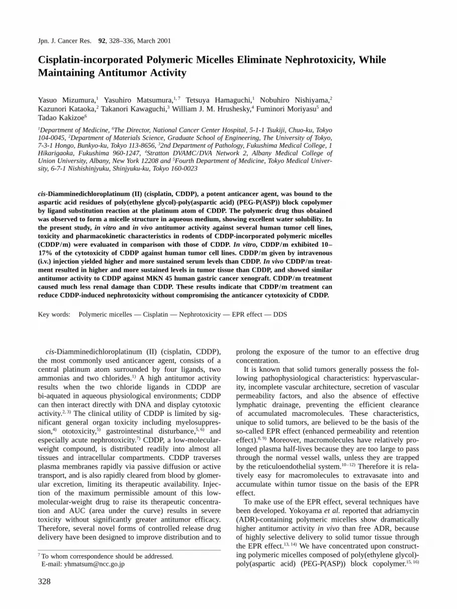
Fig. 1. Chemical structure of CDDP/m and the method ofincorporating CDDP into polymeric micelles are shown. A) ABblock copolymer is composed of poly(ethylene glycol) andpoly(aspartic acid) segments. B) CDDP is bonded to AB-blockcopolymer through ligand substitution between Pt atom and Aspresidues of the PEG-P(ASP) block. denotes CDDP. C) poly-mer-metal complexes spontaneously and easily form micelles inaqueous media owing to cohesive forces between polymer andmetal complexes.
Jpn. J. Cancer Res. 92, March 2001
330
BR-3, SST, T-47-D) and 2 lung cancer cell lines (A549,PC-14) were used in this study. All the cell lines weremaintained in monolayer cultures in Dulbecco’s modifiedEagle’s medium (DMEM) containing 10% fetal bovineserum in a humidified atmosphere containing 5% CO2 at37°C. For cytotoxicity analysis, the 3-(4,5-dimethylthia-zol-2-yl)-2,5-diphenyltetrazolium bromide (MTT) assaywas used. Ten thousand cells of each cell line in 198 µl ofculture medium were plated in 96-well plates 24 h prior todrug treatment. Then 2 µl of various doses of CDDP orCDDP/m was added. Cells were exposed to the indicateddrug concentration in triplicate for 24, 48 or 72 h.Plasma clearance and distribution Male C57BL/6Nmice were inoculated subcutaneously with 106 viableLewis lung carcinoma cells. Seven days later, tumor sizehad reached approximately 50–70 mm2, measured as theproduct of two orthogonal diameters. Animals were i.v.injected with free CDDP (150 µg/mouse) or CDDP/m atan equivalent dose of CDDP, and sacrificed in groups of 4at 1, 4, 8, 24 h after injection of each drug. The mainorgans were dissected, and blood was collected from theinferior vena cava. These samples were analyzed for totalplatinum content in the tissue and serum using an atomicabsorption spectrometer as described previously.19)
In vivo antitumor activity Antitumor activity was evalu-ated using nude mice implanted with the human gastriccancer cell line MKN-45. BALB/c nu /nu female mice (6-week-old) were inoculated at a subcutaneous (s.c.) site on
the abdominal skin with one million tumor cells. Fourdays later, when the tumor diameter reached approxi-mately 3 mm, the tumor-bearing mice were allocatedrandomly to drug treatment groups of 5 animals each.Treatment groups were as follows: free CDDP at a dosagelevel of 5 mg/kg; CDDP/m at an equivalent dose ofCDDP; saline as control. Drugs in a volume of 0.2 mlwere injected into a tail vein daily for 3 days starting onday 4 after tumor inoculation. The antitumor effect wasevaluated in terms of the tumor size by measuring twoorthogonal diameters (a×b: a, long diameter; b, shortdiameter) at days 0, 5, 8, 13, 15, 18 after initial treatment.Toxicity of free CDDP and CDDP/m Body weight change: The toxicities of CDDP and CDDP/m were evaluated by measuring body weight changes ofnude mice following i.v. administration of either saline oreach of these drugs. The first injection was followed by asecond 4 days later and a third 7 days later, and mice wereweighed on days 1, 3, 8, 10, 11 after the first injection.Body weight measurements were stopped on day 11because 3 toxic deaths occurred in CDDP-injected mice onday 15, the next scheduled day of body weight determina-tion. These body weight change data were analyzed usingrepeated-measures two-way ANOVA.Nephrotoxicity and pathological changes: Three groups of5 Sprague-Dawley male rats (8-week-old, 225–250 g ini-tial weight) were given a single injection of either CDDP(10 mg/kg) or CDDP/m at an equivalent dose, or saline.
Table I. IC50 Values (µM) of CDDP and CDDP/m in Various Cell Lines
Exposure time
24 h 48 h 72 h
CDDP CDDP/m CDDP CDDP/m CDDP CDDP/m
Colonic cancerColo 201 >100 >100 >100 >100 35 >100Colo 320 45 >100 11 72 5.0 34DLD-1 >100 >100 11 >100 9.3 57HT-29 >100 >100 38 >100 23 >100Lovo 32 >100 8.0 57 3.0 19
Gastric cancerKATO-III >100 >100 16 >100 9.0 49MKN-28 >100 >100 >100 >100 7.1 41MKN-45 >100 >100 7.1 53 5.8 30.3MKN-74 >100 >100 >100 >100 >100 >100TMK-1 >100 >100 31 >100 21 92
Breast cancerMCF-7 >100 >100 36 >100 4.6 264A4 37 >100 14 >100 8.7 52T-47-D >100 >100 >100 >100 29 >100SST >100 >100 62 >100 42 >100
Each cell line was treated in triplicate for 24, 48, and 72 h.MTT assay was used for obtaining IC50 values.
Cisplatin-incorporated Polymeric Micelles
331
After injection of each drug, samples of blood were takenat 7 days, and liver, kidney, small intestine, and colonwere collected at 7 days. These organs were immersed in10% formalin solution. In each blood sample, levels ofblood urea nitrogen (BUN) and creatinine were measuredwith a Hitachi 7170.Statistical methods Antitumor activity data and bodyweight change data are expressed as the mean±standarderror of the mean (SE). The other data are expressed as themean±standard deviation of the mean (SD). Comparativeantitumor activity data and body weight change data werecontrasted across groups using ANOVA. Other data werecompared by using the two-tailed Student’s t test. P valuesof 0.05 or less were considered statistically significant.
RESULTS
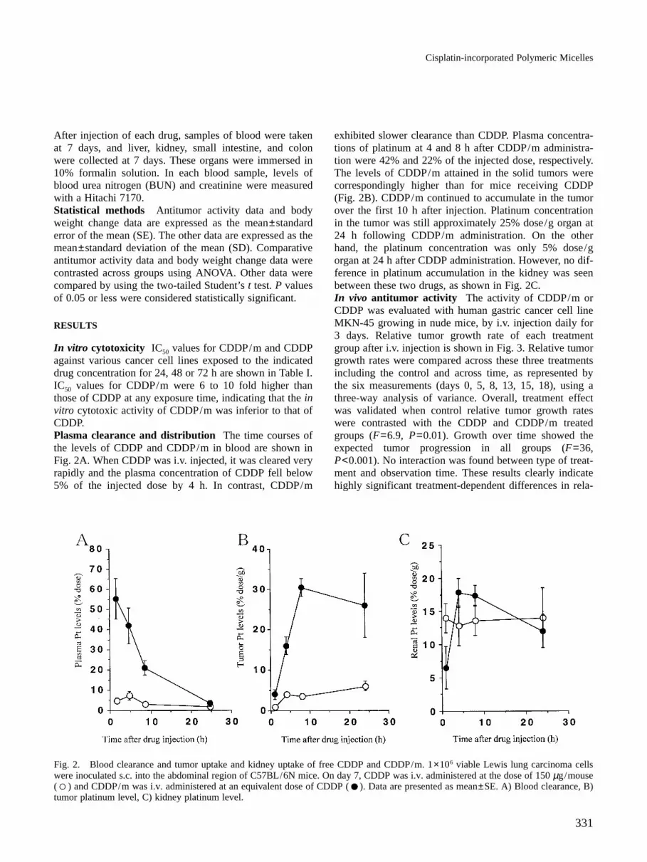
In vitro cytotoxicity IC50 values for CDDP/m and CDDPagainst various cancer cell lines exposed to the indicateddrug concentration for 24, 48 or 72 h are shown in Table I.IC50 values for CDDP/m were 6 to 10 fold higher thanthose of CDDP at any exposure time, indicating that the invitro cytotoxic activity of CDDP/m was inferior to that ofCDDP.Plasma clearance and distribution The time courses ofthe levels of CDDP and CDDP/m in blood are shown inFig. 2A. When CDDP was i.v. injected, it was cleared veryrapidly and the plasma concentration of CDDP fell below5% of the injected dose by 4 h. In contrast, CDDP/m
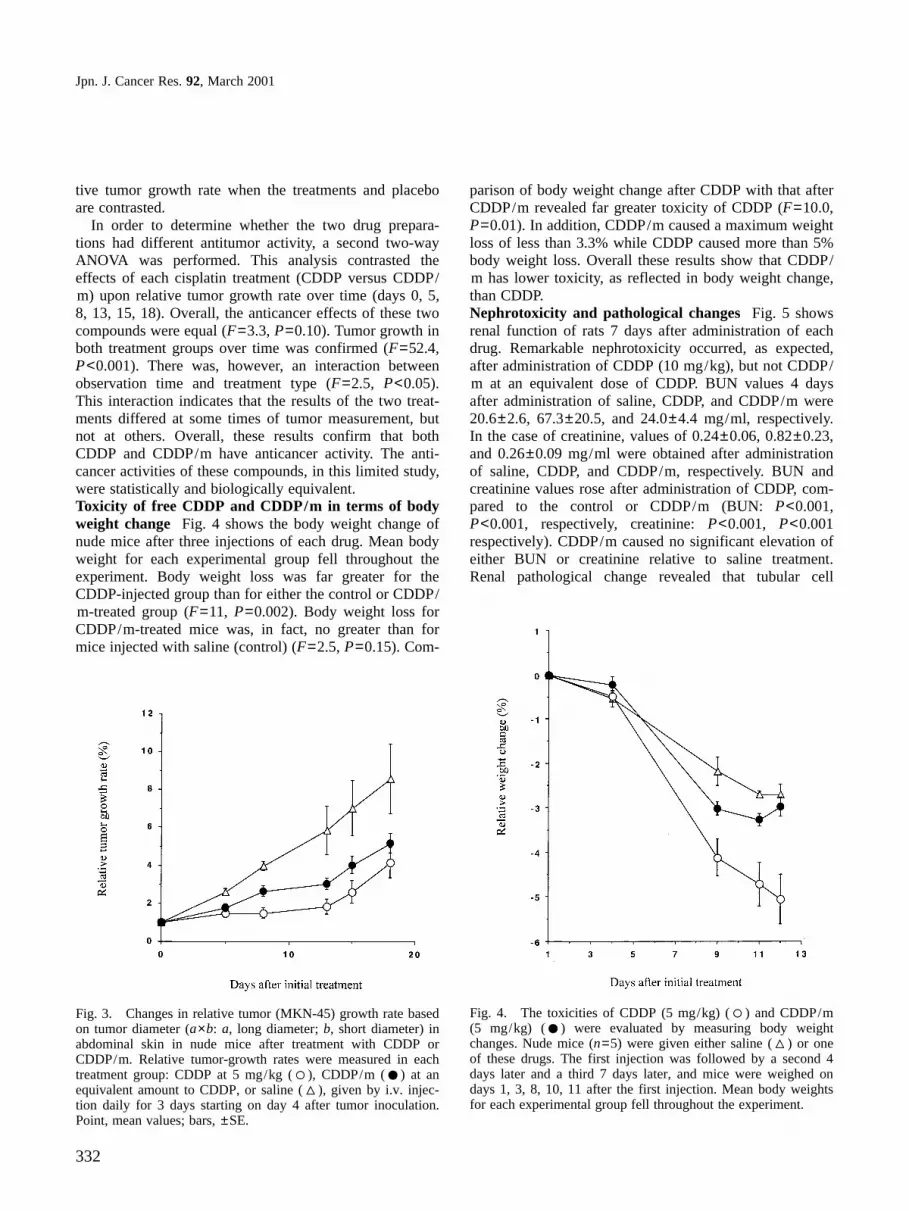
exhibited slower clearance than CDDP. Plasma concentra-tions of platinum at 4 and 8 h after CDDP/m administra-tion were 42% and 22% of the injected dose, respectively.The levels of CDDP/m attained in the solid tumors werecorrespondingly higher than for mice receiving CDDP(Fig. 2B). CDDP/m continued to accumulate in the tumorover the first 10 h after injection. Platinum concentrationin the tumor was still approximately 25% dose/g organ at24 h following CDDP/m administration. On the otherhand, the platinum concentration was only 5% dose/gorgan at 24 h after CDDP administration. However, no dif-ference in platinum accumulation in the kidney was seenbetween these two drugs, as shown in Fig. 2C.In vivo antitumor activity The activity of CDDP/m orCDDP was evaluated with human gastric cancer cell lineMKN-45 growing in nude mice, by i.v. injection daily for3 days. Relative tumor growth rate of each treatmentgroup after i.v. injection is shown in Fig. 3. Relative tumorgrowth rates were compared across these three treatmentsincluding the control and across time, as represented bythe six measurements (days 0, 5, 8, 13, 15, 18), using athree-way analysis of variance. Overall, treatment effectwas validated when control relative tumor growth rateswere contrasted with the CDDP and CDDP/m treatedgroups (F=6.9, P=0.01). Growth over time showed theexpected tumor progression in all groups (F=36,P<0.001). No interaction was found between type of treat-ment and observation time. These results clearly indicatehighly significant treatment-dependent differences in rela-
Fig. 2. Blood clearance and tumor uptake and kidney uptake of free CDDP and CDDP/m. 1×10⁶ viable Lewis lung carcinoma cellswere inoculated s.c. into the abdominal region of C57BL/6N mice. On day 7, CDDP was i.v. administered at the dose of 150 µg/mouse( ) and CDDP/m was i.v. administered at an equivalent dose of CDDP ( ). Data are presented as mean±SE. A) Blood clearance, B)tumor platinum level, C) kidney platinum level.
Jpn. J. Cancer Res. 92, March 2001
332
tive tumor growth rate when the treatments and placeboare contrasted.
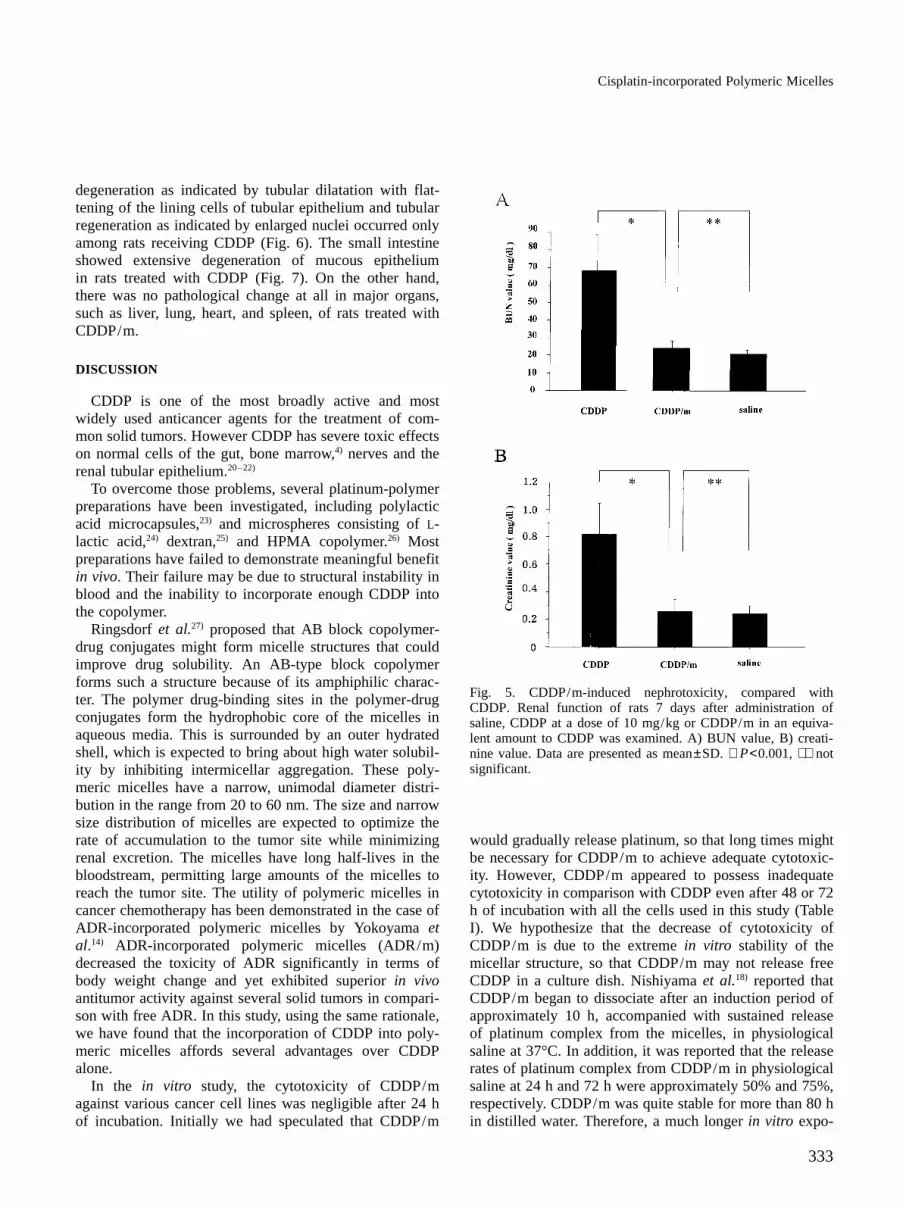
In order to determine whether the two drug prepara-tions had different antitumor activity, a second two-wayANOVA was performed. This analysis contrasted theeffects of each cisplatin treatment (CDDP versus CDDP/m) upon relative tumor growth rate over time (days 0, 5,8, 13, 15, 18). Overall, the anticancer effects of these twocompounds were equal (F=3.3, P=0.10). Tumor growth inboth treatment groups over time was confirmed (F=52.4,P<0.001). There was, however, an interaction betweenobservation time and treatment type (F=2.5, P<0.05).This interaction indicates that the results of the two treat-ments differed at some times of tumor measurement, butnot at others. Overall, these results confirm that bothCDDP and CDDP/m have anticancer activity. The anti-cancer activities of these compounds, in this limited study,were statistically and biologically equivalent.Toxicity of free CDDP and CDDP/m in terms of bodyweight change Fig. 4 shows the body weight change ofnude mice after three injections of each drug. Mean bodyweight for each experimental group fell throughout theexperiment. Body weight loss was far greater for theCDDP-injected group than for either the control or CDDP/m-treated group (F=11, P=0.002). Body weight loss forCDDP/m-treated mice was, in fact, no greater than formice injected with saline (control) (F=2.5, P=0.15). Com-
parison of body weight change after CDDP with that afterCDDP/m revealed far greater toxicity of CDDP (F=10.0,P=0.01). In addition, CDDP/m caused a maximum weightloss of less than 3.3% while CDDP caused more than 5%body weight loss. Overall these results show that CDDP/m has lower toxicity, as reflected in body weight change,than CDDP.Nephrotoxicity and pathological changes Fig. 5 showsrenal function of rats 7 days after administration of eachdrug. Remarkable nephrotoxicity occurred, as expected,after administration of CDDP (10 mg/kg), but not CDDP/m at an equivalent dose of CDDP. BUN values 4 daysafter administration of saline, CDDP, and CDDP/m were20.6±2.6, 67.3±20.5, and 24.0±4.4 mg/ml, respectively.In the case of creatinine, values of 0.24±0.06, 0.82±0.23,and 0.26±0.09 mg/ml were obtained after administrationof saline, CDDP, and CDDP/m, respectively. BUN andcreatinine values rose after administration of CDDP, com-pared to the control or CDDP/m (BUN: P<0.001,P<0.001, respectively, creatinine: P<0.001, P<0.001respectively). CDDP/m caused no significant elevation ofeither BUN or creatinine relative to saline treatment.Renal pathological change revealed that tubular cell
Fig. 3. Changes in relative tumor (MKN-45) growth rate basedon tumor diameter (a×b: a, long diameter; b, short diameter) inabdominal skin in nude mice after treatment with CDDP orCDDP/m. Relative tumor-growth rates were measured in eachtreatment group: CDDP at 5 mg/kg ( ), CDDP/m ( ) at anequivalent amount to CDDP, or saline ( ), given by i.v. injec-tion daily for 3 days starting on day 4 after tumor inoculation.Point, mean values; bars, ±SE.
Fig. 4. The toxicities of CDDP (5 mg/kg) ( ) and CDDP/m(5 mg/kg) ( ) were evaluated by measuring body weightchanges. Nude mice (n=5) were given either saline ( ) or oneof these drugs. The first injection was followed by a second 4days later and a third 7 days later, and mice were weighed ondays 1, 3, 8, 10, 11 after the first injection. Mean body weightsfor each experimental group fell throughout the experiment.
Cisplatin-incorporated Polymeric Micelles
333
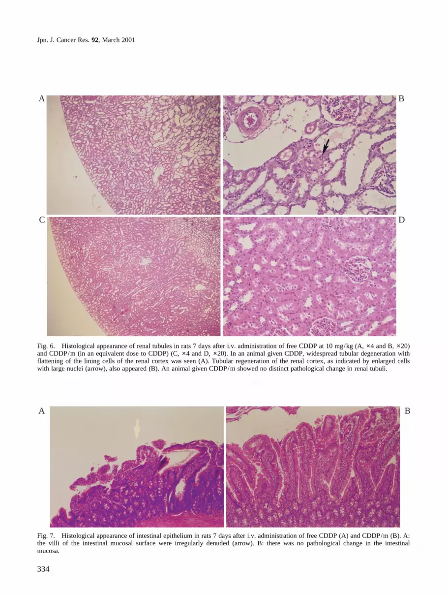
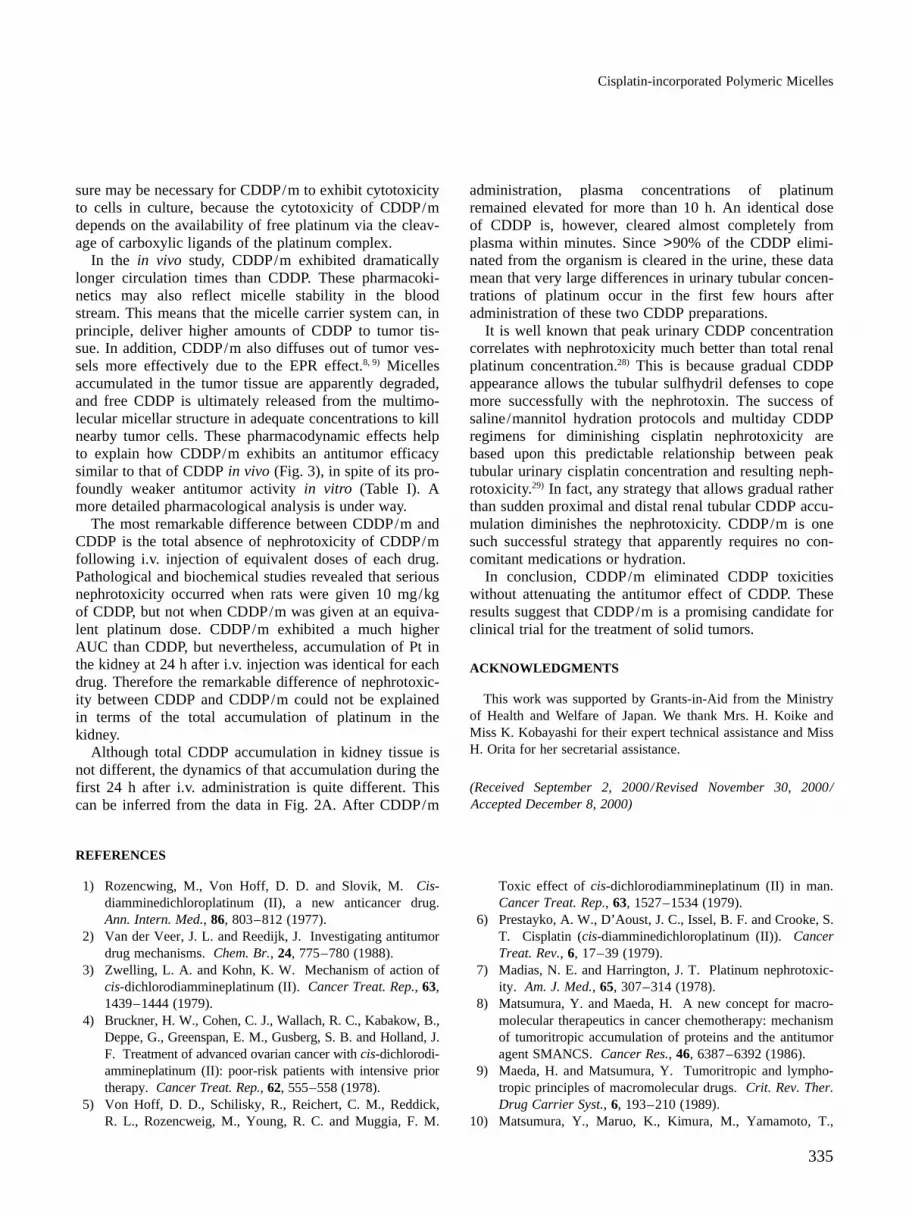
degeneration as indicated by tubular dilatation with flat-tening of the lining cells of tubular epithelium and tubularregeneration as indicated by enlarged nuclei occurred onlyamong rats receiving CDDP (Fig. 6). The small intestineshowed extensive degeneration of mucous epitheliumin rats treated with CDDP (Fig. 7). On the other hand,there was no pathological change at all in major organs,such as liver, lung, heart, and spleen, of rats treated withCDDP/m.
DISCUSSION
CDDP is one of the most broadly active and mostwidely used anticancer agents for the treatment of com-mon solid tumors. However CDDP has severe toxic effectson normal cells of the gut, bone marrow,4) nerves and therenal tubular epithelium.20–22)
To overcome those problems, several platinum-polymerpreparations have been investigated, including polylacticacid microcapsules,23) and microspheres consisting of L-lactic acid,24) dextran,25) and HPMA copolymer.26) Mostpreparations have failed to demonstrate meaningful benefitin vivo. Their failure may be due to structural instability inblood and the inability to incorporate enough CDDP intothe copolymer.
Ringsdorf et al.27) proposed that AB block copolymer-drug conjugates might form micelle structures that couldimprove drug solubility. An AB-type block copolymerforms such a structure because of its amphiphilic charac-ter. The polymer drug-binding sites in the polymer-drugconjugates form the hydrophobic core of the micelles inaqueous media. This is surrounded by an outer hydratedshell, which is expected to bring about high water solubil-ity by inhibiting intermicellar aggregation. These poly-meric micelles have a narrow, unimodal diameter distri-bution in the range from 20 to 60 nm. The size and narrowsize distribution of micelles are expected to optimize therate of accumulation to the tumor site while minimizingrenal excretion. The micelles have long half-lives in thebloodstream, permitting large amounts of the micelles toreach the tumor site. The utility of polymeric micelles incancer chemotherapy has been demonstrated in the case ofADR-incorporated polymeric micelles by Yokoyama etal.14) ADR-incorporated polymeric micelles (ADR/m)decreased the toxicity of ADR significantly in terms ofbody weight change and yet exhibited superior in vivoantitumor activity against several solid tumors in compari-son with free ADR. In this study, using the same rationale,we have found that the incorporation of CDDP into poly-meric micelles affords several advantages over CDDPalone.
In the in vitro study, the cytotoxicity of CDDP/magainst various cancer cell lines was negligible after 24 hof incubation. Initially we had speculated that CDDP/m
would gradually release platinum, so that long times mightbe necessary for CDDP/m to achieve adequate cytotoxic-ity. However, CDDP/m appeared to possess inadequatecytotoxicity in comparison with CDDP even after 48 or 72h of incubation with all the cells used in this study (TableI). We hypothesize that the decrease of cytotoxicity ofCDDP/m is due to the extreme in vitro stability of themicellar structure, so that CDDP/m may not release freeCDDP in a culture dish. Nishiyama et al.18) reported thatCDDP/m began to dissociate after an induction period ofapproximately 10 h, accompanied with sustained releaseof platinum complex from the micelles, in physiologicalsaline at 37°C. In addition, it was reported that the releaserates of platinum complex from CDDP/m in physiologicalsaline at 24 h and 72 h were approximately 50% and 75%,respectively. CDDP/m was quite stable for more than 80 hin distilled water. Therefore, a much longer in vitro expo-
Fig. 5. CDDP/m-induced nephrotoxicity, compared withCDDP. Renal function of rats 7 days after administration ofsaline, CDDP at a dose of 10 mg/kg or CDDP/m in an equiva-lent amount to CDDP was examined. A) BUN value, B) creati-nine value. Data are presented as mean±SD. ∗ P<0.001, ∗∗ notsignificant.
Jpn. J. Cancer Res. 92, March 2001
334
A
C
B
D
Fig. 6. Histological appearance of renal tubules in rats 7 days after i.v. administration of free CDDP at 10 mg/kg (A, ×4 and B, ×20)and CDDP/m (in an equivalent dose to CDDP) (C, ×4 and D, ×20). In an animal given CDDP, widespread tubular degeneration withflattening of the lining cells of the renal cortex was seen (A). Tubular regeneration of the renal cortex, as indicated by enlarged cellswith large nuclei (arrow), also appeared (B). An animal given CDDP/m showed no distinct pathological change in renal tubuli.
A B
Fig. 7. Histological appearance of intestinal epithelium in rats 7 days after i.v. administration of free CDDP (A) and CDDP/m (B). A:the villi of the intestinal mucosal surface were irregularly denuded (arrow). B: there was no pathological change in the intestinalmucosa.
Cisplatin-incorporated Polymeric Micelles
335
sure may be necessary for CDDP/m to exhibit cytotoxicityto cells in culture, because the cytotoxicity of CDDP/mdepends on the availability of free platinum via the cleav-age of carboxylic ligands of the platinum complex.
In the in vivo study, CDDP/m exhibited dramaticallylonger circulation times than CDDP. These pharmacoki-netics may also reflect micelle stability in the bloodstream. This means that the micelle carrier system can, inprinciple, deliver higher amounts of CDDP to tumor tis-sue. In addition, CDDP/m also diffuses out of tumor ves-sels more effectively due to the EPR effect.8, 9) Micellesaccumulated in the tumor tissue are apparently degraded,and free CDDP is ultimately released from the multimo-lecular micellar structure in adequate concentrations to killnearby tumor cells. These pharmacodynamic effects helpto explain how CDDP/m exhibits an antitumor efficacysimilar to that of CDDP in vivo (Fig. 3), in spite of its pro-foundly weaker antitumor activity in vitro (Table I). Amore detailed pharmacological analysis is under way.
The most remarkable difference between CDDP/m andCDDP is the total absence of nephrotoxicity of CDDP/mfollowing i.v. injection of equivalent doses of each drug.Pathological and biochemical studies revealed that seriousnephrotoxicity occurred when rats were given 10 mg/kgof CDDP, but not when CDDP/m was given at an equiva-lent platinum dose. CDDP/m exhibited a much higherAUC than CDDP, but nevertheless, accumulation of Pt inthe kidney at 24 h after i.v. injection was identical for eachdrug. Therefore the remarkable difference of nephrotoxic-ity between CDDP and CDDP/m could not be explainedin terms of the total accumulation of platinum in thekidney.
Although total CDDP accumulation in kidney tissue isnot different, the dynamics of that accumulation during thefirst 24 h after i.v. administration is quite different. Thiscan be inferred from the data in Fig. 2A. After CDDP/m
administration, plasma concentrations of platinumremained elevated for more than 10 h. An identical doseof CDDP is, however, cleared almost completely fromplasma within minutes. Since >90% of the CDDP elimi-nated from the organism is cleared in the urine, these datamean that very large differences in urinary tubular concen-trations of platinum occur in the first few hours afteradministration of these two CDDP preparations.
It is well known that peak urinary CDDP concentrationcorrelates with nephrotoxicity much better than total renalplatinum concentration.28) This is because gradual CDDPappearance allows the tubular sulfhydril defenses to copemore successfully with the nephrotoxin. The success ofsaline/mannitol hydration protocols and multiday CDDPregimens for diminishing cisplatin nephrotoxicity arebased upon this predictable relationship between peaktubular urinary cisplatin concentration and resulting neph-rotoxicity.29) In fact, any strategy that allows gradual ratherthan sudden proximal and distal renal tubular CDDP accu-mulation diminishes the nephrotoxicity. CDDP/m is onesuch successful strategy that apparently requires no con-comitant medications or hydration.
In conclusion, CDDP/m eliminated CDDP toxicitieswithout attenuating the antitumor effect of CDDP. Theseresults suggest that CDDP/m is a promising candidate forclinical trial for the treatment of solid tumors.
ACKNOWLEDGMENTS
This work was supported by Grants-in-Aid from the Ministryof Health and Welfare of Japan. We thank Mrs. H. Koike andMiss K. Kobayashi for their expert technical assistance and MissH. Orita for her secretarial assistance.
(Received September 2, 2000/Revised November 30, 2000/Accepted December 8, 2000)
REFERENCES
1) Rozencwing, M., Von Hoff, D. D. and Slovik, M. Cis-diamminedichloroplatinum (II), a new anticancer drug.Ann. Intern. Med., 86, 803–812 (1977).
2) Van der Veer, J. L. and Reedijk, J. Investigating antitumordrug mechanisms. Chem. Br., 24, 775–780 (1988).
3) Zwelling, L. A. and Kohn, K. W. Mechanism of action ofcis-dichlorodiammineplatinum (II). Cancer Treat. Rep., 63,1439–1444 (1979).
4) Bruckner, H. W., Cohen, C. J., Wallach, R. C., Kabakow, B.,Deppe, G., Greenspan, E. M., Gusberg, S. B. and Holland, J.F. Treatment of advanced ovarian cancer with cis-dichlorodi-ammineplatinum (II): poor-risk patients with intensive priortherapy. Cancer Treat. Rep., 62, 555–558 (1978).
5) Von Hoff, D. D., Schilisky, R., Reichert, C. M., Reddick,R. L., Rozencweig, M., Young, R. C. and Muggia, F. M.
Toxic effect of cis-dichlorodiammineplatinum (II) in man.Cancer Treat. Rep., 63, 1527–1534 (1979).
6) Prestayko, A. W., D’Aoust, J. C., Issel, B. F. and Crooke, S.T. Cisplatin (cis-diamminedichloroplatinum (II)). CancerTreat. Rev., 6, 17–39 (1979).
7) Madias, N. E. and Harrington, J. T. Platinum nephrotoxic-ity. Am. J. Med., 65, 307–314 (1978).
8) Matsumura, Y. and Maeda, H. A new concept for macro-molecular therapeutics in cancer chemotherapy: mechanismof tumoritropic accumulation of proteins and the antitumoragent SMANCS. Cancer Res., 46, 6387–6392 (1986).
9) Maeda, H. and Matsumura, Y. Tumoritropic and lympho-tropic principles of macromolecular drugs. Crit. Rev. Ther.Drug Carrier Syst., 6, 193–210 (1989).
10) Matsumura, Y., Maruo, K., Kimura, M., Yamamoto, T.,
Jpn. J. Cancer Res. 92, March 2001
336
Konno, T. and Maeda, H. Kinin-generating cascade inadvanced cancer patients and in vitro study. Jpn. J. CancerRes., 82, 732–741 (1991).
11) Folkman, J. Angiogenesis in cancer, vascular, rheumatoidand other diseases. Nat. Med., 1, 27–31 (1995).
12) Dvorak, H. F., Nagy, J. A., Dvorak, J. T. and Dvorak, A.M. Identification and characterization of the blood vesselsof solid tumors that are leaky to circulating macromole-cules. Am. J. Pathol., 133, 95–109 (1988).
13) Yokoyama, M., Miyauchi, M., Yamada, N., Okano, T.,Sakurai, Y., Kataoka, K. and Inoue, S. Characterizationand anticancer activity of micelle-forming polymeric antitu-mor drug adriamicin-conjugated poly(ethylene glycol)-poly(aspartic acid) block copolymer. Cancer Res., 50,1693–1700 (1990).
14) Yokoyama, M., Okano, T., Sakurai, Y., Ekimoto, H.,Shibazaki, C. and Kataoka, K. Toxicity and antitumoractivity against solid tumors of micelle-forming polymericantitumor drug and its extremely long circulation in blood.Cancer Res., 51, 3229–3236 (1991).
15) Yokoyama, M., Inoue, S., Kataoka, K., Yui, N., Okano, T.and Sakurai, Y. Molecular design for missile drug: synthe-sis of adriamycin conjugated with immunoglobulin G usingpoly(ethylene glycol)-block-poly(aspartic acid) as interme-diate carrier. Makromol. Chem., 190, 2041–2054 (1989).
16) Yokoyama, M., Miyauchi, M., Yamada, N., Okano, T.,Sakurai, Y., Kataoka, K. and Inoue, S. Polymeric micellesas novel drug carrier: adriamycin-conjugated poly(ethyleneglycol)-poly(aspartic acid) block copolymer. J. ControlledRelease, 11, 269–278 (1990).
17) Yokoyama, M., Okano, T., Sakurai, Y., Suwa, S. andKataoka, K. Introduction of cisplatin into micelle. J. Con-trolled Release, 39, 351–356 (1996).
18) Nishiyama, N., Yokoyama, M., Aoyagi, T., Okano, T.,Sakurai, Y. and Kataoka, K. Preparation and characteriza-tion of self-assembled polymer-metal complex micelle fromcis-dichlorodiammineplatinum (II) and poly(ethylene gly-col)-poly(α,β-aspartic acid) block copolymer in an aqueousmedium. Langmuir, 15, 377–383 (1999).
19) Newman, M. S., Colbern, G. T., Working, P. K., Engbers,
C. and Amantea, M. A. Comparative pharmacokinetics, tis-sue distribution, and therapeutic effectiveness of cisplatinencapsulated in long circulating, pegylated liposomes (SPI-077) in tumor-bearing mice. Cancer Chemother. Pharma-col., 43, 1–7 (1999).
20) Singh, G. A possible cellular mechanism of cisplatin-induced nephrotoxicity. Toxicology, 58, 71–80 (1989).
21) Fillastre, J. P. and Raguenez-Viotte, G. Cisplatin nephro-toxicity. Toxicol. Lett., 46, 163–175 (1989).
22) Ban, M., Hettich, D. and Huguet, N. Nephrotoxicity mech-anism of cis-platinum (II) diamine dichloride in mice. Tox-icol. Lett., 71, 161–168 (1994).
23) Araki, H., Tani, T. and Kodama, M. Antitumor effect ofcisplatin incorporated into polylactic acid microcapsules.Artif. Organs, 23, 161–168 (1999).
24) Kumagai, S., Sugiyama, T., Nishida, T., Ushijima, K. andYakushiji, M. Improvement of intraperitoneal chemother-apy for rat ovarian cancer using cisplatin-containing micro-spheres. Jpn. J. Cancer Res., 87, 412–417 (1996).
25) Ohya, Y., Masunaga, T., Baba, T. and Ouchi, T. Synthesisand cytotoxic activity of dextran-immobilizing platinum (II)complex through chelate-type coordination bond. JMSPure Appl. Chem., A33, 1005–1016 (1996).
26) Gianasi, E., Wasil, M., Evagorou, E. G., Keddle, A.,Wilson, G. and Duncan, R. HPMA copolymer platinates asnovel antitumour agents: in vitro properties, pharmacokinet-ics and antitumour activity in vivo. Eur. J. Cancer, 35,994–1002 (1999).
27) Bader, H., Ringsdorf, H. and Schmidt, B. Water-solublepolymers in medicine. Angew. Makromol. Chem., 123/124,457–485 (1984).
28) Levi, F. A., Hrushesky, W. J. M., Borch, R. F., Pleasants,M. E., Kennedy, B. J. and Halberg, F. Cisplatin urinarypharmacokinetics and nephrotoxicity: a common circadianmechanism. Cancer Treat. Rep., 66, 1933–1938 (1982).
29) Levi, F. A., Hrushesky, W. J. M., Halberg, F., Langerin, T.R., Haus, E. and Kennedy, B. J. Lethal nephrotoxicity andhematologic toxicity of cis-diamminedichloroplatinum ame-liorated by optimal circadian timing and hydration. Eur. J.Cancer Oncol., 18, 471–477 (1982).


