Embed Size (px)
Citation preview
Regulation of sialic acid transport and catabolism inHaemophilus influenzae
Jason W. Johnston,1 Anthony Zaleski,1
Simon Allen,2 Joe M. Mootz,1 David Armbruster,3,†
Bradford W. Gibson,2 Michael A. Apicella1* andRobert S. Munson Jr3,4
1Department of Microbiology, University of Iowa, IowaCity, IA 52242, USA.2Buck Institute for Age Research, 8001 Redwood Blvd.,Novato, CA 94945, USA.3Center for Microbial Pathogenesis, ColumbusChildren’s Research Institute and 4The Ohio StateUniversity, Columbus, OH 43205, USA.
Summary
Virulence of nontypeable Haemophilus influenzae(NTHi) is dependent on the decoration of lipooligosac-charide with sialic acid. This sugar must be derivedfrom the host, as NTHi cannot synthesize sialic acids.NTHi can also use sialic acid as a carbon source. Thegenes encoding the sialic acid transporter and thegenes encoding the catabolic activities are localized totwo divergently transcribed operons, the siaPT operonand the nan operon respectively. In this study, weidentified SiaR as a repressor of sialic acid transportand catabolism in NTHi. Inactivation of siaR resulted inthe unregulated expression of the genes in bothoperons. Unregulated catabolism of sialic acid in thesiaR mutant resulted in the reduction of surface sialy-lation and an increase in serum sensitivity. In additionto SiaR-mediated repression, CRP, the cAMP receptorprotein, was shown to activate expression of the siaPToperon but not the nan operon. We describe a model inwhich SiaR and CRP work to modulate intracellularsialic acid levels. Our results demonstrate the impor-tance of SiaR-mediated regulation to balance therequirement of surface sialylation and the toxic accu-mulation of intracellular sialic acid.
Introduction
Haemophilus influenzae is a common inhabitant of theupper respiratory tract of humans and is a frequent cause
of upper and lower respiratory tract infections. Encapsu-lated H. influenzae strains have the ability to cause inva-sive diseases including meningitis while unencapsulatednontypeable H. influenzae (NTHi) generally causemucosal infections including otitis media in children andbronchitis in patients with chronic obstructive pulmonarydisease. The lipooligosaccharide (LOS) is known to be amajor virulence factor of both encapsulated and non-typeable strains.
Sialic acid (5-N-acetylneuraminic acid, Neu5Ac) is incor-porated into the LOS as a terminal non-reducing sugar.Sialylation of the LOS is believed to contribute to NTHisurvival in the host by mimicking human glycosphingolipids(Mandrell et al., 1992) and protecting NTHi against killingmediated by the alternative complement pathway (Hoodet al., 1999; 2001; Figueira et al., 2007) although themechanisms of protection in NTHi are unknown. The addi-tion of sialic acid to the LOS by the sialyltransferaseencoded by lic3A is phase-variable (Weiser et al., 1990).Organisms isolated from infected chinchillas are predomi-nately lic3A-on phase variants and have sialic acid presenton their LOS, suggesting that sialylation provides a selec-tive advantage to NTHi in vivo (Bouchet et al., 2003).Inactivation of siaB, the gene encoding the CMP-Neu5Acsynthetase, results in the inability of the mutant to producesialic acid-containing glycoforms. These organisms areserum sensitive and completely avirulent in the chinchillamodel for otitis media (Bouchet et al., 2003). Sialic acid-containing glycoconjugates are also known to be a majorcomponent of NTHi biofilms and are required for biofilmformation in the chinchilla middle ear (Greiner et al., 2004;Jurcisek et al., 2005). NTHi is unable to synthesize sialicacids; therefore, decoration of NTHi glycoconjugates withsialic acid is dependent on exogenous sialic acids. Inacti-vation of the sialic acid transporter in NTHi prevents incor-poration of sialic acid into the NTHi glycoconjugates andalso leads to reduced viability of biofilms grown in vitro(Allen et al., 2005).
Sialic acid can be used as a carbon source byH. influenzae (Fig. 1A) (Vimr et al., 2000; 2004). InEscherichia coli, the genes encoding for sialic acidcatabolism are found in two separate operons: one thatencodes for NanT (the sialic acid transporter), NanA,NanE and NanK while the genes encoding for NagA andNagB are found elsewhere on the chromosome (Plum-bridge and Vimr, 1999; Ringenberg et al., 2003; Vimr
Accepted 19 July, 2007. *For correspondence. [email protected]; Tel. (+1) 319 335 7807; Fax (+1) 319335 9006. †Present address: Department of Biochemistry andMolecular Biology, Medical School of South Carolina, Charleston, SC29425, USA.
Molecular Microbiology (2007) 66(1), 26–39 doi:10.1111/j.1365-2958.2007.05890.x
© 2007 The AuthorsJournal compilation © 2007 Blackwell Publishing Ltd

et al., 2004). In NTHi, sialic acid is transported into the cellby a tripartite ATP-independent periplasmic (TRAP) trans-porter that is encoded by siaPT (Allen et al., 2005; Severiet al., 2005). In contrast to the gene arrangement inE. coli, the enzymes required for the complete conversionof sialic acid to fructose-6-phosphate are encoded by thegenes of a single operon (Fig. 1B). The siaPT and the nanoperons of NTHi are adjacent to each other and diver-gently transcribed (Vimr et al., 2000; 2004). The nanoperon also contains a gene that encodes for a putativetranscriptional regulator, HI0143 (Vimr et al., 2000). It haspreviously been speculated that the protein encoded byHI0143 may function to regulate the expression of thesialic acid metabolism and transport operons (Vimr et al.,2004) although no work has been published to supportthis hypothesis.
We examined the role of HI0143 in regulation of theNeu5Ac catabolic pathway. This transcriptional regulator,which we have designated SiaR, was found to repress theoperons that encode the sialic acid transporter and thegenes involved in sialic acid catabolism. Expression ofthe transporter, but not the catabolic pathway, wasinduced by cAMP via CRP in the absence of SiaR. Inac-tivation of siaR increased sensitivity to human serum. Thisdemonstrates the necessity of stringent control of nanoperon expression for protection of the organism fromcomplement-mediated killing.
Results
Construction of a mutant deficient in the putativetranscriptional regulator SiaR and identification of genesregulated by SiaR
HI0143 was identified as a potential transcriptional regu-lator due to its homology to RpiR family transcriptional
regulators. Members of the RpiR family are characterizedby the presence of both a helix–turn–helix DNA-bindingdomain and a sugar isomerase (SIS) domain (Bateman,1999). The SIS domain is found in a wide variety ofproteins and functions to bind a phosphosugar molecule(Bateman, 1999). In E. coli, RpiR represses rpiB whichencodes for a ribose phosphate isomerase (Sorensenand Hove-Jensen, 1996). Other members of this familyhave not been characterized. HI0143 differs significantlyfrom NanR, the sialic acid-dependent transcriptional regu-lator found in E. coli, a member of the FadR/GntR familyof transcriptional regulators (Kalivoda et al., 2003). Basedon the protein sequence HI0143 and NanR are nothomologous (15% similarity). As HI0143 is located in theoperon that encodes for the enzymes involved in Neu5Accatabolism (Fig. 1B), it seems likely that HI0143 regulatessialic acid catabolism.
To determine the role of HI0143, which we have namedsiaR, in the regulation of sialic acid catabolism we con-structed a siaR mutant. The mutant was constructed byreplacing the siaR gene with a promoterless kanamycinresistance cassette. Microarray analysis was used toinvestigate the role of SiaR in the regulation of geneexpression. Changes in gene expression were examinedusing competitive hybridizations between labelled cDNAprepared from RNA samples isolated from early to mid-logarithmic phase cultures of wild-type 2019 and 2019siaRgrown in sRPMI-based medium containing 100 mMNeu5Ac. Microarray comparisons were performed withthree independent pairs of RNA preparations. Each pair ofRNA preparations was hybridized to two slides. Theexpression profile for the 2019siaR mutant revealed thatthe expression of three putative operons was increased inthe absence of SiaR: HI0145–HI0140, HI0146–HI0149and HI1537–HI1540 (Table 1). Using reverse transcription
Fig. 1. A. Sialic acid transport andmetabolism pathway of H. influenzae. Sialicacid is transported by the SiaPT transporter.The genes of the nan operon encode forthe enzymes that convert Neu5Ac intofructose-6-phosphate, which can then enterglycolysis.B. Organization of sialic acid transport andmetabolism genes in H. influenzae. The genenumber corresponds to the HI number basedon the H. influenzae KW-20 Rd genomeannotation.
NANA NANASiaPT NanA
ManNAc + pyr
NanK
ManNAc-6-P
NanE
GlcNAc-6-P
NagA
GlcN-6-P + acetate
NagB
Fru-6-PExterior Interior
nagA nagB nanA siaR nanK nanE siaP siaT0140 0141 0142 0143 0144 0145 0146 0147 0148
SiaR regulation of sialic acid transport and catabolism 27
© 2007 The AuthorsJournal compilation © 2007 Blackwell Publishing Ltd, Molecular Microbiology, 66, 26–39

polymerase chain reaction (RT-PCR) it was determinedthat HI0145–HI0140 and HI0146–HI0149 form two sepa-rate operons (data not shown). These operons encode forthe proteins involved in Neu5Ac catabolism and transport.The HI1537–HI1540 gene cluster encodes for enzymesinvolved in the transport and addition of phosphorylcholine(ChoP) to the LOS. Expression of the genes of the threeoperons ranged from a 2.3-fold increase for HI0149 to a15.8-fold increase for HI0146 (Table 1). QuantitativeRT-PCR (qRT-PCR) performed on the same three pairs ofRNA samples used for the microarray studies was used toconfirm the results of the microarray analysis using primerand probe sets for HI0145 (nanE), HI0146 (siaP) andHI1537 (licA), which encode for the first gene of eachputative operon. The results of qRT-PCR analysis were ingood concordance with the microarray data (Table 1).Further analysis demonstrated that the apparent regula-tion of the lic1 gene cluster was due to phase variationrather than SiaR-mediated regulation as detailed in theSupplementary material.
Complementation of the siaR mutation by the introduc-tion of a single copy of siaR ectopically into the genomewas able to partially restore the repression of nanE andsiaP as observed by qRT-PCR. Analyses were performedfrom cultures grown in the same conditions used in themicroarray analysis. The complemented siaR mutant hada 5.2 � 0.2-fold and 3.9 � 0.5-fold reduction in theexpression of nanE and siaP when compared with the2019siaR mutant strain. These data indicate that SiaR isresponsible for the regulation of the nanE- and siaP-containing operons.
SiaR binds to the nanE-siaP intergenic region
The transcriptional analysis indicated that both the nanand siaPT operons were upregulated in the siaR mutant.As the operons are divergently transcribed from a
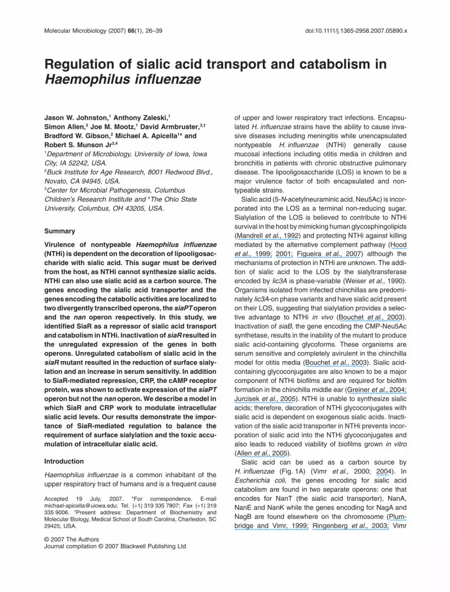
common intergenic region, it appears that SiaR regulatesthe expression of both operons from a single intergenicsite. An electrophoretic mobility shift assay (EMSA) wasused to demonstrate the interaction of SiaR with thisintergenic region. SiaR was expressed with a C-terminalHis-tag fusion using the pET-24 protein expressionplasmid. Probes for the entire intergenic region betweennanE and siaP, the region upstream of licA and the P2porin promoter were generated by PCR. The P2 poringene is believed to be constitutively expressed in NTHiand thus this promoter region was used as a negativecontrol (Munson and Tolan, 1989). The binding of purifiedSiaR to these DNA probes was examined using EMSA(Fig. 2A). SiaR was found to bind only to the nanE-siaPpromoter region and not to either the licA or P2 promoters,demonstrating that the interaction of SiaR with DNA issite-specific. The inability of SiaR to bind to the licA pro-moter further supports our finding that SiaR does notregulate the expression of the lic1 operon. A preliminaryanalysis using additional DNA fragments that span thenan and siaPT operons did not reveal the presence of anyadditional SiaR binding sites.
It has previously been demonstrated that the NanRregulator of E. coli is displaced from its operator by theaddition of Neu5Ac (Kalivoda et al., 2003). The ability ofSiaR to bind to the nanE-siaP intergenic region wasexamined in the presence of a 1000-fold excess (1 mM)of Neu5Ac, N-acetylmannosamine, N-acetylglucosamineand N-acetylglucosamine-6-phosphate (Fig. 2B). In thepresence of the sugars tested, SiaR was still able to bindto the DNA probe. This suggests that either these sugarsare unable to induce expression of SiaR-regulated genesor derepression occurs by an alternative mechanism.
DNase I footprinting was used to further map the locationof the SiaR binding site in the nanE-siaP intergenic region.SiaR was found to protect a 53 bp region located 113 bpfrom the start codon of nanE and 185 bp from the start
Table 1. Transcriptional analysis of 2019siaR mutant.
Gene ID Gene Function
Expression ratio (2019siaR/2019)
Microarray analysis Real-time RT-PCR
HI0140 nagA N-acetylglucosamine-6-phosphate deacetylase 4.9 NDHI0141 nagB Glucosamine-6-phosphate isomerase 11.9 NDHI0142 nanA N-acetylneuraminate lyase 14.3 NDHI0144 nanK N-acetylmannosamine kinase 7.2 NDHI0145 nanE N-acetylmannosamine-6-phosphate 2-epimerase 10.4 20.9 � 2.7HI0146 siaP Sialic acid transporter, periplasmic solute receptor 15.8 14.3 � 1.7HI0147 siaT Sialic acid transporter, integral membrane component 12.3 NDHI0148 Conserved hypothetical protein 6.8 NDHI0148.1 Hypothetical protein 7.9 NDHI0149 Hypothetical protein 2.3 NDHI1537 licA Choline kinase 11.2 14.7 � 3.6HI1538 licB Choline transporter 3.8 NDHI1539 licC CTP-phosphorylcholine cytidylyltransferase 6.5 NDHI1540 licD NDP-choline transferase 4.4 ND
28 J. W. Johnston et al.
© 2007 The AuthorsJournal compilation © 2007 Blackwell Publishing Ltd, Molecular Microbiology, 66, 26–39
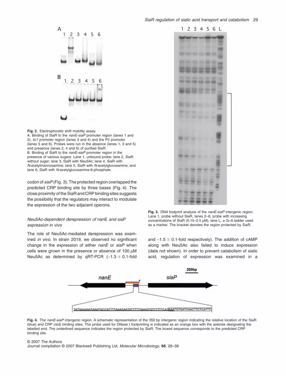
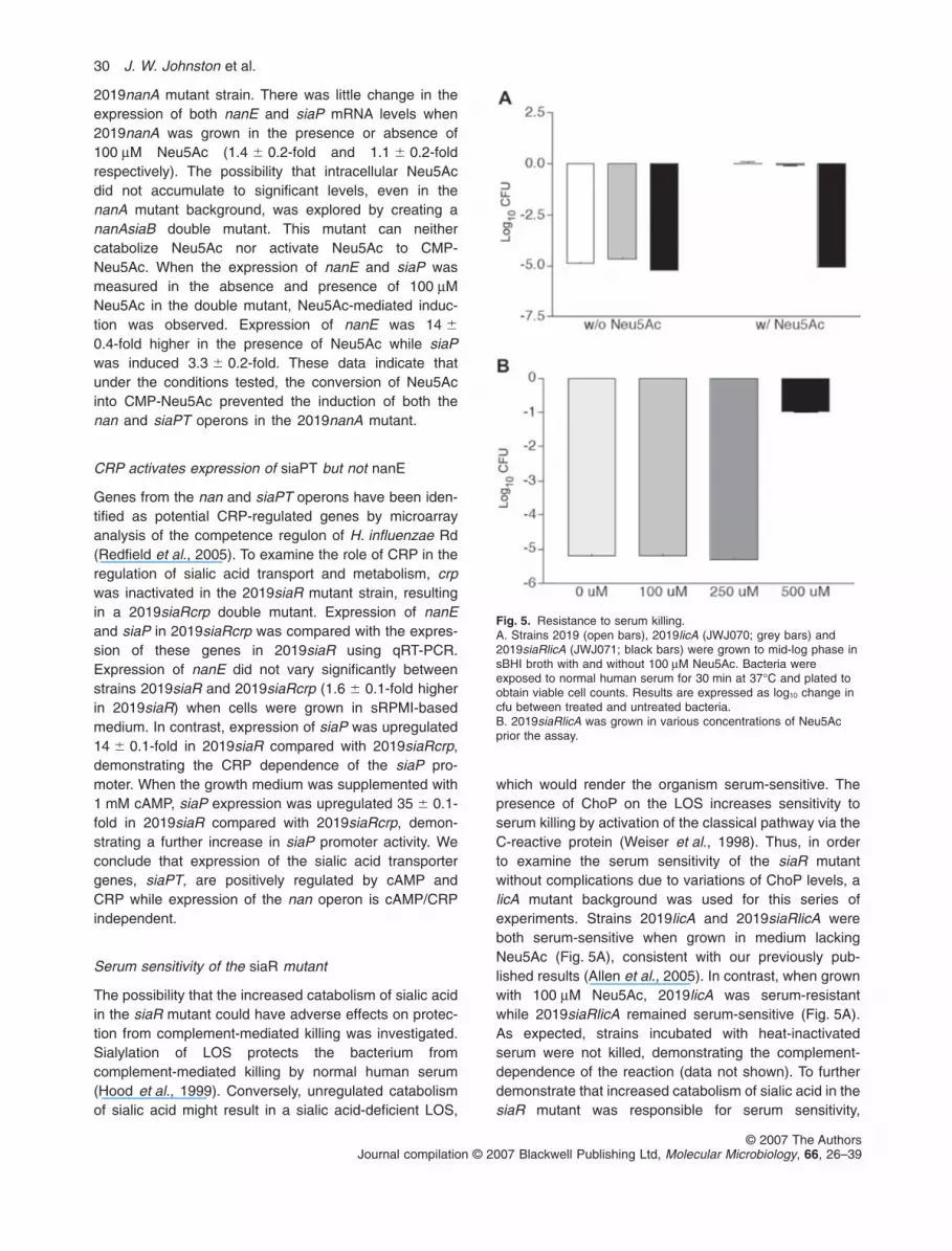
codon of siaP (Fig. 3). The protected region overlapped thepredicted CRP binding site by three bases (Fig. 4). Theclose proximity of the SiaR and CRP binding sites suggeststhe possibility that the regulators may interact to modulatethe expression of the two adjacent operons.
Neu5Ac-dependent derepression of nanE and siaPexpression in vivo
The role of Neu5Ac-mediated derepression was exam-ined in vivo. In strain 2019, we observed no significantchange in the expression of either nanE or siaP whencells were grown in the presence or absence of 100 mMNeu5Ac as determined by qRT-PCR (-1.3 � 0.1-fold
and -1.5 � 0.1-fold respectively). The addition of cAMPalong with Neu5Ac also failed to induce expression(data not shown). In order to prevent catabolism of sialicacid, regulation of expression was examined in a
Fig. 2. Electrophoretic shift mobility assay.A. Binding of SiaR to the nanE-siaP promoter region (lanes 1 and2), lic1 promoter region (lanes 3 and 4) and the P2 promoter(lanes 5 and 6). Probes were run in the absence (lanes 1, 3 and 5)and presence (lanes 2, 4 and 6) of purified SiaR.B. Binding of SiaR to the nanE-siaP promoter region in thepresence of various sugars. Lane 1, unbound probe; lane 2, SiaRwithout sugar; lane 3, SiaR with Neu5Ac; lane 4, SiaR withN-acetylmannosamine; lane 5, SiaR with N-acetylglucosamine; andlane 6, SiaR with N-acetylglucosamine-6-phosphate.
Fig. 4. The nanE-siaP intergenic region. A schematic representation of the 350 bp intergenic region indicating the relative location of the SiaR(blue) and CRP (red) binding sites. The probe used for DNase I footprinting is indicated as an orange box with the asterisk designating thelabelled end. The underlined sequence indicates the region protected by SiaR. The boxed sequence corresponds to the predicted CRPbinding site.
Fig. 3. DNA footprint analysis of the nanE-siaP intergenic region.Lane 1, probe without SiaR, lanes 2–6, probe with increasingconcentrations of SiaR (0.15–2.5 mM), lane L, a G+A ladder usedas a marker. The bracket denotes the region protected by SiaR.
SiaR regulation of sialic acid transport and catabolism 29
© 2007 The AuthorsJournal compilation © 2007 Blackwell Publishing Ltd, Molecular Microbiology, 66, 26–39
2019nanA mutant strain. There was little change in theexpression of both nanE and siaP mRNA levels when2019nanA was grown in the presence or absence of100 mM Neu5Ac (1.4 � 0.2-fold and 1.1 � 0.2-foldrespectively). The possibility that intracellular Neu5Acdid not accumulate to significant levels, even in thenanA mutant background, was explored by creating ananAsiaB double mutant. This mutant can neithercatabolize Neu5Ac nor activate Neu5Ac to CMP-Neu5Ac. When the expression of nanE and siaP wasmeasured in the absence and presence of 100 mMNeu5Ac in the double mutant, Neu5Ac-mediated induc-tion was observed. Expression of nanE was 14 �
0.4-fold higher in the presence of Neu5Ac while siaPwas induced 3.3 � 0.2-fold. These data indicate thatunder the conditions tested, the conversion of Neu5Acinto CMP-Neu5Ac prevented the induction of both thenan and siaPT operons in the 2019nanA mutant.
CRP activates expression of siaPT but not nanE
Genes from the nan and siaPT operons have been iden-tified as potential CRP-regulated genes by microarrayanalysis of the competence regulon of H. influenzae Rd(Redfield et al., 2005). To examine the role of CRP in theregulation of sialic acid transport and metabolism, crpwas inactivated in the 2019siaR mutant strain, resultingin a 2019siaRcrp double mutant. Expression of nanEand siaP in 2019siaRcrp was compared with the expres-sion of these genes in 2019siaR using qRT-PCR.Expression of nanE did not vary significantly betweenstrains 2019siaR and 2019siaRcrp (1.6 � 0.1-fold higherin 2019siaR) when cells were grown in sRPMI-basedmedium. In contrast, expression of siaP was upregulated14 � 0.1-fold in 2019siaR compared with 2019siaRcrp,demonstrating the CRP dependence of the siaP pro-moter. When the growth medium was supplemented with1 mM cAMP, siaP expression was upregulated 35 � 0.1-fold in 2019siaR compared with 2019siaRcrp, demon-strating a further increase in siaP promoter activity. Weconclude that expression of the sialic acid transportergenes, siaPT, are positively regulated by cAMP andCRP while expression of the nan operon is cAMP/CRPindependent.
Serum sensitivity of the siaR mutant
The possibility that the increased catabolism of sialic acidin the siaR mutant could have adverse effects on protec-tion from complement-mediated killing was investigated.Sialylation of LOS protects the bacterium fromcomplement-mediated killing by normal human serum(Hood et al., 1999). Conversely, unregulated catabolismof sialic acid might result in a sialic acid-deficient LOS,
which would render the organism serum-sensitive. Thepresence of ChoP on the LOS increases sensitivity toserum killing by activation of the classical pathway via theC-reactive protein (Weiser et al., 1998). Thus, in orderto examine the serum sensitivity of the siaR mutantwithout complications due to variations of ChoP levels, alicA mutant background was used for this series ofexperiments. Strains 2019licA and 2019siaRlicA wereboth serum-sensitive when grown in medium lackingNeu5Ac (Fig. 5A), consistent with our previously pub-lished results (Allen et al., 2005). In contrast, when grownwith 100 mM Neu5Ac, 2019licA was serum-resistantwhile 2019siaRlicA remained serum-sensitive (Fig. 5A).As expected, strains incubated with heat-inactivatedserum were not killed, demonstrating the complement-dependence of the reaction (data not shown). To furtherdemonstrate that increased catabolism of sialic acid in thesiaR mutant was responsible for serum sensitivity,
Fig. 5. Resistance to serum killing.A. Strains 2019 (open bars), 2019licA (JWJ070; grey bars) and2019siaRlicA (JWJ071; black bars) were grown to mid-log phase insBHI broth with and without 100 mM Neu5Ac. Bacteria wereexposed to normal human serum for 30 min at 37°C and plated toobtain viable cell counts. Results are expressed as log10 change incfu between treated and untreated bacteria.B. 2019siaRlicA was grown in various concentrations of Neu5Acprior the assay.
30 J. W. Johnston et al.
© 2007 The AuthorsJournal compilation © 2007 Blackwell Publishing Ltd, Molecular Microbiology, 66, 26–39
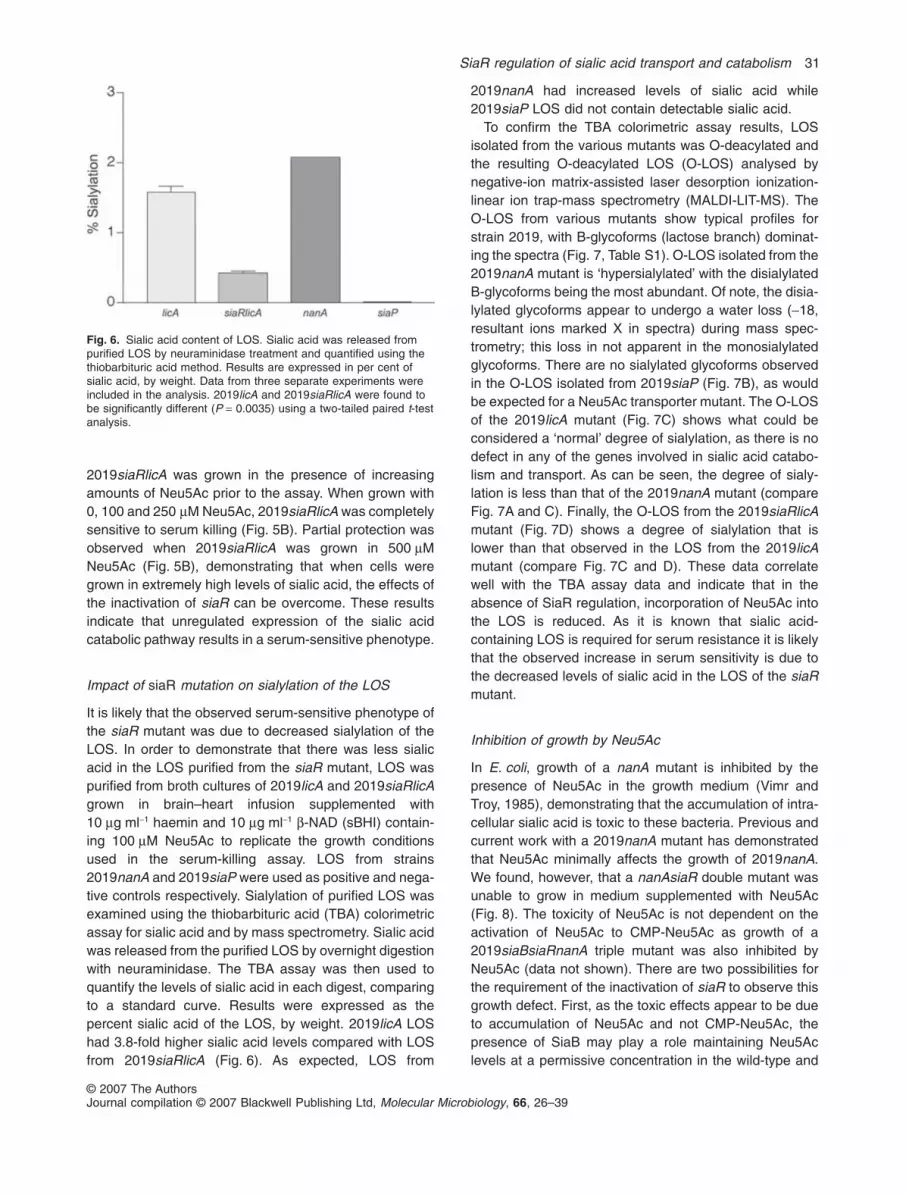
2019siaRlicA was grown in the presence of increasingamounts of Neu5Ac prior to the assay. When grown with0, 100 and 250 mM Neu5Ac, 2019siaRlicA was completelysensitive to serum killing (Fig. 5B). Partial protection wasobserved when 2019siaRlicA was grown in 500 mMNeu5Ac (Fig. 5B), demonstrating that when cells weregrown in extremely high levels of sialic acid, the effects ofthe inactivation of siaR can be overcome. These resultsindicate that unregulated expression of the sialic acidcatabolic pathway results in a serum-sensitive phenotype.
Impact of siaR mutation on sialylation of the LOS
It is likely that the observed serum-sensitive phenotype ofthe siaR mutant was due to decreased sialylation of theLOS. In order to demonstrate that there was less sialicacid in the LOS purified from the siaR mutant, LOS waspurified from broth cultures of 2019licA and 2019siaRlicAgrown in brain–heart infusion supplemented with10 mg ml-1 haemin and 10 mg ml-1 b-NAD (sBHI) contain-ing 100 mM Neu5Ac to replicate the growth conditionsused in the serum-killing assay. LOS from strains2019nanA and 2019siaP were used as positive and nega-tive controls respectively. Sialylation of purified LOS wasexamined using the thiobarbituric acid (TBA) colorimetricassay for sialic acid and by mass spectrometry. Sialic acidwas released from the purified LOS by overnight digestionwith neuraminidase. The TBA assay was then used toquantify the levels of sialic acid in each digest, comparingto a standard curve. Results were expressed as thepercent sialic acid of the LOS, by weight. 2019licA LOShad 3.8-fold higher sialic acid levels compared with LOSfrom 2019siaRlicA (Fig. 6). As expected, LOS from
2019nanA had increased levels of sialic acid while2019siaP LOS did not contain detectable sialic acid.
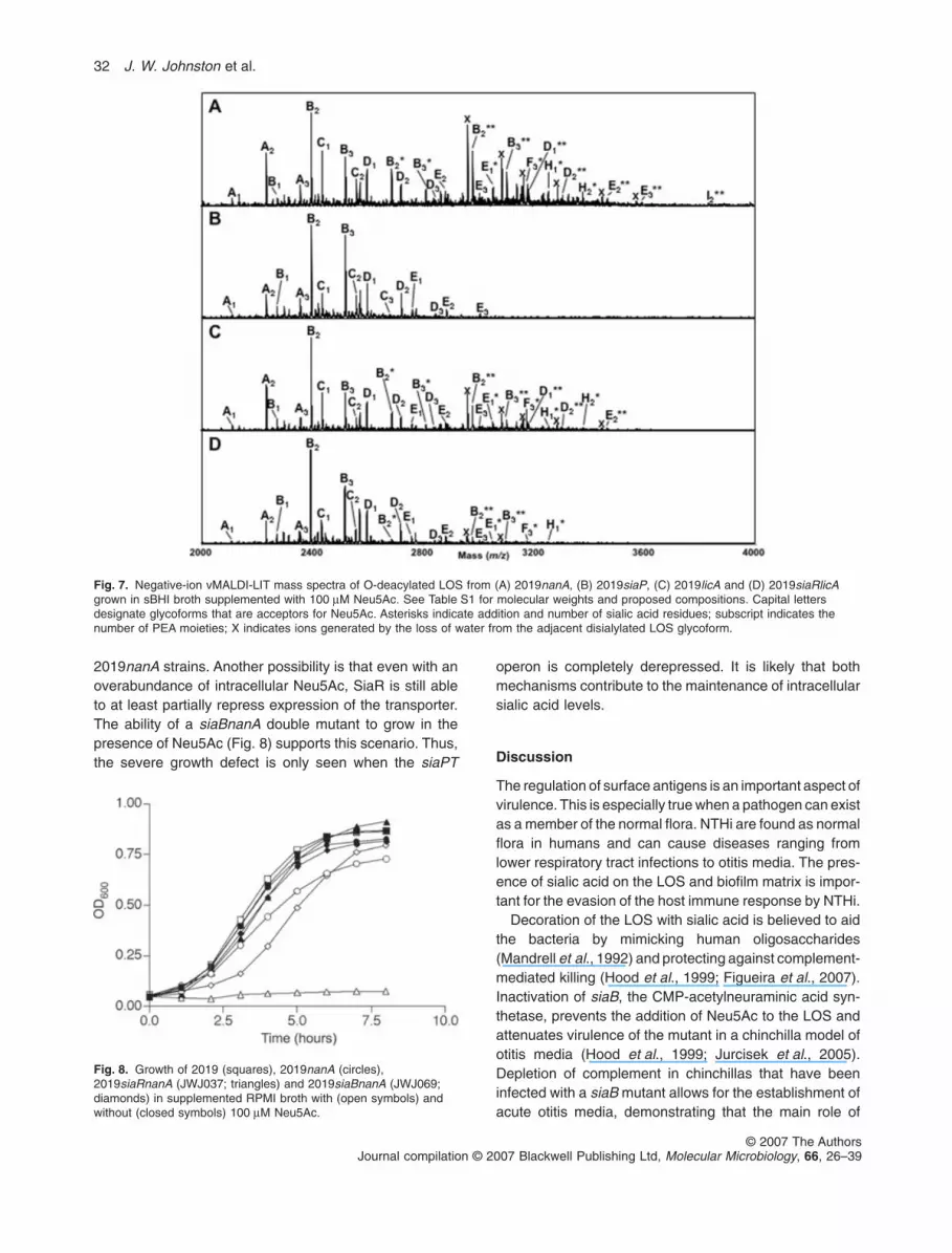
To confirm the TBA colorimetric assay results, LOSisolated from the various mutants was O-deacylated andthe resulting O-deacylated LOS (O-LOS) analysed bynegative-ion matrix-assisted laser desorption ionization-linear ion trap-mass spectrometry (MALDI-LIT-MS). TheO-LOS from various mutants show typical profiles forstrain 2019, with B-glycoforms (lactose branch) dominat-ing the spectra (Fig. 7, Table S1). O-LOS isolated from the2019nanA mutant is ‘hypersialylated’ with the disialylatedB-glycoforms being the most abundant. Of note, the disia-lylated glycoforms appear to undergo a water loss (-18,resultant ions marked X in spectra) during mass spec-trometry; this loss in not apparent in the monosialylatedglycoforms. There are no sialylated glycoforms observedin the O-LOS isolated from 2019siaP (Fig. 7B), as wouldbe expected for a Neu5Ac transporter mutant. The O-LOSof the 2019licA mutant (Fig. 7C) shows what could beconsidered a ‘normal’ degree of sialylation, as there is nodefect in any of the genes involved in sialic acid catabo-lism and transport. As can be seen, the degree of sialy-lation is less than that of the 2019nanA mutant (compareFig. 7A and C). Finally, the O-LOS from the 2019siaRlicAmutant (Fig. 7D) shows a degree of sialylation that islower than that observed in the LOS from the 2019licAmutant (compare Fig. 7C and D). These data correlatewell with the TBA assay data and indicate that in theabsence of SiaR regulation, incorporation of Neu5Ac intothe LOS is reduced. As it is known that sialic acid-containing LOS is required for serum resistance it is likelythat the observed increase in serum sensitivity is due tothe decreased levels of sialic acid in the LOS of the siaRmutant.
Inhibition of growth by Neu5Ac
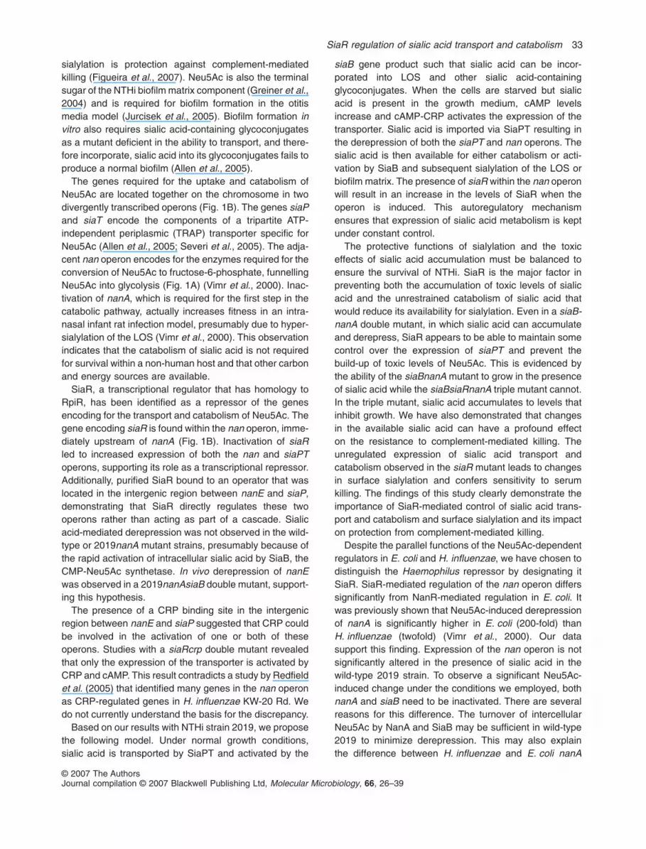
In E. coli, growth of a nanA mutant is inhibited by thepresence of Neu5Ac in the growth medium (Vimr andTroy, 1985), demonstrating that the accumulation of intra-cellular sialic acid is toxic to these bacteria. Previous andcurrent work with a 2019nanA mutant has demonstratedthat Neu5Ac minimally affects the growth of 2019nanA.We found, however, that a nanAsiaR double mutant wasunable to grow in medium supplemented with Neu5Ac(Fig. 8). The toxicity of Neu5Ac is not dependent on theactivation of Neu5Ac to CMP-Neu5Ac as growth of a2019siaBsiaRnanA triple mutant was also inhibited byNeu5Ac (data not shown). There are two possibilities forthe requirement of the inactivation of siaR to observe thisgrowth defect. First, as the toxic effects appear to be dueto accumulation of Neu5Ac and not CMP-Neu5Ac, thepresence of SiaB may play a role maintaining Neu5Aclevels at a permissive concentration in the wild-type and
Fig. 6. Sialic acid content of LOS. Sialic acid was released frompurified LOS by neuraminidase treatment and quantified using thethiobarbituric acid method. Results are expressed in per cent ofsialic acid, by weight. Data from three separate experiments wereincluded in the analysis. 2019licA and 2019siaRlicA were found tobe significantly different (P = 0.0035) using a two-tailed paired t-testanalysis.
SiaR regulation of sialic acid transport and catabolism 31
© 2007 The AuthorsJournal compilation © 2007 Blackwell Publishing Ltd, Molecular Microbiology, 66, 26–39
2019nanA strains. Another possibility is that even with anoverabundance of intracellular Neu5Ac, SiaR is still ableto at least partially repress expression of the transporter.The ability of a siaBnanA double mutant to grow in thepresence of Neu5Ac (Fig. 8) supports this scenario. Thus,the severe growth defect is only seen when the siaPT
operon is completely derepressed. It is likely that bothmechanisms contribute to the maintenance of intracellularsialic acid levels.
Discussion
The regulation of surface antigens is an important aspect ofvirulence. This is especially true when a pathogen can existas a member of the normal flora. NTHi are found as normalflora in humans and can cause diseases ranging fromlower respiratory tract infections to otitis media. The pres-ence of sialic acid on the LOS and biofilm matrix is impor-tant for the evasion of the host immune response by NTHi.
Decoration of the LOS with sialic acid is believed to aidthe bacteria by mimicking human oligosaccharides(Mandrell et al., 1992) and protecting against complement-mediated killing (Hood et al., 1999; Figueira et al., 2007).Inactivation of siaB, the CMP-acetylneuraminic acid syn-thetase, prevents the addition of Neu5Ac to the LOS andattenuates virulence of the mutant in a chinchilla model ofotitis media (Hood et al., 1999; Jurcisek et al., 2005).Depletion of complement in chinchillas that have beeninfected with a siaB mutant allows for the establishment ofacute otitis media, demonstrating that the main role of
Fig. 7. Negative-ion vMALDI-LIT mass spectra of O-deacylated LOS from (A) 2019nanA, (B) 2019siaP, (C) 2019licA and (D) 2019siaRlicAgrown in sBHI broth supplemented with 100 mM Neu5Ac. See Table S1 for molecular weights and proposed compositions. Capital lettersdesignate glycoforms that are acceptors for Neu5Ac. Asterisks indicate addition and number of sialic acid residues; subscript indicates thenumber of PEA moieties; X indicates ions generated by the loss of water from the adjacent disialylated LOS glycoform.
Fig. 8. Growth of 2019 (squares), 2019nanA (circles),2019siaRnanA (JWJ037; triangles) and 2019siaBnanA (JWJ069;diamonds) in supplemented RPMI broth with (open symbols) andwithout (closed symbols) 100 mM Neu5Ac.
32 J. W. Johnston et al.
© 2007 The AuthorsJournal compilation © 2007 Blackwell Publishing Ltd, Molecular Microbiology, 66, 26–39
sialylation is protection against complement-mediatedkilling (Figueira et al., 2007). Neu5Ac is also the terminalsugar of the NTHi biofilm matrix component (Greiner et al.,2004) and is required for biofilm formation in the otitismedia model (Jurcisek et al., 2005). Biofilm formation invitro also requires sialic acid-containing glycoconjugatesas a mutant deficient in the ability to transport, and there-fore incorporate, sialic acid into its glycoconjugates fails toproduce a normal biofilm (Allen et al., 2005).
The genes required for the uptake and catabolism ofNeu5Ac are located together on the chromosome in twodivergently transcribed operons (Fig. 1B). The genes siaPand siaT encode the components of a tripartite ATP-independent periplasmic (TRAP) transporter specific forNeu5Ac (Allen et al., 2005; Severi et al., 2005). The adja-cent nan operon encodes for the enzymes required for theconversion of Neu5Ac to fructose-6-phosphate, funnellingNeu5Ac into glycolysis (Fig. 1A) (Vimr et al., 2000). Inac-tivation of nanA, which is required for the first step in thecatabolic pathway, actually increases fitness in an intra-nasal infant rat infection model, presumably due to hyper-sialylation of the LOS (Vimr et al., 2000). This observationindicates that the catabolism of sialic acid is not requiredfor survival within a non-human host and that other carbonand energy sources are available.
SiaR, a transcriptional regulator that has homology toRpiR, has been identified as a repressor of the genesencoding for the transport and catabolism of Neu5Ac. Thegene encoding siaR is found within the nan operon, imme-diately upstream of nanA (Fig. 1B). Inactivation of siaRled to increased expression of both the nan and siaPToperons, supporting its role as a transcriptional repressor.Additionally, purified SiaR bound to an operator that waslocated in the intergenic region between nanE and siaP,demonstrating that SiaR directly regulates these twooperons rather than acting as part of a cascade. Sialicacid-mediated derepression was not observed in the wild-type or 2019nanA mutant strains, presumably because ofthe rapid activation of intracellular sialic acid by SiaB, theCMP-Neu5Ac synthetase. In vivo derepression of nanEwas observed in a 2019nanAsiaB double mutant, support-ing this hypothesis.
The presence of a CRP binding site in the intergenicregion between nanE and siaP suggested that CRP couldbe involved in the activation of one or both of theseoperons. Studies with a siaRcrp double mutant revealedthat only the expression of the transporter is activated byCRP and cAMP. This result contradicts a study by Redfieldet al. (2005) that identified many genes in the nan operonas CRP-regulated genes in H. influenzae KW-20 Rd. Wedo not currently understand the basis for the discrepancy.
Based on our results with NTHi strain 2019, we proposethe following model. Under normal growth conditions,sialic acid is transported by SiaPT and activated by the
siaB gene product such that sialic acid can be incor-porated into LOS and other sialic acid-containingglycoconjugates. When the cells are starved but sialicacid is present in the growth medium, cAMP levelsincrease and cAMP-CRP activates the expression of thetransporter. Sialic acid is imported via SiaPT resulting inthe derepression of both the siaPT and nan operons. Thesialic acid is then available for either catabolism or acti-vation by SiaB and subsequent sialylation of the LOS orbiofilm matrix. The presence of siaR within the nan operonwill result in an increase in the levels of SiaR when theoperon is induced. This autoregulatory mechanismensures that expression of sialic acid metabolism is keptunder constant control.
The protective functions of sialylation and the toxiceffects of sialic acid accumulation must be balanced toensure the survival of NTHi. SiaR is the major factor inpreventing both the accumulation of toxic levels of sialicacid and the unrestrained catabolism of sialic acid thatwould reduce its availability for sialylation. Even in a siaB-nanA double mutant, in which sialic acid can accumulateand derepress, SiaR appears to be able to maintain somecontrol over the expression of siaPT and prevent thebuild-up of toxic levels of Neu5Ac. This is evidenced bythe ability of the siaBnanA mutant to grow in the presenceof sialic acid while the siaBsiaRnanA triple mutant cannot.In the triple mutant, sialic acid accumulates to levels thatinhibit growth. We have also demonstrated that changesin the available sialic acid can have a profound effecton the resistance to complement-mediated killing. Theunregulated expression of sialic acid transport andcatabolism observed in the siaR mutant leads to changesin surface sialylation and confers sensitivity to serumkilling. The findings of this study clearly demonstrate theimportance of SiaR-mediated control of sialic acid trans-port and catabolism and surface sialylation and its impacton protection from complement-mediated killing.
Despite the parallel functions of the Neu5Ac-dependentregulators in E. coli and H. influenzae, we have chosen todistinguish the Haemophilus repressor by designating itSiaR. SiaR-mediated regulation of the nan operon differssignificantly from NanR-mediated regulation in E. coli. Itwas previously shown that Neu5Ac-induced derepressionof nanA is significantly higher in E. coli (200-fold) thanH. influenzae (twofold) (Vimr et al., 2000). Our datasupport this finding. Expression of the nan operon is notsignificantly altered in the presence of sialic acid in thewild-type 2019 strain. To observe a significant Neu5Ac-induced change under the conditions we employed, bothnanA and siaB need to be inactivated. There are severalreasons for this difference. The turnover of intercellularNeu5Ac by NanA and SiaB may be sufficient in wild-type2019 to minimize derepression. This may also explainthe difference between H. influenzae and E. coli nanA
SiaR regulation of sialic acid transport and catabolism 33
© 2007 The AuthorsJournal compilation © 2007 Blackwell Publishing Ltd, Molecular Microbiology, 66, 26–39

mutants in their sensitivity to Neu5Ac. It also appears thatNanR in E. coli is more responsive to Neu5Ac than SiaRas indicated by higher levels of nan operon induction inE. coli as compared with induction in NTHi. This may bedue to fundamental differences between these tworepressors. There is very little sequence similaritybetween NanR and SiaR even though they both areshown to relieve repression in vivo in the presence ofNeu5Ac. Additionally, SiaR remained bound to the opera-tor even in the presence of Neu5Ac, unlike NanR, indicat-ing that the mechanism of induction may be differentbetween these two systems. This may also be a result ofa difference in the relative affinity for Neu5Ac and requiresfurther investigation. Also, the possibility of a co-repressorthat works to antagonize derepression may exist.
The differences between NanR and SiaR highlight therelative importance of sialic acid to the respective bac-terial species. Sialylation of the LOS is a critical require-ment for the survival of most, if not all, NTHi strains. Wehave demonstrated that slight changes in sialylation cangreatly impact the susceptibility of NTHi 2019 to serumkilling. The E. coli strains that have been studied to date
have the ability to synthesize sialic acid (Vimr et al.,1995). As H. influenzae strains lack a biosyntheticpathway for sialic acid biosynthesis, they must scavengesialic acid from their environment. The relatively lowlevels of free sialic acid available within the host neces-sitate that the bacterium efficiently controls sialic acidcatabolism in order to sialylate the LOS and other gly-coconjugates.
Experimental procedures
Bacterial strains, media and growth
The strains used in this study are listed in Table 2. E. coli wasgrown at 37°C in Luria–Bertani (LB) medium with or withoutagar (1.5%) and supplemented with antibiotics as needed.NTHi strain 2019 (Campagnari et al., 1987) and derivativesthereof were used in this study. H. influenzae was grownat 37°C in the presence of 5% CO2 on sBHI agar (DifcoLaboratories, Detroit, MI). Kanamycin-resistant H. influenzaewere selected on sBHI agar containing 15 mg ml-1 ribostamy-cin in the absence of additional CO2. Chloramphenicol andspectinomycin were added to sBHI at concentrations of2 mg ml-1 and 25 mg ml-1 respectively. RPMI 1640 medium
Table 2. Strains and plasmids.
Strain or plasmid Genotype, relevant phenotype or selection marker Source or reference
StrainsE. coli DH5a F– fdlacZDM15 D(lacZYA-argF) U169 recA1 endA1 hsdR17(rK
–mK+) phoA
supE44 l-thi-1 gyrA96 relA1Invitrogen
E. coli BL21 Star InvitrogenNTHi 2019 Clinical respiratory isolate Campagnari et al. (1987)NTHi 2019nanA NTHi 2019nanA mutant, kanamycin resistantNTHi 2019siaB NTHi 2019siaB mutant, spectinomycin resistantJWJ022 NTHi 2019siaR mutant, kanamycin resistant This studyJWJ037 NTHi 2019siaRnanA mutant, kanamycin resistant This studyJWJ050 NTHi 2019crp mutant, spectinomycin resistant This studyJWJ052 NTHi 2019siaRcrp mutant, kanamycin and spectinomycin resistant This studyJWJ069 NTHi 2019siaBnanA mutant, kanamycin and spectinomycin resistant This studyJWJ070 NTHi 2019licA mutant, chloramphenicol resistant This studyJWJ071 NTHi 2019siaRlicA mutant, kanamycin and chloramphenicol resistant This studyJWJ074 NTHi 2019siaBsiaRnanA mutant, kanamycin and spectinomycin resistant This studyJWJ075 NTHi 2019siaR complemented mutant, kanamycin and spectinomycin resistant This study
PlasmidspGEM-T Easy PCR-cloning vector PromegapGEM-T PCR-cloning vector PromegapCR2.1-TOPO PCR-cloning vector InvitrogenpET-24(+) Expression vector NovagenpUC19 General cloning vector New England BiolabspUC18K3 Kanamycin resistance cassette Menard et al. (1993)pUC19K3 Kanamycin resistance cassette This studypSPECR Spectinomycin resistance cassette Whitby et al. (1998)pUC19nanAr_Kn nanA knockout vector Greiner et al. (2004)pCR601.1 HI0601.1 region in pCR2.1 This studyp601.1-Sp2 Ectopic complementation vector This studypJJ150 siaR knockout vector This studypJJ169 siaRnanA knockout vector This studypJJ175 crp region This studypJJ181 crp knockout vector This studypJJ185 C-terminal his tag SiaR expression vector This studypJJ237 cat reporter vector This studypJJ245 licA knockout/cat reporter vector This studypJJ256 siaR complementation vector This study
34 J. W. Johnston et al.
© 2007 The AuthorsJournal compilation © 2007 Blackwell Publishing Ltd, Molecular Microbiology, 66, 26–39
(Invitrogen, Carlsbad, CA) was used as a sialic acid-freechemically defined medium. Supplemented RPMI (sRPMI)was prepared with protoporphyrin IX (1 mg ml-1), hypoxan-thine (0.1 mg ml-1), uracil (0.1 mg ml-1), b-NAD (10 mg ml-1)and sodium pyruvate (0.8 mM). Neu5Ac (100 mM) was addedas indicated.
Construction of mutants
Genetic manipulations were performed using establishedtechniques. Restriction enzymes, Antarctic Phosphatase andT4 polymerase were obtained from New England Biolabs(Beverly, MA) and were used following established protocols.The Expand High Fidelity PCR System (Roche AppliedScience, Indianapolis, IN) was used for PCR reactions. Oli-gonucleotide primers were designed and ordered from Inte-grated DNA Technologies (Coralville, IA). Plasmids weretransformed and maintained in MAX Efficiency DH5a Chemi-cally Competent cells (Invitrogen). Competent H. influenzaecells were prepared using the M-IV method and transformedas described previously (Herriott et al., 1970).
A siaR (HI0143) mutant was constructed in NTHi strain2019 to study the role of SiaR in the regulation of sialic acidtransport and metabolism. Regions upstream and down-stream of siaR were amplified by PCR using primers listed inTable S2 and cloned into pGEM-T Easy (Promega, Madison,WI). Primers were designed to add restriction sites to bothends of the inserts to allow for subcloning. These fragmentswere then sequentially subcloned into pUC18K3 (Menardet al., 1993) and arranged around the kanamycin resistancecassette, aphA3, resulting in pJJ150. The aphA-3 gene ofpUC18K3 lacks both a promoter and transcriptional termina-tor and should allow for the expression of downstream genesof an operon to be controlled by the native promoter. pJJ150was linearized and transformed into NTHi 2019, creatingstrain JWJ022. The correct integration of the cassette andinactivation of siaR was confirmed by PCR (results notshown).
To study the role of CRP in the expression of the nan andsiaPT operons, crp and siaRcrp mutants were constructed. Aregion spanning crp was amplified by PCR and cloned intopGEM-T Easy, resulting in pJJ175. Restriction enzymesBseRI and BmgBI were used to excise a 400 bp fragmentfrom within the gene and replace it with the spectinomycinresistance cassette from pSPECR (Whitby et al., 1998), cre-ating pJJ181. pJJ181 was linearized with EcoRI and trans-formed into strains 2019 and JWJ022 to create strainsJWJ050 and JWJ052 respectively. The correct integration ofthe cassette was confirmed by PCR (results not shown).
A siaRnanA double mutant was constructed. Fragmentsamplified with primers for regions upstream of siaR anddownstream of nanA were subcloned into pUC19K3.pUC19K3 is a derivative of pUC18K3 with the aphA-3 cas-sette in the opposite orientation relative to the lacZ promoter.The resulting construct, pJJ169, was used to replace bothsiaR and nanA with aphA-3. pJJ169 was linearized withEcoRI and HindIII and transformed into NTHi 2019, resultingin strain JWJ037. The correct integration of the cassette wasconfirmed by PCR (results not shown).
2019siaB was transformed with linearized plasmidspUC19_nanAr_Kn (Greiner et al., 2004) and pJJ169 to
create 2019siaBnanA (JWJ069) and 2019siaBsiaRnanA(JWJ043) double and triple mutants respectively. Correctintegration of respective cassettes and confirmation of thesiaB mutation were confirmed by PCR.
Complementation of the siaR mutant
The open reading frame (ORF) HI0601.1 contains an authen-tic frameshift in the KW-20 genome. We used this region toinsert a copy of siaR to complement the 2019siaR mutant.PCR was used to amplify a 2342 bp fragment from NTHi2019 genomic DNA using primers 601.1-1 and 601.1-2. Thisfragment was cloned into pCR2.1-TOPO (Invitrogen) andsequenced. Sequencing confirmed that HI0601.1 waspresent in NTHi 2019 and that it also contained a frameshiftthat resulted in a severe truncation of the ORF. This plasmidwas designated pCR601.1.
PCR was used to amplify a 1061 bp fragment containingthe spectinomycin cassette and its upstream promoter, butnot the downstream transcription terminator, from pSPECRusing primers SprupBsrGI and SprdnMCS. The product wascloned into pCR2.1-TOPO, and sequenced. The sequenceconfirmed that the expected DNA fragment had been cloned.This plasmid was designated pCRSp2. The 1108 bp EcoRI(blunt ended)/BsrGI fragment from pCRSp2 was ligated intopCR601.1 which had been digested with BsgI (blunt ended)/BsrGI. This plasmid was designated p601.1-Sp2. The con-struct was verified by sequencing.
PCR was used to amplify a fragment containing siaR. ThePCR fragment was cloned into pGEM-T (Promega) and sub-sequently subcloned into p601.1-Sp2 using SmaI sites. Theresulting construct, pJJ256 (Fig. S1), was linearized andtransformed into the siaR mutant, JWJ022. Correct integra-tion of the construct was confirmed by PCR.
RNA extraction and transcriptional analysis
RNA was extracted using the hot acid phenol method asdescribed previously (Peterson et al., 2000). DNA wasremoved from extracted RNA by digestion with DNase I (NewEngland Biolabs) and cleaned up with the RNeasy Mini Kit(Qiagen, Valencia, CA). RNA quality was assessed with anAgilent 2100 Bioanalyser (Agilent, Santa Clara, CA) andconcentration was determined using a NanoDrop ND-1000Spectrophotometer (NanoDrop Technologies, Wilmington,DE). For real-time RT-PCR analysis, primer/probe sets wereobtained using the Custom TaqMan Gene ExpressionService (Applied Biosystems, Foster City, CA). Primer/probesets were designed using the sequence of HI0145, HI0146and HI1537 from H. influenzae 2019. A primer/probe set forthe 16S rRNA of H. influenzae was designed and used asa control. The TaqMan One-Step RT-PCR Master MixReagents (Applied Biosystems) were used following themanufacturer’s protocol. Reactions were set up in triplicateusing 20 ng of RNA. Reactions were carried out using an ABIPrism 7700 Sequence Detector (Perkin Elmer, Wellesley,MA) with ABI Prism 7700 v1.7 analysis software. Resultswere calculated using the comparative CT method to deter-mine the relative expression ratio between RNA samples.The primer and probe set for HI16S rRNA was used as the
SiaR regulation of sialic acid transport and catabolism 35
© 2007 The AuthorsJournal compilation © 2007 Blackwell Publishing Ltd, Molecular Microbiology, 66, 26–39
endogenous reference to normalize the results. Data areexpressed as mean � SD.
Microarray analysis
The DNA microarray and relevant protocols used in thisstudy were described previously (Harrison et al., 2007). Thearray was prepared from commercial oligonucleotide probesbased on the H. influenzae KW20 Rd genome (Operon Bio-technologies, Huntsville, AL). cDNA was synthesized andlabelled using an indirect labelling method similar to thatdescribed in Hegde et al. (2000). Two replicate slides wereperformed for each biological replicate, and three biologicalreplicates were performed per experiment, resulting in 24data points per gene. Dye intensity normalization wasapplied by performing LOWESS (subgrid) algorithm to allsignificant features including exogenous spike bacterial con-trols. Analysis of variance and replicate averaging was per-formed using the NIA/NIH ANOVA tool. A False DiscoveryRate (FDR) > 0.05 was used to exclude false positives,where FDR = P*N/rank, P is P-value, N is total number ofgenes and rank is the gene ordered in increasing P-value.Absent spots, empty spots and positive control spots wereexcluded from the ANOVA analysis.
Expression and purification of SiaR
SiaR was expressed and purified for further study. Primerswere used to amplify siaR with the restriction sites HindIII andXhoI on the 5′ and 3′ ends, respectively. The 70 bases imme-diately upstream of siaR were included to ensure that thenative bacterial translation signals were present. The down-stream primer included the last codon of the siaR ORF,excluding the stop codon, to allow for the fusion of a multiple-histidine tag. The PCR product was cloned into pGEM-T andsubsequently subcloned into pET-24(+) (Novagen, Madison,WI) using the HindIII and XhoI sites. The resulting plasmid,pJJ185, was expected to express SiaR with a carboxy-terminal His-tag.
Protein expression was induced using the OvernightExpress Autoinduction System 1 (Novagen) grown at 20°Cfor 48 h. Expressed protein was purified using the BD TALONMetal Affinity Resin (BD Biosciences, Palo Alto, CA). Purifi-cation was performed in native conditions as follows. Cellsfrom a 100 ml of expression culture were pelleted,re-suspended in 20 ml of equilibration buffer (300 mM NaCl,50 mM sodium phosphate, pH 7.0) containing 2.5 mg ml-1
DNase I (Sigma) and lysed with a French press. Cell debriswas removed by ultracentrifugation at 150 000 g for 1 h. TheBD TALON resin was prepared following the manufacturer’sprotocol and the lysate was loaded and allowed to bind for 1 hat room temperature. The resin was washed three times with10 volumes of a high-salt wash buffer (1150 mM NaCl, 5 mMimidazole, 50 mM sodium phosphate, pH 7.0). Bound proteinwas eluted by washing three times with two volumes ofelution buffer (300 mM NaCl, 500 mM imidazole, 50 mMsodium phosphate, pH 7.0). Eluted fractions were examinedby SDS-PAGE and fractions containing SiaR were pooledand dialysed into a buffer that prevents the precipitation ofpurified protein (10 mM Tris, pH 8.0, 150 mM KCl, 1 mM DTT,
1 mM EDTA, 5 mM MgCl2, 10% glycerol, 0.1% CHAPS). Ali-quots of the purified protein were frozen in liquid nitrogen andstored at -80°C until needed. The protein was unstable andprecipitated after purification, precluding the determination ofa dissociation constant for the binding reaction.
Electrophoretic mobility shift assay
Electrophoretic mobility shift assay was used to study thebinding of purified SiaR to potential promoter sequences.Probes for EMSA were amplified by PCR using primer pairsP146F1 and P146R3 (siaP-nanE intergenic region), licAUF1and licAL3 (lic1 promoter region) and P2F1 and P2R1 (P2promoter). Binding reactions were prepared using the EMSAKit (Molecular Probes, Eugene, OR) following the manufac-turer’s directions with some modifications. Pilot experimentswere performed to determine the amount of SiaR required toshift all of the DNA probe. Binding reactions consisted of thebinding buffer (150 mM KCl, 0.1 mM DTT, 0.1 mM EDTA,10 mM Tris, pH 7.4), the DNA probe (15 nM) and 1 mM SiaR.Control reactions without SiaR were set up for each probe.Reactions were incubated at room temperature for 20 min.After incubation, 6¥ EMSA gel-loading solution was addedand reactions were loaded onto a 6% DNA Retardation Gel(Invitrogen) with pre-chilled 0.5¥ TBE buffer and run at 200 Vfor 60 min. After electrophoresis, the gel was stained withSYBR Green EMSA gel stain and bands were visualized byUV transillumination. Images were captured using a KodakEDAS 120 camera with an EDAS 590 mm filter (EastmanKodak Company, Rochester, NY). To test the ability of varioussugars to prevent SiaR binding, reactions were set up withthe addition of 1 mM Neu5Ac, N-acetylmannosamine, N-acetylglucosamine or N-acetylglucosamine-6-phosphate.
DNase I footprint analysis
DNase I footprinting analysis of the nanE-siaP intergenicregion was examined using the procedure of Johnson et al.(1979), with some modifications. The probe was prepared byPCR amplification with primers P146F1 and P146R4, result-ing in a 257 bp fragment. The PCR product was cleaned upusing the QIAquick PCR Purification Kit (Qiagen) and thendigested with ApoI. The digested fragment was electrophore-sed and a 209 bp fragment was purified using the MinEluteGel Extraction Kit (Qiagen). The purified fragment was end-labelled with [a-32P]-dATP (GE Healthcare Bio-Sciences, Pis-cataway, NJ) and Klenow Fragment (3′-5′ exo-; New EnglandBiolabs). Probe was cleaned up using the MinElute ReactionCleanup Kit (Qiagen). Approximately 100 ng of probe(800 000 cpm) were used per reaction.
DNase I footprinting reactions were set up in binding buffer(10 mM Tris, pH 8.0, 150 mM NaCl, 0.1 mM DTT, 1 mMEDTA, 10% glycerol and 0.1% CHAPS) containing 25 ng ml-1
salmon sperm DNA (Sigma), 1 mg ml-1 BSA (Sigma) and100 ng of the probe. Purified SiaR was added to the reaction(90 ml total volume) at concentrations ranging from 0 to2.5 mM and reactions were incubated at room temperature for20 min to allow for DNA binding. DNase I (Sigma; 50 ng perreaction) and 10¥ digestion buffer (400 mM Tris, pH 7.5,150 mM MgCl2 and 50 mM CaCl2) were added and reactions
36 J. W. Johnston et al.
© 2007 The AuthorsJournal compilation © 2007 Blackwell Publishing Ltd, Molecular Microbiology, 66, 26–39
were incubated at room temperature for 1 min prior to theaddition of 100 ml of stop solution (60 mM Tris, pH 8.0,100 mM EDTA). Reaction was extracted with an equalvolume of phenol-chloroform and DNA was precipitated withsodium acetate and ethanol with 5 mg of glycogen (Sigma)added as a carrier. Dried DNA was re-suspended in 5 ml ofwater and 2¥ formamide loading dye (98% deionized forma-mide, 10 mM EDTA, 0.025% xylene cyanol FF, 0.025% bro-mophenol blue) was added. Reactions were loaded onto an8% sequencing gel with a G+A sequencing ladder made fromthe same probe. After electrophoresis, the gel was dried andexposed to film at -70°C.
Serum killing assay
NTHi strains 2019, JWJ070 and JWJ071 were grown to earlylog phase, OD600 of 0.2, in sBHI broth with and without100 mM Neu5Ac. Bacteria were diluted to 1 ¥ 107 cells per mlwith phosphate-buffered salt solution (PBSS) consisting of10 mM K2HPO4, 10 mM KH2PO4, 136 mM NaCl, 5 mM KCl,1 mM CaCl2, 0.3 mM·MgCl2·6H2O, 1 mM MgSO4·7H2O and0.01% BSA, pH 7.0.
The serum killing assay, modified from that reported byAndreoni et al. (1993), was carried out in a 96-well plate witha 200 ml final volume. Pooled normal human serum (PNHS),a 20-donor pool of serum from human volunteers with noprevious history of serious infections, was diluted to 10% inPBSS. A control containing PNHS that had been heat-inactivated for 30 min at 56°C was included in eachexperiment. Ten microlitres (1 ¥ 105 organisms) of there-suspended bacteria were diluted into 190 ml of PBSS andserial 10-fold dilutions made in PBSS. Twenty microlitres ofeach dilution was spread on sBHI with or without appropriateantibiotic selection and grown overnight at 37°C in 5% CO2.Colonies were counted and used as the initial colony-formingunits (cfu). Ten microlitres of the bacterial stock was incu-bated in the diluted serum at 37°C for 30 min with shaking at200 r.p.m. Serial 10-fold dilutions of the reaction mixtures inPBSS were plated on sBHI with or without appropriate anti-biotic selection. The plates were grown overnight at 37°C in5% CO2 and emerging colonies counted the next day. Theresulting cfu value was that recorded after 30 min. Killing wasassessed by comparing the number of cfu from the 30 minserum incubation with the number of the initial cfu. Resultswere expressed as the log10 change in cfu at 30 min com-pared with the initial cfu. Statistical analysis of the data frombactericidal assays was carried out using the paired t-testand analysis of variance functions found in Graphpad Prism,version 4, San Diego, CA.
LOS preparation and neuraminidase treatment
Strains 2019nanA, 2019siaP, JWJ070 and JWJ071 weregrown in sBHI containing 100 mM Neu5Ac. Five hundredmillilitres of cultures were started with an OD600 of 0.05 andgrown to an OD600 of 0.5 prior to harvesting the cells bycentrifugation. Cell pellets were washed twice with PBS andthen extracted with 25 ml of water-saturated phenol after bothwere equilibrated to 65°C. This mixture was cooled on ice for1 h and separated by low-speed centrifugation. The top
aqueous layer was removed and saved. The phenol layer wasback extracted once with water at 65°C, cooled, centrifuged,and the second aqueous layer added to the first. The residualphenol was removed from the LOS by precipitating the LOStwice using 0.3 M sodium acetate (final concentration) and twovolumes of 100% ethanol. This was incubated at -20°C over-night and then centrifuged at 15 000 g for 30 min. To removeany contaminating lipoproteins, the LOS pellets werere-suspended in 8 ml of buffer A (60 mM Tris, 10 mM EDTA,2.0% SDS, pH 6.8), and incubated in a boiling water bath for5–10 min. The samples were allowed to cool, and proteinaseK (Sigma) was added to a final concentration of 12.5 mg ml-1.The samples were incubated at 37°C for 16–24 h. The LOSwas precipitated as described above. The LOS was washedthree times by precipitation, as above, with ethanol to removeany residual SDS. After the last precipitation, the LOS waswashed twice with water and centrifugation at 100 000 g for75 min. The pellets were re-suspended in water, frozen andthen lyophilized. The dry LOS was stored at room temperature.
Quantification of total surface sialic acid
The TBA method of Warren (1959) was used to quantitativelycompare the levels of sialic acid from purified LOS. PurifiedLOS was dissolved in a 100 mM sodium acetate, pH 5.6,buffer containing 0.05 U neuraminidase (Roche AppliedScience), and incubated at 37°C overnight. At the end of theincubation the samples were centrifuged at 20 000 g toremove any precipitant and the supernatant was recoveredfor sialic acid analysis using the TBA method. Measurementswere performed on three separate occasions with a standardcurve performed with each set of samples.
Preparation of O-LOS
To make the LOS more amenable for mass spectrometricanalysis, O-linked fatty acids were removed from the lipid Amoiety as previously described (Gibson et al., 1997). Thehighly purified LOS (~0.1 mg) was incubated in anhydroushydrazine (50 ml; Aldrich) at 37°C for 35 min with mixing every10 min. Samples were cooled on ice prior to and after theaddition of ice-cold acetone (250 ml; Aldrich), then transferredto -20°C for 2 h. The quenched reaction mixture was centri-fuged (12 000 g) for 45 min at 4°C. The supernatant wasremoved and the pelleted O-LOS was dissolved in MilliQ H2O(50 ml) and evaporated on a speed vacuum system (Savant).To remove salts and other low-molecular-weight contami-nants the O-LOS (~20–30 mg) was suspended on a nitrocel-lulose membrane (type VS, 0.025 mm; Millipore) over waterfor approximately 1 h. The desalted O-LOS was removedfrom the membrane concentrated with a speed vacuumsystem, and analysed by MALDI-LIT-MS.
Mass spectrometry of O-LOS
Aliquots of O-LOS were desalted by mixing with Dowex 50beads (100–200 mesh, NH4
+ form; Bio-Rad, Hercules, CA)prior to mass spectrometry. Mass spectrometric data werecollected on a FinniganTM LTQTM linear ion trap-mass (LIT)spectrometer coupled to a vMALDI ion source (Thermo-
SiaR regulation of sialic acid transport and catabolism 37
© 2007 The AuthorsJournal compilation © 2007 Blackwell Publishing Ltd, Molecular Microbiology, 66, 26–39
Electron, San Jose, CA). The vMALDI ion source operates at170mTorr and uses an SI nitrogen laser (337.7 nm). Aliquotsof the O-LOS (0.5 ml) were spotted onto a 96-well sampleplate and allowed to dry. Dried samples were overlaid with0.5 ml of 50 mg ml-1 2, 5 dihydroxybenzoic acid (DHB) in 50%acetonitrile. Data were collected in negative mode over twomass ranges: m/z 800–4000 and m/z 2000–4000. MS2 datawere collected in negative mode to confirm sialylation, usinga precursor isolation width of 3 m/z, normalized collisionenergy of 40% (RF amplitude in per cent used for fragmentions), activation Q of 0.25 and an activation time of 30 ms.Spectra were recorded using automatic gain control (AGC) tocontrol the number of laser shots and the automatic spectrumfilter (ASF) tool.
Acknowledgements
This work was supported by funding from NIAID GrantsAI24616 and AI30040.
References
Allen, S., Zaleski, A., Johnston, J.W., Gibson, B.W., andApicella, M.A. (2005) Novel sialic acid transporter ofHaemophilus influenzae. Infect Immun 73: 5291–5300.
Andreoni, J., Kayhty, H., and Densen, P. (1993) Vaccinationand the role of capsular polysaccharide antibody in preven-tion of recurrent meningococcal disease in late comple-ment component-deficient individuals. J Infect Dis 168:227–231.
Bateman, A. (1999) The SIS domain: a phosphosugar-binding domain. Trends Biochem Sci 24: 94–95.
Bouchet, V., Hood, D.W., Li, J., Brisson, J.R., Randle, G.A.,Martin, A., et al. (2003) Host-derived sialic acid is incorpo-rated into Haemophilus influenzae lipopolysaccharide andis a major virulence factor in experimental otitis media.Proc Natl Acad Sci USA 100: 8898–8903.
Campagnari, A.A., Gupta, M.R., Dudas, K.C., Murphy, T.F.,and Apicella, M.A. (1987) Antigenic diversity of lipooli-gosaccharides of nontypable Haemophilus influenzae.Infect Immun 55: 882–887.
Figueira, M.A., Ram, S., Goldstein, R., Hood, D.W., Moxon,E.R., and Pelton, S.I. (2007) Role of complement indefense of the middle ear revealed by restoring thevirulence of nontypeable Haemophilus influenzae siaBmutants. Infect Immun 75: 325–333.
Gibson, B.W., Engstrom, J.J., John, C.M., Hines, W., andFalick, A.M. (1997) Characterization of bacterial lipooli-gosaccharides by delayed extraction matrix-assisted laserdesorption time-of-flight mass spectrometry. J Am SocMass Spectrom 8: 645–658.
Greiner, L.L., Watanabe, H., Phillips, N.J., Shao, J., Morgan,A., Zaleski, A., et al. (2004) Nontypeable Haemophilusinfluenzae strain 2019 produces a biofilm containingN-acetylneuraminic acid that may mimic sialylated O-linkedglycans. Infect Immun 72: 4249–4260.
Harrison, A., Ray, W.C., Baker, B.D., Armbruster, D.W.,Bakaletz, L.O., and Munson, R.S., Jr (2007) The OxyRregulon in nontypeable Haemophilus influenzae.J Bacteriol 189: 1004–1012.
Hegde, P., Qi, R., Abernathy, K., Gay, C., Dharap, S.,Gaspard, R., et al. (2000) A concise guide to cDNAmicroarray analysis. Biotechniques 29: 548–550,552–544,556 passim.
Herriott, R.M., Meyer, E.M., and Vogt, M. (1970) Definednongrowth media for stage II development of competencein Haemophilus influenzae. J Bacteriol 101: 517–524.
Hood, D.W., Makepeace, K., Deadman, M.E., Rest, R.F.,Thibault, P., Martin, A., et al. (1999) Sialic acid in thelipopolysaccharide of Haemophilus influenzae: strain dis-tribution, influence on serum resistance and structuralcharacterization. Mol Microbiol 33: 679–692.
Hood, D.W., Cox, A.D., Gilbert, M., Makepeace, K., Walsh,S., Deadman, M.E., et al. (2001) Identification of alipopolysaccharide alpha-2,3-sialyltransferase from Hae-mophilus influenzae. Mol Microbiol 39: 341–350.
Johnson, A.D., Meyer, B.J., and Ptashne, M. (1979) Interac-tions between DNA-bound repressors govern regulation bythe lambda phage repressor. PNAS 76: 5061–5065.
Jurcisek, J., Greiner, L., Watanabe, H., Zaleski, A., Apicella,M.A., and Bakaletz, L.O. (2005) Role of sialic acid andcomplex carbohydrate biosynthesis in biofilm formationby nontypeable Haemophilus influenzae in the chinchillamiddle ear. Infect Immun 73: 3210–3218.
Kalivoda, K.A., Steenbergen, S.M., Vimr, E.R., and Plum-bridge, J. (2003) Regulation of sialic acid catabolism by theDNA binding protein NanR in Escherichia coli. J Bacteriol185: 4806–4815.
Mandrell, R.E., McLaughlin, R., Aba Kwaik, Y., Lesse, A.,Yamasaki, R., Gibson, B., et al. (1992) Lipooligosaccha-rides (LOS) of some Haemophilus species mimic humanglycosphingolipids, and some LOS are sialylated. InfectImmun 60: 1322–1328.
Menard, R., Sansonetti, P.J., and Parsot, C. (1993) Nonpolarmutagenesis of the ipa genes defines IpaB, IpaC, and IpaDas effectors of Shigella flexneri entry into epithelial cells.J Bacteriol 175: 5899–5906.
Munson, R., Jr, and Tolan, R.W., Jr (1989) Molecular cloning,expression, and primary sequence of outer membraneprotein P2 of Haemophilus influenzae type b. Infect Immun57: 88–94.
Peterson, S., Cline, R.T., Tettelin, H., Sharov, V., and Morri-son, D.A. (2000) Gene expression analysis of the Strepto-coccus pneumoniae competence regulons by use of DNAmicroarrays. J Bacteriol 182: 6192–6202.
Plumbridge, J., and Vimr, E. (1999) Convergent pathwaysfor utilization of the amino sugars N-acetylglucosamine,N-acetylmannosamine, and N-acetylneuraminic acid byEscherichia coli. J Bacteriol 181: 47–54.
Redfield, R.J., Cameron, A.D., Qian, Q., Hinds, J., Ali, T.R.,Kroll, J.S., and Langford, P.R. (2005) A novel CRP-dependent regulon controls expression of competencegenes in Haemophilus influenzae. J Mol Biol 347: 735–747.
Ringenberg, M.A., Steenbergen, S.M., and Vimr, E.R. (2003)The first committed step in the biosynthesis of sialic acid byEscherichia coli K1 does not involve a phosphorylatedN-acetylmannosamine intermediate. Mol Microbiol 50:961–975.
Severi, E., Randle, G., Kivlin, P., Whitfield, K., Young, R.,Moxon, R., et al. (2005) Sialic acid transport in Haemophi-
38 J. W. Johnston et al.
© 2007 The AuthorsJournal compilation © 2007 Blackwell Publishing Ltd, Molecular Microbiology, 66, 26–39

lus influenzae is essential for lipopolysaccharide sialylationand serum resistance and is dependent on a novel tripartiteATP-independent periplasmic transporter. Mol Microbiol58: 1173–1185.
Sorensen, K.I., and Hove-Jensen, B. (1996) Ribose catabo-lism of Escherichia coli: characterization of the rpiB geneencoding ribose phosphate isomerase B and of the rpiRgene, which is involved in regulation of rpiB expression.J Bacteriol 178: 1003–1011.
Vimr, E.R., and Troy, F.A. (1985) Identification of an induciblecatabolic system for sialic acids (nan) in Escherichia coli.J Bacteriol 164: 845–853.
Vimr, E., Steenbergen, S., and Cieslewicz, M. (1995) Biosyn-thesis of the polysialic acid capsule in Escherichia coli K1.J Ind Microbiol 15: 352–360.
Vimr, E., Lichtensteiger, C., and Steenbergen, S. (2000)Sialic acid metabolism’s dual function in Haemophilusinfluenzae. Mol Microbiol 36: 1113–1123.
Vimr, E.R., Kalivoda, K.A., Deszo, E.L., and Steenbergen,S.M. (2004) Diversity of microbial sialic acid metabolism.Microbiol Mol Biol Rev 68: 132–153.
Warren, L. (1959) The thiobarbituric acid assay of sialic acids.J Biol Chem 234: 1971–1975.
Weiser, J.N., Maskell, D.J., Butler, P.D., Lindberg, A.A.,and Moxon, E.R. (1990) Characterization of repetitivesequences controlling phase variation of Haemophilus
influenzae lipopolysaccharide. J Bacteriol 172: 3304–3309.
Weiser, J.N., Pan, N., McGowan, K.L., Musher, D., Martin, A.,and Richards, J. (1998) Phosphorylcholine on thelipopolysaccharide of Haemophilus influenzae contributesto persistence in the respiratory tract and sensitivity toserum killing mediated by C-reactive protein. J Exp Med187: 631–640.
Whitby, P.W., Morton, D.J., and Stull, T.L. (1998) Construc-tion of antibiotic resistance cassettes with multiple pairedrestriction sites for insertional mutagenesis of Haemophilusinfluenzae. FEMS Microbiol Lett 158: 57–60.
Supplementary material
The following supplementary material is available for thisarticle:
This material is available as part of the online article from:http://www.blackwell-synergy.com/doi/abs/10.1111/j.1365-2958.2007.05890.x(This link will take you to the article abstract).
Please note: Blackwell Publishing is not responsible for thecontent or functionality of any supplementary materials sup-plied by the authors. Any queries (other than missing material)should be directed to the corresponding author for the article.
SiaR regulation of sialic acid transport and catabolism 39
© 2007 The AuthorsJournal compilation © 2007 Blackwell Publishing Ltd, Molecular Microbiology, 66, 26–39


