Embed Size (px)
Citation preview

Carbohydrate &sea&, 173 (1988) 65-74 65 Elsevier Science Publishers B.V., Amsterdam - Printed in The Netherlands
DETERMINATION OF THE STRUCI’URE OF THE Escherichia coli KlOO CAPSULAR POLYSACCHARIDE, CROSS-REACTIVE WITH THE CAPSULE FROM TYPE B Huemophilus infruenzae
FAI-PO TSUI, Wm EGAN*, MICHAEL F. SUMMERS, R. ANDREW BYRD,
Biophysics Laboratory, Office of Biologics Research and Review, Food and Drug Administration, 88&I Rockville Pike, Bethesda, Maryland 20892 (U.S. A.)
RACHEL SCHNEERSON, AND JOHN B. ROBBINS
Laboratory of Developmental and Mokcuku Immunity, National Institute of Child Health and Human Development, National Institutes of Health, Bethesda, MD 20892 (U.S.A.)
(Received January 12th, 1987; accepted for publication in revised form, August lSth, 1987)
ABSTRACT
The structure of the Escherichiu coli KlOO capsular polysaccharide, cross- reactive with that from type b Haemophilus influenzue, was determined by using a combination of chemical and spectroscopic techniques. The structure of the KlOO repeating unit was found to be ~3)-gD-Ribf-(1~2)-D-ribitol-S-(PO~. The KlOO polysaceharide is thus identical in composition to, but different in linkage from, the H. influenzue type b capsular polysaccharide, which has PD-Ribf-(l+l)-D-ribitol linkages.
INTRODUCTION
Considerable evidence supports the view that capsular polysaccharides are necessary for causation of severe, invasive disease by Haemophilus influenzue type b organisms (as well as, inter alia, Neisseria meningitidis, and Streptococcus pneumoniue)‘. The Escherichiu coZi KlOO capsular polysaccharide is antigenically and immunologically cross-reactive with that from* H. influenzue type b; despite this similarity, E. coli KlOO strains are not known to cause invasive disease in hu- mans3. Indeed, the normal enteric occurrence of KlOO strains may contribute to the establishment of the natural immunity against H. influenzue type b disease found in the majority of adults2. It has, moreover, been suggested4 that the purified KlOO capsular polysaccharide could serve as a vaccine against type b H. influenzue. Be- cause of its potential use as a vaccine, either by itself or chemically linked to a protein carrier, and in order to gain insight into the chemical basis of cross- immunogenicity, we have studied the structure of the KlOO capsular polysacchar- ide, and report herein the results of these studies.
*To whom correspondence should be addressed.
OUO&6215/88/$03.50 @ 1988 Ekevier Science Publishers B.V.

66 F.-P. TSUI, W. EGAN, M. F. SUMMERS, R. A. BYRD, R. SCHNEERSON, J. B. ROBBINS
RESULTS
FolIowing acid-catalyzed hydrolysis, ribose was detected as the sole reducing sugar by partition chromatography, and gas-liquid chromatography revealed ribose, ribitol, and anhyclroribitol (as ribitol is converted into anhydroribitol under the conditions of hydrolysiG). Chemical analysis showed that ribose, ribitol, and phosphate were present in the polysaccharide in equimolar, or approximately equimolar, proportions (see Materials and Methods); this composition was con- firmed by 13C-n.m.r. spectroscopy. Additional constituents, such as acetyl or pyruvic acetal groups, were not present, as established by n.m.r. spectroscopy (both ‘H and 13C). The nature of the phosphate linkage in the intact polysaccharide as phosphoric diester was established, as previously described6 for the H. influenzae type f capsular polysaccharide, by pH titration and by its behavior on base- catalyzed hydrolysis followed by alkaline phosphatase-catalyzed hydrolysis of the resultant phosphoric monoesters.
The 13C-n.m.r. spectrum of the KlOO polysaccharide (see Table I; cf. Fig. 2) displayed ten resonances of approximately equal, integrated absorption intensity, five of which had scalar couplings to phosphorus, splittings due to scalar couplings being identified as such by recording the spectrum at different magnetic field strengths of -2.3 and 9.4 T. The 13C-n.m.r. spectrum is thus in accord with the chemical composition reported. Only one resonance was observed in the anomeric region of the spectrum (at 6 108.75), consistent with the presence of only one reduc- ing sugar unit; the extreme downfield position of this resonance is indicative of ribose glycosidated at C-l and present in the polymer in the pfuranoid form7.
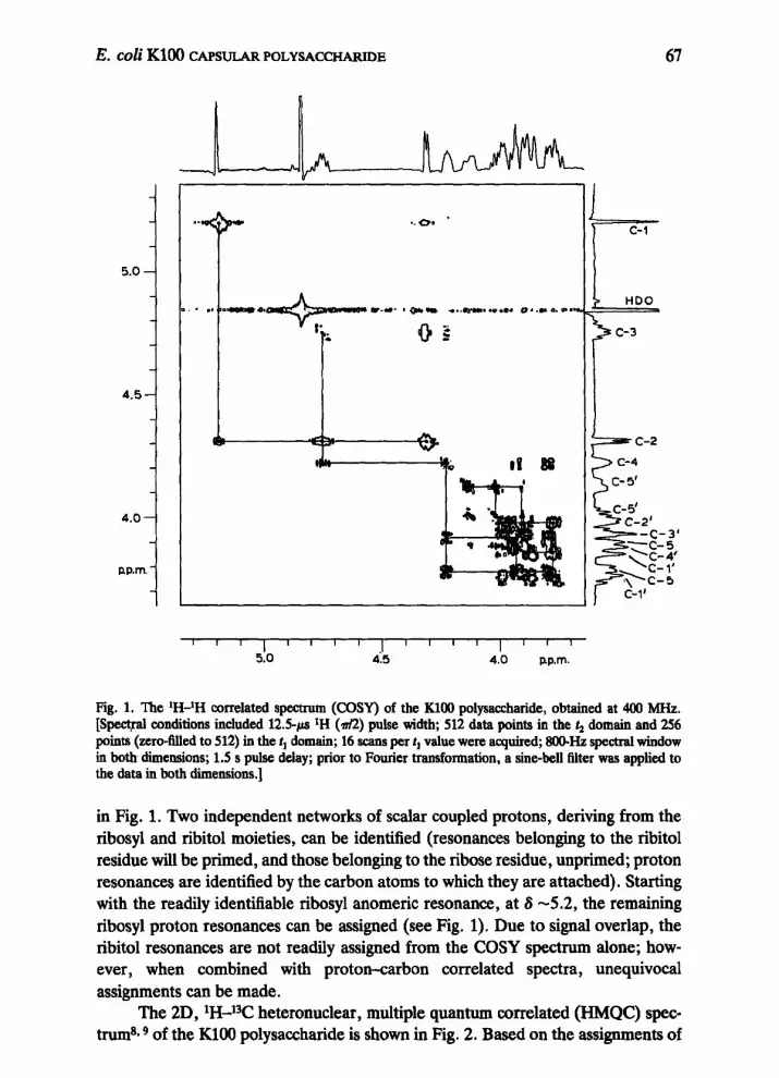
Polysaccharide linkage-sites may be readily determined by using a combina- tion of one- and two-dimensional n.m.r. techniques, as recently discussed8yg. The proton-proton correlated spectrum (COSY) of the KlOO polysaccharide is shown
TABLE I
IT-N.M.R. CHEMICAL SHIFIS FOR THE Escherichia coli KlOO CAPSULAR POLYSACCHARIDE’
Carbon atom Chemical shift
Ribose C-l c-2 c-3 C-4 c-5
Ribitol C-l c-2 c-3 c-4 C-5
108.8 76.9 (2.4) 76.1(5.5) 84.4 (6.1) 63.8
63.0 82.5 72.6 73.0 (7.3) 6!?.7 (5.5)
‘y3hemical shifts are relative to sodium 2,2,3,3-tetradeuterio-4,4dimethyl-4-silapentanoate as the internal standard. Values of 31P-13C coupling constants are given in parentheses.
E. Coli KlOO CAPSULAR POLYSACCHARIDE
5.0 -
4.5-
4.0-
nP.m. -
67
> c- 5’
c-5' C-2'
F -C-3' TC-5,
\:I;?
+c-5 C-l'
Fig. 1. The ‘H-lH correlated spectrum (COW) of the KlOO polysaccharide, obtained at 400 MHZ. [Spectral conditions included 12.59.~ *H (&?) pulse width; 512 data points in the 4 domain and 256 points (zero-tiled to 512) in the tI domain; 16 scans per t, value were acquired; SO@Hz spectral window in both dimensions; 1.5 s pulse delay; prior to Fourier transformation, a sine-bell filter was applied to the data in both dimensions.]
in Fig. 1. Two independent networks of scalar coupled protons, deriving from the ribosyl and ribitol moieties, can be identified (resonances belonging to the ribitol residue will be primed, and those belonging to the ribose residue, unprimed; proton resonances are identified by the carbon atoms to which they are attached). Starting with the readily identifiable ribosyl anomeric resonance, at S -5.2, the remaining ribosyl proton resonances can be assigned (see Fig. 1). Due to signal overlap, the ribitol resonances are not readily assigned from the COSY spectrum alone; how- ever, when combined with proton-carbon correlated spectra, unequivocal assignments can be made.
The 2D, lH-13C heteronuclear, multiple quantum correlated (HMQC) spec- trum8~ 9 of the KlOO polysaccharide is shown in Fig. 2. Based on the assignments of

68 F.-P. TSUI, W. EGAN, M. F. SUMMERS, R. A. BYRD, R. SCH~ERSON. J. B. ROBBJNS
p.p.m.
60
Ill~llll~llll~lTI1)1111~1111~1111~11ll(ll11
5.0 4.6 4.2 3.8 u.p.m.
Fig. 2. The LH-z3C muItiple quantum correlated (HMQC) spectrum of the KlOO capsular polysacchar- ide. me spectrometer had a nominal proton frequency of 400 MHz. The pulse sequence was as described in ref. 9 and in the Materials and Methods section. Acquisition parameters included: X2.5-@ (&) tH pulse; 70-w (704 1% pulse; 512 data points in the rr dimension and 64 points in the t, dimension (zero-filled to 128 poins); 16 transients were collected for each 1, value; 0.8 s pulse-delay; 6 and 12 Hz Gaussian broadening in rZ and tt domains, respectively, prior to Fourier transformation.]
the ribosyl protons deriving from the COSY spectrum, ribosyl carbon resonances are unambiguously assigned V& the HMQC experiment9. Carbon atoms 1’ and 5’ of the ribitol residue each exhibits two correlation peaks in the 2D-HMQC spec- trum, because each of these carbon atoms is directly bonded to two chemically shifted protons (and similarly for C-5 of the ribosyl residue). This multiplicity feature of the HMQC ~xpc~ment thus identifies the C-l ’ and C-5’ resonances of ribitol and their respective proton resonances; the remaining resonance assign- ments, namely, those for the C-2’, C-3’, and C-4’ atoms and their directly attached

E. coli KlOO CAPSULAR POLYSACCHARIDE 69
z 8 n I
I -- J
-70
c-4’ 00 c-4’ -
*
c-3 0 &C-3 -
J -80
c-2’0 1 c-210 1
c-4 9 c-4 Q
, 0 I -90
-100 6
0
I ” ” I ” ” 1 ““I”’
c ”
C-l'0 - p.P.m.
5.5 5.0 4.5 4.0 p.p.m.
Fig. 3. The 1H-13C multiple bond correlation (HMBC) spectrum of the KlOO pulysaccharide. [The spectrometer had a nominal proton frequency of 400 MHz. Acquisition parameters included 12.5-c (T&I) IH pulse; 45+s (m’2) 13C pulse; 512 and 128 data points were collected in the tz and 1, dimensions, respectively. 1600 and 8333 Hz spectral windows in l2 and t, dimensions; 3.4 ms AI and 50 ms A2 delay values; sine bell and Gaussian (30 Hz) filters were applied in 4 and t,, respectively.]
protons, can now be established relative to those for C-l’ and C-5’ through the
combined use of COSY and HMQC spectra. The n.m.r. experiments establish conmcrivities; they do not, however, distinguish between the diastereoisomeric possibilities resulting from attachment of ribose (or phosphate) at the pro-(R) or pro-(S) sites of ribitol, a meso-alditol (except, trivially, by comparison of n.m.r. signals with those given by authentic compounds). In the following discussion of

70 F.-P. TSUI, W. EGAN, M. F. SUMMERS, R. A. BYRD, R. SCHNEERSON, J. B. ROBBINS
I I I I I I I I I I I I I I , , I
5.0 4.5 4.0 ppm. 3.5
Fig. 4. The IH-n.m.r. spectrum (400 MHz) of the KlOO capsular polysaccharide (25 mg in 0.6 mL) at 20” and pH 7.0 (top tracing); spin-echo difference, ‘H-n.m.r. spectrum of the same sample (bottom tracing).
the determination of linkage sites is presented one assignment set, which will be justified through chemical degradation studies (see later).
To establish the ribosyl-ribitol linkage, a heteronuclear, multiple-bond, cor- relation spectrum (HMBC) was obtained. In this experiment*lg, correlations are established through long-range (i.e., two- and three-bond) lH-13C scalar cou- pling@. The HMBC results are shown in Fig. 3. The majority of the observed cross- peaks result from intraresidue, long-range C-H scalar couplings. Additionally, however, a strong interresidue correlation peak (as marked by the arrow in Fig. 3) is observed between the ribosyl H-l atom and C-2’ of ribitol. This correlation un- ambiguously establishes that the ribosyl C-l atom is linked to C-2’ of the ribitol residue. This linkage site could, alternatively, have been established by using a one-dimensional, selective INEPT experiment, as was done for the H. influenzue type b capsular polysaccharide ia. The HMBC experiment has the additional feature, relative to the selective INEPT experiment, that it can simultaneously confirm resonance assignments established by the COSY and HMQC experiments; however, the HMBC experiment is more exacting in terms of spectrometer hardware than is the selective INEPT experiment (see Materials and Methods and refs. 8 and 9).

E. Co~iKl~CAPSULARPOLYSACCHARIDE 71
The sites of attachment of the phosphate group were determined by using a spin-echo difference (SED) technique. In this experiment (see ref. 9 and citations therein), the observed lH resonances derive from protons that are scalar coupled to phosphorus (1 b-1 Hz), namely, those on the carbon atoms contiguous with the phosphate group. Figure 4 shows a normal 1D spectrum of the KlOO polysaccharide (bottom tracing) and an SED spectrum of the same sample (top tracing). The SED spectrum clearly shows that the phosphate group is linked to C-3 of ribose and C5’ of ribitol.
Although the aforementioned combination of n.m.r. techniques is useful for resonance assignments and for the determination of linkage sites, it does not directly establish (i) the ring form (pyranoid or furanoid), (ii) the absolute stereochemistry, and (iii) the anomeric stereochemistry. We now address these structural features. Using all of the 13C-n.m.r. resonance assignments already provided and the chemical-shift correlations with those of model compounds7, it was readily established that the ribosyl residue is in the furanoid form and, moreover, that it has the /3-stereochemistry at C-1. The presence of cross-peaks between the ribosyl H-l and the ribosyl C-3 and C-4 in the HMBC spectrum is consistent with ribose having the furanoid, as opposed to the pyranoid, forn$O.
The ribose was determined to have the D absolute configuration, as does the H. influenzae type b polymerll. The procedure of Leontein et al.‘*, wherein the mixture of glycosides formed by condensation of ribose with optically active 2- octanol is analyzed by g.l.c., was used to determine the absolute configuration of the ribose. As established by the n.m.r. experiments, D-ribose is linked to the penultimate carbon atom of ribitol; however, as noted, the n.m.r. experiments do not distinguish between linkage to pro-(R) or pro-(S) carbon atoms, i.e., C-4’ or C-2’ of D-ribitol. The distinction was made by chemical degradation, as described for a ribitol teichoic acid by Archibald et aZ.13, except that, in the present study, the intact polymer was oxidized and the absolute configuration of the resultant glyceric acid was established by n.m.r. spectroscopy. Thus, the KlOO polymer was oxidized with sodium metaperiodate, and the resulting aldehydic functionalities were further oxidized to carboxylates by means of aqueous Br,; following acid-catalyzed hydro- lysis of this oxidized material, L-glyceric acid was liberated, establishing that the ribose is linked to O-2’ of D-ribitol. L-Glyceric acid, as the ester formed with optically active S-(-)-2-octanol, was identified by ‘H-n.m.r. spectroscopy by com- parison with authentic D- and L-glyceric acid esters with S-( -)-Zoctanol. It may be noted that glyceric acid could be produced from C-3-C-5 of ribose in residues that were not phosphorylated at C-3 (such as end groups); however, the resultant glyceric acid would have the D absolute configuration. We may now write the struc- ture of the KlOO polysaccharide as ~3)-gD-Ribf-(1~2)-D-ribitol-5-(PO,,.
The results of additional chemical studies, particularly those involving periodate oxidation, were in accord with the structure depicted. These studies were noted in a review7*, and are not further discussed.
The H. injhenzae type b capsular polysaccharide has the repeat unit struc-

72
-
-
HO-
b-OH
d E.coli k 100 -I
F.-P. TSUI, W. EGAN, M. F. SUMMERS, R. A. BYRD, R. SCHNEERSON, J. B. ROBBINS
H
6 -OH
P,
H. influenzae. type b
ture” shown. The KlOO and type b polysaccharides are thus seen to be very similar in structure, differing only in the linkage from the ribosyl anomeric carbon atom to
the ribitol. This slight variation in structure is consistent with the high degree of immunologic relatedness.
MATERIALS AND METHODS
Isolation of the polysaccharides. - The capsular polysaccharide from E. coli strains “Easter” (075:KlOOLH5), “89” (075:KlOO:H5), and “HB-20” (07:K100:H5)2 were isolated as described 14. Analysis for protein, nucleic acids, endotoxin, and moisture are summarized as follows: for strain “Easter”, the strain
that was utilized for studies reported herein: Kd (Sepharose 4B) 0.46; moisture (% w/w) 15; nucleic acid (% , w/w) 0.1; protein (%, w/w) 0.14; phosphorus (pg/mg)
71.7; ribose (&mg) 360; ribitol + anhydroribitol (&mg) 349. The polysacchar-
ides from the two other strains were compared by *jC-n.m.r. spectroscopy and found to be identical to strain “Easter”.
Polysaccharide sugar analysis. - Hydrolysis of the native and sodium
borohydride-reduced materials were conducted as described, using methane- sulfonic acid15. Neutral, reducing sugars were analyzed underivatized in an auto-

E. co/i KlOO CAPSULARPOLYSACCHARIDE 73
mated sugar analyzer 15. The acid hydrolyzate was also examined by g.1.c.; sugars were identified as their trimethylsilyl derivatives by comparison with authentic samples (Pfanstiehl Laboratories, Inc., Waukegan, IL); a Varian Associates gas- liquid chromatograph equipped with a methylsilicone glass capillary column (SP- 2100) was used.
Phosphorus analysis. - The phosphorus content of the KlOO polysaccharide was determined as inorganic phosphate according to the method of Chen et a1.16.
Synthesis of S-2-octyl-D and -bgZycerates. -D-(+)- and r_-(-)-Glyceric acids (purchased as the hemi-calcium salts; Sigma Chemical Co., St. Louis, MO) were, in separate experiments, esterified with S-(-)-2-octanol (Aldrich Chemical Co., Milwaukee, WI), according to the procedure of Mathias”, involving the CuO- catalyzed activation of the alcohol with dicyclohexylcarbodiimide (DCC). Follow- ing acetylation with A@-pyridine the resulting esters were purified by preparative g.1.c. on a column (183 cm x 6.35 mm) of 15% of SE-30. The material collected was examined by ‘H-n.m.r. spectroscopy.
Preparation of S-2-octyl +glycerate from the KloO polysaccharide. - The KlOO polysaccharide was oxidized with an excess of NaIO,, and then with13 aqueous Br,. Following acid-catalyzed hydrolysis, the resultant glyceric acid was treated with the DCC-S-2-octanol isourea. Following acetylation, and removal of the excess of reagents with a stream of nitrogen gas, the desired ester was purified by preparative g.l.c., as already described. The absolute configuration of the glyceric acid was established as L, based on the ‘H-n.m.r. spectrum of the ester.
N.m. r. spectroscopy. - The HMQC, HMBC, and SED spectra were re- corded with a JEOL GX-400 n.m.r. spectrometer (nominal proton frequency, 400 MHz). The GX-400 n.m.r. spectrometer required several minor modifications in order to perform the proton-detected, carbon-decoupled n.m.r. experiments. Inter- ference between the *H-lock channel and the i3C-decoupling channel was minimized by (i) gating the lock receiver off during ‘3C-irradiation; (ii) inserting tumable band-pass and band-reject filters (K & L Microwave, Salisbury, MD) into the 13C-irradiation and lock channels (before the lock pre-amplifier), respectively; and (iii) rtioving the narrow band-pass crystal filters in the lock receiver of the spectrometer. Additionally, the 13C-irradiation was routed out of the spectrometer at an intermediate stage of amplification; this enabled a selectable (l-25 W), but constant, amplitude, radiofrequency power level for the 13C-pulses and decoupling. These measures were essential to obtaining proper signal cancellations in the ‘H-de- coupled HMQC spectra. ‘H-N.m.r. spectra at 300 MHz and 13C-n.m.r. spectra at 25 MHz were recorded with a Bruker WM-300 and a JEOL FX-100 n.m.r. spectro- meter, respectively.
Specific details of the collection of data are to be found in the Figure legends.
REFERENCES
1 J. B. ROBBINS, R. SCHNEF~RSON, AND M. PITTMAN, in R. GERMANIER (Ed.), Brzcterial Vaccines, Academic Press, New York, 1984, pp. 289-316.

74 F.-P. TSUI, W. EGAN, M. F. SUMMERS, R. A. BYRD, R. SCHNEERSON, 1. B. ROBBINS
2 R. SCHNEERSON, M. BRADSHAW, J. K. WHISNANT, R. L. MYEROWITZ, J. C. PARKE, AND J. B. ROBBIIVS, J. Immunol., 108 (1972) 1551-1562.
3 J. B. ROBBINS, R. SCHN~ERSON, J. C. PARKE, T.-Y. LIU, Z. T. HANDZEL, 1. ~RSKOV. AND F. Q)RSKOV, in R. F. BEERS AND E. BASSETT (Eds.), The Role of Immunological Factors in Infectious, AIlergic, and Autoimmune D&eases, Raven Press, New York, 1976, pp. 103-120.
4 R. SCHNEERSONAND J. B. ROBBINS, New England 1. Med., 292 (1975) 1093-1096. 5 J. BADDILEY, J. G. BUCHANAN, AND B. CARSS, J. Chem. Sot., (1957) 4058-4063. 6 W. EGAN, F.-P. Tsur, AND R. SCHNEERSON, Carbohydr. Res., 79 (1980) 271-277. 7 (a) W. EGAN, Map. Reson. Biob, l(l980) 198-258; (b) E. BARRETO-BERGTER AND P. A. J. GORIN,
Adv. Carbohydr. Chem. Biochem., 41 (1983) 67-103. 8 L. LERNERAND A. BAX, Carbohydr. Res., 166 (1987) 35-46. 9 R. A. BYRD, W. EGAN, M. F. QJMMERS, AND A. BAX, Carbohydr. Res., 166 (1987) 47-58.
10 A. BAX, W. EGAN. AND P. KovAcs, J. Carbohydr. Chem., 3 (1984) 593-611. 11 P. BRANEFORS-HELANDER, C. ERBING, L. KENNE. AND B. LINDBERG, Acru Chem. &and., Ser. B.,
30 (1976) 276-277. 12 K. LEONTEIN, B. LINDBERG, AND J. LONNGREN, Carbohydr. Res., 62 (1978) 359-362. 13 A. R. ARCHIBALD, J. BADDILEY, AND J. G. BUCHANAN, Biochem. J., 81 (1961) 126134. 14 E, C. GOTSCHLICH, M. REY, C. ETIENNE, W. R. SANBORN, R. TRIAN, AND B. C~EJTANOVIC, Prog.
Immunol. Stand., 5 ( 1972) 485-491. 15 R. A. BOYKINS AND T.-Y. LIU, Biochem. Biophys. Metho& 2 (1980) 71-78. 16 P. S. CHEN, T. Y. TORIBARA. AND H. WARNER, Anal. Chem., 28 (1956) 17564758. 17 L. J. MATHIAS, Synthesis, (1979) 561-576.


