Embed Size (px)
Citation preview

Biochem. J. (2000) 347, 321–337 (Printed in Great Britain) 321
REVIEW ARTICLE
Regulation of cytochrome P450 (CYP) genes by nuclear receptorsPaavo HONKAKOSKI*1 and Masahiko NEGISHI†*Department of Pharmaceutics, University of Kuopio, P. O. Box 1627, FIN-70211 Kuopio, Finland, and †Pharmacogenetics Section,Laboratory of Reproductive and Developmental Toxicology, NIEHS, National Institutes of Health, Research Triangle Park, NC 27709, U.S.A.
Members of the nuclear-receptor superfamily mediate crucial
physiological functions by regulating the synthesis of their target
genes. Nuclear receptors are usually activated by ligand binding.
Cytochrome P450 (CYP) isoforms often catalyse both formation
and degradation of these ligands. CYPs also metabolize many
exogenous compounds, some of which may act as activators of
nuclear receptors and disruptors of endocrine and cellular
INTRODUCTION
Overview of the cytochrome P450 (CYP) superfamily
CYPs constitute a superfamily of haem-thiolate proteins present
in prokaryotes and throughout the eukaryotes. CYPs act as
mono-oxygenases, with functions ranging from the synthesis and
degradation of endogenous steroid hormones, vitamins and fatty
acid derivatives (‘endobiotics ’) to the metabolism of foreign
compounds such as drugs, environmental pollutants, and
carcinogens (‘xenobiotics ’) [1]. At present, 17 mammalian CYP
gene families collectively encode about 60 distinct CYP forms in
any given species [2], a number expected to rise by the completion
of genome-wide sequencing projects. On the basis of crystal
structures of bacterial P450s, molecular modelling and site-
directed mutagenesis, the overall structure of mammalian
membrane-bound P450 has been deduced and residues required
for substrate binding, electron transfer and haem binding have
been identified [3,4].
CYPs in gene families 1–4 exhibit broad, but overlapping,
substrate and product specificities that may vary between cor-
responding forms from different species [1,5]. Their ability to
metabolize a wide array of xenobiotics [6], the inducibility of
many CYP forms by xenobiotics [7–9] and documented gene
polymorphisms [10,11] have all contributed to an explosion of
literature on CYP-dependent drug metabolism. To name just a
few examples, differences in the amounts and intrinsic capacities
of CYP forms to metabolize a particular drug or chemical
may influence profoundly drug–drug interactions, drug or car-
cinogen activation and detoxification, or species differences
in CYP-catalysed reactions of toxic chemicals. The CYPs in
families 1–4 also metabolize endogenous compounds, including
steroids and bile acids, fatty acids, prostaglandins and other
eicosanoids, and retinoids [6,12–15]. They also display complex
Abbreviations used: AF-1 and AF-2, activation functions 1 and 2; AhR, aryl-hydrocarbon receptor ; Arnt, AhR nuclear translocator ; CAR, constitutivelyactive receptor ; CPF, CYP7A promoter-binding factor ; CYP, cytochrome P450; DBD, DNA-binding domain; DEX, dexamethasone; DRn, IRn and ERn,direct, inverted and everted repeat with n bp spacing; ER, oestrogen receptor ; FXR, farnesoid X receptor ; GH, growth hormone; GR, glucocorticoidreceptor ; HNF-4, hepatocyte nuclear factor 4 ; LBD, ligand-binding domain; LXR, liver X receptor ; NFI, nuclear factor I ; NR, nuclear receptor ; PB,phenobarbital ; PBREM, PB-responsive enhancer module ; PCN, pregnenolone 16α-carbonitrile ; PPAR, peroxisome proliferator-activated receptor ;PXR, pregnane X receptor ; RA, retinoic acid ; RAR, retinoic acid receptor ; RXR, retinoid X receptor ; SF-1, steroidogenic factor 1 ; SHP, shortheterodimerization partner ; TCDD, 2,3,7,8-tetrachlorodibenzo-p-dioxin ; TR, thyroid-hormone receptor ; VDR, vitamin D receptor.
1 To whom correspondence should be addressed (e-mail paavo.honkakoski!uku.fi).
homoeostasis. This review summarizes recent findings that in-
dicate that major classes of CYP genes are selectively regulated
by certain ligand-activated nuclear receptors, thus creating tightly
controlled networks.
Key words: endobiotic metabolism, gene expression, gene tran-
scription, ligand-activated, xenobiotic metabolism.
sex-, tissue- and development-specific expression patterns which
are controlled by hormones or growth factors [16], suggesting
that these CYPs may have critical roles, not only in elimination
of endobiotic signalling molecules, but also in their production
[17]. Data from CYP gene disruptions and natural mutations
support this view (see e.g. [18,19]).
Other mammalian CYPs have a prominent role in biosynthetic
pathways. CYPs belonging to gene families 5 and 8A are involved
in thromboxane and prostacyclin synthesis, CYPs from families
11, 17, 19, and 21 are required for steroid-hormone biosynthesis,
CYPs from families 7, 8B, 24, 27, 46, and 51 catalyse reactions
in the pathways leading to the biosynthesis of bile acid, vitamin
D$and cholesterol and CYP26 is involved in retinoid metabolism
[1,2]. These CYPs usually have selective substrate specificities
and they are subject to tight tissue-specific and hormone-
dependent regulation [20,21]. In addition, mutations in the
structural genes for these CYPs underlie some common and
severe inherited diseases [22,23].
Thus CYP genes are uniquely positioned (i) to respond to both
endogenous and exogenous signals by changes in CYP gene
expression, and (ii) to modulate the strength and duration of
these signals and even to form new signalling molecules through
CYP-mediated metabolism (Figure 1). These signalling molecules
may then exert their function via the ligand-dependent nuclear
receptors described below.
Overview of the nuclear receptor (NR) superfamily
The NR superfamily codes for transcription factors that trans-
form extracellular and intracellular signals into cellular responses
by triggering the transcription of NR target genes. NRs share
significant similarity with classical steroid-hormone and thyroid-
hormone receptors (TRs) in their DNA-binding domain (DBD)
# 2000 Biochemical Society
322 P. Honkakoski and M. Negishi
CYP genesX, E XO, EO
Othergenes
CYP genes
Othergenes
NRs
CYP-dependent metabolismof xenobiotics and endobiotics
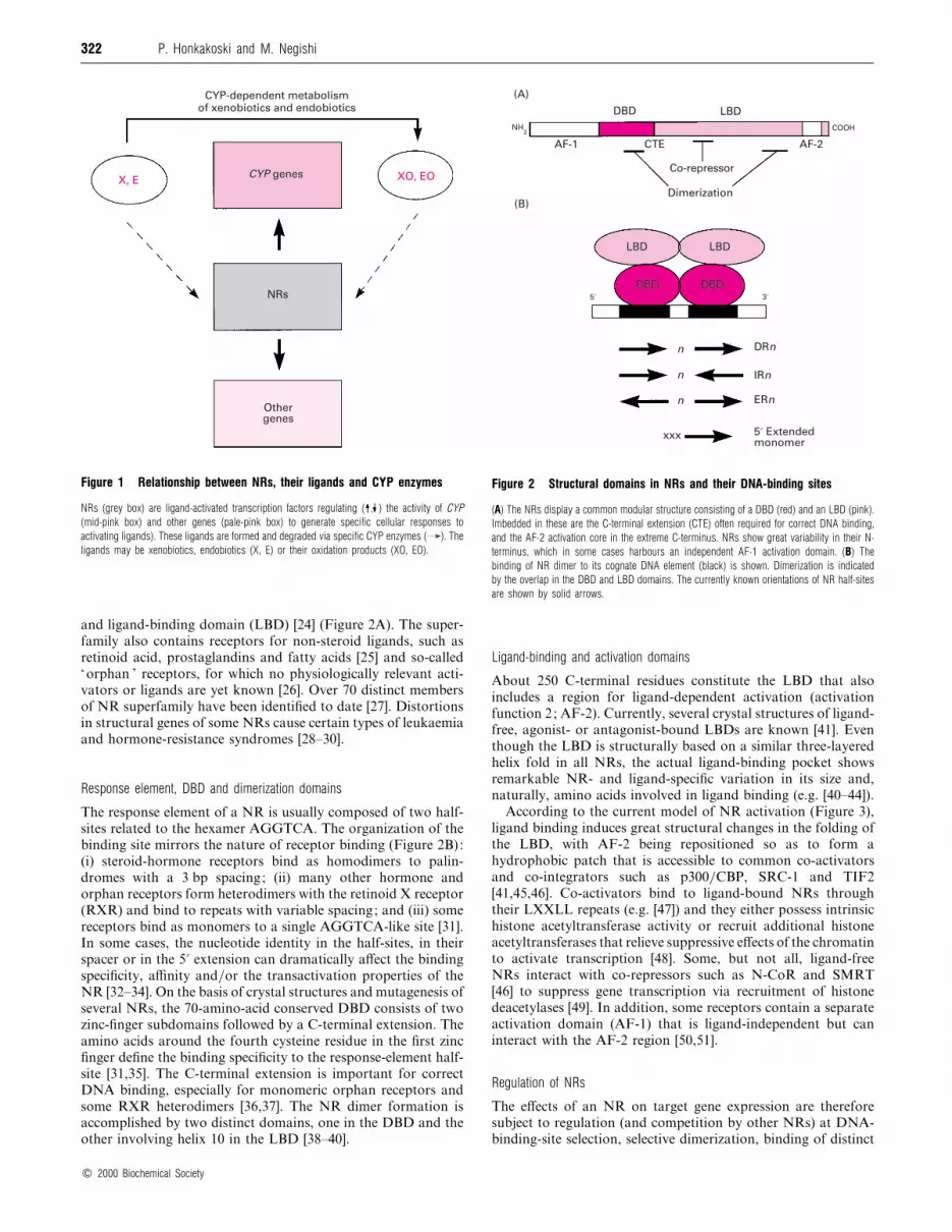
Figure 1 Relationship between NRs, their ligands and CYP enzymes
NRs (grey box) are ligand-activated transcription factors regulating ( , ) the activity of CYP(mid-pink box) and other genes (pale-pink box) to generate specific cellular responses to
activating ligands). These ligands are formed and degraded via specific CYP enzymes ( ). The
ligands may be xenobiotics, endobiotics (X, E) or their oxidation products (XO, EO).
and ligand-binding domain (LBD) [24] (Figure 2A). The super-
family also contains receptors for non-steroid ligands, such as
retinoid acid, prostaglandins and fatty acids [25] and so-called
‘orphan ’ receptors, for which no physiologically relevant acti-
vators or ligands are yet known [26]. Over 70 distinct members
of NR superfamily have been identified to date [27]. Distortions
in structural genes of some NRs cause certain types of leukaemia
and hormone-resistance syndromes [28–30].
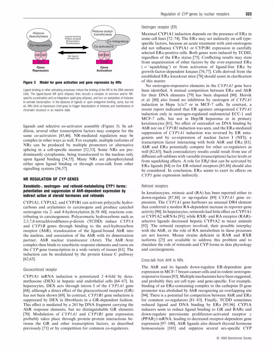
Response element, DBD and dimerization domains
The response element of a NR is usually composed of two half-
sites related to the hexamer AGGTCA. The organization of the
binding site mirrors the nature of receptor binding (Figure 2B):
(i) steroid-hormone receptors bind as homodimers to palin-
dromes with a 3 bp spacing; (ii) many other hormone and
orphan receptors form heterodimers with the retinoid X receptor
(RXR) and bind to repeats with variable spacing; and (iii) some
receptors bind as monomers to a single AGGTCA-like site [31].
In some cases, the nucleotide identity in the half-sites, in their
spacer or in the 5« extension can dramatically affect the binding
specificity, affinity and}or the transactivation properties of the
NR [32–34]. On the basis of crystal structures and mutagenesis of
several NRs, the 70-amino-acid conserved DBD consists of two
zinc-finger subdomains followed by a C-terminal extension. The
amino acids around the fourth cysteine residue in the first zinc
finger define the binding specificity to the response-element half-
site [31,35]. The C-terminal extension is important for correct
DNA binding, especially for monomeric orphan receptors and
some RXR heterodimers [36,37]. The NR dimer formation is
accomplished by two distinct domains, one in the DBD and the
other involving helix 10 in the LBD [38–40].
LBD LBD
DBD DBD
AF-2CTEAF-1
COOH
LBDDBD
(A)
(B)
NH2
LBD LBD
DBD DBD
DRn
IRn
ERn
n
n
n
xxx 5« Extendedmonomer
5« 3«
Co-repressor
Dimerization
Figure 2 Structural domains in NRs and their DNA-binding sites
(A) The NRs display a common modular structure consisting of a DBD (red) and an LBD (pink).
Imbedded in these are the C-terminal extension (CTE) often required for correct DNA binding,
and the AF-2 activation core in the extreme C-terminus. NRs show great variability in their N-
terminus, which in some cases harbours an independent AF-1 activation domain. (B) The
binding of NR dimer to its cognate DNA element (black) is shown. Dimerization is indicated
by the overlap in the DBD and LBD domains. The currently known orientations of NR half-sites
are shown by solid arrows.
Ligand-binding and activation domains
About 250 C-terminal residues constitute the LBD that also
includes a region for ligand-dependent activation (activation
function 2; AF-2). Currently, several crystal structures of ligand-
free, agonist- or antagonist-bound LBDs are known [41]. Even
though the LBD is structurally based on a similar three-layered
helix fold in all NRs, the actual ligand-binding pocket shows
remarkable NR- and ligand-specific variation in its size and,
naturally, amino acids involved in ligand binding (e.g. [40–44]).
According to the current model of NR activation (Figure 3),
ligand binding induces great structural changes in the folding of
the LBD, with AF-2 being repositioned so as to form a
hydrophobic patch that is accessible to common co-activators
and co-integrators such as p300}CBP, SRC-1 and TIF2
[41,45,46]. Co-activators bind to ligand-bound NRs through
their LXXLL repeats (e.g. [47]) and they either possess intrinsic
histone acetyltransferase activity or recruit additional histone
acetyltransferases that relieve suppressive effects of the chromatin
to activate transcription [48]. Some, but not all, ligand-free
NRs interact with co-repressors such as N-CoR and SMRT
[46] to suppress gene transcription via recruitment of histone
deacetylases [49]. In addition, some receptors contain a separate
activation domain (AF-1) that is ligand-independent but can
interact with the AF-2 region [50,51].
Regulation of NRs
The effects of an NR on target gene expression are therefore
subject to regulation (and competition by other NRs) at DNA-
binding-site selection, selective dimerization, binding of distinct
# 2000 Biochemical Society
323Regulation of CYP genes by nuclear receptors
NR
DNA
NR NR NR
DNA
Histonedeacetylases
Co-repressors
Histone acetyl-transferases
Co-activators
Co-integrators
NR NR
DNA
GeneActivation
Co-repressors
NR
DNA
GeneRepression
NR No ligandAntagonist
Ligand bindingProtein kinases
DimerizationTranslocation
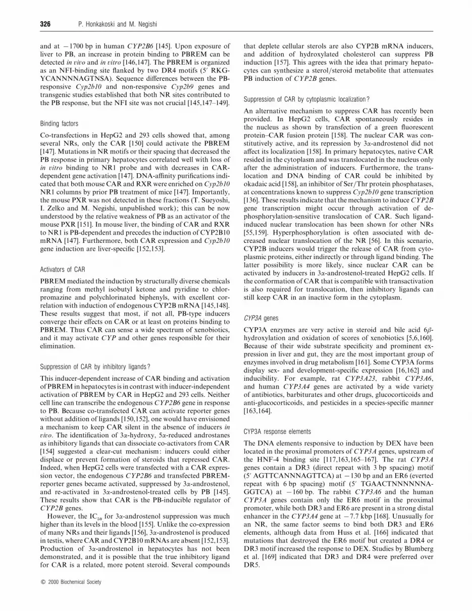
Figure 3 Model for gene activation and gene repression by NRs
Ligand binding or other activating processes induce the binding of the NR to the DNA element
(red). The ligand-bound NR (pink ellipses) then recruits a complex of common and/or NR-
specific co-activators and co-integrators (pale-grey ellipses), and turn on acetylation of histones
to activate transcription. In the absence of ligands or upon antagonist binding, some, but not
all, NRs bind co-repressors (mid-grey) to trigger deacetylation of histones and maintenance of
chromatin structure in an inactive state.
ligands and selective co-activator assembly (Figure 3). In ad-
dition, several other transcription factors may compete for the
same co-activators [45,46]. NR-mediated regulation may be
complex in other ways as well. For example, multiple isoforms of
NRs can be produced by multiple promoters or alternative
splicing in a cell-specific manner [52,53]. Some NRs are pre-
dominantly cytoplasmic, being translocated to the nucleus only
upon ligand binding [54,55]. Many NRs are phosphorylated
either upon ligand binding or through cross-talk from other
signalling systems [56,57].
NR REGULATION OF CYP GENES
Xenobiotic-, oestrogen- and retinoid-metabolizing CYP1 forms :potentiation and suppression of AhR-dependent expression byindirect action of steroid hormones and retinoids
CYP1A1, CYP1A2, and CYP1B1 can activate polycyclic hydro-
carbons and arylamines to carcinogens and produce catechol
oestrogens via 2- and 4-hydroxylation [6,58–60], reactions con-
tributing to carcinogenesis. Polyaromatic hydrocarbons such as
2,3,7,8-tetrachlorodibenzo-p-dioxin (TCDD) induce CYP1A
and CYP1B genes through binding to the aryl-hydrocarbon
receptor (AhR), translocation of the ligand-bound AhR into
the nucleus, and association of the AhR with its dimerization
partner, AhR nuclear translocator (Arnt). The AhR[Arnt
complex then binds to xenobiotic response elements and turns on
the CYP gene transcription in a wide variety of tissues [61]. The
induction can be modulated by the protein kinase C pathway
[62,63].
Glucocorticoid receptor
CYP1A1 mRNA induction is potentiated 2–4-fold by dexa-
methasone (DEX) in hepatic and endothelial cells [64–67]. In
hepatocytes, DEX acts through intron I of the CYP1A1 gene
[68], although a direct effect of the glucocorticoid receptor (GR)
has not been shown [69]. In contrast, CYP1B1 gene induction is
suppressed by DEX in fibroblasts in a GR-dependent fashion.
This effect is mediated by a 265 bp DNA fragment carrying the
AhR response elements, but no distinguishable GR elements
[70]. Modulation of CYP1A1 and CYP1B1 gene expression
probably takes place through protein–protein interactions be-
tween the GR and other transcription factors, as described
previously [71] or by competition for common co-regulators.
Oestrogen receptor (ER)
Maximal CYP1A1 induction depends on the presence of ERα in
some cell lines [72–74]. The ERα may act indirectly on cell type-
specific factors, because an acute treatment with anti-oestrogens
did not influence CYP1A1 or CYP1B1 expression in carefully
selected ERα-positive cells. Both genes were induced by TCDD,
regardless of the ERα status [75]. Conflicting results may stem
from sequestration of other factors by the over-expressed ERα
(¯ ‘ squelching’) or from activation of ligand-free ERα by
growth-factor-dependent kinases [76,77]. Cells derived from the
established ERα knockout mice [78] should assist in clarification
of this matter.
No oestrogen-responsive elements in the CYP1A1 gene have
been identified. A mutual competition between ERα and AhR
for their DNA elements [79] has been disputed [80]. Hoivik
et al. [80] also found no inhibition by oestrogen of CYP1A1
induction in Hepa 1c1c7 or in MCF-7 cells. In contrast, a
recent report indicated that ER agonists antagonized CYP1A1
induction only in oestrogen-regulated endometrial ECC-1 and
MCF-7 cells, but not in Hep3B hepatoma or in primary
keratinocytes [81]. No effect of oestradiol on DNA binding by
AhR nor on CYP1B1 induction was seen, and the ERα-mediated
suppression of CYP1A1 induction was reversed by ER anta-
gonists and by co-expression of nuclear factor I (NFI), a
transcription factor interacting with both AhR and ERα [81].
AhR and ERα potentially compete for other co-regulators as
well [82,83]. Such contradictory results could result from use of
different cell sublines with variable (transcription) factor levels or
from squelching effects. A role for ERβ that can be activated by
ERα ligands [84] or for ER-related receptors [85,86] should also
be considered. In conclusion, ERα seems to exert its effects on
CYP1 gene expression indirectly.
Retinoid receptors
In keratinocytes, retinoic acid (RA) has been reported either to
down-regulate [87,88] or up-regulate [89] CYP1A1 gene ex-
pression. The CYP1A1 gene harbours an unusual DR4 element
that conferred a modest RA-dependent increase in reporter-gene
activity [90]. In hepatocytes, retinoids had little effect on CYP1A1
or CYP1A2 mRNAs [91], while RXR- and RA receptor (RAR)-
selective ligands decreased hepatic CYP1A2 in intact animals
[92]. The retinoid receptors involved, their possible interplay
with the AhR, or the role of RA metabolism in these processes
are not known. Mouse strains deficient in RAR and RXR
isoforms [25] are available to address this problem and to
elucidate the role of retinoids and CYP forms in skin physiology
and carcinogenesis.
Cross-talk from AhR to NRs
The AhR and its ligands down-regulate ER-dependent gene
expression in MCF-7 breast-cancer cells and in rodent oestrogen-
responsive tissues [93]. Multiplemechanisms have been suggested,
and probably they are cell type- and gene-specific. For example,
binding of an ERα-containing complex to the cathepsin D gene
promoter was abolished by AhR recognizing an overlapping site
[94]. There is a potential for competition between AhR and ERα
for common co-regulators [81–83]. Finally, TCDD sometimes
reduced ligand and DNA binding by ERα [95,96]. CYP1A
inducers seem to reduce ligand binding to GR and RARs and
down-regulate peroxisome proliferator-activated receptor γ
(PPARγ) mRNA, leading to decreased receptor-dependent gene
expression [97–100]. AhR ligands also disturb thyroid hormone
homoeostasis [101] and suppress several sex-specific CYP
# 2000 Biochemical Society

324 P. Honkakoski and M. Negishi
mRNAs [102,103]. The nature of this general suppression of
cellular functions by AhR ligands is not clear. It may reflect the
limiting cellular amounts of co-activators and probably contri-
butes to TCDD toxicity.
The disruption of AhR gene leads to a marked liver disease
[104] and elevated hepatic levels of retinoids due to a decrease in
CYP-mediated catabolism of RA [18]. Intriguingly, this defect is
not due to the RA-inducible 4-hydroxylase CYP26 [18]. CYP1A
enzymes are known to metabolize retinoids [105,106], and intact
AhR is required for the basal expression of CYP1A2 [104]. Even
though the mouse CYP1A2 does not metabolize RA [18], it may
metabolize other retinoids [14], and thus the loss of CYP1A2
might contribute to retinoid accumulation in AhR null mice. This
hypothesis could be tested in Cyp1a2 null mice [107].
Steroid-metabolizing liver- and sex-specific CYP2A, CYP2C,CYP2D, and CYP3A forms : hepatocyte nuclear factor 4 (HNF-4)and orphan receptors contribute to basal expression and ERαgoverns sexual dimorphism
Members of families CYP2 and CYP3A metabolize efficiently
both xenobiotics and endogenous compounds [6,7,12]. They also
display complex developmental, species-dependent and sexually
dimorphic patterns of regulation that may substantially differ
even between closely related CYP isoforms [16]. Several P450s in
this group exhibit liver-specific or liver-predominant expression
driven by distinct liver-enriched transcription factors [108,109].
Intriguingly, several sex-specific and growth- hormone (GH)-
dependent CYP mRNAs is suppressed by chemicals that can
activate AhR or NRs [102,103,110].
HNF-4
Comparison of CYP2A, CYP2C, and CYP2D gene sequences
indicated that many promoters contain a motif (HPF1) around
®100 bp that is important for binding of HepG2-specific proteins
and transcriptional activity of rabbit CYP2C genes [111]. The
consensus HPF-1 motif (5« RRRNCAAAGKNCANYY; see
Table 1) resembled the binding site for hepatocyte nuclear factor
4 (HNF-4), an liver-enriched orphan receptor that binds to DNA
as a homodimer [24,112]. In �itro competition and antibody
supershift assays, and co-transfection of HNF-4, indicated that
HNF-4 was the major regulator for rabbit CYP2C genes in liver
[113,114]. HNF-4 or HPF-1 motifs were also shown important
for activation of human CYP2C9 [115], mouse Cyp2a4 [116], rat
CYP3A [117,118] and mouse Cyp2d9 genes [119].
Orphan receptors
Related rat CYP2C7, CYP2C11, CYP2C12 and CYP2C13 genes
seem less dependent on HNF-4 [120,121]. Although HNF-4
could recognize the HPF-1-like elements in almost every CYP2C
gene, most of the binding in rat liver nuclear extracts was not due
to HNF-4; mutation of HPF-1 motifs did not significantly
reduce CYP2C promoter activity in HepG2 cells ; and co-
expression of HNF-4 in non-hepatic cells had only a marginal
effect on the promoter activity [120,121]. However, the sequences
of virtually all rat CYP2C elements do not completely conform
to the HPF-1 motif or HNF-4 site [111,112], but differ at critical
positions [111,114]. It appears that orphan receptors such as ear-
2 and ear-3 work in concert with other transcription factors to
regulate the basal CYP2C13 gene expression [120]. The same or
related orphan receptors may influence CYP3A gene expression
as well [117,118].
Even though the activities of rabbit CYP2C gene promoters
correlated well with their HNF-4-binding affinity, they did not
correspond to CYP2C mRNA levels in �i�o [114]. This suggests
that, in addition to HNF-4, other regulators are also important.
Nevertheless, the majority of the liver-specific CYP2A, CYP2C,
CYP2D and CYP3A genes are regulated by HNF-4 and related
orphan receptors. Small sequence variations within the binding
sites may lead to distinct binding preferences and, therefore, to
gene-specific regulation by various NRs.
ER
A new role for NRs in CYP gene transcription has recently
been discovered [122]. The sex-specific transcription of CYP2C11
and CYP2C12 genes is regulated by sexually distinct patterns of
GH secretion in the rat that can be abolished by neonatal
gonadectomy or hypophysectomy [123,124]. In the male mouse,
GH governs the expression pattern of Cyp2a4 and Cyp2d9 genes,
while in the female mouse, GH has little effect [125]. The GH-
elicited nuclear localization of transcription factor STAT5b in
the male mouse has emerged as the major regulator of the sex-
specific CYP gene expression [126]. The presence of a functional
ERα is absolutely required for proper sex-specific expression of
Cyp2a4 and Cyp2d9 genes in the liver [122]. Normally, STAT5b
is not detectable in nuclei from female liver. Disruption of ERα
leads to nuclear localization of STAT5b in females and appear-
ance of CYP2D9 mRNA; this masculinization can be abolished
by hypophysectomy. Thus ERα is probably needed for prog-
ramming of the hypophysis that involves sex-dependent expr-
ession of neuronal aromatase (CYP19) [127]. The recent Cyp19-
null mouse [128] could be useful in tackling this problem: even
though ERα can be activated by mechanisms other than ligand
binding [76,77], the lack of the natural ligand for ERα and ERβ
should shed new light on the role of oestrogen in sexual
differentiation.
Steroid- and fatty acid-metabolizing CYP2B, CYP3A and CYP4Aforms : inducible expression is governed by ligand-activatednuclear receptors CAR (constitutively active receptor), PXR(pregnane X receptor) and PPARα
CYP2B genes
CYP2B is a large gene subfamily that encodes versatile catalysts
of xenobiotic and steroid hydroxylation [7,129–131]. Even closely
related isoforms display distinct sex- and tissue-specific regulation
[132,133]. A hallmark for CYP2B gene regulation is the strong
inducibility of some isoforms by structurally diverse xenobiotics,
including industrial solvents, barbiturates, antimycotics, and
pesticides [7]. These chemicals are typified by phenobarbital (PB)
and they can up-regulate several hepatic enzymes involved in
xenobiotic metabolism [7] and other genes as well [134]. PB
induction does not require on-going protein synthesis, but it
involves an okadaic acid-sensitive dephosphorylation process
[135–138].
CYP2B response elements
Data from the CYP2B2-transgenic mice [139] and mutant rats
deficient in CYP2B2 induction [140] strongly suggested that PB-
responsive elements do not reside near the transcriptional start
site in CYP2B promoters. PB-responsive primary hepatocyte
cultures, in situ DNA injection techniques, and the availability of
active and inactive derivatives of a powerful inducer, 1,4-bis-[2-
(3,5-dichloropyridyloxy)]benzene [141] facilitated the discovery
of PB-responsive DNA elements (PB-responsive enhancer
module ; PBREM). PBREM-like elements are located at around
®2300 bp in rat CYP2B2 and mouse Cyp2b10 genes [142–144]
# 2000 Biochemical Society

325Regulation
ofCYP
genesby
nuclearreceptors
Table 1 Examples of interactions between CYP genes and nuclear receptors
Abbreviations : NSAIDS, non-steroidal anti-inflammatory drugs ; MR, mineralocorticoid receptor.
CYP substrates/products that can NR affected by the CYP NRs known to Response Cell specificity
CYP gene serve as NR ligands substrate/product regulate CYP gene* element† NR effect‡ of NR action
CYP1A Oestrogens, retinoids ERs, RARs, RXRs ER, GR Indirect ER, GR # $ Ubiquitous
RAR DR4? RAR # $ Ubiquitous?
CYP1B Oestrogens ERs ER, GR Indirect ER # $ , GR $ Ubiquitous
CYP2A Androgens AR HNF-4 DR1 HNF-4 # Hepatocytes
CYP2B Xenobiotics§ Many CAR (PXR) DR4 CAR # Hepatocytes
Methoxychlor
Androgens§, oestrogens, retinoids AR, ER, RARs, RXRs GR GRE half-sites? GR # Hepatocytes?
CYP2C Xenobiotics§ Many HNF-4, orphan DR1 HNF-4 # , orphan # $ Hepatocytes
NSAIDs RAR Unknown RAR # Hepatocytes
Methoxychlor
Androgens, retinoids, AR, ER, RARs, RXRs
fatty acid derivatives PPARs
CYP2D Xenobiotics§ Many ER Indirect ER governs GH secretion Hypophysis
Androgens, oestrogens, AR, ER VDR Unknown VDR $ Hepatocytes
vitamin D3 HNF-4 DR1 HNF-4 # Hepatocytes
CYP3A Xenobiotics§ Many PXR (CAR) DR3, ER6 PXR # Hepatocytes, intestine
Methoxychlor HNF-4, orphan DR1 HNF-4 # , orphan # $ Hepatocytes
Androgens, corticoids, oestrogens,
pregnanes§AR, ER, GR GR GRE half sites? GR # Hepatocytes?
CYP4A Fatty acid derivatives PPARs PPAR, (orphan) Extended DR1 PPAR # , orphan $ Hepatocytes
CYP26 Retinoic acid RARs, RXRs RAR Unknown RAR # Ubiquitous
CYP27B1 Vitamin D VDR Unknown VDR $ Kidney
CYP24 Vitamin D VDR VDR (RXR, orphan) DR3 VDR # , orphan $ Ubiquitous
Steroidogenic CYPs Androgens, oestrogens, corticoids,
pregnanes§AR, ER, GR, MR SF-1, orphan Extended monomer SF-1 # , orphan # and $ Adrenals, gonads
CYP7A Precursors for steroids CPF Extended monomer CPF # Hepatocytes
LXR, FXR DR4 LXR # , FXR $ Hepatocytes
Bile-acid-forming CYPs Oxysterols LXR, FXR LXR?, FXR? Unknown
CYP51 Sterols LXR Unknown Unknown
* Nuclear receptors in parentheses have a tentative role.
† Binding elements related to AGGTCA motifs ( ? means role of the element is tentative or not clear ; indirect or unknown effects described in the text).
‡ Activating ( # ), repressing ( $ ) or conflicting ( # $ ) effects on CYP gene expression.
§ Many compounds in this class are activators of PXR, CAR or PPAR.
#2000
Biochem
icalSociety
326 P. Honkakoski and M. Negishi
and at ®1700 bp in human CYP2B6 [145]. Upon exposure of
liver to PB, an increase in protein binding to PBREM can be
detected in �i�o and in �itro [146,147]. The PBREM is organized
as an NFI-binding site flanked by two DR4 motifs (5« RKG-
YCANNNNAGTNSA). Sequence differences between the PB-
responsive Cyp2b10 and non-responsive Cyp2b9 genes and
transgenic studies established that both NR sites contributed to
the PB response, but the NFI site was not crucial [145,147–149].
Binding factors
Co-transfections in HepG2 and 293 cells showed that, among
several NRs, only the CAR [150] could activate the PBREM
[147]. Mutations in NR motifs or their spacing that decreased the
PB response in primary hepatocytes correlated well with loss of
in �itro binding to NR1 probe and with decreases in CAR-
dependent gene activation [147]. DNA-affinity purifications indi-
cated that both mouse CAR and RXR were enriched on Cyp2b10
NR1 columns by prior PB treatment of mice [147]. Importantly,
the mouse PXR was not detected in these fractions (T. Sueyoshi,
I. Zelko and M. Negishi, unpublished work); this can be now
understood by the relative weakness of PB as an activator of the
mouse PXR [151]. In mouse liver, the binding of CAR and RXR
to NR1 is PB-dependent and precedes the induction of CYP2B10
mRNA [147]. Furthermore, both CAR expression and Cyp2b10
gene induction are liver-specific [152,153].
Activators of CAR
PBREM mediated the induction by structurally diverse chemicals
ranging from methyl isobutyl ketone and pyridine to chlor-
promazine and polychlorinated biphenyls, with excellent cor-
relation with induction of endogenous CYP2B mRNA [145,148].
These results suggest that most, if not all, PB-type inducers
converge their effects on CAR or at least on proteins binding to
PBREM. Thus CAR can sense a wide spectrum of xenobiotics,
and it may activate CYP and other genes responsible for their
elimination.
Suppression of CAR by inhibitory ligands?
This inducer-dependent increase of CAR binding and activation
of PBREM in hepatocytes is in contrast with inducer-independent
activation of PBREM by CAR in HepG2 and 293 cells. Neither
cell line can transcribe the endogenous CYP2B6 gene in response
to PB. Because co-transfected CAR can activate reporter genes
without addition of ligands [150,152], one would have envisioned
a mechanism to keep CAR silent in the absence of inducers in
�i�o. The identification of 3α-hydroxy, 5α-reduced androstanes
as inhibitory ligands that can dissociate co-activators from CAR
[154] suggested a clear-cut mechanism: inducers could either
displace or prevent formation of steroids that repressed CAR.
Indeed, when HepG2 cells were transfected with a CAR expres-
sion vector, the endogenous CYP2B6 and transfected PBREM-
reporter genes became activated, suppressed by 3α-androstenol,
and re-activated in 3α-androstenol-treated cells by PB [145].
These results show that CAR is the PB-inducible regulator of
CYP2B genes.
However, the IC&!
for 3α-androstenol suppression was much
higher than its levels in the blood [155]. Unlike the co-expression
of many NRs and their ligands [156], 3α-androstenol is produced
in testis, where CAR and CYP2B10 mRNAs are absent [152,153].
Production of 3α-androstenol in hepatocytes has not been
demonstrated, and it is possible that the true inhibitory ligand
for CAR is a related, more potent steroid. Several compounds
that deplete cellular sterols are also CYP2B mRNA inducers,
and addition of hydroxylated cholesterol can suppress PB
induction [157]. This agrees with the idea that primary hepato-
cytes can synthesize a sterol}steroid metabolite that attenuates
PB induction of CYP2B genes.
Suppression of CAR by cytoplasmic localization?
An alternative mechanism to suppress CAR has recently been
provided. In HepG2 cells, CAR spontaneously resides in
the nucleus as shown by transfection of a green fluorescent
protein–CAR fusion protein [158]. The nuclear CAR was con-
stitutively active, and its repression by 3α-androstenol did not
affect its localization [158]. In primary hepatocytes, native CAR
resided in the cytoplasm and was translocated in the nucleus only
after the administration of inducers. Furthermore, the trans-
location and DNA binding of CAR could be inhibited by
okadaic acid [158], an inhibitor of Ser}Thr protein phosphatases,
at concentrations known to suppress Cyp2b10 gene transcription
[136]. These results indicate that the mechanism to induce CYP2B
gene transcription might occur through activation of de-
phosphorylation-sensitive translocation of CAR. Such ligand-
induced nuclear translocation has been shown for other NRs
[55,159]. Hyperphosphorylation is often associated with de-
creased nuclear translocation of the NR [56]. In this scenario,
CYP2B inducers would trigger the release of CAR from cyto-
plasmic proteins, either indirectly or through ligand binding. The
latter possibility is more likely, since nuclear CAR can be
activated by inducers in 3α-androstenol-treated HepG2 cells. If
the conformation of CAR that is compatible with transactivation
is also required for translocation, then inhibitory ligands can
still keep CAR in an inactive form in the cytoplasm.
CYP3A genes
CYP3A enzymes are very active in steroid and bile acid 6β-
hydroxylation and oxidation of scores of xenobiotics [5,6,160].
Because of their wide substrate specificity and prominent ex-
pression in liver and gut, they are the most important group of
enzymes involved in drug metabolism [161]. Some CYP3A forms
display sex- and development-specific expression [16,162] and
inducibility. For example, rat CYP3A23, rabbit CYP3A6,
and human CYP3A4 genes are activated by a wide variety
of antibiotics, barbiturates and other drugs, glucocorticoids and
anti-glucocorticoids, and pesticides in a species-specific manner
[163,164].
CYP3A response elements
The DNA elements responsive to induction by DEX have been
located in the proximal promoters of CYP3A genes, upstream of
the HNF-4 binding site [117,163,165–167]. The rat CYP3A
genes contain a DR3 (direct repeat with 3 bp spacing) motif
(5« AGTTCANNNAGTTCA) at ®130 bp and an ER6 (everted
repeat with 6 bp spacing) motif (5« TGAACTNNNNNNA-
GGTCA) at ®160 bp. The rabbit CYP3A6 and the human
CYP3A genes contain only the ER6 motif in the proximal
promoter, while both DR3 and ER6 are present in a strong distal
enhancer in the CYP3A4 gene at ®7.7 kbp [168]. Unusually for
an NR, the same factor seems to bind both DR3 and ER6
elements, although data from Huss et al. [166] indicated that
mutations that destroyed the ER6 motif but created a DR4 or
DR3 motif increased the response to DEX. Studies by Blumberg
et al. [169] indicated that DR3 and DR4 were preferred over
DR5.
# 2000 Biochemical Society

327Regulation of CYP genes by nuclear receptors
Binding factors
The cloning of the mouse PXR [170] led to the discovery that
not only glucocorticoids, but also antiglucocorticoids such as
pregnenolone 16α-carbonitrile (PCN), activated the PXR,
matching the profile of known CYP3A inducers. The mouse
PXR could bind to and transactivate the rat CYP3A DR3 motif
in response to natural and synthetic pregnanes [170]. Later on, the
identification of human PXR}SXR}PAR-1 [151,169,171], its
ability to bind to both ER6 and DR3 motifs, and their co-
expression with CYP3A isoforms strongly indicated that PXR
can regulate CYP3A genes.
PXR ligands
Both human and mouse PXR can activate reporter genes in
response to many steroids, antimycotics and antibiotics. The
species-specific induction of CYP3A forms by, for example,
rifampicin or PCN [163] can now be explained by their ability
to activate only the human or mouse PXR, respectively
[151,169,171]. This selective ligand binding can be understood by
relatively low amino acid conservation in the LBD between the
mouse andhumanPXR.Therefore PXRmaybe awide-specificity
receptor for both steroids in general and xenobiotics, although
the possibility of a potent endogenous ligand has not been ruled
out.
Cross-talk to CAR and PXR
The DNA binding preferences of CAR and PXR are quite
similar, so it is possible that these NRs may activate both
CYP3A and CYP2B genes. CAR can transactivate the CYP3A4
ER6 [145]. The contribution of PXR to CYP2B induction is
unknown, but rat CYP2B1 gene is inducible by PB in intestine
[173], where PXR is more abundant than CAR [150,152,170].
DEX, a ligand for mouse PXR [170], can induce CYP2B forms
in hepatocytes [136,174]. However, DEX does not activate CAR
[147] or PBREM in primary hepatocytes, and PXR was not
detected in NR1-affinity-purified fractions (P. Honkakoski,
T. Sueyoshi, I. Zelko and M. Negishi, unpublished work). Ap-
parently, CAR does not bind DEX, and PXR does not sig-
nificantly bind to PBREM in �i�o. These data agree with findings
that rat CYP2B and CYP3A gene induction by PB and DEX
proceed by distinct mechanisms [151,175]. Some overlap in CAR
and PXR targets may exist, but the establishment of relative
roles of CAR and PXR in induction of CYP2B, CYP3A and
other genes in �i�o awaits detailed studies on their DNA-
and ligand-binding preferences, and ultimately, the generation
of CAR- and PXR-null mice.
DEX may act on CYP2B genes through another site. A
functional GR element has been described in the CYP2B2 gene
at ®1350 bp [176], but its role in regulation of CYP2B genes is
not known. Similarly, human CYP3A5 and rat CYP3A1 contain
elements in their upstream regions that can bind the GR and
transactivate in response to DEX [177,178]. Because DEX can
synergize the induction by PB and PCN [65,179], these elements
could work in concert with PXR. Another explanation for
synergism could be the increase of PXR mRNA by DEX [180].
It should be borne in mind that DEX or glucocorticoids are
required for expression of other CYP genes and for the main-
tenance of hepatocytes [7,65,69,136], so the DEX effects may not
be gene-specific but rather depend on maintenance of the overall
transcription capacity.
Thyroid hormone has been reported to suppress PB induction
of CYP2B and CYP3A genes [7,181]. The actual targets are not
known, but thyroid hormone does not influence PBREM activity
[182]. The effects of retinoids on CYP2B and CYP3A gene
expression are inconclusive [91,92]. The 25-hydroxycholesterol
that suppressed CYP2B induction [157] can also activate liver X
receptors (LXRs) [172], which may then interfere with CAR
signalling. Some orphan receptors seem to bind to the same
elements as PXR [117,118]. SHP (short heterodimerization
partner) is a liver-enriched NR lacking a DBD which has been
reported to suppress CAR-mediated transactivation [183]. Its
role in CYP2B gene induction has not been studied, but a
specific role seems unlikely, because the promiscuousness of SHP
[183,184].
CYP4A genes
CYP4A isoforms are active in ω-hydroxylation of fatty acid
derivatives such as arachidonic acid, prostaglandin A, and other
eicosanoids, while they have less activity towards xenobiotics
[15,185]. Their expression in liver and kidney can be increased by
industrially used phthalate esters, lipid-lowering fibrate drugs,
and other chemicalswhich share the property of being peroxisome
proliferators [185,186].
Binding factors
The factor activated by peroxisome proliferators is PPARα [187].
PPARα was characterized before the binding elements respon-
sible for induction of CYP4A genes were discovered. Mice
lacking PPARα are refractory to induction of genes encoding
CYP4A and other fatty-acid-metabolizing enzymes, peroxisome
proliferation and carcinogenesis [188]. Two other PPAR isoforms
(PPARδ and PPARγ), with distinct tissue specificities and
functions, are also known [189].
Response element
The binding and activation of CYP4A promoter by PPARα has
been well studied in rabbit CYP4A6 [190]. This gene harbours
several PPAR sites, among which a cluster of four imperfect
AGGTCA motifs at around ®660 bp constitutes the major
response element. The motifs 2 and 3 are organized as a typical
PPARα[RXR DR1 element (where DR1 is a direct repeat with
1 bp spacing), similar to that in rat CYP4A1 gene [191]. Both
binding affinity and specificity of PPARα[RXR for the CYP4A6
DR1 motif were improved by 5«-flanking sequences that are
conserved in other PPAR-responsive genes (5« AWCTRGGN-
CANAGKTCA). The extra 5« nucleotides seem to decrease the
binding of RXR[RXR dimers and other NRs [192] that have
affinity for the single DR1 motif [193]. With use of natural PPAR
response elements, it was established that the 5« extension
sequence of AWCT is crucial for PPARα and PPARγ binding
and transactivation [34]. A less strict dependence on the 5«extension was found in a PCR-based binding-site-selection study
[194]. The similarity of PPAR response elements to the binding
sites of monomeric NRs [36] indicates that the C-terminal
extension of the PPARα DBD can discriminate among different
response elements to generate a specific cellular response [37].
PPAR ligands
In addition to peroxisome proliferators, endogenous compounds
such as fatty acids, leukotriene B%
and 8(S)-hydroxyeicosa-
tetraenoic acid bind to PPARα [195–197]. The ligand specificity
of PPAR isoforms seems to be distinct, but overlapping, e.g.
several prostaglandins activate all PPARs [197], PPARα and
# 2000 Biochemical Society

328 P. Honkakoski and M. Negishi
PPARδ isoforms are activated by fatty acids [198], and non-
steroidal anti-inflammatory drugs prefer PPARα and PPARγ
[199]. Species-specific ligand binding for PPARα has been
described as well [200,201]. This broad ligand specificity of
PPARs can now be understood by the large ligand-binding
pocket and unique ligand access cleft in PPARγ [44]. It is again
possible that the function of PPARα is to sense a wide spectrum
of low-affinity ligands.
Cross-talk to PPARα
The PPARα-dependent expression is also subject to cross-talk by
other NRs expressed in liver such as TAK1, LXR, TR and
PPARδ [202–205] and possibly by other DR1-recognizing
receptors such as ARP-1 and HNF-4 [193]. The mechanisms
seem to involve competition for both binding sites and common
co-activators and ‘ inactive ’ heterodimer formation. In �i�o
induction of CYP4A2 gene was suppressed by physiological
levels of thyroid hormone, while that of CYP4A1 and CYP4A3
required higher doses [206].
The co-expression of CYP4A genes and PPARα [190] and data
from PPARα-null mice [207] indicate that other PPAR isoforms
do not regulate CYP4A genes in the liver. However, there is some
PPARα-independent expression of CYP4A genes in PPARα-null
mice [208]. Both PPARα and PPARγ are able to activate the
CYP4A6 promoter in transient transfections [209]. Thus it is
conceivable that other PPAR isoforms may regulate other
CYP4A genes with similar binding sites as well. CYP4A sub-
strates often are PPAR ligands [189], so it is not unreasonable to
look for PPAR-dependent CYP4A (or other CYP) genes in
tissues involved in lipid metabolism.
Retinoid-metabolizing CYP forms : synthesis by CYP1A andretinoid-inducible catabolism by CYP26 and CYP2C7
Retinoids are a groupof vitaminAderivatives that have profound
effects on cell growth and differentiation [25,210]. They utilize
two distinct NR signalling pathways, the RARs and the RXRs,
which bind to DR2}DR5 and DR1 response elements respect-
ively. RARs and RXRs are thought to be activated by all-trans-
RA and 9-cis-RA respectively [31], although additional ligands
are being found.
Formation of retinoids
RAs are synthesized from vitamin A by sequential reactions
involving alcohol dehydrogenases or short-chain dehydro-
genases, aldehyde dehydrogenases and several CYP isoforms
[14]. CYP2B and CYP2C isoforms primarily convert retinoids
into presumably inactive 4-hydroxy derivatives while rabbit
CYP1A forms can catalyse both RA formation and 4-hydroxyl-
ation [105,106,211]. CYP2J4 also produces RA [212]. Novel 4-
oxo acid and aldehyde derivatives are also powerful and abundant
RAR ligands [213,214], and they may be produced by CYP1A2
[106].
Catabolism of retinoids
A novel RA 4-hydroxylase gene, CYP26, is highly expressed in
liver and brain and present in several cell lines [215,216]. CYP26
mRNA is induced by RA via action of RARγ[RXRα hetero-
dimers through as-yet-undefined DNA elements [217]. Because
of its RA-dependent regulation and wide expression, CYP26 is
thought to control the level and the activity of RA [215,217].
However, this form is highly specific to all-trans-RA, so elim-
ination of other active retinoids requires the action of other CYP
enzymes. This view is supported by the fact that the presence of
CYP26 cannot prevent accumulation of retinoids in AhR-null
mice [18]. Of interest are the known RA and retinol 4-
hydroxylases, rat CYP2C7 and human CYP2C8 [211,218,219].
Rat CYP2C7 gene is down-regulated by vitamin A deficiency
and up-regulated by retinoids through the RAR pathway
[220,221]. Intriguingly, all-trans-RA-dependent induction could
be abolished by the CYP inhibitor ketoconazole, and 4-oxo-RA
was an inducer of CYP2C7 mRNA [221]. This controlled
regulation by retinoids suggests a special role for CYP2C7 in
hepatic retinoid metabolism. The site of RAR action and its
potential interplay with HNF-4 and orphan receptors [120] on
CYP2C7 promoter are yet not known.
Vitamin D-metabolizing CYP forms : synthesis by CYP27A1 andCYP2D25, and vitamin D-inducible catabolism by CYP24
Vitamin D is a precursor for biologically active 1α,25-dihydroxy-
vitamin D$, which has a central role in regulation of calcium
homoeostasis and cell differentiation [21,222]. The 1α,25-
dihydroxyvitamin D$
binds to the vitamin D receptor (VDR),
VDR[RXR heterodimer then binds to DR3 elements present in
target genes and it activates their transcription via AF-2- and co-
activator-dependent mechanisms [223,224]. Some VDR targets
such as genes for parathyroid hormone and CYP27B1 are down-
regulated, but the ability of VDR to bind co-repressors or to
interfere with positively acting transcription factors is not yet
resolved [21,225]. Formation of VDR homodimers and interplay
with thyroid-hormone signalling has been suggested [226], but
their physiological role has not yet been established. With respect
to CYP forms not involved in vitamin D metabolism, CYP3A4
mRNA is induced by 1α,25-dihydroxyvitamin D$in Caco-2 cells
[227], although the direct binding of VDR to the CYP3A4
promoter or its DR3 element have not been established.
Formation of 1α,25-dihydroxyvitamin D3
1α,25-Dihydroxyvitamin D$
is formed by 25-hydroxylation in
liver and 1α-hydroxylation in kidney. The first reaction can be
catalysed by CYP27A1, the mitochondrial sterol 27-hydroxylase
[228,229]. Biochemical studies and evidence from CYP27-
deficient patients suggested that another microsomal CYP form
is the physiologically more important 25-hydroxylase [21]. This
was supported by the fact that CYP27A1-null mice have normal
plasma levels of 1α,25-dihydroxyvitamin D$[230]. The cloning of
the microsomal 25-hydroxylase rather unexpectedly indicated
that this form is CYP2D25 [231]. This emphasizes again that
CYPs in families 1–4 have important physiological functions.
The 25-hydroxylation can be suppressed by 1α,25-dihydroxy-
vitamin D$
by an unknown mechanism [232].
1α-Hydroxylation is catalysed by renal CYP27B1 which is
down-regulated by 1α,25-dihydroxyvitamin D$and tightly regu-
lated by other factors [21,233]. The down-regulation does not
take place in VDR-null mice [233]. No distinct VDR response
elements in CYP27B1 genes have yet been identified. The mouse
CYP27B1 ®1.7 kbp promoter was unresponsive [234], while
elements within the ®1100 bp of the human CYP27B1 gene
mediated activation by parathyroid hormone that was reduced
by 1α,25-dihydroxyvitamin D$
[235,236].
Catabolism of 1α,25-dihydroxyvitamin D3
The mitochondrial 1α,25-dihydroxyvitamin D$
24-hydroxylase,
CYP24 is a well-characterized vitamin D target gene [237].
# 2000 Biochemical Society
329Regulation of CYP genes by nuclear receptors
CYP24 inactivates vitamin D to calcitroic acid [238]. The critical
role for CYP24 in vitamin Dcatabolism was proved by generation
of Cyp24-null mice which exhibited hypercalcaemia and ab-
normal bone histology [21,239]. The CYP24 gene is expressed in
many vitamin D target tissues, and it is up-regulated by its
substrate through VDR[RXR-dependent binding to two con-
served response elements at about ®300 bp and at ®150 bp
[240,241]. Retinoids can increase CYP24 gene expression by
RAR[RXRbinding toVDRresponse elements [242]. The orphan
receptor TR4 which is co-expressed with VDR can down-regulate
CYP24 expression [243].
Steroidogenic CYP forms : complex tissue-specific and cAMP-inducible expression is controlled by steroidogenic factor 1 (SF-1)and gene-specific regulators
Steroid hormones are important endocrine factors that regulate
sexual differentiation and maintenance, ion balance and carbo-
hydrate and lipid metabolism. Steroid hormones act through
their classical ligand-activated receptors, which in turn modify
the expression of their target genes [24].
Tissue-specific expression
Steroid hormones are produced from cholesterol via the action
of six CYP enzymes (CYP11A, CYP11B1, CYP11B2, CYP17,
CYP19, CYP21), aided by 3β- and 17β-hydroxysteroid dehydro-
genases [244]. These CYPs are expressed in a cell-specific manner.
For example, adrenal cortex that produces gluco- and mineralo-
corticoids, but not oestrogen, expresses all but the CYP19 gene,
and oestrogen-forming ovary expresses CYP11A, CYP17 and
CYP19 genes [20,244]. Also brain, adipose tissue, placenta
and some fetal tissues express selected steroidogenic CYPs,
probably reflecting a local requirement for hormones [244–246].
The expression pattern of CYP genes correlates with that of
the SF-1 [247,248]. SF-1 is an orphan receptor that binds to
extended AGGTCA-like half sites (5« YCAAGGYC or RRAG-
GTCA) present in all steroidogenic CYP proximal promoters
[247,249]. Mutation of these SF-1 binding sites can decrease the
CYP promoter activity in cells of adrenal or gonadal origin
[32,250–254]. SF-1 also directs the organogenesis of adrenals and
gonads, because SF-1-null mice lack these organs [247,255].
Oxysterols were found to activate the SF-1 [256], but their role in
physiological regulation of CYPs has been questioned [257,258].
SF-1 activates genes through AF-2 [259] and p300}CBP co-
regulators [260].
SF-1 is not the sole cell-specific regulator of steroidogenic
CYP genes, because adrenals or gonads that express SF-1 lack
CYP19 or CYP21 and CYP11B mRNAs respectively. This idea
is reinforced by findings that CYP11A mRNA is present in
placenta, skin, and hindgut of SF-1-null foetuses [246,261] and
that mRNAs for several CYPs, but not SF-1, are detectable in
several brain regions [245]. Contribution of other NRs that have
affinity for SF-1 binding sites [32,85,262,263] or unrelated factors
[264,265] requires further studies.
Cyclic AMP-induced expression
The steroidogenic CYP genes are positively regulated in response
to trophic peptide hormones through activation of cAMP path-
way [266]. Several studies have indicated SF-1 as one cAMP
target for CYP11B1, CYP11A, CYP17 and CYP19 genes
[250,252,253,259,267,268]. However, SF-1 confers only a partial
cAMP response, indicating a need for other transcription factors
[250,268] such as Sp1 [253,260,269]. For example, corticotropin
(ACTH) induces binding of another orphan receptor that binds
to the SF-1 site at ®65 bp, resulting in increased CYP21 gene
transcription [32]. Other regions of the CYP21 gene seem to
contribute to the cAMP response [270]. Loss of trophic hormones
down-regulates steroidogenic CYP levels without affecting SF-1
[271]. Recently, phosphorylation of SF-1 by mitogen-activated
kinases was suggested to confer maximal transactivation [51].
There is no clear evidence for increased up-regulation or phos-
phorylation of SF-1 in response to trophic hormones, with the
possible exception of CYP19 regulation [267,268]. Therefore
cAMP-dependent regulation probably requires factors unique to
each steroidogenic CYP gene.
Cholesterol-metabolizing CYP forms : bile acid synthesis iscontrolled by ligand-activated LXR, farnesoid X receptor (FXR)and a liver-specific receptor
Cholesterol synthesis and CYP51
The metabolism of cholesterol is regulated at the biosynthetic
pathway by oxysterols that prevent activation of SREBP family
of regulators [272]. The biosynthetic pathway involves only one
CYP enzyme, the lanosterol 14α-demethylase CYP51 [273].
CYP51 produces so-called meiosis-activating sterols [274] that
can activate LXRα [275] and possibly the ubiquitously expressed
LXRβ [172] as well. The human CYP51 mRNA was suppressed
by oxysterols in HepG2 and adrenal cells [276], while the rat
CYP51 was induced by gonadotropin in the ovary [277], remi-
niscent of steroidogenic CYP regulation. Both mechanisms are
currently unknown, but may involve NRs.
Cholesterol disposal, CYP7A and CYP7B
The level of cholesterol is controlled also by catabolism to bile
acids [278]. Bile acids are formed from cholesterol via two routes.
The first is the liver-specific 7α-hydroxylation catalysed by
CYP7A [279] which is down-regulated by the ultimate product
chenodeoxycholic acid and activated by dietary cholesterol
[278,280]. The second route involves CYP7B, the oxysterol 7α-
hydroxylase that is expressed predominantly in brain and liver
[281,282]. The substrates for CYP7B can be produced at least
by sterol 27-hydroxylase CYP27A1 [228,229], a brain-specific
24(S)-hydroxylase CYP46 [283] and a non-CYP cholesterol
25-hydroxylase [284].
The up-regulation of CYP7A activity by excess cholesterol
involves the liver-predominant LXRα receptor. LXRα could be
activated by several oxysterols, including the 24(S)-OH derivative
[275]. LXRα could bind to, and activate, the CYP7A promoter
via the DR4 motif at ®74 bp, while the related LXRβ was not
able to bind to DR4 well [285]. The DR4 motif is contained
within the proximal of the two elements crucial for the bile acid
response [286]. These findings were confirmed by the report that
LXRα-null mice could not up-regulate their Cyp7a gene when
challenged with high dietary cholesterol levels [287].
The tight liver-specificity of CYP7A gene expression cannot be
determined by LXRα alone. A liver-specific nuclear receptor,
CYP7A-promoter-binding factor (CPF) [288], was shown to
bind and activate the CYP7A promoter. The monomeric
AGGTCA-like binding site at ®136 bp for CPF is contained
within the second of the bile acid response elements identified by
the Chiang group [286,288]. This suggests that both CPF and
LXRα are needed for CYP7A gene regulation. Interestingly,
CPF is very similar to SF-1 [247,288]. Because some oxysterols
can activate SF-1 [256], it is conceivable that CPF and LXRα
regulate CYP7A gene expression in parallel in response to
oxysterols.
# 2000 Biochemical Society
330 P. Honkakoski and M. Negishi
The third part of CYP7A regulation, the suppression by bile
acids, is also being unravelled. First, the disruption of the
Cyp27a gene and subsequent decrease in bile acid synthesis leads
to a compensatory up-regulation of CYP7A mRNA [230].
Secondly, bile acids such as chenodeoxycholic acid have now
been established as ligands for FXR [289,290], an NR expressed
mainly in liver and kidney [33,291]. The ligand-bound FXR will
suppress the CYP7A gene, as expected from in �i�o studies. On
the other hand, it can activate the gene for bile-acid-binding
protein [292] or reporter genes driven by heterologous promoters
[290]. LXRα-mediated transcription from the original LXR DR4
motif [33] can be repressed by ligand-bound FXR [290], sug-
gesting a direct communication between the two receptors. It is
not yet known by which mechanism the FXR-mediated sup-
pression occurs. FXR could interfere also with CPF [288] or with
other NRs potentially binding to CYP7A promoter [293], al-
though the role of the latter proteins in CYP7A gene expression
is disputable.
Oxysterol-forming CYP27A1 and CYP46 forms
CYP27A1 catalyses several hydroxylation reactions on the
cholesterol side chain [228,229,294]. The regulation of this form
has not been studied in detail, but rabbit CYP27A1 was up-
regulated by cholesterol [295], while the effects of bile acids point
towards down-regulation [296,297]. The function of CYP46 is
the removal of extra cholesterol from the brain [298] via
production of the 24(S)-OH derivative [283]. The presence of
LXRβ in the brain and its activation by the 24(S)-OH cholesterol
suggest a potential activation mechanism for CYP46 gene that
remains to be tested.
Bile acid-forming CYP8B and other CYP forms
CYP8B constitutes a further step in bile acid synthesis by
catalysing liver-specific 12α-hydroxylation [278]. CYP8B mRNA
is suppressed at the pretranslational level by thyroid hormone
[299] and up-regulated by bile acid sequestration, concomitant
with CYP7A mRNA increase [300]. Further metabolism of bile
acids by side-chain and 6β-hydroxylation are catalysed by
CYP3A and CYP2B enzymes [131,160] that are regulated
by PXR and CAR. The above reports suggest that similar NR-
dependent mechanisms that control CYP7A gene expression
may also regulate other enzymes involved in cholesterol and bile
acid metabolism. Results testing this hypothesis should be
forthcoming.
CONCLUSIONS
Feedback regulation
As detailed in previous sections, the CYP genes and nuclear
receptors form a complex network that may involve feedback
regulation (see Figure 1 and Table 1). First, several CYPs
degrade ligands that activate the NR responsible for regulation
of this specific CYP form, thus creating a direct feedback loop.
Clear examples of this can be recognized in CYPs metabolizing
vitamin D (CYP24 and VDR) and retinoids (CYP26, CYP2C7
and RAR). PPARα-, PXR- and CAR-activated CYP4A,
CYP3A and CYP2B genes fall into this category: fatty acids and
steroid hormones serve as ligands for PPARα and PXR which in
turn increase ligand elimination by CYP4A-mediated ω-hydroxy-
lation [185] and CYP3A-catalysed 6β-hydroxylation respectively
[170]. Methoxychlor activates CAR and stimulates its own
metabolism by CYP2B [145,301]. Candidates for this class might
include CYP2A isoforms which are stereospecific androgen 7α-
hydroxylases [302], present in testis and liver that are
sites for androgen production and elimination respectively.
Secondly, steroidogenic, vitamin D (CYP2D25) and cholesterol-
metabolizing CYPs produce ligands for major classes of NRs,
but the immediate CYP product is not a ligand for the suppressor.
Instead, a more complex loop, either through the pituitary via
action of trophic hormones (e.g. sex steroids and CYP17, CYP19,
sex-specific hepatic CYPs) or through suppression by ultimate
products (e.g. bile acids and CYP7A), is employed. Thirdly, some
CYPs may ensure a steady elimination of a wide range of
endobiotics or xenobiotics. This is made possible by prominent
basal and liver-specific CYP gene expression that is controlled by
HNF-4 or related orphan receptors. Certain CYPs in gene
families CYP2A, CYP2C, CYP2D and CYP3A belong to this
class. For these, no activating ligand or feedback loop are
evident. However, they may be indirectly suppressed by CYP
inducers or NR ligands (e.g. [102,103,110]).
Formation of NR ligands
In addition to the above feedback regulation, CYPs can often
produce metabolites that act as ligands for unrelated NRs.
Especially in the case of xenobiotics, this can result in production
of potential disruption of cellular homoeostasis. For examples,
CYP1A2 and CYP2C19 enzymes form oestrogenic metabolites
from methoxychlor [303], and a metabolite of PPARα ligand can
activate the PPARγ [201]. In addition, inefficient removal of
xenobiotics by CYPs may disrupt NR signalling (e.g. [93,98–100].
In the case of endobiotics, production of such ‘unrelated ’ ligands
may be less frequent, but formation of LXRα-activating sterols
by CYP51 and CYP46 and retinoid formation by CYP1A may
be such instances, and the androgen action in the brain is
mediated by the CYP-dependent metabolism to oestrogen and
ERα [122,127]. More details of this interwoven regulation
and metabolism of CYP genes by endobiotics and xenobiotics
are clearly needed.
The past decade has brought new evidence that enhances the
role of CYP enzymes as active participants in the control of
cellular homoeostasis, instead of passive, low-specificity enzymes
for general chemical elimination. CYP enzymes are involved in
regulation of cellular levels of many endocrine and intracrine
compounds that act via nuclear receptors. The regulatory triangle
involving ligands, CYPs and nuclear receptors is completed by
recent findings that ligand-activated nuclear receptors are re-
sponsible for the expression of major classes of CYP enzymes.
We apologize for the fact that, because of space constraints, full citation of all relevantarticles has not been possible. We thank our colleagues at the NIEHS forcollaboration and discussions, and Dr. Risto Juvonen and M. S. Maarit Korhonenfor their comments on the manuscript before its submission. P. H. is a Senior Fellowof the Academy of Finland (award no. 66655).
REFERENCES
1 Nelson, D. R., Koymans, L., Kamataki, T., Stegeman, J. J., Feyereisen, R., Waxman,
D. J., Waterman, M. R., Gotoh, O., Coon, M. J., Estabrook, R. W. et al. (1996) P450
superfamily : update on new sequences, gene mapping, accession numbers, and
nomenclature. Pharmacogenetics 6, 1–42
2 Nelson, D. R. (1999) Cytochrome P450 and the individuality of species. Arch.
Biochem. Biophys. 369, 1–10
3 Negishi, M., Uno, T., Darden, T. A., Sueyoshi, T. and Pedersen, L. P. (1996)
Structural flexibility and functional versatility of mammalian P450 enzymes. FASEB J.
10, 683–689
4 Peterson, J. A. and Graham, S.E. (1998) A close family resemblance : the importance
of structure in understanding cytochromes P450. Structure 6, 1079–1085
5 Guengerich, F. P. (1997) Comparisons of catalytic selectivity of cytochrome P450
subfamily enzymes from different species. Chem.–Biol. Interact. 106, 161–182
# 2000 Biochemical Society

331Regulation of CYP genes by nuclear receptors
6 Rendic, S. and DiCarlo, F. J. (1997) Human cytochrome P450 enzymes : a status
report summarizing their reactions, substrates, inducers, and inhibitors. Drug Metab.
Rev. 29, 413–580
7 Waxman, D. J. and Azaroff, L. (1992) Phenobarbital induction of cytochrome
P450 gene expression. Biochem. J. 281, 577–592
8 Denison, M. S. and Whitlock, J. P. (1995) Xenobiotic-inducible transcription of
cytochrome P450 genes. J. Biol. Chem. 270, 18175–18178
9 Waxman, D. J. (1999) P450 gene induction by structurally diverse xenochemicals :
central role of nuclear receptors CAR, PXR, and PPAR. Arch. Biochem. Biophys. 369,11–23
10 Meyer, U. A. and Zanger, U. M. (1997) Molecular mechanisms of genetic
polymorphisms of drug metabolism. Annu. Rev. Pharmacol. Toxicol. 37, 269–296
11 Taningher, M., Malacarne, D., Izzotti, A., Ugolini, D. and Parodi, S. (1999) Drug
metabolism polymorphisms as modulators of cancer susceptibility. Mutat. Res. 436,227–261
12 Waxman, D. J. (1988) Interactions of hepatic cytochromes P450 with steroid
hormones. Biochem. Pharmacol. 37, 71–84
13 Rifkind, A. B., Lee, C., Chang, T. K. and Waxman, D. J. (1995) Arachidonic acid
metabolism by human cytochrome P450s 2C8, 2C9, 2E1, and 1A2 : regioselective
oxygenation and evidence for a role for CYP2C enzymes in arachidonic epoxygenation
in human liver microsomes. Arch. Biochem. Biophys. 320, 380–389
14 Duester, G. (1996) Involvement of alcohol dehydrogenase, short chain
dehydrogenase/reductase, aldehyde dehydrogenase, and cytochrome P450 in the
control of retinoid signaling by activation of retinoic acid synthesis. Biochemistry 35,12221–12227
15 Harder, D. R., Campbell, W. B. and Roman, R. J. (1995) Role of cytochrome P450
enzymes and metabolites of arachidonic acid in the control of vascular tone. J. Vasc.
Res. 32, 79–82
16 Waxman, D. J. and Chang, T. K. H. (1995) Hormonal regulation of liver cytochrome
P450 enzymes. In Cytochrome P450 : Structure, Mechanism, and Biochemistry, 2nd
edn. (Ortiz de Montellano, P. R., ed.), pp. 391–417, Plenum Press, New York
17 Nebert, D. W. (1991) Proposed role of drug-metabolizing enzymes : regulation of
steady state levels of the ligands that effect growth, homeostasis, differentiation, and
neuroendocrine functions. Mol. Endocrinol. 5, 1203–1214
18 Andreola, F., Fernandez-Salguero, P. M., Chiantore, M. V., Petkovich, M. P., Gonzalez,
F. J. and De Luca, L. M. (1997) Aryl hydrocarbon receptor knockout mice
(AhR®/®) exhibit liver retinoid accumulation and reduced retinoic acid metabolism.
Cancer Res. 57, 2835–2838
19 Plasilova, M., Stoilov, I., Sarfarasi, M., Kadasi, L., Ferakova, E. and Ferak, V. (1999)
Identification of a single ancestral CYP1B1 mutation in Slovak gypsies (Rom) affected
with primary congenital glaucoma. J. Med. Genet. 36, 290–294
20 Keeney, D. S. and Waterman, M. R. (1993) Regulation of steroid hydroxylase gene
expression : importance to physiology and disease. Pharmacol. Ther. 58, 301–317
21 Jones, G., Strugnell, S. A. and DeLuca, H. F. (1998) Current understanding of the
molecular actions of vitamin D. Physiol. Rev. 78, 1193–1231
22 White, P. C. (1994) Genetic diseases of steroid metabolism. Vitam. Horm. 49,131–195
23 Setchell, K. D. R., Schwarz, M., O’Connell, N. C., Lund, E. G., Davis, D. L., Lathe, R.,
Thompson, H. R., Tyson, R. W., Sokol, R. J. and Russell, D. W. (1998) Identification
of a new inborn error in bile acid synthesis : mutation of the oxysterol 7α-
hydroxylase gene causes severe neonatal liver disease. J. Clin. Invest. 102,1690–1703
24 Mangelsdorf, D. J., Thummel, C., Beato, M., Herrlich, P., Schu$ tz, G., Umesono, K.,
Blumberg, B., Kastner, P., Mark, M., Chambon, P. and Evans, R. M. (1995) The
nuclear receptor superfamily : the second decade. Cell 83, 835–839
25 Kastner, P., Mark, M. and Chambon, P. (1995) Nonsteroid nuclear receptors : what
are genetic studies telling us about their role in real life ? Cell 83, 859–869
26 Enmark, E. and Gustafsson, J.-A/ . (1996) Orphan nuclear receptors – the first eight
years. Mol. Endocrinol. 10, 1293–1307
27 Nuclear Receptors Nomenclature Committee (1999) A unified nomenclature system
for the nuclear receptor superfamily. Cell 97, 161–163
28 Forrest, D., Golarai, G., Connor, J. and Curran, T. (1996) Genetic analysis of thyroid
hormone receptors in development and disease. Recent Prog. Horm. Res. 51, 1–22
29 Minucci, S. and Pelicci, P. G. (1999) Retinoid receptors in health and disease : co-
regulators and the chromatin connection. Semin. Cell Dev. Biol. 10, 215–225
30 Yong, E. L., Tut, T. G., Ghadessy, F. J., Prins, G. and Ratnam, S. S. (1998) Partial
androgen insensitivity and correlations with the predicted three dimensional structure
of the androgen receptor ligand-binding domain. Mol. Cell. Endocrinol. 137, 41–50
31 Mangelsdorf, D. J. and Evans, R. M. (1995) The RXR heterodimers and orphan
receptors. Cell 83, 841–850
32 Wilson, T. E., Mouw, A. R., Weaver, C. A., Milbrandt, J. and Parker, K. L. (1993) The
orphan nuclear receptor NGFI-B regulates expression of the gene encoding steroid
21-hydroxylase. Mol. Cell. Biol. 13, 861–868
33 Willy, P., Umesono, K., Ong, E. S., Evans, R. M., Heyman, R. A. and Mangelsdorf,
D. J. (1995) LXR, a nuclear receptor that defines a distinct retinoid response
pathway. Genes Dev. 9, 1033–1045
34 Juge-Aubry, C., Pernin, A., Favez, T., Burger, A. G., Wahli, W., Meier, C. A. and
Desvergne, B. (1997) DNA binding properties of peroxisome proliferator-activated
receptor subtypes on various natural peroxisome proliferator response elements.
J. Biol. Chem. 272, 25252–25259
35 Rastinejad, F., Perlmann, T., Evans, R. M. and Sigler, P. B. (1995) Structural
determinants of nuclear receptor assembly on DNA direct repeats. Nature (London)
375, 203–211
36 Meinke, G. and Sigler, P. B. (1999) DNA-binding mechanism of the monomeric
orphan nuclear receptor NGFI-B. Nat. Struct. Biol. 6, 471–477
37 Hsu, M. H., Palmer, C. N., Song, W., Griffin, K. J. and Johnson, E. F. (1998) A
carboxyl-terminal extension of the zinc finger domain contributes to the specificity
and polarity of peroxisome proliferator-activated receptor DNA binding. J. Biol. Chem.
273, 27988–27997
38 Zechel, C., Shen, X. Q., Chambon, P. and Gronemeyer, H. (1994) The dimerization
interfaces formed between the DNA binding domains of RXR, RAR, and TR determine
the binding specificity and polarity of the full-length receptors to direct repeats.
EMBO J. 13, 1414–1424
39 Perlmann, T., Umesono, K., Rangarajan, P. N., Forman, B. M. and Evans, R. M.
(1996) Two distinct dimerization interfaces differentially modulate target gene
specificity of nuclear hormone receptors. Mol. Endocrinol. 10, 958–966
40 Tanenbaum, D. M., Wang, Y., Williams, S. P. and Sigler, P. B. (1998) Crystallographic
comparison of the estrogen and progesterone receptor ’s ligand binding domains.
Proc. Natl. Acad. Sci. U.S.A. 95, 5998–6003
41 Moras, D. and Gronemeyer, H. (1998) The nuclear receptor ligand-binding domain :
structure and function. Curr. Opin. Cell Biol. 10, 384–391
42 Wagner, R. L., Apriletti, J. W., McGrath, M. E., West, B. L., Baxter, J. D. and
Fletterick, R. J. (1995) A structural role for hormone in the thyroid hormone receptor.
Nature (London) 378, 690–697
43 Brzozowski, A. M., Pike, A. C. W., Dauter, Z., Hubbard, R. E., Bonn, T., Engstro$ m, O.,
O$ hman, L., Greene, G. L., Gustafsson, J.-A/ . and Carlquist, M. (1997) Molecular basis
of agonism and antagonism in the oestrogen receptor. Nature (London) 389,753–758
44 Nolte, R. T., Wisely, G. B., Westin, S., Cobb, J. E., Lambert, M. H., Kurokawa, R.,
Rosenfeld, M. G., Willson, T. M., Glass, C. K. and Milburn, M. V. (1998) Ligand
binding and co-activator assembly of the peroxisome proliferator-activated receptor-γ.
Nature (London) 395, 137–143
45 Glass, C. K., Rose, D. W. and Rosenfeld, M. G. (1997) Nuclear receptor coactivators.
Curr. Opin. Cell Biol. 9, 222–232
46 Xu, L., Glass, C. K. and Rosenfeld, M. G. (1999) Coactivator and corepressor
complexes in nuclear receptor function. Curr. Opin. Genet. Dev. 9, 140–147
47 Darimont, B. D., Wagner, R. L., Apriletti, J. W., Stallcup, M. R., Kushner, P. J., Baxter,
J. D., Fletterick, R. J. and Yamamoto, K. R. (1998) Structure and specificity of nuclear
receptor–coactivator interactions. Genes Dev. 12, 3343–3356
48 Grant, P. A. and Berger, S. L. (1999) Histone acetyltransferase complexes. Semin.
Cell Dev. Biol. 10, 169–177
49 Johnson, C. A. and Turner, B. M. (1999) Histone deacetylases : complex transducers
of nuclear signals. Semin. Cell Dev. Biol. 10, 179–188
50 Shao, D., Rangwala, S. M., Bailey, S. T., Krakow, S. L., Reginato, M. J. and Lazar,
M. A. (1998) Interdomain communication regulating ligand binding by PPAR-γ.
Nature (London) 396, 377–380
51 Hammer, G. D., Krylova, I., Zhang, Y., Darimont, B. D., Simpson, K., Weigel, N. L.
and Ingraham, H. A. (1999) Phosphorylation of the nuclear receptor SF-1 modulates
cofactor recruitment : integration of the hormone signaling in reproduction and stress.
Mol. Cell 3, 521–526
52 Crofts, L. A., Hancock, M. S., Morrison, N. A. and Eisman, J. A. (1998) Multiple
promoters direct the tissue-specific expression of novel N-terminal variant human
vitamin D receptor gene transcripts. Proc. Natl. Acad. Sci. U.S.A. 95, 10529–10534
53 Fasco, M. J. (1998) Estrogen receptor mRNA splice variants produced from the distal
and proximal promoter transcripts. Mol. Cell. Endocrinol. 138, 51–59
54 Guiochon-Mantel, A., Delabre, K., Lescop, P. and Milgrom, E. (1996) Intracellular
traffic of steroid hormone receptors. J. Steroid Biochem. Mol. Biol. 56, 1–6
55 Zhu, X.-G., Hanover, J. A., Hager, G. L. and Cheng, S. (1998) Hormone-induced
translocation of thyroid hormone receptors in living cells using a green fluorescent
protein chimera. J. Biol. Chem. 273, 27058–27063
56 Weigel, N. L. (1996) Steroid hormone receptors and their regulation by
phosphorylation. Biochem. J. 319, 657–667
57 Shao, D. and Lazar, M. A. (1998) Modulating nuclear receptor function : may the
phos be with you. J. Clin. Invest. 103, 1617–1618
58 Kim, J. H., Stansbury, K. H., Walker, N. J., Trush, M. A., Strickland, P. T. and Sutter,
T. R. (1998) Metabolism of benzo[a]pyrene and benzo[a]pyrene-7,8-diol by human
cytochrome P450 1B1. Carcinogenesis 19, 1847–1853
59 Martucci, C. P. and Fishman, J. (1993) P450 enzymes of estrogen metabolism.
Pharmacol. Ther. 57, 237–257
# 2000 Biochemical Society

332 P. Honkakoski and M. Negishi
60 Hayes, C. L., Spink, D. C., Spink, B. C., Cao, J. Q., Walker, N. J. and Sutter, T. R.
(1996) 17β-Estradiol hydroxylation catalyzed by human cytochrome P450 1B1.
Proc. Natl. Acad. Sci. U.S.A. 93, 9776–9781
61 Hankinson, O. (1995) The aryl hydrocarbon receptor complex. Annu. Rev. Pharmacol.
Toxicol. 35, 307–340
62 Chen, Y. H. and Tukey, R. H. (1996) Protein kinase C modulates regulation of the
CYP1A1 gene by the aryl hydrocarbon receptor. J. Biol. Chem. 271,26261–26266
63 Carrier, F., Owens, R. A., Nebert, D. W. and Puga, A. (1992) Dioxin-dependent
activation of murine Cyp1a-1 gene transcription requires protein kinase C-
dependent phosphorylation. Mol. Cell. Biol. 12, 1856–1863
64 Mathis, J. M., Prough, R. A., Hines, R. N., Bresnick, E. and Simpson, E. R. (1986)
Regulation of cytochrome P450c by glucocorticoids and polycyclic aromatic
hydrocarbons in cultured fetal rat hepatocytes. Arch. Biochem. Biophys. 246,439–448
65 Sidhu, J. S. and Omiecinski, C. J. (1995) Modulation of xenobiotic-inducible
cytochrome P450 gene expression by dexamethasone in primary rat hepatocytes.
Pharmacogenetics 5, 24–36
66 Celander, M., Weisbrod, R. and Stegeman, J. J. (1997) Glucocorticoid potentiation of
cytochrome P4501A1 induction by 2,3,7,8-tetrachlorodibenzo-p-dioxin in porcine and
human endothelial cells in culture. Biochem. Biophys. Res. Commun. 232, 749–753
67 Sherratt, A. J., Banet, D. E., Linder, M. W. and Prough, R. A. (1989) Potentiation of
3-methylcholanthrene induction of cytochrome P450IA1 by dexamethasone in vivo.J. Pharmacol. Exp. Ther. 249, 667–672
68 Mathis, J. M., Houser, W. H., Bresnick, E., Cidlowski, J. A., Hines, R. N., Prough,
R. A. and Simpson, E. R. (1989) Glucocorticoid receptor regulation of the rat
cytochrome P450c (P450IA1) gene : receptor binding within intron I. Arch.
Biochem. Biophys. 269, 93–105
69 Linder, M. W., Falkner, K. C., Srinivasan, G., Hines, R. N. and Prough, R. A. (1999)
Role of canonical glucocorticoid responsive elements in modulating expression of
genes regulated by the arylhydrocarbon receptor. Drug Metab. Rev. 31, 247–271
70 Brake, P. B., Zhang, L. and Jefcoate, C. R. (1998) Aryl hydrocarbon receptor
regulation of cytochrome P4501B1 in rat mammary fibroblasts : evidence for
transcriptional repression by glucocorticoids. Mol. Pharmacol. 54, 825–833
71 Ko$ nig, H., Ponta, H., Rahmsdorf, H. J. and Herrlich, P. (1992) Interference between
pathway-specific transcription factors : glucocorticoids antagonize phorbol ester-
induced AP-1 activity without altering AP-1 site occupation in vivo. EMBO J. 11,2241–2246
72 Thomsen, J. S., Wang, X., Hines, R. N. and Safe, S. (1994) Restoration of aryl
hydrocarbon (Ah) responsiveness in MDA-MB-231 human breast cancer cells by
transient expression of the estrogen receptor. Carcinogenesis 15, 933–937
73 Spink, B. C., Fasco, M. J., Gierthy, J. F. and Spink, D. C. (1998) 12-O-tetra-
decanoylphorbol 13-acetate upregulates the Ah receptor and differentially alters
CYP1B1 and CYP1A1 expression in MCF-7 breast cancer cells. J. Cell. Biochem. 70,289–296
74 Jana, N. R., Sarkar, S., Ishizuka, M., Yonemoto, J., Tohyama, C. and Sone, H. (1999)
Role of estrogen receptor-α in differential expression of 2,3,7,8-tetrachlorodibenzo-p-dioxin-inducible genes in the RL95-2 and KLE human endometrial cancer cell lines.
Arch. Biochem. Biophys. 368, 31–39
75 Angus, W. G., Larsen, M. C. and Jefcoate, C. R. (1999) Expression of CYP1A1 and
CYP1B1 depends on cell-specific factors in human breast cancer cell lines : role of
estrogen receptor status. Carcinogenesis 20, 947–955
76 Ignar-Trowbridge, D. M., Nelson, K. G., Bidwell, M. C., Curtis, S. W., Washburn, T. F.,
McLachlan, J. A. and Korach, K. S. (1992) Coupling of dual signaling pathways :
epidermal growth factor action involves the estrogen receptor. Proc. Natl. Acad. Sci.
U.S.A. 89, 4658–4662
77 Bunone, G., Briand, P.-A., Miksicek, R. J. and Picard, D. (1996) Activation of the
unliganded estrogen receptor by EGF involves the MAP kinase pathway and direct
phosphorylation. EMBO J. 15, 2174–2183
78 Korach, K. S., Couse, J. F., Curtis, S. W., Washburn, T. D., Lindzey, J., Kimbro, K. S.,
Eddy, E. M., Migliaccio, S., Snedeker, S. M., Lubahn, D. B. et al. (1996) Estrogen
receptor gene disruption : molecular characterization and experimental and clinical
phenotypes. Recent Prog. Horm. Res. 51, 159–186
79 Kharat, I. and Saatcioglu, F. (1996) Antiestrogenic effects of 2,3,7,8-tetrachloro-
dibenzo-p-dioxin are mediated by direct interference with the liganded estrogen
receptor. Crosstalk between aryl hydrocarbon- and estrogen-mediated signaling.
J. Biol. Chem. 271, 10533–10537
80 Hoivik, D., Willett, K., Wilson, C. and Safe, S. (1997) Estrogen does not inhibit
2,3,7,8-tetrachlorodibenzo-p-dioxin-mediated effects in MCF-7 and Hepa 1c1c7 cells.
J. Biol. Chem. 272, 30270–30274
81 Ricci, M. S., Toscano, D. G., Mattingly, C. J. and Toscano, Jr., W. A. (1999) Estrogen
receptor reduces CYP1A1 induction in cultured human endometrial cells. J. Biol.
Chem. 274, 3430–3438
82 Kumar, M. B., Tarpey, R. W. and Perdew, G. H. (1999) Differential recruitment of
coactivator RIP140 by Ah and estrogen receptors. Absence of a role for LXXLL
motifs. J. Biol. Chem. 274, 22155–22164
83 Nguyen, T. A., Hoivik, D., Lee, J. E. and Safe, S. (1999) Interactions of nuclear
receptor coactivator/corepressor proteins with the aryl hydrocarbon receptor
complex. Arch. Biochem. Biophys. 367, 250–257
84 Ma$ kela$ , S., Savolainen, H., Aavik, E., Mylla$ rniemi, M., Strauss, L., Taskinen, E.,
Gustafsson, J.-A/ . and Ha$ yry, P. (1999) Differentiation between vasculoprotective and
uterotrophic effects of ligands with different binding affinities to estrogen receptors
α and β. Proc. Natl. Acad. Sci. U.S.A. 96, 7077–7082
85 Vanacker, J. M., Pettersson, K., Gustafsson, J.-A/ . and Laudet, V. (1999)
Transcriptional targets shared by estrogen receptor-related receptors (ERRs) and
estrogen receptor (ER) α, but not by ERβ. EMBO J. 18, 4270–4279
86 Vanacker, J. M., Bonnelye, E., Chopin-Delannoy, S., Delmarre, C., Cavailles, V. and
Laudet, V. (1999) Transcriptional activities of the orphan nuclear receptor ERRα(estrogen receptor-related receptor α). Mol. Endocrinol. 13, 764–773
87 Li, X.-Y., A/ strom, A., Duell, E. A., Qin, L., Griffiths, C. E. and Voorhees, J. J. (1995)
Retinoic acid antagonizes basal as well as coal tar- and glucocorticoid-induced
cytochrome P450 1A1 expression in human skin. Carcinogenesis 16, 519–525
88 Wanner, R., Brommer, S., Czarnetzki, B. M. and Rosenbach, T. (1995) The
differentiation-related upregulation of aryl hydrocarbon receptor transcript levels is
suppressed by retinoic acid. Biochem. Biophys. Res. Commun. 209, 706–711
89 Vecchini, F., Mace, K., Magdalou, J., Mahe, Y., Bernard, B. A. and Shroot, B.
(1995) Constitutive and inducible expression of drug metabolizing enzymes in
cultured human keratinocytes. Br. J. Dermatol. 132, 14–21
90 Vecchini F., Lenoir-Viale, M. C., Cathelineau, C., Magdalou, J., Bernard, B. A. and
Shroot, B. (1994) Presence of a retinoid responsive element in the promoter region
of the human cytochrome P4501A1 gene. Biochem. Biophys. Res. Commun. 201,1205–1212
91 Jurima-Romet, M., Neigh, S. and Casley, W. L. (1997) Induction of cytochrome
P4503A by retinoids in rat hepatocyte culture. Hum. Exp. Toxicol. 16, 198–203
92 Howell, S. R., Shirley, M. A. and Ulm, E. H. (1998) Effects of retinoid treatment of
rats on hepatic microsomal metabolism and cytochromes P450. Correlation between
retinoic acid receptor/retinoid X receptor selectivity and effects on metabolic
enzymes. Drug Metab. Dispos. 26, 234–239
93 Safe, S., Wang, F., Porter, W., Duan, R. and McDougal, A. (1998) Ah receptor
agonists as endocrine disruptors : antiestrogenic activity and mechanisms. Toxicol.
Lett. 102–103, 343–347
94 Krishnan, V., Porter, W., Santostefano, M., Wang, X. and Safe, S. (1995) Molecular
mechanism of inhibition of estrogen-induced cathepsin D gene expression by
2,3,7,8-tetrachlorodibenzo-p-dioxin (TCDD) in MCF-7 cells. Mol. Cell. Biol. 15,6710–6719
95 Wang, X., Porter, W., Krishnan, V., Narasimhan, T. R. and Safe, S. (1993)
Mechanism of 2,3,7,8-tetrachlorodibenzo-p-dioxin (TCDD)-mediated decrease of the
nuclear estrogen receptor in MCF-7 human breast cancer cells. Mol. Cell.
Endocrinol. 96, 159–166
96 Gierthy, J. F., Spink, B. C., Figge, H. L., Pentecost, B. T. and Spink, D. C. (1996)
Effects of 2,3,7,8-tetrachlorodibenzo-p-dioxin, 12-O-tetradecanoylphorbol 13-acetate
and 17α-estradiol on estrogen receptor regulation in MCF-7 human breast cancer
cells. J. Cell. Biochem. 60, 173–184
97 Sunahara, G. I., Guenat, C. and Grieu, F. (1989) Characterization of 3-methyl-
cholanthrene effects on the rat glucocorticoid receptor in vivo. Cancer Res.
49, 3535–3541
98 Lin, F. J., Stohs, S. J., Birnbaum, L. S., Clark, G., Lucier, G. W. and Goldstein, J. A.
(1991) The effects of 2,3,7,8-tetrachlorodibenzo-p-dioxin (TCDD) on the hepatic
estrogen and glucocorticoid receptors in congenic strains of Ah responsive and Ahnonresponsive C57BL/6J mice. Toxicol. Appl. Pharmacol. 108, 129–139
99 Lorick, K. L., Toscano, D. L. and Toscano, Jr., W. A. (1998) 2,3,7,8-tetrachloro-
dibenzo-p-dioxin alters retinoic acid receptor function in human
keratinocytes. Biochem. Biophys. Res. Commun. 243, 749–752
100 Alexander, D. L., Ganem, L. G., Fernandez-Salguero, P., Gonzalez, F. and Jefcoate,
C. R. (1998) Aryl hydrocarbon receptor is an inhibitory regulator of lipid synthesis
and of commitment to adipogenesis. J. Cell Sci. 111, 3311–3322
101 Sewall, C. H., Flagler, N., Vanden Heuvcel, J. P., Clark, G. C., Tritscher, A. M.,
Maronpot, R. M. and Lucier, G. W. (1995) Alterations in thyroid function in female
Sprague–Dawley rats following chronic treatment with 2,3,7,8-tetrachlorodibenzo-p-dioxin. Toxicol. Appl. Pharmacol. 132, 237–244
102 Emi, Y. and Omura, T. (1988) Synthesis of sex-specific forms of cytochrome P450
in rat liver is transiently suppressed by hepatic monooxygenase inducers.
J. Biochem. (Tokyo) 104, 40–43
103 Yeowell, H. N., Waxman, D. J., Wadhera, A. and Goldstein, J. A. (1987) Suppression
of the constitutive, male-specific rat hepatic cytochrome P450 2c and its mRNA by
3,4,5,3«,4«,5«-hexachlorobiphenyl and 3-methylcholanthrene. Mol. Pharmacol. 32,340–347
# 2000 Biochemical Society

333Regulation of CYP genes by nuclear receptors
104 Fernandez-Salguero, P. M., Pineau, T., Hilbert, D. M., McPhail, T., Lee, S. S.,
Kimura, S., Nebert, D. W., Rudikoff, S., Ward, J. M. and Gonzalez, F. J. (1995)
Immune system impairment and hepatic fibrosis in mice lacking the dioxin-binding
Ah receptor. Science 268, 722–726
105 Roberts, E. S., Vaz, A. D. and Coon, M. J. (1992) Role of isozymes of rabbit
microsomal cytochrome P450 in the metabolism of retinoic acid, retinol, and retinal.
Mol. Pharmacol. 41, 427–433
106 Raner, G. M., Vaz, A. D. and Coon, M. J. (1996) Metabolism of all-trans, 9-cis, and
13-cis isomers of retinal by purified isozymes of microsomal cytochrome P450
and mechanism-based inhibition of retinoid oxidation by citral. Mol. Pharmacol. 49,515–522
107 Liang, H. C., Li, H., McKinnon, R. A., Duffy, J. J., Potter, S. S., Puga, A. and
Nebert, D. W. (1996) Cyp1a2 (®/®) null mutant mice develop normally but
show deficient drug metabolism. Proc. Natl. Acad. Sci. U.S.A. 93, 1671–1676
108 Gonzalez, F. J. and Lee, Y.-H. (1996) Constitutive expression of hepatic cytochrome
P450 genes. FASEB J. 10, 1112–1117
109 Park, Y. and Kemper, B. (1996) The CYP2B1 proximal promoter contains a
functional C/EBP regulatory element. DNA Cell Biol. 15, 693–701
110 Corton, J. C., Fan, L. Q., Brown, S., Anderson, S. P., Bocos, C., Cattley, R. C. and
Gustafsson, J.-A/ . (1998) Down-regulation of cytochrome P450 2C family members
and positive acute-phase response gene expression by peroxisome proliferator
chemicals. Mol. Pharmacol. 54, 463–473
111 Venepally, P., Chen, D. and Kemper, B. (1992) Transcriptional regulatory elements
for basal expression of cytochrome P450IIC genes. J. Biol. Chem. 267,17333–17338
112 Fraser, J. D., Martinez, V., Straney, R. and Briggs, M. R. (1998) DNA binding and
transcription activation specificity of hepatocyte nuclear factor 4. Nucleic Acids Res.
26, 2702–2706
113 Chen, D., Lepar, G. and Kemper, B. (1994) A transcriptional regulatory element
common to a large family of hepatic cytochrome P450 genes is a functional
binding site of the orphan receptor HNF-4. J. Biol. Chem. 269, 5420–5427
114 Chen, D., Park, Y. and Kemper, B. (1994) Differential protein binding and
transcriptional activities of HNF-4 elements in three closely related CYP2C genes.
DNA Cell Biol. 13, 771–779
115 Ibeanu, G. C. and Goldstein, J. A. (1995) Transcriptional regulation of human
CYP2C genes : functional comparison of CYP2C9 and CYP2C18 promoter
regions. Biochemistry 34, 8028–8036
116 Yokomori, N., Nishio, K., Aida, K. and Negishi, M. (1997) Transcriptional regulation
by HNF-4 of the steroid 15α-hydroxylase P450 (Cyp2a-4) gene in mouse liver.
J. Steroid Biochem. Mol. Biol. 62, 307–314
117 Huss, J. M. and Kasper, C. B. (1998) Nuclear receptor involvement in the regulation
of rat P450 3A23 expression. J. Biol. Chem. 273, 16155–16162
118 Ogino, M., Nagata, K., Miyata, M. and Yamazoe, Y. (1999) Hepatocyte nuclear
factor 4-mediated activation of rat CYP3A1 gene and its modes of modulation by
apolipoprotein AI regulatory protein I and v-ErbA-related protein 3. Arch. Biochem.
Biophys. 362, 32–37
119 Yoshioka, H., Lang, M., Wong, G. and Negishi, M. (1990) A specific cis-actingelement regulates in vitro transcription of sex-dependent mouse steroid 16α-
hydroxylase (C-P45016α) gene. J. Biol. Chem. 265, 14612–14617
120 Legraverend, C., Eguchi, H., Stro$ m, A., Lahuna, O., Mode, A., Tollet, P., Westin, S.
and Gustafsson, J.-A/ . (1994) Transactivation of the rat CYP2C13 gene promoter
involves HNF-1, HNF-3, and members of the orphan receptor subfamily.
Biochemistry 33, 9889–9897
121 Stro$ m, A., Westin, S., Eguchi, H., Gustafsson, J-A/ . and Mode, A. (1995)
Characterization of orphan nuclear receptor binding elements in sex-differentiated
members of the CYP2C gene family expressed in rat liver. J. Biol. Chem. 270,11276–11281
122 Sueyoshi, T., Yokomori, N., Korach, K. S. and Negishi, M. (1999) Developmental
action of estrogen receptor-α feminizes the growth hormone-Stat5b pathway and
expression of Cyp2a4 and Cyp2d9 genes in mouse liver. Mol. Pharmacol. 56,473–477
123 Sundseth, S. S., Alberta, J. A. and Waxman, D. J. (1992) Sex-specific, growth
hormone-regulated transcription of the cytochrome P450 2C11 and 2C12 genes.
J. Biol. Chem. 267, 3907–3914
124 Legraverend, C., Mode, A., Westin, S., Stro$ m, A., Eguchi, H., Zaphiropoulos, P. G.
and Gustafsson, J.-A/ . (1992) Transcriptional regulation of rat P-450 2C gene
subfamily members by the sexually dimorphic pattern of growth hormone secretion.
Mol. Endocrinol. 6, 259–266
125 Noshiro, M. and Negishi, M. (1986) Pretranslational regulation of sex-dependent
testosterone hydroxylases by growth hormone in mouse liver. J. Biol. Chem. 261,15923–15927
126 Udy, G. B., Towers, R. P., Snell, R. G., Wilkins, R. J., Park, S.-H., Ram, P. A.,
Waxman, D. J. and Davey, H. W. (1997) Requirement of STAT5b for sexual
dimorphism of body growth rates and liver gene expression. Proc. Natl. Acad. Sci.
U.S.A. 94, 7239–7344
127 Roselli, C. E. and Klosterman, S. A. (1998) Sexual differentation of aromatase
activity in the rat brain : effects of perinatal steroid exposure. Endocrinology
(Baltimore) 139, 3193–3201
128 Fisher, C. R., Graves, K. H., Parlow, A. F. and Simpson, E. R. (1998)
Characterization of mice deficient in aromatase (ArKO) because of targeted
disruption of the cyp19 gene. Proc. Natl. Acad. Sci. U.S.A. 95, 6965–6970
129 Yang, C. S. and Lu, A. Y. H. (1988) The diversity of substrates for cytochrome
P450. In Mammalian Cytochromes P450, vol. 2 (Guengerich, F. P., ed.), pp. 1–18,
CRC Press, Boca Raton
130 Honkakoski, P. and Lang, M. A. (1989) The mouse liver phenobarbital-inducible
P450 system. Arch. Biochem. Biophys. 273, 42–57
131 Furster, C. and Wikvall, K. (1999) Identification of CYP3A4 as the major enzyme
responsible for 25-hydroxylation of 5β-cholestane-3α,7α,12α-triol in human liver
microsomes. Biochim. Biophys. Acta 1437, 46–52
132 Omiecinski, C. J. (1986) Tissue-specific expression of rat mRNAs homologous to
cytochromes P450b and P450e. Nucleic Acids Res. 14, 1525–1539
133 Honkakoski, P., Kojo, A. and Lang, M. A. (1992) Regulation of mouse liver P450
2B subfamily by sex hormones and phenobarbital. Biochem. J. 285, 979–983
134 Frueh, F. W., Zanger, U. M. and Meyer, U. A. (1997) Extent and character of
pheno-barbital-induced changes in gene expression in the liver. Mol. Pharmacol.
51, 363–369
135 Sidhu, J. S. and Omiecinski, C. J. (1998) Protein synthesis inhibitors exhibit a
nonspecific effect on phenobarbital-inducible cytochrome P450 gene expression in
primary rat hepatocytes. J. Biol. Chem. 273, 4769–4775
136 Honkakoski, P. and Negishi, M. (1998) Protein serine/threonine phosphatase
inhibitors suppress phenobarbital-induced Cyp2b10 gene transcription in mouse
primary hepatocytes. Biochem. J. 330, 889–895
137 Sidhu, J. S. and Omiecinski, C. J. (1997) An okadaic acid-sensitive pathway
involved in the phenobarbital-mediated induction of CYP2B gene expression in
primary rat hepatocyte cultures. J. Pharmacol. Exp. Ther. 282, 1122–1129
138 Honkakoski, P. and Negishi, M. (1998) Regulatory DNA elements of phenobarbital-
responsive cytochrome P450 CYP2B genes. J. Biochem. Mol. Toxicol. 12, 3–9
139 Ramsden, R, Sommer, K. M. and Omiecinski, C. J. (1993) Phenobarbital induction
and tissue-specific expression of the rat CYP2B2 gene in transgenic mice. J. Biol.
Chem. 268, 21722–21726
140 Hashimoto, T., Matsumoto, T., Nishizawa, M., Kawabata, S., Morohashi, K., Handa,
S. and Omura, T. (1988) A mutant rat strain deficient in induction of a
phenobarbital-inducible form of cytochrome P450 in liver microsomes. J. Biochem.
(Tokyo) 103, 487–492
141 Kende, A. S., Ebetino, F. H., Drendel, W. B., Sundaralingam, M., Glover, E. and
Poland, A. (1985) Structure--activity relationship of bispyridyloxybenzene for
induction of mouse hepatic aminopyrine N-demethylase activity. Mol. Pharmacol. 28,445–453
142 Trottier, E., Belzil, A., Stoltz, C. and Anderson, A. (1995) Localization of a
phenobarbital-responsive element (PBRE) in the 5«-flanking region of the rat
CYP2B2 gene. Gene 158, 263–268
143 Park, Y., Li, H. and Kemper, B. (1996) Phenobarbital induction mediated by a distal
CYP2B2 sequence in rat liver transiently transfected in situ. J. Biol. Chem. 271,23725–23728
144 Honkakoski, P. and Negishi, M. (1997) Characterization of a phenobarbital-
responsive enhancer module in mouse P450 Cyp2b10 gene. J. Biol. Chem. 272,14943–14949
145 Sueyoshi, T., Kawamoto, T., Zelko, I., Honkakoski, P. and Negishi, M. (1999) The
repressed nuclear receptor CAR responds to phenobarbital in activating the human
CYP2B6 gene. J. Biol. Chem. 274, 6043–6046
146 Kim, J. and Kemper, B. (1997) Phenobarbital alters protein binding to the
CYP2B1/2 phenobarbital-responsive unit in native chromatin. J. Biol. Chem.
272, 29423–29435
147 Honkakoski, P., Zelko, I., Sueyoshi, T. and Negishi, M. (1998) The orphan nuclear
receptor CAR-retinoid X receptor heterodimer activates the phenobarbital-responsive
enhancer module of the CYP2B gene. Mol. Cell. Biol. 18, 5652–5658
148 Honkakoski, P., Moore, R., Washburn, K. and Negishi, M. (1998) Activation by
diverse xenochemicals of the 51-base pair phenobarbital-responsive enhancer
module in the CYP2B10 gene. Mol. Pharmacol 53, 597–601
149 Ramsden, R., Beck, N. B., Sommer, K. M. and Omiecinski, C. J. (1999)
Phenobarbital responsiveness conferred by the 5«-flanking region of the rat
CYP2B2 gene in transgenic mice. Gene 228, 169–179
150 Baes, M., Gulick, T., Choi, H. S., Martinoli, M. G., Simha, D. and Moore, D. D.
(1994) A new orphan member of the nuclear hormone receptor superfamily that
interacts with a subset of retinoic acid response elements. Mol. Cell. Biol. 14,1544–1551
151 Lehmann, J. M., McKee, D. D., Watson, M. A., Willson, T. M., Moore, T. and
Kliewer, S. A. (1998) The human orphan nuclear receptor PXR is activated by
compounds that regulate CYP3A4 gene expression and cause drug interactions.
J. Clin. Invest. 102, 1016–1023
# 2000 Biochemical Society

334 P. Honkakoski and M. Negishi
152 Choi, H. S., Chung, M., Tzameli, I., Simha, D., Lee, Y. K., Seol, W. and Moore,
D. D. (1997) Differential transactivation by two isoforms of the orphan nuclear
hormone receptor CAR. J. Biol. Chem. 272, 23565–23571
153 Honkakoski, P., Moore, R., Gynther, J. and Negishi, M. (1996) Characterization of
phenobarbital-inducible mouse Cyp2b10 gene transcription in primary
hepatocytes. J. Biol. Chem. 271, 9746–9753
154 Forman, B. M., Tzameli, I., Choi, H. S., Chen, J., Simha, D., Seol, W., Evans, R. M.
and Moore, D. D. (1998) Androstane metabolites bind to and deactivate the nuclear
receptor CARβ. Nature (London) 395, 612–615
155 Gower, D. B. and Ruparelia, B. A. (1993) Olfaction in humans with special reference
to odorous 16-androstenes : their occurrence, perception and possible social,
psychological and sexual impact. J. Endocrinol. 137, 167–187
156 Kliewer, S. A., Lehmann, J. and Willson, T. M. (1999) Orphan nuclear receptors :
shifting endocrinology into reverse. Science 284, 757–760
157 Kocarek, T. A., Kraniak, J. M. and Reddy, A. B. (1998) Regulation of rat hepatic
cytochrome P450 expression by sterol biosynthesis inhibition : inhibitors of squalene
synthase are potent inducers of CYP2B expression in primary cultured rat
hepatocytes and rat liver. Mol. Pharmacol. 54, 474–484
158 Kawamoto, T., Sueyoshi, T., Zelko, I., Moore, R., Washburn, K. and Negishi, M.
(1999) Phenobarbital-responsive nuclear translocation of the receptor CAR in
induction of the CYP2B gene. Mol. Cell. Biol. 19, 6318–6322
159 DeFranco, D. B., Qi, M., Borror, K. C., Garabedian, M. J. and Brautigan, D. L. (1991)
Protein phosphatase types 1 and/or 2A regulate nucleocytoplasmic shuttling of
glucocorticoid receptors. Mol. Endocrinol. 5, 1215–1228
160 Chang, T. K., Teixeira, J., Gil, G. and Waxman, D. J. (1993) The lithocholic acid
6β-hydroxylase cytochrome P-450, CYP 3A10, is an active catalyst of steroid
hormone 6β-hydroxylation. Biochem. J. 291, 429–433
161 Thummel, K. E. and Wilkinson, G. R. (1998) In vitro and in vivo drug interactions
involving human CYP3A. Annu. Rev. Pharmacol. Toxicol. 38, 389–430
162 Schuetz, J. D., Beach, D. L. and Guzelian, P. S. (1994) Selective expression of
cytochrome P450 CYP3A mRNAs in embryonic and adult human liver.
Pharmacogenetics 4, 11–20
163 Barwick, J. L., Quattrochi, L. C., Mills, A. S., Potenza, C., Tukey, R. H. and Guzelian,
P. S. (1996) Trans-species gene transfer for analysis of glucocorticoid-inducible
transcriptional activation of transiently expressed human CYP3A4 and rabbit
CYP3A6 in primary cultures of adult rat and rabbit hepatocytes. Mol. Pharmacol.
50, 10–16
164 Schuetz, E. G., Brimer, C. and Schuetz, J. D. (1998) Environmental xenobiotics and
the anti-hormones cyproterone acetate and spironolactone use the nuclear hormone
pregnenolone X receptor to activate the CYP3A23 hormone response element.
Mol. Pharmacol. 54, 1113–1117
165 Quattrochi, L. C., Mills, A. S., Barwick, J. L., Yockey, C. B. and Guzelian, P. S.
(1995) A novel cis-acting element in a liver cytochrome P450 3A gene confers
synergistic induction by glucocorticoids plus antiglucocorticoids. J. Biol. Chem. 270,28917–28923
166 Huss, J. M., Wang, S. I., Astrom, A., McQuiddy, P. and Kasper, C. B. (1996)
Dexamethasone responsiveness of a major glucocorticoid-inducible CYP3A gene
is mediated by elements unrelated to a glucocorticoid receptor binding motif. Proc.
Natl. Acad. Sci. U.S.A. 93, 4666–4670
167 Pascussi, J.-M., Jounaidi, Y., Drocourt, L., Domergue, J., Balabaud, C., Maurel, P.
and Vilarem, M.-J. (1999) Evidence for the presence of a functional pregnane X
receptor response element in the CYP3A7 gene promoter. Biochem. Biophys.
Res. Commun. 260, 377–381
168 Goodwin, B., Hodgson, E. and Liddle, C. (1999) The orphan human pregnane X
receptor mediates the transcriptional activation of CYP3A4 by rifampicin through
a distal enhancer module. Mol. Pharmacol. 56, 1329–1339
169 Blumberg, B., Sabbagh, W., Juguilon, H., Bolado, J., van Meter, C. M., Ong, E. S.
and Evans, R. M. (1998) SXR, a novel steroid and xenobiotic-sensing nuclear
receptor. Genes Dev. 12, 3195–3205
170 Kliewer, S. A., Moore, J. T., Wade, L., Staudinger, J. L., Watson, M. A., Jones, S. A.,
McKee, D. D., Oliver, B. B., Willson, T. M., Zetterstrom, R. H., Perlmann, T. and
Lehmann, J. M. (1998) An orphan nuclear receptor activated by pregnanes defines
a novel steroid signaling pathway. Cell 92, 73–82
171 Bertilsson, G., Heidrich, J., Svensson, K., A/ sman, M., Jendeberg, L., Sydow-
Ba$ ckman, M., Ohlsson, R., Postlind, H., Blomquist, P. and Berkenstam, A. (1998)
Identification of a human nuclear receptor defines a new signaling pathway for
CYP3A induction. Proc. Natl. Acad. Sci. U.S.A. 95, 12208–12213
172 Janowski, B. A., Grogan, M. J., Jones, S. A., Wisely, G. B., Kliewer, S. A., Corey,
E. J. and Mangelsdorf, D. J. (1999) Structural requirements of ligands for the
oxysterol liver X receptors LXRα and LXRβ. Proc. Natl. Acad. Sci. U.S.A. 96,266–271
173 Traber, P. G., Wang, W., McDonnell, M. and Gumucio, J. J. (1990) P450IIB gene
expression in rat small intestine : cloning of intestinal CYPIIB1 mRNA using
the polymerase chain reaction and transcriptional regulation of induction.
Mol. Pharmacol. 37, 810–819
174 Meehan, R. R., Forrester, L. M., Stevenson, K., Hastie, N. D., Buchmann, A., Kunz,
H. W. and Wolf, C. R. (1988) Regulation of phenobarbital-inducible cytochrome
P450s in rat and mouse liver following dexamethasone administration and
hypophysectomy. Biochem. J. 254, 789–797
175 Kocarek, T. A., Schuetz, E. G. and Guzelian, P. S. (1990) Differentiated induction of
cytochrome P450b/e and P450p mRNAs by dose of phenobarbital in primary
cultures of rat hepatocytes. Mol. Pharmacol. 38, 440–444
176 Jaiswal, A. K., Haaparanta, T., Luc, P.-V., Schembri, J. and Adesnik, M. (1990)
Glucocorticoid regulation of a phenobarbital-inducible P450 gene : the presence of a
functional glucocorticoid response element in the 5«-flanking region of the
CYP2B2 gene. Nucleic Acids Res. 18, 4237–4242
177 Schuetz, J. D., Schuetz, E. G., Thottassery, J. V., Guzelian, P. S., Strom, S. and Sun,
D. (1996) Identification of a novel dexamethasone responsive enhancer in the
human CYP3A5 gene and its activation in human and rat liver cells.
Mol. Pharmacol. 49, 63–72
178 Pereira, T. M., Carlstedt-Duke, J., Lechner, M. C. and Gustafson, J.-A/ . (1998)
Identification of a functional glucocorticoid response element in the
CYP3A1/IGC2 gene. DNA Cell Biol. 17, 39–49
179 Burger, H.-J., Schuetz, J. D., Schuetz, E. G. and Guzelian, P. S. (1992) Paradoxical
transcriptional activation of rat liver cytochrome P450 3A1 by dexamethasone and
the antiglucocorticoid pregnenolone 16α-carbonitrile : analysis by transient
transfection into primary monolayer cultures of adult rat hepatocytes. Proc. Natl.
Acad. Sci. U.S.A. 89, 2145–2149
180 Zhang, H., LeCluyse, E., Liu, L., Hu, M., Matoney, L., Zhu, W. and Yan, B. (1999)
Rat pregnane X receptor : molecular cloning, tissue distribution, and xenobiotic
regulation. Arch. Biochem. Biophys. 368, 14–22
181 Liddle, C., Goodwin, B. J., George, J., Tapner, M. and Farrell, G. C. (1998) Separate
and interactive regulation of cytochrome P450 3A4 by triiodothyronine,
dexamethasone, and growth hormone in cultured hepatocytes. J. Clin. Endocrinol.
Metab. 83, 2411–2416
182 Ganem, L. G., Trottier, E., Anderson, A. and Jefcoate, C. R. (1999) Phenobarbital
induction of CYP2B1/2 in primary hepatocytes : endocrine regulation and evidence
for a single pathway for multiple inducers. Toxicol. Appl. Pharmacol. 155, 32–42
183 Seol, W., Choi, H. S. and Moore, D. D. (1996) An orphan nuclear hormone receptor
that lacks a DNA binding domain and heterodimerizes with other receptors. Science
272, 1336–1339
184 Johansson, L., Thomsen, J. S., Damdimopoulos, A. E., Sryrou, G., Gustafsson, J.-A/ .and Treuter, E. (1999) The orphan nuclear receptor SHP inhibits agonist-dependent
transcriptional activity of estrogen receptors ERα and ERβ. J. Biol. Chem. 274,345–353
185 Roman, L. J., Palmer, C. N. A., Clark, J. E., Muerhoff, A. S., Griffin, K. J., Johnson,
E. F. and Masters, B. S. S. (1993) Expression of rabbit cytochromes P4504A which
catalyze the ω-hydroxylation of arachidonic acid, fatty acids, and prostaglandins.
Arch. Biochem. Biophys. 307, 57–65
186 Sharma, R., Lake, B. G. and Gibson, G. G. (1988) Co-induction of microsomal
cytochrome P-452 and the peroxisomal fatty acid beta-oxidation pathway in the rat
by clofibrate and di-(2-ethylhexyl)phthalate. Dose–response studies. Biochem.
Pharmacol. 37, 1203–1206
187 Issemann, I. and Green, S. (1990) Activation of a member of the steroid hormone
receptor superfamily by peroxisome proliferators. Nature (London) 347, 645–650
188 Gonzalez, F. J., Peters, J. M. and Cattley, R. C. (1998) Mechanism of action of the
nongenotoxic peroxisome proliferators : role of the peroxisome proliferator-activator
receptor α. J. Natl. Cancer Inst. 90, 1702–1709
189 Varnecq, J. and Latruffe, N. (1999) Medical significance of peroxisome proliferator-
activated receptors. Lancet 354, 141–148
190 Johnson, E. F., Palmer, C. N. A, Griffin, K. J. and Hsu, M. H. (1996) Role of the
peroxisome proliferator-activated receptor in cytochrome P450 4A gene regulation.
FASEB J. 10, 1241–1248
191 Aldridge, T. C., Tugwood, J. D. and Green, S. (1995) Identification and
characterization of DNA elements implicated in the regulation of CYP4A1
transcription. Biochem. J. 306, 473–479
192 Palmer, C. N. A, Hsu, M. H., Griffin, H. J. and Johnson, E. F. (1995) Novel
sequence determinants in peroxisome proliferator signaling. J. Biol. Chem. 270,16114–16121
193 Nakshatri, H. and Bhat-Nakshatri, P. (1998) Multiple parameters determine the
specificity of transcriptional response by nuclear receptors HNF-4, ARP-1, PPAR,
RAR and RXR through common response elements. Nucleic Acids Res. 26,2491–2499
194 Castelein, H., Declercq, P. E. and Baes, M. (1997) DNA binding preferences of
PPARα/RXRα heterodimers. Biochem. Biophys. Res. Commun. 233, 91–95
195 Go$ ttlicher, M, Widmark, E., Li, Q. and Gustafsson, J.-A/ . (1992) Fatty acids activate
a chimera of the clofibric acid-activated receptor and the glucocorticoid receptor.
Proc. Natl. Acad. Sci. U.S.A. 89, 4653–4657
# 2000 Biochemical Society

335Regulation of CYP genes by nuclear receptors
196 Devchand, P. R., Keller, H., Peters, J. M., Vazquez, M., Gonzalez, F. J. and Wahli,
W. (1996) The PPARα–leukotriene B4 pathway to inflammation control. Nature
(London) 384, 39–43
197 Yu, K., Bayona, W., Kallen, C. B., Harding, H. P., Ravera, C. P., McMahon, G.,
Brown, M. and Lazar, M. A. (1995) Differential activation of peroxisome proliferator-
activated receptors by eicosanoids. J. Biol. Chem. 270, 23975–23983
198 Forman, B. M., Chen, J. M. and Evans, R. M. (1997) Hypolipidemic drugs,
polyunsaturated fatty acids, and eicosanoids are ligands for peroxisome proliferator-
activated receptors α and δ. Proc. Natl. Acad. Sci. U.S.A. 94, 4312–4317
199 Lehmann, J. M., Lenhard, J. M., Oliver, B. B., Ringold, G. M. and Kliewer, S. A.
(1997) Peroxisome proliferator-activated receptors α and γ are activated by
indomethacin and other non-steroidal anti-inflammatory drugs. J. Biol. Chem. 272,81–94
200 Keller, H., Devchand, P. R., Perroud, M. and Wahli, W. (1997) PPARα structure-
function relationships derived from species-specific differences in responsiveness to
hypolipidemic agents. Biol. Chem. 378, 651–655
201 Maloney, E. K. and Waxman, D. J. (1999) Trans-activation of PPARα and PPARγby structurally diverse environmental chemicals. Toxicol. Appl. Pharmacol. 161,209–218
202 Yan, Z. H., Karam., W. G., Staudinger, J. L., Medvedev, A., Ghanayem, B. I. and
Jetten, A. M. (1998) Regulation of peroxisome proliferator-activated receptor α-
induced transactivation by the nuclear orphan receptor TAK1/TR4. J. Biol. Chem.
273, 10948–10957
203 Miyata, K. S., McCaw, S.E., Patel, H. V., Rachubinski, R. A. and Capone, J. P.
(1996) The orphan nuclear hormone receptor LXRα interacts and inhibits
peroxisome proliferator signaling. J. Biol. Chem. 271, 9189–9192
204 Chu, R., Madison, L. D., Lin, Y., Kopp, P., Rao, M. S., Jameson, J. L. and Reddy,
J. K. (1995) Thyroid hormone (T3) inhibits ciprofibrate-induced transcription of
genes encoding beta-oxidation enzymes : cross talk between peroxisome proliferator
and T3 signaling pathways. Proc. Natl. Acad. Sci. U.S.A. 92, 11593–11597
205 Jow, L. and Mukherjee, R. (1995) The human peroxisome proliferator-activated
receptor (PPAR) subtype NUC1 represses the activation of hPPARα and thyroid
hormone receptors. J. Biol. Chem. 270, 3836–3840
206 Webb, S. J., Xiao, G. H., Geoghegan, T. E. and Prough, R. A. (1996) Regulation of
CYP4A expression in rat by dehydroepiandrosterone and thyroid hormone. Mol.
Pharmacol. 49, 276–287
207 Lee, S. S., Pineau, T., Drago, J., Lee, E. J., Owens, J. W., Kroetz, D. L., Fernandez-
Salguero, P., Westphal, H. and Gonzalez, F. J. (1995) Targeted disruption of the αisoform of the peroxisome proliferator-activated receptor gene in mice results in
abolishment of the pleiotropic effects of peroxisome proliferators. Mol. Cell. Biol. 15,3012–3022
208 Peters, J. M., Zhou, Y. C., Ram, P. A., Lee, S. S., Gonzalez, F. J. and Waxman, D. J.
(1996) Peroxisome proliferator-activated receptor α required for gene induction by
dehydroepiandrosterone-3β-sulfate. Mol. Pharmacol. 50, 67–74
209 Palmer, C. N. A., Hsu, M. H., Muerhoff, A. S., Griffin, K. J. and Johnson, E. F.
(1994) Interaction of the peroxisome proliferator-activated receptor α with the
retinoid X receptor α unmasks a cryptic peroxisome proliferator response element.
J. Biol. Chem. 269, 18083–18089
210 Chambon, P. (1996) A decade of molecular biology of retinoic acid receptors.
FASEB J. 10, 940–954
211 Leo, M. A. and Lieber, C. S. (1985) New pathway for retinol metabolism in liver
microsomes. J. Biol. Chem. 260, 5228–5231
212 Zhang, Q. Y., Raner, G., Ding, X., Dunbar, D., Coon, M. J. and Kaminsky, L. S.
(1998) Characterization of the cytochrome P450 CYP2J4 : expression in rat small
intestine and role in retinoic acid biotransformation from retinal. Arch. Biochem.
Biophys. 353, 257–264
213 Blumberg, B., Bolado, J., Derguini, F., Craig, A. G., Moreno, T., Chakravarti, D.,
Heyman, R. A., Buck, J. and Evans, R. M. (1996) Novel retinoic acid receptor
ligands in Xenopus embryos. Proc. Natl. Acad. Sci. U.S.A. 93, 4873–4878
214 Pijnappel, W. W. M., Folkers, G. E., de Jonge, W. J., Verdegem, P. J. E., de Laat,
S. W., Lugtenburg, J., Hendriks, H. F. J., van der Saag, P. T. and Durston, A. J.
(1998) Metabolism to a response pathway selective retinoid ligand during axial
pattern formation. Proc. Natl. Acad. Sci. U.S.A. 95, 15424–15429
215 White, J. A., Beckett-Jones, B., Guo, Y.-D., Dilworth, F. J., Bonasoro, J., Jones, G.
and Petkovich, M. (1997) Identification of the retinoic acid-inducible all-trans-retinoicacid 4-hydroxylase. J. Biol. Chem. 272, 18538–18541
216 Ray, W. J., Bain, G., Yao, M. and Gottlieb, D. I. (1997) CYP26, a novel
mammalian cytochrome P450, is induced by retinoic acid and defines a new family.
J. Biol. Chem. 272, 18702–18708
217 Abu-Abed, S. S., Beckett, B. R., Chiba, H., Chithalen, J. V., Jones, G., Metzger, D.,
Chambon, P. and Petkovich, M. (1998) Mouse P450RAI (CYP26) expression and
retinoic acid-inducible retinoic acid metabolism in F9 cells are regulated by retinoic
acid receptor γ and and retinoid X receptor α. J. Biol. Chem. 273, 2409–2415
218 Leo, M. A., Iida, S. and Lieber, C. S. (1984) Retinoic acid metabolism by a system
reconstituted with cytochrome P450. Arch. Biochem. Biophys. 234, 305–312
219 Leo, M. A., Lasker, J. M., Raucy, J. L., Kim, C. I., Black, M. and Lieber, C. S.
(1989) Metabolism of retinol and retinoic acid by human liver cytochrome
P450IIC8. Arch. Biochem. Biophys. 269, 305–312
220 Westin, S., Mode, A., Murray, M., Chen, R. and Gustafsson, J.-A/ . (1993) Growth
hormone and vitamin A induce P4502C7 mRNA expression in primary rat
hepatocytes. Mol. Pharmacol. 44, 997–1002
221 Westin, S., Sonneveld, E., van der Leede, B. M., van der Saag, P. T., Gustafsson,
J.-A/ . and Mode, A. (1997) CYP2C7 expression in rat liver and hepatocytes :
regulation by retinoids. Mol. Cell. Endocrinol. 129, 169–179
222 Carlberg, C. and Polly, P. (1998) Gene regulation by vitamin D3. Crit. Rev.
Eukaryotic Gene Expression 8, 19–42
223 Umesono, K., Murakami, K. K., Thompson, C. C. and Evans, R. M. (1991) Direct
repeats as selective response elements for the thyroid hormone, retinoic acid, and
vitamin D3 receptors. Cell 65, 1255–1266
224 vom Baur, E., Zechel, C., Heery, D., Heine, M. J. S., Garnier, J. M., Vivat, V., Le
Douarin, B., Gronemeyer, H., Chambon, P. and Losson, R. (1996) Differential ligand-
dependent interactions between the AF-2 activating domain of nuclear receptors and
the putative transcriptional intermediary factors mSUG1 and TIF1. EMBO J. 15,110–124
225 Alroy, I., Towers, T. L. and Freedman, L. P. (1995) Transcriptional repression of the
interleukin-2 gene by vitamin D3 : direct inhibition of NFATp/AP-1 complex formation
by a nuclear hormone receptor. Mol. Cell. Biol. 15, 5789–5799
226 Carlberg, C. (1995) Mechanisms of nuclear signalling by vitamin D3. Interplay with
retinoid and thyroid hormone signalling. Eur. J. Biochem. 231, 517–527
227 Schmeidlin-Ren, P., Thummel, K. E., Fisher, J. M., Paine, M. F., Lown, K. S. and
Watkins, P. B. (1997) Expression of enzymatically active CYP3A4 by Caco-2 cells
grown on extracellular matrix-coated permeable supports in the presence of 1α,25-
dihydroxyvitamin D3. Mol. Pharmacol. 51, 741–754
228 Cali, J. J. and Russell, D. W. (1991) Characterization of human sterol 27-
hydroxylase, a mitochondrial P450 that catalyzes multiple oxidation reactions in bile
acid biosynthesis. J. Biol. Chem. 266, 7774–7778
229 Axen, E., Postlind, H., Sjo$ berg, H. and Wikvall, K. (1994) Liver mitochondrial
cytochrome P450 CYP27 and recombinant-expressed human CYP27 catalyze 1α-
hydroxylation of 25-hydroxyvitamin D3. Proc. Natl. Acad. Sci. U.S.A. 91,10014–10018
230 Rosen, H., Reshef, A., Maeda, N., Lippoldt, A., Shpizen, S., Triger, L., Eggertsen, G.,
Bjo$ rkhem, I. and Leitersdorf, E. (1998) Markedly reduced bile acid synthesis but
maintained levels of cholesterol and vitamin D metabolites in mice with disrupted
sterol 27-hydroxylase gene. J. Biol. Chem. 273, 14805–14812
231 Postlind, H., Axen, E., Bergman, T. and Wikvall, K. (1997) Cloning, structure, and
expression of a cDNA encoding vitamin D3 25-hydroxylase. Biochem. Biophys. Res.
Commun. 241, 491–497
232 Reinholz, G. G. and DeLuca, H. F. (1998) Inhibition of 25-hydroxyvitamin D3
production by 1,25-dihydroxyvitamin D3 in rats. Arch. Biochem. Biophys. 355,77–83
233 Takeyama, K. Kitanaka, S., Sato, T., Kobori, M., Yanagisawa, J. and Kato, S. (1997)
25-Hydroxyvitamin D3 1α-hydroxylase and vitamin D synthesis. Science 277,1827–1830
234 Brenza, H. L., Kimmel-Jehan, C., Jehan, F., Shinki, T., Wakino, S., Anazawa, H.,
Suda, T. and DeLuca, H. F. (1998) Parathyroid hormone activation of the
25-hydroxyvitamin D3 1α-hydroxylase gene promoter. Proc. Natl. Acad. Sci. U.S.A.
95, 1387–1391
235 Kong, X. F., Zhu, X. H., Pei, Y. L., Jackson, D. M. and Holick, M. F. (1999)
Molecular cloning, characterization, and promoter analysis of the human
25-hydroxyvitamin D3 1α-hydroxylase gene. Proc. Natl. Acad. Sci. U.S.A.
96, 6988–6993
236 Murayama, A., Takeyama, K., Kitanaka, S., Kodera, Y., Hosoya, T. and Kato, S.
(1998) The promoter of the human 25-hydroxyvitamin D3 1α-hydroxylase gene
confers positive and negative responsiveness to PTH, calcitonin, and 1α,25(OH)2D3.
Biochem. Biophys. Res. Commun. 249, 11–16
237 Ohyama, Y. and Okuda, K. (1991) Isolation and characterization of a cytochrome
P-450 from rat kidney mitochondria that catalyzes the 24-hydroxylation of 25-
hydroxyvitamin D3. J. Biol. Chem. 266, 8690–8695
238 Beckman, M., Talikonda, P., Werner, E., Prahl, J. M., Yamada, S. and DeLuca, H. F.
(1996) Human 25-hydroxyvitamin D3 24-hydroxylase, a multicatalytic enzyme.
Biochemistry 35, 8465–8472
239 St. Arnaud, R. (1999) Targeted inactivation of vitamin D hydroxylases in mice. Bone
25, 127–129
240 Ohayama, Y., Ozono, K., Uchida, M., Shinki, T., Kato, S., Suda, T., Yamamoto, O.,
Noshiro, M. and Kato, Y. (1994) Identification of a vitamin D-responsive element in
the 5«-flanking region of the rat 25-hydroxyvitamin D3 24-hydroxylase gene. J. Biol.
Chem. 269, 10545–10550
241 Chen, K. S. and DeLuca, H. F. (1995) Cloning of the human 1α,25-dihydroxyvitamin
D3 24-hydroxylase gene promoter and identification of two vitamin D-responsive
elements. Biochim. Biophys. Acta 1263, 1–9
# 2000 Biochemical Society

336 P. Honkakoski and M. Negishi
242 Zou, A., Elgort, M. G. and Allegretto, E. A. (1997) Retinoid X receptor (RXR) ligands
activate the human 25-dihydroxyvitamin D3 24-hydroxylase promoter via RXR
heterodimer binding to two vitamin D-responsive elements and elicit additive effects
with 1,25-dihydroxyvitamin D3. J. Biol. Chem. 272, 19027–19034
243 Lee, Y.-F., Young, W.-J., Lin, W.-J., Shyr, C.-R. and Chang, C. (1999) Differential
regulation of direct repeat 3 vitamin D3 and direct repeat 4 thyroid hormone
signaling pathways by the human TR4 orphan receptor. J. Biol. Chem. 274,16198–16205
244 Hanukoglu, I. (1992) Steroidogenic enzymes : structure, function, and role in
regulation of steroid hormone biosynthesis. J. Steroid Biochem. Mol. Biol. 43,779–804
245 Stromstedt, M. and Waterman, M. R. (1995) Messenger RNAs encoding
steroido-genic enzymes are expressed in rodent brain. Brain Res. Mol. Brain Res.
34, 75–88
246 Keeney, D. S., Ikeda, Y., Waterman, M. R. and Parker, K. L. (1995) Cholesterol side-
chain cleavage cytochrome P450 gene expression in the primitive gut of the mouse
embryo does not require steroidogenic factor 1. Mol. Endocrinol. 9, 1091–1098
247 Lala, D. S., Ikeda, Y., Luo, X., Baity, L. A., Meade, J. C. and Parker, K. L. (1995) A
cell-specific nuclear receptor regulates the steroid hydroxylases. Steroids 60, 10–14
248 Honda, S., Morohashi, K., Nomura, M., Takeya, H., Kitayama, M. and Omura, T.
(1993) Ad4BP regulating steroidogenic P-450 gene is a member of steroid hormone
receptor superfamily. J. Biol. Chem. 268, 7494–7502
249 Morohashi, K., Honda, S., Inomata, Y., Handa, H. and Omura, T. (1992) A common
trans-acting factor, Ad4-binding protein, to the promoters of steroidogenic P450s.
J. Biol. Chem. 267, 17913–17919
250 Honda, S., Morohashi, K. and Omura, T. (1990) Novel cAMP regulatory elements in
the promoter region of bovine P-450(11β) gene. J. Biochem. (Tokyo) 108,1042–1049
251 Lynch, J. P., Lala, D. S., Peluso, J. J., Luo, W., Parker, K. L. and White, B. L.
(1993) Steroidogenic factor 1, an orphan receptor, regulates the expression of the
rat aromatase gene in gonadal tissues. Mol. Endocrinol. 7, 776–786
252 Bakke, M. and Lund, J. (1995) Mutually exclusive interactions of two nuclear
orphan receptors determine activity of a cyclic adenosine 3«,5«-monophosphate-
responsive sequence in the bovine CYP17 gene. Mol. Endocrinol. 9, 327–339
253 Liu, Z. and Simpson, E. R. (1997) Steroidogenic factor 1 (SF-1) and Sp1 are
required for regulation of bovine CYP11A gene expression in bovine luteal and
adrenal Y1 cells. Mol. Endocrinol. 11, 127–137
254 Chau, Y. M., Crawford, P. A., Woodson, K. G., Polish, J. A., Olson, L. M. and
Sadovsky, Y. (1997) Role of steroidogenic factor 1 in basal and 3«,5«-cyclicadenosine monophosphate-mediated regulation of cytochrome P450 side-chain
cleavage enzyme in the mouse. Biol. Reprod. 57, 765–771
255 Luo, X., Ikeda, Y. and Parker, K. L. (1994) A cell-specific nuclear receptor is
essential for adrenal and gonadal development and sexual differentiation. Cell 77,481–490
256 Lala, D. S., Syka, P. M., Lazarchik, S. B., Mangelsdorf, D. J., Parker, K. L. and
Heyman, R. A. (1995) Activation of the orphan nuclear receptor steroidogenic factor
1 by oxysterols. Proc. Natl. Acad. Sci. U.S.A. 94, 4895–4900
257 Mellon, S. H. and Bair, S. R. (1998) 25-hydroxycholesterol is not a ligand for the
orphan nuclear receptor steroidogenic factor-1 (SF-1). Endocrinology (Baltimore)
139, 3026–3029
258 Christenson, L. K., McAllister, J. M., Martin, K. O., Javitt, N. B., Osborne, T. F. and
Strauss, J. F. (1998) Oxysterol regulation of steroidogenic acute regulatory protein
gene expression. Structural specificity and transcriptional and posttranscriptional
actions. J. Biol. Chem. 273, 30729–30735
259 Jacob, A. L. and Lund, J. (1998) Mutations in the activation function-2 core domain
of steroidogenic factor-1 dominantly suppresses PKA-dependent transactivation of
the bovine CYP17 gene. J. Biol. Chem. 273, 13391–13394
260 Liu, Z. and Simpson, E. R. (1999) Molecular mechanism for cooperation between
Sp1 and steroidogenic factor-1 (SF-1) to regulate bovine CYP11A gene
expression. Mol. Cell. Endocrinol. 153, 183–196
261 Sadovsky, Y., Crawford, P. A., Woodson, K. G., Polish, J. A., Clements, M. A.,
Tourtellotte, L. M., Simburger, K. and Milbrandt, J. (1995) Mice deficient in the
orphan receptor steroidogenic factor 1 lack adrenal glands and gonads but express
P450 side-chain cleavage enzyme in the placenta and have normal embryonic
serum levels of corticosteroids. Proc. Natl. Acad. Sci. U.S.A. 92, 10939–10943
262 Zhang, P. and Mellon, S. H. (1997) Multiple orphan nuclear receptors converge to
regulate rat P450c17 gene transcription : novel mechanisms for orphan nuclear
receptor action. Mol. Endocrinol. 11, 891–904
263 Yang, C., Zhou, C. and Chen, S. (1998) Modulation of aromatase expression in the
breast tissue by ERRα1 orphan receptor. Cancer Res. 58, 5695–5700
264 Zhang, P., Hammer, F., Bair, S., Wang, J., Reeves, W. H. and Mellon, S. H. (1999)
Ku autoimmune antigen is involved in placental regulation of rat P450c17 gene
transcription. DNA Cell Biol. 18, 197–208
265 Mukai, K, Mitani, F., Shimada, H. and Ishimura, Y. (1995) Involvement of an AP-1
complex in zone-specific expression of the CYP11B1 gene in the rat adrenal
cortex. Mol. Cell. Biol. 15, 6003–6012
266 Waterman, M. R. and Bischof, L. J. (1996) Mechanisms of ACTH (cAMP)-dependent
transcription of adrenal steroid hydroxylases. Endocr. Res. 22, 615–620
267 Michael, M. D., Kilgore, M. W., Morohashi, K. and Simpson, E. R. (1995)
Ad4BP/SF-1 regulates cyclic AMP-induced transcription from the proximal promoter
(PII) of the human aromatase P450 (CYP19 ) gene in the ovary. J. Biol. Chem.
270, 13561–13566
268 Carlone, D. L. and Richards, J. S. (1997) Functional interactions, phosphorylation,
and levels of 3«,5«-cyclic adenosine monophosphate-regulatory element binding
protein and steroidogenic factor-1 mediate hormone-regulated and constitutive
expression of aromatase in gonadal cells. Mol. Endocrinol. 11, 292–304
269 Ahlgren, R., Suske, G., Waterman, M. R. and Lund, J. (1999) Role of Sp1 in cAMP-
dependent transcriptional regulation of the bovine CYP11A gene. J. Biol. Chem.
274, 19422–19428
270 Kagawa, N. and Waterman, M. R. (1992) Purification and characterization of a
transcription factor which appears to regulate cAMP responsiveness of the human
CYP21B gene. J. Biol. Chem. 267, 25213–25219
271 Nomura, M., Kawabe, K., Matsushita, S., Oka, S., Hatano, O., Harada, N., Nawata,
H. and Morohashi, K. (1998) Adrenocortical and gonadal expression of the
mammalian Ftz-F1 gene encoding Ad4BP/SF-1 is independent of pituitary control.
J. Biochem. (Tokyo) 124, 217–224
272 Brown, M. S. and Goldstein, J. L. (1997) The SREBP pathway : regulation of
cholesterol metabolism by proteolysis of a membrane-bound transcription factor. Cell
89, 331–340
273 Rozman, D., Stromstedt, M., Tsui, L. C., Scherer, S. W. and Waterman, M. R. (1996)
Structure and mapping of the human lanosterol 14α-demethylase gene (CYP51)encoding the cytochrome P450 involved in cholesterol biosynthesis : comparison of
exon/intron organization with other mammalian and fungal CYP genes. Genomics
38, 371–381
274 Stromstedt, M., Waterman, M. R., Haugen, T. B., Tasken, K., Parvinen, M. and
Rozman, D. (1998) Elevated expression of lanosterol 14α-demethylase (CYP51) and
the synthesis of oocyte meiosis-activating sterols in postmeiotic germ cells of male
rats. Endocrinology (Baltimore) 139, 2314–2321
275 Janowski, B. A., Willy, P. J., Devi, T. R., Falck, J. R. and Mangelsdorf, D. J. (1996)
An oxysterol signalling pathway mediated by the nuclear receptor LXR α. Nature
(London) 383, 728–731
276 Stromstedt, M., Rozman, D. and Waterman, M. R. (1996) The ubiquitously
expressed human CYP51 encodes lanosterol 14α-demethylase, a cytochrome
P450 whose expression is regulated by oxysterols. Arch. Biochem. Biophys. 329,73–81
277 Yoshida, Y., Yamashita, C., Noshiro, M., Fukuda, M. and Aoyama, Y. (1996) Sterol
14-demethylase P450 activity expressed in rat gonads : contribution to the formation
of mammalian meiosis-activating sterol. Biochem. Biophys. Res. Commun. 223,534–538
278 Russell, D. W. and Setchell, K. D. R. (1992) Bile acid biosynthesis. Biochemistry
31, 4737–4749
279 Noshiro, M., Nishimoto, M., Morohashi, K. and Okuda, K. (1989) Molecular cloning
of cDNA for cholesterol 7α-hydroxylase from rat liver microsomes. Nucleotide
sequence and expression. FEBS Lett. 257, 97–100
280 Russell, D. W. (1999) Nuclear orphan receptors control cholesterol catabolism. Cell
97, 539–542
281 Rose, K. A., Stapleton, G., Dott, K., Kieny, M. P., Best, R., Schwarz, M., Russell,
D. W., Bjo$ rkhem, I., Seckl, J. and Lathe, R. (1997) Cyp7b, a novel brain cytochrome
P450, catalyzes the synthesis of neurosteroids 7α-hydroxydehydroepiandrosterone
and 7α-hydroxypregnenolone. Proc. Natl. Acad. Sci. U.S.A. 94, 4925–4930
282 Schwarz, M., Lund, E. G., Lathe, R., Bjo$ rkhem, I. and Russell, D. W. (1997)
Identification and characterization of a mouse oxysterol 7α-hydroxylase cDNA.
J. Biol. Chem. 272, 23995–24001
283 Lund, E. G., Guileyardo, J. M. and Russell, D. W. (1999) cDNA cloning of
cholesterol 24-hydroxylase, a mediator of cholesterol homeostasis in the brain.
Proc. Natl. Acad. Sci. U.S.A. 96, 7238–7243
284 Lund, E. G., Kerr, T. A., Sakai, J., Li, W.-P. and Russell, D. W. (1998) cDNA cloning
of mouse and human cholesterol 25-hydroxylases, polytopic membrane proteins that
synthesize a potent oxysterol regulator of lipid metabolism. J. Biol. Chem. 273,34316–34318
285 Lehmann, J. M., Kliewer, S. A., Moore, L. B., Smith-Oliver, T. A., Oliver, B. B., Su,
J.-L., Sundseth, S. S., Winegar, D. A., Blanchard, D. E., Spencer, T. A. and Willson,
T. M. (1997) Activation of the nuclear receptor LXR by oxysterols defines a new
hormone response pathway. J. Biol. Chem. 272, 3137–3140
286 Stroup, D., Crestani, M. and Chiang, J. Y. L. (1997) Identification of a bile acid
response element in the cholesterol 7α-hydroxylase gene CYP7A. Am. J.
Physiol. 273, G508–G517
# 2000 Biochemical Society

337Regulation of CYP genes by nuclear receptors
287 Peet, D. J., Turley, S.D., Ma, W., Janowski, B. A., Lobaccaro, J.-M. A., Hammer,
R. E. and Mangelsdorf, D. J. (1998) Cholesterol and bile acid metabolism are
impaired in mice lacking the nuclear oxysterol receptor LXR α. Cell 93, 693–704
288 Nitta, M., Ku, S., Brown, C., Okamoto, A. Y. and Shan, B. (1999) CPF : An orphan
nuclear receptor that regulates liver-specific expression of the human cholesterol
7α-hydroxylase gene. Proc. Natl. Acad. Sci. U.S.A. 96, 6660–6665
289 Wang, H., Chen, J., Hollister, K., Sowers, L. C. and Forman, B. M. (1999)
Endogenous bile acids are ligands for the nuclear receptor FXR/BAR. Mol. Cell 3,543–553
290 Parks, D. J., Blanchard, S. G., Bledsoe, R. K., Chandra, G., Consler, T. G., Kliewer,
S. A., Stimmel, J. B., Willson, T. M., Zavacki, A. M., Moore, D. D. and Lehmann,
J. M. (1999) Bile acids : natural ligands for an orphan nuclear receptor. Science
284, 1365–1368
291 Forman, B. M., Goode, E., Chen, J., Oro, A. E., Bradley, D. J., Perlmann, T., Noonan,
D. J., Burka, L. T., McMorris, T., Kozak, C. A., Lamph, W. W., Evans, R. M. and
Weinberger, C. (1995) Identification of a nuclear receptor that is activated by
farnesol metabolites. Cell 81, 687–693
292 Makishima, M., Okamoto, A. Y., Repa, J. J., Tu, H., Learned, R. M., Luk, A., Hull,
M. V., Lustig, K. D., Mangelsdorf, D. J. and Shan, B. (1999) Identification of a
nuclear receptor for bile acids. Science 284, 1362–1365
293 Stroup, D., Crestani, M. and Chiang, J. Y. L. (1997) Orphan receptors chicken
ovalbumin upstream promoter transcription factor II (COUP-TFII) and retinoid X
receptor (RXR) activate and bind the rat cholesterol 7α-hydroxylase gene
(CYP7A). J. Biol. Chem. 272, 9833–9839
294 Pikuleva, I. A., Babiker, A., Waterman, M. R. and Bjo$ rkhem, I. (1998) Activities of
recombinant human cytochrome P450c27 (CYP27) which produce intermediates of
alternative bile acid biosynthetic pathways. J. Biol. Chem. 273, 18153–18160
295 Xu, G., Salen, G., Shefer, S., Tint, G. S., Nguyen, L. B., Chen, T. S. and Greenblatt,
D. (1999) Increasing dietary cholesterol induces different regulation of classic and
alternative bile acid synthesis. J. Clin. Invest. 103, 89–95
296 Twisk, J., de Wit, E. C. and Princen, H. M. (1995) Suppression of sterol
27-hydroxylase mRNA and transcriptional activity by bile acids in cultured rat
hepatocytes. Biochem. J. 305, 505–511
297 Stravitz, R. T., Vlahcevic, Z. R., Russell, T. L., Heizer, M. L., Avadhani, N. G. and
Hylemon, P. B. (1996) Regulation of sterol 27-hydroxylase and an alternative
pathway of bile acid biosynthesis in primary cultures of rat hepatocytes. J. Steroid
Biochem. Mol. Biol. 57, 337–347
298 Bjo$ rkhem, I., Diczfalusy, U. and Lutjohann, D. (1999) Removal of cholesterol from
extrahepatic sources by oxidative mechanisms. Curr. Opin. Lipidol. 10, 161–165
299 Andersson, U., Yang, Y.-Z., Bjo$ rkhem, I., Einarsson, C., Eggertsen, G. and Ga/ fvels,M. (1999) Thyroid hormone suppresses hepatic sterol 12α-hydroxylase (CYP8B1)
activity and messenger ribonucleic acid in rat liver : failure to define known thyroid
response elements in the gene. Biochim. Biophys. Acta 1438, 167–184
300 Ishida, H., Kuruta, Y., Gotoh, O., Yamashita, C., Yoshida, Y. and Noshiro, M. (1999)
Structure, evolution, and liver-specific expression of sterol 12α-hydroxylase P450
(CYP8B). J. Biochem. (Tokyo) 126, 19–25
301 Li, H. C., Dehai, S. S. and Kupfer, D. (1995) Induction of the hepatic CYP2B and
CYP3A enzymes by the proestrogenic pesticide methoxychlor and by DDT in the
rat. Effects on methoxychlor metabolism. J. Biochem. Toxicol. 10, 51–61
302 Honkakoski, P. and Negishi, M. (1997) The structure, function, and regulation of
cytochrome P450 2A enzymes. Drug Metab. Rev. 29, 977–996
303 Stresser, D. M. and Kupfer, D. (1998) Human cytochrome P450-catalyzed
conversion of the proestrogenic pesticide methoxychlor into an estrogen. Role of
CYP2C19 and CYP1A2 in O-demethylation. Drug. Metab. Dispos. 26, 868–874
# 2000 Biochemical Society


![Metabolic activation of benzo[a]pyrene in vitro by hepatic cytochrome P450 contrasts with detoxification in vivo: experiments with hepatic cytochrome P450 reductase null mice](https://img.dokumen.tips/doc/110x75/6357cde43cd558f04e05031c/metabolic-activation-of-benzoapyrene-in-vitro-by-hepatic-cytochrome-p450-contrasts.jpg)


