Embed Size (px)
Citation preview
Laboratory Investigation
Reduced glioma infiltration in Src-deficient mice
Caren V. Lund1,2, Mai T.N. Nguyen1,2, Geoffrey C. Owens1,2, Andrew J. Pakchoian1,2, Ashkaun Shaterian1,2
Carol A. Kruse1,2 and Brian P. Eliceiri11Division of Cancer Biology La Jolla Institute for Molecular Medicine, San Diego, CA, 92121, USA; 2 TheNeurosciences Institute, 92121, San Diego, CA, USA
Key words: glioblastoma multiforme, glioma invasion, mouse model, vascular permeability, VEGF
Summary
Malignant brain tumors, such as glioblastoma, are characterized by extensive angiogenesis and permeability of theblood-brain barrier (BBB). The infiltration of glioma cells away from the primary tumor mass is a pathologicalcharacteristic of glial tumors. The infiltrating tumor cells represent a significant factor in tumor recurrence followingsurgical debulking, radiation, and chemotherapy treatments. Vascular endothelial growth factor (VEGF)-mediatedvascular permeability (VP) has been associated with the progression of glioma tumor growth and infiltration intosurrounding normal brain parenchyma.While VEGF induces a robust VP response in controlmice (src+/+ or src+/)),theVP response is blocked in src)/)mice that demonstrate a ‘leakage-resistant phenotype’ in the brain.Weused theSrc-deficient mouse model to determine the role of Src in the maintenance of the BBB following orthotopic implantationand growth of glioma cells in the brain. Although solid tumor growth was the same in control and src)/) mice, theinfiltrating component of glioma growth was reduced in src)/) mice. Characterization of the expression and locali-zation of the extracellularmatrix (ECM) protein fibrinogenwas evaluated to determine the effect of a Src-mediatedVPdefect in the host compartment. These studies indicate that the reduced VP of host brain blood vessels of src)/) micemediates a reduction in glioma cell invasion in a mouse brain tumor xenograft model.
Abbreviations: VEGF – vascular endothelial growth factor; VP – vascular permeability; BBB – blood-brain barrier;VEGFR-2 – VEGF receptor 2; WHO – World Health Organization; ECM – extracellular matrix
Introduction
The blood brain barrier (BBB) is characterized by spe-cialized endothelial cells that form a continuous barrierwith low paracellular permeability, ultimately control-ling the accessibility of molecules to the brain [1–3].Tight junctions are a hallmark of the BBB and areformed between endothelial cells in association withother cell types including astrocytes, perivascular mac-rophages, pericytes, and with the basement membrane[4,5]. Growth of World Health Organization (WHO)grade IV malignant gliomas is associated with changesin the expression and remodeling of perivascularextracellular matrix (ECM) proteins, leading to dis-ruption of the BBB [6–8]. In glioblastoma the compro-mise of the BBB is associated with increased tumorgrowth and infiltration [9–12]. Glioma cells are distinctfrom other tumor cell types in that they rarely metas-tasize outside of the brain, however, they aggressivelyinvade the surrounding normal brain parenchyma[13–16]. The migration of glioma cells away from theprimary tumor mass prevents the complete surgicalremoval of tumor cells and represents a significantchallenge in the treatment of brain tumors [17–22]. Forexperimental in vivo models of brain tumor growth, asignificant challenge has been the ability to detect gli-oma cells infiltrating away from the primary solid tumor
[23,24]. Previous studies have demonstrated the appli-cability of cell-labeling techniques to detect and measurethe infiltratation of glioma cells in vivo [23,25–28]. Adirect comparison between fluorescent microscopy andimmunohistochemistry or hematoxylin/eosin staining,has shown that fluorescence or b-galactosidase detectionof glioma cells is essential for the detection and char-acterization of the pathophysiological hallmark of gli-oma tumor growth, as individual infiltrating gliomacells present in the parenchyma and/or perivascularspaces.
While previous studies have characterized gliomatumor growth, host-mediated mechanisms by whichglioma cells migrate and invade the brain is still unclear[29–31]. Like most solid tumors, gliomas express andsecrete vascular endothelial growth factor/vascular per-meability factor (VEGF/VPF) that induces proliferationand migration of neighboring endothelial cells [32–36].Remodeling of the BBB vasculature by VEGF disruptsthe formation of tight junctions, increases vascularpermeability (VP), and results in a disordered vascula-ture characteristic of tumor-induced angiogenesis[34,37–39]. Our previous studies have demonstrated thatthe non-receptor tyrosine kinase, Src, is essential for themaintenance of the BBB [40]. VEGF-induced VP ofblood vessels of the brain and other organs is reduced inSrc-deficient mice. We have shown that Src-deficient
Journal of Neuro-Oncology (2006) � Springer 2006DOI 10.1007/s11060-005-9068-y

mice undergo reduced hematogenous metastasis fromthe skin to the lung, which is associated with reduced VPand downstream signaling in the blood vessels ofthe murine host [41]. In this study we examine theconsequence of a Src-mediated reduction of tumor-in-duced permeability of the BBB, an endothelial barrierthat is more restrictive than the vascular beds of otherorgans. While the breakdown of the BBB is associatedwith the progression of glioma tumors, the capacity forchanges in VP to mediate ECM remodeling throughhematogenous mechanisms (i.e. perivascular fibrinaccumulation) or glioma infiltration through non-hematogenous mechanisms is unknown. Therefore, inthis study we have characterized several glioma cell ex-plants at low passage and subsequently implanted theminto the brains of Src-deficient (src)/)) and control mice(src+/+ or src+/)) to determine the role of Src in thehost compartment during tumor-induced VP, neovas-cularization, tumor growth, and tumor infiltration in anorthotopic model.
Materials and methods
Src knockout mice
The generation of Src-deficient mice has been previouslydescribed [41] and are commercially available back-crossed into the C57BL/6 genetic background (JacksonLabs, Bar Harbor, ME). For these xenograft studies, theSrc-deficient mice have been backcrossed into a Rag2immunodeficient background [42–44]. Mice were geno-typed by PCR analysis for Src and Rag2. All mice wereverified to be free of the enteric pathogen Helicobacterpylori. All animal studies including surgeries and anes-thesia were performed in accordance with institutionalanimal care and use committee regulations.
Glioma tumor cells
The 02-11-MG (DBTRG) and 13-06-MG glioma cellexplants [45] at low passage were used for implantationinto src)/) or src+/) mice. Each of these cell explantswere derived from patients with grade IV glioblastoma.Their av integrin expression and ECM interactions havealso been assayed ([45] and unpublished observations).Tumor cells were maintained in Dulbecco minimalessential media (Invitrogen, Carlsbad, CA) supple-mented with 10% fetal calf serum. Cells were labeledwith yellow fluorescent protein (YFP) by transducingthem with a lentivirus encoding the YFP gene (LIONII-YFP; kindly provided by Dr. Gary Nolan, StanfordUniversity). Lentiviral particles were generated bytransiently transfecting 293T cells with the transfervector along with plasmids encoding gag/pol and VSV-G envelope genes [46]. Tumor cells were infected in vitro,and cells expressing high levels of YFP were isolated byone round of fluorescence-activated cell sorting. Multi-ple vials of low passage YFP-positive glioma cells (<6passages) were stored and used for stereotactic braininjection.
Intracranial stereotactic injections
Low passage YFP-labeled 02–11-MG glioma cells wereprepared from cell culture at 2�108 cells/mL inHepes-buffered saline. Mice were anesthetized with iso-flurane, and after verifying adequate anesthesia, skullswere exposed by a 1 cm longitudinal incision. A burrhole was created 1 mm anterior and 1 mm lateral to thebregma using a hand held drill equipped with a 1/32-inch high-speed cutting bit (Dremel, Racine, WI). Gli-oma cells, (1�106 in 5 ll) were injected into the frontallobe over 5 min using a microsyringe (Hamilton, Reno,NV) mounted on a stereotactic frame (Kopf Instru-ments, Tujunga, CA). The incision was closed withveterinary adhesive and topical lidocaine administered.After 21 days, mice were subjected to systemic intra-cardiac perfusion with heparin-saline, and serial sec-tioned to yield 1 mm brain slices suitable for en faceimaging on a glass slide using an Olympus Fluoview1000 laser scanning confocal microscope (Olympus,Melville, NY). Confocal micrographs in these studieswere acquired with 2�/0.08 N.A., 10�/0.4 N.A. or 20�/0.7 N.A. dry objective lenses on a BX61 microscope(Olympus). Quantitation of tumor area in serial braintumor sections was determined with the image analysissoftware Volocity (Improvision, Lexington, MA) tomeasure the sum of pixel values coincident with theYFP-positive tumor area from each of 4–6 brain slicesfrom each of the mice (n=8). A Student t-test wasperformed to determine the statistical significance ofeach data set. The legends provide details on the samplesize for each analysis.
Immunohistochemistry and VP assay
Indirect immunofluorescence was performed on cryo-sections (10 lm) of tumor samples using anti-fibrinogen(DAKO, Carpinteria, CA) and anti-CD31 (BectonDickinson, Franklin Lakes, NJ) antibodies. The anti-YFP/GFP antibody (A.v., Becton-Dickinson) useddetects both GFP and YFP and was used as a comple-ment to the direct imaging of YFP in fresh tissue sec-tions. Alexa fluor-conjugated secondary antibodies werefrom Molecular Probes (Eugene, OR). The detection ofYFP by indirect immunofluorescence in Figures 1b andof YFP and von Willebrand factor (vWF) in the Sup-plementary data was performed in paraffin sectionsfollowing antigen retrieval with citrate (pH 6.0, 10 mM)in a pressure cooker. The detection of CD31 andfibrinogen in Figures 3 and 4, respectively wereperformed in tissue cryosections. Micrographs fromrandom fields within the tumor of control (src+/) orsrc+/+) and src)/) mice from at least 6 separate animalswere analyzed. Quantification of fluorescence intensityof CD31 and fibrinogen immunohistochemistry wasperformed using Volocity to measure fluorescenceintensity in blood vessels. The sum of pixel intensities ineach fluorescent image field was obtained using matchedconfocal acquisition settings between Src-knockout andcontrol tissues. The relative image intensity values ofCD31 and fibrinogen were measured with a measuring
algorithm in Volocity on sections in which the identitywas blinded during the analysis. Tumor-induced VP wasdetermined by intravenous injection into the tail veinwith 70 kDa rhodamine (TRITC)-dextran (Sigma, StLouis, MO). Fifteen minutes after injection, animalswere systemically perfused by intracardiac injection with
heparin/saline as previously described [41]. Micrographswere captured from fresh 1 mm brain sections using aconfocal microscope to directly detect YFP labeled cellsas well as TRITC-dextran, and then subjected toquantitation using the Volocity software as describedabove.
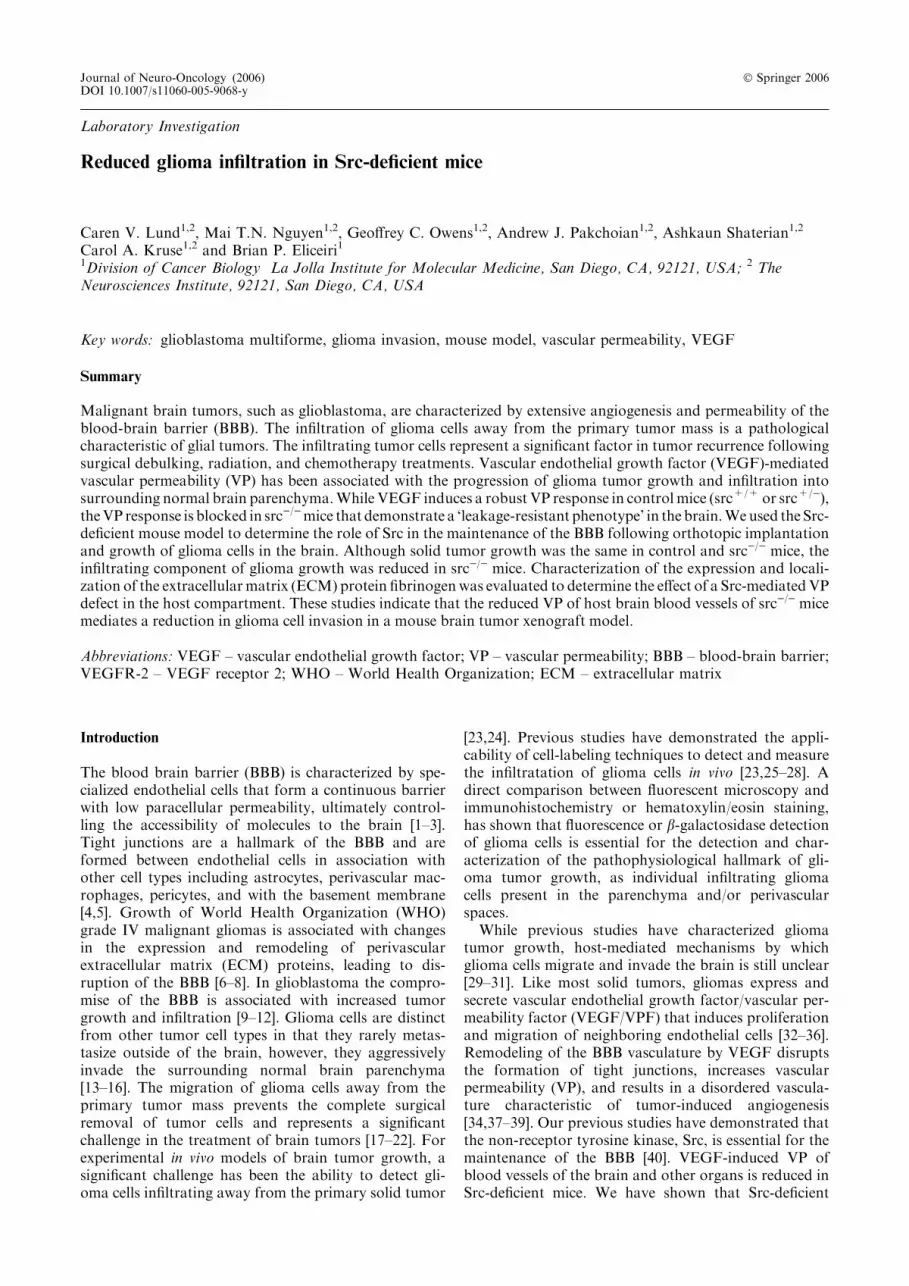
Figure 1. Characterization of Pyk2 and FAK expression by glioma cells in vitro and in vivo. (a) Lysates of cultured low passage glial cells, 02-11-
MG and 13-06-MG, were prepared as described in the Materials and Methods and subjected to immunoblotting with anti-Pyk2, anti-FAK and
anti-VEGF antibodies. Protein-antibody complexes were detected using ECL detection. (b) Infiltration of yellow fluorescent protein (YFP)-
labeled 02-11-MG tumor cells was examined in the fixed brain sections by indirect immunofluorescence with an anti-YFP/GFP antibody (middle
panel). DAPI staining labels nuclei (left panel) and a merge of the nuclei and tumor cells is also shown (right panel). Bar=200 lm. (c) Fluorescent
micrographs embedded in illustrated serial brain sections from mice bearing YFP-labeled 02-11-MG tumor cells, with src+/) sections in the top
row and src)/) sections in the bottom row. Tumors were stereotactically injected and allowed to grow for 21 days. Upon harvest, brains were
sectioned and images of unfixed serial sections were acquired with a laser scanning confocal microscope. (d) Tumor volume was calculated for 02-
11-MG tumors in src+/) and src)/) brain sections. Tumor area was quantitated by imaging the faces of serial 1.0 mm brain slices from sibling-
matched mice (n=8).

Immunoblotting
Cell lysates were collected in RIPA buffer (100 mM TrispH 7.5, 150 mM NaCl, 1 mM EDTA, 1% deoxycholicacid, 1% Triton X-100, 0.1% SDS with freshly added1 mM orthovanadate, 50 mM NaF, and proteaseinhibitors cocktail (Roche, Indianapolis, IN). Proteinconcentration was determined with a BCA ProteinAssay (Pierce, Rockford, IL). Twenty micrograms ofcell lysate for each sample was separated on a 10% SDS-polyacrylamide gel, transferred to a nitrocellulosemembrane, and probed with anti-FAK, anti-Pyk2, oranti-VEGF (Santa Cruz Biotechnologies, Santa Cruz,CA) antibodies. Secondary antibodies were anti-mouseor anti-rabbit antibodies conjugated to horseradishperoxidase. Visualization of antibody binding wasdetected using Supersignal West Pico ChemiluminescentSubstrate (Pierce).
Results
Characterization of chimeric human brain tumor/mousebrain xenograft model
The migratory phenotype of glioma cells into sur-rounding brain parenchyma is a defining characteristicfor malignant gliomas. While the mechanisms regulatingglioma tumor invasion are poorly understood, a studyof the related tyrosine kinases, Pyk2 and focal adhesionkinase (FAK), in glioma cell migration has shown thatelevated Pyk2 expression correlates with increased gli-oma cell migration in vitro [47]. We characterized theexpression of FAK, Pyk2, and VEGF (Figure 1a), aswell as the expression of various integrins and othersignaling intermediates ([45] and data not shown) of twohuman glioma explants obtained from patients diag-nosed with grade IV glioblastoma multiforme (GBM)[45]. The expression of FAK was similar in both 02-11-MG and 13-06-MG gliomas. In contrast, the expressionof Pyk2 was significantly reduced in 13-06-MG gliomacells compared to the 02-11-MG glioma cells. Both gli-oma cells express VEGF (Figure 1a, bottom row).Based on the correlation of Pyk2 expression and gliomacell migration, and an infiltrative phenotype that hasbeen shown to be characteristic of advanced stage glio-mas, we selected 02–11-MG cells to provide a physio-logically relevant model of glial tumor growth in axenograft model of brain tumor growth.
We examined the orthotopic growth of 02-11-MGglioma cells in src)/) vs. control mice (src+/+ or src+/)).The use of src)/) mice was based on the previouslycharacterized phenotype of src)/) mice that includes
reduced blood vessel VP in response to VEGF or tumor-induced VP [40]. Although many parameters influencingglioma growth and infiltration have been previouslyexamined, the role of tumor-induced VP of brain bloodvessels had not been examined in a defined geneticmodel. Therefore, we examined the orthotopic growthof 02-11-MG cells in src)/) and src+/) mice. We havepreviously shown that src+/+ or src+/) have similarVEGF- and tumor-induced responses [41], therefore wehave primarily used src+/) mice in our subsequentstudies. To facilitate the detection and measurement ofglioma tumor growth, 02-11-MG glioma tumor cellswere labeled with yellow-fluorescent protein (YFP) (SeeMaterials and methods). Figure 1b demonstrates theinvasive behavior of the 02-11 glioma cells followingstereotactic injection, 21 day incubation and detectionby indirect immunofluorescence with anti-YFP anti-body. To compare the tumor burden of 02-11 throughmultiple brain sections, src)/) and src+/) mice wereinjected with YFP-labeled 02-11 glioma cells, and fol-lowing a 21 day incubation, mouse brains were har-vested and serial 1 mm sections prepared for imageacquisition with a laser scanning confocal microscope(Figure 1c). A representative set of brain slices fromsrc)/) and src+/) mice are shown in Figure 1c. The useof YFP-labeled tumor cells enables the quantitation oftumor burden from the face of each brain slice fromeach animal. Variation between slices and between ani-mals were averaged to evaluate the tumor volume insrc)/) and src+/) mice. The data from tumor volumequantitation is shown in Figure 1d. We determined thatthere was no significant difference in gross tumor burdenbetween src)/) and src+/) mice. However, when weexamined the tumor margin at higher magnification, aSrc-mediated effect of the host on tumor infiltration wasobserved.
Reduced GBM tumor invasion in Src-deficient host mousebrains
Although the gross tumor burden of glioma cellsimplanted in src)/) and control mice was unchanged(Figure 1d), clinical manifestations of gliomas areassociated with the diffuse and infiltrative growth ofsecondary tumor foci into ipsilateral and contralateralhemispheres [13]. Labeling of glioma tumor cells hasbeen shown to be essential for monitoring the invasionand growth of single glioma cells in vivo [23,25–28,48].Therefore, we examined the growth and invasion ofYFP-labeled tumor cells 200–1000 lm from the marginof the primary tumor mass established near the injectionsite. 02-11-MG glioma tumor cells were injected into the
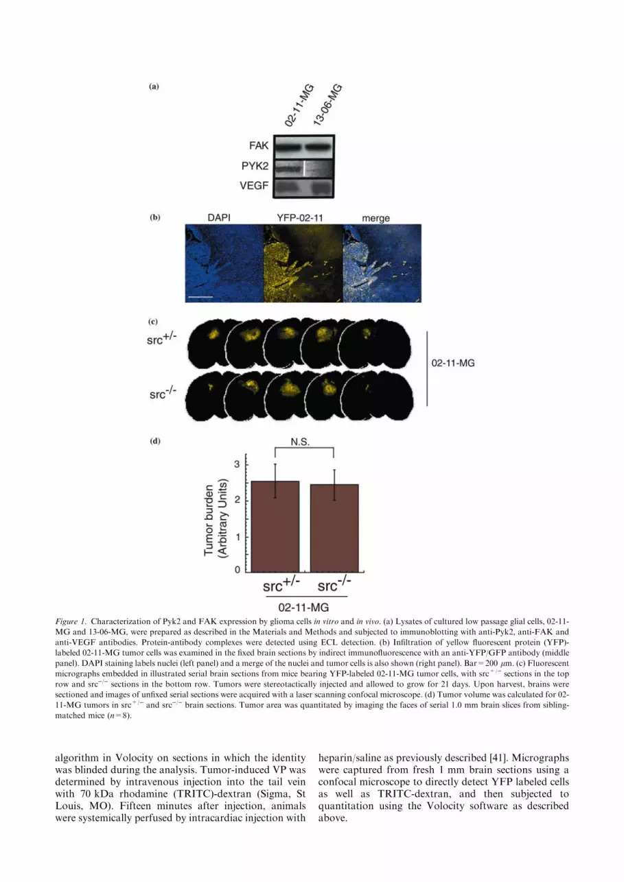
Figure 2. Reduced perivascular extension of 02-11-MG glioma cells into src+/) vs. src)/) mouse brains. (a) Low power photomicrographs from
hematoxylin/eosin stained brain tumor sections at the boundary of 02-11-MG tumor growth in representative brain section from src+/) (left
panel) and src)/) (right panel) mice. Arrows indicate perivascular tumor foci. Bar=200lm. (b) Confocal micrographs at 20� of src+/) and src)/)
mice bearing YFP-labeled 02-11-MG with tumor foci indicated with arrows. The left two panels are representative images of src+/) brain sections
and the right two panels are representative of src)/) brain sections. Arrows indicate tumor foci. Micrographs were imaged at a distance of 200–
1000 lm from the primary solid tumor margin. Bar=200 lm. (c) Quantitation of tumor foci observed in src+/) and src)/) mice. Asterisks
indicate a statistically significant difference (n=8, P<0.05).
c

brains of src)/) and src+/) mice, incubated for 21 days,and imaged using standard histological techniques aswell as confocal microscopy to detect YFP-labeled cellsin fresh brain tissue sections. Hematoxylin/eosin (H/E)staining of sections of tumor-bearing src+/) and src)/)
mice suggested that there were differences in the tumormargins between the host mice (Figure 2a). Arrowheadsmark infiltrating tumor cells in src+/) brain sections,whereas, src)/) brain sections had a reduction of infil-trating tumor cells. To more definitively evaluate theextent of glioma cell infiltration, we analyzed YFP-labeled 02-11-MG glioma cells using confocal micros-copy. In src+/) mice, 02-11-MG tumor cells exhibitedcharacteristics of grade IV glioma cells with extensiveinfiltration of the brain parencyhma (arrowheads).However, an examination of the parenchyma of tumor-bearing src)/) mice revealed a decrease in YFP-labeledglioma cell infiltration and formation of infiltrating tu-mor foci (Figure 2b). We examined the tumor burdenfrom three matched sets of src)/) and src+/) mice andquantitated the number of YFP-positive cells distinctfrom the primary glioma tumor. Quantitation of thesedata is presented in Figure 2c, and demonstrates a sig-nificant reduction in glioma tumor cell infiltration insrc)/) mice (n=8, P<0.05). Therefore, although thegross tumor volume was not reduced in src)/) vs. src+/)
mice (Figure 1c), a Src-defect in the host led to a sig-nificant reduction in glioma cell infiltration through thebrain parenchyma.
Src)/) mice have reduced glioma-induced VP, but normalneovascularization
While glioma growth is associated with increased VP,the mechanism(s) regulating glioma-induced VPremain poorly understood. We have previously shownthat VEGF-induced VP is reduced in the brains ofSrc-deficient mice [40]. Therefore, we examined whetherVP induced by a VEGF-expressing glioma tumor wouldbe reduced in the brains of Src-deficient mice. Followingthe implantation and incubation of 02-11-MG gliomacell in src)/) and src+/) mice, as described above, weinjected 70 kDa TRITC-dextran intravenously 15 minbefore harvest. After systemic intracardiac perfusion toremove intravascular FITC-dextran, we detected FITC-dextran in the perivascular tissue and the YFP-fluores-cence in 1 mm brain sections by confocal microscopyimmediately upon harvest. Representative micrographsof sibling-matched mice revealed a decrease in 02-11-MG glioma-induced VP in src)/) vs. src+/) mice (Fig-ure 3a). Differences in VP were quantitated and areshown in Figure 3b. Despite the Src-mediated decreasein glioma-induced VP observed in the src)/) brain,neovascularization in response to the brain tumorsgrown in src)/) and src+/) mice, as monitored by bloodvessel staining using anti-CD31 immunofluorescence,was unchanged (Figure 3c). Quantitation of CD31staining is shown in Figure 3d. These data demonstratethe distinction between tumor-induced VP of the hostthat is mediated by Src vs. tumor-induced angiogenesisthat is independent of a Src defect in the host.
Reduction in tumor-induced fibrinogen accumulationin Src-deficient mice
Endothelial cell migration and increased VP have beenassociated with remodeling of the ECM and are pro-cesses required for tumor progression. The extravasa-tion of plasma fibrinogen, and subsequent cleavage andformation of perivascular fibrin and cross-linked fibrin[collectively referred to as fibrin(-ogen)], are hallmarksof tumor growth [49–51], as well as a pathologicallyrelevant endogenous indicator of VP [41]. Therefore, weexamined src)/) and src+/) mice for changes in thedistribution of tumor-associated fibrin(-ogen) duringglioma growth (Figure 4a). Following implantation andincubation of gliomas in src)/) and src+/) mice, tumor-bearing brains were harvested, cryosectioned, and sub-jected to immunohistochemical analysis. Src)/) miceshowed reduced fibrin(-ogen) staining compared withsrc+/) mice. The Src-mediated reduction in tumor-associated fibrin was quantitated from micrographs offibrin-stained tumor bearing src)/) and src+/) brainsections (Figure 4b). Fibrin(-ogen) associated withCD31-positive blood vessels was also decreased in src)/)
mice. These results suggest that reduced accumulation ofperivascular fibrin(-ogen) is associated with reducedtumor VP in the brain, and may be an important markerof VP-mediated remodeling of the ECM microenviron-ment in response to tumor growth.
Discussion
Brain tumors are characterized by an invasive pheno-type that prevents complete surgical removal of thetumor mass. Infiltration of glioma cells into normalbrain tissue, combined with subsequent tumor growthand increased intracranial pressure, results in a meansurvival time of 12–14 months for patients diagnosedwith grade IV glioblastoma, the most malignant form ofbrain tumor [26,52,53]. Recent studies of a cranialtumor model show that increased VP is one of the firstevents following tumor implantation [54]. Increased VPhas been associated with the progression of malignanttumor growth and invasion in the BBB as well as inother tissues, however the molecular basis is unclear.Gliomas express a range of growth factors andcytokines, however VEGF is unique among thesegrowth factors in that it induces VP as well as neovas-cularization [55–58]. Intra-cranial administration ofrecombinant VEGF to mice has been shown to increaseVP and result in breakdown of the BBB, however themechanisms regulating this process remain poorlyunderstood [59–61]. Therefore, in this study we haveexamined Src-deficient mice, which we have previouslyshown to have a ‘leakage-resistant’ phenotype inresponse to VEGF. We have used src)/) and src+/) miceto examine the effect of reduced VP of the host vascu-lature, as well as decreased Src expression in allnon-tumor cells, during the growth and infiltration ofglioma cells implanted intracranially.
Preventing the infiltration of glioma cells away fromthe primary tumor is important in any metastasis model.

Figure 3. Reduction of tumor-induced VP but not angiogenesis in Src-knockout mice. (a) Tumor-induced VP was determined in src+/) (top left
panel) and src)/) (top right panel) mice bearing intracranial 02-11-MG tumor cells using extravasation of TRITC-70 kDa dextran. YFP-labeled
brain tumor burden in src+/) (bottom left panel) and src)/) (bottom right panel) mice was obtained by imaging with a confocal microscope as
described in the Materials and Methods. Mice were subjected to intravenous injection of TRITC-70 kDa dextran, perfused with heparin/saline to
remove intravascular dextran, and the harvested brains subjected to serial 1 mm sectioning and imaging. Micrographs were imaged at 2� and
dextran fluorescence detected. Bar= 1000 lm. (b) Quantitation of VP as detected by fluorescence of TRITC-70 kDa dextran as described in the
Materials and Methods. (n=6; P<0.05). (c) Indirect immunofluorescence of cryosections from cranial 02-11-MG tumors grown in src+/) (left
panel) and src)/) (right panel) mice. Immunostaining with an anti-CD31 antibody was used to localize blood vessels in the tumor. Bar=100 lm.
(d) Quantitation of CD31 staining as described in the Materials and methods. (n=6).

However, the unique environment of the BBB distin-guishes glioma growth and infiltration from othermodels of metastasis in that glioma cells rarely metas-tasize out of the brain [62], yet are extensively infiltrativewithin the brain [15]. Our analysis of the growth andinfiltration of low passage glioma cells in src)/) mousebrains reveals that the Src-defect of the host can mediatea decrease in glioma cell infiltration of the brainparenchyma, while the overall gross tumor burden andangiogenesis of the primary tumor is unchanged. Wehave based our studies on the use of low passage gliomacells that exhibit tumor growth and invasion phenotypessimilar to infiltrative human gliomas. To evaluate theextent of migration of single or multiple glioma cellsaway from the primary tumor in vivo, glioma cellsexpressing fluorescent proteins have been shown toprovide imaging of glioma cell infiltration [27,28,48]. Weshow in this work that tumor growth and infiltration canbe monitored in serial sections and tumor burdeninclusive of solid growth and infiltrative cells can bequantitated to determine gross tumor burden. Further-more, since we are focused on the role of the hostcompartment in mediating specific tumor-induced vas-cular responses (i.e. Src-mediated breakdown of theBBB), the implantation of YFP-labeled tumor cellsprovides a defined demarcation of tumor cells versushost cells in the xenograft tumor model.
Previous studies have shown that a systemic reductionin VEGF reduces brain tumor growth and infiltration inan orthotopic intracerebral mouse model [63]. However,in contrast to the role of VEGF in both angiogenesisand VP, we have focused on the role of VP, specificallyusing a Src-knockout model to characterize mechanismsof tumor-induced VP in tumor growth and invasion.While we cannot exclude the possibility that othertumor-expressed factors may induce VP, we have shownthat although VEGF-induced VP is blocked in Src-deficient mice, VEGF expression in the brain or fibrin-ogen expression in the plasma is unchanged in src+/) vs.src)/) mice ([40,60] and data not shown). We proposethat the reduction of tumor-induced VP in the brainvascular bed of src)/) mice mediates a reduction in braintumor infiltration indirectly by regulating the remodel-ing of the perivascular ECM. For example, fibrin(-ogen)is part of the haemostatic system mediating vasculardamage, angiogenesis, and wound repair. In response toVP, plasma fibrinogen undergoes extravasation andcleavage to form perivascular fibrin(-ogen) [50]. Accu-mulated perivascular fibrin(-ogen) serves as a substratefor endothelial and tumor cell migration and has beenassociated with tumor extension into normal brain tissue[64–66]. In this study we provide evidence of reducedperivascular fibrin(-ogen) deposition in brain tumor-bearing src)/) mice, suggesting that fibrin(-ogen) may bean example of Src-regulated ECM remodeling that canpromote intracranial glioma tumor migration through aVP-dependent mechanism that does not involve theintravasation of tumor cells. Since glioma cells do notinfiltrate the brain parenchyma through a hematoge-nous mechanism, Src-mediated VP may influence theextravasation of plasma fibrinogen, and subsequentcleavage of it to perivascular fibrin(-ogen) to provide a
Figure 4. Reduced colocalization of perivascular fibrin and CD31
staining in tumor-bearing src)/) mouse brains. (a) Indirect immu-
nofluorescence of cryosections of 02-11-MG tumor-bearing src+/)
(left) and src)/) brains (right) immunostained with an anti-fi-
brin(-ogen) antibody. Bar = 100 lm. (b) Quantitation of fibrinogen
immunostaining in 02-11-MG tumor-bearing src+/) and src)/)
brains, as described in the Materials and methods. (n=6, P<0.05).
(c) Localization of CD31-positive blood vessels and fibrin(-ogen) in
02-11-MG tumor-bearing src+/) (left) and src)/) brains (right).
Bar = 100 lm.

provisional ECM support for glioma cell migration. Theobservation of the diffuse infiltration of glioma cells isconsistent with a model in which a VP defect affectstumor cell invasion, though not necessarily affectingangiogenesis of the primary tumor or satellite tumors.
While in vitro studies have shown that Src can regu-late many different molecules including intracellularsignaling intermediates, integrins, matrix metallopro-teases, and ECM proteins, the specific role of Src in vivois poorly understood [67–76]. Based on knockout stud-ies, it is known that other Src-related family kinases cancompensate for the absence of Src particularly duringdevelopment. However, studies have shown that defectsin VP cannot be compensated for by other Src familymembers, and that defects in VP are a phenotype spe-cific for Src-deficient mice [40]. The molecular basis forthe role of Src in VEGF-induced VP has been investi-gated in both brain and lung endothelium and has beenshown to require the association of Src with focaladhesion kinase (FAK) [41,77]. However, downstreamtargets of this Src-mediated signaling pathway have notbeen identified. Further characterization of the signalingpathway through Src during VEGF-induced VP, ortumor-induced VP, will identify specific mechanismsinvolved in the process of glioma infiltration in relationto VP. While our previous studies showed that hema-togenous metastasis to other organs is reduced in src)/)
mice [41], in the brain, the inhibition of tumor-inducedVP leads to a change in the perivascular ECM, whichmay reveal a role for these molecules in mediating theinfiltration of brain tumor cells.
Acknowledgements
We thank Mario A. Bourdon and Carole A. Banka forcritical reading of this manuscript. Grant support fromthe NHLBI (B.P.E) and the NINDS (C.A.K.)
References
1. Ballabh P, Braun A, Nedergaard M: The blood-brain barrier: an
overview: structure, regulation, and clinical implications. Neuro-
biol Dis 16: 1–13, 2004
2. Nag S: Morphology and molecular properties of cellular compo-
nents of normal cerebral vessels. Methods Mol Med 89: 3–36, 2003
3. Smith QR: A review of blood-brain barrier transport techniques.
Methods Mol Med 89: 193–208, 2003
4. Bradbury MW: The blood-brain barrier. Transport across the
cerebral endothelium. Circ Res 57: 213–222, 1985
5. Gloor SM, Wachtel M, Bolliger MF, Ishihara H, Landmann R,
Frei K: Molecular and cellular permeability control at the blood-
brain barrier. Brain Res Brain Res Rev 36: 258–264, 2001
6. Gladson CL: The extracellular matrix of gliomas: modulation of
cell function. J Neuropathol Exp Neurol 58: 1029–1040, 1999
7. Mahesparan R, Read TA, Lund-Johansen M, Skaftnesmo KO,
Bjerkvig R, Engebraaten O: Expression of extracellular matrix
components in a highly infiltrative in vivo glioma model. Acta
Neuropathol (Berl) 105: 49–57, 2003
8. Davies DC: Blood-brain barrier breakdown in septic encepha-
lopathy and brain tumours. J Anat 200: 639–646, 2002
9. Coomber BL, Stewart PA, Hayakawa K, Farrell CL, Del Maestro
RF: Quantitative morphology of human glioblastoma multiforme
microvessels: structural basis of blood-brain barrier defect. J
Neurooncol 5: 299–307, 1987
10. Nag S: Pathophysiology of blood-brain barrier breakdown.
Methods Mol Med 89: 97–119, 2003
11. Oliveira R, Christov C, Guillamo JS, Debouard S, Palfi S,
Venance L, Tardy M, Peschanski M: Contribution of gap junc-
tional communication between tumor cells and astroglia to the
invasion of the brain parenchyma by human glioblastomas. BMC
Cell Biol 6: 7, 2005
12. Henson JW, Gaviani P, Gonzalez RG: MRI in treatment of adult
gliomas. Lancet Oncol 6: 167–175, 2005
13. Demuth T, Berens ME: Molecular mechanisms of glioma cell
migration and invasion. J Neurooncol 70: 217–228, 2004
14. Giese A, Bjerkvig R, Berens ME, Westphal M: Cost of migration:
invasion of malignant gliomas and implications for treatment. J
Clin Oncol 21: 1624–1636, 2003
15. Mourad PD, Farrell L, Stamps LD, Chicoine MR, Silbergeld DL:
Why are systemic glioblastoma metastases rare? Systemic and
cerebral growth of mouse glioblastoma. Surg Neurol 63: 511–519,
2005 discussion 519
16. Bolteus AJ, Berens ME, Pilkington GJ: Migration and invasion in
brain neoplasms. Curr Neurol Neurosci Rep 1: 225–232, 2001
17. Bellail AC, Hunter SB, Brat DJ, Tan C, Van Meir EG: Microre-
gional extracellular matrix heterogeneity in brain modulates gli-
oma cell invasion. Int J Biochem Cell Biol 36: 1046–1069, 2004
18. Desjardins A, Rich JN, Quinn JA, Vredenburgh J, Gururangan S,
Sathornsumetee S, Reardon DA, Friedman AH, Bigner DD,
Friedman HS: Chemotherapy and novel therapeutic approaches in
malignant glioma. Front Biosci 10: 2645–2668, 2005
19. Fujimaki T: Surgical treatment of brain metastasis. Int J Clin
Oncol 10: 74–80, 2005
20. Gilbert MR, Loghin M: The Treatment of Malignant Gliomas.
Curr Treat Options Neurol 7: 293–303, 2005
21. Nabors LB, Fiveash J: Treatment of adults with recurrent malig-
nant glioma. Expert Rev Neurother 5: 509–514, 2005
22. Berens ME, Giese A: ‘‘...those left behind.’’ Biology and oncology
of invasive glioma cells. Neoplasia 1: 208–219, 1999
23. Lampson LA, Lampson MA, Dunne AD: Exploiting the lacZ
reporter gene for quantitative analysis of disseminated tumor
growth within the brain: use of the lacZ gene product as a tumor
antigen, for evaluation of antigenic modulation, and to facilitate
image analysis of tumor growth in situ. Cancer Res 53: 176–182,
1993
24. Owens GC, Orr EA, DeMasters BK, Muschel RJ, Berens ME,
Kruse CA: Overexpression of a transmembrane isoform of neural
cell adhesion molecule alters the invasiveness of rat CNS-1 glioma.
Cancer Res 58: 2020–2028, 1998
25. Pedersen PH, Edvardsen K, Garcia-Cabrera I, Mahesparan R,
Thorsen J, Mathisen B, Rosenblum ML, Bjerkvig R: Migratory
patterns of lac-z transfected human glioma cells in the rat brain.
Int J Cancer 62: 767–771, 1995
26. Prados MD, Berger MS, Wilson CB: Primary central nervous
system tumors: advances in knowledge and treatment. CA Cancer
J Clin 48: 331–360, 321, 1998
27. MacDonald TJ, Tabrizi P, Shimada H, Zlokovic BV, Laug WE:
Detection of brain tumor invasion and micrometastasis in vivo by
expression of enhanced green fluorescent protein. Neurosurgery
43: 1437–1442, 1998discussion 1442–1433
28. Mourad PD, Farrell L, Stamps LD, Santiago P, Fillmore HL,
Broaddus WC, Silbergeld DL: Quantitative assessment of glio-
blastoma invasion in vivo. Cancer Lett 192: 97–107, 2003
29. Bello L, Giussani C, Carrabba G, Pluderi M, Costa F, Bikfalvi A:
Angiogenesis and invasion in gliomas. Cancer Treat Res 117: 263–
284, 2004

30. Tonn JC, Goldbrunner R: Mechanisms of glioma cell invasion.
Acta Neurochir Suppl 88: 163–167, 2003
31. Lefranc F, Brotchi J, Kiss R: Possible future issues in the treat-
ment of glioblastomas: special emphasis on cell migration and the
resistance of migrating glioblastoma cells to apoptosis. J Clin
Oncol 23: 2411–2422, 2005
32. Jensen RL: Growth factor-mediated angiogenesis in the malignant
progression of glial tumors: a review. Surg Neurol 49: 189–195,
1998 discussion 196
33. Berkman RA, Merrill MJ, Reinhold WC, Monacci WT, Saxena A,
Clark WC, Robertson JT, Ali IU, Oldfield EH: Expression of the
vascular permeability factor/vascular endothelial growth factor
gene in central nervous system neoplasms. J Clin Invest 91: 153–
159, 1993
34. Machein MR, Plate KH: VEGF in brain tumors. J Neurooncol 50:
109–120, 2000
35. Ferrara N, Gerber HP, LeCouter J: The biology of VEGF and its
receptors. Nat Med 9: 669–676, 2003
36. Autiero M, Waltenberger J, Communi D, Kranz A, Moons L,
Lambrechts D, Kroll J, Plaisance S, De Mol M, Bono F et al.:
Role of PlGF in the intra- and intermolecular cross talk be-
tween the VEGF receptors Flt1 and Flk1. Nat Med 9: 936–943,
2003
37. Vaquero J, Zurita M, Morales C, Cincu R, Oya S: Expression of
vascular permeability factor in glioblastoma specimens: correla-
tion with tumor vascular endothelial surface and peritumoral
edema. J Neurooncol 49: 49–55, 2000
38. Harhaj NS, Antonetti DA: Regulation of tight junctions and loss
of barrier function in pathophysiology. Int J Biochem Cell Biol 36:
1206–1237, 2004
39. Schneider SW, Ludwig T, Tatenhorst L, Braune S, Oberleithner H,
Senner V, Paulus W: Glioblastoma cells release factors that disrupt
blood-brain barrier features. Acta Neuropathol (Berl) 107: 272–
276, 2004
40. Eliceiri BP, Paul R, Schwartzberg PL, Hood JD, Leng J, Cheresh
DA: Selective requirement for Src kinases during VEGF-induced
angiogenesis and vascular permeability. Mol Cell 4: 915–924, 1999
41. Criscuoli ML, Nguyen M, Eliceiri BP: Tumor metastasis but not
tumor growth is dependent on Src-mediated vascular permeability.
Blood 105: 1508–1514, 2005
42. Chen J, Lansford R, Stewart V, Young F, Alt FW: RAG-2-defi-
cient blastocyst complementation: an assay of gene function in
lymphocyte development. Proc Natl Acad Sci U S A 90: 4528–
4532, 1993
43. Hochedlinger K, Blelloch R, Brennan C, Yamada Y, KimM, Chin
L, Jaenisch R: Reprogramming of a melanoma genome by nuclear
transplantation. Genes Dev 18: 1875–1885, 2004
44. Mazurier F, Fontanellas A, Salesse S, Taine L, Landriau S,
Moreau-Gaudry F, Reiffers J, Peault B, Di Santo JP, de Verneuil
H: A novel immunodeficient mouse model – RAG2 x common
cytokine receptor gamma chain double mutants – requiring
exogenous cytokine administration for human hematopoietic stem
cell engraftment. J Interferon Cytokine Res 19: 533–541, 1999
45. Mattern RH, Read SH, Pierschbacher MD, Sze CI, Eliceiri B,
Kruse CA: Glioma cell integrin expression and their interactions
with integrin antagonists. Cancer Therapy 3: 325–340, 2005
46. Curran MA, Kaiser SM, Achacoso PL, Nolan GP: Efficient
transduction of nondividing cells by optimized feline immunode-
ficiency virus vectors. Mol Ther 1: 31–38, 2000
47. Lipinski CA, Tran NL, Menashi E, Rohl C, Kloss J, Bay RC,
Berens ME, Loftus JC: The tyrosine kinase pyk2 promotes
migration and invasion of glioma cells. Neoplasia 7: 435–445, 2005
48. Zhang X, Li X, Wu JW, Gao DK, Liang JW, Liu XZ: Experiment
and observation on invasion of brain glioma in vivo. J Clin
Neurosci 9: 668–671, 2002
49. Degen JL, Palumbo JS: Mechanisms linking hemostatic factors to
tumor growth in mice. Pathophysiol Haemost Thromb 33(Suppl
1): 31–35, 2003
50. Staton CA, Brown NJ, Lewis CE: The role of fibrinogen and
related fragments in tumour angiogenesis and metastasis. Expert
Opin Biol Ther 3: 1105–1120, 2003
51. Wojtukiewicz MZ, Sierko E, Rak J: Contribution of the hemo-
static system to angiogenesis in cancer. Semin Thromb Hemost 30:
5–20, 2004
52. Stupp R, van den Bent MJ, Hegi ME: Optimal role of temozol-
omide in the treatment of malignant gliomas. Curr Neurol Neu-
rosci Rep 5: 198–206, 2005
53. Thorsen F, Tysnes BB: Brain tumor cell invasion, anatomical and
biological considerations. Anticancer Res 17: 4121–4126, 1997
54. Hansen-Algenstaedt N, Joscheck C, Schaefer C, Lamszus K,
Wolfram L, Biermann T, Algenstaedt P, Brockmann MA, Heintz
C, Fiedler W et al.: Long-term observation reveals time-course-
dependent characteristics of tumour vascularisation. Eur J Cancer
41: 1073–1085, 2005
55. Brockmann MA, Ulbricht U, Gruner K, Fillbrandt R, Westphal
M, Lamszus K: Glioblastoma and cerebral microvascular
endothelial cell migration in response to tumor-associated growth
factors. Neurosurgery 52: 1391–1399, 2003 discussion 1399
56. Kaur B, Tan C, Brat DJ, Post DE, Van Meir EG: Genetic and
hypoxic regulation of angiogenesis in gliomas. J Neurooncol 70:
229–243, 2004
57. Lamszus K, Heese O, Westphal M: Angiogenesis-related growth
factors in brain tumors. Cancer Treat Res 117: 169–190, 2004
58. Dunn IF, Heese O, Black PM: Growth factors in glioma angio-
genesis: FGFs, PDGF, EGF, and TGFs. J Neurooncol 50: 121–
137, 2000
59. Zhang ZG, Zhang L, Jiang Q, Zhang R, Davies K, Powers C,
Bruggen N, Chopp M: VEGF enhances angiogenesis and pro-
motes blood–brain barrier leakage in the ischemic brain. J Clin
Invest 106: 829–838, 2000
60. Paul R, Zhang ZG, Eliceiri BP, Jiang Q, Boccia AD, Zhang RL,
Chopp M, Cheresh DA: Src deficiency or blockade of Src activity
in mice provides cerebral protection following stroke. Nat Med 7:
222–227, 2001
61. Zhang ZG, Zhang L, Tsang W, Soltanian-Zadeh H, Morris D,
Zhang R, Goussev A, Powers C, Yeich T, Chopp M: Correlation
of VEGF and angiopoietin expression with disruption of blood-
brain barrier and angiogenesis after focal cerebral ischemia. J
Cereb Blood Flow Metab 22: 379–392, 2002
62. Armanios MY, Grossman SA, Yang SC, White B, Perry A, Burger
PC, Orens JB: Transmission of glioblastoma multiforme following
bilateral lung transplantation from an affected donor: case study
and review of the literature. Neurooncol 6: 259–263, 2004
63. Kunkel P, Ulbricht U, Bohlen P, Brockmann MA, Fillbrandt R,
Stavrou D, Westphal M, Lamszus K: Inhibition of glioma
angiogenesis and growth in vivo by systemic treatment with a
monoclonal antibody against vascular endothelial growth factor
receptor-2. Cancer Res 61: 6624–6628, 2001
64. Bardos H, Molnar P, Csecsei G, Adany R: Fibrin deposition in
primary and metastatic human brain tumours. Blood Coagul
Fibrinolysis 7: 536–548, 1996
65. Yumitori K, Handa H, Teraura T, Yamashita J, Yamamura K:
Metastatic brain tumour and fibrinopeptides. Acta Neurochir
(Wien) 89: 43–47, 1987
66. Sawaya R, Mandybur T, Ormsby I, Tew JM, Westhoff MA,
Serrels B, Fincham VJ, Frame MC, Carragher NO: SRC-mediated
phosphorylation of focal adhesion kinase couples actin and
adhesion dynamics to survival signaling. Mol Cell Biol 24: 8113–
8133, 2004
68. Campbell ID: Modular proteins at the cell surface. Biochem Soc
Trans 31: 1107–1114, 2003
69. Shattil SJ: Integrins and Src: dynamic duo of adhesion signaling.
Trends Cell Biol 15: 399–403, 2005
70. Courter DL, Lomas L, Scatena M, Giachelli CM: Src kinase
activity is required for integrin alphaVbeta3-mediated activation
of nuclear factor-kappaB. J Biol Chem 280: 12145–12151, 2005
71. PlayfordMP, SchallerMD: The interplay between Src and integrins
in normal and tumor biology. Oncogene 23: 7928–7946, 2004
72. Wu X, Gan B, Yoo Y, Guan JL: FAK-Mediated Src Phosphor-
ylation of Endophilin A2 Inhibits Endocytosis of MT1-MMP and
Promotes ECM Degradation. Dev Cell 9: 185–196, 2005

73. Labrecque L, Nyalendo C, Langlois S, Durocher Y, Roghi C,
Murphy G, Gingras D, Beliveau R: Src-mediated tyrosine phos-
phorylation of caveolin-1 induces its association with membrane
type 1 matrix metalloproteinase. J Biol Chem 279: 52132–52140,
2004
74. Sounni NE, Roghi C, Chabottaux V, Janssen M, Munaut C,
Maquoi E, Galvez BG, Gilles C, Frankenne F, Murphy G et al.:
Up-regulation of vascular endothelial growth factor-A by active
membrane-type 1 matrix metalloproteinase through activation of
Src-tyrosine kinases. J Biol Chem 279: 13564–13574, 2004
75. Nadav L, Katz BZ: The molecular effects of oncogenesis on cell-
extracellular matrix adhesion (review). Int J Oncol 19: 237–246,
2001
76. Yang Y, Dang D, Mogi S, Ramos DM: Tenascin-C deposition
requires beta3 integrin and Src. Biochem Biophys Res Commun
322: 935–942, 2004
77. Eliceiri BP, Puente XS, Hood JD, Stupack DG, Schlaepfer DD,
Huang XZ, Sheppard D, Cheresh DA: Src-mediated coupling of
focal adhesion kinase to integrin alpha(v)beta5 in vascular
endothelial growth factor signaling. J Cell Biol 157: 149–160,
2002
Address for offprints: Brian P. Eliceiri, Division of Cancer Biology La
Jolla Institute for Molecular Medicine, 4570 Executive Drive, Suite
100, San Diego, CA, 92121, USA; Tel.: +1-858-587-8788; Fax:
+1-858-587-6742; E-mail: [email protected]


![Salford 345. IMMUNIZATION WITH AUTOLOGOUS GLIOMA CELLS ]](https://img.dokumen.tips/doc/110x75/634785cf6e55ee279302cc25/salford-345-immunization-with-autologous-glioma-cells-.jpg)


