Embed Size (px)
Citation preview
International Journal of
Molecular Sciences
Article
Epinephrine Infiltration of Adipose Tissue ImpactsMCF7 Breast Cancer Cells and Total Lipid Content
Pierre Avril 1,†, Luciano Vidal 1,†, Sophie Barille-Nion 2 , Louis-Romée Le Nail 1,Françoise Redini 1, Pierre Layrolle 1 , Michelle Pinault 3, Stéphane Chevalier 3, Pierre Perrot 1,4,*and Valérie Trichet 1
1 INSERM, Université de Nantes, UMR1238, Phy-Os, Sarcomes osseux et remodelage des tissus calcifiés,F-44035 Nantes, France; [email protected] (P.A.); [email protected] (L.V.);[email protected] (L.-R.L.N.); [email protected] (F.R.); [email protected] (P.L.);[email protected] (V.T.)
2 CRCINA, INSERM, Université d’Angers, Université de Nantes, F-44035 Nantes, France;[email protected]
3 INSERM Université de Tours, UMR1069, Nutrition, Croissance et Cancer, F-37032 Tours, France;[email protected] (M.P.); [email protected] (S.C.)
4 CHU de Nantes, Service de Chirurgie Plastique et des Brûlés, F-44035 Nantes, France* Correspondence: [email protected]; Tel.: +33-2-40-08-73-02† These authors contributed equally to this work.
Received: 6 October 2019; Accepted: 4 November 2019; Published: 11 November 2019�����������������
Abstract: Background: Considering the positive or negative potential effects of adipocytes, dependingon their lipid composition, on breast tumor progression, it is important to evaluate whether adiposetissue (AT) harvesting procedures, including epinephrine infiltration, may influence breast cancerprogression. Methods: Culture medium conditioned with epinephrine-infiltrated adipose tissue wastested on human Michigan Cancer Foundation-7 (MCF7) breast cancer cells, cultured in monolayeror in oncospheres. Lipid composition was evaluated depending on epinephrine-infiltration forfive patients. Epinephrine-infiltrated adipose tissue (EI-AT) or corresponding conditioned medium(EI-CM) were injected into orthotopic breast carcinoma induced in athymic mouse. Results: EI-CMsignificantly increased the proliferation rate of MCF7 cells Moreover EI-CM induced an output ofthe quiescent state of MCF7 cells, but it could be either an activator or inhibitor of the epithelialmesenchymal transition as indicated by gene expression changes. EI-CM presented a significantlyhigher lipid total weight compared with the conditioned medium obtained from non-infiltrated-AT ofpaired-patients. In vivo, neither the EI-CM or EI-AT injection significantly promoted MCF7-inducedtumor growth. Conclusions: Even though conditioned media are widely used to mimic the secretomeof cells or tissues, they may produce different effects on tumor progression, which may explain someof the discrepancy observed between in vitro, preclinical and clinical data using AT samples.
Keywords: adipose tissue; breast cancer; epinephrine; breast reconstruction
1. Introduction
Adipose tissue (AT) is a biologically active tissue, which releases soluble growth factors (vascularendothelium growth factor, insulin growth factor) inducing tissue revascularization, but also produceshormones (leptin, adiponectin), cytokines (interleukin 6) and insoluble fatty acids, which all interactthrough complex networks within a tumor microenvironment. Different in vitro and pre-clinical studieshave demonstrated that AT including mature adipocytes and stem cells, promotes the proliferation,invasion and survival of breast cancer cells through different secreted factors [1–5].
Int. J. Mol. Sci. 2019, 20, 5626; doi:10.3390/ijms20225626 www.mdpi.com/journal/ijms
Int. J. Mol. Sci. 2019, 20, 5626 2 of 15
In contrast specific lipids content in peritumoral AT of breast cancer patients were correlatedwith therapeutic benefits. Decreased levels of two polyunsaturated n-3 fatty acids (n-3 PUFA),docosahexaenoic and eicosapentaenoice acids (DHA 22:6n-3 and EPA 20:5n-3), in peritumoral AT ofwomen were associated with aggressive multifocal tumors compared to unifocal ones. Moreover itwas shown that DHA and EPA decreased resistance of experimental mammary tumors to taxanes,anthracyclines or radio-therapy. These preclinical results are supported by the improved outcomeof chemotherapy on metastatic breast cancer that was observed in a phase II clinical study includingdietary supplementation with n-3 PUFA [6–11].
In addition, AT transfer does not increase the risk of recurrence of breast cancer as recentlysuggested by large retrospective clinical studies [12–14]. However one retrospective study observedthat patients presenting either ductal or lobular intraepithelial neoplasia, had an increased risk of localevents in the group who had undergone lipofilling [15]. The method to harvest AT is one of the mostimportant steps governing the success of AT transplantation. Different methods have been describedin the literature studying the variables (fat harvesting technique, infiltration solution, donor site, fatprocessing, etc.) that influence adipocyte survival and the AT engraftment [16–19]. However, the breastcancer recurrence risk related to the AT harvesting method has not yet been investigated. To harvestfat tissue, some surgical teams use for fat harvesting, an infiltration solution that contains epinephrine,to induce vasoconstriction [20], but it is worth noting that epinephrine may enhance lipolysis in AT [21].
In our study, the ephinephrine-lactated Ringer’s infiltrated solution adipose tissue conditionedmedium (EI-CM) was tested on proliferative and quiescent human Michigan Cancer Foundation-7(MCF7) breast cancer cells. The lipid composition of conditioned media of non-infiltrated or epinephrineinfiltrated-AT from five donors was investigated. EI-CM and epinephrine-infiltrated adipose tissue(EI-AT) were injected into orthotopic induced breast carcinoma in athymic mouse.
2. Results
2.1. MCF7 Cell Proliferation was Enhanced by Epinephrine-Infiltrated Adipose Tissue Conditioned Medium
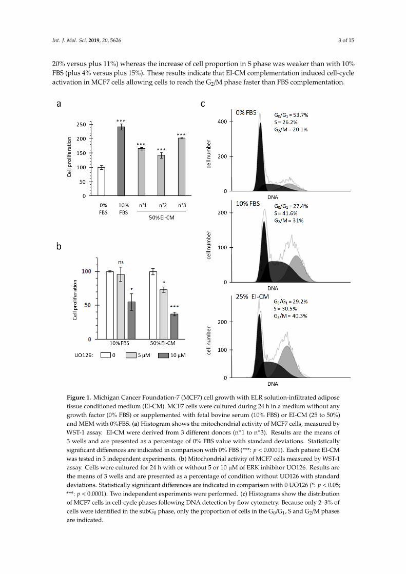
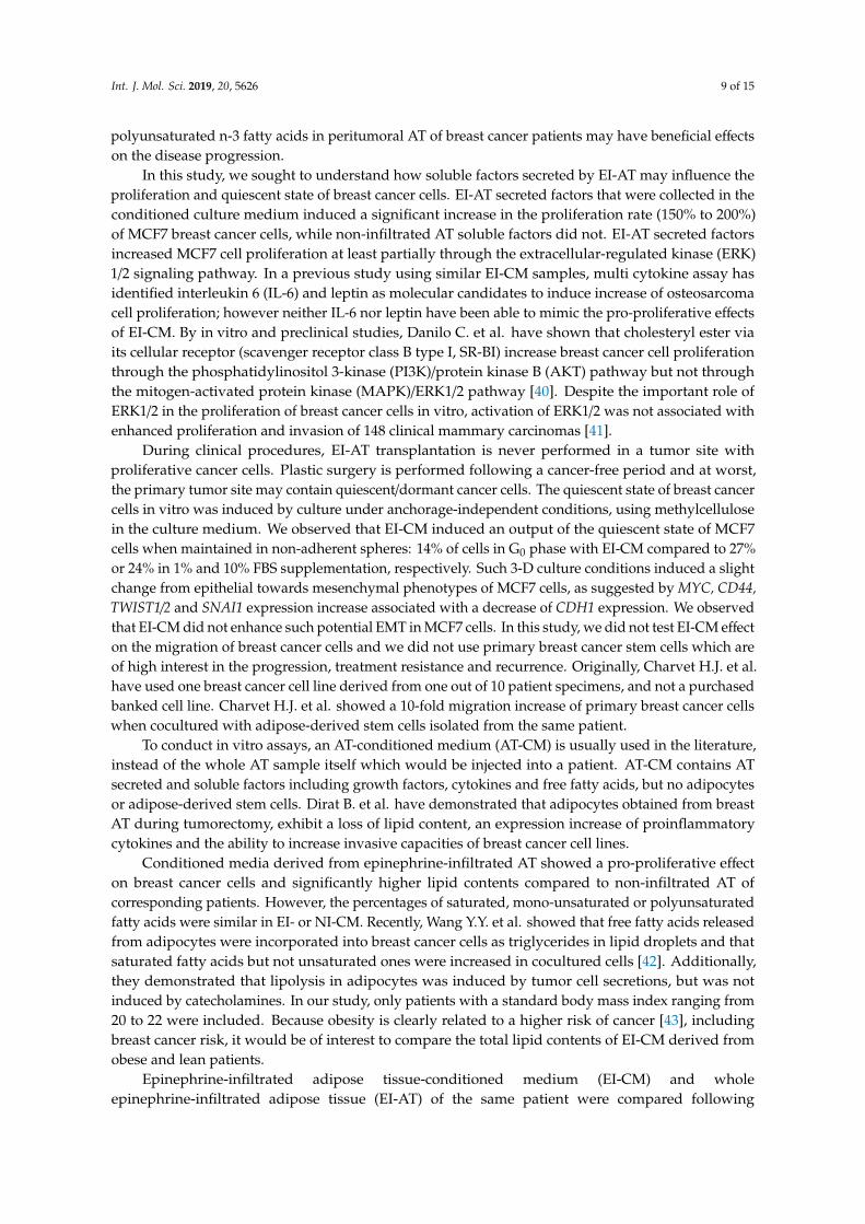
Proliferation of MCF7 breast carcinoma cells was analyzed in adherent culture conditions bymeasuring mitochondrial activity (WST-1 assay) and cell viability was controlled by cell counting withtrypan blue staining. As cancer cells lost their adherence to plastic when whole epinephrine-infiltratedadipose tissue (EI-AT) was used, EI-AT was replaced by EI-CM to complement cell culture mediumin order to mimic secreted factors by EI-AT. Before being treated, cells were cultured overnight(16 h) in a standard medium without fetal bovine serum (FBS) in order to observe a synchronizedresponse to growth factor stimulation. FBS supplementation (10%) enhanced MCF7 mitochondrialactivity up to 240% compared to that without FBS (Figure 1a). Similarly supplementation with 50%epinephrine-infiltrated adipose tissue conditioned medium (EI-CM) and 50% MEM α medium (0%FBS) from three different donors enhanced MCF7 mitochondrial activity from 150 to 200% (Figure 1a).The increases of mitochondrial activity were correlated with an increase in cell number by trypan bleucounting. Moreover, independent experiments performed with 50% or 25% of EI-CM of patients n◦1 to3 showed similar increases of MCF7 cell proliferation. The inhibition of the ERK1/2 signaling pathwayusing 5 µM UO126 induced a 30% decrease of the MCF7 proliferation with EI-CM, whereas it did notchange the MCF7 proliferation with 10% FBS (Figure 1b). These results indicate that EI-CM increasesMCF7 cell proliferation at least partially through the ERK1/2 signaling pathway.
Cell distribution in each cell-cycle phases was observed by flow cytometry after DNA stainingwith propidium iodide. During culture without FBS, at least half of the MCF7 cells were in G0/G1
phase (54% in Figure 1c, top panel). FBS treatment decreased the proportion of cells in G0/G1 phaseby half and increased the proportion of cells preparing their mitosis and those replicating their DNA(Figure 1c, middle panel). When MCF7 cells were treated with 25% EI-CM (Figure 1c, low panel),the proportion of cells in G0/G1 phase was also reduced by half compared to 0% FBS culture condition.With 25% EI-CM, a higher increase in cells in G2/M phase was observed compared to 10% FBS (plus
Int. J. Mol. Sci. 2019, 20, 5626 3 of 15
20% versus plus 11%) whereas the increase of cell proportion in S phase was weaker than with 10%FBS (plus 4% versus plus 15%). These results indicate that EI-CM complementation induced cell-cycleactivation in MCF7 cells allowing cells to reach the G2/M phase faster than FBS complementation.
Int. J. Mol. Sci. 2019, 20, x FOR PEER REVIEW 3 of 16
20% versus plus 11%) whereas the increase of cell proportion in S phase was weaker than with 10% FBS (plus 4% versus plus 15%). These results indicate that EI-CM complementation induced cell-cycle activation in MCF7 cells allowing cells to reach the G2/M phase faster than FBS complementation.
Figure 1. Michigan Cancer Foundation-7 (MCF7) cell growth with ELR solution-infiltrated adipose tissue conditioned medium (EI-CM). MCF7 cells were cultured during 24 h in a medium without any growth factor (0% FBS) or supplemented with fetal bovine serum (10% FBS) or EI-CM (25 to 50%) and MEM with 0%FBS. (a) Histogram shows the mitochondrial activity of MCF7 cells, measured by WST-1 assay. EI-CM were derived from 3 different donors (n°1 to n°3). Results are the means of 3 wells and are presented as a percentage of 0% FBS value with standard deviations. Statistically significant differences are indicated in comparison with 0% FBS (***: p < 0.0001). Each patient EI-CM was tested in 3 independent experiments. (b) Mitochondrial activity of MCF7 cells measured by WST-1 assay. Cells were cultured for 24 h with or without 5 or 10 µM of ERK inhibitor UO126. Results are the means of 3 wells and are presented as a percentage of condition without UO126 with standard deviations. Statistically significant differences are indicated in comparison with 0 UO126 (*: p < 0.05; ***: p < 0.0001). Two independent experiments were performed. (c) Histograms show the distribution of MCF7 cells in cell-cycle phases following DNA detection by flow cytometry. Because only 2–3% of cells were identified in the subG0 phase, only the proportion of cells in the G0/G1, S and G2/M phases are indicated.
Figure 1. Michigan Cancer Foundation-7 (MCF7) cell growth with ELR solution-infiltrated adiposetissue conditioned medium (EI-CM). MCF7 cells were cultured during 24 h in a medium without anygrowth factor (0% FBS) or supplemented with fetal bovine serum (10% FBS) or EI-CM (25 to 50%)and MEM with 0%FBS. (a) Histogram shows the mitochondrial activity of MCF7 cells, measured byWST-1 assay. EI-CM were derived from 3 different donors (n◦1 to n◦3). Results are the means of3 wells and are presented as a percentage of 0% FBS value with standard deviations. Statisticallysignificant differences are indicated in comparison with 0% FBS (***: p < 0.0001). Each patient EI-CMwas tested in 3 independent experiments. (b) Mitochondrial activity of MCF7 cells measured by WST-1assay. Cells were cultured for 24 h with or without 5 or 10 µM of ERK inhibitor UO126. Results arethe means of 3 wells and are presented as a percentage of condition without UO126 with standarddeviations. Statistically significant differences are indicated in comparison with 0 UO126 (*: p < 0.05;***: p < 0.0001). Two independent experiments were performed. (c) Histograms show the distributionof MCF7 cells in cell-cycle phases following DNA detection by flow cytometry. Because only 2–3% ofcells were identified in the subG0 phase, only the proportion of cells in the G0/G1, S and G2/M phasesare indicated.
Int. J. Mol. Sci. 2019, 20, 5626 4 of 15
2.2. MCF7 Cell Quiescence was Increased by Sphereoid Culture and Reduced by Epinephrine-InfiltratedAdipose Tissue Conditioned Medium
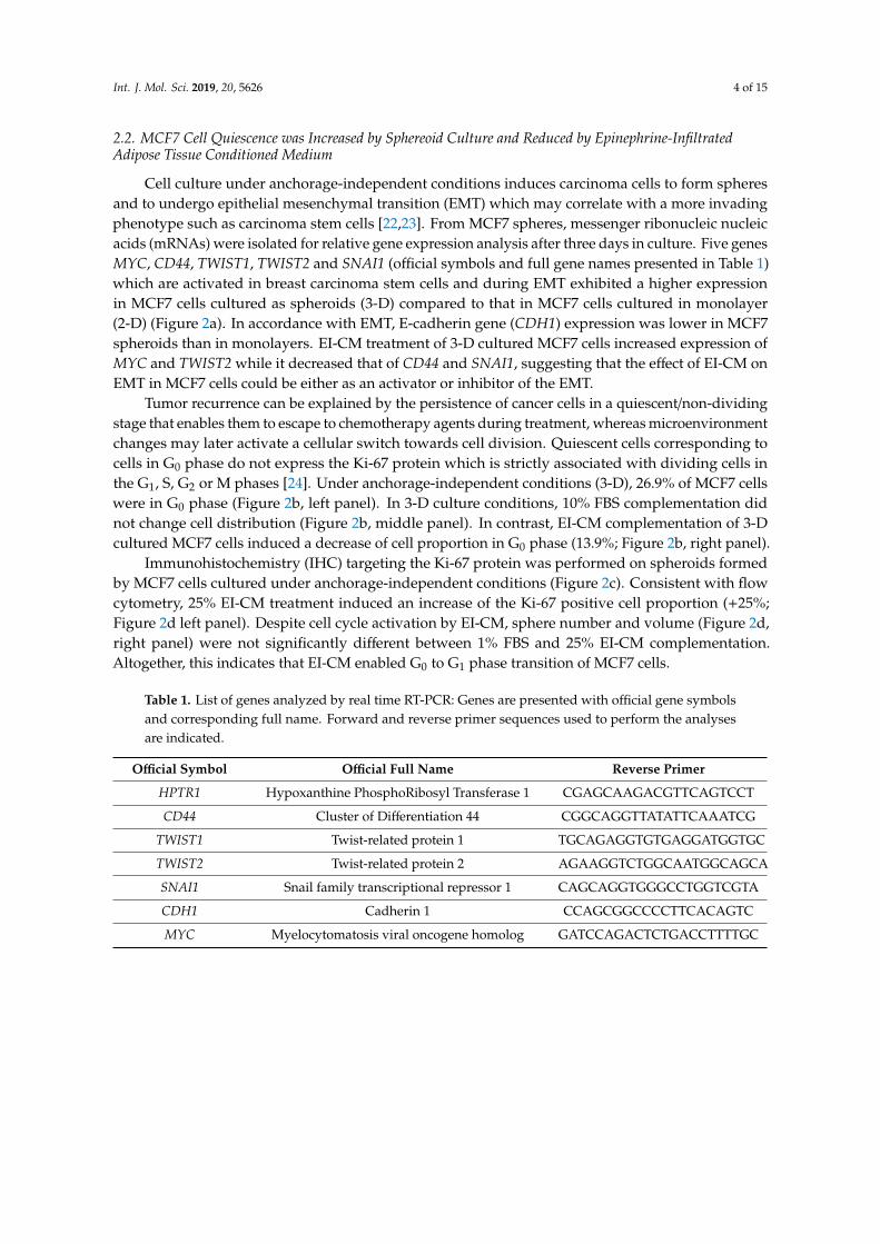
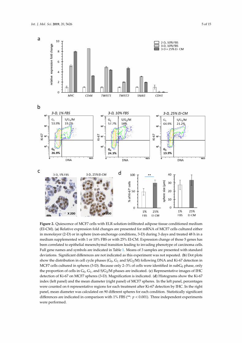
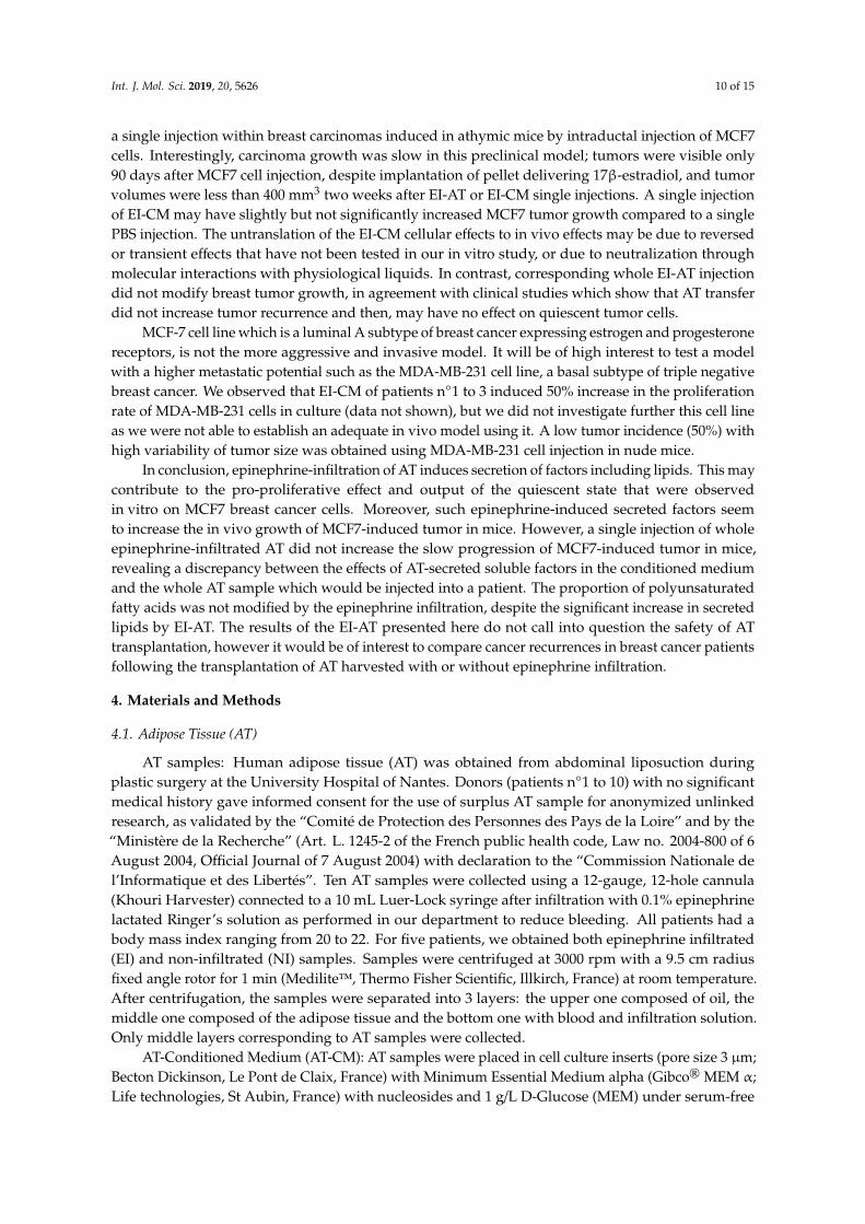
Cell culture under anchorage-independent conditions induces carcinoma cells to form spheresand to undergo epithelial mesenchymal transition (EMT) which may correlate with a more invadingphenotype such as carcinoma stem cells [22,23]. From MCF7 spheres, messenger ribonucleic nucleicacids (mRNAs) were isolated for relative gene expression analysis after three days in culture. Five genesMYC, CD44, TWIST1, TWIST2 and SNAI1 (official symbols and full gene names presented in Table 1)which are activated in breast carcinoma stem cells and during EMT exhibited a higher expressionin MCF7 cells cultured as spheroids (3-D) compared to that in MCF7 cells cultured in monolayer(2-D) (Figure 2a). In accordance with EMT, E-cadherin gene (CDH1) expression was lower in MCF7spheroids than in monolayers. EI-CM treatment of 3-D cultured MCF7 cells increased expression ofMYC and TWIST2 while it decreased that of CD44 and SNAI1, suggesting that the effect of EI-CM onEMT in MCF7 cells could be either as an activator or inhibitor of the EMT.
Tumor recurrence can be explained by the persistence of cancer cells in a quiescent/non-dividingstage that enables them to escape to chemotherapy agents during treatment, whereas microenvironmentchanges may later activate a cellular switch towards cell division. Quiescent cells corresponding tocells in G0 phase do not express the Ki-67 protein which is strictly associated with dividing cells inthe G1, S, G2 or M phases [24]. Under anchorage-independent conditions (3-D), 26.9% of MCF7 cellswere in G0 phase (Figure 2b, left panel). In 3-D culture conditions, 10% FBS complementation didnot change cell distribution (Figure 2b, middle panel). In contrast, EI-CM complementation of 3-Dcultured MCF7 cells induced a decrease of cell proportion in G0 phase (13.9%; Figure 2b, right panel).
Immunohistochemistry (IHC) targeting the Ki-67 protein was performed on spheroids formedby MCF7 cells cultured under anchorage-independent conditions (Figure 2c). Consistent with flowcytometry, 25% EI-CM treatment induced an increase of the Ki-67 positive cell proportion (+25%;Figure 2d left panel). Despite cell cycle activation by EI-CM, sphere number and volume (Figure 2d,right panel) were not significantly different between 1% FBS and 25% EI-CM complementation.Altogether, this indicates that EI-CM enabled G0 to G1 phase transition of MCF7 cells.
Table 1. List of genes analyzed by real time RT-PCR: Genes are presented with official gene symbolsand corresponding full name. Forward and reverse primer sequences used to perform the analysesare indicated.
Official Symbol Official Full Name Reverse Primer
HPTR1 Hypoxanthine PhosphoRibosyl Transferase 1 CGAGCAAGACGTTCAGTCCT
CD44 Cluster of Differentiation 44 CGGCAGGTTATATTCAAATCG
TWIST1 Twist-related protein 1 TGCAGAGGTGTGAGGATGGTGC
TWIST2 Twist-related protein 2 AGAAGGTCTGGCAATGGCAGCA
SNAI1 Snail family transcriptional repressor 1 CAGCAGGTGGGCCTGGTCGTA
CDH1 Cadherin 1 CCAGCGGCCCCTTCACAGTC
MYC Myelocytomatosis viral oncogene homolog GATCCAGACTCTGACCTTTTGC
Int. J. Mol. Sci. 2019, 20, 5626 5 of 15Int. J. Mol. Sci. 2019, 20, x FOR PEER REVIEW 5 of 16
Figure 2. Quiescence of MCF7 cells with ELR solution-infiltrated adipose tissue conditioned medium (EI-CM). (a) Relative expression fold changes are presented for mRNA of MCF7 cells cultured either in monolayer (2-D) or in sphere (non-anchorage conditions, 3-D) during 3 days and treated 48 h in a medium supplemented with 1 or 10% FBS or with 25% EI-CM. Expression change of those 5 genes has been correlated to epithelial mesenchymal transition leading to invading phenotype of carcinoma cells. Full gene names and symbols are indicated in Table 1. Means of 3 samples are presented with standard deviations. Significant differences are not indicated as this experiment was not repeated. (b) Dot plots show the distribution in cell cycle phases (G0, G1 and S/G2/M) following DNA and Ki-67 detection in MCF7 cells cultured in spheres (3-D). Because only 2–3% of cells were identified in subG0 phase, only the proportion of cells in G0, G1, and S/G2/M phases are indicated. (c) Representative images of IHC detection of Ki-67 on MCF7 spheres (3-D). Magnification is indicated. (d) Histograms show the Ki-67 index (left panel) and the mean diameter (right panel) of MCF7 spheres. In the left panel, percentages were counted on 6 representative regions for each treatment after Ki-67 detection by IHC. In the right panel, mean diameter was calculated on 90 different spheres for each condition. Statistically significant differences are indicated in comparison with 1% FBS (**: p < 0.001). Three independent experiments were performed.
Figure 2. Quiescence of MCF7 cells with ELR solution-infiltrated adipose tissue conditioned medium(EI-CM). (a) Relative expression fold changes are presented for mRNA of MCF7 cells cultured eitherin monolayer (2-D) or in sphere (non-anchorage conditions, 3-D) during 3 days and treated 48 h in amedium supplemented with 1 or 10% FBS or with 25% EI-CM. Expression change of those 5 genes hasbeen correlated to epithelial mesenchymal transition leading to invading phenotype of carcinoma cells.Full gene names and symbols are indicated in Table 1. Means of 3 samples are presented with standarddeviations. Significant differences are not indicated as this experiment was not repeated. (b) Dot plotsshow the distribution in cell cycle phases (G0, G1 and S/G2/M) following DNA and Ki-67 detection inMCF7 cells cultured in spheres (3-D). Because only 2–3% of cells were identified in subG0 phase, onlythe proportion of cells in G0, G1, and S/G2/M phases are indicated. (c) Representative images of IHCdetection of Ki-67 on MCF7 spheres (3-D). Magnification is indicated. (d) Histograms show the Ki-67index (left panel) and the mean diameter (right panel) of MCF7 spheres. In the left panel, percentageswere counted on 6 representative regions for each treatment after Ki-67 detection by IHC. In the rightpanel, mean diameter was calculated on 90 different spheres for each condition. Statistically significantdifferences are indicated in comparison with 1% FBS (**: p < 0.001). Three independent experimentswere performed.
Int. J. Mol. Sci. 2019, 20, 5626 6 of 15
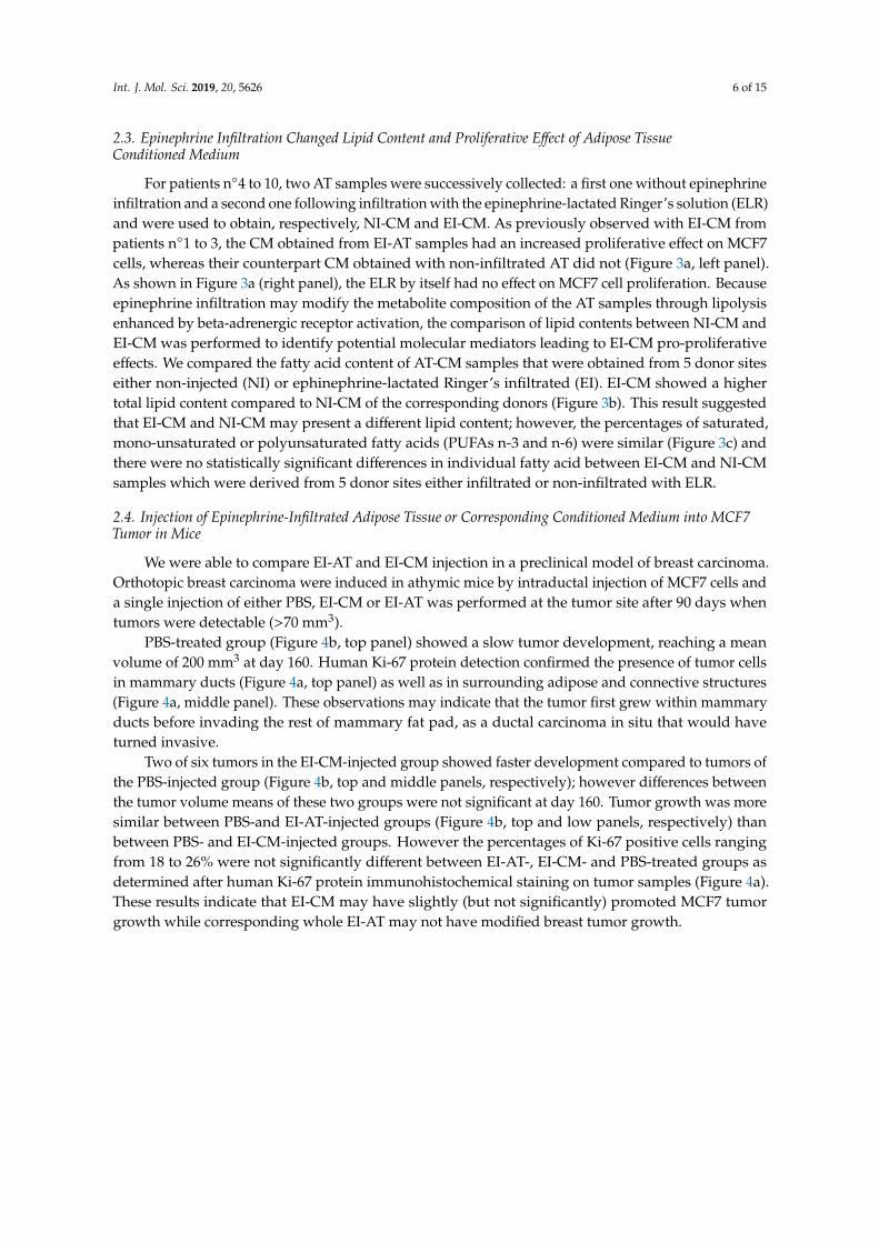
2.3. Epinephrine Infiltration Changed Lipid Content and Proliferative Effect of Adipose TissueConditioned Medium
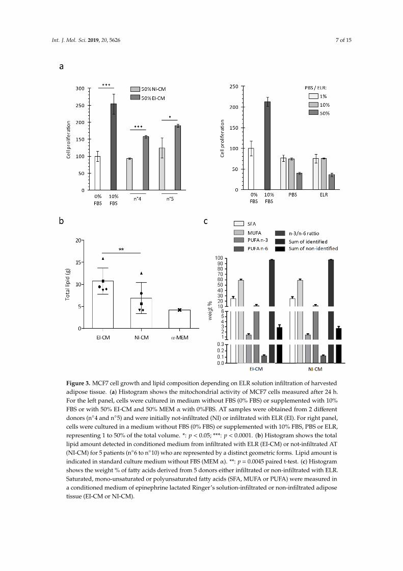
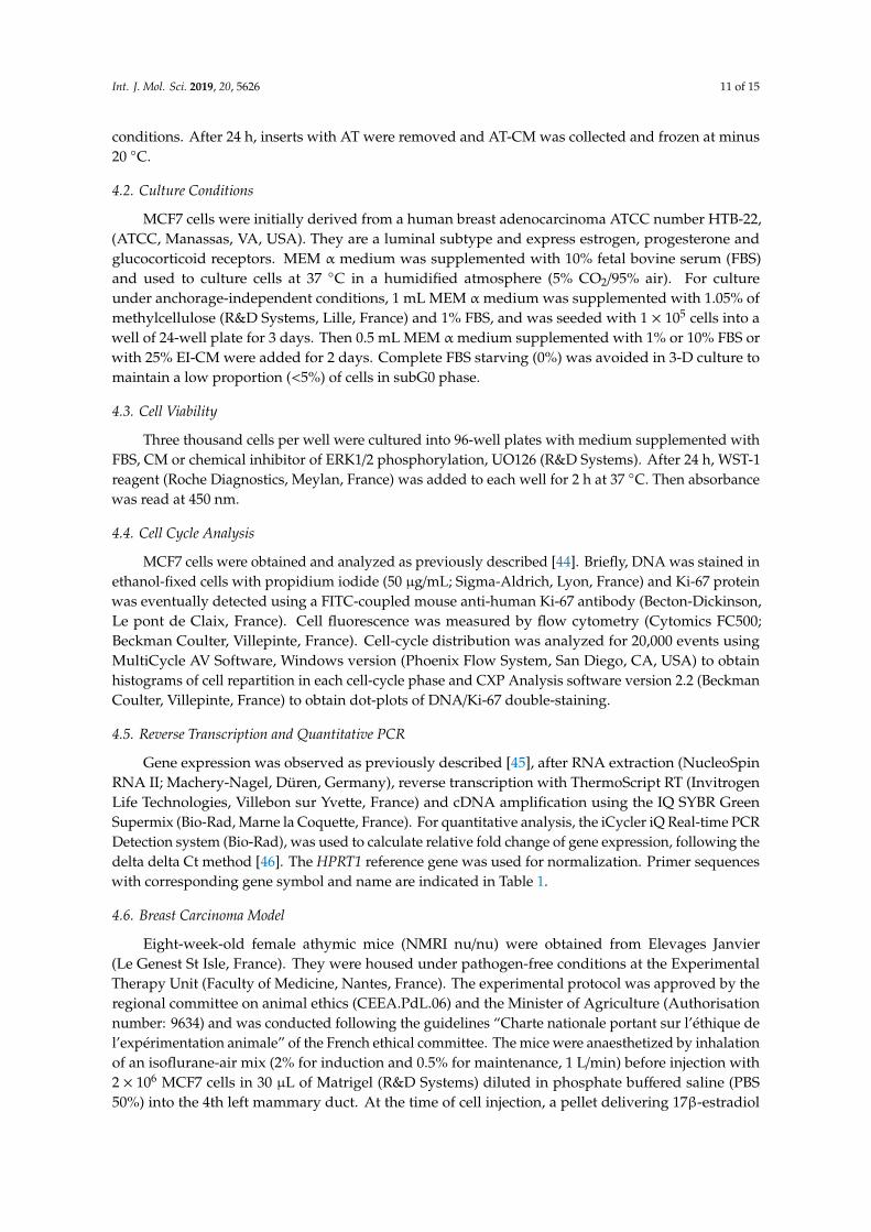
For patients n◦4 to 10, two AT samples were successively collected: a first one without epinephrineinfiltration and a second one following infiltration with the epinephrine-lactated Ringer’s solution (ELR)and were used to obtain, respectively, NI-CM and EI-CM. As previously observed with EI-CM frompatients n◦1 to 3, the CM obtained from EI-AT samples had an increased proliferative effect on MCF7cells, whereas their counterpart CM obtained with non-infiltrated AT did not (Figure 3a, left panel).As shown in Figure 3a (right panel), the ELR by itself had no effect on MCF7 cell proliferation. Becauseepinephrine infiltration may modify the metabolite composition of the AT samples through lipolysisenhanced by beta-adrenergic receptor activation, the comparison of lipid contents between NI-CM andEI-CM was performed to identify potential molecular mediators leading to EI-CM pro-proliferativeeffects. We compared the fatty acid content of AT-CM samples that were obtained from 5 donor siteseither non-injected (NI) or ephinephrine-lactated Ringer’s infiltrated (EI). EI-CM showed a highertotal lipid content compared to NI-CM of the corresponding donors (Figure 3b). This result suggestedthat EI-CM and NI-CM may present a different lipid content; however, the percentages of saturated,mono-unsaturated or polyunsaturated fatty acids (PUFAs n-3 and n-6) were similar (Figure 3c) andthere were no statistically significant differences in individual fatty acid between EI-CM and NI-CMsamples which were derived from 5 donor sites either infiltrated or non-infiltrated with ELR.
2.4. Injection of Epinephrine-Infiltrated Adipose Tissue or Corresponding Conditioned Medium into MCF7Tumor in Mice
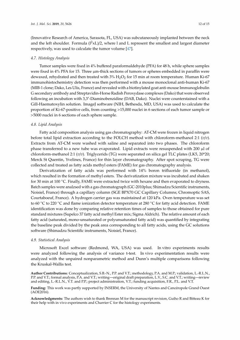
We were able to compare EI-AT and EI-CM injection in a preclinical model of breast carcinoma.Orthotopic breast carcinoma were induced in athymic mice by intraductal injection of MCF7 cells anda single injection of either PBS, EI-CM or EI-AT was performed at the tumor site after 90 days whentumors were detectable (>70 mm3).
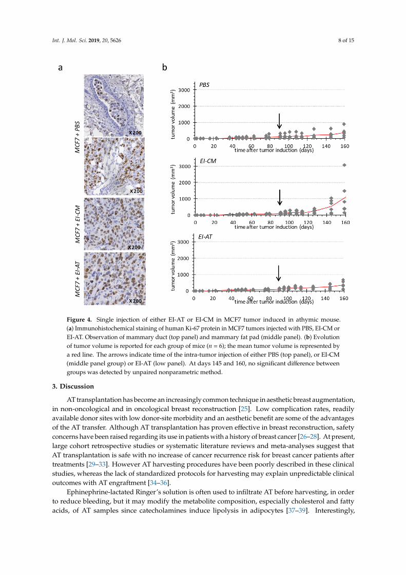
PBS-treated group (Figure 4b, top panel) showed a slow tumor development, reaching a meanvolume of 200 mm3 at day 160. Human Ki-67 protein detection confirmed the presence of tumor cellsin mammary ducts (Figure 4a, top panel) as well as in surrounding adipose and connective structures(Figure 4a, middle panel). These observations may indicate that the tumor first grew within mammaryducts before invading the rest of mammary fat pad, as a ductal carcinoma in situ that would haveturned invasive.
Two of six tumors in the EI-CM-injected group showed faster development compared to tumors ofthe PBS-injected group (Figure 4b, top and middle panels, respectively); however differences betweenthe tumor volume means of these two groups were not significant at day 160. Tumor growth was moresimilar between PBS-and EI-AT-injected groups (Figure 4b, top and low panels, respectively) thanbetween PBS- and EI-CM-injected groups. However the percentages of Ki-67 positive cells rangingfrom 18 to 26% were not significantly different between EI-AT-, EI-CM- and PBS-treated groups asdetermined after human Ki-67 protein immunohistochemical staining on tumor samples (Figure 4a).These results indicate that EI-CM may have slightly (but not significantly) promoted MCF7 tumorgrowth while corresponding whole EI-AT may not have modified breast tumor growth.
Int. J. Mol. Sci. 2019, 20, 5626 7 of 15Int. J. Mol. Sci. 2019, 20, x FOR PEER REVIEW 7 of 16
Figure 3. MCF7 cell growth and lipid composition depending on ELR solution infiltration of harvested adipose tissue. (a) Histogram shows the mitochondrial activity of MCF7 cells measured after 24 h. For the left panel, cells were cultured in medium without FBS (0% FBS) or supplemented with 10% FBS or with 50% EI-CM and 50% MEM α with 0%FBS. AT samples were obtained from 2 different donors (n°4 and n°5) and were initially not-infiltrated (NI) or infiltrated with ELR (EI). For right panel, cells were cultured in a medium without FBS (0% FBS) or supplemented with 10% FBS, PBS or ELR, representing 1 to 50% of the total volume. *: p < 0.05; ***: p < 0.0001. (b) Histogram shows the total lipid amount detected in conditioned medium from infiltrated with ELR (EI-CM) or not-infiltrated AT (NI-CM) for 5 patients (n°6 to n°10) who are represented by a distinct geometric forms. Lipid amount is indicated in standard culture medium without FBS (MEM α). **: p = 0.0045 paired t-test. (c) Histogram shows the weight % of fatty acids derived from 5 donors either infiltrated or non-infiltrated with ELR. Saturated, mono-unsaturated or polyunsaturated fatty acids (SFA, MUFA or PUFA) were measured in a conditioned medium of epinephrine lactated Ringer’s solution-infiltrated or non-infiltrated adipose tissue (EI-CM or NI-CM).
Figure 3. MCF7 cell growth and lipid composition depending on ELR solution infiltration of harvestedadipose tissue. (a) Histogram shows the mitochondrial activity of MCF7 cells measured after 24 h.For the left panel, cells were cultured in medium without FBS (0% FBS) or supplemented with 10%FBS or with 50% EI-CM and 50% MEM α with 0%FBS. AT samples were obtained from 2 differentdonors (n◦4 and n◦5) and were initially not-infiltrated (NI) or infiltrated with ELR (EI). For right panel,cells were cultured in a medium without FBS (0% FBS) or supplemented with 10% FBS, PBS or ELR,representing 1 to 50% of the total volume. *: p < 0.05; ***: p < 0.0001. (b) Histogram shows the totallipid amount detected in conditioned medium from infiltrated with ELR (EI-CM) or not-infiltrated AT(NI-CM) for 5 patients (n◦6 to n◦10) who are represented by a distinct geometric forms. Lipid amount isindicated in standard culture medium without FBS (MEM α). **: p = 0.0045 paired t-test. (c) Histogramshows the weight % of fatty acids derived from 5 donors either infiltrated or non-infiltrated with ELR.Saturated, mono-unsaturated or polyunsaturated fatty acids (SFA, MUFA or PUFA) were measured ina conditioned medium of epinephrine lactated Ringer’s solution-infiltrated or non-infiltrated adiposetissue (EI-CM or NI-CM).
Int. J. Mol. Sci. 2019, 20, 5626 8 of 15
Int. J. Mol. Sci. 2019, 20, x FOR PEER REVIEW 8 of 16
2.4. Injection of Epinephrine-Infiltrated Adipose Tissue or Corresponding Conditioned Medium into MCF7 Tumor in Mice
We were able to compare EI-AT and EI-CM injection in a preclinical model of breast carcinoma. Orthotopic breast carcinoma were induced in athymic mice by intraductal injection of MCF7 cells and a single injection of either PBS, EI-CM or EI-AT was performed at the tumor site after 90 days when tumors were detectable (>70 mm3).
PBS-treated group (Figure 4b, top panel) showed a slow tumor development, reaching a mean volume of 200 mm3 at day 160. Human Ki-67 protein detection confirmed the presence of tumor cells in mammary ducts (Figure 4a, top panel) as well as in surrounding adipose and connective structures (Figure 4a, middle panel). These observations may indicate that the tumor first grew within mammary ducts before invading the rest of mammary fat pad, as a ductal carcinoma in situ that would have turned invasive.
Two of six tumors in the EI-CM-injected group showed faster development compared to tumors of the PBS-injected group (Figure 4b, top and middle panels, respectively); however differences between the tumor volume means of these two groups were not significant at day 160. Tumor growth was more similar between PBS-and EI-AT-injected groups (Figure 4b, top and low panels, respectively) than between PBS- and EI-CM-injected groups. However the percentages of Ki-67 positive cells ranging from 18 to 26% were not significantly different between EI-AT-, EI-CM- and PBS-treated groups as determined after human Ki-67 protein immunohistochemical staining on tumor samples (Figure 4a). These results indicate that EI-CM may have slightly (but not significantly) promoted MCF7 tumor growth while corresponding whole EI-AT may not have modified breast tumor growth.
Figure 4. Single injection of either EI-AT or EI-CM in MCF7 tumor induced in athymic mouse. (a) Immunohistochemical staining of human Ki-67 protein in MCF7 tumors injected with PBS, EI-CM or Figure 4. Single injection of either EI-AT or EI-CM in MCF7 tumor induced in athymic mouse.(a) Immunohistochemical staining of human Ki-67 protein in MCF7 tumors injected with PBS, EI-CM orEI-AT. Observation of mammary duct (top panel) and mammary fat pad (middle panel). (b) Evolutionof tumor volume is reported for each group of mice (n = 6); the mean tumor volume is represented bya red line. The arrows indicate time of the intra-tumor injection of either PBS (top panel), or EI-CM(middle panel group) or EI-AT (low panel). At days 145 and 160, no significant difference betweengroups was detected by unpaired nonparametric method.
3. Discussion
AT transplantation has become an increasingly common technique in aesthetic breast augmentation,in non-oncological and in oncological breast reconstruction [25]. Low complication rates, readilyavailable donor sites with low donor-site morbidity and an aesthetic benefit are some of the advantagesof the AT transfer. Although AT transplantation has proven effective in breast reconstruction, safetyconcerns have been raised regarding its use in patients with a history of breast cancer [26–28]. At present,large cohort retrospective studies or systematic literature reviews and meta-analyses suggest thatAT transplantation is safe with no increase of cancer recurrence risk for breast cancer patients aftertreatments [29–33]. However AT harvesting procedures have been poorly described in these clinicalstudies, whereas the lack of standardized protocols for harvesting may explain unpredictable clinicaloutcomes with AT engraftment [34–36].
Ephinephrine-lactated Ringer’s solution is often used to infiltrate AT before harvesting, in orderto reduce bleeding, but it may modify the metabolite composition, especially cholesterol and fattyacids, of AT samples since catecholamines induce lipolysis in adipocytes [37–39]. Interestingly,
Int. J. Mol. Sci. 2019, 20, 5626 9 of 15
polyunsaturated n-3 fatty acids in peritumoral AT of breast cancer patients may have beneficial effectson the disease progression.
In this study, we sought to understand how soluble factors secreted by EI-AT may influence theproliferation and quiescent state of breast cancer cells. EI-AT secreted factors that were collected in theconditioned culture medium induced a significant increase in the proliferation rate (150% to 200%)of MCF7 breast cancer cells, while non-infiltrated AT soluble factors did not. EI-AT secreted factorsincreased MCF7 cell proliferation at least partially through the extracellular-regulated kinase (ERK)1/2 signaling pathway. In a previous study using similar EI-CM samples, multi cytokine assay hasidentified interleukin 6 (IL-6) and leptin as molecular candidates to induce increase of osteosarcomacell proliferation; however neither IL-6 nor leptin have been able to mimic the pro-proliferative effectsof EI-CM. By in vitro and preclinical studies, Danilo C. et al. have shown that cholesteryl ester viaits cellular receptor (scavenger receptor class B type I, SR-BI) increase breast cancer cell proliferationthrough the phosphatidylinositol 3-kinase (PI3K)/protein kinase B (AKT) pathway but not throughthe mitogen-activated protein kinase (MAPK)/ERK1/2 pathway [40]. Despite the important role ofERK1/2 in the proliferation of breast cancer cells in vitro, activation of ERK1/2 was not associated withenhanced proliferation and invasion of 148 clinical mammary carcinomas [41].
During clinical procedures, EI-AT transplantation is never performed in a tumor site withproliferative cancer cells. Plastic surgery is performed following a cancer-free period and at worst,the primary tumor site may contain quiescent/dormant cancer cells. The quiescent state of breast cancercells in vitro was induced by culture under anchorage-independent conditions, using methylcellulosein the culture medium. We observed that EI-CM induced an output of the quiescent state of MCF7cells when maintained in non-adherent spheres: 14% of cells in G0 phase with EI-CM compared to 27%or 24% in 1% and 10% FBS supplementation, respectively. Such 3-D culture conditions induced a slightchange from epithelial towards mesenchymal phenotypes of MCF7 cells, as suggested by MYC, CD44,TWIST1/2 and SNAI1 expression increase associated with a decrease of CDH1 expression. We observedthat EI-CM did not enhance such potential EMT in MCF7 cells. In this study, we did not test EI-CM effecton the migration of breast cancer cells and we did not use primary breast cancer stem cells which areof high interest in the progression, treatment resistance and recurrence. Originally, Charvet H.J. et al.have used one breast cancer cell line derived from one out of 10 patient specimens, and not a purchasedbanked cell line. Charvet H.J. et al. showed a 10-fold migration increase of primary breast cancer cellswhen cocultured with adipose-derived stem cells isolated from the same patient.
To conduct in vitro assays, an AT-conditioned medium (AT-CM) is usually used in the literature,instead of the whole AT sample itself which would be injected into a patient. AT-CM contains ATsecreted and soluble factors including growth factors, cytokines and free fatty acids, but no adipocytesor adipose-derived stem cells. Dirat B. et al. have demonstrated that adipocytes obtained from breastAT during tumorectomy, exhibit a loss of lipid content, an expression increase of proinflammatorycytokines and the ability to increase invasive capacities of breast cancer cell lines.
Conditioned media derived from epinephrine-infiltrated AT showed a pro-proliferative effecton breast cancer cells and significantly higher lipid contents compared to non-infiltrated AT ofcorresponding patients. However, the percentages of saturated, mono-unsaturated or polyunsaturatedfatty acids were similar in EI- or NI-CM. Recently, Wang Y.Y. et al. showed that free fatty acids releasedfrom adipocytes were incorporated into breast cancer cells as triglycerides in lipid droplets and thatsaturated fatty acids but not unsaturated ones were increased in cocultured cells [42]. Additionally,they demonstrated that lipolysis in adipocytes was induced by tumor cell secretions, but was notinduced by catecholamines. In our study, only patients with a standard body mass index ranging from20 to 22 were included. Because obesity is clearly related to a higher risk of cancer [43], includingbreast cancer risk, it would be of interest to compare the total lipid contents of EI-CM derived fromobese and lean patients.
Epinephrine-infiltrated adipose tissue-conditioned medium (EI-CM) and wholeepinephrine-infiltrated adipose tissue (EI-AT) of the same patient were compared following
Int. J. Mol. Sci. 2019, 20, 5626 10 of 15
a single injection within breast carcinomas induced in athymic mice by intraductal injection of MCF7cells. Interestingly, carcinoma growth was slow in this preclinical model; tumors were visible only90 days after MCF7 cell injection, despite implantation of pellet delivering 17β-estradiol, and tumorvolumes were less than 400 mm3 two weeks after EI-AT or EI-CM single injections. A single injectionof EI-CM may have slightly but not significantly increased MCF7 tumor growth compared to a singlePBS injection. The untranslation of the EI-CM cellular effects to in vivo effects may be due to reversedor transient effects that have not been tested in our in vitro study, or due to neutralization throughmolecular interactions with physiological liquids. In contrast, corresponding whole EI-AT injectiondid not modify breast tumor growth, in agreement with clinical studies which show that AT transferdid not increase tumor recurrence and then, may have no effect on quiescent tumor cells.
MCF-7 cell line which is a luminal A subtype of breast cancer expressing estrogen and progesteronereceptors, is not the more aggressive and invasive model. It will be of high interest to test a modelwith a higher metastatic potential such as the MDA-MB-231 cell line, a basal subtype of triple negativebreast cancer. We observed that EI-CM of patients n◦1 to 3 induced 50% increase in the proliferationrate of MDA-MB-231 cells in culture (data not shown), but we did not investigate further this cell lineas we were not able to establish an adequate in vivo model using it. A low tumor incidence (50%) withhigh variability of tumor size was obtained using MDA-MB-231 cell injection in nude mice.
In conclusion, epinephrine-infiltration of AT induces secretion of factors including lipids. This maycontribute to the pro-proliferative effect and output of the quiescent state that were observedin vitro on MCF7 breast cancer cells. Moreover, such epinephrine-induced secreted factors seemto increase the in vivo growth of MCF7-induced tumor in mice. However, a single injection of wholeepinephrine-infiltrated AT did not increase the slow progression of MCF7-induced tumor in mice,revealing a discrepancy between the effects of AT-secreted soluble factors in the conditioned mediumand the whole AT sample which would be injected into a patient. The proportion of polyunsaturatedfatty acids was not modified by the epinephrine infiltration, despite the significant increase in secretedlipids by EI-AT. The results of the EI-AT presented here do not call into question the safety of ATtransplantation, however it would be of interest to compare cancer recurrences in breast cancer patientsfollowing the transplantation of AT harvested with or without epinephrine infiltration.
4. Materials and Methods
4.1. Adipose Tissue (AT)
AT samples: Human adipose tissue (AT) was obtained from abdominal liposuction duringplastic surgery at the University Hospital of Nantes. Donors (patients n◦1 to 10) with no significantmedical history gave informed consent for the use of surplus AT sample for anonymized unlinkedresearch, as validated by the “Comité de Protection des Personnes des Pays de la Loire” and by the“Ministère de la Recherche” (Art. L. 1245-2 of the French public health code, Law no. 2004-800 of 6August 2004, Official Journal of 7 August 2004) with declaration to the “Commission Nationale del’Informatique et des Libertés”. Ten AT samples were collected using a 12-gauge, 12-hole cannula(Khouri Harvester) connected to a 10 mL Luer-Lock syringe after infiltration with 0.1% epinephrinelactated Ringer’s solution as performed in our department to reduce bleeding. All patients had abody mass index ranging from 20 to 22. For five patients, we obtained both epinephrine infiltrated(EI) and non-infiltrated (NI) samples. Samples were centrifuged at 3000 rpm with a 9.5 cm radiusfixed angle rotor for 1 min (Medilite™, Thermo Fisher Scientific, Illkirch, France) at room temperature.After centrifugation, the samples were separated into 3 layers: the upper one composed of oil, themiddle one composed of the adipose tissue and the bottom one with blood and infiltration solution.Only middle layers corresponding to AT samples were collected.
AT-Conditioned Medium (AT-CM): AT samples were placed in cell culture inserts (pore size 3 µm;Becton Dickinson, Le Pont de Claix, France) with Minimum Essential Medium alpha (Gibco® MEM α;Life technologies, St Aubin, France) with nucleosides and 1 g/L D-Glucose (MEM) under serum-free
Int. J. Mol. Sci. 2019, 20, 5626 11 of 15
conditions. After 24 h, inserts with AT were removed and AT-CM was collected and frozen at minus20 ◦C.
4.2. Culture Conditions
MCF7 cells were initially derived from a human breast adenocarcinoma ATCC number HTB-22,(ATCC, Manassas, VA, USA). They are a luminal subtype and express estrogen, progesterone andglucocorticoid receptors. MEM α medium was supplemented with 10% fetal bovine serum (FBS)and used to culture cells at 37 ◦C in a humidified atmosphere (5% CO2/95% air). For cultureunder anchorage-independent conditions, 1 mL MEM αmedium was supplemented with 1.05% ofmethylcellulose (R&D Systems, Lille, France) and 1% FBS, and was seeded with 1 × 10⁵ cells into awell of 24-well plate for 3 days. Then 0.5 mL MEM αmedium supplemented with 1% or 10% FBS orwith 25% EI-CM were added for 2 days. Complete FBS starving (0%) was avoided in 3-D culture tomaintain a low proportion (<5%) of cells in subG0 phase.
4.3. Cell Viability
Three thousand cells per well were cultured into 96-well plates with medium supplemented withFBS, CM or chemical inhibitor of ERK1/2 phosphorylation, UO126 (R&D Systems). After 24 h, WST-1reagent (Roche Diagnostics, Meylan, France) was added to each well for 2 h at 37 ◦C. Then absorbancewas read at 450 nm.
4.4. Cell Cycle Analysis
MCF7 cells were obtained and analyzed as previously described [44]. Briefly, DNA was stained inethanol-fixed cells with propidium iodide (50 µg/mL; Sigma-Aldrich, Lyon, France) and Ki-67 proteinwas eventually detected using a FITC-coupled mouse anti-human Ki-67 antibody (Becton-Dickinson,Le pont de Claix, France). Cell fluorescence was measured by flow cytometry (Cytomics FC500;Beckman Coulter, Villepinte, France). Cell-cycle distribution was analyzed for 20,000 events usingMultiCycle AV Software, Windows version (Phoenix Flow System, San Diego, CA, USA) to obtainhistograms of cell repartition in each cell-cycle phase and CXP Analysis software version 2.2 (BeckmanCoulter, Villepinte, France) to obtain dot-plots of DNA/Ki-67 double-staining.
4.5. Reverse Transcription and Quantitative PCR
Gene expression was observed as previously described [45], after RNA extraction (NucleoSpinRNA II; Machery-Nagel, Düren, Germany), reverse transcription with ThermoScript RT (InvitrogenLife Technologies, Villebon sur Yvette, France) and cDNA amplification using the IQ SYBR GreenSupermix (Bio-Rad, Marne la Coquette, France). For quantitative analysis, the iCycler iQ Real-time PCRDetection system (Bio-Rad), was used to calculate relative fold change of gene expression, following thedelta delta Ct method [46]. The HPRT1 reference gene was used for normalization. Primer sequenceswith corresponding gene symbol and name are indicated in Table 1.
4.6. Breast Carcinoma Model
Eight-week-old female athymic mice (NMRI nu/nu) were obtained from Elevages Janvier(Le Genest St Isle, France). They were housed under pathogen-free conditions at the ExperimentalTherapy Unit (Faculty of Medicine, Nantes, France). The experimental protocol was approved by theregional committee on animal ethics (CEEA.PdL.06) and the Minister of Agriculture (Authorisationnumber: 9634) and was conducted following the guidelines “Charte nationale portant sur l’éthique del’expérimentation animale” of the French ethical committee. The mice were anaesthetized by inhalationof an isoflurane-air mix (2% for induction and 0.5% for maintenance, 1 L/min) before injection with2 × 106 MCF7 cells in 30 µL of Matrigel (R&D Systems) diluted in phosphate buffered saline (PBS50%) into the 4th left mammary duct. At the time of cell injection, a pellet delivering 17β-estradiol
Int. J. Mol. Sci. 2019, 20, 5626 12 of 15
(Innovative Research of America, Sarasota, FL, USA) was subcutaneously implanted between the neckand the left shoulder. Formula (l2xL)/2, where l and L represent the smallest and largest diameterrespectively, was used to calculate the tumor volume [47].
4.7. Histology Analysis
Tumor samples were fixed in 4% buffered paraformaldehyde (PFA) for 48 h, while sphere sampleswere fixed in 4% PFA for 15. Three µm-thick sections of tumors or spheres embedded in paraffin weredewaxed, rehydrated and then treated with 3% H2O2 for 15 min at room temperature. Human Ki-67immunohistochemistry detection was then performed with a mouse monoclonal anti-human Ki-67(MIB-1 clone; Dako, Les Ulis, France) and revealed with a biotinylated goat anti-mouse ImmunoglobulinG secondary antibody and Streptavidin-Horse Radish Peroxydase complexes (Dako) that were observedfollowing an incubation with 3,3′-Diaminobenzidine (DAB, Dako). Nuclei were counterstained with aGill-Haematoxylin solution. ImageJ software (NIH, Bethesda, MD, USA) was used to calculate theproportion of Ki-67-positive cells, from counting >15,000 nuclei in 6 sections of each tumor sample or>5000 nuclei in 6 sections of each sphere sample.
4.8. Lipid Analysis
Fatty acid composition analysis using gas chromatography: AT-CM were frozen in liquid nitrogenbefore total lipid extraction according to the FOLCH method with chloroform-methanol 2:1 (v/v).Extracts from AT-CM were washed with saline and separated into two phases. The chloroformphase transferred to a new tube was evaporated. Lipid extracts were resuspended with 200 µl ofchloroform-methanol 2:1 (v/v). Triglycerids (TG) were separated on silica gel TLC plates (LK5, 20*20;Merck St Quentin, Yvelines, France) for thin layer chromatography. After spot scraping, TG werecollected and treated as fatty acids methyl esters (FAME) for gas chromatography analysis.
Derivatization of fatty acids was performed with 14% boron trifluoride (in methanol),which resulted in the formation of methyl esters. The derivatization mixture was incubated and shakenfor 30 min at 100 ◦C. Finally, FAME were extracted twice with hexane and then evaporated to dryness.Batch samples were analzsed with a gas chromatograph (GC-2010plus; Shimadzu Scientific instruments,Noisiel, France) through a capillary column (SGE BPX70 GC Capillary Columns; Chromoptic SAS,Courtaboeuf, France). A hydrogen carrier gas was maintained at 120 kPa. Oven temperature was setto 60 ◦C to 220 ◦C and flame ionization detector temperature at 280 ◦C for fatty acid detection. FAMEidentification was done by comparing relative retention times of samples to those obtained for purestandard mixtures (Supelco 37 fatty acid methyl Ester mix; Sigma Aldrich). The relative amount of eachfatty acid (saturated, mono-unsaturated or polyunsaturated fatty acid) was quantified by integratingthe baseline peak divided by the peak area corresponding to all fatty acids, using the GC solutionssoftware (Shimadzu Scientific instruments, Noisiel, France).
4.9. Statistical Analysis
Microsoft Excel software (Redmond, WA, USA) was used. In vitro experiments resultswere analyzed following the analysis of variance t-test. In vivo experimentation results wereanalyzed with the unpaired nonparametric method and Dunn’s multiple comparisons followingthe Kruskal-Wallis test.
Author Contributions: Conceptualization, S.B.-N., P.P. and V.T.; methodology, P.A. and M.P.; validation, L.-R.L.N.,P.P. and V.T.; formal analysis, P.A. and V.T.; writing—original draft preparation, L.V., S.C. and V.T.; writing—reviewand editing, L.-R.L.N., V.T. and P.P.; project administration, V.T.; funding acquisition, F.R., P.L. and V.T.
Funding: This work was partly supported by INSERM, the University of Nantes and Cancéropole Grand Ouest(AOE2016).
Acknowledgments: The authors wish to thank Brennan M for the manuscript revision, Guiho R and Bitteau K fortheir help with in vivo experiments and Charrier C for the histology experiments.
Int. J. Mol. Sci. 2019, 20, 5626 13 of 15
Conflicts of Interest: The authors declare no conflicts of interest. The funders had no role in the design of thestudy; in the collection, analyses, or interpretation of data; in the writing of the manuscript, or in the decision topublish the results.
References
1. Wolfson, B.; Eades, G.; Zhou, Q. Adipocyte activation of cancer stem cell signaling in breast cancer. World J.Biol. Chem. 2015, 6, 39–47. [CrossRef]
2. D’Esposito, V.; Liguoro, D.; Ambrosio, M.R.; Collina, F.; Cantile, M.; Spinelli, R.; Raciti, G.A.; Miele, C.;Valentino, R.; Campiglia, P.; et al. Adipose microenvironment promotes triple negative breast cancer cellinvasiveness and dissemination by producing CCL5. Oncotarget 2016, 7, 24495–24509.
3. Nieman, K.M.; Romero, I.L.; Van Houten, B.; Lengyel, E. Adipose tissue and adipocytes supportstumorigenesis and metastasis. Biochim. Biophys. Acta 2013, 1831, 1533–1541. [CrossRef]
4. Iyengar, P.; Combs, T.P.; Shah, S.J.; Gouon-Evans, V.; Pollard, J.W.; Albanese, C.; Flanagan, L.; Tenniswood, M.P.;Guha, C.; Lisanti, M.P.; et al. Adipocyte-secreted factors synergistically promote mammary tumorigenesisthrough induction of anti-apoptotic transcriptional programs and proto-oncogene stabilization. Oncogene2003, 22, 6408–6423. [CrossRef]
5. Dirat, B.; Bochet, L.; Dabek, M.; Daviaud, D.; Dauvillier, S.; Majed, B.; Wang, Y.Y.; Meulle, B.; Salles, B.;Gonidec, S.L.; et al. Cancer-associated adipocytes exhibit an activated phenotype and contribute to breastcancer invasion. Cancer Res. 2011, 71, 2455–2465. [CrossRef] [PubMed]
6. Kornfeld, S.; Goupille, C.; Vibet, S.; Chevalier, S.; Pinet, A.; Lebeau, J.; Tranquart, F.; Bougnoux, P.; Martel, E.;Maurin, A.; et al. Reducing endothelial NOS activation and interstitial fluid pressure with n-3 PUFA offsettumor chemoresistance. Carcinogenesis 2012, 33, 260–267. [CrossRef]
7. Ouldamer, L.; Goupille, C.; Vildé, A.; Arbion, F.; Body, G.; Chevalier, S.; Cottier, J.P.; Bougnoux, P. N-3Polyunsaturated Fatty Acids of Marine Origin and Multifocality in Human Breast Cancer. PLoS ONE 2016,11, e0147148. [CrossRef]
8. Chauvin, L.; Goupille, C.; Blanc, C.; Pinault, M.; Domingo, I.; Guimaraes, C.; Bougnoux, P.; Chevalier, S.;Maheo, K. Long chain n-3 polyunsaturated fatty acids increase the efficacy of docetaxel in mammary cancercells by downregulating Akt and PKCε/δ-induced ERK pathways. Biochim. Biophys. Acta 2016, 1861, 380–390.[CrossRef] [PubMed]
9. Biondo, P.D.; Brindley, D.N.; Sawyer, M.B.; Field, C.J. The potential for treatment with dietary long-chainpolyunsaturated n-3 fatty acids during chemotherapy. J. Nutr. Biochem. 2008, 19, 787–796. [CrossRef]
10. Calviello, G.; Serini, S.; Piccioni, E.; Pessina, G. Antineoplastic effects of n-3 polyunsaturated fatty acids incombination with drugs and radiotherapy: Preventive and therapeutic strategies. Nutr. Cancer 2009, 61,287–301. [CrossRef]
11. Bougnoux, P.; Hajjaji, N.; Maheo, K.; Couet, C.; Chevalier, S. Fatty acids and breast cancer: Sensitization totreatments and prevention of metastatic re-growth. Prog. Lipid. Res. 2010, 49, 76–86. [CrossRef]
12. Kronowitz, S.J.; Mandujano, C.C.; Liu, J.; Kuerer, H.M.; Smith, B.; Garvey, P.; Jagsi, R.; Hsu, L.; Hanson, S.;Valero, V. Lipofilling of the breast does not increase the risk of recurrence of breast cancer: A matchedcontrolled study. Plast. Reconstr. Surg. 2016, 137, 385–393. [CrossRef]
13. Charvet, H.J.; Orbay, H.; Wong, M.S.; Sahar, D.E. The oncologic safety of breast fat grafting and contradictionsbetween basic science and clinical studies: A systematic review of the recent literature. Ann. Plast. Surg.2015, 75, 471–479. [CrossRef]
14. Gentile, P.; Casella, D.; Palma, E.; Calabrese, C. Engineered fat graft enhanced with adipose-derived stromalvascular fraction cells for regenerative medicine: Clinical, histological and instrumental evaluation in breastreconstruction. J. Clin. Med. 2019, 8, 504. [CrossRef]
15. Petit, J.Y.; Rietjens, M.; Botteri, E.; Rotmensz, N.; Bertolini, F.; Curigliano, G.; Rey, P.; Garusi, F.; De Lorenzi, S.;Martella, S.; et al. Evaluation of fat grafting safety in patients with intraepithelial neoplasia: A matched-cohortstudy. Ann. Oncol. Off. J. Eur. Soc. Med. Oncol. 2013, 24, 1479–1484. [CrossRef]
16. Geissler, P.J.; Davis, K.; Roostaeian, J.; Unger, J.; Huang, J.; Rohrich, R.J. Improving fat transfer viability:The role of aging, body mass index, and harvest site. Plast. Reconstr. Surg. 2014, 134, 227–232. [CrossRef]
Int. J. Mol. Sci. 2019, 20, 5626 14 of 15
17. Gentile, P.; De Angelis, B.; Di Pietro, V.; Amorosi, V.; Scioli, M.G.; Orlandi, A.; Cervelli, V. Gentle Is Better:The original “Gentle Technique” for fat placement in breast lipofilling. J. Cutan. Aesthet. Surg. 2018, 11,120–126. [CrossRef] [PubMed]
18. Gentile, P.; Orlandi, A.; Scioli, M.G.; Di Pasquali, C.; Bocchini, I.; Curcio, C.B.; Floris, M.; Fiaschetti, V.;Floris, R.; Cervell, V. A comparative translational study: The combined use of enhanced stromal vascularfraction and platelet-rich plasma improves fat grafting maintenance in breast reconstruction. Stem CellsTransl. Med. 2012, 1, 341–351. [CrossRef]
19. Gentile, P.; Scioli, M.G.; Orlandi, A.; Cervelli, V. Breast reconstruction with enhanced stromal vascularfraction fat grafting: What is the best method? Plast. Reconstr. Surg. Glob. Open 2015, 3, e406. [CrossRef]
20. Hamza, A.; Lohsiriwat, V.; Rietjens, M. Lipofilling in breast cancer surgery. Gland Surg. 2013, 2, 7–14.[PubMed]
21. Large, V.; Hellström, L.; Reynisdottir, S.; Lönnqvist, F.; Eriksson, P.; Lannfelt, L.; Arner, P. Human beta-2adrenoceptor gene polymorphisms are highly frequent in obesity and associate with altered adipocyte beta-2adrenoceptor function. J. Clin. Investig. 1997, 100, 3005–3013. [CrossRef] [PubMed]
22. Girard, Y.K.; Wang, C.; Ravi, S.; Howell, M.C.; Mallela, J.; Alibrahim, M.; Green, R.; Hellemann, G.;Mohapatra, S.S.; Mohapatra, S. A 3D fibrous scaffold inducing tumoroids: A platform for anticancer drugdevelopment. PLoS ONE 2013, 8, e75345. [CrossRef]
23. Al-Hajj, M.; Wicha, M.S.; Benito-Hernandez, A.; Morrison, S.J.; Clarke, M.F. Prospective identification oftumorigenic breast cancer cells. Proc. Natl. Acad. Sci. USA 2003, 100, 3983–3988. [CrossRef]
24. Cuylen, S.; Blaukopf, C.; Politi, A.Z.; Müller-Reichert, T.; Neumann, B.; Poser, I.; Ellenberg, J.; Hyman, A.A.;Gerlich, D.W. Ki-67 acts as a biological surfactant to disperse mitotic chromosomes. Nature 2016, 535, 308–312.[CrossRef]
25. Niddam, J.; Vidal, L.; Hersant, B.; Meningaud, J.P. Primary Fat Grafting to the Pectoralis Muscle duringLatissimus Dorsi Breast Reconstruction. Plast. Reconstr. Surg. Glob. Open 2016, 4, 1059. [CrossRef]
26. Lohsiriwat, V.; Curigliano, G.; Rietjens, M.; Goldhirsch, A.; Petit, J.Y. Autologous fat transplantation inpatients with breast cancer: «silencing» or “fueling” cancer recurrence? Breast Edinb. Scotl. 2011, 20, 351–357.[CrossRef] [PubMed]
27. Charvet, H.J.; Orbay, H.; Harrison, L.; Devi, K.; Sahar, D.E. In vitro effects of adipose-derived stem cells onbreast cancer cells harvested from the same patient. Ann. Plast. Surg. 2016, 76, S241–S245. [CrossRef]
28. Fraser, J.K.; Hedrick, M.H.; Cohen, S.R. Oncologic risks of autologous fat grafting to the breast. Aesthet. Surg.J. 2011, 31, 68–75. [CrossRef]
29. Silva-Vergara, C.; Fontdevila, J.; Weshahy, O.; Yuste, M.; Descarrega, J.; Grande, L. Breast cancer recurrence isnot increased with lipofilling reconstruction: A case-controlled study. Ann. Plast. Surg. 2017, 79, 243–248.[CrossRef] [PubMed]
30. Largo, R.D.; Tchang, L.A.H.; Mele, V.; Scherberich, A.; Harder, Y.; Wettstein, R.; Schaefer, D. Efficacy, safetyand complications of autologous fat grafting to healthy breast tissue: A systematic review. J. Plast. Reconstr.Aesthetic Surg. 2014, 67, 437–448. [CrossRef]
31. Cohen, O.; Lam, G.; Karp, N.; Choi, M. Determining the oncologic safety of autologous fat grafting as areconstructive modality: An institutional review of breast cancer recurrence rates and surgical outcomes.Plast. Reconstr. Surg. 2017, 140, 382e–392e. [CrossRef]
32. Wazir, U.; El Hage Chehade, H.; Headon, H.; Oteifa, M.; Kasem, A.; Mokbel, K. Oncological safety oflipofilling in patients with breast cancer: A meta-analysis and update on clinical practice. Anticancer Res.2016, 36, 4521–4528. [CrossRef]
33. Gigli, S.; Amabile, M.I.; Pastena, F.D.; De Luca, A.; Gulia, C.; Manganaro, L.; Monti, M.; Ballesio, L. Lipofillingoutcomes mimicking breast cancer recurrence: Case report and update of the literature. Anticancer. Res. 2017,37, 5395–5398.
34. Suszynski, T.M.; Sieber, D.A.; Van Beek, A.L.; Cunningham, B.L. Characterization of adipose tissue forautologous fat grafting. Aesthet. Surg. J. 2015, 35, 194–203. [CrossRef]
35. Spear, S.L.; Coles, C.N.; Leung, B.K.; Gitlin, M.; Parekh, M.; Macarios, D. The safety, effectiveness, andefficiency of autologous fat grafting in breast surgery. Plast. Reconstr. Surg. Glob. Open 2016, 4, e827.[CrossRef]
36. Waked, K.; Colle, J.; Doornaert, M.; Cocquyt, V.; Blondeel, P. Systematic review: The oncological safety ofadipose fat transfer after breast cancer surgery. Breast Edinb. Scotl. 2017, 31, 128–136. [CrossRef]
Int. J. Mol. Sci. 2019, 20, 5626 15 of 15
37. Lafontan, M.; Berlan, M. Fat cell adrenergic receptors and the control of white and brown fat cell function.J. Lipid Res. 1993, 34, 1057–1091.
38. Bougnères, P.; Stunff, C.L.; Pecqueur, C.; Pinglier, E.; Adnot, P.; Ricquier, D. In vivo resistance of lipolysis toepinephrine. A new feature of childhood onset obesity. J. Clin. Investig. 1997, 99, 2568–2573. [CrossRef]
39. Jocken, J.W.E.; Blaak, E.E. Catecholamine-induced lipolysis in adipose tissue and skeletal muscle in obesity.Physiol. Behav. 2008, 94, 219–230. [CrossRef]
40. Danilo, C.; Gutierrez-Pajares, J.L.; Mainieri, M.A.; Mercier, I.; Lisanti, M.P.; Frank, P.G. Scavenger receptorclass B type I regulates cellular cholesterol metabolism and cell signaling associated with breast cancerdevelopment. Breast Cancer Res. 2013, 15, R87. [CrossRef]
41. Milde-Langosch, K.; Bamberger, A.M.; Rieck, G.; Grund, D.; Hemminger, G.; Müller, V.; Longing, T. Expressionand prognostic relevance of activated extracellular-regulated kinases (ERK1/2) in breast cancer. Br. J. Cancer2005, 92, 2206–2215. [CrossRef]
42. Wang, Y.Y.; Attané, C.; Milhas, D.; Dirat, B.; Dauvillier, S.; Guerard, A.; Gilhodes, G.; Lazar, I.; Alet, N.;Laurent, V.; et al. Mammary adipocytes stimulate breast cancer invasion through metabolic remodeling oftumor cells. JCI. Insight. 2017, 2, e87489. [CrossRef]
43. Basen-Engquist, K.; Chang, M. Obesity and cancer risk: Recent review and evidence. Curr. Oncol. Rep. 2011,13, 71–76. [CrossRef]
44. Chipoy, C.; Brounais, B.; Trichet, V.; Battaglia, S.; Berreur, M.; Oliver, L.; Juin, P.; Redini, F.; Heymann, D.;Blanchard, F. Sensitization of osteosarcoma cells to apoptosis by oncostatin M depends on STAT5 and p53.Oncogene 2007, 26, 6653–6664. [CrossRef]
45. Avril, P.; Le Nail, L.R.; Brennan, M.Á.; Rosset, P.; De Pinieux, G.; Layrolle, P.; Layrolle, P.; Heymann, D.;Trichet, V.; Perrot, P. Mesenchymal stem cells increase proliferation but do not change quiescent state ofosteosarcoma cells: Potential implications according to the tumor resection status. J. Bone Oncol. 2015, 5,5–14. [CrossRef]
46. Livak, K.J.; Schmittgen, T.D. Analysis of relative gene expression data using real-time quantitative PCR andthe 2(-Delta Delta C(T)) Method. Methods 2001, 25, 402–408. [CrossRef]
47. Gernapudi, R.; Yao, Y.; Zhang, Y.; Wolfson, B.; Roy, S.; Duru, N.; Eades, G.; Yang, P.; Zhou, Q. Targetingexosomes from preadipocytes inhibits preadipocyte to cancer stem cell signaling in early-stage breast cancer.Breast Cancer Res. Treat. 2015, 150, 685–695. [CrossRef]
© 2019 by the authors. Licensee MDPI, Basel, Switzerland. This article is an open accessarticle distributed under the terms and conditions of the Creative Commons Attribution(CC BY) license (http://creativecommons.org/licenses/by/4.0/).


