Embed Size (px)
Citation preview
ORIGINAL RESEARCH ARTICLEpublished: 07 May 2014
doi: 10.3389/fmicb.2014.00202
Pyoverdine synthesis by the Mn(II)-oxidizing bacteriumPseudomonas putida GB-1Dorothy L. Parker1, Sung-Woo Lee2*, Kati Geszvain2, Richard E. Davis2, Christelle Gruffaz3,
Jean-Marie Meyer3, Justin W. Torpey4 and Bradley M. Tebo2
1 Geosciences Research Division, Scripps Institution of Oceanography, University of California San Diego, La Jolla, CA, USA2 Division of Environmental and Biomolecular Systems, Oregon Health and Science University, Beaverton, OR, USA3 Laboratoire de Génétique Moléculaire, Génomique et Microbiologie, Université de Strasbourg, Strasbourg, France4 Biomolecular Mass Spectrometry Facility, Department of Chemistry and Biochemistry, University of California San Diego, La Jolla, CA, USA
Edited by:
Partha Basu, Duquesne University,USA
Reviewed by:
John Senko, The University ofAkron, USAAl Crumbliss, Duke University, USA
*Correspondence:
Sung-Woo Lee, Division ofEnvironmental and BiomolecularSystems, Oregon Health andScience University, 20000 NWWalker Rd., Beaverton, OR 97006,USAe-mail: [email protected]
When iron-starved, the Mn(II)-oxidizing bacteria Pseudomonas putida strains GB-1 andMnB1 produce pyoverdines (PVDGB 1 and PVD ),− MnB1 siderophores that both influenceiron uptake and inhibit manganese(II) oxidation by these strains. To explore the propertiesand genetics of a PVD that can affect manganese oxidation, LC-MS/MS, and varioussiderotyping techniques were used to identify the peptides of PVDGB−1 and PVDMnB1 asbeing (for both PVDs): chromophore-Asp-Lys-OHAsp-Ser-Gly-aThr-Lys-cOHOrn, resemblinga structure previously reported for P. putida CFML 90-51, which does not oxidize Mn. Allthree strains also produced an azotobactin and a sulfonated PVD, each with the peptidesequence above, but with unknown regulatory or metabolic effects. Bioinformatic analysisof the sequenced genome of P. putida GB-1 suggested that a particular non-ribosomalpeptide synthetase (NRPS), coded by the operon PputGB1_4083-4086, could produce thepeptide backbone of PVDGB 1. To verify this prediction, plasmid integration disruption of−PputGB1_4083 was performed and the resulting mutant failed to produce detectable PVD.In silico analysis of the modules in PputGB1_4083-4086 predicted a peptide sequenceof Asp-Lys-Asp-Ser-Ala-Thr-Lsy-Orn, which closely matches the peptide determined byMS/MS. To extend these studies to other organisms, various Mn(II)-oxidizing andnon-oxidizing isolates of P. putida, P. fluorescens, P. marincola, P. fluorescens-syringaegroup, P. mendocina-resinovorans group, and P. stutzerii group were screened for PVDsynthesis. The PVD producers (12 out of 16 tested strains) were siderotyped and placedinto four sets of differing PVD structures, some corresponding to previously characterizedPVDs and some to novel PVDs. These results combined with previous studies suggestedthat the presence of OHAsp or the flexibility of the pyoverdine polypeptide may enableefficient binding of Mn(III).
Keywords: siderophore, pyoverdine, azotobactin, manganese oxidation, iron
INTRODUCTIONThe global manganese oxidation-reduction cycle, which dependson microbial activities that increase the manganese oxidation rateby up to 5 orders of magnitude (Hastings and Emerson, 1986;Tebo et al., 2004), strongly influences the cycling of organic com-pounds, pollutants, and many elements including carbon, arsenic,uranium, and chromium (Tebo et al., 2004). Among the mostprevalent Mn(II)-oxidizing bacteria are various Pseudomonasspecies, which oxidize soluble Mn2+ to insoluble Mn(IV)oxidesthat accumulate in late logarithmic and early stationary growthphases (Toner et al., 2005). These Mn oxides coat the cells withdark brown precipitates of nanoparticulate MnO2, birnessite-type minerals that exhibit large surface areas and efficient adsorp-tion of toxic metals and organics (Villalobos et al., 2003, 2006),contributing to the environmental importance of this process.Oxidation of Mn2+ by all tested pseudomonads is enzymaticand utilizes oxygen as an electron acceptor (Okazaki et al., 1997;Brouwers et al., 1999; Francis and Tebo, 2001).
The model Mn(II)-oxidizing Pseudomonas putida strains GB-1and MnB1 are typical representatives of the widely-distributedand diverse group of several “fluorescent Pseudomonas”species, which synthesize fluorescent iron-chelating compounds(siderophores) called pyoverdines (PVDs) to scavenge iron iniron-starved conditions (Budzikiewicz, 1993; Albrecht-Garyet al., 1994; Schalk et al., 2002). However, PVDs also form strongcomplexes with Mn(III) and can inhibit the enzymatic formationof MnO2 by P. putida GB-1 and MnB1 (Parker et al., 2004,2007), raising several interesting questions about the interplay ofiron and manganese metabolisms in these organisms and in theenvironment.
The PVDs of various fluorescent pseudomonads sharethe same chromofluorophore [(1S)-5-amino-2,3-dihydro-8,9-di-hydroxy-1H-pyrimido-[1,2-a]quinoline-1-carboxylic acid], butcan differ in an attached peptide chain that is recognizedby a strain-specific PVD uptake receptor on the cell surface(Fuchs et al., 2001; Clement et al., 2004; Schons et al., 2005,
www.frontiersin.org May 2014 | Volume 5 | Article 202 | 1
Parker et al. Siderophore synthesis by a Mn(II)-oxidizing bacterium
Shen et al., 2005). Most isolates synthesize and recognize a suiteof PVDs, usually with the same peptide but with various modifi-cations including the addition of acyl chains to the chromophore,sulfonation of the chromophore, or the formation of azotobactin,in which an extra 5-membered ring is added to the PVD chro-mophore (Fuchs et al., 2001). Both the peptide that comprisesthe backbone of a PVD and the chromophore are synthesized bynon-ribosomal peptide synthetases (NRPSs) (Ravel and Cornelis,2003). NRPSs are large enzymes containing multiple modules;the number and order of these modules generally correlate tothe number and order of (modified or unmodified) amino acidsin the peptides (Ravel and Cornelis, 2003). Among the domainsfound within each of the modules, the adenylation domainsspecifically recognize and activate corresponding amino acids.This property enables in silico predictions concerning the aminoacid sequence corresponding to a particular NRPS (Rausch et al.,2005). The sequence of the peptide backbone of a PVD can alsobe indirectly achieved by siderotyping. Siderotyping comparesunknown PVDs with standard PVDs in terms of isoelectric focus-ing, microbiological uptake studies with 59Fe-PVD standards ofdiffering structures (which defines the specificity of a strain’s PVDuptake receptor) and, if necessary, mass spectrometric (MS) tech-niques adapted to PVDs (Fuchs and Budzikiewicz, 2001; Fuchset al., 2001; Meyer et al., 2008). Among other things, siderotypingrapidly screens whether a strain produces a previously-describedor a novel PVD and can aid in determining the sequence of thepeptide backbone, confirmed by MS/MS.
The present study aims: (1) to identify NRPSs responsiblefor synthesis of the peptide backbone of the PVD producedby P. putida GB-1, using genomic and genetic analyses; (2) todescribe the structure of this PVD based on in silico predic-tions coupled with siderotyping and MS/MS determinations; and(3) assess whether there is any correlation between PVD struc-ture or siderotype and the ability to oxidize Mn(II). Additionally,siderotyping of the PVDs synthesized by several other Mn(II)-oxidizing Pseudomonas species from diverse environments wasalso included to define a set of PVDs with varying peptidecomposition and differing uptake receptor specificity for usein future investigations of PVD effects on Mn(II) oxidation inpseudomonads.
MATERIALS AND METHODSIN SILICO ANALYSISFor phylogenetic analysis, a maximum likelihood tree was con-structed with NRPS sequences of known capabilities. Sequencesused include the PvdI, J and D proteins of P. aeruginosa PAO1(PA2399-2402), Psyr_1957-1960 from P. syringae pv. syringaeB728a (Ravel and Cornelis, 2003), and the SypC protein ofP. syringae pv. syringae B301D (Scholz-Schroeder et al., 2003).The adenylation modules from each protein were identified bythe NRPSpredictor (http://www-ab.informatik.uni-tuebingen.
de/software/NRPSpredictor) (Rausch et al., 2005), which wasalso used to predict the identity of the amino acid incor-porated into the peptide by each domain, for a total of 35modules of ∼150 amino acids, including the 8 modules fromP. putida GB-1. Translated sequences were aligned using theMUSCLE multiple alignment program (Edgar, 2004) using
the default parameters. A maximum likelihood phylogenetictree was then calculated with the PROML program in thePHYLIP package (Felsenstein, 2005) using the Jones-Taylor-Thornton probability model (Jones et al., 1994). One hun-dred bootstrap replicates were calculated using these sameconditions.
MASS SPECTROMETRIC ANALYSISFor mass spectrometric determinations, 72-h cultures (3–4 L)of P. putida CFML 90-51 (Sultana et al., 2000), P. putidaMnB1(Caspi et al., 1998), and P. putida GB-1 (Corstjens et al.,1992), which had been grown at 20–25◦C with shaking in low-iron casamino acids medium (Meyer et al., 1997), were cen-trifuged, filtered, and adjusted to pH 5.5 with HNO3. Eachfiltrate was adsorbed to a 35 cc SepPak C18 column, washedwith 5 volumes of Milli-Q deionized water (18.2 M�), elutedwith 50% methanol in deionized water, evaporated to drynessin a SpeedVac freeze-drier (Savant Corp.), and resuspended to2–4 mM PVD in MilliQ water. The PVDs from strains CFML90-51 and GB-1 were each diluted 10,000-fold with MilliQ waterand mixed 50:50 with α-cyano-4-hydroxycinnamic acid (AgilentG2037A). Each mixture was spotted (1 μL) on a stainless steelMALDI plate and analyzed on a 4800 MALDI-TOF/TOF massspectrometer (Applied Biosystems) in reflector positive mode.Tandem mass spectrometry was acquired using 2 kV collisionenergy with collision-induced dissociation. The PVD of strainMnB1, which was analyzed at a different time, was diluted 1000-fold using 50% acetonitrile in 0.1% formic acid and analyzed byelectrospray ionization on a QSTAR hybrid QqTOF mass spec-trometer (Applied Biosystems) infused at 10 μL·min−1 in positivemode.
PLASMID INTEGRATION INTO PputGB1_4083Strains and plasmids used are summarized in Table 1. To gener-ate a plasmid integration disruption mutation of PputGB1_4083(PputGB1_4083::pKG220), a homolog of pvdI, an ∼1 kbregion within the gene was amplified using primer fliF_2-R(ACGATGTCCAGGCGCACC). This primer was fortuitously dis-covered to anneal near the 3′ end of PputGB1_4083 on oppo-site strands of the DNA, producing a 1 kb product. The PCRproduct was first cloned into the specialized PCR cloning plas-mid pJET1.2/blunt (Fermentas) then subcloned into pKG161,a derivative of pEX18Gm (Table 1) from which the sacB genehad been deleted by digestion with MscI/SnaBI and self-ligationof the plasmid backbone. This plasmid (pKG220) was movedby conjugation into P. putida GB-1 (Geszvain and Tebo, 2010)and transconjugants were screened for Gm resistance. GmR
colonies were screened for homologous recombination betweenthe plasmid and the chromosome, resulting in integration ofthe plasmid into the chromosome, by isolating genomic DNAfrom candidate colonies and screening by PCR using the M13-F primer, which anneals within the plasmid, and the 4083_1-F primer (GGGCCGACCATCAGGTGAAAG), which annealswithin PputGB1_4083 immediately upstream of the regionpresent on pKG220. The ability to amplify an ∼1 kb product withthese primers indicated that the plasmid had integrated into thechromosome to generate PputGB1_4083::pKG220.
Frontiers in Microbiology | Microbiological Chemistry May 2014 | Volume 5 | Article 202 | 2

Parker et al. Siderophore synthesis by a Mn(II)-oxidizing bacterium
Table 1 | Bacterial strains and plasmids used in genetic studies in this
work.
Characteristics References
P. putida GB-1 STRAINS
GB-1 Wild type Corstjens et al., 1992
KG163 PputGB1_4083::pKG220, GmR This work
KG165 glmS::pKG222, GmR This work
PLASMIDS
pEX18Gm Gene replacement vector, GmR,oriT, sacB
Hoang et al., 1998
pJET1.2/blunt Commercial cloning vector Fermentas
pKG161 pEX18Gm with MscI/SnaBIfragment removed
This work
pKG220 pKG161 with ∼1 kb internalfragment from PputGB1_4083cloned into the BamHI site
This work
pKG222 pKG161 with ∼300 bp from theattTn7 region cloned into theBamHI site
This work
To address the concern that the presence of the plasmid back-bone itself could affect the phenotype of the bacteria, we alsogenerated a plasmid integration strain in which the plasmid wasinserted downstream from PputGB1_5427 (glmS) in the attTn7region of the chromosome. This region commonly tolerates inser-tions without affecting cell growth/behavior (Choi et al., 2005).The attTn7 region was amplified using primers glmS-F (GTTGGTTGTGTTCGCCGACG) and glmS-R (TTCAAGGCAGCGGAGGGG), and then cloned into pKG161 to generate plasmidpKG222 as described above. Plasmid integrants were gener-ated as above and screened via PCR with the M13-F primerand the attTn7 region primer glmS_3-F (GCGCCGAACAACGAACTGC).
TEST OF NRPS MUTANTThe wild-type equivalent, glmS::pKG222, and NRPS mutant,pvdI::pKG220, were grown in LB containing 50 μg ml−1 gentam-icin and streaked out on LB plates supplemented with 36 μMFeSO4 with either 100 μM or 1 mM 2′-2′ dipyridyl (Lehouxet al., 2000), CAS-CAA (Matthijs et al., 2004), or succinatemedium (Meyer et al., 1997) to determine production of PVDsor siderophores.
COMPARISON OF Mn-OXIDIZING STRAINSTable 2 lists the strains examined. For tests of siderophore pro-duction, each organism was serially transferred three times ineach of two media: low-iron casamino acids medium (Meyeret al., 1997) and succinate minimal medium without added iron(Meyer et al., 1997). Chrom azurol S (CAS) tests of generalsiderophore presence in centrifuged and filtered culture super-natants were performed by the standard shuttle method (Schwynand Neilands, 1987). For PVD detection, culture supernatants(ca. pH 7.7) were adjusted to pH 5, 6, or 8 with HCl or NaOH,transferred to quartz cuvettes (1 cm path length) and examinedin a SpectraMax M2 scanning spectrophotometer-fluorimeter
(Molecular Devices). PVD was identified by its known fluores-cence (excitation at 405 nm; emission read at 470 and 535 nm,pH 8) and by its characteristic UV-vis absorbance properties asa function of pH (Albrecht-Gary et al., 1994; Parker et al., 2004,2007), with screening for absorbance maxima at ca. 364 (pH 5),380 (pH 6), and 400–405 nm (pH 7.5–8). Isoelectric focusingand biological uptake of 59Fe-PVD standards were as previouslydescribed (Fuchs et al., 2001; Meyer et al., 2007), with PVDstandards from: Pseudomonas putida CFML 90-44, Pseudomonassp. G76, Pseudomonas sp. G4, P. putida CFML 90-51, P. putidaGS43, P. costantinii CFBP 5705T, P. fluorescens W, P. monteiliiCFML 90-54, P. putida GS37, P. aeruginosa Pa6, Pseudomonassp. 2908, P. putida WCS358, P. fluorescens PL7, Pseudomonas sp.B10, P. fluorescens 51W, P. fluorescens Pflii, Pseudomonas sp. CFML96-188, P. fluorescens Pfl12, Pseudomonas sp. D47, P. thivervalen-sis ML45, P. fluorescens Pf0-1, P. putida AP3, Pseudomonas sp.G85, Pseudomonas sp. F317, and Pseudomonas sp. F360 (abbre-viations: CFML, Collection de la Faculté de Médecine de Lille,France; CFBP, Collection Française de Bactéries Phytopathogènes,Angers, France).
RESULTSIN SILICO IDENTIFICATION OF THE PUTATIVE PVD SYNTHESIS OPERONIN P. putida GB-1The genome of P. putida GB-1 encodes an NRPS operoncomprised of the genes PputGB1_4086 through PputGB1_4083(Figure 1) (Markowitz et al., 2008). These four genes are anno-tated as encoding NRPSs and have homology to the P. aeruginosaPAO1 PVD synthesis genes pvdI/J and D. Furthermore, down-stream of this operon is a putative TonB-dependent siderophorereceptor gene (PputGB1_4082). A gene encoding a homologof PvdO (PputGB1_4081) which appears to have some role inPVD formation (Yeterian et al., 2010) was also found to bepresent (Figure 1A). Upstream of the first gene in the putativeoperon—PputGB1_4086—is a sequence with a perfect match tothe PvdS sigma recognition site (TAAAT-N16-CGT) (Ochsneret al., 2002) (Figure 1B). The alternative sigma factor PvdSis an iron-responsive extracytoplasmic function (ECF) sigma(Leoni et al., 2000), suggesting that expression of the genesPputGB1_4086-4083 is regulated by iron concentration as wouldbe expected for a PVD synthesis operon. A PvdS recognition siteis located upstream of the pvdI (PA2402) and pvdD (PA2399)genes of P. aeruginosa PAO1 as well (Ochsner et al., 2002).PputGB1_3810, a homolog of pvdS (PA2424), was found alongwith PputGB1_3809, a homolog of pvdL/psvA (PA2424), a puta-tive NRPS for chromophore synthesis (Mossialos et al., 2002)(Figure 1A). Also found in this operon along with NRPSs forPVD peptide backbone synthesis is a homolog of a gene encod-ing SyrP (PputGB1_4087) which is an Asp hydroxylase requiredfor synthesis of syringomycin (Singh et al., 2008).
GENERATION OF AN NRPS MUTANTIf the PputGB1_4086-4083 operon encodes the synthetic machin-ery of the PVD peptide in P. putida GB-1, disruption of thisoperon should lead to a loss of PVD synthesis, as has been shownto occur following mutation of the PVD peptide NRPS genespvdI and pvdD in P. aeruginosa PAO1 (Merriman et al., 1995;
www.frontiersin.org May 2014 | Volume 5 | Article 202 | 3

Parker et al. Siderophore synthesis by a Mn(II)-oxidizing bacterium
Table 2 | Properties of various Pseudomonas sp. strains examined.
Strain Pseudomonas species Forms CAS reactionb Sidero- 59Fe-PVD Isolated frome
or group (gp), MnO2 (PVD reaction)b typec uptake (% of (references
from 16S rRNAa homologous uptake)d in footnote)
CFML 90-45 putida No CAS+ (PVD+) 1 CFML 90-51 (>90%) Clinical specimen
CFML 90-48 putida No CAS+ (PVD+) 1 CFML 90-51 (>90%) Clinical specimen
CFML 90-49 putida No CAS+ (PVD+) 1 CFML 90-51 (>90%) Clinical specimen
CFML 90-50 putida No CAS+ (PVD+) 1 CFML 90-51 (>90%) Clinical specimen
CFML 90-51 putida No CAS+ (PVD+) 1 CFML 90-51 (100%) Clinical specimen
GB-1 putida Yes CAS+ (PVD+) 1 CFML 90-51 (95%) Freshwater sediment
MnB1 putida Yes CAS+ (PVD+) 1 CFML 90-51 (104%) Freshwater pipe
KT2440 putida Yes, at low O2 CAS+ (PVD+) 2 F317 (91%) Soil, toluate deg
ATCC 55241 fluorescens biotype II BNL-WVC No CAS+ (PVD+) 3 No match to known PVD Radiowaste leachate
ISO6 fluorescens-syringae gp. Yes, at low O2 CAS+ (PVD+) 4 Metallogenium particles
PCP1 fluorescens-syringae gp. Yes CAS+ (PVD+) 4 D47, SB8.3 (∼50% each) Sediment, mine drainage
MG1 fluorescens-syringae gp. Yes CAS+ (PVD+) NTf Metallogenium particles
ISO1 fluorescens-syringae gp. Yes CAS+ (PVD−) NAg Metallogenium particles
GP11 stutzeri gp. Yes CAS− (PVD−) NA Pulpmill effluent
SI85-2B marincola Yes CASNT (PVD−) NA Marine bay, suboxic
PCP2 mendocina-resinovorans gp. Yes CAS− (PVD−) NA Sediment, mine effluent
aBased on 16S rRNA sequence (Francis and Dodge, 1998; Francis and Tebo, 2001; Meyer et al., 2007).bThe chrom azurol S (CAS) method, which depends on the ability of siderophores to displace Fe from its CAS complex, is a general assay for siderophores (Schwyn
and Neilands, 1987). The presence of pyoverdine-group siderophores (PVD) was detected from UV-vis absorption and fluorescence spectra (Parker et al., 2007). A
strain was scored positive (+) if a CAS reaction or PVD was detected.cBased on the isolectric focusing pattern of each strain’s fluorescent PVD and confirmed by each strain’s uptake of 59Fe-PVD from 34 standard strains, using methods
in Fuchs et al. (2001), but with assigning of our own siderotype numbers.d FePVD standard that was taken up in greatest amount. (% uptake compared to that of the homologous standard strain).eClinical specimen or from associated medical environment, Collection de la Faculté de Médecine de Lille, France (Meyer et al., 2007); freshwater pipe encrusted
with MnO2, Germany (Schweisfurth, 1973); freshwater sediment, Green Bay of Lake Michigan, USA (Francis and Tebo, 2001); laboratory variant selected by Brandy
Toner in the Garrison Sposito laboratory, University of California Berkeley, USA; marine fjord, oxic-anoxic interface, Saanich Inlet, Vancouver Island, BC, Canada
(Emerson et al., 1982; Francis and Tebo, 2001; Romanenko et al., 2008); Metallogenium particles from Horsetooth Reservoir, Fort Collins, CO, USA (Francis and
Tebo, 2001); pulpmill effluent, Grande Prairie, AB, Canada (Francis and Tebo, 2001); radiowaste leachate, low-level radioactive waste leachate, Brookhaven Natl. Lab.,
USA (Francis and Dodge, 1998); sediment, mine drainage, Pinal Creek, Globe, AZ, USA, downstream from a Cu mine (Fuller and Harvey, 2000; Francis and Tebo,
2001); soil, toluate deg, soil enrichment for degradation of toluate, Osaka, Japan (Nakazawa, 2002; Regenhardt et al., 2002).f NT, not tested. Strain MG-1 did not grow at the standard conditions used for IEF analysis and 59Fe PVD uptake.gNA, not applicable because that organism does not make PVD.
Lehoux et al., 2000). Plasmid integration disruption was there-fore performed on PputGB1_4083, which encodes a homolog topvdI (PA2402 in P. aeruginosa PAO1). As would be expected, theplasmid integration mutant (KG163) did not fluoresce and lackedyellow-green pigments whereas a control strain in which the plas-mid was integrated into the attTn7 site (KG165) did, suggestingthe NRPS mutant KG163 did not produce PVDs (Figures 2A,B).
KG163 was also grown in increasing amounts of the iron-chelator dipyridyl to verify the decreased ability to synthesizePVD, which is needed to compete for iron under these conditions.As shown in Figure 2C, the growth of the NRPS mutant is inhib-ited by increasing amounts of dipyridyl while growth of KG165was not. This result supports the conclusion that the inabilityto acquire iron and not toxicity of dipyridyl was responsible forthe growth defect of the NRPS mutant, KG163. Based on thesefindings, we conclude that PputGB1_4083 is required for PVDsynthesis.
IN SILICO ANALYSES OF ADENYLATION DOMAINS OF NRPSsTo determine the peptide sequence of PVDGB−1, in silico anal-yses were performed using the NRPSpredictor website (http://www-ab.informatik.uni-tuebingen.de/software/NRPSpredictor)(Rausch et al., 2005). In silico analyses predicted 8 adenylationmodules within the 4 genes of the putative PVD peptide synthesisoperon (PputGB1_4086-4083) (Figure 1B). Also, it was possibleto generate a preliminary prediction of the sequence of aminoacids in the peptide: Asp1-Xxx2-Asp3-Ser4-Ala5-Xxx6-Thr7-Xxx8
(where Xxx indicates amino acids that could not be predictedusing NRPSpredictor). To further investigate the nature of theamino acids recognized by each of the adenylation domains inthe putative PVD peptide synthesis operon of P. putida GB-1,we compared the amino acid sequence of each domain to that ofother pseudomonad PVD synthesis proteins that produce PVDswith known structures. In this analysis, most of the adenylationmodules from P. putida GB-1 fell into distinct clusters with
Frontiers in Microbiology | Microbiological Chemistry May 2014 | Volume 5 | Article 202 | 4

Parker et al. Siderophore synthesis by a Mn(II)-oxidizing bacterium
FIGURE 1 | (A) Clusters of putative genes likely to be involved in PVDsynthesis in P. putida GB-1. Identification is based on in silico comparisonto known PVD-related genes, as described in the text. Genes areannotated according to Ravel and Cornelis (2003). Color schemes arebased on Ravel and Cornelis (2003). Numbers on top are locus tags forP. putida GB-1 (PputGB1 numbers). Length is not to scale. (B) The 8
predicted adenylation domains found in the putative NRPSs(PputGB1_4083-4086) in P. putida GB-1, and the resulting peptides ofeach adenylation domain predicted using NRPSpredictor and clusteranalysis in comparison with the reported peptide sequence ofPVDCFML90−51. The NRPS genes are shown in green with the adenylationdomains boxed in blue.
FIGURE 2 | KG163 (PVD synthesis mutant of P. putida GB-1) and KG165
(WT equivalent) grown in succinate medium and visualized under
normal light (A) and UV (B). KG163 and 165 grown on LB supplementedwith 36 μM FeSO4 and different amounts of 2′-2′ dipyridyl (C). Plate on theleft has 100 μM and the right has 1 mM dipyridyl while the left side of eachplate is the WT equivalent and on the right side of each plate is the NRPSmutant.
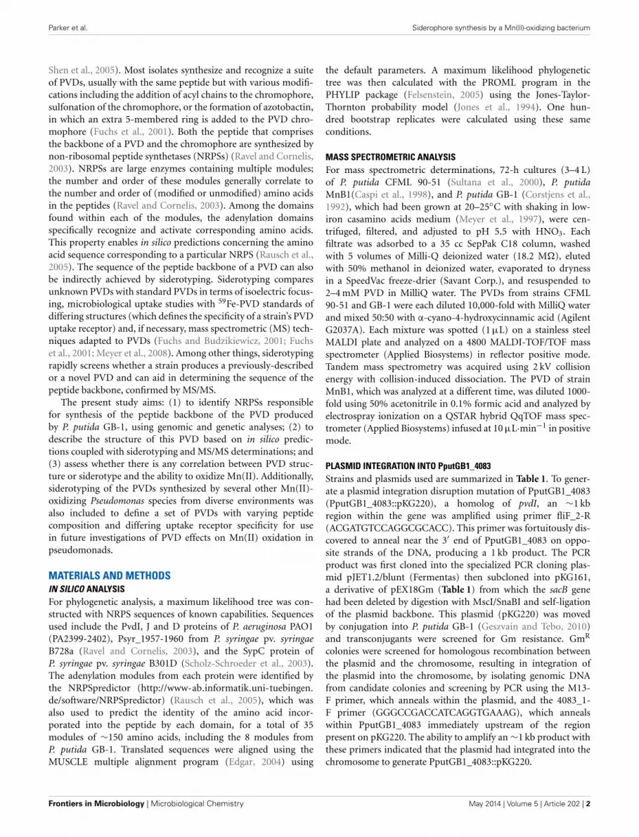
known adenylation modules in other organisms (Figure 3). Thecluster analysis confirmed the conclusions of NRPS predictor formodules m1 (Asp), m3 (Asp), and m4 (Ser), but suggested a Lysat m7 instead of the Thr chosen by NRPS predictor (Figure 1B).For the three cases in which NRPS predictor failed to select anamino acid, the identifications by cluster analysis were: m2 (Lys),m6 (Thr), and m8 (Orn) (Figure 1B). More precisely, module
m6 clusters with other Thr-incorporating modules from fivedifferent pseudomonads, and it appears to be distinct from theallo-Thr incorporating modules of SypC, although these belongto the more distantly related organism Pseudomonas syringaepv. syringae B301D. Module m8 falls into a cluster with twoP. aeruginosa PAO1 modules that incorporate N5-formyl-N5-hydroxyornithine (hfOrn) but is located on a long branch andtherefore might direct incorporation of a different amino acid.However, the P. putida GB-1 genome encodes a homolog toPvdA (PputGB1_2120), which is an enzyme responsible for themodification of ornithine to OHOrn (Visca et al., 2007) and thusstrain GB-1 is predicted to have the biochemical potential togenerate OHOrn. Module m5 appears to cluster somewhat nearseveral Ala-incorporating modules from Pseudomonas syringaepv. syringae B301D, but lies on a long branch outside of thatcluster, which may reflect a lack of sufficiently homologoussequences to accurately root this branch on the tree.
CHEMICAL COMPOSITION AND SIDEROTYPING OF PVDGB−1 ANDPVDMnB1
The PVDs of strains P. putida GB-1 and MnB1 were siderotypedin comparison to those of several isolates of known PVD structure(Table 2). Strains GB-1 and MnB1 were found to be in the samesiderotype as P. putida CFML 90-51, which has been reportedto have the following PVD peptide sequence: chromophore-Asp-Lys-OHAsp-Ser-Gly-aThr-Lys-cOHOrn (D-amino acids under-lined) (Sultana et al., 2000). Since this sequence was strikinglysimilar to that predicted for PVDGB−1 by cluster analysis, it isincluded in Figure 1A for comparison. To determine whether thissequence also applied to strains GB-1 and MnB1, mass spec-trophotometric (MS/MS) analysis was used to obtain molecularweights and also to identify the secondary MS/MS fragments of
www.frontiersin.org May 2014 | Volume 5 | Article 202 | 5
Parker et al. Siderophore synthesis by a Mn(II)-oxidizing bacterium
FIGURE 3 | Phylogenetic cluster analysis using MUSCLE performed for selected adenylation domains among pseudomonads along with 8
adenylation domains found in the genome of P. putida GB-1 in PputGB1_4086-4083.
each PVD in partially purified preparations from the P. putidastrains GB-1 and MnB1, in comparison to parallel preparationsfrom P. putida CFML 90-51 (Table 3). The PVDs of all threeorganisms shared a major MS peak at the monoisotopic m/z of1250–1251 Da (Table 3). This value, as well as the weights ofsecondary MS/MS fragmentation products, corresponded to thepeptide sequence given above. Our data agreed with the previ-ous report for PVDCFML90−51 (Sultana et al., 2000), except thatthe variable acyl side chain in our samples from strain GB-1 wasmalic acid amide (mala) instead of malic acid (mal) and the acylside chain in our CFML 90-51 preparation was ∼50% mala and∼50% mal, in contrast to the previous report in which only malwas found with CFML 90-51 (Sultana et al., 2000). Interestingly,MS/MS samples from all three strains, CFML 90-51, MnB1, andGB-1, also contained a major monoisotopic peak at m/z 1161 Da,which was identified to contain the same PVD peptide as inthe 1250 Da peak, but with the PVD chromophore replaced byan azotobactin chromophore, a structure that is frequently co-produced with pyoverdine (Fuchs et al., 2001). Table 2 lists theMS apparent relative abundance of the peaks at m/z of 1250.4and 1161.4 Da, expressed as a percentage of the total peak areaof PVD-type siderophores produced by each strain. The samplesalso showed minor amounts of sulfonated PVD at 1333.5–1335.5Da (2 and 6% of the total peak areas, respectively), and two minorpeaks at 1232.5 and 1144 Da, which appear to represent water-loss reactions involving β elimination at various hydroxyl groupsof the peptide, with the formation of double bonds conjugated toa carbonyl. Material from each peak at m/z 1250, 1161, and 1333Da was further fragmented and analyzed by MS/MS. Since thesesecondary fragments from the three tested siderotype n◦ 1 strains
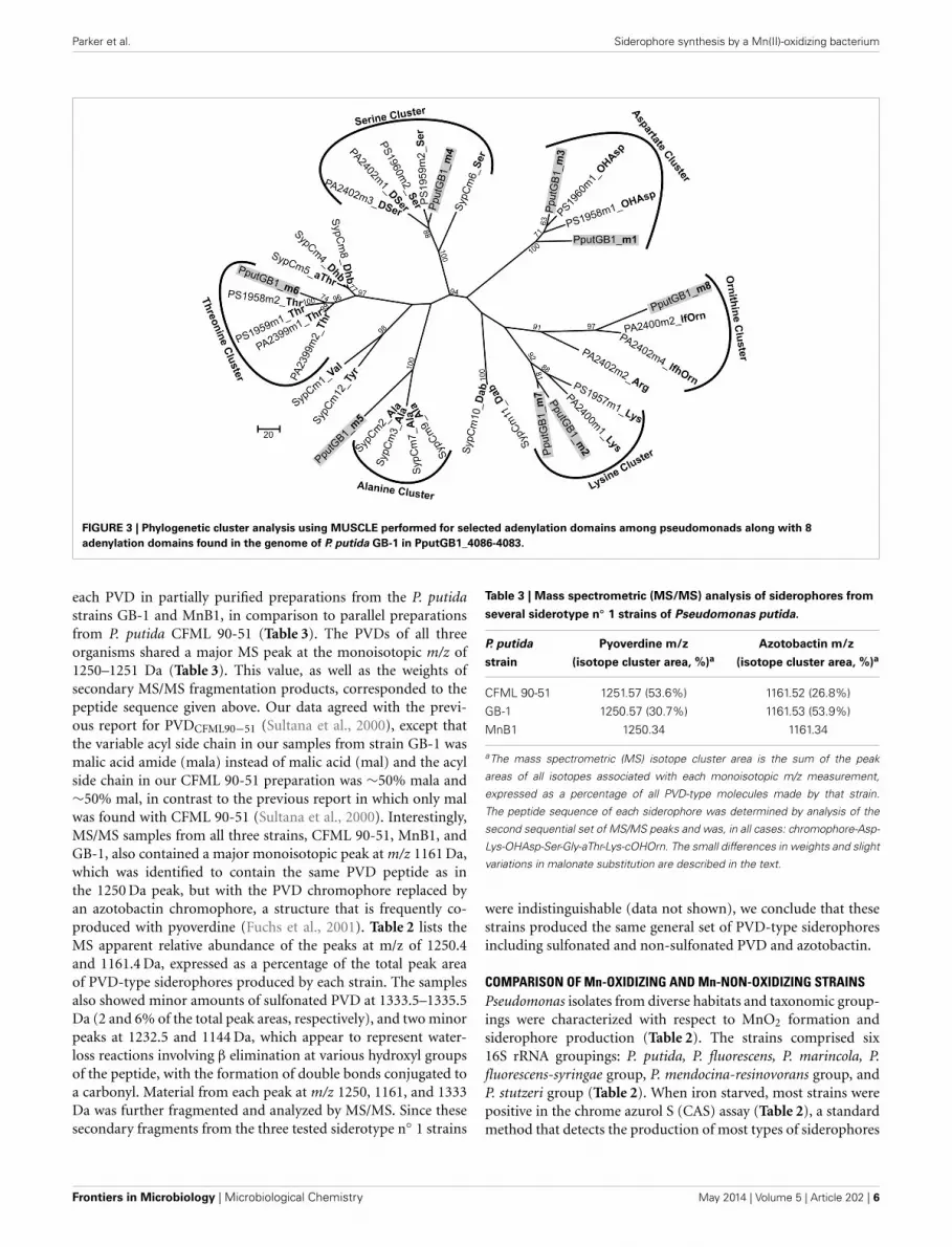
Table 3 | Mass spectrometric (MS/MS) analysis of siderophores from
several siderotype n◦ 1 strains of Pseudomonas putida.
P. putida Pyoverdine m/z Azotobactin m/z
strain (isotope cluster area, %)a (isotope cluster area, %)a
CFML 90-51 1251.57 (53.6%) 1161.52 (26.8%)
GB-1 1250.57 (30.7%) 1161.53 (53.9%)
MnB1 1250.34 1161.34
aThe mass spectrometric (MS) isotope cluster area is the sum of the peak
areas of all isotopes associated with each monoisotopic m/z measurement,
expressed as a percentage of all PVD-type molecules made by that strain.
The peptide sequence of each siderophore was determined by analysis of the
second sequential set of MS/MS peaks and was, in all cases: chromophore-Asp-
Lys-OHAsp-Ser-Gly-aThr-Lys-cOHOrn. The small differences in weights and slight
variations in malonate substitution are described in the text.
were indistinguishable (data not shown), we conclude that thesestrains produced the same general set of PVD-type siderophoresincluding sulfonated and non-sulfonated PVD and azotobactin.
COMPARISON OF Mn-OXIDIZING AND Mn-NON-OXIDIZING STRAINSPseudomonas isolates from diverse habitats and taxonomic group-ings were characterized with respect to MnO2 formation andsiderophore production (Table 2). The strains comprised six16S rRNA groupings: P. putida, P. fluorescens, P. marincola, P.fluorescens-syringae group, P. mendocina-resinovorans group, andP. stutzeri group (Table 2). When iron starved, most strains werepositive in the chrome azurol S (CAS) assay (Table 2), a standardmethod that detects the production of most types of siderophores
Frontiers in Microbiology | Microbiological Chemistry May 2014 | Volume 5 | Article 202 | 6
Parker et al. Siderophore synthesis by a Mn(II)-oxidizing bacterium
(Schwyn and Neilands, 1987). The majority also formed PVD, asindicated by their release of green fluorescent compounds intothe medium under iron-limiting, but not iron-replete, conditions(Table 2). The presence of PVD in culture fluids was confirmedby examination of fluorescence and absorbance spectra at pH 5,6, and 8, both in the absence and presence of iron (Parker et al.,2004, 2007); in all positive cases the spectra were those expectedfor PVD (data not shown because they were so standard).
When the above strains were siderotyped based on the iso-electric focusing (IEF) patterns of their PVDs (Fuchs et al.,2001; Meyer, 2007), seven P. putida isolates showed identical IEFpatterns (Table 2); these strains included two Mn(II) oxidizers(strains GB-1 and MnB1) and five isolates that did not detectablyoxidize Mn(II) (the five CFML strains in Table 2). These sevenstrains also incorporated 59Fe-PVD from the reference organ-ism CFML 90-51 as efficiently as their own 59Fe-PVD (Table 2),but did not internalize 24 other 59Fe-PVD standards of othersiderotypes (see Methods). Based on our data and previous char-acterizations of the five CFML strains (Meyer et al., 2007), weconclude that these 7 isolates are of the same siderotype, here des-ignated siderotype number 1 (n◦ 1), which is equivalent to n◦ 30in Fuchs et al. (2001), siderovar n◦ 15 in Meyer et al. (2007), or n◦32 in Meyer et al. (2008).
The other tested strains were assigned to three new siderotypes(Table 2) that did not correspond to any grouping in Fuchs et al.(2001). P. putida KT2440 is here designated as siderotype n◦ 2, forwhich a PVD structure has been deduced from genome sequence(Ravel and Cornelis, 2003; Meyer et al., 2007). Previously, thissiderotype was also assigned as siderovar n◦ 25 (Meyer et al.,2007) or as type 3 (Matthijs et al., 2009).
Siderotype n◦ 3, P. fluorescens ATCC 55241, did not take up anytested 59Fe-PVD standard and appears to have an as yet unchar-acterized PVD (Table 2). The siderotype n◦ 4 strains were Pfluorescens-syringae group Mn(II) oxidizers ISO6 and PCP1 whichpartially took up 59Fe-PVD of two reference strains, P. fluorescensSB8.3 and Pseudomonas sp. D47 (Table 2). PVD of Pseudomonassp. D47 was previously classified as n◦ 29 and P. fluorescens sp.SB8.3 (=Ps4a) as n◦ 7 (Meyer et al., 2008). Although we have notdetermined the PVD structures of siderotypes n◦ 3 and n◦ 4, itis clear that they are not the same as those of siderotypes n◦ 1or n◦ 2.
DISCUSSIONP. putida strains GB-1 and MnB1 are Mn(II)-oxidizing bacteriathat produce pyoverdines (PVDs), siderophores that affect ironuptake but may also substantially influence the metabolism ofother metals, including Mn. To facilitate future studies of thepotentially multi-faceted roles such as inhibiting Mn(II) oxida-tion played by PVD in the oxidation of Mn by pseudomonads, wehave examined both the genetics of PVD production in strain GB-1 and the composition of the pyoverdines produced by P. putidaGB-1, as well as several other Mn(II)-oxidizing strains.
Within the genome sequence of P. putida GB-1, a putativenon-ribosomal peptide synthase (NRPS) operon capable of beinginvolved in the synthesis of the PVDGB−1 peptide was iden-tified: PputGB1_4086-4083. Disruption of PputGB1_4086-4083resulted in a defect in PVD synthesis similar to that reported
in other Pseudomonas strains with mutations in comparableNRPSs (Merriman et al., 1995; Lehoux et al., 2000). Furthermore,the peptide sequence based on in silico analyses of the adeny-lation domains of NRPS PputGB1_4086-4083 was consistentwith the actual peptide sequence determined from MS/MS ofPVDGB−1 samples. Therefore, it can be safely concluded thatPputGB1_4086-4083 is the NRPS that governs PVD peptide syn-thesis in P. putida GB-1. In silico examination of the P. putidaGB-1 genome also identified candidates (Figure 1) for manyof the other genes needed for the synthesis, modification, andexpression of PVDGB−1, including a NRPS for chromophore syn-thesis (PputGB1_3809), a TonB-dependent siderophore receptorgene (PputGB1_4082), an Asp hydroxylase (PputGB1_4087), anOrn hydroxylase (PputGB1_2120), and an iron-responsive ECFalternative sigma factor (PputGB1_3810) with recognition sitesfound in the promoters upstream of most of the above genes.
Based on siderotyping followed by MS/MS analysis(Tables 2, 3), we have determined the peptide sequences ofPVDGB−1 and PVDMnB1 to be: chromophore-Asp-Lys-OHAsp-Ser-Gly-aThr-Lys-cOHOrn, identical to that reported (Sultanaet al., 2000) and confirmed here (Table 3) for PVDCFML90−51.The OHAsp and the aThr in PVDCFML90−51 are known to be Disomers (Sultana et al., 2000). PVDGB−1 and PVDMnB1 probablyalso contain these two D amino acids because the degree ofuptake of 59Fe-labeled PVDCFML90−51 by strains GB-1, MnB1,and CFML 90-51 was similar (Table 2), suggesting that the cel-lular uptake receptors of strains GB-1 and MnB1 did not detecta steric difference between PVDCFML90−51 and the endogenousPVD of each strain. However, the presence of D amino acids inPVDGB−1 and PVDMnB1 has not been directly tested.
The peptide sequence above suggests a metal-binding pocketformed by three moieties: (1) the catecholate of the chromophore,(2) the cyclic hydroxamate from cOHOrn, and (3) the α-OH-carboxylate from OHAsp. It is not yet clear how this structure,including the presence of a (OH)carboxylate donor group, leadsto the higher thermodymamic stability constants for Mn(III), ascompared to Fe(III), reported for these siderophores at physio-logical and alkaline pH (Parker et al., 2004; Harrington et al.,2012). However, an influence of OHcarboxylate on the preferen-tial binding of Mn(III) has been recently suggested by an inves-tigation showing that siderophores containing solely hydroxam-ates (e.g., desferrioxamine B, DFOB) or solely catecholates (e.g.,protochelin) bind Fe(III) more strongly than Mn(III), whereasrhizoferrin, which complexes metals via a mixture of carboxy-late and (OH)carboxylate groups, binds Mn(III) more stronglythan Fe(III) (Harrington et al., 2012), as do several aminocar-boxylate ligands (Hamm and Suwyn, 1967; Ahrland et al., 1990;Martell and Smith, 2003). Based on K-edge EXAFS compar-isons of various Fe(III)- and Mn(III)-siderophore complexes insolution, Harrington et al. (2012) proposed that the greaterflexibility of carboxylates vis-à-vis hydroxamates or catecholatesallows the former to accommodate the Jahn-Teller-distorted coor-dination that is characteristic of Mn(III) but not Fe(III). Thisresult suggests that (OH)carboxylates within siderophores, per-haps including the siderotype n◦ 1 PVDs studied here, may affectthe preferential binding of Mn(III). However, the situation forpyoverdines probably also involves additional factors, because two
www.frontiersin.org May 2014 | Volume 5 | Article 202 | 7
Parker et al. Siderophore synthesis by a Mn(II)-oxidizing bacterium
mixed-moiety pyoverdines (PVDCFML90−51 and PVDPa1) bothpreferentially bound Mn(III) and both seemed to accommodateJahn-Teller distortion, even though PVDCFML90−51 contains a(OH)carboxylate but PVDPa1 does not (Harrington et al., 2012).Perhaps these PVDs gain flexibility from their mixed donorgroups, their polypeptide structure, or some other factor.
MS/MS analysis of the PVDs from P. putida strains GB-1 andMnB1 also indicated that each strain produced a set of three PVD-type siderophores sharing the same peptide tail but with differ-ently modified chromophores: “classical” PVD, sulfonated PVD,and azotoactin (Table 3). Since all three are strongly fluorescentand since fluorescence was undetectable in the mutant KG163(Figure 2B), it is likely that the peptide tail of all three PVD typesin strain GB-1 is synthesized through the same NRPS operon,PputGB1_4083-4086, which makes sense since the peptides of allthree PVD types showed identical MSMS fragmentation patterns.However, subsequent modifications could be subject to differingregulatory or catalytic pathways. It is currently unknown whetherthese three differing PVD types affect Mn metabolism or thecomplexation of Mn vis á vis Fe similarly or differently.
Azotobactin and PVD are both known to complex variousmetal cations (Braud et al., 2009; Wichard et al., 2009). However,azotobactin can also bind oxyanions such as molybdate and vana-date (Wichard et al., 2009). In contrast, the predominant PVD ofP. aeruginosa PAO1 is not able to form complexes with vanadate,whereas the other main siderophore of strain PAO1, pyochelin,can (Baysse et al., 2000). This observation is consistent with otherreports that PVDs do not play an important role with oxyanions(Wichard et al., 2009). Therefore, one function of azotobactin inP. putida GB-1 and related strains might be to complex oxyan-ions for uptake or detoxification, as was suggested for Azotobactervinelandii (Wichard et al., 2009). Alternatively, sulfonated PVDsand azotobactin could be precursors or byproducts of PVD syn-thesis (Fuchs et al., 2001; Baysse et al., 2002). Further studiesneed to be performed to elucidate the respective roles of the mul-tiple siderophores of these organisms, especially with regard tomanganese oxidation.
Since the ability to oxidize Mn(II) occurs very commonly, butnonetheless sporadically, among a wide variety of Pseudomonasspecies (Francis and Tebo, 2001), it was no surprise that multiplePVD siderotypes were identified among the phylogenetically-diverse Mn(II)-oxidizing pseudomonads tested here (Table 2).Within P. putida, two siderotypes were identified (Table 2): n◦ 1including strains CFML 90-51 and GB-1 and n◦ 2 consisting ofstrain KT2440. Although not included in this research, P. putidaATCC 12663 is also capable of oxidizing Mn(II), is closely relatedto P. putida GB-1 based on 16S data (Francis and Tebo, 2001), butproduces a PVD that has been previously shown to be differentfrom PVDCFML90−51 (Meyer et al., 2007). Since PVDCFML90−51
and PVDGB−1are indistinguishable by MS/MS (Table 3) andsiderotyping (Table 2), PVDGB−1 and PVDATCC12663 cannot bethe same. Therefore, even among the Mn(II)-oxidizing P. putidaat least three differing PVDs exist: PVDGB−1, PVDKT2440, andPVDATCC12663. This situation is in agreement with the conclusionof Meyer et al. (2007) that P. putida as currently defined is het-erogeneous with respect to siderotype. It is also notable that thesiderotype of a Mn oxidizer can be the same as that of a strain that
does not oxidize Mn(II), as for P. putida GB-1 and CFML 90-51(Table 2).
In summary, this study has combined in silico, genetic andchemical (siderotyping and MS/MS) approaches to explore thesynthesis and nature of the suite of related PVDs (“classic”PVD, azotobactin, and sulfonated PVD) that were producedby the model Mn(II)-oxidizing organism Pseudomonas putidaGB-1 at our growth conditions. In silico analysis indicated thatposition PputGB1_4083-4086 of the GB-1 genome containedNRPSs that could synthesize a peptide chain consistent with thePVDGB−1 peptide determined by MS/MS (chromophore-Asp-Lys-OHAsp-Ser-Gly-aThr-Lys-cOHOrn). Furthermore, muta-tion at PputGB1_4083 prevented PVD synthesis. A diverseselection of Mn-oxidizing Pseudomonas species were found tocomprise at least three distinct PVD siderotypes, indicating dif-ferences in PVD structure and PVD uptake specificity that canbe exploited in future studies concerning the ways that variousPVDs can influence Mn metabolism, especially Mn(II) oxidation,in pseudomonads and other bacteria.
ACKNOWLEDGMENTSWe thank Katherine Barbeau, Elizabeth Komives, The ScrippsInstitution of Oceanography, and the U.C.S.D. Molecular MassSpectrometry Facility for helpful discussions, technical assistance,and access to instruments. This publication was made possible bygrants from the National Science Foundation (MCB-0630355 andOCE-1154307) and from the National Institute of EnvironmentalHealth Sciences (P42ES010337).
REFERENCESAhrland, S., Dahlgren, Ã. S., and Persson, I. (1990). Stabilities and hydrolysis of
some iron(III) and manganese(III) complexes with chelating ligands. Acta Agric.Scand. 40, 101–111. doi: 10.1080/00015129009438008
Albrecht-Gary, A. M., Blanc, S., Rochel, N., Ocaktan, A. Z., and Abdallah, M. A.(1994). Bacterial iron transport - coordination properties of pyoverdin Paa, apeptidic siderophore of Pseudomonas aeruginosa. Inorg. Chem. 33, 6391–6402.doi: 10.1021/ic00104a059
Baysse, C., Budzikiewicz, H., Uría Fernández, D., and Cornelis, P. (2002). Impairedmaturation of the siderophore pyoverdine chromophore in Pseudomonas fluo-rescens ATCC 17400 deficient for the cytochrome c biogenesis protein CcmC.FEBS Lett. 523, 23–28. doi: 10.1016/S0014-5793(02)02915-0
Baysse, C., De Vos, D., Naudet, Y., Vandermonde, A., Ochsner, U., Meyer, J.-M.,et al. (2000). Vanadium interferes with siderophore-mediated iron uptake inPseudomonas aeruginosa. Microbiology 146, 2425–2434.
Braud, A., Hoegy, F., Jezequel, K., Lebeau, T., and Schalk, I. J. (2009). Newinsights into the metal specificity of the Pseudomonas aeruginosa pyoverdine-iron uptake pathway. Environ. Microbiol. 11, 1079–1091. doi: 10.1111/j.1462-2920.2008.01838.x
Brouwers, G. J., De Vrind, J. P., Corstjens, P. L., Cornelis, P., Baysse, C., and DeVrind-De Jong, E. W. (1999). cumA, a gene encoding a multicopper oxidase,is involved in Mn2+ oxidation in Pseudomonas putida GB-1. Appl. Environ.Microbiol. 65, 1762–1768.
Budzikiewicz, H. (1993). Secondary metabolites from fluorescent pseudomonads.FEMS Microbiol. Rev. 104, 209–228. doi: 10.1111/j.1574-6968.1993.tb05868.x
Caspi, R., Tebo, B. M., and Haygood, M. G. (1998). c-type cytochromes and man-ganese oxidation in Pseudomonas putida MnB1. Appl. Environ. Microbiol. 64,3549–3555.
Choi, K.-H., Gaynor, J. B., White, K. G., Lopez, C., Bosio, C. M., Karkhoff-Schweizer, R. R., et al. (2005). A Tn7-based broad-range bacterial cloning andexpression system. Nat. Methods 2, 443–448. doi: 10.1038/nmeth765
Clement, E., Mesini, P. J., Pattus, F., and Schalk, I. J. (2004). The binding mech-anism of pyoverdin with the outer membrane receptor FpvA in Pseudomonas
Frontiers in Microbiology | Microbiological Chemistry May 2014 | Volume 5 | Article 202 | 8
Parker et al. Siderophore synthesis by a Mn(II)-oxidizing bacterium
aeruginosa is dependent on its iron-loaded status. Biochemistry 43, 7954–7965.doi: 10.1021/bi049768c
Corstjens, P. L., De Vrind, J. P., Westbroek, P., and De Vrind-De Jong, E. W. (1992).Enzymatic iron oxidation by Leptothrix discophora: identification of an iron-oxidizing protein. Appl. Environ. Microbiol. 58, 450–454.
Edgar, R. C. (2004). MUSCLE: multiple sequence alignment with high accuracy andhigh throughput. Nucleic Acids Res. 32, 1792–1797. doi: 10.1093/nar/gkh340
Emerson, S., Kalhorn, S., Jacobs, L., Tebo, B. M., Nealson, K. H., and Rosson, R.A. (1982). Environmental oxidation rate of manganese(II): bacterial catalysis.Geochim. Cosmochim. Acta. 46, 1073–1079. doi: 10.1016/0016-7037(82)90060-6
Felsenstein, J. (2005). PHYLIP (Phylogeny Inference Package) Version 3.6.Distributed by the author. Seattle, WA: Department of Genome Sciences,University of Washington.
Francis, A. J., and Dodge, C. J. (1998). Remediation of soils and wastes contami-nated with uranium and toxic metals. Environ. Sci. Technol. 32, 3993–3998. doi:10.1021/es9803310
Francis, C. A., and Tebo, B. M. (2001). cumA multicopper oxidase genes fromdiverse Mn(II)-oxidizing and non-Mn(II)-oxidizing Pseudomonas strains. Appl.Environ. Microbiol. 67, 4272–4278. doi: 10.1128/AEM.67.9.4272-4278.2001
Fuchs, R., and Budzikiewicz, H. (2001). Structural sutides of pyoverdins bymass spectrometry. Curr. Org. Chem. 5, 265–288. doi: 10.2174/1385272013375562
Fuchs, R., Schafer, M., Geoffroy, V., and Meyer, J. M. (2001). Siderotyping a power-ful tool for the characterization of pyoverdines. Curr. Top. Med. Chem. 1, 31–57.doi: 10.2174/1568026013395542
Fuller, C. C., and Harvey, J. W. (2000). Reactive uptake of trace metals inthe hyporheic zone of a mining-contaminated stream, Pinal Creek, Arizona.Environ. Sci. Technol. 34, 1150–1155. doi: 10.1021/es990714d
Geszvain, K., and Tebo, B. M. (2010). Identification of a two-component regula-tory pathway essential for Mn(II) oxidation in Pseudomonas putida GB-1. Appl.Environ. Microbiol. 76, 1224–1231. doi: 10.1128/AEM.02473-09
Hamm, R. E., and Suwyn, M. A. (1967). Preparation and characterization of someamino-polycarboxylate complexes of manganese(III). Inorg. Chem. 6, 139–142.doi: 10.1021/ic50047a032
Harrington, J. M., Parker, D. L., Bargar, J. R., Jarzecki, A. A., Tebo, B. M.,Sposito, G., et al. (2012). Structural dependence of Mn complexation bysiderophores: donor group dependence on complex stability and reactivity.Geochim. Cosmochim. Acta. 88, 106–119. doi: 10.1016/j.gca.2012.04.006
Hastings, D., and Emerson, S. (1986). Oxidation of manganese by spores ofa marine Bacillus - kinetic and thermodynamic considerations. Geochim.Cosmochim. Acta. 50, 1819–1824. doi: 10.1016/0016-7037(86)90141-9
Hoang, T. T., Karkhoff-Schweizer, R. R., Kutchma, A. J., and Schweizer, H. P.(1998). A broad-host-range Flp-FRT recombination system for site-specificexcision of chromosomally-located DNA sequences: application for isola-tion of unmarked Pseudomonas aeruginosa mutants. Gene 212, 77–86. doi:10.1016/S0378-1119(98)00130-9
Jones, D. T., Taylor, W. R., and Thornton, J. M. (1994). A mutation data matrixfor transmembrane proteins. FEBS Lett. 339, 269–275. doi: 10.1016/0014-5793(94)80429-X
Lehoux, D. E., Sanschagrin, F., and Levesque, R. C. (2000). Genomics of the 35-kbpvd locus and analysis of novel pvdIJK genes implicated in pyoverdine biosyn-thesis in Pseudomonas aeruginosa. FEMS Microbiol. Lett. 190, 141–146. doi:10.1111/j.1574-6968.2000.tb09276.x
Leoni, L., Orsi, N., De Lorenzo, V., and Visca, P. (2000). Functional analysis ofPvdS, an iron starvation sigma factor of Pseudomonas aeruginosa. J. Bacteriol.182, 1481–1491. doi: 10.1128/JB.182.6.1481-1491.2000
Markowitz, V. M., Chen, I. M. A., Palaniappan, K., Chu, K., Szeto, E., Grechkin,Y., et al. (2008). The integrated microbial genomes system: an expand-ing comparative analysis resource. Nucleic Acids Res. 38, D382–D390. doi:10.1093/nar/gkp887
Martell, A. E., and Smith, R. M. (2003). Determination and Use of StabilityConstants. Gaithersburg, MD: National Institute of Science and Technology(NIST).
Matthijs, S., Baysse, C., Koedam, N., Tehrani, K. A., Verheyden, L., Budzikiewicz,H., et al. (2004). The Pseudomonas siderophore quinolobactin is synthe-sized from xanthurenic acid, an intermediate of the kynurenine pathway. Mol.Microbiol. 52, 371–384. doi: 10.1111/j.1365-2958.2004.03999.x
Matthijs, S., Laus, G., Meyer, J.-M., Abbaspour-Tehrani, K., Schäfer, M.,Budzikiewicz, H., et al. (2009). Siderophore-mediated iron acquisition in the
entomopathogenic bacterium Pseudomonas entomophila L48 and its close rela-tive Pseudomonas putida KT2440. Biometals 22, 951–964. doi: 10.1007/s10534-009-9247-y
Merriman, T. R., Merriman, M. E., and Lamont, I. L. (1995). Nucleotide sequenceof pvdD, a pyoverdine biosynthetic gene from Pseudomonas aeruginosa: PvdDhas similarity to peptide synthetases. J. Bacteriol. 177, 252–258.
Meyer, J.-M. (2007). “Siderotyping and bacterial taxonomy: a siderophore bankfor a rapid identification at the species level of fluorescent and non-fluorescentpseudomonas,” in Soil Biology 12: Microbial Siderophores, eds A. Varma and S.Chincholkar (Berlin: Springer Verlag), 43–65.
Meyer, J.-M., Gruffaz, C., Raharinosy, V., Bezverbnaya, I., Schäfer, M., andBudzikiewicz, H. (2008). Siderotyping of fluorescent Pseudomonas: molecularmass determination by mass spectrometry as a powerful pyoverdine siderotyp-ing method. Biometals 21, 259–271. doi: 10.1007/s10534-007-9115-6
Meyer, J.-M., Gruffaz, C., Tulkki, T., and Izard, D. (2007). Taxonomic heterogeneity,as shown by siderotyping, of strains primarily identified as Pseudomonas putida.Int. J. Syst. Evol. Microbiol. 57, 2543–2556. doi: 10.1099/ijs.0.65233-0
Meyer, J.-M., Stintzi, A., De Vos, D., Cornelis, P., Tappe, R., Taraz, K., et al. (1997).Use of siderophores to type pseudomonads: the three Pseudomonas aeruginosapyoverdine systems. Microbiology 143, 35–43. doi: 10.1099/00221287-143-1-35
Mossialos, D., Ochsner, U., Baysse, C., Chablain, P., Pirnay, J.-P., Koedam, N.,et al. (2002). Identification of new, conserved, non-ribosomal peptide syn-thetases from fluorescent pseudomonads involved in the biosynthesis of thesiderophore pyoverdine. Mol. Microbiol. 45, 1673–1685. doi: 10.1046/j.1365-2958.2002.03120.x
Nakazawa, T. (2002). Travels of a Pseudomonas, from Japan around the world.Environ. Microbiol. 4, 782–786. doi: 10.1046/j.1462-2920.2002.00310.x
Ochsner, U. A., Wilderman, P. J., Vasil, A. I., and Vasil, M. L. (2002). GeneChipexpression analysis of the iron starvation response in Pseudomonas aerugi-nosa: identification of novel pyoverdine biosynthesis genes. Mol. Microbiol. 45,1277–1287. doi: 10.1046/j.1365-2958.2002.03084.x
Okazaki, M., Sugita, T., Shimizu, M., Ohode, Y., Iwamoto, K., De Vrind-De Jong,E. W., et al. (1997). Partial purification and characterization of manganese-oxidizing factors of Pseudomonas fluorescens GB-1. Appl. Environ. Microbiol. 63,4793–4799.
Parker, D. L., Morita, T., Mozafarzadeh, M. L., Verity, R., Mccarthy, J. K., and Tebo,B. M. (2007). Inter-relationships of MnO2 precipitation, siderophore-Mn-(III)complex formation, siderophore degradation, and iron limitation in Mn-(II)-oxidizing bacterial cultures. Geochim. Cosmochim. Acta. 71, 5672–5683. doi:10.1016/j.gca.2007.03.042
Parker, D. L., Sposito, G., and Tebo, B. M. (2004). Manganese(III) binding toa pyoverdine siderophore produced by a manganese(II)-oxidizing bacterium.Geochim. Cosmochim. Acta. 68, 4809–4820. doi: 10.1016/j.gca.2004.05.038
Rausch, C., Weber, T., Kohlbacher, O., Wohlleben, W., and Huson, D. H. (2005).Specificity prediction of adenylation domains in nonribosomal peptide syn-thetases (NRPS) using transductive support vector machines (TSVMs). NucleicAcids Res. 33, 5799–5808. doi: 10.1093/nar/gki885
Ravel, J., and Cornelis, P. (2003). Genomics of pyoverdine-mediated ironuptake in pseudomonads. Trends Microbiol. 11, 195–200. doi: 10.1016/S0966-842X(03)00076-3
Regenhardt, D., Heuer, H., Heim, S., Fernandez, D. U., Strompl, C., Moore, E. R. B.,et al. (2002). Pedigree and taxonomic credentials of Pseudomonas putida strainKT2440. Environ. Microbiol. 4, 912–915. doi: 10.1046/j.1462-2920.2002.00368.x
Romanenko, L. A., Uchino, M., Tebo, B. M., Tanaka, N., Frolova, G. M., andMikhailov, V. V. (2008). Pseudomonas marincola sp. nov., isolated from marineenvironments. Int. J. Syst. Evol. Microbiol. 58, 706–710. doi: 10.1099/ijs.0.65406-0
Schalk, I. J., Abdallah, M. A., and Pattus, F. (2002). Recycling of pyoverdin on theFpvA receptor after ferric pyoverdin uptake and dissociation in Pseudomonasaeruginosa. Biochemistry 41, 1663–1671. doi: 10.1021/bi0157767
Scholz-Schroeder, B. K., Soule, J. D., and Gross, D. C. (2003). The sypA,sypB, and sypC synthetase genes encode twenty-two modules involvedin the nonribosomal peptide synthesis of syringopeptin by Pseudomonassyringae pv. syringae B301D. Mol. Plant Microbe Interact. 16, 271–280. doi:10.1094/MPMI.2003.16.4.271
Schons, V., Atkinson, R. A., Dugave, C., Graff, R., Mislin, G. L. A., Rochet, L.,et al. (2005). The structure-activity relationship of ferric pyoverdine bound toits outer membrane transporter: implications for the mechanism of iron uptake.Biochemistry 44, 14069–14079. doi: 10.1021/bi051155s
www.frontiersin.org May 2014 | Volume 5 | Article 202 | 9
Parker et al. Siderophore synthesis by a Mn(II)-oxidizing bacterium
Schweisfurth, R. (1973). Manganoxydierende Bakterien. I. Isolierung undBestimmung einiger Stamme von Manganbacterien. Z. Allg. Mikrobiol. 13,341–347. doi: 10.1002/jobm.3630130408
Schwyn, B., and Neilands, J. B. (1987). Universal chemical assay for the detec-tion and determination of siderophores. Anal. Biochem. 160, 47–57. doi:10.1016/0003-2697(87)90612-9
Shen, J.-S., Geoffroy, V., Neshat, S., Jia, Z., Meldrum, A., Meyer, J.-M., et al. (2005).FpvA-mediated ferric pyoverdine uptake in Pseudomonas aeruginosa: identifi-cation of aromatic residues in FpvA implicated in ferric pyoverdine bindingand transport. J. Bacteriol. 187, 8511–8515. doi: 10.1128/JB.187.24.8511-8515.2005
Singh, G. M., Fortin, P. D., Koglin, A., and Walsh, C. T. (2008). β-Hydroxylation ofthe aspartyl residue in the phytotoxin syringomycin E: characterization of twocandidate hydroxylases AspH and SyrP in Pseudomonas syringae. Biochemistry47, 11310–11320. doi: 10.1021/bi801322z
Sultana, R., Siddiqui, B. S., Taraz, K., Budzikiewicz, H., and Meyer, J.-M. (2000).A pyoverdine from Pseudomonas putida CFML 90-51 with a Lys e-aminolink in the peptide chain. Biometals 13, 147–152. doi: 10.1023/A:1009212729408
Tebo, B. M., Bargar, J. R., Clement, B. G., Dick, G. J., Murray, K. J.,Parker, D., et al. (2004). Biogenic manganese oxides: properties and mech-anisms of formation. Annu. Rev. Earth Planet. Sci. 32, 287–328. doi:10.1146/annurev.earth.32.101802.120213
Toner, B., Fakra, S., Villalobos, M., Warwick, T., and Sposito, G. (2005).Spatially resolved characterization of biogenic manganese oxide productionwithin a bacterial biofilm. Appl. Environ. Microbiol. 71, 1300–1310. doi:10.1128/AEM.71.3.1300-1310.2005
Villalobos, M., Lanson, B., Manceau, A., Toner, B., and Sposito, G. (2006).Structural model for the biogenic Mn oxide produced by Pseudomonas putida.Am. Mineral. 91, 489–502. doi: 10.2138/am.2006.1925
Villalobos, M., Toner, B., Bargar, J., and Sposito, G. (2003). Characterization ofthe manganese oxide produced by Pseudomonas putida strain MnB1. Geochim.Cosmochim. Acta. 67, 2649–2662. doi: 10.1016/S0016-7037(03)00217-5
Visca, P., Imperi, F., and Lamont, I. (2007). “Pyoverdine synthesis and its regulationin fluorescent pseudomonads,” in Microbial Siderophores, eds A. Varma and S.B. Chincholkar (Berlin; Heidelberg: Springer-Verlag), 135–163.
Wichard, T., Bellenger, J.-P., Morel, F. O. M. M., and Kraepiel, A. M. L. (2009). Roleof the siderophore azotobactin in the bacterial acquisition of nitrogenase metalcofactors. Environ. Sci. Technol. 43, 7218–7224. doi: 10.1021/es8037214
Yeterian, E., Martin, L. W., Guillon, L., Journet, L., Lamont, I. L., and Schalk,I. J. (2010). Synthesis of the siderophore pyoverdine in Pseudomonas aerug-inosa involves a periplasmic maturation. Amino Acids 38, 1447–1459. doi:10.1007/s00726-009-0358-0
Conflict of Interest Statement: The authors declare that the research was con-ducted in the absence of any commercial or financial relationships that could beconstrued as a potential conflict of interest.
Received: 18 March 2014; accepted: 16 April 2014; published online: May 2014.Citation: Parker DL, Lee S-W, Geszvain K, Davis RE, Gruffaz C, Meyer J-M, TorpeyJW and Tebo BM (2014) Pyoverdine synthesis by the Mn(II)-oxidizing bacteriumPseudomonas putida GB-1. Front. Microbiol. 5:202. doi: 10.3389/fmicb.2014.00202This article was submitted to Microbiological Chemistry, a section of the journalFrontiers in Microbiology.Copyright © 2014 Parker, Lee, Geszvain, Davis, Gruffaz, Meyer, Torpey and Tebo.This is an open-access article distributed under the terms of the Creative CommonsAttribution License (CC BY). The use, distribution or reproduction in other forums ispermitted, provided the original author(s) or licensor are credited and that the originalpublication in this journal is cited, in accordance with accepted academic practice. Nouse, distribution or reproduction is permitted which does not comply with these terms.
Frontiers in Microbiology | Microbiological Chemistry May 2014 | Volume 5 | Article 202 | 10
07


