Embed Size (px)
Citation preview
Protection of Spanish Ibex (Capra pyrenaica) againstBluetongue Virus Serotypes 1 and 8 in a SubclinicalExperimental InfectionCristina Lorca-Oro1*, Joan Pujols1,2, Ignacio Garcıa-Bocanegra3, Gregorio Mentaberre4, Jose
Enrique Granados5,6, David Solanes1, Paulino Fandos7, Ivan Galindo1,8, Mariano Domingo1,8,
Santiago Lavın4, Jorge Ramon Lopez-Olvera4
1 Centre de Recerca en Sanitat Animal (CReSA), UAB-IRTA, Campus de la Universitat Autonoma de Barcelona, Barcelona, Spain, 2 Institut de Recerca i Tecnologia
Agroalimentaries (IRTA), Barcelona, Spain, 3 Departamento de Sanidad Animal. Facultad de Veterinaria, UCO, Campus Universitarios de Rabanales, Cordoba, Spain, 4 Servei
d’Ecopatologia de Fauna Salvatge (SEFaS), Departament de Medicina i Cirurgia Animals, Universitat Autonoma de Barcelona (UAB), Barcelona, Spain, 5 Parque Nacional y
Parque Natural de Sierra Nevada, Carretera Antigua de Sierra Nevada, Pinos Genil, Granada, Spain, 6 Agencia de Medio Ambiente y Agua, Granada, Spain, 7 Agencia de
Medio Ambiente y Agua, Isla de la Cartuja, Sevilla, Spain, 8 Departament de Sanitat i Anatomia Animals, Universitat Autonoma de Barcelona, Barcelona, Spain
Abstract
Many wild ruminants such as Spanish ibex (Capra pyrenaica) are susceptible to Bluetongue virus (BTV) infection, whichcauses disease mainly in domestic sheep and cattle. Outbreaks involving either BTV serotypes 1 (BTV-1) and 8 (BTV-8) arecurrently challenging Europe. Inclusion of wildlife vaccination among BTV control measures should be considered in certainspecies. In the present study, four out of fifteen seronegative Spanish ibexes were immunized with a single dose ofinactivated vaccine against BTV-1, four against BTV-8 and seven ibexes were non vaccinated controls. Seven ibexes (fourvaccinated and three controls) were inoculated with each BTV serotype. Antibody and IFN-gamma responses wereevaluated until 28 days after inoculation (dpi). The vaccinated ibexes showed significant (P,0.05) neutralizing antibodylevels after vaccination compared to non vaccinated ibexes. The non vaccinated ibexes remained seronegative untilchallenge and showed neutralizing antibodies from 7 dpi. BTV RNA was detected in the blood of non vaccinated ibexesfrom 2 to the end of the study (28 dpi) and in target tissue samples obtained at necropsy (8 and 28 dpi). BTV-1 wassuccessfully isolated on cell culture from blood and target tissues of non vaccinated ibexes. Clinical signs were unapparentand no gross lesions were found at necropsy. Our results show for the first time that Spanish ibex is susceptible andasymptomatic to BTV infection and also that a single dose of vaccine prevents viraemia against BTV-1 and BTV-8 replication.
Citation: Lorca-Oro C, Pujols J, Garcıa-Bocanegra I, Mentaberre G, Granados JE, et al. (2012) Protection of Spanish Ibex (Capra pyrenaica) against Bluetongue VirusSerotypes 1 and 8 in a Subclinical Experimental Infection. PLoS ONE 7(5): e36380. doi:10.1371/journal.pone.0036380
Editor: Martin Beer, Friedrich-Loeffler-Institut, Germany
Received December 13, 2011; Accepted April 3, 2012; Published May 30, 2012
Copyright: � 2012 Lorca-Oro et al. This is an open-access article distributed under the terms of the Creative Commons Attribution License, which permitsunrestricted use, distribution, and reproduction in any medium, provided the original author and source are credited.
Funding: This work was supported by the project FAU2008-00019-C03-01, from the Instituto Nacional de Investigacion y Tecnologıa Agroalimentaria (INIA). Thefunders had no role in study design, data collection and analysis, decision to publish, or preparation of the manuscript.
Competing Interests: The authors have declared that no competing interests exist.
* E-mail: [email protected]
Introduction
Domestic and wild ruminants are thought to be susceptible to
bluetongue virus (BTV) infection, which causes bluetongue (BT),
a disease that has a high economic impact on animal health. BTV
belongs to the genus Orbivirus (family Reoviridae) and is trans-
mitted by blood-feeding midges of the genus Culicoides (Diptera,
Ceratopogonidae) [1,2]. There are at least 24 different BTV serotypes,
and two putative new serotypes, the 25th named Toggenburg
orbivirus [3,4] and a 26th [5], coinciding with the distribution of
competent vectors in all continents except Antarctica. BT is
considered an emerging and re-emerging disease in Europe. Since
1998, at least eight serotypes (BTV-1, -2, -4, -6, -8, -9, -11 and -16)
have been detected in Europe, where BT has expanded its
geographical range northwards [6–9].
The spreading of BTV-8 through Europe since its introduction
in 2006 caused severe disease, mainly in cattle, but also in sheep,
and heavy financial losses in animal industry. Previously, BTV-1
infections caused epizootics in southern Europe. BTV-1 and -8
were detected in livestock in Spain in 2007 and 2008, respectively.
For safety reasons, immunization against BTV-1 was carried out
together with a mass vaccination campaign against BTV-8 using
inactivated vaccines to control the expansion of these serotypes in
the affected countries of Europe. The target of the vaccination
program was to achieve at least 80% coverage of susceptible
ruminants [6,8,10].
The origin of BT is probably African, and wild ruminants are
the natural hosts of BTV, although it is thought that cattle have
replaced antelope as BTV maintenance host [11]. Information on
the role of wild ruminants in the maintenance and spread of BTV
is still limited. Several studies have been performed in wild
ruminants from North America, where a range of species are
frequently infected with BTV [12]. However, studies on the
susceptibility of native wild ruminant species are scarce in Europe.
From 2006 to 2010, antibodies against BTV-1, -4, and -8 have
been found in red deer (Cervus elaphus), fallow deer (Dama dama),
mouflon (Ovis aries musimon), roe deer (Capreolus capreolus), aoudad
(Amotragus lervia) and Spanish ibex (Capra pyrenaica) in Spain [13–
PLoS ONE | www.plosone.org 1 May 2012 | Volume 7 | Issue 5 | e36380
19]. Although BTV infection is often subclinical or unapparent in
some wild ruminants, bighorn sheep (Ovis canadiensis) and mouflon
can develop fatal clinical disease, as do closely related domestic
sheep [17,20,21]. Experimental infection of pronghorn antelope
(Antilocapra americana), American bison (Bison bison) and Affrican
buffalo (Syncerus caffer) also produced clinical disease [22,23],
whereas blesbock (Damaliscus pygarus) [24] and mountain gazelle
(Gazella gazella) [25] did not show clinical signs after natural or
experimental infection. Recent studies observed susceptibility to
experimental infection with BTV-8 in red deer [26]. Red deer
vaccination against BTV-1 and BTV-8 has proved to be safe and
effective to prevent viraemia in experimentally inoculated deer
[27].
Spanish ibex is an endemic species from Spain, with populations
widespread throughout the southern and eastern regions of the
country [28]. This wild mountain ungulate has a great value for its
conservation as it has been listed as threatened and currently of
least concern in the IUCN Red List of Threatened Species [29]. In
the last decades, contagious diseases such as sarcoptic mange,
habitat fragmentation, illegal hunting, loss of genetic diversity,
local overabundance and disequilibrium in the population sex
ratio and age structure have also contributed to a significant
decline of its populations [30–33]. Spanish ibex frequently share
the same habitat with domestic ruminants, especially in summer
months when exploiting the summer high mountain pastures [28].
Allochthonous wild ungulate species, such as aoudad, fallow deer
and mouflon, also suppose a threat to Spanish ibex and increase
the potential risk of shared diseases transmission [34–35].
The aim of the present study is to evaluate the efficacy of
commercial inactivated BTV vaccines in Spanish ibex, a potential
BTV susceptible species.
Results
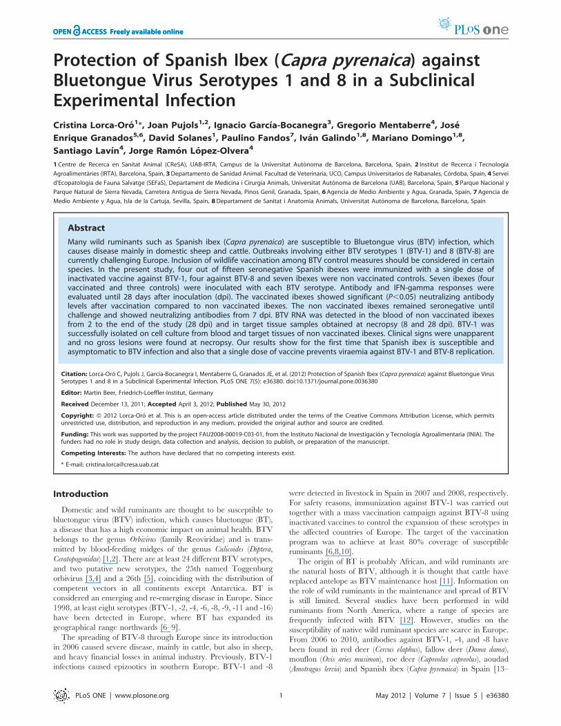
Antibody response to vaccination and infectionNon vaccinated ibexes were seronegative until challenge. BTV-
specific antibodies measured by ELISA increased significantly
(P,0.05) by 23 days after vaccination (25 dpi) in the vaccinated
ibexes, which showed protective antibody levels along the
challenge. Conversely, BTV antibodies increased from 4–7 dpi
in the non vaccinated ibexes, reaching its maximum at 17 dpi for
BTV-1, and at 9 dpi for BTV-8, which showed a shorter and
faster dynamics than BTV-1. Mean and standard deviation of
percentage values of VP7 ELISA assays before and after BTV
challenge are shown in Figure 1.
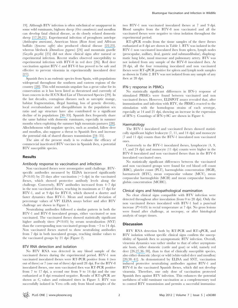
Neutralizing antibodies followed a similar pattern in both the
BTV-1 and BTV-8 inoculated groups, either vaccinated or non
vaccinated. The vaccinated ibexes showed statistically significant
higher antibody titres (P,0.05) by serum neutralization (SNT)
than the corresponding non vaccinated groups from 25 to 14 dpi.
Non vaccinated ibexes started to show neutralizing antibodies
from 7 dpi in both inoculated groups, reaching similar values to
the vaccinated groups by 21 dpi (Figure 2).
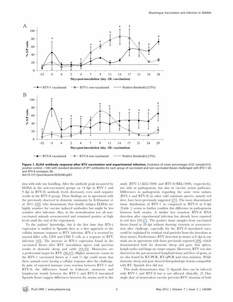
BTV RNA detection and isolationNo BTV RNA was detected in any blood sample of the
vaccinated ibexes during the experimental period. BTV-1 non
vaccinated inoculated ibexes were RT-PCR positive from 4 (two
out of three) or 7 (one out of three) dpi until 28 dpi. For the BTV-8
inoculated ibexes, one non vaccinated ibex was RT-PCR positive
from 7 to 17 dpi, a second one from 9 to 14 dpi and the one
euthanized at 8 dpi remained negative. Results of RT-qPCR are
shown as Ct values and estimated titres in Figure 3. BTV was
successfully isolated in Vero cells only from blood samples of the
two BTV-1 non vaccinated inoculated ibexes at 7 and 9 dpi.
Blood samples from the BTV-8 non vaccinated and all the
vaccinated ibexes were negative to virus isolation throughout the
experimental period.
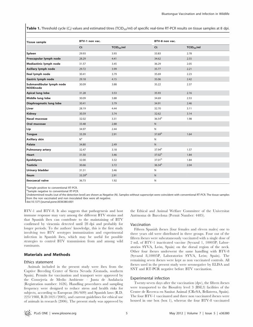
RT-qPCR results from the tissue samples of the three ibexes
euthanized at 8 dpi are shown in Table 1. BTV was isolated in the
BTV-1 non vaccinated inoculated ibex from spleen, lymph nodes
(prescapular, axillary, ileal, gastric and submandibular), diaphrag-
matic lung lobe, nasal mucosae and pulmonary artery. BTV was
not isolated from any sample of the BTV-8 inoculated ibex. At
28 dpi, all the four remaining inoculated and non vaccinated
ibexes were RT-qPCR positive for spleen and lymph node samples
as shown in Table 2. BTV was not isolated from any sample of any
ibex at 28 dpi.
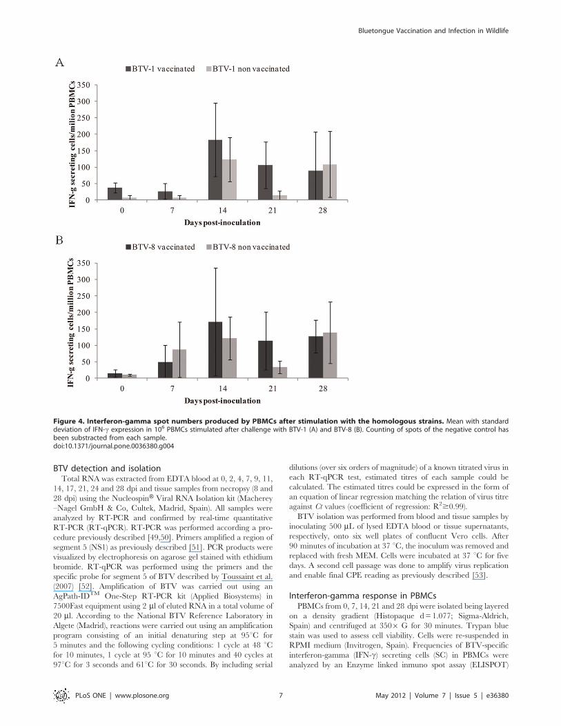
IFN-c response in PBMCsNo statistically significant differences in IFN-c response of
stimulated PBMCs were found between vaccinated and non
vaccinated ibexes. However, ELISPOT assays revealed that, after
immunization and infection with BTV, the PBMCs reacted to the
stimulation with the homologous strains of each serotype,
especially at 14 and 21 dpi, showing an increase in the expression
of IFN-c. Countings of IFN-c-SC are shown in Figure 4.
HaematologyThe BTV-1 inoculated and vaccinated ibexes showed statisti-
cally significant higher leukocyte (7, 11, and 14 dpi) and monocyte
(7 and 11 dpi) counts than the BTV-1 inoculated non vaccinated
ones.
Conversely to the BTV-1 inoculated ibexes, lymphocyte (4, 9,
17, and 24 dpi) and monocyte (11 dpi) counts were higher in the
BTV-8 inoculated and non vaccinated ibexes than in the BTV-8
inoculated vaccinated ones.
No statistically significant differences between the vaccinated
and non vaccinated groups were found for red blood cell count
(RBC), platelet count (PLT), haemoglobin concentration (HGB),
haematocrit (HTC), mean corpuscular volume (MCV), mean
corpuscular haemoglobin (MCH) and mean corpuscular haemo-
globin concentration (MCHC).
Clinical signs and histopathological examinationNo clear clinical signs compatible with BTV infection were
detected throughout after inoculation (from 0 to 28 dpi). Only the
non vaccinated ibexes inoculated with BTV-1 had a punctual
increase (P.0.05) in rectal temperature at 7 dpi. No gross lesions
were found after challenge, at necropsy, or after histological
analysis of target tissues.
Discussion
BTV RNA detection both by RT-PCR and RT-qPCR, and
BTV isolation without specific clinical signs confirm the suscep-
tibility of Spanish ibex to asymptomatic BTV infection. Overall,
viraemia dynamics was rather similar to that of other asymptom-
atic hosts, either domestic (cattle and goat) or wild, namely red
deer [26,27,36–38], than to that of clinically susceptible species,
also either domestic (sheep) or wild (white-tailed deer and mouflon)
[20,38–41]. As demonstrated by ELISA and SNT, vaccination
induced protective neutralizing antibodies against BTV-1 and
BTV-8 in the vaccinated Spanish ibexes, which did not develop
viraemia. Therefore, one only dose of vaccination protected
Spanish ibex against BTV infection. This enhances the potential
usefulness of wild ruminant vaccination as a complementary tool
to control BTV transmission and permits a successful immuniza-
Bluetongue Vaccination and Infection in Wildlife
PLoS ONE | www.plosone.org 2 May 2012 | Volume 7 | Issue 5 | e36380
tion with only one handling. After the antibody peak measured by
ELISA in the non-vaccinated groups (at 14 dpi in BTV-1 and
9 dpi in BTV-8) antibody levels decreased, even until negative
results in the BTV-8 group. These findings are in agreement with
the previously observed in domestic ruminants by Echbaumer et
al. 2011 [42], who demonstrate that double antigen ELISAs are
highly sensitive for vaccine induced antibodies but might be less
sensitive after infection. Also, in the neutralization test all non-
vaccinated animals seroconverted and remained positive at high
levels until the end of the experiment.
To the authors’ knowledge, this is the first time that IFN-cexpression is studied in Spanish ibex as a first approach to the
cellular immune response to BTV infection. IFN-c is secreted by
natural killer cells, CD4 and CD8 T cells as a response to BTV
infection [43]. The increase in IFN-c expression found in the
vaccinated ibexes after BTV inoculation agrees with previous
results in domestic sheep and cattle [44,45]. Monocytes are
a preferential target for BTV [46,47]. Higher monocyte counts in
the BTV-1 vaccinated ibexes at 7 and 11 dpi could mean that
these animals were having a cellular response after the challenge.
In spite of reported immune cross reaction between BTV-1 and
BTV-8, the differences found in leukocyte, monocyte and
lymphocyte trends between the BTV-1 and BTV-8 inoculated
Spanish ibexes suggest differences between the strains used in this
study (BTV-1/ALG/2006 and BTV-8/BEL/2006, respectively),
not only in pathogenesis, but also in vaccine action pathways.
Differences in pathogenesis regarding the same virus isolates
(BTV-1 and BTV-8) in other wild ruminant species, namely red
deer, have been previously suggested [27]. The more disseminated
tissue distribution of BTV-1 as compared to BTV-8 at 8 dpi
(Table 1) seems to further confirm this difference in pathogenesis
between both strains. A similar low transient BTV-8 RNA
detection after experimental infection has already been reported
in red deer [26,27]. The positive tissue samples from vaccinated
ibexes found at 28 dpi without showing viraemia or seroconver-
sion after challenge, especially for the BTV-8 inoculated ones,
could be explained by residual viral particles from the inoculum at
those tissues. Furthermore, BTV detection in tissues at 8 dpi in our
study are in agreement with those previously reported [48], which
demonstrated both for domestic sheep and goat, that spleen,
lymph nodes and lungs are target organs. Moreover, BTV was also
detected in the gut-associated lymphoid tissue and liver of goats, as
we also found by RT-PCR, RT-qPCR and virus isolation. While
domestic sheep and goat showed histopahologic lesions compatible
with BT, Spanish ibex did not.
This study demonstrates that: (1) Spanish ibex can be infected
with BTV-1 and BTV-8 but is not affected clinically; (2) One
single dose of monovalent vaccine prevents BTV viraemia of both
Figure 1. ELISA antibody response after BTV vaccination and experimental infection. Evolution of mean percentages (O.D. sample/O.D.positive control 6100) with standard deviation of VP7 antibodies for each group of vaccinated and non vaccinated ibexes challenged with BTV-1 (A)and BTV-8 serotypes (B).doi:10.1371/journal.pone.0036380.g001
Bluetongue Vaccination and Infection in Wildlife
PLoS ONE | www.plosone.org 3 May 2012 | Volume 7 | Issue 5 | e36380
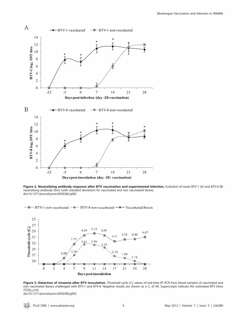
Figure 2. Neutralizing antibody response after BTV vaccination and experimental infection. Evolution of mean BTV-1 (A) and BTV-8 (B)neutralizing antibody titres (with standard deviation) for vaccinated and non vaccinated ibexes.doi:10.1371/journal.pone.0036380.g002
Figure 3. Detection of viraemia after BTV inoculation. Threshold cycle (Ct) values of real-time RT-PCR from blood samples of vaccinated andnon vaccinated ibexes challenged with BTV-1 and BTV-8. Negative results are shown as a Ct of 40. Superscripts indicate the estimated BTV titres(TCID50/ml).doi:10.1371/journal.pone.0036380.g003
Bluetongue Vaccination and Infection in Wildlife
PLoS ONE | www.plosone.org 4 May 2012 | Volume 7 | Issue 5 | e36380
BTV-1 and BTV-8. It also suggests that pathogenesis and host
immune response may vary among the different BTV strains and
that Spanish ibex can contribute to the maintaining of BTV
confirmed by viraemia detected until 28 dpi and probably for
longer periods. To the authors’ knowledge, this is the first study
involving two BTV serotypes immunization and experimental
infection in Spanish ibex, which may be useful for possible
strategies to control BTV transmission from and among wild
ruminants.
Materials and Methods
Ethics statementAnimals included in the present study were ibex from the
Captive Breeding Center of Sierra Nevada (Granada, southern
Spain). Permits for vaccination and transport were approved by
the Consejerıa de Medio Ambiente – Junta de Andalucıa
(Registration number: 1626). Handling procedures and sampling
frequency were designed to reduce stress and health risks for
subjects, according to European (86/609) and Spanish laws (R.D.
223/1988, R.D.1021/2005), and current guidelines for ethical use
of animals in research (2006). The present study was approved by
the Ethical and Animal Welfare Committee of the Universitat
Autonoma de Barcelona (Permit Number: 4485).
VaccinationFifteen Spanish ibexes (four females and eleven males) one to
three years old were distributed in three groups. Four out of the
fifteen ibexes were subcutaneously vaccinated with a single dose of
2 mL of BTV-1 inactivated vaccine (Syvazul 1, 10003P; Labor-
atorios SYVA, Leon, Spain) on the dorsal region of the neck.
Other four ibexes underwent the same handling with BTV-8
(Syvazul 8,10005P, Laboratorios SYVA, Leon; Spain). The
remaining seven ibexes were kept as non vaccinated controls. All
ibexes used in the present study were seronegative by ELISA and
SNT and RT-PCR negative before BTV vaccination.
Experimental infectionTwenty-seven days after the vaccination (dpv), the fifteen ibexes
were transported to the Biosafety level 3 (BSL3) facilities of the
Centre de Recerca en Sanitat Animal (CReSA, Bellaterra, Spain).
The four BTV-1 vaccinated and three non vaccinated ibexes were
housed in one box (box 1), whereas the four BTV-8 vaccinated
Table 1. Threshold cycle (Ct) values and estimated titres (TCID50/ml) of specific real-time RT-PCR results on tissue samples at 8 dpi.
Tissue sample BTV-1 non vac. BTV-8 non vac.
Ct TCID50/ml Ct TCID50/ml
Spleen 29.93 3.93 33.83 2.78
Prescapular lymph node 28.29 4.41 34.62 2.55
Mediastinic lymph node 31.57 3.45 36.29 2.05
Axillary lymph node 29.72 3.99 35.77 2.21
Ileal lymph node 30.41 3.79 35.69 2.23
Gastric lymph node 29.18 4.15 35.06 2.42
Submandibular lymph nodeNODEnode
30.09 3.88 35.22 2.37
Apical lung lobe 31.28 3.53 35.93 2.16
Middle lung lobe 30.09 3.88 34.69 2.53
Diaphragmatic lung lobe 30.41 3.79 34.91 2.46
Liver 28.19 4.44 32.70 3.11
Kidney 30.59 3.74 32.62 3.14
Nasal mucosae 32.02 3.31 36.54b 1.98
Oral mucosae 33.48 2.88 N
Lip 34.97 2.44 N
Tongue 33.39 2.91 37.69b 1.64
Axillary skin Na N
Palate 34.80 2.49 N
Pulmonary artery 32.47 3.18 37.94b 1.57
Heart 31.50 3.46 37.02b 1.84
Epididymis 32.00 3.32 37.01b 1.84
Testicle 30.66 3.72 36.34b 2.04
Urinary bladder 31.51 3.46 N
Ileum 33.39b 2.91 N
Ileocaecal valve 36.73 1.92 N
aSample positive to conventional RT-PCR.bSample negative to conventional RT-PCR.Undetermined results (out of the detection level) are shown as Negative (N). Samples without superscript were coincident with conventional RT-PCR. The tissue samplesfrom the non vaccinated and non inoculated ibex were all negative.doi:10.1371/journal.pone.0036380.t001
Bluetongue Vaccination and Infection in Wildlife
PLoS ONE | www.plosone.org 5 May 2012 | Volume 7 | Issue 5 | e36380

ibexes were housed in another box (box 2) with four non
vaccinated ibexes. After an adaptation period of five days, all the
ibexes except one non vaccinated ibex in box 2, were challenged
against BTV-1 (box 1) or BTV-8 (box 2) serotypes with 2 mL of
BTV viral suspension in the jugular vein. Viral inocula consisted of
infected Vero (African green monkey kidney) E6 culture super-
natants of BTV-1/ALG/2006/E6 strain (six passages) with 106.5
TCID50/mL (50% tissue culture infective doses) and BTV-8/
BEL/2006/E6 strain (five passages) with 106.6 TCID50/mL. The
viruses were given one passage on embryonated chicken eggs, one
passage on baby hamster kidney cells and three (BTV-8) or four
(BTV-1) on Vero cells.
Blood samples (with and without EDTA) were collected by
jugular puncture, and clinical signs and rectal temperature were
measured at days 0, 2, 4, 7, 9, 11, 14, 17, 21, 24 and 28 post-
inoculation (dpi). Heparinized blood was collected at 0, 7, 14, 21
and 28 dpi to obtain peripheral blood mononuclear cells (PBMCs).
Body weights were measured at 0 dpv (233 dpi) and at necropsy
(8 or 28 dpi). Sera was extracted from whole blood tubes after
centrifugation (3006 G for 15 minutes) and stored at 220uC.
EDTA blood was stored at 4uC until analysis.
At 8 dpi, three non vaccinated ibexes (one inoculated with
BTV-1, one with BTV-8 and the one non inoculated ibex) were
anesthetized with xylazine (Xilagesic 20%, Laboratorios Calier,
1 mg/kg) and euthanized with an overdose of barbiturate
(intravenous infusion of pentobarbital at 100 mg/kg) to study
BTV lesions at viraemia peak period. By 28 dpi the remaining
twelve ibexes were euthanized using the same protocol. At
necropsy, ordinary sampling was performed, including tissue
collection (spleen, lung, liver, kidney, bowel, skin, tongue, lip, skin,
nasal and oral mucosae, palate, pulmonary artery, heart,
epididymis, testicle, urinary bladder, ileum, ileocaecal valve, and
mediastinal, mesenteric, axillary and iliac lymph nodes) for BTV
RNA detection, BTV isolation and histopathological studies.
SerologySera before vaccination (233 dpi) and at 25, 0, 2, 4, 7, 9, 11,
14, 17, 21, 24 and 28 dpi were analyzed for the presence of
specific antibodies against the BTV major core protein VP7, using
a commercial double-antigen ELISA assay (Ingezim BTV
DR12.BTV.KO Ingenasa, Spain).
Serotype specific antibodies were detected by means of serum
neutralization test (SNT) as described previously [49]. Briefly,
serum samples were inactivated at 56 uC for 30 minutes prior to
analysis. Sera were diluted 1:2 to 1:4096 in microplates (CostarHCat. Nu 3915, Cultek, Madrid, Spain) using MEM Earle (Eagle’s
minimum essential medium with Earle salts) and mixed with 100
TCID50% of each reference strain (BTV-1 and BTV-8). Samples
were tested against both BTV-1 and BTV-8 to determine
a possible cross-neutralization of BTV serotypes. Mixtures were
incubated for one hour at 37 uC, and 100 ml of a Vero E6 cell
suspension in MEM supplemented with 15% foetal bovine serum
(FBS; PAA Laboratories GmbH, Austria), 300 mg/l-glutamine/
ml, 300 U penicillin/ml and 300 mg streptomycin/ml, were added
to a final concentration of 1.56104/well. The mixture was further
incubated for 6 days at 37 uC, plate readings for cytopathic effect
(CPE) were done at 4 and 6 days. Developing CPE was compared
with control wells containing 100 TCID50% of virus and negative
control wells (without virus). Only samples that showed neutral-
ization (absence of CPE) at dilutions $1:4 were considered
positive to avoid false positive results from unspecific reactions of
sera.
Table 2. Threshold cycle (Ct) values and estimated titres (TCID50/ml) of specific real-time RT-PCR results on tissue samples atnecropsy at the end of the study (28 dpi).
TreatmentIbexnum. Tissue sample
Spleen Prescapular lymph node Mediastinic lymph node
Ct TCID50/ml Ct TCID50/ml Ct TCID50/ml
BTV-1 nonvac.
215 30.41 3.79 26.47 4.95 31.17 3.56
220 30.60 3.73 28.24 4.43 30.96 3.63
BTV-1 vac.
227 N 35.80 2.20 N
306 N N N
310 N N N
312 N N N
BTV-8 nonvac.
217 30.52 3.76 31.65 3.42 33.71 2.81
307 33.03 3.02 34.18 2.68 36.37 2.03
BTV-8 vac.
225 N 33.06 3.01 39.24 1.48
303 N 37.55 1.68 N
304 34.70 2.52 29.75 3.98 N
305 N 31.05 3.60 N
Undetermined results (out of the detection level) are shown as Negative (N).doi:10.1371/journal.pone.0036380.t002
Bluetongue Vaccination and Infection in Wildlife
PLoS ONE | www.plosone.org 6 May 2012 | Volume 7 | Issue 5 | e36380
BTV detection and isolationTotal RNA was extracted from EDTA blood at 0, 2, 4, 7, 9, 11,
14, 17, 21, 24 and 28 dpi and tissue samples from necropsy (8 and
28 dpi) using the NucleospinH Viral RNA Isolation kit (Macherey
–Nagel GmbH & Co, Cultek, Madrid, Spain). All samples were
analyzed by RT-PCR and confirmed by real-time quantitative
RT-PCR (RT-qPCR). RT-PCR was performed according a pro-
cedure previously described [49,50]. Primers amplified a region of
segment 5 (NS1) as previously described [51]. PCR products were
visualized by electrophoresis on agarose gel stained with ethidium
bromide. RT-qPCR was performed using the primers and the
specific probe for segment 5 of BTV described by Toussaint et al.
(2007) [52]. Amplification of BTV was carried out using an
AgPath-IDTM One-Step RT-PCR kit (Applied Biosystems) in
7500Fast equipment using 2 ml of eluted RNA in a total volume of
20 ml. According to the National BTV Reference Laboratory in
Algete (Madrid), reactions were carried out using an amplification
program consisting of an initial denaturing step at 95uC for
5 minutes and the following cycling conditions: 1 cycle at 48 uCfor 10 minutes, 1 cycle at 95 uC for 10 minutes and 40 cycles at
97uC for 3 seconds and 61uC for 30 seconds. By including serial
dilutions (over six orders of magnitude) of a known titrated virus in
each RT-qPCR test, estimated titres of each sample could be
calculated. The estimated titres could be expressed in the form of
an equation of linear regression matching the relation of virus titre
against Ct values (coefficient of regression: R2$0.99).
BTV isolation was performed from blood and tissue samples by
inoculating 500 mL of lysed EDTA blood or tissue supernatants,
respectively, onto six well plates of confluent Vero cells. After
90 minutes of incubation at 37 uC, the inoculum was removed and
replaced with fresh MEM. Cells were incubated at 37 uC for five
days. A second cell passage was done to amplify virus replication
and enable final CPE reading as previously described [53].
Interferon-gamma response in PBMCsPBMCs from 0, 7, 14, 21 and 28 dpi were isolated being layered
on a density gradient (Histopaque d = 1.077; Sigma-Aldrich,
Spain) and centrifuged at 3506 G for 30 minutes. Trypan blue
stain was used to assess cell viability. Cells were re-suspended in
RPMI medium (Invitrogen, Spain). Frequencies of BTV-specific
interferon-gamma (IFN-c) secreting cells (SC) in PBMCs were
analyzed by an Enzyme linked inmuno spot assay (ELISPOT)
Figure 4. Interferon-gamma spot numbers produced by PBMCs after stimulation with the homologous strains. Mean with standarddeviation of IFN-c expression in 106 PBMCs stimulated after challenge with BTV-1 (A) and BTV-8 (B). Counting of spots of the negative control hasbeen substracted from each sample.doi:10.1371/journal.pone.0036380.g004
Bluetongue Vaccination and Infection in Wildlife
PLoS ONE | www.plosone.org 7 May 2012 | Volume 7 | Issue 5 | e36380
using commercial monoclonal antibodies (mAbs) (Bovine IFN-cAM05875PU-N and AM05867BT-N, Acris, AntibodyBcn, Spain).
Briefly, ELISA plates (Costar 3590, Corning, USA) were coated
overnight at 4uC with 5 mg/mL of IFN-c capture antibody
(AM05875PU-N) diluted in carbonate–bicarbonate buffer
(pH 9.6). Plates were then washed and blocked for 1 hour at
37uC with 150 ml of PBS with 1% of bovine serum albumin. After
removal of the blocking solution, 2.56105 live PBMC were
dispensed per well in triplicates and stimulated with phytohae-
magglutinin (PHA) (10 mg/ml) (Sigma-Aldrich, Spain) and BTV-1
or BTV-8 strains at 0.04 of multiplicity of infection (moi). The
BTV strains were the same used previously at challenge. Non
stimulated cells (only RPMI) were kept as background controls.
After 20 hours of incubation at 37uC in a 5% CO2 atmosphere,
cells were removed, and the biotinylated detection antibody
(AM05867BT-N) was added at 2.5 mg/mL (50 mL) and incubated
for 1 hour at 37uC. The reaction was revealed by sequential
incubation of plates with streptavidin-peroxidase at 0.5 mg/mL for
1 hour and insoluble 3,39,5,59-Tetramethylbenzidine (TMB;
Sigma-Aldrich, Spain). To calculate the BTV-specific frequencies
of IFN-c-SC, counts of spots in non stimulated wells were
subtracted from counts in virus-stimulated wells. Frequencies of
IFN-c-SC were expressed as responding cells in 106 PBMCs.
HaematologyErythrocytic parameters (RBC, HGB, HTC, MCV, MCH and
MCHC), WBC and PLT were determined by a semi-automated
haematologic counter (Horiba ABX ABC Vet Hematology
Analyzers, Scil Vet abc, Divasa-Farmavic, Spain). Differential
leukocyte count was performed by identifying 200 leukocytes on
blood smears stained with a commercial Diff-Quick-like stain
(Quimica Clınica Aplicada, Spain).
Statistical analysesA repeated measures analysis of the variance was performed to
detect statistical differences regarding specific BTV antibodies
(tested by ELISA and SNT), body temperatures, IFN-c-SC and
haematological parameters, using the PROC MIXED COVT-
EST procedure of SAS 9.1. (SAS Institute Inc., Cary, NC, USA).
The main factor was vaccine (vaccinated or non-vaccinated) and
the repeated factor was DPV (day post vaccination). Differences
were considered statistically significant when P-value,0.05.
Acknowledgments
The authors would like to thank Syva Laboratories for providing the
vaccines and challenge viruses. The authors are also very grateful to the
rangers and staff of the National and Natural Park of Sierra Nevada and
Agencia de Medio Ambiente y Agua of the Junta de Andalucıa working on
the Spanish Ibex Management Program.
Author Contributions
Conceived and designed the experiments: CLO JRLO JP IGB MD.
Performed the experiments: CLO DS JRLO IGB GM. Analyzed the data:
JRLO CLO JP. Contributed reagents/materials/analysis tools: SL JP JEG.
Wrote the paper: CLO JRLO IGB. Responsible of biosecurity facilities at
CReSA: DS. Handling of animals during vaccination and experimental
infection: GM JRLO CLO IGB DS. Responsible for providing the Spanish
ibexes, and arrangement of all permissions and conditions for animal
cession and transport: PF JEG. Performed all necropsies and all histological
analysis to look for compatible lesions with bluetongue: IG.
References
1. Mellor PS, Wittmann EJ (2002) Bluetongue virus in the Mediterranean Basin
1998–2001. Vet J 164: 20–37.
2. Mertens PP, Diprose J, Maan S, Singh KP, Attoui H, et al. (2004) Bluetongue
virus replication, molecular and structural biology. Vet Ital 40: 426–437.
3. Hofmann MA, Renzullo S, Mader M, Chaignat V, Worwa G, et al. (2008)
Genetic characterization of toggenburg orbivirus, a new bluetongue virus, fromgoats, Switzerland. Emerg Infect Dis 14: 1855–1861.
4. Chaignat V, Worwa G, Scherrer N, Hilbe M, Ehrensperger F, et al. (2009)
Toggenburg Orbivirus, a new bluetongue virus: initial detection, firstobservations in field and experimental infection of goats and sheep. Vet
Microbiol 138: 11–19.
5. Maan S, Maan NS, Nomikov K, Veronesi E, Bachanek-Bankowska K, et al.
(2011) Complete genome characterization of a novel 26th bluetongue virusserotype from Kuwait. PLoS One 6(10): e26147. Doi:10.1371/journal.-
pone.0026147.
6. Enserink M (2008) Animal disease – Exotic disease of farm animals tests
Europe’s responses. Science 319: 710–711.
7. Purse BV, Brown HE, Harrup L, Mertens PPC, Rogers DJ (2008) Invasion of
bluetongue and other orbivirus infections into Europe: the role of biological andclimatic processes. Rev Sci Tech OIE 27: 427–442.
8. Rodrıguez-Sanchez B, Iglesias-Martın I, Martınez-Aviles M, Sanchez-Vizcaıno JM (2008) Orbiviruses in the Mediterranean basin: updated
epidemiological situation of Bluetongue and new methods for the detection of
BTV serotype 4. Transbound Emerg Dis 55: 205–214.
9. Eschbaumer M, Hoffmann B, Konig P, Teifke JP, Gethmann JM, et al. (2009)
Efficacy of three inactivated vaccines against bluetongue virus serotype 8 insheep. Vaccine 27: 4169–4175.
10. Hateley G (2009) Bluetongue in northern Europe: the story so far. In practice31: 202–209.
11. Gerdes GH (2004) A South African overview of the virus, vectors, survellianceand unique features of bluetongue. Vet Ital 40: 39–42.
12. Stallknecht DE, Howerth EW (2004) Epidemiology of bluetongue and epizootichaemorrhagic disease in wildlife: surveillance methods. Vet Ital 40: 203–307.
13. Falconi C, Lopez-Olvera JR, Gortazar C (2011) BTV infection in wildruminants, with emphasis on red deer: A review. Vet Microbiol, 151: 209–219.
14. Garcıa-Bocanegra I, Arenas-Montes A, Lorca-Oro C, Pujols J, Gonzalez MA, etal. (2011) Role of wild ruminants in the epidemiology of bluetongue virus
serotypes 1, 4 and 8 in Spain. Vet Res 42: 88.
15. Lorca-Oro C, Pujols J, Arenas A, Gomez-Guillamon F, Zorrilla I, et al. (2011)
Epidemiological surveillance of bluetongue virus serotypes 1, 4 and 8 in Spanishibex (Capra pyrenaica hispanica) in southern Spain. Vet Mircobiol 149: 230–235.
16. Rodrıguez-Sanchez B, Gortazar C, Ruiz-Fons F, Sanchez-Vizcaıno JM (2010)
Bluetongue Virus Serotypes 1 and 4 in Red Deer, Spain. Emerg Infect Dis 16:518–520.
17. Rodrıguez-Sanchez B, Sanchez-Cordon PJ, Molina V, Risalde MA, deDiego AC, et al. (2008) Detection of bluetongue serotype 4 in mouflons (Ovis
aries musimon) from Spain. Vet Microbiol 141: 164–167.
18. Garcıa I, Napp S, Casal J, Perea A, Allepuz A, et al. (2009) Bluetongue
epidemiology in wild ruminants from Southern Spain. Eur J Wildl Res 55:
173–178.
19. Ruiz-Fons F, Reyes-Garcıa AR, Alcaide V, Gortazar C (2008) Spatial and
temporal evolution of bluetongue virus in wild ruminants, Spain. Emerg InfectDis 14: 951–953.
20. Fernandez-Pacheco P, Fernandez-Pinero J, Aguero M, Jimenez-Clavero MA(2008) Bluetongue virus serotype 1 in wild mouflons in Spain. Vet Rec 162:
659–660.
21. Robinson RM, Hailey TL, Livingston CW, Thomas JW (1967) Bluetongue in
the desert bighorn sheep. J Wildl Manage 31: 165–168.
22. Howerth EW, Stallknecht DE, Kirkland PD (2001) Bluetongue, epizootic
haemorrhagic disease, and other orbivirus-related diseases. In: Williams ES,Barker IK (Eds) Infect Dis Wild Mammals Iowa. State Univ Press, Ames.
23. Tessaro SV, Clavijo A (2001) Duration of bluetongue viremia in experimentallyinfected American bison. J Wildl Dis 37: 722–729.
24. Bender LC, Hong L, Bruce Thomson C, Horrow PC, Valdez R (2003)Infectious disease of gemsbok in New Mexico. J Wildl Dis 39: 722–778.
25. Barzilai TE, Tadmor A (1972) Experimental infection of the Mountain gazelle(Gazella gazella) with bluetongue virus. Refuah Veterinarith 29: 45–50.
26. Lopez-Olvera JR, Falconi C, Fernandez-Pacheco P, Fernandez-Pinero J,
Sanchez MA, et al. (2010) Experimental infection of European red deer (Cervuselaphus) with bluetongue virus serotypes 1 and 8. Vet Microbiol 145: 148–152.
27. Lorca-Oro C, Lopez-Olvera JR, Fernandez-Sirera L, Solanes D, Navarro N, etal. (2012) Evaluation of the efficacy of commercial vaccines against bluetongue
virus serotypes 1 and 8 in experimentally infected red deer (Cervus elaphus). VetMicrobiol 154: 240–246.
28. Perez JM, Granados JE, Soriguer RC, Fandos P, Marquez FJ, et al. (2002)Distribution, status and conservation problems of the Spanish Ibex, Capra
pyrenaica (Mammalia: Artiodactyla). Mammal Review 32: 26–39.
29. IUCN Red List of Threatened Species website. Available: www.iucnredlist.org.
Version 2011.2. Accessed 2011 Nov, 29.
30. Gonzalez-Candela M, Cubero MJ, Martin-Atance P, Leon-Vizcaıno L (2006)
Potential pathogens carried by Spanish ibex (Capra pyrenaica hispanica) in southernSpain. J Wildl Dis 42: 325–334.
Bluetongue Vaccination and Infection in Wildlife
PLoS ONE | www.plosone.org 8 May 2012 | Volume 7 | Issue 5 | e36380
31. Leon-Vizcaıno L, Ruiz de Ybanez MR, Cubero MJ, Ortiz JM, Espinosa J, et al.
(1999) Sarcoptic mange in Spanish ibex from Spain. J Wildl Dis 35: 647–659.32. Granados JE, Perez JM, Marquez FJ, Serrano E, Soriguer RC, et al. (2001) La
cabra montes (Capra pyrenaica, Schinz 1838). Galemys 13: 3–37.
33. Perez JM (2001) Distribucion, genetica y estatus sanitario de las poblacionesandaluzas de la cabra montes. Universidad de Jaen, Consejerıa de Medio
Ambiente.34. Acevedo P, Cassinello J, Hortal J, Gortazar C (2007) Invasive exotic aoudad
(Ammotragus lervia) as a major threat to native Iberian ibex (Capra pyrenaica):
a habitat suitability model approach. Diversity and Distributions 13: 587–597.35. Fandos P, Reig S (1992) Problems associated with mouflon and barbary sheep
introductions in Spain. In: Bobek B, Pierzanowski K, Regelin W, eds. GlobalTrends in Wildlife Management Swiat Press, Krakow-Warzawa, Poland. pp
139–140.36. Backx A, Heutink CG, Van Rooij EMA, Van Rijn PA (2011) Clinical signs of
bluetongue virus serotype 8 infection in sheep and goats. Vet Rec 161: 591–592.
37. Breard E, Belbis G, Hamers C, Moulin V, Lilin T (2011) Evaluation of humoralresponse and protective efficacy of two inactivated vaccines against bluetongue
virus after vaccination of goats. Vaccine 29: 2495–2502.38. Puentes E, Fernandez-Pinero J, Fernandez-Pacheco P, Sanchez MA, Varo I, et
al. (2008) Inactivated vaccines against bluetongue: protection of cattle and sheep
from experimental challenge with bluetongue virus serotypes 1, 4 and 8. In:Epizone 2008 Bluetongue Satellite Symposium Abstract book, Brescia, Italy.
39. Vosdingh RA, Trainer DO, Easterday BC (1968) Experimental bluetonguedisease in white-tailed deer. Can J Comp Med Vet Sci, 32: 382–387.
40. Murray JO, Trainer DO (1970) Bluetongue virus in North American elk. J WildlDis, 6: 144–148.
41. Ellis JA, Coen ML, Maclachlan NJ, Wilson WC, Williams ES, et al. (1993)
Prevalence of bluetongue virus expression in leukocytes from experimentallyinfected ruminants. Am J Vet Res, 54: 1452–1456.
42. Eschbaumer M, Schulz C, Wackerlin R, Gaulyn M, Beer M, et al. (2011)Limitations of sandwich ELISAs for bluetongue virus antibody detection. Vet
Rec 168: 643.
43. Hemati B, Contreras V, Urien C, Bonneau M, Takamatsu H-H, et al. (2009)
Bluetongue virus targets conventional dendritic cells in skin lymph. J Virol 83:8789–8799.
44. Hund A, Gollnick N, Souter-Louis C, Neubauer-Juric A, Lahm H, et al. (2012)
A two year BTV-8 vaccination follow up: molecular diagnostics and assessmentof humoral and cellular immune reactions. Vet Microbiol 154: 247–256.
45. Umeshappa CS, Singh KP, Pandey AB, Singh RP, Nanjundappa RH (2010)Cell-mediated immune response and cross-protective efficacy of binary
ethylenimine-inactivated bluetongue virus serotype-1 vaccine in sheep. Vaccine
28: 2522–2531.46. Barratt-Boyes SM, Rossitto PV, Stott JL, MacLachlan NJ (1992) Flow
cytometric analysis from in vitro bluetongue virus infection of bovine bloodmononuclear cells. J Gen Virol 73: 1953–1960.
47. Whetter LE, MacLachlan NJ, Gebhard DH, Heidner HW, Moore PF (1989)Bluetongue virus infection of bovine monocytes. J Gen Virol 70: 1663–1676.
48. Sanchez-Cordon PJ, Rodrıguez-Sanchez B, Risalde MA, Molina V, Pedrera M,
et al. (2010) Immunohistochemical detection of bluetongue virus in fixed tissues.J Comp Path 143: 20–28.
49. OIE website. Bluetongue and Epizootic Haemorrhagic Disease. Chapter 2.1.3.,(2009) Available: http://www.oie.int/fileadmin/Home/eng/Health_standards/
tahm/2008/pdf/2.01.03_BLUETONGUE.pdf. Accessed 2011 April 18.
50. Aguero M, Arias M, Romero LJ, Zamora MJ, Sanchez-Vizcaıno JM (2002)Molecular differentiation between NS1 gene of a field strain Bluetongue virus
serotype 2 (BTV-2) and NS1 gene of an attenuated BTV-2 vaccine. VetMicrobiol 86: 337–341.
51. Katz JB, Alstad AD, Gustafson GA, Moser KM (1993) Sensitive identification ofbluetongue virus serogroup by a colorimetric dual oligonucleotide sorbent assay
of amplified viral nucleic acid. J Clin Microbiol 31: 3028–3030.
52. Toussaint JF, Sailleau C, Breard E, Zientara S, De Clercq K (2007) Bluetonguevirus detection by two real-time RT-qPCRs targeting two different genomic
segments. J Virol Methods 140: 115–123.53. Clavijo A, Heckert RA, Dulac GC, Afshar A (2000) Isolation and identification
of bluetongue virus. J. Virol. Methods 87: 13–23.
Bluetongue Vaccination and Infection in Wildlife
PLoS ONE | www.plosone.org 9 May 2012 | Volume 7 | Issue 5 | e36380


