Embed Size (px)
Citation preview
____________________________________________________________________________________________
*Corresponding author: E-mail: [email protected];
Advances in Research2(1): 12-23, 2014, Article no. AIR.2014.002
SCIENCEDOMAIN internationalwww.sciencedomain.org
Prevalence and Conjugal Transfer ofVancomycin Resistance among Clinical
Isolates of Staphylococcus aureus
Rehab Mahmoud Abd El-Baky1*, Hala Rady Ahmed1
and Gamal Fadl Mahmoud Gad1
1Microbiology Department, Faculty of Pharmacy, Minia University, Minia, Egypt.
Authors’ contributions
This work was carried out in collaboration between all authors. Authors GFMG and RMAEdesigned the study, wrote the protocol, and wrote the first draft of the manuscript. AuthorRMAE and HRA managed the literature searches, collection of samples, identification of
VRSA, Antimicrobial susceptibility testing and the molecular studies. All authors read andapproved the final manuscript.
Received 25th September 2013Accepted 27th October 2013
Published 16th November 2013
ABSTRACT
Aims: Emergence of vancomycin resistant Staphylococcus aureus (VRSA) has been ofgreat concern in clinical settings. Our study aimed to detect vancomycin resistance andthe possibility of its conjugal transfer among clinical isolates of S. aureus.Study Design: Medical microbiology.Place and Duration of Study: This study was carried out in the department ofmicrobiology, Faculty of pharmacy, between October 2011 and January 2013.Methodology: Two hundreds and seven samples were collected from different types ofinfections and examined for Staphylococcus aureus using standard bacteriologicalprocedures, Antimicrobial susceptibility testing against vancomycin and someantimicrobials was done by agar dilution method, resistance to methicillin was determinedby disc diffusion test using cefoxitin (30µg), Vancomycin resistance gene transfer wastested by broth matting procedure and confirmed by plasmid and DNA product analysis.Results: Sixty-three samples were positive for Staphylococcus aureus. All isolates wereresistant to penicillin while the lowest resistance was to amikacin and vancomycin. Oneisolate was VRSA (MIC ≥ 16 µg/ml and positive for vanA gene 1032bp) while 5 isolates
Original Research Article
Abd El-Baky et al.; AIR, Article no. AIR.2014.002
13
were VISA. All VRSA and VISA isolates were MRSA and showed a high resistance toother examined antibiotics. Vancomycin resistance was plasmid mediated andsuccessfully transferred from VRSA donor to a vancomycin sensitive recipient isolate(MIC 2 µg/ml). The MIC of vancomycin for the transconjugant strain was 32 µg/ml. PCRproduct analysis for the transconjugate was positive for vanA gene.Conclusion: Low incidence of vancomycin resistance, All VRSA and VISA weremethicillin resistant, and mostly isolated from skin infections. Also, vancomycin resistancewas plasmid mediated and successfully transferred to susceptible strains which isalarming. Skin colonization by VRSA strains harboring plasmid carrying resistance genesmay result in widespread of these strains that could lead to outbreaks of staphylococcaldiseases in both hospital and community.
Keywords: Vancomycin; resistance; Staphylococcus aureus; vanA gene; plasmid.
1. INTRODUCTION
Clinicians are continually being challenged by infections caused by Staphylococcus aureus.Not only is S. aureus a major cause of both community-acquired and health care–associatedinfections, but the treatment of suspected S. aureus infections is becoming increasinglymore complicated [1].
It is reported that the incidence of S. aureus nosocomial infections associated with oxacillinand methicillin resistance surpassed 50% in 1999 among patients in the intensive care unitaccording to the Centers for Disease Control and Prevention’s (CDC) and NationalNosocomial Infections Surveillance (NNIS) system. Of great concern are reports ofcommunity-acquired infections associated with MRSA, such as those that contributed to thedeaths of 4 children in 1999 [2]. Also, the emergence of high levels of resistance tomethicillin, oxacillin, nafcillin, macrolides, tetracyclines, and aminoglycosides has made thetherapy of staphylococcal disease a global challenge [1,3].
In 1980s, due to the widespread occurrence of methicillin-resistant Staphylococcus aureus(MRSA), empiric therapy for staphylococcal infections (particularly nosocomial sepsis) waschanged to vancomycin in many health-care institutions. Vancomycin use in many countriesalso increased during this period because of the growing numbers of infections withClostridium difficile and coagulase-negative staphylococci in health-care facilities [4]. Thus,the early 1990s saw a discernible increase in vancomycin use. As a consequence, selectivepressure was established that eventually led to the emergence of strains of S. aureus andother species of staphylococci with decreased susceptibility to vancomycin.
As a result of antibiotic resistance, vancomycin has been the drug of last resort.Vancomycin, a glycopeptide antibiotic, acts against Gram-positive bacteria only, by inhibitingthe incorporation of NAM-NAG-polypeptide into the growing peptidoglycan (PG) chain. Itinhibits this process by reacting with D-Ala-D-Ala, which consequently blocks the release ofterminal D-Ala and intrachain bond formation [5]. Vancomycin-resistant Enterococcusfaecium harbors the vanA operon, which contains five genes, van S, -R, -H, -A and -X [6].vanS and vanR are the regulator genes [7]. VanH is a D-hydroxy acid dehydrogenase thatreduces pyruvate to D-lactate [8], which could be used by VanA ligase in conjunction withATP and D-Ala to make a D-Ala-D-lactate depsipeptide, which is incorporated into thepeptidoglycan layer. Vancomycin binds to N-acyl- D-Ala-D-lactate with an affinity 1000-fold
Abd El-Baky et al.; AIR, Article no. AIR.2014.002
14
lower than that of N-acyl-D-Ala-D-Ala [9]. VanX is a dipeptidase required for the hydrolysis ofD-Ala-D-Ala [10].
Conjugative transfer of high-level vancomycin resistance from Enterococcus faecalis toStaphylococcus aureus [11], and transfer of glycopeptide- and macrolide-resistance genesby transconjugation among enterococci and from E. faecalis to S. aureus [12] have beenreported. Vancomycin-resistance gene (vanA gene) acquisition by S. aureus from E. faeciumin the clinical environment has also been reported by Weigel et al. [13].
In 1997, the first clinical isolate of S. aureus with reduced susceptibility to vancomycin wasreported from Japan [14]. The vancomycin minimum inhibitory concentration (MIC) resultsreported for this isolate was in the intermediate range (8 μg/mL) using interpretive criteriadefined by the National Committee for Clinical Laboratory Standards (NCCLS) [15]. Thisreport was quickly followed by similar ones from other countries, including the United States[16], Belgium [17], Germany [18]. These strains were called vancomycin-intermediate S.aureus (VISA). The first clinical infection with vancomycin-resistant S. aureus (VRSA) (MIC ≥32μg/mL) was reported in July 2002 from Michigan [19] with a second case in Pennsylvaniareported shortly thereafter [20]. Though, there have been only a few reports of VRSA, thehigh prevalence of MRSA and vancomycin use, both thought to be risk factors for VRSA,make the widespread dissemination of these organisms an alarmingly realistic possibility[21]. Such resistance could result in serious clinical and public health consequencesbecause, currently, few licensed alternatives to vancomycin are available to treat seriousresistant S. aureus infections [22]. Furthermore, there is an equally alarming threat of therisk of transmission of these organisms between patients [23].
The emergence of VRSA underscores the need for programs to prevent the spread ofantimicrobial resistant microorganisms and to control the use of antimicrobial drugs inhealth-care settings.
In this study, we have shown the emergence of vancomycin resistance in our country, Egyptand its ability to undergo a conjugative transfer from one clinical strain to another.
2. MATERIALS AND METHOD
2.1 Isolation and Identification
The study included 63 strains of S. aureus isolated from clinical specimens obtained from207 patients with wound infections, respiratory tract infection, urinary tract infections and eyeinfections. Identification of S. aureus was based upon colony morphology, positive Gramstain, DNase, catalase and coagulase tests, and fermentation of Mannitol [24,25].
2.2 Antibiotic Susceptibility Testing and MIC Determination
Antimicrobial susceptibility for methicillin was determined by the disk agar diffusion (DAD)technique using cefoxitin (30 µg) discs according to the guidelines recommended by CLSI[26].
MIC determination and antimicrobial susceptibility testing for the following antibiotics:Penicillin, ampicillin/sulbactam, amoxicillin/clavulanic acid, cefazolin, cefuroxime, gentamicin,
Abd El-Baky et al.; AIR, Article no. AIR.2014.002
15
amikacin, ciprofloxacin, levofloxacin, vancomycin were performed using agar dilution methodas recommended by CLSI [26].
2.3 Isolation of Plasmid DNAs
Plasmids were isolated by alkaline lysis techniques, separated by electrophoresis on 0.8%agarose (Sigma), and visualized with UV light after treatment with ethidium bromide [27].
2.4 Transfer of Vancomycin Resistance by Mating Procedure
Broth mating was performed as follows. Single colonies of donor and recipient cells wereinoculated separately in Luria–Bertani (LB) broth and grown overnight at 37°C with shaking.These overnight cultures were diluted 1:100 in fresh medium, and each was grown to earlyexponential phase (OD600˷0.6). Mating mixture was prepared by adding 0.1ml of donor cellsto 0.9ml of recipient cells, and was swirled gently for a few minutes and then incubated at37°C for 6h (without shaking), followed by plating (0.2ml per plate) on Luria Bertani agar (LB)medium containing appropriate selective antibiotics (16µg/ml vancomycin and 2.5µg/mlciprofloxacin). Colonies were picked up after 48-72h of incubation. Donor and recipient cellswere also plated separately to check their disability to grow on the vancomycin plusciprofloxacin plate, because the donor was ciprofloxacin-sensitive and the recipient wassusceptible to vancomycin and ciprofloxacin resistant [28]. Then, MIC determination forvancomycin and plasmid profile was done for the transconjugate.
2.5 PCR- based Detection of Vancomycin Resistance Genes (vanA) in VRSAStrain and the Transconjugate Strain
Bacterial cultures on selective media plates were collected and suspended in sterile,deionized distilled water and heated in a boiling water bath for 10 min. The samples werecooled immediately on ice for 5-10 min and centrifuged at 13000 × g for 5 min. Thesupernatants were used as DNA templates for PCR [5]. Oligonucleotide primers for vanA(vanA F 5'CATGAATAGAATAAAAGTTGCAATA3') and (vanA R5'CCCCTTTAACGCTAATACGACGATCAA3') gene.
2.5.1 PCR amplification of vanA
The PCR amplification mixture contained the following components: Phusion GC buffercontaining 1.5 mM MgCl2, 200 mm each dNTP, 2 mm each primer, 0.1 mg template DNA,3% (v/v) DMSO and 1 U Phusion DNA polymerase (Finnzymes). The amplificationconditions were initial denaturation at 98◦C for 2 min, followed by 35 cycles of denaturation at98◦C for 10 s; annealing at 50◦C for 1 min; polymerization at 72 ◦C for 60 s for vanA, and finalextension at 72◦C for 5 min for all. The PCR product (5 μl aliquot) was separated byelectrophoresis in 1 % agarose gel at 100V for 40 minutes in Tris-acetate buffer visualizedby ethidium bromide staining illuminated by UV transilluminator [29,30].
3. RESULTS
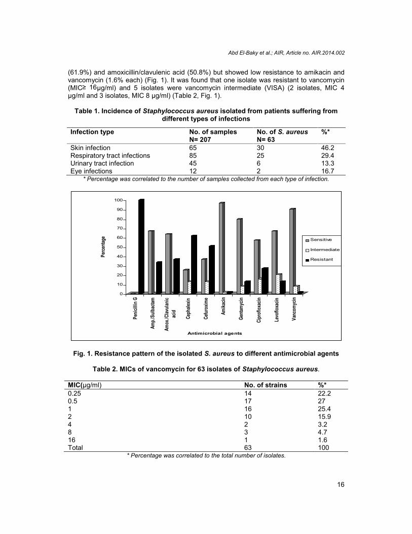
A total of 63 S. aureus isolates were obtained from 207 samples collected from differentinfections (Table 1). Most of the isolated S. aureus isolates were isolated from skin infectionsfollowed by respiratory tract infections. Antibiotic resistance pattern revealed that all S.aureus isolates were penicillin resistant. Isolates showed high resistance to cefuroxime
Abd El-Baky et al.; AIR, Article no. AIR.2014.002
16
(61.9%) and amoxicillin/clavulenic acid (50.8%) but showed low resistance to amikacin andvancomycin (1.6% each) (Fig. 1). It was found that one isolate was resistant to vancomycin(MIC≥ 16µg/ml) and 5 isolates were vancomycin intermediate (VISA) (2 isolates, MIC 4µg/ml and 3 isolates, MIC 8 µg/ml) (Table 2, Fig. 1).
Table 1. Incidence of Staphylococcus aureus isolated from patients suffering fromdifferent types of infections
* Percentage was correlated to the number of samples collected from each type of infection.
Fig. 1. Resistance pattern of the isolated S. aureus to different antimicrobial agents
Table 2. MICs of vancomycin for 63 isolates of Staphylococcus aureus.
MIC(µg/ml) No. of strains %*0.25 14 22.20.5 17 271 16 25.42 10 15.94 2 3.28 3 4.716 1 1.6Total 63 100
* Percentage was correlated to the total number of isolates.
0
10
20
30
40
50
60
70
80
90
100
Perce
ntage
Penic
illin G
Amp./
Sulba
ctam
Amox
./Clav
ulanic
acid
Ceph
alexin
Cefur
oxim
e
Amika
cin
Genta
mycin
Cipr
oflox
acin
Levo
floxa
cin
Vanc
omyc
in
Antimicrobial agents
Sensitive
Intermediate
Resistant
Infection type No. of samplesN= 207
No. of S. aureusN= 63
%*
Skin infection 65 30 46.2Respiratory tract infections 85 25 29.4Urinary tract infection 45 6 13.3Eye infections 12 2 16.7
Abd El-Baky et al.; AIR, Article no. AIR.2014.002
17
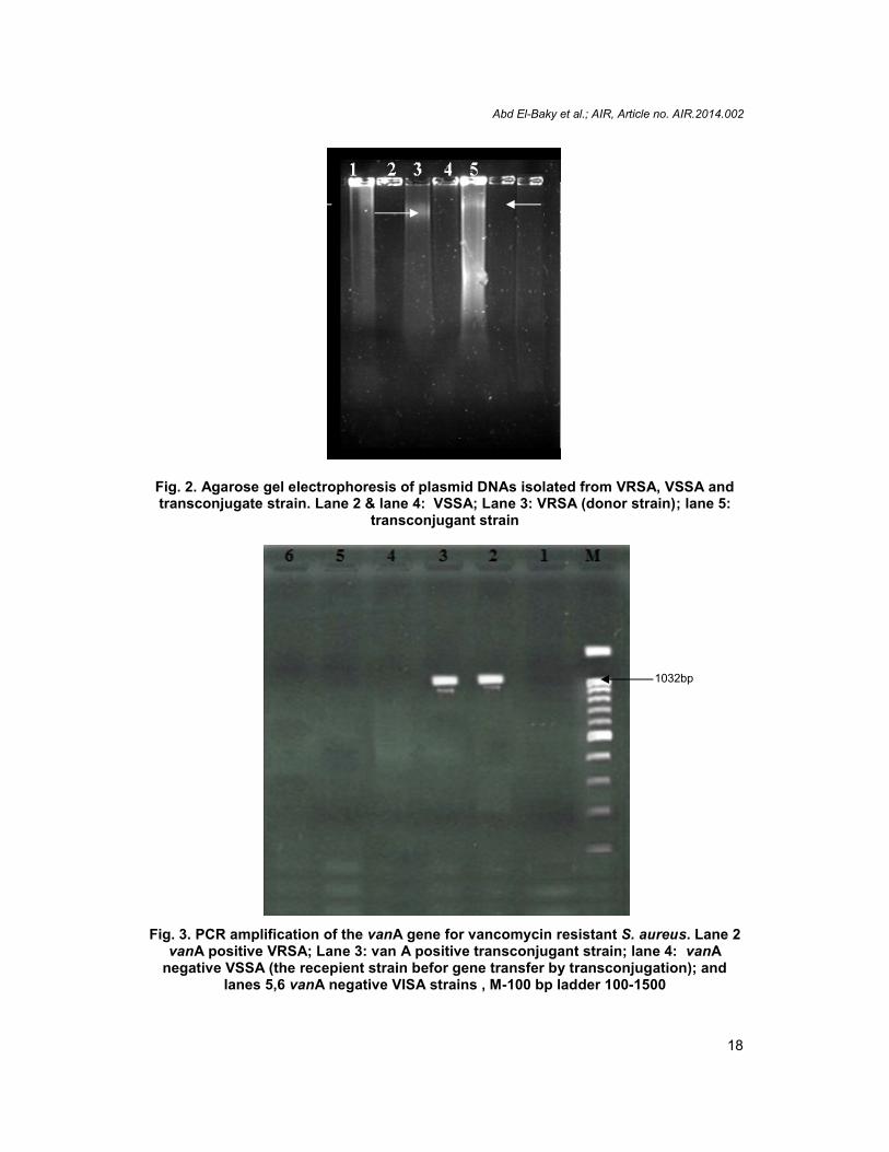
Plasmid analysis for VRSA revealed the presence of one plasmid (Fig. 2) which is confirmedby the presence of PCR product band positive for vanA gene at 1032bp (Fig. 3).
Disc agar diffusion test using Cefoxitin (30µg) discs revealed that out of 63 S. aureusisolates, 16 (25.4%) were methicillin resistant (≤ 21mm). Table 3 showed the resistancepattern of VRSA and VISA to other antimicrobials. As it revealed that VRSA isolate (Skinswab) and VISA isolates were all methicillin resistant. It also showed that these strains wereall resistant to all tested β-lactams antibiotics. VRSA was found to be susceptible tociprofloxacin and amikacin. The table showed also that VRSA and VISA were mostlyisolated from skin.
Table 3. Resistance pattern of VISA and VRSA isolates to the tested antimicrobial agent
StrainsNo.
MICVancomycin(µg/ml)
Specimens Resistant Susceptible
VISA1 4 Throatswab
Pen., Amox.\calv., Amp.\sulb.,Gen., Cip., Cefa., Cefu., Meth.
Amik., Levo.
VISA2 4 Urine Pen., Amox.\calv., Amp.\sulb.,Cefa., Cefu., Meth.,Amik., Levo.
Gen., Cipro.
VISA3 8 Skin swab Pen., Amox.\calv., Amp.\sulb.,Gen., Cip., Cefa., Cefu., Meth.,Levo.
Amik.
VISA4 4 Throatswab
Pen., Amox.\calv., Amp.\sulb.,Gen., Cip., Cefa., Cefu., Meth.,Amik.
Levo.
VISA5 8 Skin swab Pen., Amox.\calv., Amp.\sulb.,Gen., Cefa., Cefu., Meth., Livo.
Amik.,Cipro.
VRSA1 16 Skin swab Pen., Amox.\calv., Amp.\sulb. Cip., Amik.Pen: penicillin; Amox.\calv: amoxcillin\calvulanic acid; Amp.\sulb: ampicillin\sulbactam; Amik: amikacin;
Gen: gentamycin; Cip: ciprofloxacin; Lev: levofloxacin; Cefa: cefazolin; Cefu: cefuroxime; Meth:methicillin.
Transconjugant colonies were found on LB plates containing appropriate selective antibioticstaking VRSA (ciprofloxacin-sensitive) as a donor, and vancomycin-sensitive S. aureus(ciprofloxacin-resistant) as a recipient. No growth of recipient S. aureus strain and the donorstrain VRSA, was observed on the above LB medium when inoculated separately. MIC ofvancomycin was determined for the transconjugate and it was found that it increased from 2µg/ml to 32 µg/ml. on the other hand, plasmid profile for transconjugate revealed thepresence of one plasmid which was not found in the recipient strain before conjugal transfer(Fig. 2).
A selected transconjugant colony was picked up for PCR amplification that revealed thepresence of a band for vanA gene at 1032bp (Fig. 3).
Abd El-Baky et al.; AIR, Article no. AIR.2014.002
18
Fig. 2. Agarose gel electrophoresis of plasmid DNAs isolated from VRSA, VSSA andtransconjugate strain. Lane 2 & lane 4: VSSA; Lane 3: VRSA (donor strain); lane 5:
transconjugant strain
Fig. 3. PCR amplification of the vanA gene for vancomycin resistant S. aureus. Lane 2vanA positive VRSA; Lane 3: van A positive transconjugant strain; lane 4: vanA
negative VSSA (the recepient strain befor gene transfer by transconjugation); andlanes 5,6 vanA negative VISA strains , M-100 bp ladder 100-1500
1032bp
Abd El-Baky et al.; AIR, Article no. AIR.2014.002
19
4. DISCUSSION
As strains of S. aureus with reduced susceptibility continue to emerge and evolve, perhapsto full resistance, there is a clinical need to fully characterize them and conduct welldesigned research and epidemiological studies. The current vancomycin resistantstaphylococci in hospital as well as in community are alarming situation to the clinicians. Thedevelopment of antibiotic resistance in developing countries like ours seems to be very muchrelated to the irrational antibiotic usage due to its easy availability at the drug store withoutprescription, injudicious use in hospitals and uncontrolled use in agriculture, animalhusbandry and fisheries [31]. Widespread use of vancomycin to treat infections caused byMRSA and other gram-positive cocci has led to the emergence of vancomycin resistance.The large scale of development and subsequent spread of resistance to vancomycin hasbeen perceived as a fearsome threat to the already challenging therapy of MRSA.
The true mechanism of vancomycin resistance in S. aureus is not known. It was initiallyfeared that S. aureus would acquire the van gene that code for vancomycin resistance inEnterococcus spp; this phenomenon was successfully accomplished in the laboratory [11].Further, Showsh et al. [32] have demonstrated the presence of sex pheromone in S. aureusthat promotes plasmid transfer in Enterococcus spp. Release of these pheromones by S.aureus with proximity to vancomycin-resistant enterococci causes the transfer of plasmidsencoding van gene to the S. aureus.
The present study encountered one vancomycin resistant strain (1.5%) with MIC 16µg\mlthat was confirmed by PCR (showed band positive for vanA gene) and 5 isolates were VISA(7.9%). Many researchers reported vancomycin resistance; Bathaineh has reported VRSAstrains from Jordan [33]. Assadulla et al. [34] have reported some strains of vancomycinintermediate S. aureus (VISA) from India. Song et al. [35] have also reported the emergenceof heterogeneous vancomycin resistant S. aureus strains from India and its neighboringcountries. Also, Saha et al. [5] reported that out of 57 S. aureus strains, one isolate wasVRSA which agree with our results. On the other hand, Saderi et al. [36] reported that 3.5%of S. aureus isolates were VRSA which is higher than that reported in our study while Hakimet al. [37] reported a higher incidence of VISA (13%). It was found that methicillin resistancerepresented 25.4% of isolates that is lower than reported by Japooni et al. [38]. vancomycinresistant isolate and all vancomycin intermediate isolates were resistant to methicillin andmostly isolated from skin infection. Similar results were obtained by Denis et al. [17] andAlzolibani et al. [39]. On the hand, Dhanalakshmi, et al. reported that no VISA or VRSA werefound among methicillin resistant strains which represent 31.3% of staphylococcal isolates[40]. VRSA and VISA showed resistance to penicillin, amoxicillin/clavulanic,ampicillin/sulbactam, cefazolin, cefuroxime. Also, VRSA showed resistance to gentamicinand levofloxacin but showed susceptibility to ciprofloxacin and amikacin. Multi-drugresistance of VRSA and VISA was also reported by many studies [5,37,41].
Six major phenotypes of vancomycin resistance (VanA, VanB, VanC, VanD, VanE, andVanG) were known [42,43]. VanA and VanB phenotypes are both common and transferable.The VanA phenotype confers high level resistance to vancomycin and teicoplanin, whereasthe VanB phenotype exhibits variable levels of resistance to vancomycin, but not teicoplanin.The VanC phenotype is not transferable and is limited to Enterococcus gallinarum andEnterococcus casseliflavus (Enterococcus flavescens). VanD, VanE, and VanG phenotypesare uncommon [42,44].
Abd El-Baky et al.; AIR, Article no. AIR.2014.002
20
Resistance in VISA isolates is typically mediated via mechanisms that develop in thepresence of vancomycin and are not readily transferrable to other strains. The potential forspread of these isolates is low in the absence of vancomycin therapy. In contrast, VRSAisolates uniformly contain the vanA gene derived from Enterococcus. The VanA phenotypeis transferable to other MRSA strains and across microbial species, with a much greaterpotential for spread, even in the absence of vancomycin therapy [45]. These results agreewith our study which showed that vancomycin resistant isolate carried a plasmid that agreewith Shriram et al. [46] who proved that vancomycin resistance was plasmid mediated asupon curing test they became sensitive to low concentrations of the antibiotic. Our studyshowed that plasmid mediated VRSA was positive for vanA gene and by testing the ability ofresistance gene transfer, a conjugative transfer of vancomycin resistance gene from oneclinical strain to another was observed. Transconjugate showed an increase in the MIC ofvancomycin and was positive for vanA gene which agrees with the results obtained by Sahaet al. [5].
5. CONCLUSION
Our study showed that vancomycin resistant and intermediate strains were mostly isolatedfrom skin infections, multi-drug resistant. Also resistance genes were plasmid mediated(transferred successfully to sensitive strains) which is alarming and may lead to rapiddissemination of these strains among health care workers and patients by direct contact.This may soon become a global problem, unless antimicrobial agents are used moreprudently. So, it becomes a must to start to implement infection-control precautions toprevent the spread of VRSA.
COMPETING INTERESTS
Authors have declared that no competing interests exist.
REFERENCES
1. Lowy FD. Staphylococcus aureus infections. N Engl J Med. 1998;339:520–32.2. Centers for Disease Control and Prevention. Four pediatric deaths from community-
acquired methicillin-resistant Staphylococcus aureus: Minnesota and North Dakota,1997–1999. MMWR Morb Mortal Wkly Rep. 1999;48:707–10.
3. Maranan MC, Moreira B, Boyle-Vavra S, Daum RS. Antimicrobial resistance instaphylococci. Epidemiology, molecular mechanisms, and clinical relevance. Infect DisClin North Am. 1997;11:813 – 849.
4. Ena J, Dick RW, Jones RN, Wenzel RP. The epidemiology of intravenous vancomycinusage in a university hospital: a 10-year study. JAMA. 1993;269:598 – 602.
5. Saha B, Singh AK, Ghosh A, Bal M. Identification and characterization of avancomycin resistant Staphylococcus aureus isolated from Kolkata (South Asia). JMedical Microbiol 2008;57:72–79.
6. Arthur M, Molinas C, Depardieu F, Courvalin P. Characterization of Tn1546, a Tn3-related transposon conferring glycopeptide resistance by synthesis of depsipeptidepeptidoglycan precursors in Enterococcus faecium BM4147. J Bacteriol1993;175:117–127.
Abd El-Baky et al.; AIR, Article no. AIR.2014.002
21
7. Wright GD, Holman TR, Walsh CT. Purification and characterization of VanR and thecytosolic domain of VanS: a two component regulatory system required forvancomycin resistance in Enterococcus faecium BM4147. Biochemistry1993;32:5057–5063.
8. Bugg TDH, Wright GD, Dutka-Malen S, Arthur M, Courvalin P, Walsh CT. Molecularbasis for vancomycin resistance in Enterococcus faecium BM4147: biosynthesis of adepsipeptide peptidoglycan precursor by vancomycin resistance proteins VanH andVanA. Biochemistry. 1991a;30:10408–10415.
9. Bugg TDH, Dutka-Malen S, Arthur M, Courvalin P, Walsh CT. Identification ofvancomycin resistance protein VanA as a D-alanine : D-alanine ligase of alteredsubstrate specificity. Biochemistry. 1991b;30:2017–2021.
10. Reynolds PE, Depardieu F, Dutka-Malen S, Arthur M, Courvalin P. Glycopeptideresistance mediated by enterococcal transposon Tn1546 requires production of vanXfor hydrolysis of D-alanyl-D-alanine. Mol Microbiol 1994;13:1065–1070.
11. Noble WC, Virani Z, Cree RG. Co-transfer of vancomycin and other resistance genesfrom Enterococcus faecalis NCTC 12201 to Staphylococcus aureus. FEMS MicrobiolLett. 1992;72:195–198.
12. Młynarczyk A, Młynarczyk G & Łuczak M. Conjugative transfer of glycopeptide andmacrolide resistant genes among enterococci and from Enterococcus faecalis toStaphylococcus aureus. Med Dosw Mikrobiol. 2002;54:21–28.
13. Weigel LM, Donlan RM, Shin DH, Jensen B, Clark NC, McDougal LK, Zhu W, MusserKA, Thompson J, other authors. High-level vancomycin-resistant Staphylococcusaureus isolates associated with a polymicrobial biofilm. Antimicrob Agents Chemother.2007;51:231–238.
14. Hiramatsu K, Hanaki H, Ino T, Yabuta K, Oguri T, Tenover FC. Methicillin-resistantStaphylococcus aureus clinical strain with reduced vancomycin susceptibility. JAntimicrob Chemother. 1997;40:135–136.
15. National Committee for Clinical Laboratory Standards. Methods for dilutionantimicrobial susceptibility tests for bacteria that grow aerobically. 6th ed. Approvedstandard, M7-A6. Wayne, Pennsylvania. 2003
16. Smith TL, Pearson ML, Wilcox KR, et al. Emergence of vancomycin resistance inStaphylococcus aureus: Glycopeptide-Intermediate Staphylococcus aureus WorkingGroup. N Engl J Med. 1999, 340:493 – 501.
17. Denis O, Nonhoff C, Byl B, Knoop C, Bobin-Dubreux S, Struelens MJ. Emergence ofvancomycin-intermediate Staphylococcus aureus in a Belgian hospital: microbiologicaland clinical features. J Antimicrob Chemother. 2002;50:383 – 391.
18. Bierbaum G, Fuchs K, Lenz W, Szekat C, Sahl HG. Presence of Staphylococcusaureus with reduced susceptibility to vancomycin in Germany. Eur J Clin MicrobiolInfect Dis. 1999;18:691–696.
19. No authors listed. Staphylococcus aureus resistant to vancomycin—the United States,MMWR Morb Mortal Wkly Rep. 2002;51:565 – 567.
20. No authors listed. Vancomycin-resistant Staphylococcus aureus—Pennsylvania,.MMWR Morb Mortal Wkly Rep. 2002;51:902.
21. Perl TM. The threat of vancomycin resistance. Am J Med. 1999;106:26S – 37S.22. Schweiger ES, Scheinfeld NS, Tischler HR, Weinberg JM. Linezolid and
quinupristin/dalfopristin: novel antibiotics for Gram-positive infections of the skin. JDrugs Dermatol. 2003;2:378 – 383.
23. Srinivasan A, Dick JD, Perl TM. Vancomycin resistance in staphylococci. ClinMicrobiol Rev. 2002;15:430–438.
Abd El-Baky et al.; AIR, Article no. AIR.2014.002
22
24. Bannerman TL. Staphylococcus, Micrococcus, other Catalase positive cocci that growaerobically. In Manual of Clinical Microbiology. 2003;384–404. Edited by PR. Murray,EJ Baron, JH Jorgensen, MA Pfaller, RH Yolken. Washington, DC: ASM Press.
25. Turk DC, Porter IA. A Short Textbook of Medical Microbiology, 4th edn. London; 1978.Hodder and Stoughton.
26. Clinical and laboratory standards institutes. Performance standards for antimicrobialsusceptibility testing. Twenty first informational supplement M100-S21; 2011. Wayne,PA: CLSI.
27. Birnboim. A rapid alkaline extraction method for the isolation of plasmid DNA, Meth.Enzymol. 1983;100:243-255.
28. Philippon AM, Paul GC, Jacoby GA. Properties of PSE- 2beta-lactamase and geneticbasis for its production in Pseudomonas aeroginosa. Antimicrob AgentsChemother 1983;24:362–369.
29. Clark NC, Cooksey RC, Hill BC, Swenson JM, Tenover FC. Characterization ofglycopeptide-resistant enterococci from U. S. hospitals. Antimicob Agents Chemother1993;37:2311-2317.
30. Cohen ML. Epidemiology of drug resistance: implications for a post-antimicrobial era.Science. 1992;257:1050-1055.
31. Holloway K. Antimicrobial resistance: the facts. Essential Drug Monitor WHO,2000;28&29:7-8.
32. Showsh SA, De Boever RH, Clewell DB. Vancomycin resistance plasmid inEnterococcus faecalis that encodes sensitivity to a sex pheromone also produced byStaphylococcus aureus. Antimicrob Agents Chemother. 2001;45:2177-2178.
33. Bataineh AB. Resistance of Staphylococcus aureus to Vancomycin in Zarqa, Jordan.Pak J Med Sci. 2006;22:144-148.
34. Assadullah S, Kakru DK, Thoker MA, Bhat FA, Hussain N, Shah A: Emergence of lowlevel vancomycin resistance in MRSA. Indian J Med Microbiol. 2003;21:196-198.
35. Song JH, Hiramatsu K, Suh JY, Ko KS, Ito T, Kapi M, Kiem S, Kim YS,Oh WS, PeckKR, Lee NY. Asian Network for Surveillance of Resistant Pathogens Study Group:Emergence in Asian countries of Staphylococcus aureus with reduced susceptibility tovancomycin. Antimicrob Agents Chemother. 2004;48:4926-4928.
36. Saderi H, Owlia P, Shahrbanooie R. Vancomycin resistance among clinical isolates ofStaphylococcus aureus. Arch Iranian Med; 2005;8:100–103.
37. Hakim ST, Arshed S, Iqbal M, Javaid SG. Vancomycin sensitivity of Staphylococcusaureus isolates from hospital patients in Karachi, Pakistan. Libyan J Med. 2008;176-179.
38. Japooni A, Alborzi A, Orafa F, Rasouli M, Farshad S. Distribution patterns ofmethicillin resistance genes (mecA) in Staphylococcus aureus isolated from clinicalspecimens. Iran Biomed J. 2004;8:173-78.
39. Alzolibani AA, Robaee AAA, Shobaili HAA, Bilal JA, Ahmad MI, Bin Saif G.Documentation of vancomycin-resistant Staphylococcus aureus (VRSA) amongchildren with atopic dermatitis in the Qassim region, Saudi Arabia ActaDermatovenerol APA. 2012;21:51-53.
40. Dhanalakshmi TA, Umapathy BL, Mohan DR. Prevalence of methicillin, vancomycinand multi drug resistance among Staphylococcus aureus. J Clin Diag Res.2012;6:974-977
41. Wootton M, Howe RA, Walsh TR, Bennett PM, MacGowan AP. In vitro activity of 21antimicrobials against vancomycin-resistant Staphylococcus aureus (VRSA) andhetero-VRSA (hVRSA). J Antimicrob Chemother. 2002;50:760–761.
42. Krcmery Jr V, Sefton A. Vancomycin resistance in Gram-positive bacteria.International J Antimicrob Agents. 2000;14:99–105.
Abd El-Baky et al.; AIR, Article no. AIR.2014.002
23
43. Courvalin P. Vancomycin resistance in Grampositive cocci. Clin Infect Dis.2006;42:S25–S34.
44. Murray BE. Vancomycin-resistant enterococcal infections. N Engl J Med.2000;342:710–721.
45. Hageman JC, et al. Investigation and control of vancomycin-intermediate and -resistant Staphylococcus aureus: A Guide for Health Departments and InfectionControl Personnel; 2006. Atlanta, GA. Available at: http://www.cdc.gov/ncidod/dhqp/pdf/ar/visa_vrsa_guide.pdf.
46. Shriram V, Kumar V, Mulla J, Latha C. Curing of plasmid - mediated antibioticresistance in multi drug resistant pathogens using Alpinia galanga rhizome extract,Advanced Bio Tech.; 2013. vol1.www.advancedbiotech.in.
_________________________________________________________________________© 2014 Abd El-Baky et al.; This is an Open Access article distributed under the terms of the Creative CommonsAttribution License (http://creativecommons.org/licenses/by/3.0), which permits unrestricted use, distribution, andreproduction in any medium, provided the original work is properly cited.
Peer-review history:The peer review history for this paper can be accessed here:
http://www.sciencedomain.org/review-history.php?iid=322&id=31&aid=2424


