Embed Size (px)
Citation preview

Werner et al. Antimicrobial Resistance and Infection Control 2012, 1:21http://www.aricjournal.com/content/1/1/21
RESEARCH Open Access
Vancomycin-resistant vanB-type Enterococcusfaecium isolates expressing varying levels ofvancomycin resistance and being highlyprevalent among neonatal patients in a singleICUGuido Werner1*, Ingo Klare1, Carola Fleige1, Uta Geringer1, Wolfgang Witte1, Heinz-Michael Just2
and Renate Ziegler2
Abstract
Background: Vancomycin-resistant isolates of E. faecalis and E. faecium are of special concern and patients at risk ofacquiring a VRE colonization/infection include also intensively-cared neonates. We describe here an ongoing highprevalence of VanB type E. faecium in a neonatal ICU hardly to identify by routine diagnostics.
Methods: During a 10 months’ key period 71 E. faecium isolates including 67 vanB-type isolates from 61 patientswere collected non-selectively. Vancomycin resistance was determined by different MIC methods (brothmicrodilution, VitekW 2) including two EtestW protocols (McFarland 0.5/2.0. on Mueller-Hinton/Brain Heart Infusionagars). Performance of three chromogenic VRE agars to identify the vanB type outbreak VRE was evaluated(BrillianceTM VRE agar, chromIDTM VRE agar, CHROMagarTM VRE). Isolates were genotyped by SmaI- and CeuI-macrorestriction analysis in PFGE, plasmid profiling, vanB Southern hybridisations as well as MLST typing.
Results: Majority of vanB isolates (n = 56, 79%) belonged to a single ST192 outbreak strain type showing anidentical PFGE pattern and analyzed representative isolates revealed a chromosomal localization of a vanB2-Tn5382cluster type. Vancomycin MICs in cation-adjusted MH broth revealed a susceptible value of ≤4 mg/L for 31 (55%) ofthe 56 outbreak VRE isolates. EtestW vancomycin on MH and BHI agars revealed only two vanB VRE isolates with asusceptible result; in general EtestW MIC results were about 1 to 2 doubling dilutions higher than MICs assessed inbroth and values after the 48 h readout were 0.5 to 1 doubling dilutions higher for vanB VRE. Of all vanB type VREonly three, three and two isolates did not grow on BrillianceTM VRE agar, chromIDTM VRE agar and CHROMagarTM
VRE, respectively. Permanent cross contamination via the patients’ surrounding appeared as a possible risk factor forpermanent VRE colonization/infection.
Conclusions: Low level expression of vanB resistance may complicate a proper routine diagnostics of vanB VRE andmask an ongoing high VRE prevalence. A high inoculum and growth on rich solid media showed the highestsensitivity in identifying vanB type resistance.
* Correspondence: [email protected] FG13 Nosocomial Infections, Robert Koch-Institute Wernigerode,Wernigerode, GermanyFull list of author information is available at the end of the article
© 2012 Werner et al.; licensee BioMed Central Ltd. This is an Open Access article distributed under the terms of the CreativeCommons Attribution License (http://creativecommons.org/licenses/by/2.0), which permits unrestricted use, distribution, andreproduction in any medium, provided the original work is properly cited.

Werner et al. Antimicrobial Resistance and Infection Control 2012, 1:21 Page 2 of 11http://www.aricjournal.com/content/1/1/21
BackgroundA recent Health Care report of the European Center forDisease Prevention and Control lists Enterococcus spp. asthe second most prevalent ICU-acquired bloodstream andurinary tract infection pathogen [1]. Special problems arelinked to multi- and vancomycin-resistant variants hardlyto treat by standard antibiotic regimes. The reservoirof acquired vancomycin resistance is in the speciesE. faecium. Altogether eight types of acquired vanco-mycin resistance genotypes vanA-vanN are known in en-terococci with vanA the worldwide most prevalentgenotype followed by vanB [2]. VanB type resistance ischaracterized by resistance to vancomycin and susceptibil-ity to other glycopeptides like teicoplanin since only theformer antibiotic is capable of inducing the vanB resistancetype. Levels of expression of vancomycin resistance aregenerally higher in VanA than in VanB type strains mean-ing MICs against vancomycin are several dilution stepshigher in VanA (commonly 16–512 mg/L) than in VanBstrains (4–64 mg/L). Low level vancomycin resistance ex-pression especially in VanB strains may complicate per-formance of diagnostic assays assessing the resistancephenotype and predicting the corresponding genotype.During recent years, clusters of infections and coloniza-tions with vanB genotype E. faecium increased in a numberof European countries. Sweden experienced several out-breaks with vanB type VRE [3] and country-wide surveil-lance of VRE notified an increasing number of vanB-typeVRE in low prevalence countries like France [4]. The PCR-determined vanB type ligase gene appears in three allelicvariants with vanB2 as the most frequent. The vanB2 geneis part of similar “Integrative and Conjugative Elements”(ICE) of the Tn5382- and Tn1549-types and thus transfer-able as well [5]. Common vanB type clusters were mostoften chromosomally located as, for instance, described forthe E. faecalis type strain V583 [6]. The recent outbreaksof VanB strains in Sweden were linked to vanB2-Tn5382elements located on transferable pRUM-like plasmids [7,8].In Germany, different surveillance schemes exist in paral-
lel assessing also numbers of clinical cases and VRE/entero-coccal infections in general. Despite having differentdenominators and foci, they mainly report similar frequen-cies of vancomycin resistance among E. faecium from colo-nizations and infections in hospital patients being around10–15% in recent years [9,10]. The vanA genotype was themost wide-spread for many years in Germany and clustersof infections with vanB type VRE remained exceptional;however, this has changed since 2008 [10]. In a backgroundof similar levels of vancomycin resistance, data of a numberof surveillance schemes showed a permanent decrease infrequency of teicoplanin resistance since 2008, a marker ofVanB type resistance. This has been confirmed by a geno-typic assessment of resistance genotypes and is representedin the growing number of VanB strains sent to the German
Focal laboratory for enterococci; from 2009 to 2011 morevanB type VRE than vanA type VRE were received repre-senting outbreaks or an ongoing high overall prevalence ofinfections and colonisations with vanB type E. faecium inmore than 20 university hospitals countrywide (Klare et al.,unpublished data). Here we report a molecular-epidemiological investigation of a cluster of vanB type VREcases in neonatal patients of a single ICU during an10 months’ period. VRE were identified by a non-selectivestool sample screening and subsequent VRE identification/confirmation. From this key period altogether 71 VRE weresubjected to a deeper molecular analysis.
MethodsHospital settingThe neonatal ICU belongs to a hospital of tertiary care(2.300 beds) located in South-Western Germany. It con-sists actually of two units with one containing 16 beds infour rooms and an additional one containing 20 beds in5 rooms. Unit 1 offers the possibility of mechanical ven-tilation; patients are regularly transferred between thetwo units. The setting is conceptually a mixed wardallowing also older, pediatric patients to be admitted intotwo of the rooms. The neonatal ICU is a so-called “Peri-neonatology level 1 Centre” with an annual number ofabout 50 neonatal patients with a low birth weight of<1.500 g.
Patient setting, bacterial isolates and primary diagnosticsDuring a period of September 2008 until June 2009altogether 598 patients attending a neonatal ICU werescreened non-selectively for enterococci. Columbia Agarwith sheep blood (COL SB; Oxoid, Wesel, Germany)was used to isolate enterococci from different clinicalsamples. E. faecium was identified by using standardmicrobiological methods including hydrolyzing esculinand growth in 6.5% NaCl and by API 20 Strep(bioMérieux, Nürtingen, Germany). Randomly chosenenterococcal isolates were subsequently tested for resist-ance to vancomycin by routine diagnostics using agardiffusion or EtestW Vancomycin (bioMérieux). Suscepti-bility interpretations followed the guidelines proposed byCLSI (S ≤4; I =8/16; R ≥32 mg/L). Vancomycin resist-ance genotypes (vanA, vanB or vanC) were determinedby a PCR and Southern hybridization based assay(GenoTypeW Enterococcus, Hain Lifescience, Nehren,Germany). Discrepancies between phenotypic (suscep-tible) and genotypic (vanB-positive) results lead to thegeneral agreement to test all isolates for vanB with an in-hibition zone of ≤18 mm around a vancomycin disk by agenotypic method. Seventy-one pre-selected E. faeciumisolates were sent for vanB type confirmation and clonalanalysis to the German focal laboratory for enterococci atthe Robert Koch Institute.

Werner et al. Antimicrobial Resistance and Infection Control 2012, 1:21 Page 3 of 11http://www.aricjournal.com/content/1/1/21
Antibiotic susceptibility testingFor all 71 E. faecium isolates antibiotic susceptibilitieswere determined for 14 antibiotics as minimal inhibitoryconcentrations (MIC) using a microdilution method incation-adjusted Mueller-Hinton broth according tointernational standards. We used the EUCAST clinicalbreakpoints when available; for other antibiotics we ap-plied breakpoints derived from CLSI, DIN and based onother criteria (e.g., for high level ciprofloxacin resistance>16 mg/L [11]). MICs were classified as resistant (inmg/L) as follows: penicillin/ampicillin >8, vancomycin>4; teicoplanin >2, erythromycin >4, linezolid >4, tetra-cycline >4, rifampicin >0.5, chloramphenicol >16, tigecy-cline >0.5, daptomycin >4, gentamicin (high-level) >128,streptomycin (high-level) >512, quinupristin/dalfopristin>4. Etest for vancomycin was performed according tothe recommendation of the manufacturer (bioMérieux).In brief, two different protocols were followed. First, astandard screening method with Mueller-Hinton agarand an inoculum equivalent to McFarland 0.5 and sec-ond, Brain Heart Infusion agar and an inoculum equiva-lent to McFarland 2.0 was used. The latter one is calledEtestW macromethod and is suggested for a confirmationof a supposed vancomycin resistance phenotype. Valuesare read after incubation at 35°C for 24 and 48 h asrecommended (EtestW application sheet for Entero-coccus/VRE and vancomycin EAS009). E. faecalisATCC29212 and E. faecium ATCC19434 were used ascontrol strains. Performance of three commercially avail-able, chromogenic VRE screening agars was evaluated;Oxoid BrillianceTM agar VRE (Thermo Scientific Fisher,Wesel, Germany); chromIDTM VRE (bioMérieux) andCHROMagarTM VRE (Mast Diagnostika, Reinfeld,Germany). Strains were streaked out on selective platesand incubated as recommended by the manufacturers.Growth as single colonies and with the equivalent colourswas rated as a positive result.
DNA isolationGenomic DNA was prepared using a DNA extraction kit(DNeasy Tissue Kit; Qiagen, Hilden, Germany) accord-ing to the manufacturer’s instructions. An initial cell walllysis step was added dissolving the cell pellet in TES buf-fer [10 mM Tris, 0.5 mM ethylene diamine tetra-aceticacid (EDTA), 10% sucrose (pH 8.0)] plus 10 mg/mLlysozyme (Roche Applied Science, Mannheim, Germany)followed by incubation at 37°C for 30 min. Plasmidswere extracted according to an alkaline lysis protocoland subsequent phenol/chloroform-based purification asdescribed recently [12].
PCRPCR was performed with a PCR master mix (ThermoFisher Scientific; St. Leon-Rot, Germany) according to the
manufacturer’s instructions. Exactly 0.5 μL of isolatedgenomic DNA (ca. 10 ng) and primers (200 nM each)were added. Amplification of fragments representingthe esp, hylEfm and vanA/B genes was performed in amultiplex PCR as described elsewhere [12]. Subtypingof vanB ligases and cluster types was done asdescribed recently [13,14]. Primers vanB-L1: 5’-GTTTGATGCAGAGGCAGACGACT and vanB-L2 5’-ACAAGTTCCCCTGTATCCAAGTGG were used toamplify a 5,959 bp product using the Expand LongTemplate PCR system and conditions set by themanufacturer (Roche Applied Science, Mannheim,Germany). Long PCR products were subsequentlydigested with BspH1 and DraI for 2 h at 37°C andresolved in 0.8% agarose gels. Plasmid replicase geneswere amplified as described [15,16]. PCR for IS16 wasperformed as described [17]. The following strainsand plasmids were used as positive control samples:plasmid pRUM (IS16, rep17 family), plasmid pLG1(hylEfm, repA-N family, new subtype), plasmid pIP816(vanA; E. faecium BM4147), E. faecium U0317 (esp),and E. faecalis V583 (vanB) and E. faecalis RE25pRE25 (inc18 rep2 family). E. faecalis OG1RF servedas a negative control sample for all PCR assays.
Mating experimentsAltogether nine ST192 strains (UW7606, UW7609,UW7611, UW7612, UW7813, UW7819, UW7835,UW7842, UW7845) were used as donors in in vitrofilter-mating experiments. The rifampicin- and fusidicacid-resistant E. faecium strain 64/3 was used as a re-cipient. Transconjugants were selected on BHI agar sup-plemented with rifampicin (30 mg/L), fusidic acid(20 mg/L) and vancomycin with various concentrationsaccording to the MIC of the donor strain (2, 4, 8 mg/L).The mating protocol was performed and mating rateswere calculated as described recently [18]. Plates wereincubated at 37°C up to 48 h. Supposed transconjugantswere grown on selective plates and analyzed phenotypic-ally (antibiotic susceptibilities) and genotypically(PCR-based marker genes and PFGE).
PFGE analysesGenomic DNA for PFGE analysis was isolated and trea-ted as described recently [18]. The agarose gel concen-tration was 1%, the CHEF-DR III apparatus (Bio-RadLaboratories, Hercules, CA, USA) was used for PFGE.SmaI-digested Staphylococcus aureus NCTC 8325 wasused as a molecular mass standard on all PFGE gels.Genomic DNA of the E. faecium isolates was digestedwith SmaI. The ramped pulsed times were as follows: 1– 11 s for 15 h and 11 – 30 s for 14 h at 14°C. Digestionof genomic DNA with I-Ceu-I linearises chromosomalDNA by recognizing the six rDNA operons in E. faecium

Werner et al. Antimicrobial Resistance and Infection Control 2012, 1:21 Page 4 of 11http://www.aricjournal.com/content/1/1/21
revealing six chromosomal bands in PFGE. GenomicDNA was digested with I-Ceu-I for 16 h at 37°C. Theramped pulsed times for I-Ceu-I gels were 5 – 30 s for22 h at 14°C [12].
Southern hybridizationsSouthern hybridization experiments were done as describedelsewhere using a PCR-generated digoxigenin-labelled vanBprobe (DIG High Prime; Roche Applied Science),hybridization chemicals and equipment from commercialkits and according to recommendations of the manufac-turer (Roche Applied Science). Immunological detectionwas done as recommended using a chemiluminescentprobe (CDP-StarTM, Roche Applied Science) and severalreadouts were taken at 10, 30, 60 and 120 min in a chemi-imager from Bio-Rad (Chemidoc XRS, Bio-Rad Labs.,Hercules, US).
MLST and DNA sequencingPCRs amplifying the seven loci used for MLST weredone according to the reference (http://efaecium.mlst.net/). Sequencing reactions were performed according tothe manufacturer’s recommendations for cycle sequen-cing of PCR products (Life Technologies/Applied Biosys-tems, Germany). Sequence files were read, evaluated,aligned and compared to the reference set of allelesusing sequencing software Lasergene 8.0 from DNA-STAR (SeqMan 8.0; EditSeq 8.0), TraceEditPro v. 1.1.1from Ridom (www.ridom.de), and via the official MLSTwebpage (http://efaecium.mlst.net/).
StatisticsStatistical analyses were performed with software pack-age EpiCompare 1.0 (Ridom).
ResultsPrimary diagnosticsDuring a period of September 2008 until June 2009 al-most 600 patients attending a neonatal ICU werescreened non-selectively for enterococci and altogether80 VRE carriers were primarily diagnosed. Seventy-oneisolates including 67 vanB-type VRE and 4 vancomycin-susceptible E. faecium were available for a detailed mo-lecular analysis. The 67 vanB isolates were from 61patients, four vanB VRE were from patients’ surround-ings (control monitor, incubator holding, incubator mat-tress) and from one patient three vanB-type E. faeciumwere sampled within 23 days. All but one patients werebetween 0–11 months old (a single case was 6 yearsold). The median age was less than one month; the ma-jority of babies had been transferred into the neonatalICU directly after birth where they were intensivelycared.
Infection control measuresSince bundle measures are known to be effective espe-cially in containing outbreaks with VRE [19] various in-tensified infection prevention measures including anintensified disinfection procedure were established soonafter the first cluster of VRE cases was identified. Thecatalogue of all these measures should only be men-tioned here in brief: (a) intensified screening for VRE todefine the extent of the scenario; (b) environmental sam-pling to identify contaminated surfaces as possiblespreading sources; (c) introduction of barrier precau-tions for medical personal (gowns, gloves, masks, etc.);(d) permanent infection prevention and control trainingfocussing on preventing environmental contaminationand spread of pathogens; (e) interdisciplinary results’discussion with medical staff and doctors; (f ) repeatedupdating of an adequate antibiotic therapy; (g) rectalscreenings of newborns directly after delivery to identifya possible introduction of VRE into the neonatal ICUfrom other wards/outside (see below); (h) extended bar-rier precautions also for family members of the neonatalpatients. Comprehensive epidemiological and infectioncontrol analyzes in the described neonatal setting wereneither able to identify the source of the vanB typestrains nor reliable routes of transmission.Recurrent introduction of VRE by patient admissions
into the neonatal ICU was evaluated as a possible riskfactor for a high VRE prevalence. Most neonatal patientswere directly admitted after delivery to the neonatal ICU(median age of 0 months). An one-month screening(rectal swabs) of 100 neonates directly after delivery andbefore attending the neonatal ICU did not reveal anypositive VRE result (but 56 with bacterial growth in gen-eral) excluding mothers and other family members aspotential sources (not described in details).After introducing extended barrier precautions also for
family members of the neonates in August 2009, VREcases dropped continuously (09-12/2008: n = 58; 2009:n = 71; 01-09/2010: n = 13) and from 2011 on, new VREcases were not noticed.
Clinical case descriptionsA retrospective analysis of clinical data of all identifiedneonatal VRE cases between 2008 and 2010 (n = 158)revealed valid and retrospectively analyzable data forabout two third of them (n = 103). Vast majority of VREcases were colonizations (anal/rectal, skin, throat); threebabies were diagnosed with an enterococcal infection:one with a catheter sepsis, one with a necrotising enter-ococolitis and one with high enterococcal yields clinic-ally linked to a ventilator-associated pneumonia (role ofEnterococcus questionable); nevertheless, ten neonatalpatients received linezolid therapy due to a suspectedVRE infection.
Werner et al. Antimicrobial Resistance and Infection Control 2012, 1:21 Page 5 of 11http://www.aricjournal.com/content/1/1/21
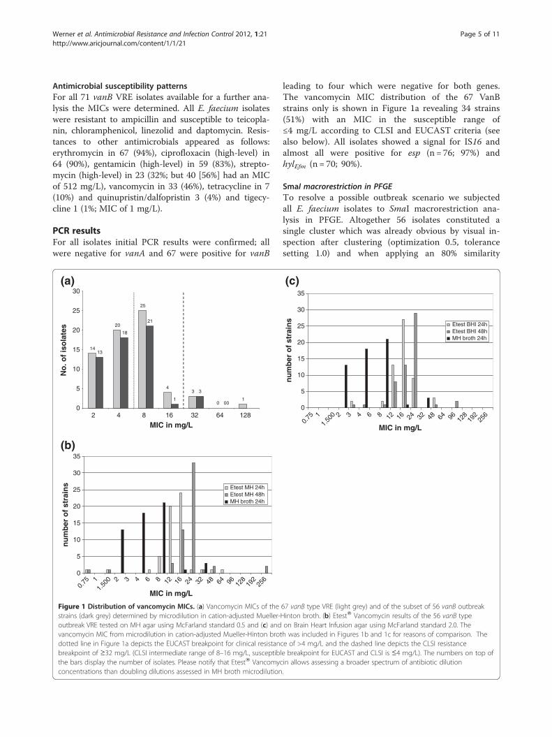
Antimicrobial susceptibility patternsFor all 71 vanB VRE isolates available for a further ana-lysis the MICs were determined. All E. faecium isolateswere resistant to ampicillin and susceptible to teicopla-nin, chloramphenicol, linezolid and daptomycin. Resis-tances to other antimicrobials appeared as follows:erythromycin in 67 (94%), ciprofloxacin (high-level) in64 (90%), gentamicin (high-level) in 59 (83%), strepto-mycin (high-level) in 23 (32%; but 40 [56%] had an MICof 512 mg/L), vancomycin in 33 (46%), tetracycline in 7(10%) and quinupristin/dalfopristin 3 (4%) and tigecy-cline 1 (1%; MIC of 1 mg/L).
PCR resultsFor all isolates initial PCR results were confirmed; allwere negative for vanA and 67 were positive for vanB
14
20
25
43
01
13
18
21
1
3
000
5
10
15
20
25
30
2 4 8 16 32 64 128
MIC in mg/L
No
. of
iso
late
s
0
5
10
15
20
25
30
35
0.75 1
1.50
0 2 3 4 6 8 12 16 24 32 48 64 96 128
192
256
MIC in mg/L
nu
mb
er o
f st
rain
s Etest MH 24hEtest MH 48hMH broth 24h
(a)
(b)
Figure 1 Distribution of vancomycin MICs. (a) Vancomycin MICs of thestrains (dark grey) determined by microdilution in cation-adjusted Mueller-outbreak VRE tested on MH agar using McFarland standard 0.5 and (c) andvancomycin MIC from microdilution in cation-adjusted Mueller-Hinton brotdotted line in Figure 1a depicts the EUCAST breakpoint for clinical resistancbreakpoint of ≥32 mg/L (CLSI intermediate range of 8–16 mg/L, susceptiblthe bars display the number of isolates. Please notify that EtestW Vancomycconcentrations than doubling dilutions assessed in MH broth microdilution
leading to four which were negative for both genes.The vancomycin MIC distribution of the 67 VanBstrains only is shown in Figure 1a revealing 34 strains(51%) with an MIC in the susceptible range of≤4 mg/L according to CLSI and EUCAST criteria (seealso below). All isolates showed a signal for IS16 andalmost all were positive for esp (n = 76; 97%) andhylEfm (n = 70; 90%).
SmaI macrorestriction in PFGETo resolve a possible outbreak scenario we subjectedall E. faecium isolates to SmaI macrorestriction ana-lysis in PFGE. Altogether 56 isolates constituted asingle cluster which was already obvious by visual in-spection after clustering (optimization 0.5, tolerancesetting 1.0) and when applying an 80% similarity
0
5
10
15
20
25
30
35
0.75 1
1.50
0 2 3 4 6 8 12 16 24 32 48 64 96 128
192
256
MIC in mg/L
nu
mb
er o
f st
rain
s
Etest BHI 24hEtest BHI 48hMH broth 24h
(c)
67 vanB type VRE (light grey) and of the subset of 56 vanB outbreakHinton broth. (b) EtestW Vancomycin results of the 56 vanB typeon Brain Heart Infusion agar using McFarland standard 2.0. Theh was included in Figures 1b and 1c for reasons of comparison. Thee of >4 mg/L and the dashed line depicts the CLSI resistancee breakpoint for EUCAST and CLSI is ≤4 mg/L). The numbers on top ofin allows assessing a broader spectrum of antibiotic dilution.

Werner et al. Antimicrobial Resistance and Infection Control 2012, 1:21 Page 6 of 11http://www.aricjournal.com/content/1/1/21
score (Figure 2); these isolates are further designatedas “outbreak strain type” or “outbreak isolates”. Iso-lates constituting the main cluster type derived from52 neonatal patients; two isolates were from environ-mental sources and three originated from the samepatient. The 15 non-outbreak strains revealed severalsmaller clusters or individual patterns, among them apair of isolates with one isolate being vanA/B-nega-tive (UW7620) and another one being vanB-positive(UW7610), the former being obviously the susceptibleprogenitor of the latter isolate having acquired a vanBgene cluster. We were unable to identify a vanB-negative progenitor isolate of the VanB type outbreakstrain type.
Strain no. MLST type sample date8002.01.226187WU
UW7835 ST192 04.12.20088002.21.016387WU
UW7611 ST192 25.09.20088002.11.214287WU8002.11.605287WU
UW7819 ST192 16.10.20088002.11.034387WU8002.11.213287WU8002.11.028287WU8002.11.710387WU8002.11.710287WU8002.90.924167WU8002.01.022187WU
UW7813 ST192 15.10.20088002.01.514187WU8002.01.815187WU8002.01.327187WU8002.01.328187WU8002.01.721287WU8002.11.312287WU
UW7826 ST192 27.10.20088002.11.517287WU8002.90.928167WU
UW7612 ST192 29.09.20088002.90.923167WU8002.90.929167WU8002.01.601267WU8002.90.925167WU8002.90.227067WU
UW7609 ST192 25.09.20088002.01.602267WU8002.01.604267WU9002.11.919287WU8002.11.622387WU8002.21.303387WU9002.20.810587WU8002.01.901687WU9002.20.824587WU9002.20.816587WU9002.30.707587WU8002.21.510487WU8002.21.221487WU
UW7842 ST192 22.12.20088002.21.223487WU8002.21.924487WU9002.10.918487WU9002.10.919487WU9002.20.825587WU
UW7845 ST192 29.12.20089002.30.203587WU9002.20.321587WU8002.90.813067WU8002.90.124067WU8002.90.225067WU
UW7606 ST192 22.09.2008UW7610 ST117 25.09.2008UW7620 ST117 29.09.2008UW7625 ST192 06.10.2008
8002.90.926167WU8002.01.603267WU9002.70.204897WU
UW7852 ST203 17.02.20099002.10.500897WU9002.60.222897WU
UW7983 ST202 24.06.2009UW7859 ST-New 16.03.2009
9002.30.610687WU9002.70.315897WU9002.70.316897WU
UW7617 ST192 29.09.2008
Figure 2 Clonal relatedness of all investigated 71 enterococcal isolatesubsequent phylogenetic analysis (Dice co-efficient using UPGMA clu1.0%). An 80% similarity line (dashed line) divides the group of the 56 relaline) from the other 15 “non-outbreak” strains (below the dotted line). Legecation-adjusted Mueller-Hinton broth; Etest VAN is the value after 24 h readNEW represents a previously unrecognised MLST type with the pattern [9-1
MLST analysisAltogether ten strains representing the outbreak straintype were MLST typed. They were from differentbranches of the PFGE tree and all revealed the same se-quence type ST192 (Figure 2). The two van-negative andvanB-positive isolates with the same PFGE pattern wereST117. Isolates representing individual PFGE patternsrevealed ST203 (2 isolates) and ST192 (“non-outbreak”ST192 type). Results of molecular screening tests werecongruent with the typing results. All isolates belongingto the outbreak strain (PFGE) type were esp- and hylEfm-positive. Prevalence of both markers varied among theother 15 non-outbreak strains confirming the diversestrain background of these isolates.
esp hylEfm genotype MIC VAN Etest VAN Patient no. Sourceelpmasloots3262Bnav++
+ + vanB 2 12 39 anal swabeniru04218Bnav++
+ + vanB 4 12 8 unknownelpmasloots03618Bnav++
bawslana13218Bnav+++ + vanB 4 16 26 stool sample
bawslana83618Bnav++elpmasloots03618Bnav++
bawslana43618Bnav++elpmasloots63618Bnav++slpmasloots72614Bnav++elpmasloots1157.02Bnav++elpmasloots912161Bnav++
+ + vanB 32 16 20 anal swabelpmasloots12614Bnav++elpmasloots22214Bnav++elpmasloots42218Bnav++elpmasloots52218Bnav++
nwonknu82618Bnav++elpmasloots92214Bnav++
+ + vanB 8 16 32 stool sampleelpmasloots33214Bnav++
gulpGCE,rotabucnI-618Bnav+++ + vanB 8 8 9 anal swab
bawsniks01614Bnav++gnidlohrenni,rotabucnI-618Bnav++
elpmasloots41212Bnav++elpmasloots21238Bnav++elpmasloots55.14Bnav++
+ + vanB 32 64 6 throat swabelpmasloots51614Bnav++elpmasloots71612Bnav++
bawslana53424Bnav++elpmasloots73618Bnav++elpmasloots03614Bnav++elpmasloots9482Bnav++
nwonknu95614Bnav++nwonknu35212Bnav++
elpmasloots5588Bnav++bawslana65212Bnav++
elpmasloots14618Bnav++elpmasloots24614Bnav++
+ + vanB 4 16 43 stool sampleelpmasloots44614Bnav++elpmasloots54614Bnav++elpmasloots84212Bnav++
bawslana94212Bnav++bawslana45212Bnav++
+ + vanB 2 8 46 stool sampleelpmasloots2584Bnav++
bawsniks05212Bnav++bawstaorht1218Bnav++
noiterceslaehcart2218Bnav++bawslana3218Bnav++
+ + vanB 32 48 4 stool sample+ - vanB 16 n.d. 7 unknown+ - - ≤1 n.d. 13 unknown+ + vanB 2 n.d. 18 stool sample
sserttam,rotabucnI-.d.n4Bnav++elpmasloots61.d.n8Bnav++elpmasloots26.d.n4Bnav++
+ - vanB 128 n.d. 51 unknown--- ≤1 n.d. 60 stool sample-+- ≤1 n.d. 61 anal swab
+ - - ≤1 n.d. 62 skin swab+ + vanB 8 n.d. 57 stool sample
nwonknu85.d.n61Bnav++bawslana36.d.n61Bnav++bawslana46.d.n8Bnav++
+ + vanB 8 n.d. - Monitor
s as based on SmaI macrorestriction analysis in PFGE andstering; BioNumerics v 6.5; settings: optimization 0.5%; toleranceted ST192 vanB-type outbreak isolates (above the horizontal dottednd: MIC VAN represents the MICs determined in microdilution inout on MH agar and using McFarland 0.5; n.d., not determined; ST--1-1-12-7-1] which is a single locus variant of ST80.

Werner et al. Antimicrobial Resistance and Infection Control 2012, 1:21 Page 7 of 11http://www.aricjournal.com/content/1/1/21
Comparison of the 56 outbreak isolatesAltogether 31 (55%) isolates of the outbreak vanBstrain type had an MIC in the susceptible range of≤4 mg/L (Figure 1) similar to results of the primaryanalysis using vancomycin disk diffusion. All 56 iso-lates were PCR-positive for genes esp, hylEfm and IS16(and vanB). In general, vancomycin EtestW MICs onMH agar were 1–2 doubling dilutions higher than inMH broth (Figure 1b,c). EtestW Vancomycin MICs onMH and BHI agar revealed two VRE (24/48 h) with asusceptible result of ≤4 mg/L only (Figure 1b,c). The48 h EtestW Vancomycin MIC of VanB strains was com-monly 0.5 - 1 doubling dilutions higher than after the 24 hreadout, whereas it remained constant for susceptiblereference isolates (Figure 1b,c). Performance of threechromogenic VRE agars to identify the 56 vanB type out-break VRE was evaluated. On BrillianceTM VRE agar, threeisolates did not grow and two showed growths of smallcolonies only. The chromIDTM VRE agar showed a similarperformance with three isolates that did not grow and oneisolate that showed growth of small colonies only. OnCHROMagarTM VRE two isolates did not grow and an-other two grew with small colonies.
Determination of the vanB subtypeThe long PCR products amplified with DNA from nineST192 outbreak isolates and three non-outbreak isolates(ST117; “non-outbreak ST192”; ST203) were subse-quently digested with BspH1/Dra1. All but one showeda unique restriction pattern which is type-specific forvanB2 subtype clusters (Additional file 1: Figure S1). A
M 1 2 3 4 5 6 7 8 9 10 11 12 13 M
(a)
Figure 3 Chromosomal localization of vanB2 in E. faecium outbreak aresolved in PFGE; (b) Southern hybridisation with a labelled vanB probe. UnST192 outbreak strain; NO, non-outbreak strains. M, S. aureus x SmaI; 1, UWUW7842(O); 7, UW7610 (NO, ST117); 8, UW7611 (O, ST192); 9, UW7835 (O);UW7852 (NO, ST203); 13, UW7859 (NO, ST-NEW).
single non-outbreak strain (ST203) possessed an identi-cal pattern but an additional band.
Localisation of vanBWe performed different analyses with a set of nineMLST typed outbreak and four non-outbreak strains. Toresolve the plasmid vs. a chromosomal localization weisolated the plasmids from 13 strains (see above; plusone additional ST203 isolate). The plasmid profiles ofthe nine outbreak strains were similar, the patterns ofthe four non-related strains differed; however, Southernhybridisations with a labelled vanB probe did not revealany signal (Additional file 2: Figure S2). Nevertheless, weperformed PCRs for the three most common E. faeciumplasmid types determined by their replicase genes, whichwere positive for rep(pRUM)(rep17 family according to[15]), rep(pRE25(rep2 family)), and rep(pLG1)(repA_Nfamily, new class according to [16]) in all outbreakST192 isolates and different for the other four isolatesrepresenting various sequence types (not shown indetails). A single Ceu-I band of ca. 260 kb hybridised tothe vanB gene probe in all PFGE lanes confirming thechromosomal localization of this determinant (Figure 3).
Conjugation experimentsAltogether nine outbreak ST192 isolates (UW7606,UW7609, UW7611, UW7612, UW7813, UW7819,UW7835, UW7842, UW7845) were used as donors forin vitro filter-mating experiments and E. faecium 64/3 as arecipient. Only one mating experiment revealed transcon-jugants (UW7606) with a very low mating rate of 1,67 x
M 1 2 3 4 5 6 7 8 9 10 11 12 13 M
(b)
nd non-outbreak strains. (a) Genomic DNA digested with I-CeuI andderlined lane numbers designate “non-outbreak strains”. Legend: O,7606(O); 2, UW7609(O); 3, UW7612(O); 4, UW7813(O); 5, UW7819(O); 6,10, UW7837 (NO, ST192); 11, UW7845(O); 11, UW7852 (NO, ST203); 12,

Werner et al. Antimicrobial Resistance and Infection Control 2012, 1:21 Page 8 of 11http://www.aricjournal.com/content/1/1/21
10e-8 (per recipient) and 7,14 x 10e-8 (per donor), re-spectively. A single transconjugant TC1 was investigatedin details. TC1 became resistant to vancomycin butremained susceptible to erythromycin, gentamicin, ampi-cillin and ciprofloxacin. It was PCR-positive for IS16 (re-cipient 64/3 is IS16-negative) most probably due toacquisition of a mobile vanB gene cluster which is com-monly flanked by copies of IS16 [5]. PFGE analysis of thedonor, the recipient and the transconjugant followed bysubsequent Southern hybridisation with a labelled vanBgene probe confirmed the clonal relatedness of the recipi-ent and the transconjugant, the latter having acquired amobile vanB gene cluster from the donor which integratedinto the chromosome (Additional file 3: Figure S3).
DiscussionIt has not been investigated in greater details until now towhich extent VanB type strains may show a low expres-sion of vancomycin resistance which could result in a dis-tinct frequency of underestimation or underreporting ofvanB type resistance in Enterococcus spp. in general.Results of our present study (Figure 1) and annual reportsfrom our Focal laboratory for enterococci already demon-strated that low resistance expression among vanB typestrains was prevalent in 10–25%, dependent on themethod and breakpoints used [10,20]. It has to be notedagain that in the present setting samples were screenednon-selectively and selected enterococcal colonies werelater tested for glycopeptide resistance phenotypically andgenotypically. Most strategies to identify vanA- or vanB-type VRE start with selective enrichment or direct selec-tion on screening plates which would certainly miss a sub-stantial amount of strains showing low vancomycinresistance expression (see below). This general strategymay already pre-select the detectable wildtype vanB popu-lation. Nevertheless, non-selective screening was success-ful in this setting with premature infants lacking anestablished intestinal flora. In adult patients VRE accountonly for a certain percentage of all intestinal enterococciand non-selective screening would most likely be unableto select (by chance) for resistant variants [21]. Also real-time based screening assays targeting only the resistancegenes vanA and vanB are not considered as a reliable al-ternative for screening since prevalence of vanB genes innon-enterococcal, intestinal colonizers complicates assayaccuracy and leads to false positive test results [22-25].The numbers are comparably similar in different parts ofthe world resulting in a generally low positive predictivevalue for VanB and the demand for a confirmation by cul-ture based methods (with the problems as describedabove)[22,26,27]. In the light of our present study results,it cannot be excluded that the generally high false-positivevanB rate of real-time based genotypic assays could alsoresult from the low performance of comparator assays to
identify VanB strains with low vancomycin resistancelevels and may thus to a lesser extent be attributed tovanB prevalent in non-enterocococcal species.We assessed the performance of three commercially
available chromogenic VRE selective agar plates in iden-tifying the 56 VanB type outbreak E. faecium isolateswith partly low level vancomycin resistance expressionand compared it to results of EtestW Vancomycin MICsbased on two different protocols. In general, MICs were1–1.5 doubling dilutions higher on solid media (EtestW)than in liquid broth (Figure 1). In line with these resultsthe three chromogenic agar media also performed com-parably well in identifying vanB type VRE. A supposedbetter vancomycin resistance expression of vanB onsolid media is an important finding, but observed hereon a collection of admittedly similar isolates and thuscannot be extrapolated to the vanB VRE population ingeneral.At least two of the 56 outbreak VRE isolates originated
from the patients’ surrounding and were clonally identi-cal to the other patients’ isolates (Figure 2). Togetherwith all the described infection control measures onemight conclude that despite all rigid disinfection, infec-tion prevention and training procedures, environmentalVRE contamination remains a possible source for thisongoing VRE prevalence over three years. It is knownfor a long time that VRE carriers contaminate their dir-ect surrounding and that VRE/enterococci are able tosurvive for weeks and months on these surfaces andpatients commodities [28,29] and are thus able to spreadto and colonise other patients. A common strategy todecolonise VRE patients and newborns is not estab-lished; however, results of recent experimental studiespinpoint towards strategies by eliminating VRE colonisa-tion with probiotic competitor strains [30].A number of European countries reported increasing
numbers of colonisations and infections with VanB typeVRE. Sweden having had extremely low overall VREprevalence over the years experienced several clusters ofcolonisations and infections in hospitals in Stockholmrecently [3,31]. Molecular analysis revealed distributionof vanB2-Tn5382 subtype clusters located on pRUM-like transferable plasmids introduced into hospital-associated E. faecium strain types [7,8]. A similar resist-ance gene cluster was shown to be prevalent amongvarious E. faecalis and E. faecium strains in 16 hospitalsin Chile [32] although in general vanA-type resistanceseemed most prevalent among Latin American VRE iso-lates (Peru, Colombia, Ecuador, Venzuela)[33]. A num-ber of reports described vanB type VRE outbreaks inrecent years among hospital patients in Spain andFrance representing European low VRE prevalencecountries according to EARS-Net data [4,34-36].Whereas the outbreaks in Spain remained at a local level
Werner et al. Antimicrobial Resistance and Infection Control 2012, 1:21 Page 9 of 11http://www.aricjournal.com/content/1/1/21
(hospital outbreaks) and did not feed substantially theoverall surveillance numbers; increasing VRE frequenciesin France in 2008 are mainly attributed to a marked in-crease in a number of vanB outbreaks in the North ofthe country [4]. Similar rates are notified for Germanyafter 2008, at least for some federal states mainly in theSouth-Western part of the country [10].During the ongoing outbreak also other non-outbreak
vanB isolates were identified. It remains to be speculatedif the identified vanB2-Tn5382 cluster as part of an ICEis capable of spreading horizontally from strain to strainand integrate into a recipients’ genome. Molecular ana-lysis revealed a similar cluster type and a chromosomallocalization in all VanB strains. Resistance spread at avery low mating rate as assessed in vitro. Nevertheless, itwas shown recently that vancomycin resistance genesare transferred successfully in vivo in mammal intestinesand that rates could be several orders of magnitudehigher than those determined in vitro [37-39].Various expression levels of vancomycin resistance in
vanB strains were known for some time. The level ofvanB gene expression and the presence of a distinctvanB allele type (vanB1 to vanB3) could not be genetic-ally linked. Grabsch et al. were unable in identifying mu-tational changes in unrelated vanB2 type VRE showing avarious level of vanB gene expression [40]. Teicoplaninheteroresistance in vanA VRE strains was described re-cently and several genomic rearrangements and dele-tions within the vanA gene cluster elements andmutational changes within the two-component regulatorgenes vanS and vanR were identified in glycopeptide-heteroresistant strains, but these changes were not (ex-perimentally proven) functionally linked to the describedheteroresistance phenotype [41-43]. We did not investi-gate the molecular background of different vanB resist-ance expression in the isolates investigated here.Nevertheless, MICs were reproducible in repeatedexperiments.
ConclusionsWe analysed an ongoing high prevalence of vanB type E.faecium in a neonatal ICU over a period of several years;ca. 80% (n= 56) of isolates collected during a key period of10 months belonged to a single outbreak strain type.Enterococcal isolates were assessed non-selectively andabout half of the isolates appeared phenotypically suscep-tible to vancomycin with MICs ≤4 mg/L. This suggeststhat a certain amount of VanB strains may show a low ex-pression of vancomycin resistance which results in an un-known frequency of underestimation or underreporting ofvanB type resistance in Enterococcus spp in general. It istempting to speculate that this phenomenon may directlysupport ongoing and increasing prevalence of(unrecognized) vanB VRE prevalence among the clinical
setting. The comparably high false-positive vanB rate ofreal-time based genotypic assays may thus only be partlyattributed to vanB prevalent in non-enterocococcal spe-cies but could also simply result from the low accuracy ofcomparator assays to identify VanB strains with low ex-pression of vancomycin resistance.
Additional files
Additional file 1: Figure S1. vanB2 subtype determination. Long PCRproducts with DNA from outbreak and non-outbreak strains weresubsequently digested with BspH1/DraI. Underlined lane numbersdesignate “non-outbreak strains”. Legend: O, ST192 outbreak strain; NO,non-outbreak strains. M, Gene Ruler 100bp Plus (Thermo Fisher Scientific);1, UW7606(O); 2, UW7609(O); 3, UW7612(O); 4, UW7813(O); 5, UW7819(O);6, UW7842(O); 7, UW7610 (NO, ST117); 8, UW7611 (O, ST192); 9, UW7835(O); 10, UW7842 (O); 11, UW7845(O); 12, UW7852 (NO, ST203) [UW7859(NO, ST203) did not reveal a long PCR product; not shown].
Additional file 2: Figure S2. Plasmid patterns of vanB2 E. faeciumoutbreak and non-outbreak strains. (a) Undigested plasmid patternsresolved in 0.8% agarose gel; (b) Southern hybridisation with a labelledvanB probe. Underlined lane numbers designate “non-outbreak strains”.Legend: O, ST192 outbreak strain; NO, non-outbreak strains; M, Roche SizeMarker III, DIG-labelled (for orientation purposes only); 1, UW7606(O); 2,UW7609(O); 3, UW7612(O); 4, UW7813(O); 5, UW7819(O); 6, UW7842(O); 7,UW7610 (NO, ST117); 8, UW7611(O, ST192); 9, UW7835 (O); 10, UW7845(O); 11, UW7852 (NO, ST203); 12, UW7859 (NO, ST203).
Additional file 3: Figure S3. (a) SmaI-digested genomic DNA resolvedin PFGE and (b) Southern hybridisation with a labelled vanB probe of avanB type E. faecium donor strain UW7706, a vancomycin-susceptiblerecipient E. faecium 64/3 and a vanB-positive transconjugant 1. Legend:M, S.aureus x SmaI. D, donor strain UW7706; R, recipent strain 64/3; T,transconjugant UW7706x64/3 TC1. Please note that the vanB positiveband in lanes D and T refers to a double band.
Competing interestsThe authors declare that they have no competing interests.
Authors’ contributionsHMJ and RZ co-ordinated, supervised and evaluated the primary diagnosticsof the clinical samples and consulted the staff at the neonatal ICU regularly.UG determined the spectrum of antimicrobial susceptibilities by brothmicrodilution and performed all confirmatory PCR screenings. CF performedall EtestsW, chromogenic agar assays, PFGE and Southern hybridisationexperiments. IK, WW and GW supervised the confirmatory diagnostic andmolecular typing experiments and interpreted and analyzed the results. GW,IK and RZ wrote the manuscript.
AcknowledgementsWe thank all the involved clinicians that did the initial clinical diagnoses andthe nurses that sampled the swabs.
Author details1Unit FG13 Nosocomial Infections, Robert Koch-Institute Wernigerode,Wernigerode, Germany. 2Institute for Clinical Hygiene and Infectiology,Hospital Nord der Stadt Nürnberg, Nuremberg, Germany.
Received: 16 February 2012 Accepted: 14 April 2012Published: 30 May 2012
References1. ECDC: Annual Epidemiological Report 2011. In Stockholm, SE, European
Centre for Disease Prevention and Control. Annual Epidemiological Report onCommunicable Diseases in Europe. 2012.
2. Werner G: Surveillance of antimicrobial resistance among Enterococcusfaecium and Enterococcus faecalis isolated from human (clinical/commensal), food animal, meat and environmental samples. In

Werner et al. Antimicrobial Resistance and Infection Control 2012, 1:21 Page 10 of 11http://www.aricjournal.com/content/1/1/21
Enterococcus and safety. Edited by Semedo-Lemsaddek T, Barreto-CrespoMT, Tenreiro R. Hauppage, N.Y: Nova Science Publishers Inc; 2011 [in press].
3. Soderblom T, Aspevall O, Erntell M, Hedin G, Heimer D, Hokeberg I, et al:Alarming spread of vancomycin resistant enterococci in Sweden since2007. Euro Surveill 2010, 15:19620.
4. Bourdon N, Fines-Guyon M, Thiolet JM, Maugat S, Coignard B, Leclercq R, etal: Changing trends in vancomycin-resistant enterococci in Frenchhospitals, 2001–2008. J Antimicrob Chemother 2011, 66:713–721.
5. Hegstad K, Mikalsen T, Coque TM, Werner G, Sundsfjord A: Mobile geneticelements and their contribution to the emergence of antimicrobialresistant Enterococcus faecalis and Enterococus faecium. Clin MicrobiolInfect 2010, 16:541–554.
6. Paulsen IT, Banerjei L, Myers GS, Nelson KE, Seshadri R, Read TD, et al: Roleof mobile DNA in the evolution of vancomycin-resistant Enterococcusfaecalis. Science 2003, 299:2071–2074.
7. Bjorkeng E, Rasmussen G, Sundsfjord A, Sjoberg L, Hegstad K, Soderquist B:Clustering of polyclonal VanB-type vancomycin-resistant Enterococcusfaecium in a low-endemic area was associated with CC17-genogroupstrains harbouring transferable vanB2-Tn5382 and pRUM-like repAcontaining plasmids with axe-txe plasmid addiction systems. APMIS 2011,119:247–258.
8. Sivertsen A, Lundblad EW, Wisell KT, Liljequist B, Billström H, Ullberg M, et al:The widespread VRE outbreak in Swedish hospitals 2007–2009 wasassociated with clonal E. faecium CC17 genogroup strains harbouringseveral virulence traits and transferable vanB pRUM-like repA plasmids.In Final Programme of the 21st ECCMID, Milano, May 7–10, 2011.. Poster P924,113.
9. Klare I, Werner G, Witte W, Fahr A-M: 4.1.3. Enterococcus spp. GERMAP 2008Antibiot Resist Use 2009, 1:41–46.
10. Klare I, Werner G, Witte W: Enterococci with vancomycin resistance fromGerman hospitals in 2008/2009 (German). Epidemiologisches Bull 2010,44:427–437.
11. Werner G, Fleige C, Ewert B, Laverde-Gomez JA, Klare I, Witte W: High-levelciprofloxacin resistance among hospital-adapted Enterococcus faecium(CC17). Int J Antimicrob Agents 2010, 35:119–125.
12. Werner G, Klare I, Fleige C, Witte W: Increasing rates of vancomycinresistance among Enterococcus faecium isolated from German hospitalsbetween 2004 and 2006 are due to wide clonal dissemination ofvancomycin-resistant enterococci and horizontal spread of vanA clusters.Int J Med Microbiol 2007, 298:515–527.
13. Dahl KH, Rokenes TP, Lundblad EW, Sundsfjord A: Nonconjugativetransposition of the vanB-containing Tn5382-like element in Enterococcusfaecium. Antimicrob Agents Chemother 2003, 47:786–789.
14. Evers S, Courvalin P: Regulation of VanB-type vancomycin resistance geneexpression by the VanS(B)-VanR (B) two-component regulatory system inEnterococcus faecalis V583. J Bacteriol 1996, 178:1302–1309.
15. Jensen LB, Garcia-Migura L, Valenzuela AJ, Lohr M, Hasman H, Aarestrup FM:A classification system for plasmids from enterococci and other Gram-positive bacteria. J Microbiol Methods 2009, 80:25–43.
16. Laverde Gomez JA, van SW, Freitas AR, Coque TM, Weaver KE, Francia MV, etal: A multiresistance megaplasmid pLG1 bearing a hyl(Efm) genomicisland in hospital Enterococcus faecium isolates. Int J Med Microbiol 2010,301:165–175.
17. Werner G, Fleige C, Geringer U, van SW, Klare I, Witte W: IS element IS16 asa molecular screening tool to identify hospital-associated strains ofEnterococcus faecium. BMC Infect Dis 2011, 11:80.
18. Werner G, Freitas AR, Coque TM, Sollid JE, Lester C, Hammerum AM, et al:Host range of enterococcal vanA plasmids among Gram-positiveintestinal bacteria. J Antimicrob Chemother 2010, 66:273–282.
19. Fournier S, Brun-Bruisson C, Jarlier V: Twenty years of antimicrobialresistance control programme in a regional multi hospital institution,with focus on emerging bacteria (VRE and CPE). Antimicrob Resist Inf Contr2012, 1:9.
20. Werner G, Klare I, Strommenger B, Witte W: Vancomycin-resistantenterococci - epidemiology, diagnostics, typing, trends. Mikrobiologe2007, 17:57–74.
21. Ubeda C, Taur Y, Jenq RR, Equinda MJ, Son T, Samstein M, et al:Vancomycin-resistant Enterococcus domination of intestinal microbiotais enabled by antibiotic treatment in mice and precedes bloodstreaminvasion in humans. J Clin Invest 2010, 120:4332–4341.
22. Young HL, Ballard SA, Roffey P, Grayson ML: Direct detection of vanB2using the Roche LightCycler vanA/B detection assay to indicatevancomycin-resistant enterococcal carriage - sensitive but not specific. JAntimicrob Chemother 2007, 59:809–810.
23. Domingo MC, Huletsky A, Giroux R, Boissinot K, Picard FJ, Lebel P, et al:High prevalence of glycopeptide resistance genes vanB, vanD, and vanGnot associated with enterococci in human fecal flora. Antimicrob AgentsChemother 2005, 49:4784–4786.
24. Marvaud JC, Mory F, Lambert T: Clostridium clostridioforme andAtopobium minutum clinical isolates with vanB-type resistance inFrance. J Clin Microbiol 2011, 49:3436–3438.
25. Ballard SA, Pertile KK, Lim M, Johnson PD, Grayson ML: Molecularcharacterization of vanB elements in naturally occurring gut anaerobes.Antimicrob Agents Chemother 2005, 49:1688–1694.
26. Stamper PD, Cai M, Lema C, Eskey K, Carroll KC: Comparison of the BDGeneOhm VanR Assay to culture for identification of vancomycin-resistant enterococci in rectal and stool specimens. J Clin Microbiol 2007,45:3360–3365.
27. Werner G, Serr A, Schütt S, Schneider C, Klare I, Witte W, et al: Comparisonof direct cultivation on a selective solid medium, polymerase chainreaction from an enrichment broth, and the BD GeneOhm™ VanR Assayfor identification of vancomycin-resistant enterococci in screeningspecimens. Diagn Microbiol Inf Dis 2011, 70:512–521.
28. Kramer A, Schwebke I, Kampf G: How long do nosocomial pathogenspersist on inanimate surfaces? A systematic review. BMC Infect Dis 2006,6:130.
29. Lemmen SW, Hafner H, Zolldann D, Stanzel S, Lutticken R: Distribution ofmulti-resistant Gram-negative versus Gram-positive bacteria in thehospital inanimate environment. J Hosp Infect 2004, 56:191–197.
30. Szachta P, Ignys I, Cichy W: An evaluation of the ability of the probioticstrain Lactobacillus rhamnosus GG to eliminate the gastrointestinalcarrier state of vancomycin-resistant enterococci in colonized children. JClin Gastroenterol 2011, 45:872–877.
31. Fang H, Nord CE, Ullberg M: Screening for vancomycin-resistantenterococci: results of a survey in Stockholm. APMIS 2010, 118:413–417.
32. Lopez M, Hormazabal JC, Maldonado A, Saavedra G, Baquero F, Silva J, et al:Clonal dissemination of Enterococcus faecalis ST201 and Enterococcusfaecium CC17-ST64 containing Tn5382-vanB2 among 16 hospitals inChile. Clin Microbiol Infect 2009, 15:586–588.
33. Panesso D, Reyes J, Rincon S, Diaz L, Galloway-Pena J, Zurita J, et al:Molecular epidemiology of vancomycin-resistant Enterococcus faecium:a prospective, multicenter study in South American hospitals. J ClinMicrobiol 2010, 48:1562–1569.
34. Torres C, Escobar S, Portillo A, Torres L, Rezusta A, Ruiz-Larrea F, et al:Detection of clonally related vanB2-containing Enterococcus faeciumstrains in two Spanish hospitals. J Med Microbiol 2006, 55:1237–1243.
35. Valdezate S, Labayru C, Navarro A, Mantecon MA, Ortega M, Coque TM, etal: Large clonal outbreak of multidrug-resistant CC17 ST17 Enterococcusfaecium containing Tn5382 in a Spanish hospital. J Antimicrob Chemother2009, 63:17–20.
36. Servais A, Mercadal L, Brossier F, Venditto M, Issad B, Isnard-Bagnis C, et al:Rapid curbing of a vancomycin-resistant Enterococcus faecium outbreakin a nephrology department. Clin J Am Soc Nephrol 2009, 4:1559–1564.
37. Dahl KH, Mater DD, Flores MJ, Johnsen PJ, Midtvedt T, Corthier G, et al:Transfer of plasmid and chromosomal glycopeptide resistancedeterminants occurs more readily in the digestive tract of mice thanin vitro and exconjugants can persist stably in vivo in the absence ofglycopeptide selection. J Antimicrob Chemother 2007, 59:478–486.
38. Moubareck C, Bourgeois N, Courvalin P, Doucet-Populaire F: Multipleantibiotic resistance gene transfer from animal to human enterococci inthe digestive tract of gnotobiotic mice. Antimicrob Agents Chemother2003, 47:2993–2996.
39. Lester CH, Hammerum AM: Transfer of vanA from an Enterococcusfaecium isolate of chicken origin to a CC17 E. faecium isolate in theintestine of cephalosporin-treated mice. J Antimicrob Chemother 2010,65:1534–1536.
40. Grabsch EA, Chua K, Xie S, Byrne J, Ballard SA, Ward PB, et al: Improveddetection of vanB2-containing Enterococcus faecium with vancomycinsusceptibility by Etest using oxgall supplementation. J Clin Microbiol 2008,46:1961–1964.

Werner et al. Antimicrobial Resistance and Infection Control 2012, 1:21 Page 11 of 11http://www.aricjournal.com/content/1/1/21
41. Alam MR, Donabedian S, Brown W, Gordon J, Chow JW, Zervos MJ, et al:Heteroresistance to vancomycin in Enterococcus faecium. J Clin Microbiol2001, 39:3379–3381.
42. Khan SA, Sung K, Layton S, Nawaz MS: Heteroresistance to vancomycinand novel point mutations in Tn1546 of Enterococcus faecium ATCC51559. Int J Antimicrob Agents 2008, 31:27–36.
43. Qu TT, Zhang JL, Zhou ZH, Wei ZQ, Yu YS, Chen YG, et al: Heteroresistanceto teicoplanin in Enterococcus faecium harboring the vanA gene. J ClinMicrobiol 2009, 47:4194–4196.
doi:10.1186/2047-2994-1-21Cite this article as: Werner et al.: Vancomycin-resistant vanB-typeEnterococcus faecium isolates expressing varying levels of vancomycinresistance and being highly prevalent among neonatal patients in asingle ICU. Antimicrobial Resistance and Infection Control 2012 1:21.
Submit your next manuscript to BioMed Centraland take full advantage of:
• Convenient online submission
• Thorough peer review
• No space constraints or color figure charges
• Immediate publication on acceptance
• Inclusion in PubMed, CAS, Scopus and Google Scholar
• Research which is freely available for redistribution
Submit your manuscript at www.biomedcentral.com/submit


