Embed Size (px)
Citation preview

Kidney transplant has been established as a superi-or method of renal replacement therapy for
patients with end-stage kidney disease.1 Many factorsthat are donor-dependent affect immediate and long-term outcomes, for example, age, hemodynamic status,cause of death, sex, time spent in the intensive careunit, or presence of infections.2,3 Other factors such asthe method of organ preservation, duration of coldischemia, and type of perfusion solution are related toorgan preservation, still other factors are recipient-dependent, such as human leukocyte antigen mis-match, presence of panel reactive antibodies, age,presence of primary kidney disease or other contribut-ing diseases, immunosuppression protocols followed,or presence of infections.4 During the past 2 decades,
short-term results of kidney transplant have improvedmarkedly thanks to better donor-recipient managementand new discoveries in immunosuppression, whichhelped to maximize the donor pool by allowing the useof marginal donors. It is believed that hemodynamicinstability of donors may cause inflammation5 and animmunologic response during reperfusion.
The introduction of machine perfusion hasimproved kidney assessment before transplant,6,7 sig-nificantly improving short-term results, reducingdelayed graft function,8 and improving outcomes fromkidneys retrieved from donors after cardiac death.9 Ithas been proven that kidneys stored by using machineperfusion have better long-term survival,8,10,11 withorgans procured from expanded criteria donors
Preservation of kidneys by machine perfusioninfluences gene expression and may limitischemia/reperfusion injury
{AUTHOR: AMA/PiT style is to avoid abbreviations in abstracts.}Context—Machine perfusion improves graft survival. Histopathologic analysisreveals a lower incidence of chronic rejection and interstitial fibrosis in kidneyspreserved with machine perfusion. Ischemic/reperfusion injury may help toexplain these findings. Objective—To assess the activation of genes correlated with ischemic/reperfusioninjury in kidneys preserved under different conditions before transplant. Design/Patients—Between 2005 and 2006, 69 kidney biopsy specimens were col-lected and patients were followed up for 5 years after that. Intervention—Before transplant, kidneys were preserved with machine perfusionor cold storage. Donors from the machine perfusion and cold storage groups didnot differ with regard to age, sex, or hemodynamic status. Recipients were dividedinto 5 groups: expanded criteria donor–machine perfusion (n = 16), standard crite-ria donor–machine perfusion (n = 10), expanded criteria donor–cold storage (n =9), and standard criteria donor–cold storage (n = 27); 7 kidneys were retrievedfrom living related donors.Main Outcome Measures—Biopsies were done 30 minutes after reperfusion.Interleukin-1!, vascular endothelial growth factor, heme oxygenase-1, and hypoxia-inducible factor–1 gene expression levels were analyzed. Results—Mean expression levels of hypoxia-inducible factor–1" were signifi-cantly higher in the cold storage groups, and lower in the machine perfusion andliving related donor groups. Five-year graft survival was significantly (P < .05)lower in the expanded criteria donor–cold storage group (66%) than in the stan-dard criteria donor–machine perfusion group (90%). Machine perfusion influencesgene expression related to hypoxia during reperfusion and may improve the long-term results of kidney transplant. (Progress in Transplantation. 2014;24:xxx-xxx)©2014 NATCO, The Organization for Transplant Professionals doi: http://dx.doi.org/10.7182/pit2014XXX
Michal Wszola, MD, PhD,Artur Kwiatkowski, MD, PhD,Piotr Domagala, MD, PhD,Agnieszka Wirkowska, PhD,Monika Bieniasz, MD, PhD,Piotr Diuwe, MD, Rafa!Kieszek, MD, MagdalenaDurlik, MD, PhD, AndrzejChmura, MD, PhDWarsaw Medical University, Poland
Corresponding author: Michal Wszola,MD, PhD, Department of General andTransplantation Surgery, Nowogrodzka59th Street, 02-006 Warsaw, Poland (e-mail: [email protected])
To purchase electronic or print reprints,contact:The InnoVision Group101 Columbia, Aliso Viejo, CA 92656Phone (800) 899-1712 (ext 532) or (949) 448-7370 (ext 532)Fax (949) 362-2049E-mail [email protected]
1 Progress in Transplantation, Vol 24, No. 1, March 2014

(ECDs) achieving the biggest benefit. Eurotransplantreports that machine perfusion reduces the risk ofdelayed graft function (DGF) by approximately 7%and improves 1-year graft survival by 12%.12
However, the mechanism by which renal function isimproved by machine perfusion is not well under-stood. Nevertheless, in our previous study,13 weshowed a lower incidence of interstitial fibrosis/tubu-lar atrophy and chronic rejection (according to Banff2005 criteria) in a group of recipients of kidneys pre-served with machine perfusion.
Ischemia/reperfusion injury also plays an impor-tant role in both early and long-term renal function.14
Activation of genes associated with ischemia/reperfu-sion injury is thought to play a role in early graft func-tion.15-17 The major factor for cellular adaptation that isactivated in response to hypoxia duringischemia/reperfusion injury is the heterodimer,hypoxia-inducible factor (HIF)-1,18 which can activatealmost 200 genes in response to hypoxia,19 includinggenes for vascular endothelial growth factor (VEGF)and transforming growth factor (TGF)-!. HIF-1 con-sists of 2 subunits, HIF-1" and HIF-1!. HIF-1!belongs to the helix-loop-helix and Per Arnt Simdomain-containing transcription factor families. HIF-1! protein is always present, whereas HIF-1" isdependent on the presence of oxygen. Under normox-ic conditions, HIF-1" binds to the von Hippel-Lindautumor suppressor protein, which leads to the proteaso-mal degradation of HIF-1". The lack of oxygenresults in stabilization of the HIF-1" protein, which isthen translocated to the nucleus where it plays a rolein the regulation of many target genes. HIF-1" activa-tion within a tissue may not only influence othergenes, it may also be an indicator of the level of oxy-genation or hypoxia within a tissue. The aim of thisstudy was to assess the activation of genes associatedwith ischemia/reperfusion injury in the cortex of renalgrafts stored by using different preservation methodsbefore transplant. The results of this study may helpelucidate the influence of machine perfusion on earlyand long-term graft function.
Patients and MethodsPopulation Description
Seventy-eight kidneys were retrieved from 39deceased, brain-dead donors (BDDs), and 7 wereobtained from living related donors (LRDs). Eightkidneys from BDDs were sent to other transplant cen-ters for transplant; another 8 kidneys procuredharvest-ed from BDDs were not sampled by biopsy per thestudy protocol upon implantation and were thusexcluded from the study. The remaining 62 kidneysfrom BDDs were preserved before transplant by eithermachine perfusion (17 donors, 26 kidneys) or coldstorage (22 donors, 36 kidneys). Fifteen of the 39
BDD donors (38%) were ECDs, identified as such ifthe donor’s age were either greater than 60 years orgreater than 50 years with 2 of the following criteriabeing met: hypertension in anamnesis, creatinine levelgreater than 1.5 mg/dL (multiply by 88.4 to convert tomicromoles per liter), or stroke as the cause of death.Characteristics of BDDs are shown in Table 1.Seventy-seven recipients underwent kidney trans-plant: 70 received kidneys from BDDs and 7 receivedkidneys from LRDs. In 8 kidneys retrieved fromBDDs, biopsy samples were not obtained duringreperfusion, mainly because of technical problems,and those kidneys were thus excluded from furtheranalysis. Short- and long-term observations wereavailable for 69 kidney recipients. All patients weredivided into 5 categories:
1. MP-ECD group (n=16): received kidneys fromECDs and kidneys were kept in machine perfusionbefore transplant
2. MP-SCD group (n=10): received kidneys fromSCDs and kidneys were kept in machine perfusionbefore transplant
3. CS-ECD group (n = 9): received kidneys fromECDs and kidneys were kept in cold storage beforetransplant
4. CS-SCD group (n = 27): received kidneys fromSCDs and kidneys were kept in cold storage beforetransplant
5. LRD group (n = 7): received kidneys fromLRDs
Recipients of kidneys from BDDs did not differbetween groups with regard to human leukocyte anti-gen match, age, sex, duration of dialysis treatmentbefore transplant, or pretransplant levels of panel reac-tive antibodies (Table 1). The most frequent causes ofend-stage renal disease were as follows: chronicglomerulonephritis (39.0%), chronic pyelonephritis(20.8%), polycystic kidney disease (7.8%), and dia-betic nephropathy (6.5%). Twelve patients underwenttransplant for a second time. Triple-drug immunosup-pression was primarily used. Induction therapy withantithymocyte globulin or daclizumab was adminis-tered to patients who underwent a second transplantand had maximum panel reactive antibodies above20% (8 cases). Cyclosporin A was used in 55%(38/69) and tacrolimus in 45% (31/69) of patients.Mycophenolate mofetil was given to 90% (62/69) andazathioprine to 10% (7/69) of patients. {AUTHOR:All percentages were checked and adjusted ifincorrect, hence minor changes here.} All patientswere administered steroids during the immunosup-pression therapy. DGF was defined recognized as aneed for dialysis in the first week after kidney trans-plant, regardless of the cause (hyperkalemia, highserum urea concentration). Primary nonfunction wasdefined as a permanent loss of graft function immedi-
Progress in Transplantation, Vol 24, No. 1, March 20142

ately after transplant. Acute rejection was confirmedby biopsy and diagnosed according to Banff 2007 cri-teria. Patient and graft survival were analyzed duringthe immediate postoperative and long-term periods.All patients completed a 5-year follow-up examina-tion.
Preservation MethodBetween 2005 and 2006, all kidneys from hemo-
dynamically unstable donors, ECD donors, or donorswith elevated serum levels of creatinine were storedby machine perfusion. In a few cases, the MPS II per-fusion solution or cassettes were unavailable, andsome nonoptimal kidneys were preserved by coldstorage (5 donors, 9 kidneys). Immediately after organrecovery and cooling to 4°C, each kidney was placedin a thermally stable container in a preservation solu-tion (simple cold storage). Thirty-six kidneys fromBDDs and 7 kidneys from LRDs were stored in thisway. Twenty-six kidneys were preserved by machineperfusion (Waters Medical Systems, RM3).
After bench surgery, kidneys were placed in ster-ile disposable cassettes (MOX 100 DCM disposablecassette, Waters Instruments Inc) in the perfusion unit(MOX-100 m2 Transport Unit, Waters InstrumentsInc) filled with 1 L of perfusion fluid (MPS II). Acooling bath was used to ensure constant fluid tem-perature (6°C) (Model 900 Constant TemperatureCirculator, Fisher Scientific). Electrolyte and pHmonitoring of the perfusion solution and subsequentbacteriology cultures were performed during and atthe completion of every perfusion procedure. A con-tinuous flow at a 1-Hz frequency (60 pulses/min) wasused. An initial perfusion pressure was set at 50 mmHg systolic pressure. During machine perfusion (at 1,2, 3, and 4 h, and every 4 h thereafter), the followingparameters were monitored: systolic/diastolic pres-sure, mean perfusion pressure, flow per minute, vas-cular resistance, pH, PO2, PCO2, HCO3# (ABL 330Radiometer), Na+, K+ (Ionometer EF Fresenius), andosmolarity (Osmometer 800 cl). Biochemical markersof ischemic organ injury also were investigated: lac-tate dehydrogenase (by spectrophotometric assay) andlactate (GM7 APR). {AUTHOR: What is APR?Isn’t the GM7 manufactured by AnaloxInstruments?} Depending on the results of the moni-tored parameters, vasodilation agents (papaverine,verapamil) were administered to reduce resistance,and bicarbonate was given to adjust the pH. Kidneyswere removed from machine perfusion immediatelybefore vascular anastomosis when the iliac fossa wasprepared for implantation. Perfusion characteristicsare shown in Table 2.
Gene Expression AnalysisBiopsy specimens were taken 30 minutes after
reperfusion and snap frozen at -75°C before urinaryanastomosis. Expression levels of interleukin 1! (IL-1 !), VEGF, heme oxygenase-1 (HO-1), and HIF-1genes were analyzed by real-time polymerase chainreaction (RT-PCR).
RNA IsolationRNA isolation was performed by using a modi-
fied Chomczynski and Sacchi method20 according tothe instructions of the RNEasy Micro Kit (Qiagen,Syngen Biotech). Each kidney biopsy specimen wasplaced in a buffer solution (RLT; QiagenRNEasyMini Kit) containing !-mercaptoethanol (100:1).Homogenization with proteinase K (concentration 20mg/mL; Sigma-Aldrich) was then performed at 55°Cfor 20 minutes. The homogenate was separated bycentrifugation at 13400 rpm for 3 minutes. The super-natant was mixed with 99% ethanol at a 2:1 ratio toallow for protein precipitation. Samples were placedon filtration columns and separated by centrifugationat 13 400 rpm for 15 seconds at room temperature.Proteins were eluted, leaving RNA on the column. Toprevent DNA contamination, the samples were incu-bated with DNAse for 15 minutes at room tempera-ture. At the end of the procedure, both RNA concen-tration and purity were assessed by measuringabsorbances at 260 nm and 280 nm (A260 and A280,respectively) by using a NanoDrop device. RNA puri-ty was satisfactory if the A260/A280 ratio was between1.8 and 2.1. Total RNA was kept at -80°C for less than14 days before complementary DNA (cDNA) tran-scription. {AUTHOR: cDNA defined correctly inpreceding sentence? Please revise as needed butdefine abbreviation before you use it.}
Reverse TranscriptionReverse transcription was performed by using the
Superscript III First-Strand Synthesis System for RT-PCR reagents (Invitrogen). Superscript UI{AUTHOR: What is UI short for?} reverse tran-scriptase synthesized cDNA at a temperature range of42°C to 55°C. Initially, all RNA was incubated withrandom hexamers and deoxynucleotide triphosphates{AUTHOR: OK how dNTPs spelled out?} (dATP,dUTP, dCTP, dGTP) for 5 minutes at 65°C. Next, anincubation mixture for cDNA synthesis containing10$ reverse transcription buffer, 25 mM MgCI2, 0.1M dithiothreitol, {AUTHOR: OK how DTT spelledout?} RNAseOUT (40 U/%L), {Greek mu} andSuperscript III reverse transcriptase (200 U/%L) wasadded. Finally, propidium iodide {AUTHOR: OKhow PI spelled out?} RNAse (Escherichia coli) wasadded and samples were incubated at 37°C for 20minutes to remove cDNA:RNA hybrids. DerivedcDNA was then either immediately used for RT-PCRor stored at -20°C.
Progress in Transplantation, Vol 24, No. 1, March 20143

4 Progress in Transplantation, Vol 24, No. 1, March 2014
RT-PCRRT-PCR was performed by using Applied
Biosystems reagents and the 7700 ABI PRISMSequence Detection System. Glyceraldehyde-3-phos-phate dehydrogenase (GAPDH) was used as a house-keeping gene. The reaction mixture consisted ofTaqMan Universal PCR Master Mix containing poly-merase and deoxynucleotide triphosphates{AUTHOR: OK how dNTPs spelled out?} (dATP,dUTP, dCTP, dGTP), template cDNA derived fromthe previous phase, and pairs of primers from 1 of thefollowing genes: IL-1!, VEGF, HO-1, HIF-1", orGAPDH. All reactions were performed in duplicatealong with a control cDNA-free reaction. RT-PCRconsisted of 4 steps: (1) initiation at 50°C for 2 min-utes, (2) initiation at 95°C for 10 minutes, (3) denatu-ration at 95°C for 15 seconds, and (4) elongation at60°C for 1 minute. The 2 final steps were repeated 40times. CT is defined as the number of cycles in whichthe quantity of produced fragments exceeds the cut-off curve (basic level). The &CT was then analyzed,which is the difference between the CT of a studiedgene and the CT of the control gene (GAPDH) withinthe same sample. The following formula was used toassess gene expression:
EXGE = 2^–&CT) of examined gene{AUTHOR: What does the ^ in the equation
mean? Should right side of equation be 2–(!CT of examined gene)?}
where EXGE is the expression of the examinedgene.21
Statistical AnalysisCategorical variables in 2 groups were compared
with the '2 test or Fisher test. A Student t test or theWilcoxon test was applied to test differences betweenmeans and medians, respectively. For long-term out-come comparisons, Kaplan-Meier analysis and thelog-rank test were applied. Observation endpoints forthe 5-year analysis were defined as either survivalwithout hemodialysis (denoted as censored events [+])or return to hemodialysis (denoted as complete [o]).The critical " level for hypothesis testing was set at.05. Statistical version 9.0 software was used foranalyses. {AUTHOR: Version 9.0 of what soft-ware?}
ResultsEarly Results
All patients survived the early period after trans-plant. The mean (SD) for cold ischemia time (CIT)was 28.79 (3.76), 28.14 (3.00), 27.9 (4.5), and 23 (7)in the MP-ECD, MP-SCD, CS-ECD, and CS-SCDgroups, respectively (not significantly different),whereas the mean (SD) for CIT in the LRD group was
0.71 (1.13) h. The mean (SD) for warm ischemia timeminus 2 (time of vascular anastomosis) was 31 (12),29 (16), 34 (10), 33 (9), and 28 (7) in the MP-ECD,MP-SCD, CS-ECD, CS-SCD, and LRD groups,respectively (not significantly different). The DGFrate was 31% (5/16), 20% (2/10), 56% (5/9), and 37%(10/27) in the MP-ECD, MP-SCD, CS-ECD, and CS-SCD groups, respectively (not significantly different),whereas the DGF rate in the LRD group was 0% (0/7).Acute rejection within 1 year after transplant wasobserved in 12% (2/16), 30% (3/10), 30% (3/10), 11%(3/27), and 0% (0/7) in the MP-ECD, MP-SCD, CS-ECD, CS-SCD, and LRD groups, respectively (notsignificantly different).
Gene Expression in BDDs and LRDsBetween 2005 and 2006, 69 kidney biopsy speci-
mens from 69 allografts were obtained. When analyz-ing messenger RNA expression in renal biopsy speci-mens taken 30 minutes after reperfusion, the mean(SD) for relative gene expression for the cold storagegroup was 1.06 (1.09) versus 0.41 (0.43) for themachine perfusion group (P<.05) and 0.46 (SD, 0.63)for the LRD group (LRD vs cold storage group; P <.05; and LRD vs machine perfusion group; P = NS).{AUTHOR: Either list the nonsignificant P valueor change to say “not significantly different”—donot use NS} The mean (SD) for gene expression forHO-1 in the cold storage group was 18.3 (8.39) versus21.44 (7.45) in the machine perfusion group and 41.76(20.92) in the LRD group, but these differences werenot statistically significant. The mean &CT andexpression of both IL-1! and VEGF were similar inall groups (data not shown).
Expression of HIF-1!The mean gene expression of HIF-1" was signif-
icantly higher in the CS-ECD and CS-SCD groupsand was lower in the MP-SCD, MP-ECD, and LRDgroups (Table 3). {AUTHOR: OK to cite Table 3here? It was not cited in the text in the uneditedversion.} Even when HIF-1" gene expression for theSCD-CS was compared with that for the ECD-MPgroup (0.98 [1.09] vs 0.49 [0.5], respectively), a sig-nificant difference was found that indicated improve-ment in the ECD-MP group (see Figure). {AUTHOR:Or should the preceding citation be for Table 3rather than a citation of the lone figure?} The mean&CT for HO-1, IL-1!, and VEGF, and hence their rel-ative gene expression levels, were similar in all groups(data not shown).
Long-Term Results{AUTHOR: Note that most percentages are
rounded off to whole numbers per AMA stylebecause the denominator used to calculate them

5 Progress in Transplantation, Vol 24, No. 1, March 2014
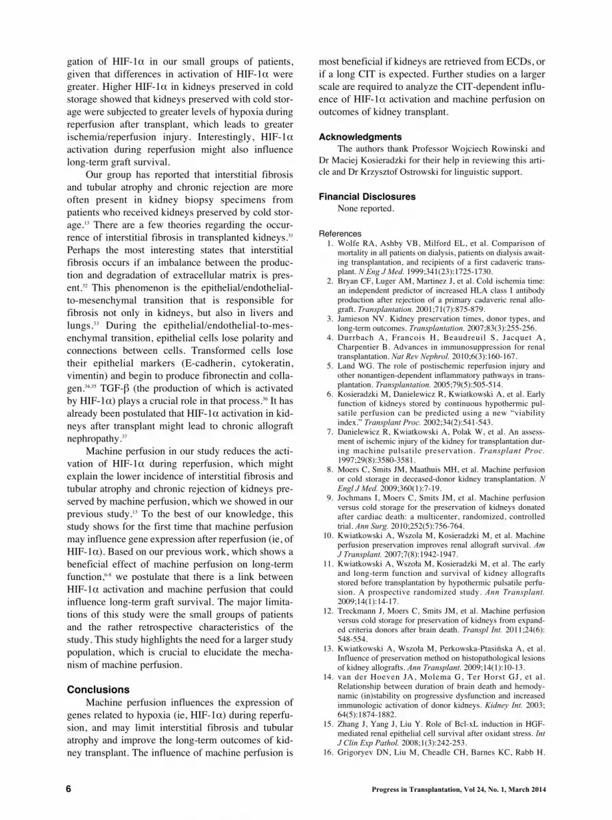
was less than 100.} The 5-year patient survival was96% (66/69). The 5-year survival in BDD kidney graftrecipients was 95% (59/62) and 100% (7/7) in LRDkidney graft recipients. Kidney graft survival of alltransplant patients was 83% (57/69). Graft survivalwas 82% (51/62) from BDDs, whereas graft survivalwas 86% (6/7) from LRDs. Five-year kidney graft sur-vival was better in the machine perfusion groups thanin the cold storage groups (88% [23/26] vs 78%[28/36], respectively), although the data were not sig-nificantly different. Five-year graft survival in each ofthe groups was 81% (22/27) for the CS-SCD group,67% (6/9) for the CS-ECD group, 90% (9/10) for theMP-SCD group, 88% (14/16) for the MP-ECD group,and 86% (6/7) for the LRD group (Fig. 2).{AUTHOR: Only one figure was provided, soshould not preceding citation say “(see Figure)”?}Statistical significance was reached in graft survivalbetween the CS-ECD and the MP-ECD groups andbetween the CS-ECD and the MP-SCD groups. Mean(SD) for serum creatinine levels 5 years after trans-plant were 1.54 (1.6), 1.39 (1.2), 1.61 (2), 1.74 (1.5),and 1.55 (1) in the CS-SCD, CS-ECD, MP-SCD, MP-ECD, and LRD groups, respectively (not significantlydifferent).
Analysis of Graft LossAfter 5 years of observation, 11 of the 69 grafts
were lost. In the MP-SCD group, 1 patient died ofischemic heart failure with a functioning graft. In theMP-ECD group, 2 patients lost grafts: 1 died becauseof infection during the deterioration of graft functionwhereas the second graft failure was due to interstitialfibrosis and tubular atrophy. In the CS-ECD group, 3patients lost their grafts: 1 because of infection, 1 diedof ischemic heart failure due to a dysfunctioning graft,and 1 because of interstitial fibrosis and tubular atro-phy. In the CS-SCD group, 5 patients lost their grafts:1 because of arteriosclerosis, 2 because of chronicglomerulopathy, and 2 because of interstitial fibrosisand tubular atrophy (1 also had signs of toxic effectsof calcineurin inhibitors). Overall, interstitial fibrosisand tubular atrophy was diagnosed in 11 lost grafts in4 different cases. Analysis of the mean HIF-1" acti-vation in the group of kidneys with interstitial fibrosisand tubular atrophy yielded 1.84 (0.88) versus 0.69(0.88) in the rest of the surviving population but didnot reach statistical significance (P = .09).
DiscussionWe investigated the possible interaction between
methods of preservation before transplant and theexpression of selected genes after reperfusion. Thistask was difficult because gene expression can beinfluenced by many factors. Pneumoperitoneum,which is performed {AUTHOR:
Pneumoperitoneum is not an action (verb) andthus cannot be performed. Please rephrase.Perhaps “created”?} during living donor nephrecto-my, may influence more than 600 genes.22 We focusedon genes related to ischemia/reperfusion injury due tohypoxia such as HIF-1", HO-1, VEGF, and IL-1!.Living kidney donations served as a control group.Our primary question was whether there was a linkbetween the method of preservation and the expres-sion of genes associated with hypoxia (which mayinfluence ischemia/reperfusion injury), and howmachine perfusion might benefit early and long-termsurvival of kidney grafts.
The effects of machine perfusion on early andlong-term results have previously been described inseveral studies by our group6,7,9,10,13 and by otherauthors.11,23 In the present study, we discovered a sta-tistically significant difference in HIF-1" geneexpression that was dependent upon both the methodof preservation and type of donor. We did not find asignificant difference in the activation of other genes(VEGF, HO-1, or IL-1!). These results are generallyconsistent with results obtained by Lemos et al,24 whoobserved that HIF-1" activation is significantly lowerin kidneys retrieved from living donors than in kid-neys procured from deceased donors and kept in coldstorage. In contrast, they reported that VEGF and HO-1 activation were significantly higher in kidneysretrieved from living donors. We did not find the samecorrelation. Furthermore, data from other groups showthat VEGF activation is dependent on HIF-1",25,26
which means that high VEGF levels should be pre-ceded by HIF-1" activation. As described by otherinvestigators in experimental models, activation ofHIF-1" during hypoxia may play a positive role inovercoming oxidative stress.27,28 The possible clinicaluse of HIF-1" stabilization agents to prevent chronickidney disease or acute kidney injury has also beeninvestigated.
Enhanced activation of VEGF, TGF-!, andhuman major histocompatibility complex class I-relat-ed chain A29 may play protective roles under stressfulconditions. Although these mechanisms might behelpful during acute episodes of hypoxia, they maynot function under posttransplant conditions.Additionally, HIF-1"-dependent growth factor activa-tion might be harmful and undesirable during kidneytransplant and in the presence of foreign immunologi-cal tissues. Higher HIF-1" activation correlates withlonger CIT, older donors, and DGF.30 In our study,CIT was longer than 24 hours because of an organiza-tional issue related to kidney/recipient transportationfor transplant and the time needed for tissue typing.Longer CIT adversely affects the results of transplant,which is reflected in the high percentage of DGF in allgroups of our study. However, it allowed an investi-
6 Progress in Transplantation, Vol 24, No. 1, March 2014
gation of HIF-1" in our small groups of patients,given that differences in activation of HIF-1" weregreater. Higher HIF-1" in kidneys preserved in coldstorage showed that kidneys preserved with cold stor-age were subjected to greater levels of hypoxia duringreperfusion after transplant, which leads to greaterischemia/reperfusion injury. Interestingly, HIF-1"activation during reperfusion might also influencelong-term graft survival.
Our group has reported that interstitial fibrosisand tubular atrophy and chronic rejection are moreoften present in kidney biopsy specimens frompatients who received kidneys preserved by cold stor-age.13 There are a few theories regarding the occur-rence of interstitial fibrosis in transplanted kidneys.31
Perhaps the most interesting states that interstitialfibrosis occurs if an imbalance between the produc-tion and degradation of extracellular matrix is pres-ent.32 This phenomenon is the epithelial/endothelial-to-mesenchymal transition that is responsible forfibrosis not only in kidneys, but also in livers andlungs.33 During the epithelial/endothelial-to-mes-enchymal transition, epithelial cells lose polarity andconnections between cells. Transformed cells losetheir epithelial markers (E-cadherin, cytokeratin,vimentin) and begin to produce fibronectin and colla-gen.34,35 TGF-! (the production of which is activatedby HIF-1") plays a crucial role in that process.36 It hasalready been postulated that HIF-1" activation in kid-neys after transplant might lead to chronic allograftnephropathy.37
Machine perfusion in our study reduces the acti-vation of HIF-1" during reperfusion, which mightexplain the lower incidence of interstitial fibrosis andtubular atrophy and chronic rejection of kidneys pre-served by machine perfusion, which we showed in ourprevious study.13 To the best of our knowledge, thisstudy shows for the first time that machine perfusionmay influence gene expression after reperfusion (ie, ofHIF-1"). Based on our previous work, which shows abeneficial effect of machine perfusion on long-termfunction,6-8 we postulate that there is a link betweenHIF-1" activation and machine perfusion that couldinfluence long-term graft survival. The major limita-tions of this study were the small groups of patientsand the rather retrospective characteristics of thestudy. This study highlights the need for a larger studypopulation, which is crucial to elucidate the mecha-nism of machine perfusion.
ConclusionsMachine perfusion influences the expression of
genes related to hypoxia (ie, HIF-1") during reperfu-sion, and may limit interstitial fibrosis and tubularatrophy and improve the long-term outcomes of kid-ney transplant. The influence of machine perfusion is
most beneficial if kidneys are retrieved from ECDs, orif a long CIT is expected. Further studies on a largerscale are required to analyze the CIT-dependent influ-ence of HIF-1" activation and machine perfusion onoutcomes of kidney transplant.
AcknowledgmentsThe authors thank Professor Wojciech Rowinski and
Dr Maciej Kosieradzki for their help in reviewing this arti-cle and Dr Krzysztof Ostrowski for linguistic support.
Financial DisclosuresNone reported.
References1. Wolfe RA, Ashby VB, Milford EL, et al. Comparison of
mortality in all patients on dialysis, patients on dialysis await-ing transplantation, and recipients of a first cadaveric trans-plant. N Eng J Med. 1999;341(23):1725-1730.
2. Bryan CF, Luger AM, Martinez J, et al. Cold ischemia time:an independent predictor of increased HLA class I antibodyproduction after rejection of a primary cadaveric renal allo-graft. Transplantation. 2001;71(7):875-879.
3. Jamieson NV. Kidney preservation times, donor types, andlong-term outcomes. Transplantation. 2007;83(3):255-256.
4. Durrbach A, Francois H, Beaudreuil S, Jacquet A,Charpentier B. Advances in immunosuppression for renaltransplantation. Nat Rev Nephrol. 2010;6(3):160-167.
5. Land WG. The role of postischemic reperfusion injury andother nonantigen-dependent inflammatory pathways in trans-plantation. Transplantation. 2005;79(5):505-514.
6. Kosieradzki M, Danielewicz R, Kwiatkowski A, et al. Earlyfunction of kidneys stored by continuous hypothermic pul-satile perfusion can be predicted using a new “viabilityindex.” Transplant Proc. 2002;34(2):541-543.
7. Danielewicz R, Kwiatkowski A, Polak W, et al. An assess-ment of ischemic injury of the kidney for transplantation dur-ing machine pulsatile preservation. Transplant Proc.1997;29(8):3580-3581.
8. Moers C, Smits JM, Maathuis MH, et al. Machine perfusionor cold storage in deceased-donor kidney transplantation. NEngl J Med. 2009;360(1):7-19.
9. Jochmans I, Moers C, Smits JM, et al. Machine perfusionversus cold storage for the preservation of kidneys donatedafter cardiac death: a multicenter, randomized, controlledtrial. Ann Surg. 2010;252(5):756-764.
10. Kwiatkowski A, Wszola M, Kosieradzki M, et al. Machineperfusion preservation improves renal allograft survival. AmJ Transplant. 2007;7(8):1942-1947.
11. Kwiatkowski A, Wszo(a M, Kosieradzki M, et al. The earlyand long-term function and survival of kidney allograftsstored before transplantation by hypothermic pulsatile perfu-sion. A prospective randomized study. Ann Transplant.2009;14(1):14-17.
12. Treckmann J, Moers C, Smits JM, et al. Machine perfusionversus cold storage for preservation of kidneys from expand-ed criteria donors after brain death. Transpl Int. 2011;24(6):548-554.
13. Kwiatkowski A, Wszo(a M, Perkowska-Ptasi)ska A, et al.Influence of preservation method on histopathological lesionsof kidney allografts. Ann Transplant. 2009;14(1):10-13.
14. van der Hoeven JA, Molema G, Ter Horst GJ, et al.Relationship between duration of brain death and hemody-namic (in)stability on progressive dysfunction and increasedimmunologic activation of donor kidneys. Kidney Int. 2003;64(5):1874-1882.
15. Zhang J, Yang J, Liu Y. Role of Bcl-xL induction in HGF-mediated renal epithelial cell survival after oxidant stress. IntJ Clin Exp Pathol. 2008;1(3):242-253.
16. Grigoryev DN, Liu M, Cheadle CH, Barnes KC, Rabb H.

7 Progress in Transplantation, Vol 24, No. 1, March 2014
Genomic profiling of kidney ischemia ischemia-reperfusionreveals expression of specific alloimmunity-associated genes:linking Linking “immune” and “non-immune” injury events.Transplant Proc. 2006;38(10):3333-3336.
17. Naesens M, Li L, Ying L, et al. Expression of complementcomponents differs between kidney allografts from living anddeceased donors. J Am Soc Nephrol. 2009;20(8):1839-1851.
18. Semenza GL. Targeting HIF-1 for cancer therapy. Nat RevCancer. 2003;3(10):721-732.
19. Semenza GL, Wang GL. A nuclear factor induced by hypox-ia via de novo protein synthesis binds to the human erythro-poietin gene enhancer at a site required for transcriptionalactivation. Moll Cell Biol. 1992;12(12):5447-5454.
20. Chomczynski P, Sacchi N. Single-step method of RNA isola-tion by acid guanidinum thiocyanate-phenol-chloroformexctraction. Anal Biochem. 1987;162(1):156-159.
21. Kosieradzki M. Wp(yw odpowiedzi zapalnej towarzysz*cej+mierci mózgu, niedokrwienia i zaburze) wynikaj*cych zreperfuzji na czynno+, przeszczepu nerki. Rozprawa habilita-cyjna WUM 2006. http://www.researchgate.net/publica-tion/36118559_Wpyw_odpowiedzi_zapalnej_u_dawcw_zmarych_niedokrwienia_oraz_reperfuzji_na_czynno_allogen-nego_przeszczepu_nerki_. Accessed August 21, 2013.{AUTHOR: Please translate the titel of this articleinto English for the purpose of this reference list.}
22. Kurian SM, Flechner SM, Kaouk J, et al. Laparoscopic donornephrectomy gene expression profiling reveals upregulationof stress and ischemia associated genes compared to controlkidneys. Transplantation. 2005;80(8):1067-1071.
23. Wight JP, Chilcott JB, Holmes MW, et al. Pulsatile machineperfusion vs. cold storage of kidneys for transplantation: arapid and systematic review. Clin Transplant.2003;17(4):293-307.
24. Lemos FBC, Ijzermans JNM, Zondervan PE, et al.Differential expression of heme oxygenase-1 and vascularendothelial growth factor in cadaveric and living donor kid-neys after ischemia-reperfusion. J Am Soc Nephrol. 2003;14(12):3278-3287.
25. Levy AP, Levy NS, Wegner S, Goldberg MA.Transcriptional regulation of the rat vascular endothelialgrowth factor gene by hypoxia. J Biol Chem.1995;270(22):13333-13340.
26. Pugh CW, Ratcliffe PJ. Regulation of angiogenesis byhypoxia: role of the HIF system. Nat Med. 2003;9(6):677-684.
27. Rosenberger C, Rosen S, Shina A, et al. Activation of hypox-ia-inducible factors ameliorates hypoxic distal tubular injuryin the isolated perfused rat kidney. Nephrol Dial Transplant.2008;23(11):3472-3478. Epub 2008 May 29.
28. Weidemann A, Bernhardt WM, Klanke B, et al. HIF activa-tion protects from acute kidney injury. J Am Soc Nephrol.2008;19(3):486-494.
29. Luo L, Lu J, Wei L, et al. The role of HIF-1 in up-regulatingMICA expression on human renal proximal tubular epithelialcells during hypoxia/reoxygenation. BMC Cell Biol.2010;11:91.
30. Rosenberger C, Pratschke J, Rudolph B, et al.Immunohistochemical detection of hypoxia-inducible factor-1" in human renal allograft biopsies. J Am Soc Nephrol.2007;18(1):343-351.
31. K-dzierska K, Doma)ski M, Sporniak-Tutak K, Do(-gowskaB, Ciechanowski K. Oxidative stress and renal interstitialfibrosis in patients after renal transplantation: current state ofknowledge. Transplant Proc. 2011;43(10):3577-3583.
32. Iwano M, Neilson EG. Mechanisms of tubulointerstitialfibrosis. Curr Opin Nephrol Hypertens. 2004;13(3):279-284.
33. Guarino M, Tosoni A, Nebuloni M. Direct contribution ofepithelium to organ fibrosis: epithelial-mesenchymal transi-tion. Hum Pathol. 2009;40(10):1365-1376.
34. de Matos AC, Câmara NO, Tonato EJ, et al. Vimentinexpression and myofibroblast infiltration are early markers ofrenal dysfunction in kidney transplantation: an early stage ofchronic allograft dysfunction? Transplant Proc. 2010;
42(9):3482-3488.35. Rastaldi MP, Ferrario F, Giardino L, et al. Epithelial-mes-
enchymal transition of tubular epithelial cells in human renalbiopsies. Kidney Int. 2002;62(1):137-146.
36. Forino M, Torregrossa R, Ceol M, et al. TGF beta1 inducesepithelial-mesenchymal transition, but not myofibroblasttransdifferentiation of human kidney tubular epithelial cellsin primary culture. Int J Exp Pathol. 2006;87(3):197-208.
37. Baan C, van Gelder T, Peeters A, et al. Living kidney donorsand hypoxia-inducible factor-1alpha. Transplantation.2003;75(4):570-571.

8 Progress in Transplantation, Vol 24, No. 1, March 2014

Table 1 Population characteristics
Machine perfusion Cold storage
Expanded criteria Standard criteria Expanded criteria Standard criteriaCharacteristic donor (n = 16) donor (n = 10) donor (n = 9) donor (n = 27)
Donors Age,a mean (SD), y 54.4 (4) 35.1 (10) 61.8 (6) 46.65 (9) Trauma, % 19 (3/16) 30 (3/10) 22 (2/9) 19 (5/27) Hemorrhagic stroke, % 75 (12/16) 50 (5/10) 67 (6/9) 63 (17/27) Days in intensive care unit, mean (SD) 4.4 (2.6) 3.1 (1) 3.77 (2) 4 (2.4) Creatinine,b mean (SD), mg/dL 2.15 (0.98) 1.78 (1.3) 1.18 (0.4) 1.02 (0.4)
Recipients Age, mean (SD), y 50 (16) 39.7 (10) 53.1 (8) 41.6 (12) Male sex, % 50 (8/16) 60 (6/10) 56 (5/9) 59 (16/27) Dialysis before kidney transplant, mean (SD), months 41 (36) 44 (43) 28 (16) 30 (25) Panel-reactive antibodies, mean (SD), % 15 (29) 15.8 (31) 12 (18) 7.5 (20) Maximum last before kidney transplant 6.5 (20) 6.9 (19) 4 (10) 2.6 (9) Human leukocyte antigen mismatch (A, B, DR), mean (SD) 1.95 (1) 2.25 (0.95) 3.45 (0.95) 2.95 (0.98)a For age, a significant difference (P < .05) was found between standard criteria donors who had machine perfusion and extended criteria donors who had cold
storage.b For creatinine level, a significant difference (P < .05) was found between extended criteria donors who had machine perfusion and extended criteria donors who had cold storage, extended criteria donors who had machine perfusion and standard criteria donors who had cold storage, standard criteria donors who had machine perfusion and extended criteria donors who had cold storage, and standard criteria donors who had machine perfusion and standard criteria donors who had cold storage.
AUTHOR: Please provide the actual P values rather than just listing as NS.
Table 2 Perfusion parameters
Machine perfusiona
Standard criteria Extended criteriaParameter donors (n = 16) donors (n = 10) P
Pressure, mm Hg 0 hour 33 (4) 36 (1) NS 4th hour 31 (1) 32 (2) NS 12th hour 30 (1.5) 32 (1.5) NS 24th hour 32 (2) 33 (2) NS
Flow, mL/min 0 hour 88 (45) 70 (43) NS 4th hour 123 (43) 107 (42) NS 12th hour 147 (59) 140 (60) NS 24th hour 159 (61) 187 (60) NS
Renal resistance index 0 hour 0.54 (0.32) 0.7 (0.5) NS 4th hour 0.29 (0.16) 0.46 (0.4) .08 12th hour 0.21 (0.14) 0.35 (0.35) NS 24th hour 0.2 (0.16) 0.38 (0.3) NS
Level of lactate dehydrogenase, 4th hour, U/L 529 (400) 985 (398) .01
Level of lactate, 4th hour, mg/dL 13.9 (6.9) 22.98 (6.5) .04
a Values are mean (SD).

Table 3 Mean relative gene expression for hypoxia-induciblefactor subunit 1! in kidney biopsies 30 minutes afterreperfusion
Relative gene expression, Groupa mean (SD)
SCD-CS 0.98 (1.09)
ECD-CS 1.57 (1.11)
SCD-MP 0.28 (0.1)
ECD-MP 0.49 (0.5)
LRD 0.46 (0.63)
Abbreviations: ECD-CS, extended criteria donor, cold storage; ECD-MP, extended criteria donor, machine perfusion; LRD, living related donor; SCD-CS, standard criteria donor, cold storage; SCD-MP, standard criteria donor, machine perfusion.a Gene expression differed significantly (P < .05) between the following groups: SCD-CS vs SCD-MP, SCD-CS vs ECD-MP, SCD-CS vs LRD, ECD-CS vs ECD-MP, ECD-CS vs SCD-MP, and ECD-CS vs. LRD. SCD-MP did not differ significantly from LRD, nor did ECD-MP differ significantly from LRD.
AUTHOR: Can you specify the actual P value for CS-ECD vs MP-SCD, rather thanjust saying that it is <.05?
Figure Five-year graft survival based on perfusion model(cold storage [CS] or machine perfusion [MP]), donor status(extended [ECD] or standard [SCD] criteria), and controlgroup of living related donors (LRD). Significant differenceswere found between CS-ECD and MP-ECD (P = .05) andCS-ECD and MP-SCD (P < .05).
1.00.90.80.70.60.50.40.30.20.10.0
Kidn
ey g
raft
surv
ival
0 500 1000 1500 2000 2500Days after kidney transplant
Kaplan-Meier analysiscomplete + cutoff
CS-SCD CS-ECD MP-SCDMP-ECD LRD


