Embed Size (px)
Citation preview
Send Orders of Reprints at [email protected]
170 Current Vascular Pharmacology, 2013, 11, 170-186
Brain Perfusion In Sepsis
Fabio Silvio Taccone*,1, Sabino Scolletta
1, Federico Franchi
2, Katia Donadello
1 and Mauro Oddo
3
1Department of Intensive Care, Erasme Hospital, Université Libre de Bruxelles (ULB), Route de Lennik, 808, 1070
Brussels, Belgium; 2Department of Anaesthesia and Intensive Care, University of Siena, Viale Bracci, 1, 53100 Siena,
Italy; 3Department of Intensive Care Medicine, Centre Hospitalier Universitaire Vaudois, Rue du Bugnon 21, 1011
Lausanne, Vaud, Switzerland
Abstract: Brain dysfunction is a frequent complication of sepsis, usually defined as “sepsis-associated encephalopathy”
(SAE). Its pathophysiology is complex and related to numerous processes and pathways, while the exact mechanisms pro-
ducing neurological impairment in septic patients remain incompletely elucidated. Alterations of the cerebral blood flow
(CBF) may represent a key component for the development of SAE. Reduction of CBF may be caused by cerebral vaso-
constriction, either induced by inflammation or hypocapnia. Endothelial dysfunction associated with sepsis leads to im-
pairment of microcirculation and cerebral metabolic uncoupling that may further reduce brain perfusion so that CBF be-
comes inadequate to satisfy brain cellular needs. The natural autoregulatory mechanisms that protect the brain from re-
duced/inadequate CBF can be impaired in septic patients, especially in those with shock or delirium, and this further con-
tributes to cerebral ischemia if blood pressure drops below critical thresholds. Sedative agents alter cerebro-vascular reac-
tivity and may significantly reduce CBF. Although disorders of brain perfusion and alteration of CBF and cerebral
autoregulation are frequently observed in humans with sepsis, their exact role in the pathogenesis of SAE remains un-
known. Brain perfusion can further become inadequate due to cerebral microcirculatory dysfunction, as evidenced in the
experimental setting. Microvascular alterations can be implicated in the development of electrophysiological abnormali-
ties observed during sepsis and contribute to neurological alterations in septic animals. The aim of this review is to pro-
vide an update on the pathophysiology of brain perfusion in sepsis, with a particular focus on human clinical investigation
and novel tools for CBF monitoring in septic patients.
Keywords: Sepsis, encephalopathy, brain dysfunction, cerebral hemodynamics, autoregulation, cerebral blood flow, carbon dioxide, microcirculation.
1. BRAIN DYSFUNCTION DURING SEPSIS
1.1. Definition of Sepsis-Associated Encephalopathy (SAE)
Septic shock and related multi-organ failure (MOF) re-main a major cause of morbidity and mortality in intensive care units (ICUs) worldwide [1]. The infectious stimuli, as-sociated with a widespread reaction characterized by the release of numerous circulating pro-inflammatory molecules, can potentially impair the function of several organs [2]. Brain dysfunction occurs early during sepsis and is com-monly characterized by the development of an altered mental state; however, cerebral abnormalities are also described in late course of sepsis, often accompanied by MOF, hypoten-sion and other systemic events [3, 4]. Hippocrates first re-ported the association between infections and cerebral dysfunction more than 2500 years ago [5], and sir William Osler also described, later on, the occurrence of “delirium” in patients with ongoing sepsis [6]. Nevertheless, concomi-tant hepatic or renal failure, electrolyte and metabolic distur-bances, altered glucose homeostasis, hypotension,
*Address correspondence to this author at the Department of Intensive Care,
Erasme University Hospital, Université Libre de Bruxelles, ULB), Route de
Lennik, 808, 1070 Brussels, BELGIUM; Tel: +322 555 5587;
Fax: +322 555 4698; E-mail: [email protected]
hypoxemia, hypothermia or neurological side-effects of differ-
ent pharmacological agents may concomitantly occur in septic patients, rendering the discrimination between sepsis-related brain dysfunction and encephalopathy from other causes a potentially difficult task [7].
Sepsis-associated encephalopathy (SAE) can be defined as a diffuse or multifocal brain dysfunction associated with an infectious illness (a) without clinical and laboratory evidence of intracranial infection and/or (b) without conditions unre-
lated to the infectious process that would significantly alter brain function [8]. The diagnosis should therefore exclude structural brain lesions or primary pathologies of the central nervous system (i.e. meningitis or stroke), other neurological
diseases (i.e. epilepsy), a toxic-metabolic cause, fat embolism syndrome and anoxic brain injury [9]. Moreover, acute in-flammatory encephalopathies, such as acute disseminated en-cephalomyelitis or acute hemorrhagic leucoencephalopathy,
should be also considered apart from SAE, because of their specific immuno-mediated inflammation of central nervous system (CNS) structures and response to corticosteroids [10].
1.2. Epidemiology, Clinical Features and Diagnosis
The occurrence of SAE is variable but is one of the most common forms of encephalopathy encountered in critically
1875-6212/13 $58.00+.00 © 2013 Bentham Science Publishers
Cerebral Hemodynamics in Sepsis Current Vascular Pharmacology, 2013, Vol. 11, No. 2 171
ill patients [11, 12]. In a large cohort of mechanically venti-lated patients, mostly septic or with acute pneumonia, altered mental state was observed in 66% of them at some point during the ICU stay [11]. In a prospective study of 1758 pa-tients admitted to a medical ICU, 217 of them experienced a neurological complication; encephalopathy was present in one third of patients, and SAE was the most frequent etiol-ogy [12]. In a series on 69 non-sedated patients with bacter-aemia, 70% of them had clinical signs of brain dysfunction and half of them showed severe encephalopathy [9]. In two large cohort studies of septic patients, acute mental state ab-normalities were described in more than 20% of them [13]. The heterogeneity of SAE rate among these studies may probably be due to the different definitions of sepsis and SAE used. As such, more than 60% of patients with neuro-logical changes during critical illness patients also had evi-dence of brain lesions abnormalities on brain magnetic reso-nance imaging [14], while abnormalities of electroencephalo-graphy (EEG), including clinically silent epileptiform pat-terns, were present in all septic patients, regardless of the presence of clinical brain dysfunction [15, 16].
The most common manifestation of SAE is an alteration of mental state, ranging, from mild disorientation or lethargy to stupor and coma [17]. Most patients have fluctuant changes in mental status, especially in the early course of the infectious process where SAE may often be the first manifes-tation of severe sepsis [18]. Usually the neurological exam does not show focal motor deficits. Myoclonus, asterixis and rigidity are rare and should raise the possibility of a toxic-metabolic encephalopathy. Focal asymmetric neurological signs mandate to rule out structural brain lesions, either pri-mary or secondary to septic ischemic emboli. Cranial nerves are usually spared whereas clinical signs of peripheral nerve alterations, like loss of tendon reflexes, should raise the pos-sibility of sepsis-associated critically ill polyneuromyopathy [19]. Finally, alteration of the neuro-endocrine axis (e.g. ad-renal and vasopressin insufficiency) and autonomic dysfunc-tion (e.g. blood pressure variations, arrhythmias, irregular breathing) may be other manifestations of SAE [20]. Given the variable clinical manifestations of SAE, the Confusion Assessment Method for the Intensive Care Unit (CAM-ICU) or the Assessment to Intensive Care Environment (ATICE) have been used to score altered mental state in this setting, however the Glasgow Coma Scale (GCS) remains the most popular tool and has been used to evaluate brain dysfunction in septic patients with MOF [3, 21, 22].
There are no diagnostic tests with high specificity for SAE, so that the clinical, electrophysiological (Electroen-cephalogram, EEG; Somato Sensory Evoked Potentials, SSEPs), biochemical (neuron-specific enolase, NSE; S-100 protein) or imaging (Magnetic Resonance Imaging, MRI) criteria may be useful. Young et al. demonstrated that EEG is the most sensitive in the diagnosis of SAE, with mild dif-fuse slow waves even in patients without clinical abnormali-ties [15]. Also, EEG could be useful in the differential diag-nosis of coma in critically ill septic patients, especially to detect unexpected clinically silent non-convulsive status epi-lepticus [23]; in a large cohort of patients continuously moni-tored with EEG, septic patients had a higher rate of seizures than those without sepsis (32% vs. 9%) and sepsis on admis-sion was the only significant predictor of seizures and of poor outcome [24]. In one study using SSEPs, an increase of
peak latencies in cortical and sub-cortical component of dor-sal column-medial lemniscus pathway (84% and 34% of all cases) was remarked. The impairment of these pathways was associated with severity of illness and had good correlation with the degree of brain dysfunction [25]. Finally, serum levels of NSE and S-100 protein have been shown to corre-late with poor outcome in septic shock patients [26]. Mag-netic resonance imaging (MRI) showed variable degrees of vasogenic edema, related to blood-brain barrier (BBB) breakdown, or ischemic lesions surrounding the Virchow-Robin spaces in septic brain [27]. Damage to the gray matter may include bilateral lesions of basal ganglia and thalamus. Although not specific for brain dysfunction during sepsis, MRI can identify patients developing structural cerebral le-sions during severe sepsis and septic shock and potentially help in patient prognostication.
1.3. Outcome and Therapy
Septic encephalopathy is not only an unpleasant confu-sion state but represents a severe organ dysfunction and may contribute to poor outcome in septic patients [3, 13, 28]. The greatest the severity of SAE, as assessed by the GCS, the highest the risk of death, with a mortality rate of 16% when GCS was 15, 20% when GCS was 13-14, 50% when GCS was 9-12, up to 63% in comatose patients (GCS < 9) [3]. In another study, septic patients with altered mental status had a higher mortality than those without [13]. In a large cohort of mechanically ventilated patients, the majority of them admit-ted for severe sepsis, it was demonstrated that ICU-related delirium was independently associated with longer hospital stay, worse cognitive recovery and higher mortality [11]. It appears that SAE may also actively participate to the devel-opment of long-term cognitive dysfunction after critical ill-ness [29]. Unfortunately, there is no specific treatment of SAE and outcome depends on the appropriate treatment of the underlying disease. Recently, the use of activated protein C appeared to be beneficial on the evolution of SAE in septic patients [30]. Several drugs, including dopexamine, inhibi-tors of inducible nitric oxide synthase (iNOS), corticoster-oids, magnesium and antioxidants, have shown promising results in experimental sepsis, however no clinical data on the efficacy of these interventions are currently available [31-34]. Given the absence of specific therapeutic interven-tions, treatment of SAE is mainly supportive, aimed to limit SAE-related secondary cerebral damage. Targeting brain perfusion and providing adequate oxygen and nutrients sup-ply seem reasonable in this context.
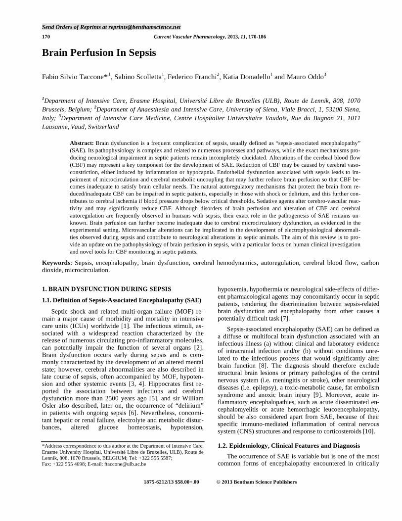
2. PATHOGENESIS OF SAE: A SHORT OVERVIEW
Sepsis-associated encephalopathy is a poorly understood condition and little is known about this clinical process. The pathophysiology is likely to be multifactorial (Fig. 1) [35]. The concept of altered brain function related to the presence in the blood, and possibly in the brain, of micro-organisms or their toxins is considered obsolete as altered CNS function is observed also in patients without bacterial blood stream in-vasion [36]. Disseminated cerebral microabscesses have been suggested as a cause of SAE [37], although this has not been reported in more recent post-mortem studies [38]. The specific role of specific bacterial products like endotoxin is unlikely, as the incidence of SAE is similar in Gram-positive
172 Current Vascular Pharmacology, 2013, Vol. 11, No. 2 Taccone et al.
and Gram-negative bacteremia, as well as fungemia and even sepsis with unidentified pathogens [13]. Encephalopathy occurs also in non-infectious conditions, such as pancreatitis or trauma, suggesting that it would be mostly related to the systemic inflammatory response [39, 40].
The role of inflammation on brain dysfunction has been widely investigated in several in vitro or experimental mod-els of sepsis; although some of the reported findings have also been confirmed in the human setting, the pathogenesis of brain dysfunction mostly relies on laboratory data. Circu-lating pro-inflammatory mediators can promote vascular permeability of the cerebral endothelium, which separates the blood from the brain parenchyma, thus permitting to sev-eral substances to exert toxic effects on neuronal cells. Tu-mor necrosis factor-alpha (TNF- ) selectively altered BBB permeability in brain microvessel endothelial cells without disrupting tight junctions [41]. Endothelial cells treated with interferon-gamma (IFN- ) also exhibited significant morpho-logical changes with increased permeability [42]. Inflamma-
tory mediators have also direct effects on brain functions; the subarachnoid injection of TNF- altered cerebral metabo-lism and increased cerebrospinal fluid lactate, by activating the production of nitric oxide (NO) [43]. Also, TNF- can upregulate the expression of aquaporin-4 channels and asso-ciated edema as well as astrocytosis through activation of its receptor [44]. The intra-cerebral administration of interleu-kin-1 and IFN- in animals induced the same slow-waves EEG patterns than those observed in patients with SAE [45, 46].
More importantly, sepsis-induced systemic inflammation can directly affect brain homeostasis by triggering the two important pathways that are implicated in the response to stress and in the immune system modulation: the circumven-tricular organs (CVOs), which lack a BBB and have a direct communication with circulating mediators of sepsis and the vagus nerve, which is triggered by visceral inflammation [7]. Once systemic and/or visceral inflammation are detected by these pathways, an abnormal activation of brain signaling
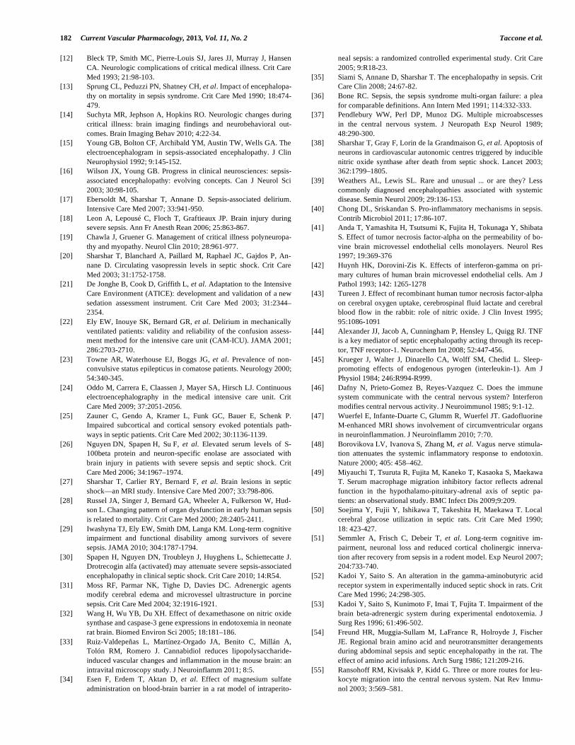
Fig. (1). Schematic representation of mechanisms involved in the pathogenesis of sepsis-associated encephalopathy. Proinflammatory cyto-
kines are released during sepsis. They can either activate vagal fibres or enter the brain causing neurologic dysfunction (see text for details).
5-HT, serotonin (5-hydroxytryptamine); Ach, acetylcholine; BBB, blood-brain barrier; CNS, central nervous system; CO, carbon monoxide;
CVOs = circumventricular organs; ECs, endothelial cells; GABA, gamma-aminobutyric acid; GLT, glutamate; HPA, hypothalamic-pituitary-
adrenocortical; HSF, heat-shock factors; IL, interleukin; LPS, lipopolysaccharide; NF-k , nuclear factor kappa B; NO, nitric oxide; NTS,
nucleus tractus solitarius; PAF, platelet activating factor; PG, prostaglandin; PVN, paraventricular nuclei; RAS, reticular activation system;
ROS, reactive oxygen species; TNF- , tumor necrosis factor-alpha.
Cerebral Hemodynamics in Sepsis Current Vascular Pharmacology, 2013, Vol. 11, No. 2 173
spread to behavioural, neuroendocrine and neurovegetative structures, affecting directly microglial and neuronal cells functions and modulating neurosecretion and neurotrasmis-sion [47, 48]. Inflammation rapidly alters the function of hypothalamus, which regulates the endogenous production of corticosteroids and can modulate the inflammatory response [49] and may significantly impair the reticular activating system functions, which control consciousness and attention [50]. The cholinergic and serotoninergic releases are altered, while an increase in the -aminobutyric acid receptor density is observed in the forebrain of septic rats [51, 52]. Brain tis-sular norepinephrine and epinephrine concentrations are de-creased in the forebrain and brain stem of septic animals together with the down-regulation of the cerebral -adrenergic system [53]. Also, a significant and early increase of plasma aromatic aminoacids during abdominal murine sepsis was found to be associated with encephalopathy de-velopment [54]. The activation of CVO also promotes the entry of leucocytes into the CNS, with further increase of local inflammation [55].
Several other mechanisms may be involved in the devel-opment of SAE. Sepsis produces an imbalance between the production of reactive oxygen species (ROS) and the corre-sponding repair system, the so-called oxidative stress. Reac-tive oxygen species trigger lipid peroxidation in the cerebral vessels and the surrounding parenchyma, which eventually cause structural membrane damage and promote inflamma-tion [56]. Cerebral oxidative stress can also be triggered by the decrease of heat-shock factors or by hyperglycemia [57, 58]. Bacterial endotoxin and inflammatory cytokines in-crease cerebral tissue levels of glutamate via the up-regulation of iNOS; glutamate can trigger brain cells damage by allowing high intracellular levels of calcium and the acti-vation of several enzymes that alter cell structures, such as components of the cytoskeleton, membrane, and DNA [59]. Within the brain parenchyma, the activation of astrocytes via the toll-like receptors by the mediators of inflammation [60] can further increase the vulnerability of neurons to glutamate and free radical-mediated injuries [56, 61, 62]. Moreover, sepsis also alters the synthesis of ascorbate, which may pro-vide antioxidant and protective effects on neurons in re-sponse to glutamate and ROS release [63]. The complement system, which normally contributes to eliminate bacteria, may be excessively activated during sepsis and have delete-rious effect through the activation of glial cells, secretion of proinflammatory cytokines and generation of other toxic products on brain function [64]. Edema formation could be further promoted by the activation of aquaporin channels, which regulate the presence of water in the brain tissue and may altered during sepsis [65]. Alterations in the glucose uptake and metabolism or regional deregulation of intracellu-lar calcium homeostasis have also been suggested to contrib-ute to the pathogenesis of SAE [50, 66]. All these pathways cause mitochondrial dysfunction by inhibiting the mitochon-drial electron transport chain and uncoupling oxidative phos-phorylation, which ultimately leads to bioenergetic failure and apoptosis of brain cells [38, 58].
The complex signaling network that is implicated in these phenomena include the production of nitric oxide (NO), through the activation of the inducible form of NO synthase (iNOS) [67, 68], the release of pro-inflammatory
cytokines and their receptors from neurons, astrocytes and microglial cells [69, 70], as well as the production of prosta-glandins, which are key mediators in the brain response to inflammatory stimuli [71, 72]. Finally, a number of other mediators are involved in the cerebral brain response to sys-temic inflammation, such as chemokines, macrophage migrating inhibitory factor, angiotensin, endothelin, platelet activating factor, superoxide radicals and carbon monoxide [73].
3. ALTERATIONS IN BRAIN PERFUSION DURING SEPSIS
A decrease in brain perfusion has been evoked as a major determinant of SAE [74]. Alterations of systemic blood flow, associated with tissue hypo-perfusion and poor oxygen dis-tribution, are a key feature of sepsis [2]; indeed, several stud-ies have tried to evaluate the link between cerebral perfusion abnormalities and brain dysfunction in sepsis. As brain per-fusion is dependent, amongst all, from mean arterial pressure (MAP), which is generally reduced in severe sepsis and sep-tic shock, it would be important to monitor the adequacy of cerebral blood flow (CBF) and oxygenation during sepsis as well as to evaluate the integrity of flow regulation, in order to adapt blood pressure levels and avoid the development of secondary brain ischemic events in such patients [38].
3.1. Cerebral Blood Flow: Normal Ranges, Physiology
and Regulation
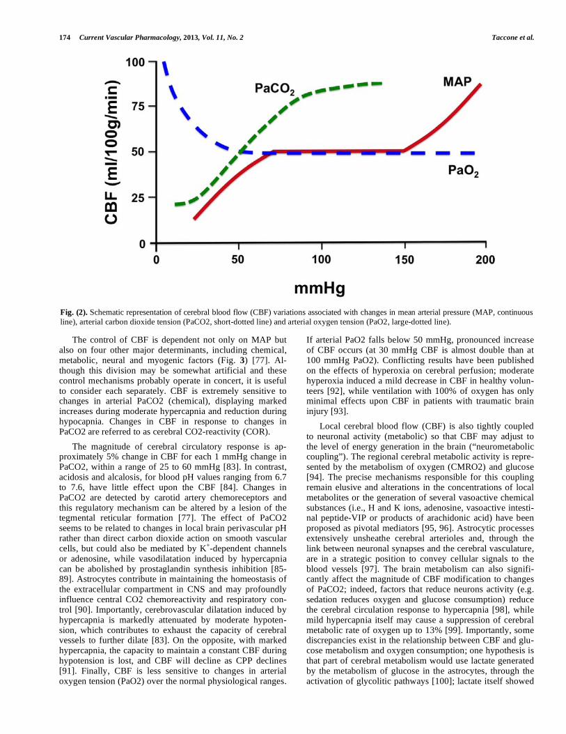
Cerebral blood flow is the blood supply to the brain in a given time. In an adult, CBF is typically 750 mL (or 50-55 mL/100 g of brain tissue/min) and represents roughly 15% of the cardiac output. It ranges from 20 mL/100g/min in the white matter to more than 70 mL/100g/min in the grey mat-ter and is tightly regulated to meet cerebral metabolic de-mands [75]. Cerebral blood flow regulation is extremely complex and is determined by a number of factors, such as viscosity of blood, the diameter of cerebral blood vessels and the net pressure of the blood flow into the brain, known as cerebral perfusion pressure (CPP), which is calculated as the difference between mean arterial pressure (MAP) and intrac-ranial pressure (ICP) or the central venous pressure (CVP), whichever is the higher [76]. Cerebral vessels are able to maintain CBF constant through a wide range of MAP (from 50 to 150 mmHg) by altering their diameters in a process called “cerebral autoregulation” (CA); in normal conditions, a raise of MAP induce vasoconstriction whereas a reduction of MAP produces vasodilatation (Fig. 2) [77]. This process is extremely important because when CBF exceed cellular requirements (a condition known as hyperemia), then the intracranial pressure (ICP) may rise and potentially damage brain tissue. On the other hand, low CBF (<18-20 mL/100g/min) is responsible for brain ischemia and tissue death when CBF falls below 8-10 mL/100g/min. Systemic administration of vasopressors would not produce dramatic effects on CBF, provided the BBB is not altered [78]. Pres-sure autoregulation is thought to be controlled by the baro-ceptive reflexes and both the upper and the lower limits of CA can be affected by many factors, including sympathetic nerve activity, arterial carbon dioxide tension (PaCO2) and pharmacologic agents [79-82].
174 Current Vascular Pharmacology, 2013, Vol. 11, No. 2 Taccone et al.
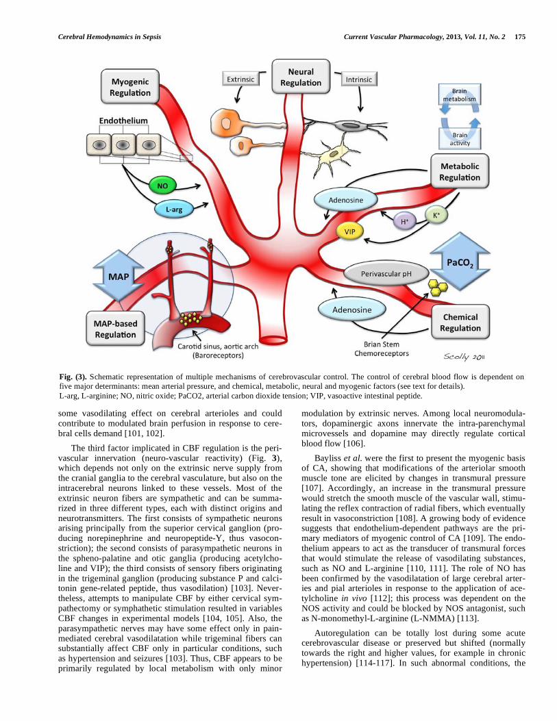
The control of CBF is dependent not only on MAP but also on four other major determinants, including chemical, metabolic, neural and myogenic factors (Fig. 3) [77]. Al-though this division may be somewhat artificial and these control mechanisms probably operate in concert, it is useful to consider each separately. CBF is extremely sensitive to changes in arterial PaCO2 (chemical), displaying marked increases during moderate hypercapnia and reduction during hypocapnia. Changes in CBF in response to changes in PaCO2 are referred to as cerebral CO2-reactivity (COR).
The magnitude of cerebral circulatory response is ap-proximately 5% change in CBF for each 1 mmHg change in PaCO2, within a range of 25 to 60 mmHg [83]. In contrast, acidosis and alcalosis, for blood pH values ranging from 6.7 to 7.6, have little effect upon the CBF [84]. Changes in PaCO2 are detected by carotid artery chemoreceptors and this regulatory mechanism can be altered by a lesion of the tegmental reticular formation [77]. The effect of PaCO2 seems to be related to changes in local brain perivascular pH rather than direct carbon dioxide action on smooth vascular cells, but could also be mediated by K
+-dependent channels
or adenosine, while vasodilatation induced by hypercapnia can be abolished by prostaglandin synthesis inhibition [85-89]. Astrocytes contribute in maintaining the homeostasis of the extracellular compartment in CNS and may profoundly influence central CO2 chemoreactivity and respiratory con-trol [90]. Importantly, cerebrovascular dilatation induced by hypercapnia is markedly attenuated by moderate hypoten-sion, which contributes to exhaust the capacity of cerebral vessels to further dilate [83]. On the opposite, with marked hypercapnia, the capacity to maintain a constant CBF during hypotension is lost, and CBF will decline as CPP declines [91]. Finally, CBF is less sensitive to changes in arterial oxygen tension (PaO2) over the normal physiological ranges.
If arterial PaO2 falls below 50 mmHg, pronounced increase of CBF occurs (at 30 mmHg CBF is almost double than at 100 mmHg PaO2). Conflicting results have been published on the effects of hyperoxia on cerebral perfusion; moderate hyperoxia induced a mild decrease in CBF in healthy volun-teers [92], while ventilation with 100% of oxygen has only minimal effects upon CBF in patients with traumatic brain injury [93].
Local cerebral blood flow (CBF) is also tightly coupled to neuronal activity (metabolic) so that CBF may adjust to the level of energy generation in the brain (“neurometabolic coupling”). The regional cerebral metabolic activity is repre-sented by the metabolism of oxygen (CMRO2) and glucose [94]. The precise mechanisms responsible for this coupling remain elusive and alterations in the concentrations of local metabolites or the generation of several vasoactive chemical substances (i.e., H and K ions, adenosine, vasoactive intesti-nal peptide-VIP or products of arachidonic acid) have been proposed as pivotal mediators [95, 96]. Astrocytic processes extensively unsheathe cerebral arterioles and, through the link between neuronal synapses and the cerebral vasculature, are in a strategic position to convey cellular signals to the blood vessels [97]. The brain metabolism can also signifi-cantly affect the magnitude of CBF modification to changes of PaCO2; indeed, factors that reduce neurons activity (e.g. sedation reduces oxygen and glucose consumption) reduce the cerebral circulation response to hypercapnia [98], while mild hypercapnia itself may cause a suppression of cerebral metabolic rate of oxygen up to 13% [99]. Importantly, some discrepancies exist in the relationship between CBF and glu-cose metabolism and oxygen consumption; one hypothesis is that part of cerebral metabolism would use lactate generated by the metabolism of glucose in the astrocytes, through the activation of glycolitic pathways [100]; lactate itself showed
Fig. (2). Schematic representation of cerebral blood flow (CBF) variations associated with changes in mean arterial pressure (MAP, continuous
line), arterial carbon dioxide tension (PaCO2, short-dotted line) and arterial oxygen tension (PaO2, large-dotted line).
Cerebral Hemodynamics in Sepsis Current Vascular Pharmacology, 2013, Vol. 11, No. 2 175
some vasodilating effect on cerebral arterioles and could contribute to modulated brain perfusion in response to cere-bral cells demand [101, 102].
The third factor implicated in CBF regulation is the peri-vascular innervation (neuro-vascular reactivity) (Fig. 3), which depends not only on the extrinsic nerve supply from the cranial ganglia to the cerebral vasculature, but also on the intracerebral neurons linked to these vessels. Most of the extrinsic neuron fibers are sympathetic and can be summa-rized in three different types, each with distinct origins and neurotransmitters. The first consists of sympathetic neurons arising principally from the superior cervical ganglion (pro-ducing norepinephrine and neuropeptide-Y, thus vasocon-striction); the second consists of parasympathetic neurons in the spheno-palatine and otic ganglia (producing acetylcho-line and VIP); the third consists of sensory fibers originating in the trigeminal ganglion (producing substance P and calci-tonin gene-related peptide, thus vasodilation) [103]. Never-theless, attempts to manipulate CBF by either cervical sym-pathectomy or symphathetic stimulation resulted in variables CBF changes in experimental models [104, 105]. Also, the parasympathetic nerves may have some effect only in pain-mediated cerebral vasodilatation while trigeminal fibers can substantially affect CBF only in particular conditions, such as hypertension and seizures [103]. Thus, CBF appears to be primarily regulated by local metabolism with only minor
modulation by extrinsic nerves. Among local neuromodula-tors, dopaminergic axons innervate the intra-parenchymal microvessels and dopamine may directly regulate cortical blood flow [106].
Bayliss et al. were the first to present the myogenic basis of CA, showing that modifications of the arteriolar smooth muscle tone are elicited by changes in transmural pressure [107]. Accordingly, an increase in the transmural pressure would stretch the smooth muscle of the vascular wall, stimu-lating the reflex contraction of radial fibers, which eventually result in vasoconstriction [108]. A growing body of evidence suggests that endothelium-dependent pathways are the pri-mary mediators of myogenic control of CA [109]. The endo-thelium appears to act as the transducer of transmural forces that would stimulate the release of vasodilating substances, such as NO and L-arginine [110, 111]. The role of NO has been confirmed by the vasodilatation of large cerebral arter-ies and pial arterioles in response to the application of ace-tylcholine in vivo [112]; this process was dependent on the NOS activity and could be blocked by NOS antagonist, such as N-monomethyl-L-arginine (L-NMMA) [113].
Autoregulation can be totally lost during some acute cerebrovascular disease or preserved but shifted (normally towards the right and higher values, for example in chronic hypertension) [114-117]. In such abnormal conditions, the
Fig. (3). Schematic representation of multiple mechanisms of cerebrovascular control. The control of cerebral blood flow is dependent on
five major determinants: mean arterial pressure, and chemical, metabolic, neural and myogenic factors (see text for details).
L-arg, L-arginine; NO, nitric oxide; PaCO2, arterial carbon dioxide tension; VIP, vasoactive intestinal peptide.
176 Current Vascular Pharmacology, 2013, Vol. 11, No. 2 Taccone et al.
upper and lower ranges of MAP/CPP or PaCO2, among which CBF remain constant, can be much narrower than in normal conditions. Such ranges may be different among pa-tients and change over time within the same subject, so that optimal MAP targets to maintain adequate brain perfusion may be difficult to predict. In sepsis, several mechanisms are implicated in altered brain perfusion and CBF regulation in septic patients.
3.2. Effects of Endothelial Dysfunction and Inflammation
on Brain During Sepsis
Alterations of brain perfusion and microcirculation dur-ing sepsis are mostly mediated by the changes induced by systemic inflammation on cerebral endothelial cells [118]. In normal conditions, the cerebral endothelium has tight inter-cellular junctions, no fenestrations and few pynocytic vesicules, which separate blood from the brain parenchyma. The abnormal brain stimulation occurring during sepsis can activate the endothelium [119, 120], causing the expression of adhesion molecules with subsequent aggregation of circu-lating white blood cells and increased permeability to vari-ous neurotoxic agents [121]. The breakdown of the endothe-lial barrier during sepsis has been directly demonstrated by the intracerebral accumulation of radioactive tracers in sev-eral experimental studies [119]. The activation of cerebral endothelial cells is mediated by endotoxin and pro-inflammatory cytokines that, through activation of nuclear factor (NF)-kB, induce the over-expression of iNOS that further increases BBB permeability [122, 123]. This would also allow high brain concentrations of vasoactive agents, acting directly on intracerebral vessels and inducing vaso-constriction and reduction of CBF [124]. Reduction of local blood flow as well as impairment of microcirculation can contribute to leucocytes accumulation and local production of lysosomial enzymes and oxygen free radicals [125]. Reac-tive products of leukocytes can interact with erythrocytes membrane and reduce their deformability, which in turn fa-cilitates red blood cells impaction in microvessels and exac-erbates cerebral hypoperfusion [126]. The result of all these events is the development of perivascular edema that con-tributes to altered brain perfusion and is associated with neu-ronal injury [127, 128].
Low brain flow levels are a feature of the response to endotoxin [129]. Early reductions of CBF during experimen-tal endotoxemia are due to cerebral vasoconstriction, both in newborn and adult animals, and these changes are not linked to hypotensive events [129, 130]. Importantly, endotoxin can suppress neuronal activity and cerebral metabolic rate [131, 132]; thus, as cerebral blood flow remains closely coupled to cerebral metabolic requirements, a concomitant reduction of brain perfusion and metabolic rates have been observed in experimental sepsis [133]. Elevated levels of TNF- occur-ring during endotoxemia have powerful vasoactive effects on cerebral vessels [134]. Injection of TNF- into the cisterna magna of rabbits produced a rapid reduction in cerebral oxy-gen uptake and a more prolonged reduction in CBF; this was accompanied by an increase in intracranial pressure and an increase in cerebrospinal fluid lactate [43]. Although TNF- can both modulate vasodilatation of superficial cerebral arte-rioles and vasoconstriction of intraparenchymal cerebral ves-sels, endotoxin can impair endothelium-dependent vasodila-
tor pathways, such as NO [135], thus promoting vessels con-striction via prostanoids and endothelin pathways [136, 137]. Persistent exposure to endotoxin may mitigate its effects on CBF [129].
The excessive production of NO by the endothelium through the activation of iNOS is responsible for the vaso-dilatation found in several vascular beds during sepsis, in-cluding the brain, which may ultimately counteract the early vasoconstrictor response [138]. This complex endothelial signaling has further been highlighted in experimental studies, which showed conflicting results on the effects of iNOS inhibition on brain perfusion during sepsis did not affect CBF [43, 139-141], suggesting that CBF is also con-trolled by other mechanisms than NO during systemic in-flammation [73].
3.3. Alteration of Brain Microcirculation During Sepsis
Microcirculatory perfusion is responsible for the fine-tuning of oxygen supply to organs [142] and microcircula-tory alterations may play a key role in the pathogenesis of sepsis-related organ dysfunction [143, 144]. Moreover, cere-bral endothelial cells play an important regulatory role in brain vasoregulation [145] and in maintaining a constant energy supply to brain cells [146]. Sepsis-associated micro-circulatory alterations have been reported in the sublingual area, as well as in striated muscle, small bowel mucosa and liver [147-150]. Vachhrajani et al. showed that microvascu-lar flow abnormalities were secondary to a marked adhesion of platelets and leukocytes to the vascular endothelium of brain venules already four hours after induced sepsis, with exaggerated response in obese mice compared to the lean animals [151]. In another study on peritonitis induced in sheep, Taccone et al. showed that cortical cerebral microcir-culation was altered and microcirculatory abnormalities be-came significant at the onset of septic shock and were not prevented by aggressive fluid administration [152]. Moreo-ver, changes in the cerebral microcirculation were not related to changes in MAP, CI or lactate, suggesting that these al-terations in the brain may occur even when systemic pressure is maintained into normal ranges. Indirect data suggest these alterations may have important consequences on brain cells and function. In endotoxic animals, microcirculatory failure occurred in the early phase of sepsis, and preceded changes in evoked potential responses, indicating that altered perfu-sion of active neurons is responsible for electrophysiological abnormalities [153]. Microcirculatory alterations in the cor-tex were associated with changes in brain tissue PO2, im-paired oxygen delivery and cell energy crisis (Taccone FS et al. Abstract - 39th SCCM Congress, 2010, Miami, USA), all of which contribute to secondary cerebral damage after sep-sis. Using intravital microscopy, Comim et al. [154] showed a progressive increase in leucocytes and platelets adhesion within brain microcirculation, especially in the deeper brain structures, and microvascular disturbances were associated with a local production of pro-inflammatory cytokines and with the development of abnormalities in locomotor func-tions in septic rats. Importantly, microvascular abnormalities were attenuated by selectively blocking adhesion molecules, such as P-selectin, CD18, or intercellular adhesion molecule (ICAM)-1, or by the administration of curcumin, which blocks the interaction of leucocytes with endothelial cells
Cerebral Hemodynamics in Sepsis Current Vascular Pharmacology, 2013, Vol. 11, No. 2 177
[151, 155]. Other therapeutical interventions, such as dex-amethasone, magnesium or hypertonic solutions significantly improved brain microcirculation in experimental sepsis, while iNOS inhibition and norepinephrine administration did not affect capillary abnormalities in endotoxemic rats [34, 156-158]. Unfortunately, brain microcirculation is still im-possible to monitor and visualize in the clinical practice without direct exposure of cerebral cortex after craniectomy and data on microvascular abnormalities are still lacking in the human setting.
3.4. Brain Perfusion and Autoregulation in Experimental
Sepsis
Several experimental papers have investigated cerebral perfusion during sepsis. In one study on dogs, CBF showed a 30% decrease within 15 min after endotoxin administration, while the arterial blood pressure was still not markedly changed [129]. In a second canine study, CBF decreased immediately after the administration of endotoxin and con-sistently remained below control values [159]. Cerebrovas-cular resistances (CVR) initially decreased, then progres-sively increased to levels significantly higher than normal and were associated with the lowest CBF levels in the later stages of shock. Nevertheless, other studies also demon-strated that CBF was unchanged during sepsis induced in rats and sheep, while others reported an increased CBF [139, 140, 160, 161]. These seemingly controversial findings may be related to different models (endotoxin vs. bacteria) and species used.
Cerebro-vascular reactivity to pressure changes was well maintained in several experimental model of sepsis [129, 138, 159], but in others reactivity to PaCO2 changes was altered [162, 163]. In one study, Hinkelbein et al. [164] found no significant difference in the global CBF between non-septic and septic animals, despite the presence of sig-nificant hypocapnia in the sepsis group. Although one possi-ble explanation to these findings is that the cerebrovascular tone becomes unresponsive to carbon dioxide stimuli, authors also suggested that, during the hyperdynamic phase of sepsis, brain hyperemia might develop, which counteracts hypocapnia-mediated reduction of CBF. The role of PaCO2 is also essential in maintaining normal CA during sepsis; in one study, septic normocapnic animals showed higher in-crease in CMRO2 than hypocapnic animals, suggesting loss of CA and uncoupling between CBF and cerebral metabo-lism during sepsis at normal or high PaCO2 levels [129].
Another important factor influencing brain autoregulation in experimental models is the local inflammation. In one study, CBF was increased with preserved autoregulation in rats with pneumococcal sepsis, even if there was a right shift of the lower threshold of MAP at which CBF was kept con-stant [165]. Importantly, if these animals had also a direct brain injury, represented by concomitant bacterial meningi-tis, CA was completely impaired, suggesting that pneumo-coccal bacteraemia itself can trigger only cerebral vasodilata-tion but does not affect CA in the absence of direct brain inflammation. The importance of inflammation on brain autoregulation was also underlined in a paper by Rosengar-ten et al. in which cerebral hyperemia induced by transient carotid compression in septic rats was significantly impaired
in those animals receiving high dose endotoxin and having lower MAP [166].
Although some controversy still exists, previous studies appear to demonstrate that sepsis significantly impair brain perfusion and CBF regulation. Whether modulating brain perfusion with hemodynamic augmentation and the use of vasopressors may improve brain perfusion after sepsis is still unclear. In one study on a model of sepsis induced by con-tinuous infusion of Pseudomonas aeruginosa [138], CBF remained stable during the hypotension phase even if a redis-tribution of cardiac output in other organs than the brain was observed. Interestingly, when norepinephrine was used to restore normal MAP, cerebral perfusion was unaffected, and the same results were found after the administration of N-monomethyl L-Arginine (L-NMMA), which inhibited the NO production from iNOS. As Meyer and Lingnau [139, 140] observed a significant decrease in CBF after NOS inhi-bition using N-nitro-L-Arginine-Methylester (L-NAME), it is still possible that more selective iNOS inhibitors like L-NMMA, when compared to L-NAME, would leave the con-stitutive NOS untouched and thus prevent excessive vaso-constriction.
Finally, a significant reduction of cortical CBF despite stable cardiac output was observed in a murine model of endotoxemic sepsis; this reduction of brain perfusion oc-curred in parallel with decreased EEG activity and was asso-ciated with reduced glucose uptake, measured by PET-scan, and increased inflammation [167]. These data suggested that an early drop of CBF could be related to regional changes in neuronal activity and energy demand and may be independ-ent from MAP changes in septic animals. Although different areas of the brain showed significant differences in cerebral metabolic changes during sepsis (i.e. an increase of 27 to 33 % in the septal nucleus and raphe nucleus and a decrease of 14 to 27 % in the auditory cortex, lateral geniculate, superior colliculus, hippocampus, parietal cortex, and locus coer-uleus) [50], the influence of cerebral metabolic changes on brain perfusion during a septic process may add a new key of interpretation.
4. BRAIN PERFUSION IN HUMAN SEPSIS
The first study supporting the concept of reduced cerebral perfusion as a major determinant in SAE development showed, in a retrospective analysis, that hypotension was the only predictor of delirium in patients developing sepsis after surgery [168]. Moreover, in an autoptic study analyzing the brain of patients who died from sepsis, multiple ischemic lesions could be identified in different areas of the brain and were attributed to hypotensive events, which may occur in presence of preexisting cerebrovascular disease as well as in case of impairment of CBF autoregulation [38].
4.1. How to Monitor Cerebral Perfusion and Autoregula-
tion in Septic Patients
Cerebral perfusion has been initially evaluated using the Kety-Schmidt technique, which applies the Fick principle (arterial and bulb jugular venous content at different time-points of a tracer is proportional to the global blood flow) to calculate CBF; different tracers, such as N2O, xenon or argon have been used [169]. Using the same Fick principle, the
178 Current Vascular Pharmacology, 2013, Vol. 11, No. 2 Taccone et al.
CMRO2 and the cerebral metabolic rate for glucose could be calculated as: [cerebral arterial oxygen (or glucose) concen-tration – cerebral venous oxygen (or glucose) concentration] x CBF [170]. Similarly, the indicator-dilution technique can also estimate CBF, through the injection of a dye solution (for example, indocyanine green) and arterial and bulb jugu-lar dye concentrations, which are used to build dilution curves and calculate the mean transit time for the indicator [171]. These methods are rather invasive and time-consuming and have also a low temporal resolution.
Neuroimaging can be used to measure global and re-gional CBF. The CT-perfusion is nowadays routinely used in clinical practice and consists of the sequential scanning of selected brain areas during the injection of a bolus of con-trast medium, when it passes through the cerebral vascula-ture. Various mathematical modeling can be used to process the raw data and compute quantitative analysis of CBF [172]. Additional imaging techniques include functional MRI and positron emission tomography (PET) that can both be used to measure global and regional CBF. In functional MRI, gadolinium contrast produce a reduction of T2 inten-sity depending on local concentration; the acquired data are processed and allow the calculation of several parameters, such as blood volume, CBF and mean transit time [173]. PET technique uses radioactive isotopes, such as oxygen or glucose, to characterize CBF and cerebral metabolic function [174]. Other techniques include single photon emission computed tomography (SPECT), which uses a gamma-emitting tracer (the 99-Technetium), and the Xenon-enhanced CT scan, which evaluates the brain distribution of Xenon, after the administration through the ventilator of a mixture of air and 28% Xenon [175].
Neuroimaging techniques are effective in measuring CBF however they cannot be used as a continuous monitoring at bedside and some side-effects or complications would be related to the exposure of patients to high doses of contrast media and/or irradiation, as well as the safety of transport from ICU to the Radiology department. In this setting, tran-scranial Doppler (TCD), although it does not directly meas-ures the CBF, is a suitable non-invasive tool to assess cere-bro-vascular reactivity to blood pressure and PaCO2 and to assess CA [176]. Other less studied non-invasive techniques include near infrared spectroscopy (NIRS) [177].
There is no general consensus on which is the best method to monitor CA. Testing CA requires to apply a hemodynamic stimulation, such as an increase of MAP through the administration of vasoactive agents, manipulat-ing the ventilator to induce PaCO2 changes, the modification of venous return (i.e., thigh cuff release, application of nega-tive body pressure, tilting test) or the compression of the carotid artery [178]. Thereafter, dynamic changes of CBF are recorded to quantify the reactivity of autoregulatory forces. This approach is limited because it only allows assessment of CA to precise time-points, i.e. during the different manipula-tions. Another option to assess CA, without the potentially harmful effects of hemodynamic manipulations, is to analyze the continuous dynamic trends of MAP and CBF over time In this setting, the most used tool to estimate CA is TCD, however some data suggest that cerebral near infrared spec-troscopy (NIRS) could also be a valuable method [177].
Brain autoregulation could be continuously assessed by cal-culating the moving correlation coefficient between MAP and middle cerebral artery velocities (MCAV) (so called, Mx index) [179], or between MAP and cerebral oxygenation estimated by NIRS (Tox index) [177]. Briefly, values of MAP and MCAV are calculated every 10 seconds by bedside softwares and Mx/Tox indexes are obtained as the moving linear correlation coefficient over the last 30 consecutive values. A positive correlation coefficient indicates a close linear relationship between pressure (MAP) and flow (MCAV), thereby suggesting pressure dependency of CBF and impaired CA, while a coefficient close to zero or nega-tive (<0.3) indicates intact CA.
Different stimuli have also been used to test the cerebro-vascular reactivity to CO2, such as altering PaCO2 by changing respiratory rate or using a breathing hold test. Re-cently, the intravenous injection of acetazolamide, the re-versible inhibitor of the enzyme carbonic anhydrase, which induces hypercapnia lasting for approximatively 20 minutes, can result in vasodilatation of cerebral arterioles and allows testing cerebro-vascular reactivity to CO2 also in septic pa-tients [180]. Other techniques, such as the blood oxygenation level dependent contrast (BOLD) MRI or the intraparenchy-mal probes to directly measure brain tissue oxygen tension (PbO2 catheters), are useful devices to estimate CBF and cerebral oxygenation in critically ill patients, however no data are available in sepsis.
4.2. Cerebral Blood Flow
Several studies on healthy volunteers or septic patients have been conducted to understand the changes of CBF dur-ing a severe infectious process, as well as brain autoregula-tory capacity and metabolism (Table 1). In experimental sepsis using endotoxin injection in healthy volunteers, Moller et al. [181] reported a reduction of CBF immediately after sepsis induction, which was attributed to hypocapnia occurring because of general symptoms of malaise. Cerebral oxidative metabolism was unchanged and no detectable cerebral flux of cytokines was observed, despite high sys-temic concentrations of these molecules. In another experi-ment on healthy volunteers, when sepsis was induced using an intravenous bolus of Escherichia coli endotoxin, CBF and CMRO2 measured few hours later were found to be pre-served, despite a drop in systemic vascular resistances [182]. In septic patients, Bowton et al. [183] showed reduced CBF values by means of the Xenon clearance technique; low brain perfusion occurred independently from MAP values. In an-other small cohort (n=6) of septic patients with MOF, CBF was within normal ranges but significantly lower than awake controls; CMRO2 was also significantly reduced than control values and these alterations were associated with a signifi-cant slowering of EEG recordings [132]. A recent study in mechanically ventilated septic patients with delirium [171], CBF was assessed by dilution technique and reported to be within normal ranges (64 ± 29 mL/100g/min). Also, cerebral oxygenation was within the normal limits in all patients. In 20 patients with septic shock, Straver et al. [184] showed a close relationship between cerebral and systemic hemody-namics. This study showed that mean MCAV measured by TCD significantly increased if the systemic vascular resis-tances (SVR) decreased, suggesting a concomitant cerebral
Cerebral Hemodynamics in Sepsis Current Vascular Pharmacology, 2013, Vol. 11, No. 2 179
and systemic vasodilatation occur inducing an increased CBF. Also TCD abnormalities were strongly related to dis-ease severity and outcome.
4.3. Cerebral Autoregulation
In a first study on 10 patients with sepsis and altered mental status, Matta et al. [185] showed that CA was intact within the first 24 hours after ICU admission, when phen-ylephrine infusion was used to increase MAP within the nor-mal pleateau of autoregulation. In opposite, Smith and colleagues [186] reported loss of CA in 15 patients with sep-tic shock, in which the changes in CBF, estimated by carotid Doppler, significantly correlated with changes in cardiac index. In a more recent study on 16 patients with different degree of sepsis severity, Pfister and colleagues [176] found disturbed CA in patients having sepsis-associated delirium but not in patients without neurological symptoms, despite similar systemic hemodynamics and baseline MAP. These alterations were associated with higher levels of inflamma-tory biomarkers (CRP and IL-6), of brain injury biomarkers (s100 ) and poorer outcome, but not to the severity of dis-ease, assessed by the APACHE II score, and to catechola-mine requirements. Also, Taccone et al. [187] showed that CA was impaired in 14 out of 21 patients with septic shock, including 7 of the 14 patients with PaCO2 < 40 mmHg and 7/7 of those with PaCO2 > 40 mmHg (p = 0.046), suggesting a possible role for normal levels of carbon dioxide in the
alterations of CA during sepsis. These findings were supported by Steiner et al. [177], who showed that CA dur-ing sepsis was significantly related to PaCO2, with higher PaCO2 levels being associated with the worse autoregulation.
4.4. Carbon Dioxide Reactivity
In mechanically ventilated septic patients [185], authors showed that there was only a moderate reduction of cere-brovascular reactivity to PaCO2 when compared to values in awake controls, but consistent with values obtained during sedation and anaesthesia. Bowton et al. [183] also reported a normal response to CO2 changes in septic patients. More recently, in ongoing sepsis for more than 48 hours, CO2 re-activity was shown to be intact in 10 patients with sepsis and undergoing mechanical ventilation [171]. Hypocapnia was associated with a reduction of CBF without affecting CMRO2, which was already within low ranges at baseline. None of the characteristics of patient population, including APACHE II, temperature, MAP, CI had any significant as-sociation with CO2 reactivity. In contrast with these find-ings, Terborg et al. [188] observed, in a small cohort of brain injured patients developing severe sepsis and septic shock, that cerebrovascular reactivity to PaCO2 was severely im-paired, independently of changes in MAP. Nevertheless, they assessed vasomotor reactivity in septic patients having all a pre-existing neurological illness, which may have affected
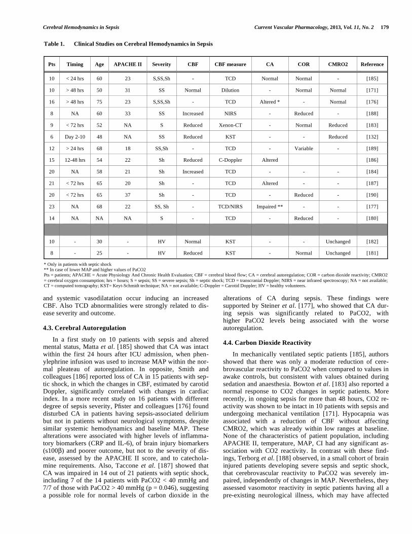
Table 1. Clinical Studies on Cerebral Hemodynamics in Sepsis
Pts Timing Age APACHE II Severity CBF CBF measure CA COR CMRO2 Reference
10 < 24 hrs 60 23 S,SS,Sh - TCD Normal Normal - [185]
10 > 48 hrs 50 31 SS Normal Dilution - Normal Normal [171]
16 > 48 hrs 75 23 S,SS,Sh - TCD Altered * - Normal [176]
8 NA 60 33 SS Increased NIRS - Reduced - [188]
9 < 72 hrs 52 NA S Reduced Xenon-CT - Normal Reduced [183]
6 Day 2-10 48 NA SS Reduced KST - - Reduced [132]
12 > 24 hrs 68 18 SS,Sh - TCD - Variable - [189]
15 12-48 hrs 54 22 Sh Reduced C-Doppler Altered [186]
20 NA 58 21 Sh Increased TCD - - - [184]
21 < 72 hrs 65 20 Sh - TCD Altered - - [187]
20 < 72 hrs 65 37 Sh - TCD - Reduced - [190]
23 NA 68 22 SS, Sh - TCD/NIRS Impaired ** - - [177]
14 NA NA NA S - TCD - Reduced - [180]
10 - 30 - HV Normal KST - - Unchanged [182]
8 - 25 - HV Reduced KST - Normal Unchanged [181]
* Only in patients with septic shock ** In case of lower MAP and higher values of PaCO2 Pts = patients; APACHE = Acute Physiology And Chronic Health Evaluation; CBF = cerebral blood flow; CA = cerebral autoregulation; COR = carbon dioxide reactivity; CMRO2
= cerebral oxygen consumption; hrs = hours; S = sepsis; SS = severe sepsis; Sh = septic shock; TCD = transcranial Doppler; NIRS = near infrared spectroscopy; NA = not available; CT = computed tomography; KST= Keyt-Schmidt technique; NA = not available; C-Doppler = Carotid Doppler; HV = healthy volunteers.
180 Current Vascular Pharmacology, 2013, Vol. 11, No. 2 Taccone et al.
the results. In another study on 12 patients, 3 of them had normal cerebrovascular reactivity, 7 had impaired vasomotor tone while in two others the response to CO2 changes ap-peared to be higher than control [189], without any associa-tion of this finding with cardiovascular status, outcome and severity of illness; also, these alterations did not affect out-come. Recently, cerebro-vascular reactivity was found to be impaired in septic patients with SAE by means of acetazola-mide injection test. Also, the reaction to the vasodilating stimulus was slower in septic patients than control [180]. Finally, the cerebrovascular reactivity to PaCO2 in patients with septic shock was lower than in control patients, with a significantly lower reactivity in patients receiving dexmede-tomidine than propofol [190].
4.5. Summary of Human Findings and Clinical Implications
Sepsis-induced brain dysfunction can directly cause brain damage by altering brain global perfusion and microcircula-tion. In clinical practice, several therapeutical protocols exist to guide the management of patients suffering from severe sepsis or septic shock, including the early-goal directed ther-apy, the prompt antimicrobial administration or the protec-tive lung ventilation; however, we still lack a specific neuro-protective approach to prevent or minimize secondary ischemic brain injuries occurring during sepsis. This is probably due to the limited data available in the literature on the mechanisms inducing brain hypoperfusion in septic pa-tients; moreover, most of studies evaluating CBF in this set-ting showed a reduced CBF but did not provide relevant data for the role of these observations in the development of SAE.
Some studies suggested that vasoconstriction occurs at the level of cerebral arterioles and contribute to reduce CBF [180]; however, it remains still unknown if this phenomenon is secondary to the effects of circulating substances that act on brain vessels or because of impairment of microcircula-tion, as suggested in animal studies. Importantly, if the in-crease in cerebrovascular resistances is dependent from a vasoconstrictor agent, it is possible that it would be released directly within the brain parenchyma. Indeed, omental arter-ies placed in plasma from septic patients had diminished response to vasopressin [191], while the administration of vasopressors poorly affected CBF in septic patients [192]. We can then hypothesize that the use of vasoactive sub-stances is unlikely to negatively influence cerebral perfusion during sepsis. Importantly, data on the cerebrovascular ef-fects of catecholamines in experimental head injury have shown that dopamine, norepinephrine and phenylephrine all increase CBF, with the most predictable effects when nore-pinephrine was used, while dopamine was associated with increased brain edema and phenylephrine with a raise in in-tracranial pressure [193]. Thus, further studies on the effects of such vasopressors on brain perfusion during both animal and human sepsis are needed.
Other studies suggested that changes in CBF were in-duced by acute hypocapnia, rather than sepsis itself [181]. Conflicting data on cerebrovascular reactivity to CO2 dur-ing sepsis exist; when COR was preserved, it was shown to be lower than in awake subjects, leaving the possibility that this difference could be due to the use of sedation rather than to the septic process [185]. Studies showing reduced
CO2 reactivity in septic patients did not strictly control PaCO2 using mechanical ventilation, used different timing of measurements and different vasoactive agents, making difficult to reconcile these data with previous studies [188, 189]. Considering that acute reduction of PaCO2 did not change cerebral oxygenation in healthy volunteers and sep-tic patients [171, 181], hypocapnia should not be actively corrected in septic patients to improve brain perfusion. Fur-thermore, normocapnic/hypercapnic patients have an im-paired autoregulation of CBF and should be considered at higher risk of brain hypoperfusion than those with hypo-capnia [177, 187].
The concomitant use of sedative agents, which reduced neural metabolic rates, may be responsible for lowering brain perfusion in septic patients, accordingly to the metabolic hypothesis of CBF regulation. Importantly, sedatives are likely to modify the reactivity of brain vasculature to physio-logical stimuli, thus limiting the capacity of CBF to autoregulate in case of abrupt changes in PaCO2 or cerebral perfusion pressure. These data further support the concept of early interruption of sedation in critically ill patients to limit improper use of anesthetic agents and prevent the occurrence of brain hypoperfusion [194].
More than the absolute measure of CBF, the evaluation of cerebral autoregulation appears to be fundamental for the hemodynamic management of septic patients; as such, im-paired CA may leave brain tissue unprotected against possi-bly harmful effects of blood pressure changes during sepsis, leading to cerebral ischemia. Cerebral autoregulation was often impaired in septic patients and this was more fre-quently altered in patients with more severe disease, such as shock or delirium [176, 187]. Thus, an early hemodynamic stabilization associated with CBF monitoring and assessment of CBF reactivity may be recommended in septic patients, especially those with the highest degree of illness severity.
Finally, we still are unable to identify the optimal thresh-old of MAP to target in a patient with septic shock to avoid brain ischemic events. As MAP is generally below normal ranges during sepsis, particularly when shock is present, brain perfusion become dependent on CA and the individual capacity to maintain stable CBF over a wide range of systemic pressure. In addition, given concomitant brain edema and elevated ICP above 15 mmHg can occur in septic patients, if MAP is maintained between 60 and 70 mm Hg as generally recommended, cerebral perfusion pressure may fall below 50 mmHg and potentially contribute to brain hypoper-fusion [195].
CONCLUSION
Altered brain perfusion is frequent after sepsis and may contribute to the pathogenesis of sepsis-associated encepha-lopathy. Although the complex pathophysiology has not been fully elucidated, main factors involve direct decrease of cerebral blood flow, alterations of cerebro-vascular autoregu-lation, endothelial dysfunction and disorders of microcircula-tion, release of vasogenic inflammatory mediators, neuro-chemical derangements and metabolic uncoupling, which may eventually lead to cerebral hypoperfusion. This may potentially expose the brain of septic patients to secondary ischemic/hypoxic insults, brain cell dysfunction and worse
Cerebral Hemodynamics in Sepsis Current Vascular Pharmacology, 2013, Vol. 11, No. 2 181
neurological recovery. Novel monitoring tools and recent clinical investigations contributed to improve our under-standing of the regulation of cerebral perfusion in patients with sepsis. These studies have underlined the impact of sedatives and vasopressors on brain perfusion. Some issues are still controversial, particularly whether hemodynamic augmentation of brain perfusion after severe sepsis or septic shock may be beneficial.
CONFLICT OF INTEREST
The authors confirm that this article content has no con-flicts of interest.
ACKNOWLEDGEMENTS
Mauro Oddo is supported by the Swiss National Science Foundation (grant nr 320030-138191).
FINANCIAL AND COMPETING INTERESTS DIS-
CLOSURE
The authors have no relevant affiliation or financial in-volvement with any organization having a financial interest with the subject discussed in this manuscript. No writing assistance was used in the preparation of this manuscript.
ABBREVIATIONS
APACHE II = Acute physiology and chronic health evaluation
ATICE = Assessment to Intensive Care environ-ment
BBB = Brain-blood barrier
BOLD = Blood oxygenation level dependent contrast
CA = Cerebral autoregulation
CAM-ICU = Confusion assessment method for the Intensive Care Unit
CBF = Cerebral blood flow
CI = Cardiac index
CMRO2 = Cerebral metabolism rate of oxygen
CNS = Central nervous system
COR = Cerebrovascular-CO2 reactivity
CPP = Cerebral perfusion pressure
CRP = C-reactive protein
CT = Computed tomography
CVOs = Circumventricular organs
CVR = Cerebrovascular resistance
EEG = Electroencephalography
GCS = Glasgow coma score
ICAM-1 = Inter-cellular adhesion molecule 1
ICU = Intensive Care Unit
ICP = Intracranial pressure
IFN- = Interferon gamma
iNOS = Inducible nitric oxide synthase
L-NAME = N-nitro-L-arginine methylester
L-NMMA = N-monomethyl-L-arginine
MAP = Mean arterial pressure
MCAV = Middle cerebral artery velocity
MOF = Multiple organ failure
MRI = Magnetic resonance imaging
NIRS = Near-infrared spectroscopy
NO = Nitric oxide
NSE = Neuron-specific enolase
PaCO2 = Arterial carbon dioxide tension
PaO2 = Arterial oxygen tension
PbO2 = Brain oxygen tension
PET = Positron emission tomography
ROS = Reactive oxygen species
SAE = Sepsis-associated encephalopathy
SPECT = Single photon emission computed tomography
SSEPs = Somato-sensitive evoked potentials
TCD = Transcranial Doppler
TNF- = Tumor necrosis factor alpha
VIP = Vasoactive intestinal peptide
REFERENCES
[1] Vincent JL, Taccone F, Schmit X. Classification, incidence and
outcome of sepsis and multiple organ failure. Contrib Nephrol
2007; 156:64-74.
[2] Gustot T. Multiple organ failure in sepsis: prognosis and role of
systemic inflammatory response. Curr Opin Crit Care 2011;
17:153-159.
[3] Eidelman LA, Putterman D, Putterman C, Sprung CL. The spec-
trum of septic encephalopathy. Definitions, etiologies and mortali-
ties. JAMA 1996; 275:470-473.
[4] Papadopoulos MC, Davies DC, Moss RF, Tighe D, Bennett ED.
Pathophysiology of septic encephalopathy: a review. Crit Care Med
2000; 28:3019-3024.
[5] Chadwick J, Mann WN. The Medical Works of Hippocrates. Ox-
ford, Blackwell, 1950.
[6] Osler W. The Principles and Practice of Medicine. New York,
Appleton, 1892.
[7] Iacobone E, Bailly-Salin J, Polito A, Friedman D, Stevens RD,
Sharshar T. Sepsis-associated encephalopathy and its differential
diagnosis. Crit Care Med 2009; 37(10 Suppl):S331-336.
[8] Wilson JX, Young GB. Sepsis-associated encephalopathy: evolving
concepts. Can J Neurol Sci 2003; 30:98-105.
[9] Young GB, Bolton CF, Austin TW, Archibald YM, Gonder J,
Wells GA. The encephalopathy associated with sepsis illness. Clin
Invest Med 1990; 13:297-304.
[10] Davies NWS, Sharief MK, Howard RS. Infection-associated en-
cephalopathies - their investigation, diagnosis and treatment. J Neu-
rol 2006; 253:833-845.
[11] Ely EW, Shintani A, Truman B, et al. Delirium as a predictor of
mortality in mechanically ventilated patients in the intensive care
unit. JAMA 2004; 291:1753-1762.
182 Current Vascular Pharmacology, 2013, Vol. 11, No. 2 Taccone et al.
[12] Bleck TP, Smith MC, Pierre-Louis SJ, Jares JJ, Murray J, Hansen
CA. Neurologic complications of critical medical illness. Crit Care
Med 1993; 21:98-103.
[13] Sprung CL, Peduzzi PN, Shatney CH, et al. Impact of encephalopa-
thy on mortality in sepsis syndrome. Crit Care Med 1990; 18:474-
479.
[14] Suchyta MR, Jephson A, Hopkins RO. Neurologic changes during
critical illness: brain imaging findings and neurobehavioral out-
comes. Brain Imaging Behav 2010; 4:22-34.
[15] Young GB, Bolton CF, Archibald YM, Austin TW, Wells GA. The
electroencephalogram in sepsis-associated encephalopathy. J Clin
Neurophysiol 1992; 9:145-152.
[16] Wilson JX, Young GB. Progress in clinical neurosciences: sepsis-
associated encephalopathy: evolving concepts. Can J Neurol Sci
2003; 30:98-105.
[17] Ebersoldt M, Sharshar T, Annane D. Sepsis-associated delirium.
Intensive Care Med 2007; 33:941-950.
[18] Leon A, Lepousé C, Floch T, Graftieaux JP. Brain injury during
severe sepsis. Ann Fr Anesth Rean 2006; 25:863-867.
[19] Chawla J, Gruener G. Management of critical illness polyneuropa-
thy and myopathy. Neurol Clin 2010; 28:961-977.
[20] Sharshar T, Blanchard A, Paillard M, Raphael JC, Gajdos P, An-
nane D. Circulating vasopressin levels in septic shock. Crit Care
Med 2003; 31:1752-1758.
[21] De Jonghe B, Cook D, Griffith L, et al. Adaptation to the Intensive
Care Environment (ATICE): development and validation of a new
sedation assessment instrument. Crit Care Med 2003; 31:2344–
2354.
[22] Ely EW, Inouye SK, Bernard GR, et al. Delirium in mechanically
ventilated patients: validity and reliability of the confusion assess-
ment method for the intensive care unit (CAM-ICU). JAMA 2001;
286:2703-2710.
[23] Towne AR, Waterhouse EJ, Boggs JG, et al. Prevalence of non-
convulsive status epilepticus in comatose patients. Neurology 2000;
54:340-345.
[24] Oddo M, Carrera E, Claassen J, Mayer SA, Hirsch LJ. Continuous
electroencephalography in the medical intensive care unit. Crit
Care Med 2009; 37:2051-2056.
[25] Zauner C, Gendo A, Kramer L, Funk GC, Bauer E, Schenk P.
Impaired subcortical and cortical sensory evoked potentials path-
ways in septic patients. Crit Care Med 2002; 30:1136-1139.
[26] Nguyen DN, Spapen H, Su F, et al. Elevated serum levels of S-
100beta protein and neuron-specific enolase are associated with
brain injury in patients with severe sepsis and septic shock. Crit
Care Med 2006; 34:1967–1974.
[27] Sharshar T, Carlier RY, Bernard F, et al. Brain lesions in septic
shock—an MRI study. Intensive Care Med 2007; 33:798-806.
[28] Russel JA, Singer J, Bernard GA, Wheeler A, Fulkerson W, Hud-
son L. Changing pattern of organ dysfunction in early human sepsis
is related to mortality. Crit Care Med 2000; 28:2405-2411.
[29] Iwashyna TJ, Ely EW, Smith DM, Langa KM. Long-term cognitive
impairment and functional disability among survivors of severe
sepsis. JAMA 2010; 304:1787-1794.
[30] Spapen H, Nguyen DN, Troubleyn J, Huyghens L, Schiettecatte J.
Drotrecogin alfa (activated) may attenuate severe sepsis-associated
encephalopathy in clinical septic shock. Crit Care 2010; 14:R54.
[31] Moss RF, Parmar NK, Tighe D, Davies DC. Adrenergic agents
modify cerebral edema and microvessel ultrastructure in porcine
sepsis. Crit Care Med 2004; 32:1916-1921.
[32] Wang H, Wu YB, Du XH. Effect of dexamethasone on nitric oxide
synthase and caspase-3 gene expressions in endotoxemia in neonate
rat brain. Biomed Environ Sci 2005; 18:181–186.
[33] Ruiz-Valdepeñas L, Martínez-Orgado JA, Benito C, Millán A,
Tolón RM, Romero J. Cannabidiol reduces lipopolysaccharide-
induced vascular changes and inflammation in the mouse brain: an
intravital microscopy study. J Neuroinflamm 2011; 8:5.
[34] Esen F, Erdem T, Aktan D, et al. Effect of magnesium sulfate
administration on blood-brain barrier in a rat model of intraperito-
neal sepsis: a randomized controlled experimental study. Crit Care
2005; 9:R18-23.
[35] Siami S, Annane D, Sharshar T. The encephalopathy in sepsis. Crit
Care Clin 2008; 24:67-82.
[36] Bone RC. Sepsis, the sepsis syndrome multi-organ failure: a plea
for comparable definitions. Ann Intern Med 1991; 114:332-333.
[37] Pendlebury WW, Perl DP, Munoz DG. Multiple microabscesses
in the central nervous system. J Neuropath Exp Neurol 1989;
48:290-300.
[38] Sharshar T, Gray F, Lorin de la Grandmaison G, et al. Apoptosis of
neurons in cardiovascular autonomic centres triggered by inducible
nitric oxide synthase after death from septic shock. Lancet 2003;
362:1799–1805.
[39] Weathers AL, Lewis SL. Rare and unusual ... or are they? Less
commonly diagnosed encephalopathies associated with systemic
disease. Semin Neurol 2009; 29:136-153.
[40] Chong DL, Sriskandan S. Pro-inflammatory mechanisms in sepsis.
Contrib Microbiol 2011; 17:86-107.
[41] Anda T, Yamashita H, Tsutsumi K, Fujita H, Tokunaga Y, Shibata
S. Effect of tumor necrosis factor-alpha on the permeability of bo-
vine brain microvessel endothelial cells monolayers. Neurol Res
1997; 19:369-376
[42] Huynh HK, Dorovini-Zis K. Effects of interferon-gamma on pri-
mary cultures of human brain microvessel endothelial cells. Am J
Pathol 1993; 142: 1265-1278
[43] Tureen J. Effect of recombinant human tumor necrosis factor-alpha
on cerebral oxygen uptake, cerebrospinal fluid lactate and cerebral
blood flow in the rabbit: role of nitric oxide. J Clin Invest 1995;
95:1086-1091
[44] Alexander JJ, Jacob A, Cunningham P, Hensley L, Quigg RJ. TNF
is a key mediator of septic encephalopathy acting through its recep-
tor, TNF receptor-1. Neurochem Int 2008; 52:447-456.
[45] Krueger J, Walter J, Dinarello CA, Wolff SM, Chedid L. Sleep-
promoting effects of endogenous pyrogen (interleukin-1). Am J
Physiol 1984; 246:R994-R999.
[46] Dafny N, Prieto-Gomez B, Reyes-Vazquez C. Does the immune
system communicate with the central nervous system? Interferon
modifies central nervous activity. J Neuroimmunol 1985; 9:1-12.
[47] Wuerfel E, Infante-Duarte C, Glumm R, Wuerfel JT. Gadofluorine
M-enhanced MRI shows involvement of circumventricular organs
in neuroinflammation. J Neuroinflamm 2010; 7:70.
[48] Borovikova LV, Ivanova S, Zhang M, et al. Vagus nerve stimula-
tion attenuates the systemic inflammatory response to endotoxin.
Nature 2000; 405: 458–462.
[49] Miyauchi T, Tsuruta R, Fujita M, Kaneko T, Kasaoka S, Maekawa
T. Serum macrophage migration inhibitory factor reflects adrenal
function in the hypothalamo-pituitary-adrenal axis of septic pa-
tients: an observational study. BMC Infect Dis 2009;9:209.
[50] Soejima Y, Fujii Y, Ishikawa T, Takeshita H, Maekawa T. Local
cerebral glucose utilization in septic rats. Crit Care Med 1990;
18: 423-427.
[51] Semmler A, Frisch C, Debeir T, et al. Long-term cognitive im-
pairment, neuronal loss and reduced cortical cholinergic innerva-
tion after recovery from sepsis in a rodent model. Exp Neurol 2007;
204:733-740.
[52] Kadoi Y, Saito S. An alteration in the gamma-aminobutyric acid
receptor system in experimentally induced septic shock in rats. Crit
Care Med 1996; 24:298-305.
[53] Kadoi Y, Saito S, Kunimoto F, Imai T, Fujita T. Impairment of the
brain beta-adrenergic system during experimental endotoxemia. J
Surg Res 1996; 61:496-502.
[54] Freund HR, Muggia-Sullam M, LaFrance R, Holroyde J, Fischer
JE. Regional brain amino acid and neurotransmitter derangements
during abdominal sepsis and septic encephalopathy in the rat. The
effect of amino acid infusions. Arch Surg 1986; 121:209-216.
[55] Ransohoff RM, Kivisakk P, Kidd G. Three or more routes for leu-
kocyte migration into the central nervous system. Nat Rev Immu-
nol 2003; 3:569–581.

Cerebral Hemodynamics in Sepsis Current Vascular Pharmacology, 2013, Vol. 11, No. 2 183
[56] Dal-Pizzol F, Ritter C, Cassol-Jr OJ, et al. Oxidative mechanisms
of brain dysfunction during sepsis. Neurochem Res 2010; 35:1-12.
[57] Christians ES, Yan LJ, Benjamin IJ. Heat shock factor 1 and heat
shock proteins: critical partners in protection against acute cell in-
jury. Crit Care Med 2002; 30(1 Suppl):S43-50.
[58] Polito A, Brouland JP, Porcher R, et al. Hyperglycaemia and apop-
tosis of microglial cells in human septic shock. Crit Care 2011;
15:R131.
[59] Korcok J, Wu F, Tyml K, Hammond RR, Wilson JX. Sepsis inhib-
its reduction of dehydroascorbic acid and accumulation of ascor-
bate in astroglial cultures: intracellular ascorbate depletion in-
creases nitric oxide synthase induction and glutamate uptake inhibi-
tion. J Neurochem 2002; 81:185-193.
[60] Laflamme N, Rivest S. Toll-like receptor 4: the missing link of the
cerebral innate immune response triggered by circulating gram-
negative bacterial cell wall components. FASEB J 2001; 15:155–
163.
[61] Fernandes A, Silva RF, Falcão AS, Brito MA, Brites D. Cytokine
production, glutamate release and cell death in rat cultured astro-
cytes treated with unconjugated bilirubin and LPS. J Neuroimmu-
nol 2004; 153:64-75.
[62] Dugan LL, Bruno VMG, Amagasu SM, et al. Glia modulate the
response of murine cortical neurons to excitotoxicity. Glia exacer-
bate AMPA neurotoxicity. J Neurosci 1995; 15: 4545-4555.
[63] Wilson JX, Dragan M. Sepsis inhibits recycling and glutamate-
stimulated export of ascorbate by astrocytes. Free Radic Biol Med
2005; 39:990-998.
[64] Jacob A, Hensley LK, Safratowich BD, Quigg RJ, Alexander JJ.
The role of the complement cascade in endotoxin-induced septic
encephalopathy. Lab Invest 2007; 87:1186-1194.
[65] Ari I, Kafa IM, Kurt MA. Perimicrovascular edema in the frontal
cortex in a rat model of intraperitoneal sepsis. Exp Neurol 2006;
198:242-249.
[66] Zhan RZ, Fujiwara N, Shimoji K. Regionally different elevation of
intracellular free calcium in hippocampus of septic rat brain. Shock
1996; 6:293-297.
[67] Eckel B, Ohl F, Bogdanski R, Kochs EF, Blobner M. Cognitive
deficits after systemic induction of inducible nitric oxide synthase:
a randomised trial in rats. Eur J Anaesthesiol 2011; 28(9):655-63.
[68] Minc-Golomb D, Tsarfaty I, Schwartz JP. Expression of inducible
nitric oxide synthase by neurones following exposure to endotoxin
and cytokine. Br J Pharmacol 1994; 112:720–722.
[69] Aloisi F, Penna G, Cerase J, Menendez Iglesias B, Adorini L. IL-12
production by central nervous system microglia is inhibited by as-
trocytes. J Immunol 1997; 159:1604–1612.
[70] Ledeboer A, Breve JJ, Wierinckx A, et al. Expression and regula-
tion of interleukin-10 and interleukin-10 receptor in rat astroglial
and microglial cells. Eur J Neurosci 2002; 16:1175–1185.
[71] Caggiano AO, Kraig RP. Prostaglandin E receptor subtypes in
cultured rat microglia and their role in reducing lipopolysaccharide-
induced interleukin-1beta production. J Neurochem 1999; 72:565–
575.
[72] Fontana A, Kristensen F, Dubs R, Gemsa D, Weber E. Production
of prostaglandin E and an interleukin-1 like factor by cultured as-
trocytes and C6 glioma cells. J Immunol 1982; 129:2413–2419.
[73] Akrout N, Sharshar T, Annane D. Mechanisms of brain signaling
during sepsis. Curr Neuropharmacol 2009; 7:296-301.
[74] Bowton DL, Bertels NH, Prough DS, et al. Cerebral blood flow is
reduced in patients with sepsis syndrome. Crit Care Med 1989;
17:399-403.
[75] Dagal A, Lam AM. Cerebral blood flow and the injured brain: how
should we monitor and manipulate it? Curr Opin Anaesthesiol
2011; 24:131-137.
[76] Peterson EC, Wang Z, Britz G. Regulation of cerebral blood flow.
Int J Vasc Med 2011; 2011:823525.
[77] Fieschi C, Mackenzie E, Lenzi GL, et al. Facts and trends in cere-
bral blood flow and metabolism: synopsis of brain '89. J Cereb
Blood Flow Metab 1989; 9:573-578.
[78] Tuor UI, Edvinsson L, McCulloch J. Catecholamines and the rela-
tionship between cerebral blood flow and glucose use. Am J
Physiol 1986; 251:H824-H833.
[79] Panerai RB, Dineen NE, Brodie FG, Robinson TG. Spontaneous
fluctuations in cerebral blood flow regulation: contribution of
PaCO2. J Appl Physiol 2010; 109:1860-1868.
[80] Lagerkranser M, Stånge K, Sollevi A. Effects of propofol on cere-
bral blood flow, metabolism, and cerebral autoregulation in the
anesthetized pig. J Neurosurg Anesthesiol 1997; 9:188-193.
[81] Preckel MP, Leftheriotis G, Ferber C, Degoute CS, Banssillon V,
Saumet JL. Effect of nitric oxide blockade on the lower limit of the
cortical cerebral autoregulation in pentobarbital-anaesthetized rats.
Int J Microcirc Clin Exp 1996; 16:277-283.
[82] Roatta S, Micieli G, Bosone D, Losano G, Bini R, Cavallini A,
Passatore M. Effect of generalised sympathetic activation by cold
pressor test on cerebral haemodynamics in healthy humans. J
Auton Nerv Syst 1998; 71:159-166.
[83] Harper AM, Glass HI. Effect of alterations in the arterial carbon
dioxide tension on the blood flow through the cerebral cortex at
normal and low arterial blood pressures. J Neurol Neurosurg Psy-
chiatry 1965; 28:449-452.
[84] McDowall DG, Harper AM. Relationship between the blood flow
and the extracellular pH of the cerebral cortex. Br J Anaesth
1967; 39:516.
[85] Kuschinsky W, Wahl M. Local chemical and neurogenic regulation
of cerebral vascular resistance. Physiol Rev 1978; 58:656-689.
[86] Winn HR, Morii S, Berne RM. The role of adenosine in autoregula-
tion of cerebral blood flow. Ann Biomed Eng 1985; 13:321-328.
[87] Nguyen TS, Winn HR, Janigro D. ATP-sensitive potassium chan-
nels may participate in the coupling of neuronal activity and cere-
brovascular tone. Am J Physiol Heart Circ Physiol 2000;
278:H878-H885.
[88] Straub SV, Girouard H, Doetsch PE, Hannah RM, Wilkerson MK,
Nelson MT. Regulation of intracerebral arteriolar tone by K(v)
channels: effects of glucose and PKC. Am J Physiol Cell Physiol
2009; 297:C788-C796.
[89] Pickard JD, MacDonell LA, Mackenzie ET, Harper AM. Prosta-
glandin-induced effects in the primate cerebral circulation. Eur J
Pharmacol 1977; 43:343-351.
[90] Erlichman JS, Leiter JC. Glia modulation of the extracellular mi-
lieu as a factor in central CO2 chemosensitivity and respiratory
control. J Appl Physiol 2010; 108:1803-1811.
[91] Aaslid R, Lindegaard KF, Sorteberg W, Nornes H. Cerebral
autoregulation dynamics in humans. Stroke 1989; 20:45–52.
[92] Bulte DP, Chiarelli PA, Wise RG, Jezzard P. Cerebral perfusion
response to hyperoxia. J Cereb Blood Flow Met 2007, 27:69-75.
[93] Rangel-Castilla L, Lara LR, Gopinath S, Swank PR, Valadka A,
Robertson C. Cerebral hemodynamic effects of acute hyperoxia
and hyperventilation after severe traumatic brain injury. J Neuro-
trauma 2010; 27:1853-1863.
[94] Leybaert L. Neurobarrier coupling in the brain: a partner of
neurovascular and neurometabolic coupling? J Cereb Blood Flow
Metab 2005; 25:2-16.
[95] Kochanek PM, Hendrich KS, Jackson EK, et al. Characterization
of the effects of adenosine receptor agonists on cerebral blood flow
in uninjured and traumatically injured rat brain using continuous
arterial spin-labeled magnetic resonance imaging. J Cereb Blood
Flow Metab 2005; 25:1596-1612.
[96] Paulson OB, Hasselbalch SG, Rostrup E, Knudsen GM, Pelligrino
D. Cerebral blood flow response to functional activation. J Cereb
Blood Flow Metab 2010; 30:2-14.
[97] Straub SV, Bonev AD, Wilkerson MK, Nelson MT. Dynamic
inositol trisphosphate-mediated calcium signals within astrocytic
endfeet underlie vasodilation of cerebral arterioles. J Gen Physiol
2006; 128:659–669.
[98] Xu F, Uh J, Brier MR, et al. The influence of carbon dioxide on
brain activity and metabolism in conscious humans. J Cereb Blood
Flow Metab 2011; 31:58-67.
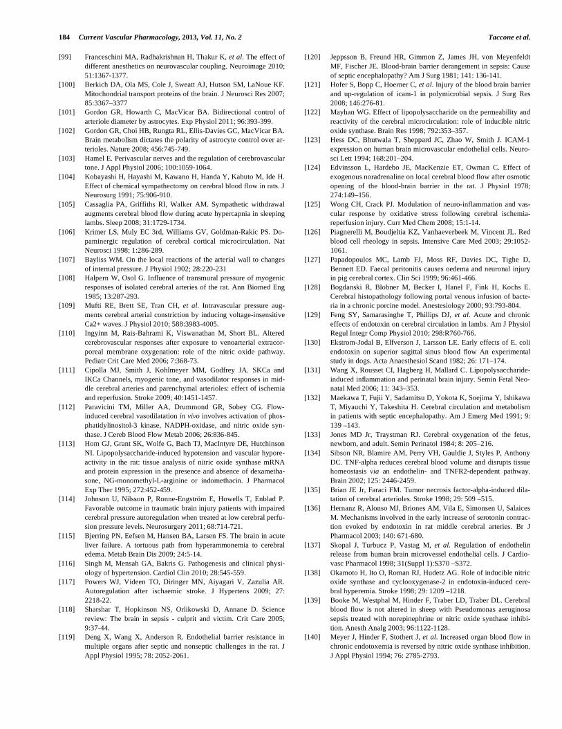
184 Current Vascular Pharmacology, 2013, Vol. 11, No. 2 Taccone et al.
[99] Franceschini MA, Radhakrishnan H, Thakur K, et al. The effect of
different anesthetics on neurovascular coupling. Neuroimage 2010;
51:1367-1377.
[100] Berkich DA, Ola MS, Cole J, Sweatt AJ, Hutson SM, LaNoue KF.
Mitochondrial transport proteins of the brain. J Neurosci Res 2007;
85:3367–3377
[101] Gordon GR, Howarth C, MacVicar BA. Bidirectional control of
arteriole diameter by astrocytes. Exp Physiol 2011; 96:393-399.
[102] Gordon GR, Choi HB, Rungta RL, Ellis-Davies GC, MacVicar BA.
Brain metabolism dictates the polarity of astrocyte control over ar-
terioles. Nature 2008; 456:745-749.
[103] Hamel E. Perivascular nerves and the regulation of cerebrovascular
tone. J Appl Physiol 2006; 100:1059-1064.
[104] Kobayashi H, Hayashi M, Kawano H, Handa Y, Kabuto M, Ide H.
Effect of chemical sympathectomy on cerebral blood flow in rats. J
Neurosurg 1991; 75:906-910.
[105] Cassaglia PA, Griffiths RI, Walker AM. Sympathetic withdrawal
augments cerebral blood flow during acute hypercapnia in sleeping
lambs. Sleep 2008; 31:1729-1734.
[106] Krimer LS, Muly EC 3rd, Williams GV, Goldman-Rakic PS. Do-
paminergic regulation of cerebral cortical microcirculation. Nat
Neurosci 1998; 1:286-289.
[107] Bayliss WM. On the local reactions of the arterial wall to changes
of internal pressure. J Physiol 1902; 28:220-231
[108] Halpern W, Osol G. Influence of transmural pressure of myogenic
responses of isolated cerebral arteries of the rat. Ann Biomed Eng
1985; 13:287-293.
[109] Mufti RE, Brett SE, Tran CH, et al. Intravascular pressure aug-
ments cerebral arterial constriction by inducing voltage-insensitive
Ca2+ waves. J Physiol 2010; 588:3983-4005.
[110] Ingyinn M, Rais-Bahrami K, Viswanathan M, Short BL. Altered
cerebrovascular responses after exposure to venoarterial extracor-
poreal membrane oxygenation: role of the nitric oxide pathway.
Pediatr Crit Care Med 2006; 7:368-73.
[111] Cipolla MJ, Smith J, Kohlmeyer MM, Godfrey JA. SKCa and
IKCa Channels, myogenic tone, and vasodilator responses in mid-
dle cerebral arteries and parenchymal arterioles: effect of ischemia
and reperfusion. Stroke 2009; 40:1451-1457.
[112] Paravicini TM, Miller AA, Drummond GR, Sobey CG. Flow-
induced cerebral vasodilatation in vivo involves activation of phos-
phatidylinositol-3 kinase, NADPH-oxidase, and nitric oxide syn-
thase. J Cereb Blood Flow Metab 2006; 26:836-845.
[113] Hom GJ, Grant SK, Wolfe G, Bach TJ, MacIntyre DE, Hutchinson
NI. Lipopolysaccharide-induced hypotension and vascular hypore-
activity in the rat: tissue analysis of nitric oxide synthase mRNA
and protein expression in the presence and absence of dexametha-
sone, NG-monomethyl-L-arginine or indomethacin. J Pharmacol
Exp Ther 1995; 272:452-459.
[114] Johnson U, Nilsson P, Ronne-Engström E, Howells T, Enblad P.
Favorable outcome in traumatic brain injury patients with impaired
cerebral pressure autoregulation when treated at low cerebral perfu-
sion pressure levels. Neurosurgery 2011; 68:714-721.
[115] Bjerring PN, Eefsen M, Hansen BA, Larsen FS. The brain in acute
liver failure. A tortuous path from hyperammonemia to cerebral
edema. Metab Brain Dis 2009; 24:5-14.
[116] Singh M, Mensah GA, Bakris G. Pathogenesis and clinical physi-
ology of hypertension. Cardiol Clin 2010; 28:545-559.
[117] Powers WJ, Videen TO, Diringer MN, Aiyagari V, Zazulia AR.
Autoregulation after ischaemic stroke. J Hypertens 2009; 27:
2218-22.
[118] Sharshar T, Hopkinson NS, Orlikowski D, Annane D. Science
review: The brain in sepsis - culprit and victim. Crit Care 2005;
9:37-44.
[119] Deng X, Wang X, Anderson R. Endothelial barrier resistance in
multiple organs after septic and nonseptic challenges in the rat. J
Appl Physiol 1995; 78: 2052-2061.
[120] Jeppsson B, Freund HR, Gimmon Z, James JH, von Meyenfeldt
MF, Fischer JE. Blood-brain barrier derangement in sepsis: Cause
of septic encephalopathy? Am J Surg 1981; 141: 136-141.
[121] Hofer S, Bopp C, Hoerner C, et al. Injury of the blood brain barrier
and up-regulation of icam-1 in polymicrobial sepsis. J Surg Res
2008; 146:276-81.
[122] Mayhan WG. Effect of lipopolysaccharide on the permeability and
reactivity of the cerebral microcirculation: role of inducible nitric
oxide synthase. Brain Res 1998; 792:353–357.
[123] Hess DC, Bhutwala T, Sheppard JC, Zhao W, Smith J. ICAM-1
expression on human brain microvascular endothelial cells. Neuro-
sci Lett 1994; 168:201–204.
[124] Edvinsson L, Hardebo JE, MacKenzie ET, Owman C. Effect of
exogenous noradrenaline on local cerebral blood flow after osmotic
opening of the blood-brain barrier in the rat. J Physiol 1978;
274:149–156.
[125] Wong CH, Crack PJ. Modulation of neuro-inflammation and vas-
cular response by oxidative stress following cerebral ischemia-
reperfusion injury. Curr Med Chem 2008; 15:1-14.
[126] Piagnerelli M, Boudjeltia KZ, Vanhaeverbeek M, Vincent JL. Red
blood cell rheology in sepsis. Intensive Care Med 2003; 29:1052-
1061.
[127] Papadopoulos MC, Lamb FJ, Moss RF, Davies DC, Tighe D,
Bennett ED. Faecal peritonitis causes oedema and neuronal injury
in pig cerebral cortex. Clin Sci 1999; 96:461-466.
[128] Bogdanski R, Blobner M, Becker I, Hanel F, Fink H, Kochs E.
Cerebral histopathology following portal venous infusion of bacte-
ria in a chronic porcine model. Anestesiology 2000; 93:793-804.
[129] Feng SY, Samarasinghe T, Phillips DJ, et al. Acute and chronic
effects of endotoxin on cerebral circulation in lambs. Am J Physiol
Regul Integr Comp Physiol 2010; 298:R760-766.
[130] Ekstrom-Jodal B, Elfverson J, Larsson LE. Early effects of E. coli
endotoxin on superior sagittal sinus blood flow An experimental
study in dogs. Acta Anaesthesiol Scand 1982; 26: 171–174.
[131] Wang X, Rousset CI, Hagberg H, Mallard C. Lipopolysaccharide-
induced inflammation and perinatal brain injury. Semin Fetal Neo-
natal Med 2006; 11: 343–353.
[132] Maekawa T, Fujii Y, Sadamitsu D, Yokota K, Soejima Y, Ishikawa
T, Miyauchi Y, Takeshita H. Cerebral circulation and metabolism
in patients with septic encephalopathy. Am J Emerg Med 1991; 9:
139 –143.
[133] Jones MD Jr, Traystman RJ. Cerebral oxygenation of the fetus,
newborn, and adult. Semin Perinatol 1984; 8: 205–216.
[134] Sibson NR, Blamire AM, Perry VH, Gauldie J, Styles P, Anthony
DC. TNF-alpha reduces cerebral blood volume and disrupts tissue
homeostasis via an endothelin- and TNFR2-dependent pathway.
Brain 2002; 125: 2446-2459.
[135] Brian JE Jr, Faraci FM. Tumor necrosis factor-alpha-induced dila-
tation of cerebral arterioles. Stroke 1998; 29: 509 –515.
[136] Hernanz R, Alonso MJ, Briones AM, Vila E, Simonsen U, Salaices
M. Mechanisms involved in the early increase of serotonin contrac-
tion evoked by endotoxin in rat middle cerebral arteries. Br J
Pharmacol 2003; 140: 671-680.
[137] Skopal J, Turbucz P, Vastag M, et al. Regulation of endothelin
release from human brain microvessel endothelial cells. J Cardio-
vasc Pharmacol 1998; 31(Suppl 1):S370 –S372.
[138] Okamoto H, Ito O, Roman RJ, Hudetz AG. Role of inducible nitric
oxide synthase and cyclooxygenase-2 in endotoxin-induced cere-
bral hyperemia. Stroke 1998; 29: 1209 –1218.
[139] Booke M, Westphal M, Hinder F, Traber LD, Traber DL. Cerebral
blood flow is not altered in sheep with Pseudomonas aeruginosa
sepsis treated with norepinephrine or nitric oxide synthase inhibi-
tion. Anesth Analg 2003; 96:1122-1128.
[140] Meyer J, Hinder F, Stothert J, et al. Increased organ blood flow in
chronic endotoxemia is reversed by nitric oxide synthase inhibition.
J Appl Physiol 1994; 76: 2785-2793.
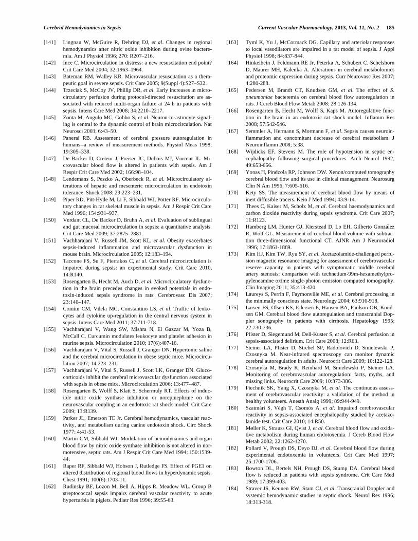
Cerebral Hemodynamics in Sepsis Current Vascular Pharmacology, 2013, Vol. 11, No. 2 185
[141] Lingnau W, McGuire R, Dehring DJ, et al. Changes in regional
hemodynamics after nitric oxide inhibition during ovine bactere-
mia. Am J Physiol 1996; 270: R207–216.
[142] Ince C. Microcirculation in distress: a new resuscitation end point?
Crit Care Med 2004; 32:1963–1964.
[143] Bateman RM, Walley KR. Microvascular resuscitation as a thera-
peutic goal in severe sepsis. Crit Care 2005; 9(Suppl 4):S27–S32.
[144] Trzeciak S, McCoy JV, Phillip DR, et al. Early increases in micro-
circulatory perfusion during protocol-directed resuscitation are as-
sociated with reduced multi-organ failure at 24 h in patients with
sepsis. Intens Care Med 2008; 34:2210–2217.
[145] Zonta M, Angulo MC, Gobbo S, et al. Neuron-to-astrocyte signal-
ing is central to the dynamic control of brain microcirculation. Nat
Neurosci 2003; 6:43–50.
[146] Panerai RB. Assessment of cerebral pressure autoregulation in
humans--a review of measurement methods. Physiol Meas 1998;
19:305–338.
[147] De Backer D, Creteur J, Preiser JC, Dubois MJ, Vincent JL. Mi-
crovascular blood flow is altered in patients with sepsis. Am J
Respir Crit Care Med 2002; 166:98–104.
[148] Lendemans S, Peszko A, Oberbeck R, et al. Microcirculatory al-
terations of hepatic and mesenteric microcirculation in endotoxin
tolerance. Shock 2008; 29:223–231.
[149] Piper RD, Pitt-Hyde M, Li F, Sibbald WJ, Potter RF. Microcircula-
tory changes in rat skeletal muscle in sepsis. Am J Respir Crit Care
Med 1996; 154:931–937.
[150] Verdant CL, De Backer D, Bruhn A, et al. Evaluation of sublingual
and gut mucosal microcirculation in sepsis: a quantitative analysis.
Crit Care Med 2009; 37:2875–2881.
[151] Vachharajani V, Russell JM, Scott KL, et al. Obesity exacerbates
sepsis-induced inflammation and microvascular dysfunction in
mouse brain. Microcirculation 2005; 12:183–194.
[152] Taccone FS, Su F, Pierrakos C, et al. Cerebral microcirculation is
impaired during sepsis: an experimental study. Crit Care 2010,
14:R140.
[153] Rosengarten B, Hecht M, Auch D, et al. Microcirculatory dysfunc-
tion in the brain precedes changes in evoked potentials in endo-
toxin-induced sepsis syndrome in rats. Cerebrovasc Dis 2007;
23:140–147.
[154] Comim CM, Vilela MC, Constantino LS, et al. Traffic of leuko-
cytes and cytokine up-regulation in the central nervous system in
sepsis. Intens Care Med 2011; 37:711-718.
[155] Vachharajani V, Wang SW, Mishra N, El Gazzar M, Yoza B,
McCall C. Curcumin modulates leukocyte and platelet adhesion in
murine sepsis. Microcirculation 2010; 17(6):407-16.
[156] Vachharajani V, Vital S, Russell J, Granger DN. Hypertonic saline
and the cerebral microcirculation in obese septic mice. Microcircu-
lation 2007; 14:223–231.
[157] Vachharajani V, Vital S, Russell J, Scott LK, Granger DN. Gluco-
corticoids inhibit the cerebral microvascular dysfunction associated
with sepsis in obese mice. Microcirculation 2006; 13:477–487.
[158] Rosengarten B, Wolff S, Klatt S, Schermuly RT. Effects of induc-
ible nitric oxide synthase inhibition or norepinephrine on the
neurovascular coupling in an endotoxic rat shock model. Crit Care
2009; 13:R139.
[159] Parker JL, Emerson TE Jr. Cerebral hemodynamics, vascular reac-
tivity, and metabolism during canine endotoxin shock. Circ Shock
1977; 4:41-53.
[160] Martin CM, Sibbald WJ. Modulation of hemodynamics and organ
blood flow by nitric oxide synthase inhibition is not altered in nor-
motensive, septic rats. Am J Respir Crit Care Med 1994; 150:1539-
44.
[161] Raper RF, Sibbald WJ, Hobson J, Rutledge FS. Effect of PGE1 on
altered distribution of regional blood flows in hyperdynamic sepsis.
Chest 1991; 100(6):1703-11.
[162] Rudinsky BF, Lozon M, Bell A, Hipps R, Meadow WL. Group B
streptococcal sepsis impairs cerebral vascular reactivity to acute
hypercarbia in piglets. Pediatr Res 1996; 39:55-63.
[163] Tyml K, Yu J, McCormack DG. Capillary and arteriolar responses
to local vasodilators are impaired in a rat model of sepsis. J Appl
Physiol 1998; 84:837-844.
[164] Hinkelbein J, Feldmann RE Jr, Peterka A, Schubert C, Schelshorn
D, Maurer MH, Kalenka A. Alterations in cerebral metabolomics
and proteomic expression during sepsis. Curr Neurovasc Res 2007;
4:280-288.
[165] Pedersen M, Brandt CT, Knudsen GM, et al. The effect of S.
pneumoniae bacteremia on cerebral blood flow autoregulation in
rats. J Cereb Blood Flow Metab 2008; 28:126-134.
[166] Rosengarten B, Hecht M, Wolff S, Kaps M. Autoregulative func-
tion in the brain in an endotoxic rat shock model. Inflamm Res
2008; 57:542-546.
[167] Semmler A, Hermann S, Mormann F, et al. Sepsis causes neuroin-
flammation and concomitant decrease of cerebral metabolism. J
Neuroinflamm 2008; 5:38.
[168] Wijdicks EF, Stevens M. The role of hypotension in septic en-
cephalopathy following surgical procedures. Arch Neurol 1992;
49:653-656.
[169] Yonas H, Pindzola RP, Johnson DW. Xenon/computed tomography
cerebral blood flow and its use in clinical management. Neurosurg
Clin N Am 1996; 7:605-616.
[170] Kety SS. The measurement of cerebral blood flow by means of
inert diffusible tracers. Keio J Med 1994; 43:9-14.
[171] Thees C, Kaiser M, Scholz M, et al. Cerebral haemodynamics and
carbon dioxide reactivity during sepsis syndrome. Crit Care 2007;
11:R123.
[172] Hamberg LM, Hunter GJ, Kierstead D, Lo EH, Gilberto González
R, Wolf GL. Measurement of cerebral blood volume with subtrac-
tion three-dimensional functional CT. AJNR Am J Neuroradiol
1996; 17:1861-1869.
[173] Kim HJ, Kim TW, Ryu SY, et al. Acetazolamide-challenged perfu-
sion magnetic resonance imaging for assessment of cerebrovascular
reserve capacity in patients with symptomatic middle cerebral
artery stenosis: comparison with technetium-99m-hexamethylpro-
pyleneamine oxime single-photon emission computed tomography.
Clin Imaging 2011; 35:413-420.
[174] Laureys S, Perrin F, Faymonville ME, et al. Cerebral processing in
the minimally conscious state. Neurology 2004; 63:916-918.
[175] Larsen FS, Olsen KS, Ejlersen E, Hansen BA, Paulson OB, Knud-
sen GM. Cerebral blood flow autoregulation and transcranial Dop-
pler sonography in patients with cirrhosis. Hepatology 1995;
22:730-736.
[176] Pfister D, Siegemund M, Dell-Kuster S, et al. Cerebral perfusion in
sepsis-associated delirium. Crit Care 2008; 12:R63.
[177] Steiner LA, Pfister D, Strebel SP, Radolovich D, Smielewski P,
Czosnyka M. Near-infrared spectroscopy can monitor dynamic
cerebral autoregulation in adults. Neurocrit Care 2009; 10:122-128.
[178] Czosnyka M, Brady K, Reinhard M, Smielewski P, Steiner LA.
Monitoring of cerebrovascular autoregulation: facts, myths, and
missing links. Neurocrit Care 2009; 10:373-386.
[179] Piechnik SK, Yang X, Czosnyka M, et al. The continuous assess-
ment of cerebrovascular reactivity: a validation of the method in
healthy volunteers. Anesth Analg 1999; 89:944-949.
[180] Szatmári S, Végh T, Csomós A, et al. Impaired cerebrovascular
reactivity in sepsis-associated encephalopathy studied by acetazo-
lamide test. Crit Care 2010; 14:R50.
[181] Møller K, Strauss GI, Qvist J, et al. Cerebral blood flow and oxida-
tive metabolism during human endotoxemia. J Cereb Blood Flow
Metab 2002; 22:1262-1270.
[182] Pollard V, Prough DS, Deyo DJ, et al. Cerebral blood flow during
experimental endotoxemia in volunteers. Crit Care Med 1997;
25:1700-1706.
[183] Bowton DL, Bertels NH, Prough DS, Stump DA. Cerebral blood
flow is reduced in patients with sepsis syndrome. Crit Care Med
1989; 17:399-403.
[184] Straver JS, Keunen RW, Stam CJ, et al. Transcranial Doppler and
systemic hemodynamic studies in septic shock. Neurol Res 1996;
18:313-318.
186 Current Vascular Pharmacology, 2013, Vol. 11, No. 2 Taccone et al.
[185] Matta BF, Stow PJ. Sepsis-induced vasoparalysis does not involve
the cerebral vasculature: indirect evidence from autoregulation and
carbon dioxide reactivity studies. Br J Anaesth 1996; 76:790-794.
[186] Smith SM, Padayachee S, Modaresi KB, Smithies MN, Bihari DJ.
Cerebral blood flow is proportional to cardiac index in patients
with septic shock. J Crit Care 1998; 13:104-109.
[187] Taccone FS, Castanares-Zapatero D, Peres-Bota D, Vincent JL,
Berre' J, Melot C. Cerebral autoregulation is influenced by carbon
dioxide levels in patients with septic shock. Neurocrit Care 2010;
12:35-42.
[188] Terborg C, Schummer W, Albrecht M, Reinhart K, Weiller C,
Röther J. Dysfunction of vasomotor reactivity in severe sepsis and
septic shock. Intensive Care Med 2001; 27:1231-1234.
[189] Bowie RA, O'Connor PJ, Mahajan RP. Cerebrovascular reactivity
to carbon dioxide in sepsis syndrome. Anaesthesia 2003; 58:261-
265.
[190] Kadoi Y, Saito S, Kawauchi C, Hinohara H, Kunimoto F. Com-
parative effects of propofol vs dexmedetomidine on cerebrovascu-
lar carbon dioxide reactivity in patients with septic shock. Br J An-
aesth 2008; 100:224-229.
[191] Hamu Y, Kanmura Y, Tsuneyoshi I, Yoshimura N. The effects of
vasopressin on endotoxin-induced attenuation of contractile re-
sponses in human gastroepiploic arteries in vitro. Anesth Analg
1999; 88:542-548.
[192] Berré J, De Backer D, Moraine JJ, Mélot C, Kahn RJ, Vincent JL.
Dobutamine increases cerebral blood flow velocity and jugular
bulb hemoglobin saturation in septic patients. Crit Care Med 1997;
25:392-398.
[193] Pfister D, Strebel SP, Steiner LA. Effects of catecholamines on
cerebral blood vessels in patients with traumatic brain injury. Eur J
Anaesthesiol Suppl 2008; 42:98-103.
[194] Dotson B. Daily interruption of sedation in patients treated with
mechanical ventilation. Am J Health Syst Pharm 2010; 67:1002-
1006.
[195] Pfister D, Schmidt B, Smielewski P, et al. Intracranial pressure in
patients with sepsis. Acta Neurochir Suppl 2008; 102:71-75.
Received: November 26, 2011 Revised: April 15, 2012 Accepted: May 05, 2012


