Embed Size (px)
Citation preview

167
15
15.1 INTRODUCTION
The brain is composed of areas of gray and white matter and consists of various regions, including the cerebral cor-tex, the thalamus, the hypothalamus, the brain stem, and the cerebellum. The sensory areas of the cerebral cortex are involved in perception of sensory information: motor areas control execution of voluntary movements and association areas deal with complex integrative functions such as mem-ory, personality traits, and intelligence. The limbic system promotes range of emotions including pleasure, pain, affec-tion, fear, and anger. The thalamus relays almost all sensory input to the cerebral cortex: it contributes to motor func-tions by transmitting information from the cerebellum and basal nuclei to motor areas of the cerebral cortex. It also plays a role in maintaining consciousness. The hypothala-mus controls and integrates activities of the autonomic ner-vous system: it regulates emotional and behavioral patterns and circadian rhythms. The cerebellum smoothes and coor-dinates contractions of skeletal muscles, regulates posture,
and balance and it may have a role in cognition and lan-guage processing.
The brain is a heterogeneous tissue that contains neurons, neuroglia, and other cell types that vary among anatomical regions. Nonneuronal cell types are broadly categorized into (1) astrocytes, (2) radial glia, (3) oligodendrocytes, (4) epen-dymal cells, and (5) microglia. The role of each cell type is well defined; moreover, their interaction is essential for the neuronal function of the central nervous system. The cellular communication is substantially involved in the establishment of the majority of neurological disorders (Pham and Gupta, 2009).
Brain function is determined by the communication between electrically excitable neurons and the surrounding glial cells, which perform many tasks in the brain:
Oligodendrocytes are one type of glial cell that form an insulating protective myelin sheath around the axons of neu-rons that enables saltatory nerve conduction. A loss of myelin in defined areas of brain leads to an impairment of axonal conduc-tance. This is what happens in many forms of myelin disorders,
Techniques and Methods of Animal Brain SurgeryPerfusion, Brain Removal, and Histological Techniques
Jihane Soueid, Amaly Nokkari, and Joelle Makoukji
CONTeNTs
15.1 Introduction ..................................................................................................................................................................... 16715.2 Materials .......................................................................................................................................................................... 168
15.2.1 Anesthesia, Perfusion, and Brain Removal ......................................................................................................... 16815.2.2 ISH Buffers .......................................................................................................................................................... 16815.2.3 Immunofluorescence Materials and Buffers ........................................................................................................ 169
15.3 Methods ........................................................................................................................................................................... 16915.3.1 Intracardial Perfusion of Mice ............................................................................................................................. 169
15.3.1.1 Anesthesia ............................................................................................................................................. 16915.3.1.2 Perfusion ............................................................................................................................................... 16915.3.1.3 Troubleshooting .....................................................................................................................................170
15.3.2 Brain Removal ......................................................................................................................................................17015.3.3 Brain Slicing .........................................................................................................................................................17115.3.4 In Situ Hybridization ............................................................................................................................................171
15.3.4.1 Protocol ................................................................................................................................................. 17215.3.5 IHC ...................................................................................................................................................................... 172
15.3.5.1 Protocol ..................................................................................................................................................17315.4 Conclusion ........................................................................................................................................................................173References ..................................................................................................................................................................................173
168 Brain Neurotrauma: Molecular, Neuropsychological, and Rehabilitation Aspects
such as multiple sclerosis, and it results in a permanent loss of neuron impulse transmission. It is evident that the demyelinated region contains inflammatory cells such as infiltrating lympho-cytes and macrophages and activated microglia. These cells might potentiate or even initiate a damage cascade leading to continuous neurodegeneration.
Microglia are the resident phagocytic cells in the brain, taking part in immune-mediated defense mechanisms and clearing damaged cell debris (Ransohoff and Cardona, 2010; Ransohoff and Perry, 2009). Previously, it was thought that microglia, in their resting state, are relatively quiescent. More recent work suggests that microglia are constantly active and surveying their surroundings (Hughes, 2012; Nimmerjahn et al., 2005). Microglia are now implicated in synapse prun-ing during both development and throughout adulthood, and therefore play a role in regulating homeostatic synaptic plas-ticity (Schafer et al., 2012).
Together with astrocytes, another type of glial cells, microglia can release neuromodulatory chemicals that influ-ence neuronal firing and intracellular signaling. When first described, astrocytes were seen merely as structural scaffold-ing to support and fill the gaps between neurons. However, recent evidence suggests that astrocytes serve as much more than a nutrient supply or supportive scaffolding to protect neural networks (Nedergaard et al., 2003). Astrocytes are highly secretory cells, participating in rapid brain commu-nication by releasing factors that modulate neurotransmis-sion (Haydon and Carmignoto, 2006; Huang et al., 2004; Pascual et al., 2012) and more recently have been suggested to possess their own repertoire of gliotransmitters (Bezzi et al., 2004; Cali et al., 2008; Cali and Bezzi, 2010; Domercq et al., 2006; Jourdain et al., 2007; Prada et al., 2011; Santello et al., 2011). Astrocytes also express a wide variety of func-tional neurotransmitter receptors essential for sensing neuro-nal activity (Verkhratsky et al., 1998).
When a local inflammatory reaction is triggered in the brain, the increased levels of pro-inflammatory mediators such tumor necrosis factor-alpha and prostaglandin 2 can deeply alter the properties of glial network and thus of neuro-nal network (Bezzi and Volterra, 2001). The important roles played by glial cells in normal and pathological brain func-tioning are growing, and a more complete picture of neuron–glia interactions is beginning to emerge.
Cellular behaviors such as proliferation, differentiation, migration, and cell death are studied during brain develop-ment and in pathological situations. To better understand the developmental processes involved, it is important to obtain information regarding the spatial and temporal patterns of gene expression.
Over the past three decades, animal models have been developed to replicate the various aspects of human brain injury to better understand the underlying pathophysiology and to explore potential treatments. Among more recent mod-els for traumatic brain injury, four specific models are widely used in research: fluid percussion injury (Dixon et al., 1987),
controlled cortical impact injury (Dixon et al., 1991; Lighthall, 1988), weight drop impact acceleration injury (Marmarou et al., 1994), and blast injury (Cernak et al., 1996; Leung et al., 2008). Rodents are mostly used in traumatic brain injury research because of their modest cost, small size, and standardized outcome measurements.
In this chapter, we will describe the method of intracardial perfusion and an appropriate method to dissect and remove the brain of a mouse. We will then describe methods for the detection of protein (immunohistochemistry, IHC) and mes-senger RNA (mRNA) (in situ hybridization, ISH) in brain sections.
15.2 MATeRIALs
15.2.1 AnesthesiA, Perfusion, And BrAin removAl
1. 1-mL syringe and 27 G needle for anesthesia 2. Anesthetic: ketamine and xylazine 3. Peristaltic pump 4. Scalpel handle and #10 blade 5 Freshly made 4% paraformaldehyde (PFA) 10–150 mL
per mouse 6. 0.9% saline (or preferred flush) 8–25 mL per mouse 7. Butterfly catheter (23G) with the needle blunted 8. Straight iris scissors 9. Curved iris scissors 10. Curved narrow pattern forceps 11. Chemical fume hood 12. Container for mouse 13. Corked surface 14. Safety glasses
15.2.2 ish Buffers
1. 10X salt buffer2 M NaCL50 mM EDTA100 mM Tris-HCl pH 7.550 mM NaH2PO4.2H2O50 mM Na2HPO4
2. Hybridization buffer50% desionized formamide10% (w/v) dextran sulphate1X Denhart’s0.1 mg/mL yeast transfer RNA1X salt buffer
3. Posthybridization buffer1X SSC50% formamide0.1% Tween 20
4. MABT buffer100 mM Maleic acid pH 7.5150 mM NaCl0.1% Tween 20NaOH
169Techniques and Methods of Animal Brain Surgery
5. NTMT buffer100 mM Tris HCl pH 9.5100 mM NaCl50 mM MgCl2
0.1% Tween 20 6. Staining buffer
0.2 mM nitroblueb tetrazolium salt0.2 mM 5-bromo-4-chloro-3indolyl-phosphateLevamisole 1 MNTMT
7. Blocking solutionMABT2% blocking reagent, Roche10% heat-inactivated sheep serum
15.2.3 immunofluorescence mAteriAls And Buffers
1. Coated slides 2. Xylene 3. Ethanol (100%, 95%, 70%, and 50%) 4. Distilled water 5. Formaldehyde 4% 6. TBS 1X 7. Citrate buffer 8. Phosphate-buffered saline (PBS)/bovine serum
albumin (BSA) 10% 9. Milk powder 10. Primary antibody 11. Secondary antibody A. TBS 1X
Tris: 121.14 gNaCl: 175.32 gHCl: to adjust pH 7.5H2O: qsp 1LAdd Tris and NaCl in a beaker with 900 mL
H2OShake the solution to dissolveAdjust pH 7.5 with HClAdd H2O up to 1L
B. Citrate buffer
Stock solutions:
• 0.1 M citric acid: − 1.92 g citric acid, anhydrous to 100 mL in
glass of distilled water• 0.1 M Na citrate:
− 14.7 g Na citrate, dihydrate to 500 mL in glass of distilled water
Working solution: (Store at 4ºC)
• 9 mL of 0.1 M citric acid• 41 mL of 0.1 M Na citrate• Bring final volume to 500 mL with glass dis-
tilled water• Adjust pH 6 with 5N NaOH
15.3 MeTHODs
15.3.1 intrAcArdiAl Perfusion of mice
Currently, the recommended techniques for brain fixation require thoracotomy and direct cardiac perfusion with fixa-tive solution under pressure. The goal of perfusion fixation is to use the vascular system of a deeply anesthetized animal to deliver fixatives to the tissues of interest. This is the optimal method of tissue preservation because the tissues are fixed before autolysis begins. The following technique is appropri-ate for harvesting brain and organs throughout circulation supplied by the left side of the heart. This method combines tissue fixation with euthanasia and can only be performed as a terminal procedure.
15.3.1.1 Anesthesia
1. Inject the mouse intraperitoneally with a ketamine/xylazine mixture. The most widely used dose of ketamine/xylazine for mouse surgery is 100 mg/kg and 10 mg/kg body weight, respectively (Flecknell, 1993).
2. Allow the mouse to rest in a cage by itself in a dark and quiet environment.
3. The withdrawal reflex must be absent in each pelvic limb before the perfusion can begin.
15.3.1.2 Perfusion
1. 4% PFA must be made fresh on the day of the pro-cedure in a chemical fume hood.
2. The perfusion process should be performed in a chemical fume hood for the best personal protection
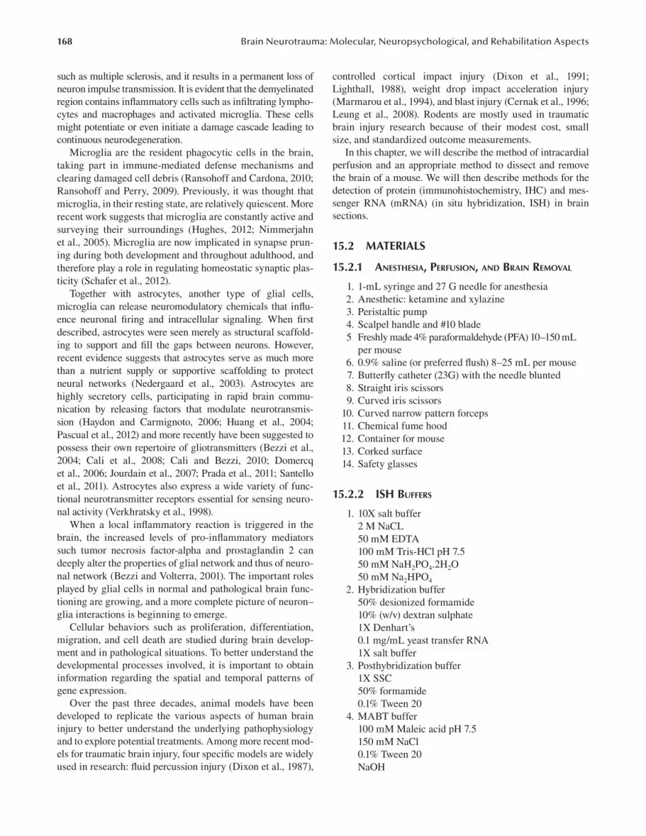
3. Place the mouse on its back. 4. Check the withdrawal reflex once more to assure
adequate depth of anesthesia. 5. Make a midline skin incision from the thoracic inlet
to the pelvis (Figure 15.1a). 6. Use scissors to carefully open the abdomen
(Figure 15.1b). 7. Grasp the xiphoid, which is the tip of the sternum,
with forceps and make an incision through the dia-phragm (Figure 15.1c), then down the costal carti-lage (Figure 15.1d).
8. Flip the thoracic cage widely enough to visualize the heart.
9. Grasp the heart gently and lift it to the midline and slightly out of the chest.
10. The left ventricle is thicker and lighter pink than the right ventricle.
11. Place butterfly needle into the apex of the left ven-tricle, and turn on the pump with a flow no higher than 0.5 mL/minute of PBS buffer (Figure 15.1e and f). Then immediately cut the right auricle to allow the perfusate to exit the circulation (Figure 15.1g and h).
AQ1

170 Brain Neurotrauma: Molecular, Neuropsychological, and Rehabilitation Aspects
12. When the fluid exiting the mouse is clear of blood, perfuse with 4% PFA.
13. Muscle contractions and blanching of the liver and mesenteric blood vessels are signs of good perfu-sion. Perfusion is complete when all muscle con-tractions have stopped, the liver and mesenteric vessels are blanched, and the desired amount of preservative has passed through the circulatory sys-tem. The mouse should be stiff. A good indication of how well the animal is being fixed is to test tail flexibility.
14. PFA and other fixatives must be collected after the perfusion and stored appropriately as hazardous chemical waste.
For immunofluorescence staining, postfixation can affect the antigen (Ag). Therefore, trials should be done with differ-ent times of postfixation. For example, postfixation for any amount of time does not seem to affect SMI-32 (unphos-phorylated neurofilaments) immunoreactivity, but prolonged postfixation times will affect most common Ags for visual-izing microglia.
15.3.1.3 Troubleshooting 1. A mouse still having a positive withdrawal reflex
before the procedure indicates inadequate anesthe-sia. Administer one-third of the original dose of anesthesia and allow the mouse to rest in a dark, quiet cage until withdrawal reflexes are absent.
2. Fluid dripping from the mouse’s nose indicates too much fluid pressure.
3. Fluid not seen dripping in the fourth chamber indi-cates that the fluid is not flowing into the mouse’s circulatory system. The butterfly needle should be repositioned.
15.3.2 BrAin removAl
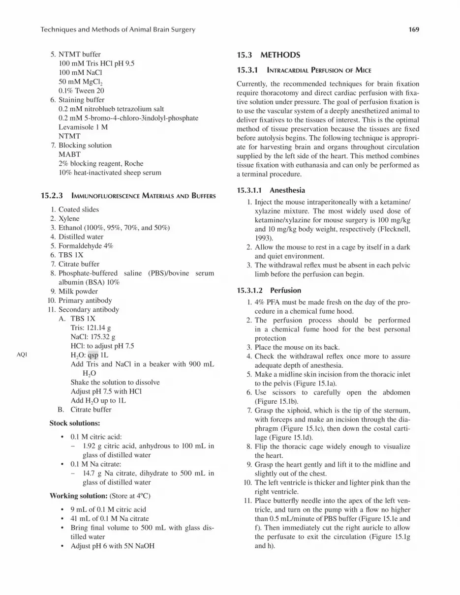
1. Make a midline incision in the skin using the scis-sors (Figure 15.2A). Flip the skin over the eyes to free the skull. Insert the scissors beneath each eye to cut the optical nerve and extract the eyes (Figure 15.2b).
2. Insert Iris scissors caudally to the interparietal bone and cut along the sagittal suture (Figure 15.2c). Incline the scissors ~45° to avoid cutting through the brain (Figure 15.2d).
3. Tilt one side of the parietal bone with the curved narrow pattern forceps and break it off (Figure 12.2e and f). Do the same with the other side to reveal the brain. Most likely the frontal bone will remain. In that case, make a firm cut through the most anterior part of the skull, between the eyes (frontal bone, Figure 15.2g). This enables to remove the brain more easily.
4. Make a small incision that enables tilting and break-ing off this bone plate. Cut the meninges that are beneath the skull.
5. When the brain is freed from meninges, slide the curved narrow pattern forceps (closed) under anterior part of the brain (olfactory bulb) and tilt the brain gently upward (Figure 15.2h). Gently place the forceps underneath the brain and sep-arate it from the underlying tissue. Break the optic nerves and other cranial nerves (Figure 15.2i and j) and gently lift the brain out of the skull (Figure 15.2j).
6. Transfer brain to a petri dish filled with PBS buffer and placed on ice to cool down the brain immedi-ately. These steps should be performed within 2 to 3 minutes.
(a) (b) (c) (d)
(h)(g)(f )(e)
FIgURe 15.1 Intracardial perfusion of mice. (a) A midline skin incision from the thoracic inlet to the pelvis is made. (b) Scissors are used to open the abdomen. (c) The xiphoid process is grasped with forceps and an incision is made through the diaphragm, then down the costal cartilage; (d) the sternum is flipped widely enough to visualize the heart. (e,f) A butterfly needle is placed into the apex of the left ventricle. (g,h) The right auricle is cut to allow the perfus-ate to exit the circulation.
(a) (b) (c) (d)
(h) (i) (j)(g)(f )
(e)
FIgURe 15.2 Brain removal. (a) A midline incision in the skin is made using the scissors. (b) The eyes are removed. (c) Iris scis-sors are inserted caudally to the interparietal bone (d) and the skull is cut along the sagittal suture. (e,f) One side of the parietal bone is tilted with the curved narrow pattern forceps. (g) The same is done with the other side to reveal the brain. (h) The curved forceps is inserted under the olfactory bulbs and the brain is gently tilted upward. (i) The brain is separated from the underlying tissue, and the optic nerves and other cranial nerves are broken. (j) The brain is lifted out of the skull.

171Techniques and Methods of Animal Brain Surgery
15.3.3 BrAin slicing
To perform histological studies, the brain must be sliced to visualize the brain cytoarchitecture. Different techniques can be used:
The cryosectioning is the slicing of a frozen brain into sec-tions as thin as a 10 µm. After fixation, the brain is immersed in 20% sucrose to cryoprotect and prevent freeze artifact and loss of tissue architecture. The brain is then embeded in fresh optimum cutting temperature compound and immersed in isopentane placed in liquid nitrogen to permit rapid freez-ing. Cryosections show high preservation of antigenicity and therefore the detection of Ags by microscopy. They can be used in a variety of procedures including immunochemistry, enzymatic detection, and in situ hybridization.
Biological samples often need to be solidified to allow fine sectioning. Thin slices improve the access of dyes, probes, and antibodies. For light microscopy, paraffin wax is the most frequently used on hard matrix for cutting. Paraffin sections for light microscopy are typically 5 μm thick. After fixation, the water must be removed from the tissue block, a process called dehydration, usually done by using Isopropyl alcohol. Before sectioning, the tissue block must be infil-trated with a material such as paraffin that acts as a support during the sectioning process. Then, the tissue is allowed to solidify in a mold, embedded within a small cube of paraf-fin. Sectioning is accomplished by using a cutting apparatus called a microtome.
Vibratome sectioning is often used for floating immu-nostaining. This technique does not require any organic sol-vents or typical dehydration steps before embedding, which preserves the tissue morphology. The brain is embedded in
agarose and a vibrating razor blade is used to cut through the tissue. This technique allows cutting of thick sections (50 μm). The disadvantage of Vibratome sections is that the sectioning process is slow and difficult with soft and poorly fixed tissues.
The major planes of sectioning are transverse, coronal, and sagittal sections:
• Transverse sections (also called horizontal sections) (Figure 15.3a) provide a view of the whole brain, from the olfactory lobes, to the cerebellum.
• Coronal sections (Figure 15.3b), if made at specific sites, allow similar areas to be examined, so that comparisons can be made between littermate con-trols and from mutant animals. Coronal sections of the entire brain can be examined in order to pick up small abnormalities.
• Sagittal sections (Figure 15.3c) are made for eas-ier viewing of each half of the brain, viewed from caudal to rostral aspects, to detect any obvious abnormalities.
15.3.4 in situ hyBridizAtion
In situ hybridization (Povlishock and Christman) is a tech-nique that allows direct analysis of gene expression through the localization of specific nucleic acid sequences to indi-vidual cells within a morphologically preserved tissue (Guiot and Rahier, 1995). Detection is carried out using nucleic acid probes that are complementary to hybridize with a particular mRNA sequence (Jin and Lloyd, 1997). Because this detection is performed on tissue sections, in situ hybridization provides
(b)
(a)(c)
FIgURe 15.3 A schematic diagram showing the three principal section planes of a brain: (a) transverse, (b) coronal, and (c) sagittal.

172 Brain Neurotrauma: Molecular, Neuropsychological, and Rehabilitation Aspects
additional morphological information on the spatial distribu-tion and heterogeneity of gene expression in complex tissues.
Identifying the precise cellular localization of genes of interest is often necessary for understanding the regulation and the function of the genes. Traditionally, ISH depends on the hybridization of the specific RNA sequence in situ to radiolabeled probes (Gall and Pardue, 1969). Currently, digoxigenin-labeled probes are more commonly used in ISH, which can be recognized with antibodies coupled with fluorophore or enzymes such as alkaline phosphatase or peroxidase (Komminoth et al., 1992). ISH can be per-formed on both frozen sections and paraffin sections, with frozen sections allowing more sensitive detection of weak signals (Wilcox, 1993). The ISH method used in this chap-ter uses digoxigenin-labeled riboprobes (complementary RNA probes) to detect specific mRNA on frozen brain sec-tions. The procedure includes hybridization of sections with digoxigenin-labeled riboprobes, posthybridization washes, incubation with alkaline phosphatase-conjugated antidigoxi-genin antibody (Margulies et al., 2009); colormetric analysis using phosphatase substrate can be used for detection.
15.3.4.1 Protocol
1. Let the slides dry at room temperature 15 minutes to 4 hours.
2. The digoxigenin-labeled probe is diluted (usually 1/200) immediately before use in hybridization buf-fer, denaturated at 65° C for 5 to 10 minutes.
3. An appropriated volume (usually 150 µL per slide) of diluted probe is placed on each slide.
4. The slides are cover slipped and placed in a humidi-fied chamber overnight at 65° C.
5. After overnight hybridization, slides are incubated in posthybridization buffer at 65° in glass Coplin jars until the coverslips slide off.
6. The slides are washed two times 30 minutes in posthybridization buffer at 65°.
7. The slides are washed two times for 30 minutes in MABT at room temperature.
8. The slides are transferred to a humidified chamber and incubated in blocking solution for 1 hour at room temperature without a coverslip.
9. AP-conjugated anti-DIG antibodies are diluted 1/2000 in blocking solution. An appropriated vol-ume is placed on each slide.
10. The slides are cover-slipped and placed in a humidi-fied chamber overnight at room temperature.
11. The slides are transferred to Coplin jars and washed five times for 20 minutes at room temperature in MABT buffer.
12. The slides are washed two times for 20 minutes at room temperature in NTMT prestaining buffer.
13. The slides are incubated in the dark at 37°C in the staining buffer until the signal reaches a satisfactory intensity (usually a few hours to overnight).
14. The slides are washed in PBS 1X and mounted.
15.3.5 ihc
The IHC technique is used in the search for cell or tissue Ags ranging from amino acids and proteins to infectious agents and specific cellular populations. IHC is an umbrella term that encompasses many methods used to determine tis-sue constituents (the Ags) with the use of specific antibodies that can be visualized through staining (Brandtzaeg, 1998; Haines and West, 2005). When used in cell preparations it is called immunocytochemistry.
The fundamental concept behind IHC is the demonstra-tion of Ags within tissue sections by means of specific anti-bodies (Abs). Once Ag–Ab binding occurs, it is demonstrated with a colored histochemical reaction visible by light micros-copy or fluorochromes with ultraviolet light. So, it is based on the binding of Abs to a specific Ag in tissue sections. The most common immunoglobulin (Haydon and Carmignoto, 2006) used in IHC is immunoglobulin G.
Fixation of tissues is necessary to (1) adequately preserve cellular components, including soluble and structural proteins; (2) prevent autolysis and displacement of cell constituents, including Ags and enzymes; (3) stabilize cellular materials against deleterious effects of subsequent procedures; and (4) facilitate conventional staining and immunostaining. Two types of fixatives are used in histopathology: cross-linking (noncoagulating) fixatives and coagulating fixatives.
Formaldehyde is the gold standard of fixatives for rou-tine histology and IHC. Formaldehyde preserves mainly peptides and the general structure of cellular organelles. It also interacts with nucleic acids, but has little effect on car-bohydrates. Many of the formalin substitutes are coagulat-ing fixatives that precipitate proteins by breaking hydrogen bonds in the absence of protein cross-linking. The typical non– cross-linking fixative is ethanol.
Fixation modifies, several times, the tertiary structure of pro-teins (Ags), making them undetected by specific Abs. This is better understood if one remembers that the reaction between the Ag and the Ab depends on the conformation of the former. One of the challenges of IHC is to develop methods that reverse changes produced during fixation. Antigen retrieval produc-ers reverse at least some of these changes. Antigen retrieval is particularly necessary when tissues are fixed in cross-linking fixatives. Approximately 85% of Ags fixed in formalin require some type of antigen retrieval to optimize the immunoreaction.
The Ag–Ab reaction cannot be seen with the light micro-scope unless it is labeled. Therefore, labels (reporter mole-cules) are attached to the primary, secondary, or tertiary Abs of a detection system to allow visualization of the immune reaction. Several labels have been used, including fluorescent compounds, enzymes, and metals. The most commonly used labels are enzymes (e.g., peroxidase, alkaline phosphatase, glucose oxidase). Enzymes in presence of a specific substrate and a chromogen will produce a colored precipitate at the site of the Ag–Ab reaction. Selection of a detection system is very important, considering that the sensitivity of an immune reaction will depend mostly on the detection system used. Detection systems are classified as direct or indirect methods.
AQ2
173Techniques and Methods of Animal Brain Surgery
Background is one of the most common problems in IHC, and it can seriously affect the interpretation of the immuno-logic reaction
15.3.5.1 Protocol 1. Dry paraffin sections (~8–10 µm) on coated slides
(eg: HistoBond) overnight at 37°C 2. Incubate slides at 60°C for 10 minutes 3. Deparaffinization/rehydration:
• Put the slides in xylene bath for 5 minutes. Repeat this step twice in a different bath.
• Put the slides in different ethanol baths (100%, 95%, 70%, and 50%) for three minutes each.
• Put the slides in distilled water for 3 minutes. 4. Fixation:
• Fix the tissue with 1 mL formaldehyde 4%.• Incubate for 20 minutes at room temperature.
5. Aspirate the fixative and wash the slides with TBS 1X for 1 minute.
6. Wash the slides 3 times with TBS 1X for 3 minutes. 7. Antigen unmasking:
• Put the slides in a jar containing 10 mM citrate buffer and immerse it in a boiling water bath (99° C) with a glass lid on.
• Incubate the slides for 10 minutes.• Remove the jar from water bath, open the lid,
and let it cool on bench for 30 minutes. 8. Wash the slides three times with TBS 1X and 2%
milk powder for 5 minutes. 9. Blocking: Incubate the slides with PBS/BSA 10%
for 20 minutes at room temperature. 10. Primary antibody:
• Decant the blocking solution and apply 1 mL of the primary antibody in its working dilution in PBS/BSA 10%.
• Incubate slides overnight at 4°C in a humid chamber.
11. Remove the primary antibody and wash the slides three times with TBS 1X diluted in 2% milk powder for 5 minutes.
12. Secondary antibody:• Apply 1 mL of the secondary antibody in its
working dilution in PBS/BSA 10%.• Incubate slides 1 hour at room temperature in
the dark. 13. Remove the secondary antibody and wash the slides
three times with TBS 1X diluted in 2% milk powder for 5 minutes.
14. Wash the slides three times with TBS 1X without milk powder for 5 minutes.
15. Mount the slides.
15.4 CONCLUsION
In conclusion, this chapter describes methods and tech-niques of animal brain surgery used on brain surgeries and can be applied on experimental brain injury models (mouse
model). The first method described is intracardial perfusion. This method consists in dissecting the thoracic cage of the mouse and injecting a fixative solution to fix and preserve the tissues. Importantly, this technique is a terminal proce-dure because it ends up with euthanasia. Then, this chapter presents an appropriate method to dissect and remove the brain with the tools and solutions necessary for an optimal result. This is followed by the description of three differ-ent techniques used for brain slicing. The first technique is called cryosectioning and consists in slicing frozen tis-sues that can be later used for immunochemistry, enzymatic detection, and in situ hybridization. The second technique is microtome sectioning, which consists in solidifying the biological samples with paraffin to allow fine slicing. The third technique is the Vibratome sectioning that is used for floating immunostaining and allows thick slicing. In addi-tion, the chapter portrays the three major types of sections: transverse, coronal, and sagittal sections. Afterward, the ISH method used for detection of mRNA in brain sections is assessed. This method allows precise cellular localiza-tion of genes of interest by using nucleic acid probes that are complementary to hybridize with a particular mRNA sequence. Of interest, ISH can be used on frozen and paraf-fin embedded sections. In this chapter the protocol described makes use of digoxigenin-labeled riboprobes to detect spe-cific mRNA on frozen brain sections. Finally, this chapter describes the IHC method used to detect protein in brain sections and provides the most appropriate protocol to be followed. IHC is mainly used to demonstrate the presence of Ags within tissue sections by means of specific Abs. Then, labels, mainly enzymes, attached to the primary, secondary, or tertiary Abs allow visualization of the immune reaction.
ReFeReNCes
Bezzi, P., V. Gundersen, J.L. Galbete, G. Seifert, C. Steinhauser, E. Pilati et al. 2004. Astrocytes contain a vesicular compart-ment that is competent for regulated exocytosis of glutamate. Nature Neuroscience. 7:613–620.
Bezzi, P. and A. Volterra. 2001. A neuron-glia signalling net-work in the active brain. Current Opinion in Neurobiology. 11:387–394.
Brandtzaeg, P. 1998. The increasing power of immunohistochem-istry and immunocytochemistry. Journal of Immunological Methods. 216:49–67.
Cali, C. and P. Bezzi. 2010. CXCR4-mediated glutamate exocytosis from astrocytes. Journal of Neuroimmunology. 224:13–21.
Cali, C., J. Marchaland, R. Regazzi, and P. Bezzi. 2008. SDF 1-alpha (CXCL12) triggers glutamate exocytosis from astrocytes on a millisecond time scale: imaging analysis at the single-vesicle level with TIRF microscopy. Journal of Neuroimmunology. 198:82–91.
Cernak, I., J. Savic, Z. Malicevic, G. Zunic, P. Radosevic, I. Ivanovic et al. 1996. Involvement of the central nervous system in the general response to pulmonary blast injury. The Journal of Trauma. 40:S100–104.
Dixon, C.E., G.L. Clifton, J.W. Lighthall, A.A. Yaghmai, and R.L. Hayes. 1991. A controlled cortical impact model of traumatic brain injury in the rat. Journal of Neuroscience Methods. 39:253–262.

174 Brain Neurotrauma: Molecular, Neuropsychological, and Rehabilitation Aspects
Dixon, C.E., B.G. Lyeth, J.T. Povlishock, R.L. Findling, R.J. Hamm, A. Marmarou et al. 1987. A fluid percussion model of experimental brain injury in the rat. Journal of Neurosurgery. 67:110–119.
Domercq, M., L. Brambilla, E. Pilati, J. Marchaland, A. Volterra, and P. Bezzi. 2006. P2Y1 receptor-evoked glutamate exocy-tosis from astrocytes: control by tumor necrosis factor-alpha and prostaglandins. The Journal of Biological Chemistry. 281:30684–30696.
Flecknell, P.A. 1993. Anaesthesia of animals for biomedical research. British Journal of Anaesthesia. 71:885–894.
Gall, J.G. and M.L. Pardue. 1969. Formation and detection of RNA-DNA hybrid molecules in cytological preparations. Proceedings of the National Academy of Sciences of the United States of America. 63:378–383.
Guiot, Y. and J. Rahier. 1995. The effects of varying key steps in the non-radioactive in situ hybridization protocol: a quantitative study. The Histochemical Journal. 27:60–68.
Haines, D.M. and K.H. West. 2005. Immunohistochemistry: forg-ing the links between immunology and pathology. Veterinary Immunology and Immunopathology. 108:151–156.
Haydon, P.G. and G. Carmignoto. 2006. Astrocyte control of syn-aptic transmission and neurovascular coupling. Physiological Reviews. 86:1009–1031.
Huang, Y.H., S.R. Sinha, K. Tanaka, J.D. Rothstein, and D.E. Bergles. 2004. Astrocyte glutamate transporters regulate metabotropic glutamate receptor-mediated excitation of hippocampal inter-neurons. The Journal of Neuroscience. 24:4551–4559.
Hughes, V. 2012. Microglia: the constant gardeners. Nature. 485:570–572.
Jin, L. and R.V. Lloyd. 1997. in situ hybridization: methods and applications. Journal of Clinical Laboratory Analysis. 11:2–9.
Jourdain, P., L.H. Bergersen, K. Bhaukaurally, P. Bezzi, M. Santello, M. Domercq et al. 2007. Glutamate exocytosis from astrocytes controls synaptic strength. Nature Neuroscience. 10:331–339.
Komminoth, P., F.B. Merk, I. Leav, H.J. Wolfe, and J. Roth. 1992. Comparison of 35S- and digoxigenin-labeled RNA and oli-gonucleotide probes for in situ hybridization. Expression of mRNA of the seminal vesicle secretion protein II and androgen receptor genes in the rat prostate. Histochemistry. 98:217–228.
Leung, L.Y., P.J. VandeVord, A.L. Dal Cengio, C. Bir, K.H. Yang, and A.I. King. 2008. Blast related neurotrauma: a review of cellular injury. Molecular & Cellular Biomechanics. 5:155–168.
Lighthall, J.W. 1988. Controlled cortical impact: a new experimen-tal brain injury model. Journal of Neurotrauma. 5:1–15.
Margulies, S., R. Hicks, and Combination Therapies for Traumatic Brain Injury Workshop Leaders. 2009. Combination therapies for traumatic brain injury: prospective considerations. Journal of Neurotrauma. 26:925–939.
Marmarou, A., M.A. Foda, W. van den Brink, J. Campbell, H. Kita, and K. Demetriadou. 1994. A new model of diffuse brain injury in rats. Part I: Pathophysiology and biomechanics. Journal of Neurosurgery. 80:291–300.
Nedergaard, M., B. Ransom, and S.A. Goldman. 2003. New roles for astrocytes: redefining the functional architecture of the brain. Trends in Neurosciences. 26:523–530.
Nimmerjahn, A., F. Kirchhoff, and F. Helmchen. 2005. Resting microglial cells are highly dynamic surveillants of brain parenchyma in vivo. Science. 308:1314–1318.
Pascual, O., S. Ben Achour, P. Rostaing, A. Triller, and A. Bessis. 2012. Microglia activation triggers astrocyte-mediated modulation of excitatory neurotransmission. Proceedings of the National Academy of Sciences of the United States of America. 109:E197–205.
Pham, K. and R. Gupta. 2009. Understanding the mechanisms of entrapment neuropathies. Review article. Neurosurgical Focus. 26:E7.
Povlishock, J.T. and C.W. Christman. 1995. The pathobiology of traumatically induced axonal injury in animals and humans: a review of current thoughts. Journal of Neurotrauma. 12:555–564.
Prada, I., J. Marchaland, P. Podini, L. Magrassi, R. D’Alessandro, P. Bezzi et al. 2011. REST/NRSF governs the expression of dense-core vesicle gliosecretion in astrocytes. The Journal of Cell Biology. 193:537–549.
Ransohoff, R.M. and A.E. Cardona. 2010. The myeloid cells of the central nervous system parenchyma. Nature. 468:253–262.
Ransohoff, R.M. and V.H. Perry. 2009. Microglial physiology: unique stimuli, specialized responses. Annual Review of Immunology. 27:119–145.
Santello, M., P. Bezzi, and A. Volterra. 2011. TNFalpha controls glutamatergic gliotransmission in the hippocampal dentate gyrus. Neuron. 69:988–1001.
Schafer, D.P., E.K. Lehrman, A.G. Kautzman, R. Koyama, A.R. Mardinly, R. Yamasaki et al. 2012. Microglia sculpt postnatal neural circuits in an activity and complement-dependent man-ner. Neuron. 74:691–705.
Verkhratsky, A., R.K. Orkand, and H. Kettenmann. 1998. Glial calcium: homeostasis and signaling function. Physiological Reviews. 78:99–141.
Wilcox, J.N. 1993. Fundamental principles of in situ hybridiza-tion. The Journal of Histochemistry and Cytochemistry. 41:1725–1733.

Author Queries
AQ1 on page 169: Please spell out qsp.AQ2 on page 172: Please spell out AP.


