Embed Size (px)
Citation preview
LUND UNIVERSITY
PO Box 117221 00 Lund+46 46-222 00 00
Perfusion Monitoring of Advancement and Rotational Flaps. Oculoplastic Surgery andLaser Speckle Contrast Imaging.
Dybelius Ansson, Cu
2021
Document Version:Publisher's PDF, also known as Version of record
Link to publication
Citation for published version (APA):Dybelius Ansson, C. (2021). Perfusion Monitoring of Advancement and Rotational Flaps. Oculoplastic Surgeryand Laser Speckle Contrast Imaging. Lund University, Faculty of Medicine.
Total number of authors:1
General rightsUnless other specific re-use rights are stated the following general rights apply:Copyright and moral rights for the publications made accessible in the public portal are retained by the authorsand/or other copyright owners and it is a condition of accessing publications that users recognise and abide by thelegal requirements associated with these rights. • Users may download and print one copy of any publication from the public portal for the purpose of private studyor research. • You may not further distribute the material or use it for any profit-making activity or commercial gain • You may freely distribute the URL identifying the publication in the public portal
Read more about Creative commons licenses: https://creativecommons.org/licenses/Take down policyIf you believe that this document breaches copyright please contact us providing details, and we will removeaccess to the work immediately and investigate your claim.
CU
DY
BELIU
S AN
SSON
Perfusion M
onitoring of Advancem
ent and Rotational Flaps
2021:38
Department of Clinical Sciences and Ophthalmology
Lund University, Faculty of Medicine Doctoral Dissertation Series 2021:38
ISBN 978-91-8021-044-7ISSN 1652-8220
Perfusion Monitoring of Advancement and Rotational FlapsOculoplastic Surgery and Laser Speckle Contrast Imaging
CU DYBELIUS ANSSON
DEPARTMENT OF CLINICAL SCIENCES AND OPHTHALMOLOGY | LUND UNIVERSITY
Perfusion Monitoring of Advancement and Rotational Flaps
Cu Dybelius Ansson was born in Vietnam, 1985. He moved to Sweden in 1991 and later to Denmark to study medicine at the University of Copenhagen. He received his medical degree in 2012. He is an ophthalmologist special ized in glaucoma and cataract surgery.
9789180
210447
NO
RDIC
SW
AN
EC
OLA
BEL
3041
090
3Pr
inte
d by
Med
ia-T
ryck
, Lun
d 20
21

1
Perfusion Monitoring of Advancement
and Rotational Flaps Oculoplastic Surgery and Laser Speckle Contrast Imaging
Cu Dybelius Ansson, MD
DOCTORAL DISSERTATION
by due permission of the Faculty of Medicine, Lund University, Sweden. To be defended at 09.00 21st May 2021, in LUX Aula, Helgonavägen 3, Lund
University
Faculty Opponent Simon Farnebo, MD, PhD
Adjunct professor, Dept. of Biomedical and Clinical Sciences, University of Linköping, Sweden
2
Organization LUND UNIVERSITY Faculty of Medicine Department of Clinical Sciences and Ophthalmology, Lund, Sweden
Document name DOCTORAL DISSERTATION
Date of issue: May 21
Author: Cu Dybelius Ansson Sponsoring organization
Title and subtitle Perfusion Monitoring of Advancement and Rotational Flaps. Oculoplastic Surgery and Laser Speckle Contrast Imaging Abstract Flaps are widely used in oculoplastic reconstructive surgery to cover defects following the removal of tumors, malformations, or trauma. Adequate blood perfusion of the flap is crucial for its survival. Knowledge about the perfusion in different type of flaps is therefore of great value. The main aim of the studies presented in this thesis was to investigate the changes in blood perfusion in advancement and rotational flaps using laser speckle contrast imaging (LSCI) on pigs and humans, in particular in oculoplastic surgery, but also to examine how the blood perfusion in these flaps is affected by the flap length and diathermic coagulation. Flaps from three different locations were used; random skin flaps on pig flank (Paper I), Hewes tarsoconjunctival eyelid flaps on pigs (Paper II) and human upper eyelid skin flaps (Papers III and IV). The results showed that blood perfusion decreased along the length of the random skin flap, and that the relationship between perfusion and distance from the base was nonlinear (Papers I, III, and IV). In human upper eyelid skin flaps the greatest decrease was seen in the first 15 mm. Beyond 15 mm the perfusion exhibited a constant low value (Papers III and IV). Stretching the nonrotated random skin flaps on the pig flank with a force of 3 N reduced the perfusion to 45%, while a force of 10 N reduced it to 29%, compared with the baseline value (Paper I). In human upper eyelids, stretching the nonrotated random skin flaps with 2 N reduced the perfusion to 43% (Paper IV). Simply rotating a random skin flap or a Hewes tarsoconjunctival flap appeared to have little effect on blood perfusion, however, the combination of rotation and stretching had considerable effects on perfusion (Papers I, II and IV). In human upper eyelid skin flaps, the combination of rotation (90°) and stretching with 2 N reduced the perfusion to 22%, compared with the baseline value (Paper IV). It was also found that diathermic coagulation had detrimental effects on blood perfusion, which was seen to decrease with increasing number of applications (Papers II and III). In conclusion, blood perfusion decreases rapidly with distance from the base of random skin flaps. Rotation combined with stretching reduced the blood perfusion significantly, and should thus be avoided in long flaps. Diathermic coagulation also reduced the blood perfusion, and its use should be carefully considered.
Key words perfusion, flap, eyelid, oculoplastic, diathermic coagulation, laser speckle contrast imaging
Classification system and/or index terms (if any)
Supplementary bibliographical information Language: English
ISSN and key title 1652-8220 ISBN 978-91-8021-044-7
Recipient’s notes Number of pages 49 Price
Security classification
I, the undersigned, being the copyright owner of the abstract of the above-mentioned dissertation, hereby grant to all reference sources permission to publish and disseminate the abstract of the above-mentioned dissertation. Signature Date 2021-03-16
3
Perfusion Monitoring of Advancement
and Rotational Flaps Oculoplastic Surgery and Laser Speckle Contrast Imaging
Cu Dybelius Ansson, MD
4
Cover photo by Cu Dybelius Ansson and Ulf Dahlstrand Copyright pp 1-49 Cu Dybelius Ansson Paper 1 © Elsevier
Paper 2 © Elsevier (Authors retain copyrights)
Paper 3 © LWW Journals
Paper 4 © LWW Journals
Lund University, Faculty of Medicine, Doctoral Dissertation Series 2021:38 ISBN 978-91-8021-044-7 ISSN 1652-8220 Printed in Sweden by Media-Tryck, Lund University Lund 2021

5
To my mother
The first and greatest victory is to conquer yourself.
- Platon
6
Contents
Papers included in this thesis ........................................................................... 8 Abstract ........................................................................................................... 9
Introduction ......................................................................................................... 11 Classification of flaps ..................................................................................... 11
Random flaps ........................................................................................ 11 Axial flaps ............................................................................................. 12 Advancement flaps ................................................................................ 12 Rotational flaps ..................................................................................... 13
Blood circulation in the skin .......................................................................... 14 Blood circulation in the human eyelid ........................................................... 15 Monitoring blood perfusion in flaps .............................................................. 16 Thesis at a glance ........................................................................................... 17
Aims .................................................................................................................... 19 General aims ................................................................................................. 19 Specific aims .................................................................................................. 19
Methods ............................................................................................................... 21 Animal studies ............................................................................................... 21 Human studies .............................................................................................. 23 LSCI equipment ............................................................................................ 25
Safety aspects ........................................................................................ 25
7
Results and discussion .......................................................................................... 27 Blood perfusion in advancement flaps............................................................ 27
Blood perfusion along the length of the advancement flap .................... 27 Blood perfusion when stretching an advancement flap .......................... 28
Blood perfusion in rotational flaps ................................................................. 29 Blood perfusion when stretching a rotational flap ................................. 30
The effects of diathermic coagulation on blood perfusion in flaps .................. 32 LSCI in reconstructive surgery ....................................................................... 33
Conclusions and future perspectives ..................................................................... 35 Conclusions .......................................................................................... 35 Future perspectives ............................................................................... 35
Populärvetenskaplig sammanfattning ................................................................... 37
Acknowledgements .............................................................................................. 41
References ............................................................................................................ 45
8
Papers included in this thesis
1. Nguyen* CD, Sheikh R, Dahlstrand U, Lindstedt S, Malmsjö M: Investigation of blood perfusion by laser speckle contrast imaging in stretched and rotated skin flaps in a porcine model. J Plast Reconstr Aesthet Surg. 2018 Apr;71(4):611-613. doi: 10.1016/j.bjps.2017.08.030. Epub 2017 Sept. 13. PMID: 28967585.
2. Ansson CD, Sheikh R, Dahlstrand U, Hult J, Lindstedt S, Malmsjö M: Blood perfusion in Hewes tarsoconjunctival flaps in pigs measured by laser speckle contrast imaging. JPRAS Open. 2018 Jul. 29;18:98-103. doi: 10.1016/j.jpra.2018.07.001. PMID: 32158843; PMCID: PMC7061646.
3. Nguyen CD, Hult J, Sheikh R, Tenland K, Dahlstrand U, Lindstedt S, Malmsjö M: Blood Perfusion in Human Eyelid Skin Flaps Examined by Laser Speckle Contrast Imaging – Importance of Flap Length and the Use of Diathermy. Ophthalmic Plast Reconstr Surg. 2018 Jul./Aug.;34(4):361-365. doi: 10.1097/IOP.0000000000001010. PMID: 29036006.
4. Ansson CD, Berggren JV, Tenland K, Sheikh R, Hult J, Dahlstrand U, Lindstedt S, Malmsjö M: Perfusion in Upper Eyelid Flaps: Effects of Rotation and Stretching Measured with Laser Speckle Contrast Imaging in Patients. Ophthalmic Plast Reconstr Surg. 2020 Sept./Oct.;36(5):481-484. doi: 10.1097/IOP.0000000000001614. PMID: 32049945.
*Study I is a case report/letter to the editor, but it describes a full study.
In 2018 the author changed his surname from Nguyen to Ansson.
9
Abstract
Flaps are widely used in oculoplastic reconstructive surgery to cover defects following the removal of tumors, malformations, or trauma. Adequate blood perfusion of the flap is crucial for its survival. Knowledge about the perfusion in different type of flaps is therefore of great value. The main aim of the studies presented in this thesis was to investigate the changes in blood perfusion in advancement and rotational flaps using laser speckle contrast imaging (LSCI) on pigs and humans, in particular in oculoplastic surgery, but also to examine how the blood perfusion in these flaps is affected by the flap length and diathermic coagulation. Flaps from three different locations were used; random skin flaps on pig flank (Paper I), Hewes tarsoconjunctival eyelid flaps on pigs (Paper II) and human upper eyelid skin flaps (Papers III and IV).
The results showed that blood perfusion decreased along the length of the random skin flap, and that the relationship between perfusion and distance from the base was nonlinear (Papers I, III, and IV). In human upper eyelid skin flaps the greatest decrease was seen in the first 15 mm. Beyond 15 mm the perfusion exhibited a constant low value (Papers III and IV).
Stretching the nonrotated random skin flaps on the pig flank with a force of 3 N reduced the perfusion to 45%, while a force of 10 N reduced it to 29%, compared with the baseline value (Paper I). In human upper eyelids, stretching the nonrotated random skin flaps with 2 N reduced the perfusion to 43% (Paper IV). Simply rotating a random skin flap or a Hewes tarsoconjunctival flap appeared to have little effect on blood perfusion, however, the combination of rotation and stretching had considerable effects on perfusion (Papers I, II and IV). In human upper eyelid skin flaps, the combination of rotation (90°) and stretching with 2 N reduced the perfusion to 22%, compared with the baseline value (Paper IV).
It was also found that diathermic coagulation had detrimental effects on blood perfusion, which was seen to decrease with increasing number of applications (Papers II and III).
In conclusion, blood perfusion decreases rapidly with distance from the base of random skin flaps. Rotation combined with stretching reduced the blood perfusion significantly, and should thus be avoided in long flaps. Diathermic coagulation also reduced the blood perfusion, and its use should be carefully considered.

10
11
Introduction
Plastic reconstructive surgery is used in the repair and reconstruction of defects following the removal of tumors, malformations or trauma. Skin flaps are widely used in plastic reconstructive surgery to cover defects. The advantages of local flaps over secondary intention healing or skin grafting include better matching of skin color and texture together with minimal wound contraction (1). The flap must be long enough to allow it to be moved from the donor site to the recipient site, while still having sufficient blood perfusion to ensure viability of the tissue. The effects of rotating and stretching a random flap on its blood perfusion have not been sufficiently investigated, particularly in oculoplastic surgery.
The work described in this thesis was carried out to investigate the change in blood perfusion in random advancement and rotational flaps, and to examine how the blood perfusion in flaps is affected by the flap length and diathermic coagulation. Laser speckle contrast imaging was used to monitor perfusion, first in a porcine model and thereafter in humans focusing mainly on the upper eyelid region.
Classification of flaps
Flaps can be classified according to their blood supply, such as random and axial flaps. But they can also be classified based on the primary movement of the flap (2). Two well-known types of tissue movement are advancement and rotation. In many cases, it is necessary to both rotate and stretch skin flaps to cover a defect. Such flaps are often described by both their blood supply and their primary movement, e.g. random advancement flaps or random rotational flaps.
Random flaps
A random skin flap, unlike axial flaps, lacks a specific vessel for vascularization. It is perfused by musculocutaneous microcirculation in the tissue, and the blood supply is derived longitudinally from the dermal plexus at the base of the flap. The capacity of
12
the dermal plexus is, however, limited since it lacks an axial arteriovenous system (3, 4). It is known that a random flap is more viable on the face than anywhere else on the body, and most skin flaps are of this type, especially in the richly vascularized periorbital region (2, 4).
Axial flaps
An axial flap has a large cutaneous arteriovenous system that provides blood supply within the skin, parallel to the skin surface. An axial pattern flap incorporates an anatomically named vascular pedicle (3, 5). Such a flap can be created in the periorbital region utilizing the superficial temporal artery. Axial flaps are not often used in oculoplastic surgery (2), and were therefore not investigated in this work.
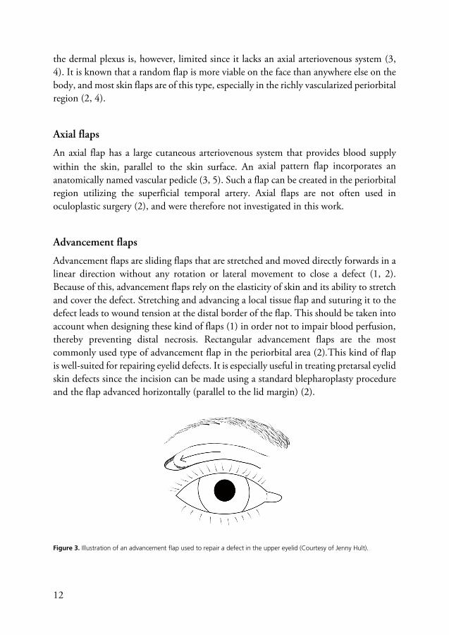
Advancement flaps
Advancement flaps are sliding flaps that are stretched and moved directly forwards in a linear direction without any rotation or lateral movement to close a defect (1, 2). Because of this, advancement flaps rely on the elasticity of skin and its ability to stretch and cover the defect. Stretching and advancing a local tissue flap and suturing it to the defect leads to wound tension at the distal border of the flap. This should be taken into account when designing these kind of flaps (1) in order not to impair blood perfusion, thereby preventing distal necrosis. Rectangular advancement flaps are the most commonly used type of advancement flap in the periorbital area (2).This kind of flap is well-suited for repairing eyelid defects. It is especially useful in treating pretarsal eyelid skin defects since the incision can be made using a standard blepharoplasty procedure and the flap advanced horizontally (parallel to the lid margin) (2).
Figure 3. Illustration of an advancement flap used to repair a defect in the upper eyelid (Courtesy of Jenny Hult).
13
Rotational flaps
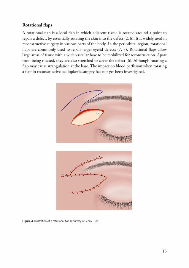
A rotational flap is a local flap in which adjacent tissue is rotated around a point to repair a defect, by essentially rotating the skin into the defect (2, 6). It is widely used in reconstructive surgery in various parts of the body. In the periorbital region, rotational flaps are commonly used to repair larger eyelid defects (7, 8). Rotational flaps allow large areas of tissue with a wide vascular base to be mobilized for reconstruction. Apart from being rotated, they are also stretched to cover the defect (6). Although rotating a flap may cause strangulation at the base. The impact on blood perfusion when rotating a flap in reconstructive oculoplastic surgery has not yet been investigated.
Figure 4. Illustration of a rotational flap (Courtesy of Jenny Hult).
14
Blood circulation in the skin
Human skin forms an external barrier surrounding the body and has three main functions: protection, sensation and thermoregulation. The skin has an outer layer, the epidermis, and an inner layer, the dermis. The epidermis is mainly composed of keratinized epithelium. The thickness is about 0.04 mm to 1.4 mm thick and varies with location on the body. It is about 0.04 mm thick in the upper eyelids (9, 10). Underneath the epidermis is the dermis, which is composed of fibroblasts and dense connective tissue that provide support and nutrients to the epidermis. It also contains hair follicles, sweat glands, nerves, lymphatic vessels, and blood vessels. The thickness of the dermis also varies with location on the body. It is about 0.6 mm thick in the eyelids, but approximately 3 mm on the palms of the hands and the soles of the feet (9).
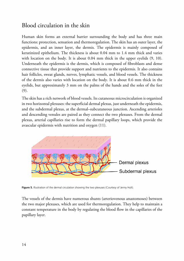
The skin has a rich network of blood vessels. Its cutaneous microcirculation is organized in two horizontal plexuses: the superficial dermal plexus, just underneath the epidermis, and the subdermal plexus, at the dermal–subcutaneous junction. Ascending arterioles and descending venules are paired as they connect the two plexuses. From the dermal plexus, arterial capillaries rise to form the dermal papillary loops, which provide the avascular epidermis with nutrition and oxygen (11).
Figure 5. Illustration of the dermal circulation showing the two plexuses (Courtesy of Jenny Hult).
The vessels of the dermis have numerous shunts (arteriovenous anastomoses) between the two major plexuses, which are used for thermoregulation. They help to maintain a constant temperature in the body by regulating the blood flow in the capillaries of the papillary layer.
15
Blood circulation in the human eyelid
The eyelids contain several anatomical structures that keep the eye lubricated and protect it from the outer world. The tarsal plates of the upper and lower eyelids consist of dense fibrous connective tissue that provide stability, and are anchored to the orbital rim by the medial and lateral canthal ligaments. The tarsal plates are 1 mm thick, and the upper plate has a height of 10-12 mm. Skin moves freely over their anterior surface, although the conjunctiva is tightly bound to the posterior surface. The conjunctiva is a thin translucent mucous membrane. It consists of a superficial conjunctival epithelium overlying a loose connective tissue stroma that contains a rich vascular network, similar to that in the eyelid. In addition, it also receives blood from the anterior ciliary arteries (12).
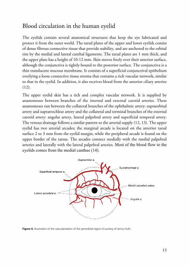
The upper eyelid skin has a rich and complex vascular network. It is supplied by anastomoses between branches of the internal and external carotid arteries. These anastomoses run between the collateral branches of the ophthalmic artery: supraorbital artery and supratrochlear artery and the collateral and terminal branches of the external carotid artery: angular artery, lateral palpebral artery and superficial temporal artery. The venous drainage follows a similar pattern to the arterial supply (12, 13). The upper eyelid has two arterial arcades; the marginal arcade is located on the anterior tarsal surface 2 to 3 mm from the eyelid margin, while the peripheral arcade is found on the upper border of the tarsus. The arcades connect medially with the medial palpebral arteries and laterally with the lateral palpebral arteries. Most of the blood flow to the eyelids comes from the medial canthus (14).
Figure 6. Illustration of the vascularization of the periorbital region (Courtesy of Jenny Hult).
16
Monitoring blood perfusion in flaps
Monitoring the blood perfusion of tissue is challenging. It is difficult to measure the microcirculation in flaps, but advances in laser-based techniques in recent decades have made it easier to monitor the perfusion of flaps. Laser speckle contrast imaging (LSCI) is a recently developed non-invasive technique. The object is illuminated by a laser beam which is spread over the surface of the flap, and the backscattered light, which forms a pattern consisting of dark and bright areas called a speckle pattern, is used to obtain a measure of the perfusion (15). If the illuminated object is static, the speckle pattern is stationary. When there is movement in the object, such as the flow of red blood cells in a tissue, the speckle pattern changes with time, allowing the blood perfusion to be quantified. The results are given in arbitrary units called perfusion units (PU). LSCI is a fast, full-field technique for the imaging of microvascular perfusion (16). Current LSCI equipment can produce up to 100 images per second of the microvascular blood perfusion on the surface of tissue over a relatively large area (up to 24 x 24 cm), with a resolution of up to 100 μm/pixel and with high reproducibility. The measurement depth of LSCI depends on factors such as vascular anatomy and the concentration of red blood cells. Previous studies have reported measurement depths of approximately 700 μm (17, 18).
LSCI is now used in several fields of medicine, such as neurology, dermatology, ophthalmology, and the treatment of burns (19, 20). LSCI has also recently been used to image blood perfusion in skin flaps (21, 22). Stewart et al. were the first to describe the use of LSCI to assess superficial blood perfusion in the surgical treatment of burns. They measured burn scar perfusion and compared laser Doppler imaging and LSCI, and concluded that LSCI performed well (23). Other reported uses of LSCI are in the prediction of brain infarction (24) and functional brain mapping (25). Many studies have been published on the use of LSCI within the field of ophthalmology, since this allows the effect on the cardiovascular system to be studied in great detail, for example, to monitor the effects of diabetes (26, 27).
Full-field monitoring of skin perfusion was one of the earliest reported uses of LSCI (28) since the skin is easy to access. LSCI has been demonstrated to be useful in quantifying the overall perfusion of the capillary bed (29). In dermatology, LSCI is used to study port-wine stain birthmarks (30), to monitor the effects of their treatment (31, 32), and as a surgical guidance tool to reduce the number of sessions required for complete port-wine stain blanching (33, 34).
17
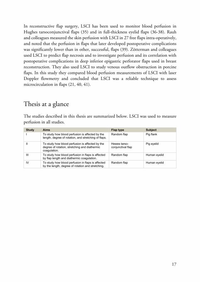
In reconstructive flap surgery, LSCI has been used to monitor blood perfusion in Hughes tarsoconjunctival flaps (35) and in full-thickness eyelid flaps (36-38). Rauh and colleagues measured the skin perfusion with LSCI in 27 free flaps intra-operatively, and noted that the perfusion in flaps that later developed postoperative complications was significantly lower than in other, successful, flaps (39). Zötterman and colleagues used LSCI to predict flap necrosis and to investigate perfusion and its correlation with postoperative complications in deep inferior epigastric perforator flaps used in breast reconstruction. They also used LSCI to study venous outflow obstruction in porcine flaps. In this study they compared blood perfusion measurements of LSCI with laser Doppler flowmetry and concluded that LSCI was a reliable technique to assess microcirculation in flaps (21, 40, 41).
Thesis at a glance
The studies described in this thesis are summarized below. LSCI was used to measure perfusion in all studies.
Study Aims Flap type Subject I To study how blood perfusion is affected by the
length, degree of rotation, and stretching of flaps. Random flap Pig flank
II To study how blood perfusion is affected by the degree of rotation, stretching and diathermic coagulation.
Hewes tarso-conjunctival flap
Pig eyelid
III To study how blood perfusion in flaps is affected by flap length and diathermic coagulation.
Random flap Human eyelid
IV To study how blood perfusion in flaps is affected by the length, degree of rotation and stretching.
Random flap Human eyelid

18
19
Aims
General aims
The main aims of the work presented in this thesis were to investigate the changes in blood perfusion in advancement and rotational flaps, and to examine how the blood perfusion in flaps is affected by the flap length and diathermic coagulation, using LSCI on a porcine model and on human upper eyelids.
Specific aims
• To study how blood perfusion in random skin flaps is affected by flap length, and the degree of rotation and stretching of the flap, using LSCI, in a porcine model.
• To study how blood perfusion in Hewes tarsoconjunctival flaps is affected by the degree of rotation and stretching of the flap and diathermic coagulation, using LSCI, in a porcine model.
• To study how blood perfusion in upper eyelid skin flaps is affected by flap length, the degree of rotation and stretching of the flap, and diathermic coagulation, using LSCI, in patients.

20
21
Methods
Animal studies
Eight domestic pigs were used to study perfusion in random skin flaps (Study I) and Hewes tarsoconjunctival flaps (Study II). The pigs had a body weight of approximately 70 kg. The experiments were performed under general anesthesia, which was maintained throughout the experiments. An intramuscular injection of Dexdomitor® (0.03 mg/kg) mixed with Zoletil® (6 mg/kg) was used for premedication. Anesthesia was then induced by intravenous Pentocur® (0.5 g mixed with 20 ml 0.9% sodium chloride solution) and Fentanyl B. Braun® (2 μg/kg). Anesthesia was maintained by continuous infusion of fentanyl, Ringer’s acetate and sodium thiopental. Blood pressure and pulse were monitored and kept within 120–140 / 80–100 mmHg and 60–80 bpm, respectively. The room temperature was maintained at 20°C. At the end of the experiments, the pigs were euthanized while still under general anesthesia.
The Ethics Committee for Animal Research at Lund University, Sweden, approved the experimental protocols for these studies. The pigs received care in compliance with the European Convention on Animal Care.
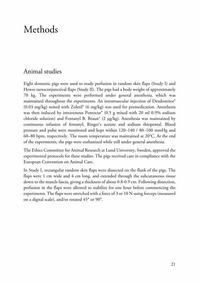
In Study I, rectangular random skin flaps were dissected on the flank of the pigs. The flaps were 1 cm wide and 4 cm long, and extended through the subcutaneous tissue down to the muscle fascia, giving a thickness of about 0.8-0.9 cm. Following dissection, perfusion in the flaps were allowed to stabilize for one hour before commencing the experiments. The flaps were stretched with a force of 3 or 10 N using forceps (measured on a digital scale), and/or rotated 45° or 90°.
22
Figure 7. Example of a random skin flap on the flank of a pig.
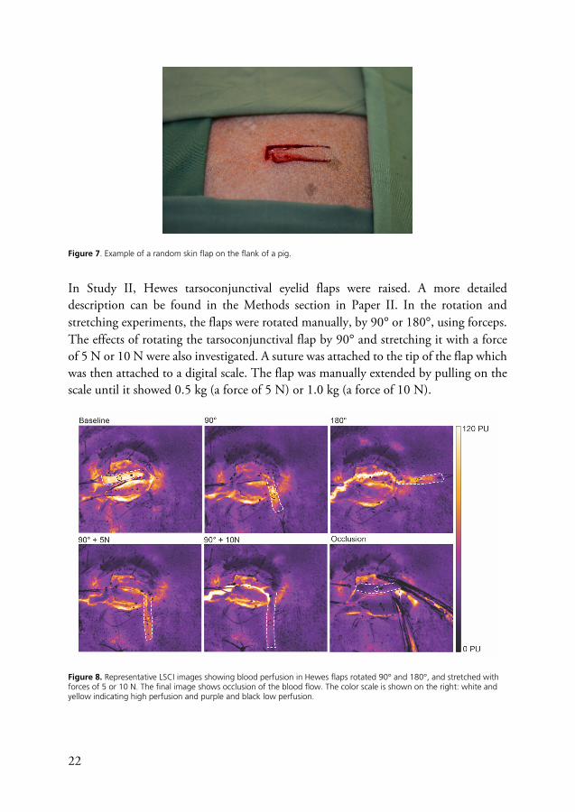
In Study II, Hewes tarsoconjunctival eyelid flaps were raised. A more detailed description can be found in the Methods section in Paper II. In the rotation and stretching experiments, the flaps were rotated manually, by 90° or 180°, using forceps. The effects of rotating the tarsoconjunctival flap by 90° and stretching it with a force of 5 N or 10 N were also investigated. A suture was attached to the tip of the flap which was then attached to a digital scale. The flap was manually extended by pulling on the scale until it showed 0.5 kg (a force of 5 N) or 1.0 kg (a force of 10 N).
Figure 8. Representative LSCI images showing blood perfusion in Hewes flaps rotated 90° and 180°, and stretched with forces of 5 or 10 N. The final image shows occlusion of the blood flow. The color scale is shown on the right: white and yellow indicating high perfusion and purple and black low perfusion.
23
In the diathermic coagulation experiments, diathermic coagulation was applied four times at a location approximately 5 mm distal from the base of the Hewes tarsoconjunctival flaps, using monopolar diathermic coagulation. The first application was at the upper edge of the base, the second at the lower edge of the base, and the third and fourth applications on the dorsal side of the base.
Blood perfusion was measured in the flaps before mechanical manipulation, using LSCI, to obtain a baseline value. When the rotation and stretching and diathermic coagulation experiments had been concluded, the blood supply to the base of the flaps was occluded to obtain a value for zero blood flow, as LSCI measures the blood perfusion in arbitrary units, rather than absolute units.
Human studies
Sixteen patients were included in the human studies: eight in Study III and fiftheen in Study IV. These patients had been referred to the Department of Ophthalmology, Skåne University Hospital, for upper eyelid blepharoplasty. They were consecutively recruited for the studies during February 2017 through May 2018. The patients’ medical records were not made available before the day of surgery. Exclusion criteria were inability to provide informed consent or physical or mental inability to cooperate during the local anesthetic procedure. No patients were excluded. Surgery was performed under local anesthesia. Lidocaine® (20 mg/ml) without adrenaline was used for local anesthesia, to prevent vasoconstriction of the vasculature and to allow perfusion measurements. Lidocaine is a weak vasodilator and may increase perfusion slightly. However, as the local anesthetic was administered in a standardized fashion in all patients, this should not have affected the outcome of the study.
All the patients participating in the studies were given information about the study, and were informed of the voluntary nature of participation. All patients gave their fully informed written consent. The protocol for these experimental studies was approved by the Ethics Committee at Lund University, Sweden.
The eyelid skin was excised as in conventional blepharoplasty, apart from one end, which remained attached to mimic a random skin flap. The flap consisted of skin only, with no orbicularis muscle. It may be argued that a flap based on the medial upper eyelid is not necessarily a random flap due to distinct branches of the angular artery. However, only the skin was dissected, avoiding the angular artery and its subsequent arcades, and was therefore considered a random flap.
24
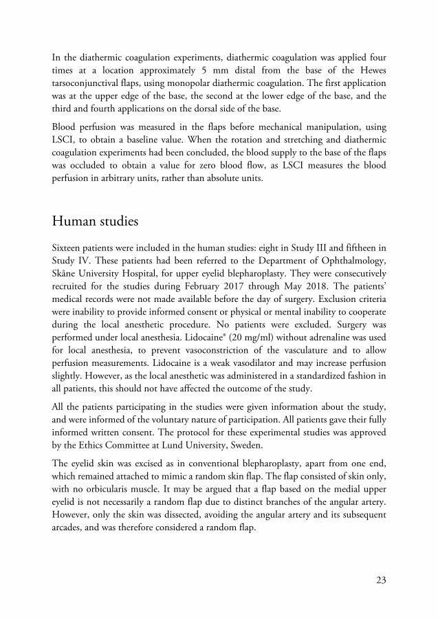
Figure 9. Photograph showing an upper eyelid skin flap. Perfusion was first measured without any rotation or stretching of the flap. Perfusion was thereafter measured under different stretching forces. Two sutures were inserted at the end of the flap, which were attached to a thin nylon line. This was threaded through a shackle mounted on a floor stand that was placed laterally to the patient’s head. Different weights were added to the end of the nylon line (0.05, 0.10, and 0.20 kg) to achieve approximate stretching forces of 0.5, 1, and 2 N. To measure perfusion during rotation, the traction device was moved from laterally to superiorly of the patient’s head.
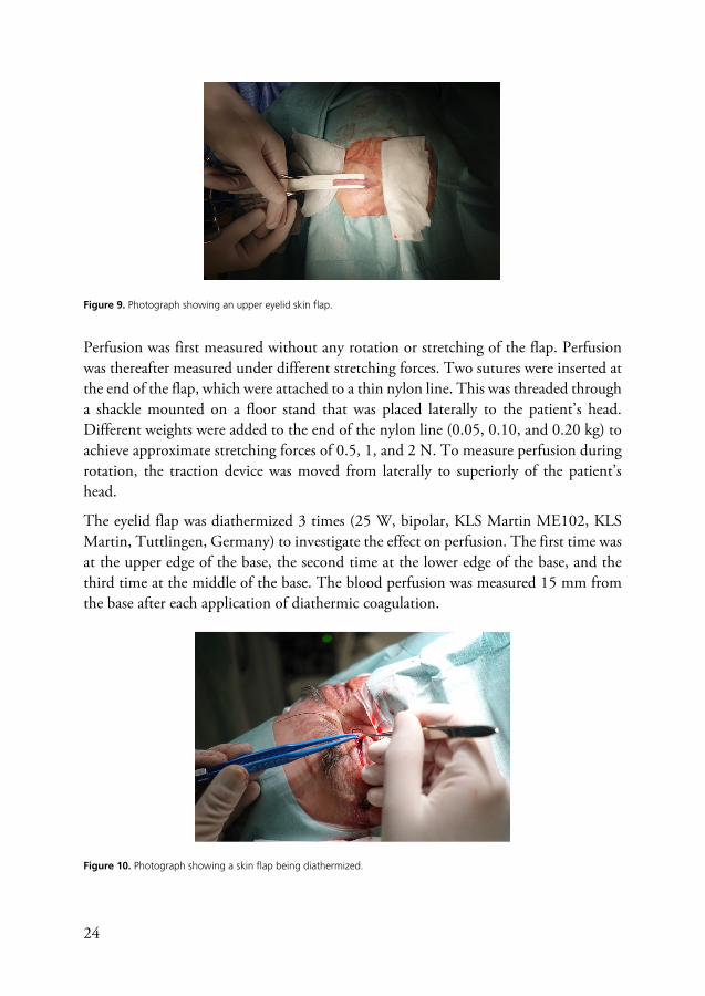
The eyelid flap was diathermized 3 times (25 W, bipolar, KLS Martin ME102, KLS Martin, Tuttlingen, Germany) to investigate the effect on perfusion. The first time was at the upper edge of the base, the second time at the lower edge of the base, and the third time at the middle of the base. The blood perfusion was measured 15 mm from the base after each application of diathermic coagulation.
Figure 10. Photograph showing a skin flap being diathermized.
25
Baseline values were obtained by measuring the perfusion at a point just outside the flap in undissected tissue, and the values recorded were set to 100%. After the LSCI measurements had been completed, the skin flap was detached, and the blepharoplastic procedure was completed according to normal clinical routine. The biological zero was measured on the detached piece of skin, and the value recorded was set to 0%.
LSCI equipment
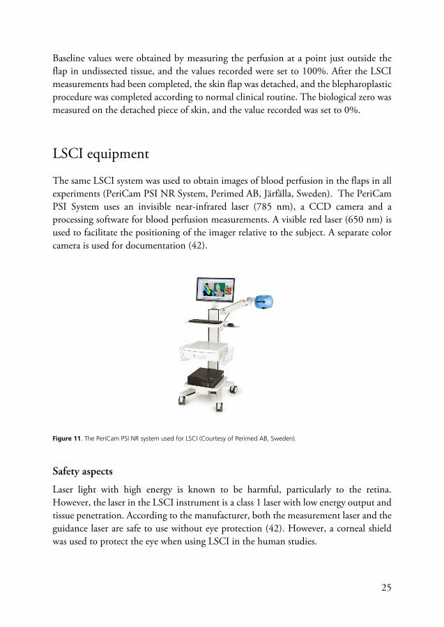
The same LSCI system was used to obtain images of blood perfusion in the flaps in all experiments (PeriCam PSI NR System, Perimed AB, Järfälla, Sweden). The PeriCam PSI System uses an invisible near-infrared laser (785 nm), a CCD camera and a processing software for blood perfusion measurements. A visible red laser (650 nm) is used to facilitate the positioning of the imager relative to the subject. A separate color camera is used for documentation (42).
Figure 11. The PeriCam PSI NR system used for LSCI (Courtesy of Perimed AB, Sweden).
Safety aspects
Laser light with high energy is known to be harmful, particularly to the retina. However, the laser in the LSCI instrument is a class 1 laser with low energy output and tissue penetration. According to the manufacturer, both the measurement laser and the guidance laser are safe to use without eye protection (42). However, a corneal shield was used to protect the eye when using LSCI in the human studies.

26
27
Results and discussion
Blood perfusion in advancement flaps
Blood perfusion along the length of the advancement flap
The flap design used in these studies reflects the design of advancement flaps commonly used in oculoplastic surgery to cover a skin defect. The results of Study I on pig flank showed that blood perfusion along the length of the random skin flap was reduced to 60% 20 mm from the base, to 37% 30 mm from the base, and to 27% 40 mm from the base, compared to the baseline value. In Studies III and IV the perfusion measurements were made along the length of the upper eyelid random skin flaps in humans. In Study III, the blood perfusion was reduced to 69% of the baseline value when measured 5 mm from the base of the flap, to 40% when measured 10 mm from the base, and to 20% when measured 15 mm from the base. At 20 mm from the base, the blood perfusion was only 13% of the baseline value. The same trend was seen in Study IV, i.e. the blood perfusion decreased gradually from the base to the tip of the flap. The flap was only well perfused in the proximal 10 mm (60% at 5 mm and 37% at 10 mm) and was minimally perfused beyond 20 mm (22%).
The decrease in blood perfusion along the length of the random flap was expected. However, the findings indicated that the relationship between perfusion and distance from the base was nonlinear. In human upper eyelids (Studies III and IV) the greatest decrease was seen in the first 15 mm. Beyond 15 mm the perfusion exhibited a constant low value. This nonlinear decrease in blood perfusion is in line with the results of Study I and other studies on full-thickness lower eyelid flaps in the pig and humans (37, 38). However, the blood perfusion was better preserved in the current study on flaps on the pig flank (Study I) compared to the thin upper eyelid skin flaps including only the skin and its subcutis in the present work (Studies III and IV). This may be because the random flaps on the pig flank are thicker, and therefore have a more extensive vascular network. In a previous study on the pig flank, it was shown that perfusion was better maintained in thick flaps than in thin flaps (43).
28
These findings indicate that, from the perspective of perfusion, the optimal length of a thin random upper eyelid skin flap with a 5 or 10 mm base is 15 mm. This is in line with previous studies on animal skin flaps. Stranc et al. (44) monitored rat dorsal skin flaps with near-infrared spectroscopy and reported that the tissue was hypoxic at distances greater than 20 mm from the flap base. In a previous study on random skin flaps on the pig flank a decrease in perfusion was reported with length, approaching zero 25 mm from the flap base (43). If larger tissue areas are required, a free graft could be used. However, the tip of a long flap is basically a partially vascularized (0%–15%) skin graft and may be preferable to a neovascularized free skin graft.
In clinical practice, it is common to limit the width:length ratio of skin flaps in oculoplastic surgery to 1:4 or 1:3, as the viable length of a flap is thought to depend on the width of its base (5). The base of the flap in Study III had a width of 5 mm, while in Study IV the base was almost 10 mm wide. The blood perfusion decreased in a similar manner in both series of flaps, regardless of the width of the base. This suggests that the distance from the base is more important than the width of the base. In a previous study on blood perfusion in random advancement porcine skin flaps, it was reported that the width:length ratio of the flap did not determine the blood flow or oxygenation (43). The viable length of a flap does not necessarily depend on the width of its base. It has also previously been demonstrated experimentally that flaps made under similar conditions survive to the same length regardless of width (45).
Blood perfusion when stretching an advancement flap
Tension is commonly applied to advancement flaps when they are used to repair defects, for example, after tumor surgery. Tension results if the flap is stretched too tightly. Stretching the random skin flaps on pig flank with a force of 3 N reduced the perfusion to 45%, while 10 N reduced the perfusion to 29%, measured 20 mm from the flap base (Study I). In human upper eyelid skin flaps, stretching with 2 N decreased perfusion to 43% at 5 mm (Study IV).
Anything compromising the blood perfusion after transposition can jeopardize flap survival. Indeed, it has been shown that stretching increases the risk of necrosis due to strangulation of the blood vessels in advancement flaps. In 1980, Stell used a porcine model to study the effect of stretching by observing the discoloration and survival of skin flaps, when subjected to different amounts of tension (no stretching, slightly stretched, and maximally stretched). The surviving length in each group did not differ significantly, but flaps under maximal tension were more likely to exhibit distal necrosis (46). However, this is a rather blunt and subjective measure of blood perfusion compared to LSCI. Zötterman et al. used LSCI to predict flap necrosis in a porcine
29
model. A decrease in perfusion to a value <25 PU in the first 30 min after surgery was a predictor of tissue morbidity 72 h after surgery. They suggested that LSCI could be a promising technique for perioperative monitoring in reconstructive flap surgery (41).
Furthermore, it is possible that perfusion may be affected after the flap has been attached to the recipient site. A recent study on bipedicle random advancement skin flaps (including skin and orbicular muscle) on eyelids in 7 patients with median length and width of the skin flaps of 13 mm (range, 8–20 mm) and 10 mm (range, 5–11 mm), respectively, showed that immediately postoperatively, the perfusion in the distal end of the flaps had fallen to 54% (47). Unfortunately, the blood perfusion in the flaps was not measured before the flaps were sutured in place. Furthermore, the degree of stretching was unknown, and it was thus impossible to compare the difference between blood perfusion before and after the flap was sutured to the recipient site.
Larrabee et al. investigated the relationship between wound-closing tension, blood flow, and flap viability in random skin flaps on pigs with laser Doppler flowmetry. In flaps with a poor blood supply, there was a significant increase in flap necrosis in flaps closed with a tension greater than 0.25 kg. Measurements with laser Doppler flowmetry showed an inverse relationship between flap tension and blood flow in these flaps, which correlated well with flap necrosis (48). Burkhardt et al. investigated the role of flap tension in primary wound closure of mucoperiosteal flaps in sixty patients. The wound closing forces were measured with an electronic tension device before suturing, and was found to vary between 0.01 and 0.4 N. A tension of 0.01-0.1 N was applied in 72% of cases, resulting in few cases of dehiscence (10%), while higher closing forces (>0.1 N) led to a significant increase in wound dehiscence (≥ 40%) (49).
Blood perfusion in rotational flaps
The effects of flap rotation on blood perfusion were studied in both pigs and humans (Studies I, II, and IV). The results of Study I showed that rotation of the random skin flaps on pig flank by up to 45° had no significant impact on blood perfusion, while rotation by 90° reduced perfusion to 54% of the baseline value. Rotating the Hewes tarsoconjunctival flaps by 90° had no significant effect on perfusion, while further rotation to 180° reduced the perfusion to 75% of the baseline value (Study II). In Study IV, on upper eyelid random skin flaps in humans, the results showed that rotating the flaps by 90° had no significant effect on the perfusion.
30
It is not unnatural to assume that rotating a skin flap would impair blood perfusion due to strangulation of the blood vessels. It has been reported in a previous study that the maximum tension in a rotational flap is found between 90° and 135° (50). However, the results of the present study show that human eyelid flaps could be rotated by 90° without the blood perfusion being significantly compromised. In Studies II and IV the base of the flaps was medial. It may be argued that a flap based on the medial upper eyelid is not necessarily a random flap due to distinct branches of the angular artery. However, the skin flaps in Study IV were dissected so as to avoid the angular artery and its subsequent arcades, and were therefore considered random flaps. Nevertheless, it cannot be ruled out that the results may have been different if the flaps had been dissected extending from the lateral canthus. Furthermore, it is possible that perfusion may be affected after the flap has been attached to the recipient site.
Blood perfusion when stretching a rotational flap
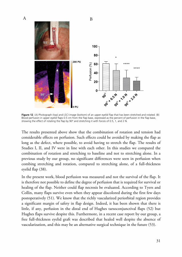
The effects of combined stretching and rotation were investigated in Studies I, II, and IV. The results showed that when stretching with a force of 3 N was applied to the already 90° rotated skin flap on the pig flank, the perfusion decreased to 26% of the baseline value (Study I). A similar investigation was carried out in Study II, using Hewes tarsoconjunctival flaps, which showed that when a stretching force of 5 N was applied to the already rotated tarsoconjunctival flap (90°), the perfusion decreased to 63% of the baseline value. Increasing the stretching force to 10 N caused the perfusion to decrease further, to 36% of the baseline value. In human upper eyelid skin flaps (Study IV) the combination of rotation (90°) and a stretching force of 2 N reduced the perfusion to 22% of the baseline value.
31
Figure 12. (A) Photograph (top) and LSCI image (bottom) of an upper eyelid flap that has been stretched and rotated. (B) Blood perfusion in upper eyelid flaps 0.5 cm from the flap base, expressed as the percent of perfusion in the flap base, showing the effect of rotating the flap by 90° and stretching it with forces of 0.5, 1, and 2 N.
The results presented above show that the combination of rotation and tension had considerable effects on perfusion. Such effects could be avoided by making the flap as long as the defect, where possible, to avoid having to stretch the flap. The results of Studies I, II, and IV were in line with each other. In this studies we compared the combination of rotation and stretching to baseline and not to stretching alone. In a previous study by our group, no significant differences were seen in perfusion when combing stretching and rotation, compared to stretching alone, of a full-thickness eyelid flap (38).
In the present work, blood perfusion was measured and not the survival of the flap. It is therefore not possible to define the degree of perfusion that is required for survival or healing of the flap. Neither could flap necrosis be evaluated. According to Tyers and Collin, many flaps survive even when they appear discolored during the first few days postoperatively (51). We know that the richly vascularized periorbital region provides a significant margin of safety in flap design. Indeed, it has been shown that there is little, if any, perfusion in the distal end of Hughes tarsoconjunctival flaps (52) but Hughes flaps survive despite this. Furthermore, in a recent case report by our group, a free full-thickness eyelid graft was described that healed well despite the absence of vascularization, and this may be an alternative surgical technique in the future (53).
A B
Perfu
sion
(%)
32
The effects of diathermic coagulation on blood perfusion in flaps
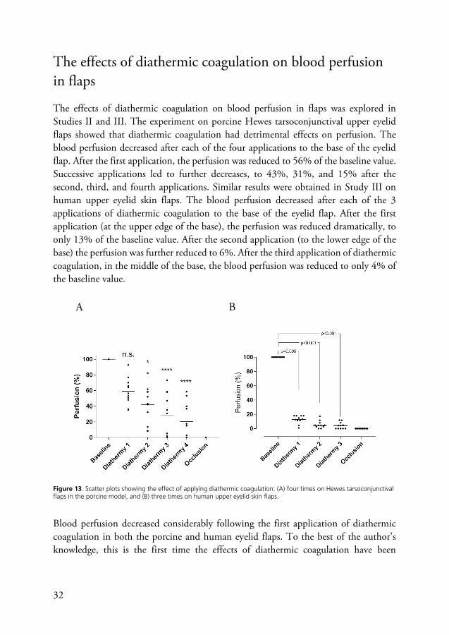
The effects of diathermic coagulation on blood perfusion in flaps was explored in Studies II and III. The experiment on porcine Hewes tarsoconjunctival upper eyelid flaps showed that diathermic coagulation had detrimental effects on perfusion. The blood perfusion decreased after each of the four applications to the base of the eyelid flap. After the first application, the perfusion was reduced to 56% of the baseline value. Successive applications led to further decreases, to 43%, 31%, and 15% after the second, third, and fourth applications. Similar results were obtained in Study III on human upper eyelid skin flaps. The blood perfusion decreased after each of the 3 applications of diathermic coagulation to the base of the eyelid flap. After the first application (at the upper edge of the base), the perfusion was reduced dramatically, to only 13% of the baseline value. After the second application (to the lower edge of the base) the perfusion was further reduced to 6%. After the third application of diathermic coagulation, in the middle of the base, the blood perfusion was reduced to only 4% of the baseline value.
Figure 13. Scatter plots showing the effect of applying diathermic coagulation: (A) four times on Hewes tarsoconjunctival flaps in the porcine model, and (B) three times on human upper eyelid skin flaps.
Blood perfusion decreased considerably following the first application of diathermic coagulation in both the porcine and human eyelid flaps. To the best of the author’s knowledge, this is the first time the effects of diathermic coagulation have been
B A
33
measured on eyelid flap blood perfusion. These observations are thought-provoking, and suggest that the use of diathermic coagulation should be carefully considered, especially in cases of a long thin flap that is poorly perfused. Repeated diathermy at the base of the flap probably causes the flap to function more like a free graft than a flap. Diathermic coagulation of the flap pedicle should thus be avoided, if possible, to ensure adequate vascularization of the flap.
Overdam et al. studied intra‐ocular diathermic coagulation on porcine cadaver eyes and observed in fundal photographs that the blood columns in the artery and vein had been interrupted by coagulation. The coagulation site and the blood vessel downstream from it showed no flow during perfusion of the ophthalmic artery with fluorescein‐stained fluid. The histology examination showed coagulated and closed retinal blood vessels following the application of diathermic coagulation (54).
The design of the studies in the present work did not allow for measurements on the days following surgery. It is therefore not known whether the low perfusion continued in the hours or days after surgery. It is possible that ischemia leads to vasodilation and an increase in the blood flow through the conjunctival vessels during a period after surgery. Tissue may be able to survive for many hours, and perhaps even days, with very little or no blood flow (55-57).
LSCI in reconstructive surgery
Laser speckle contrast imaging could easily be implemented in clinical routines in reconstructive surgery to determine blood perfusion, as it is noninvasive, and the measurements are not very time-consuming. LSCI has also shown good interobserver reliability (58). However, the use of laser-based methods is associated with some limitations due to motion other than that of the blood, which must be overcome. It has also been reported that perfusion appeared to increase with increasing tissue motion. The relation was independent of frame rate and number of image. The angle of measurement was also found to be important since perfusion appeared to decrease with increasing angle (9% at 45° and 16% at 60°). However, the perfusion did not vary significantly when measurement distance from LSCI to the tissue was between 15 and 40 cm (59). Nevertheless, artifacts will be introduced by movement resulting, for example, from breathing and blinking, and care must be taken to eliminate all sources of motion error. In the human eyelid skin flap study, the traction sutures also served to prevent movement, reducing the influence of artifacts on the results. Efforts were also made to limit motion-induced artifacts by instructing the patients to remain still during
34
the recordings. Other methods of overcoming motion artefacts, such as shorter sampling times, simultaneously recording the signal backscattered from an adjacent opaque surface, or retrospective motion correction techniques, have been suggested (60, 61). However, no definitive solution has yet been found. LSCI is unfortunately still not a quantitative method, although considerable advances have been made by Dunn et al. through the introduction of multi-exposure speckle imaging (62).
Laser speckle contrast imaging would be particularly important in cases where flaps or grafts are at risk of failure, i.e., when the geometry or size of the flap indicates a risk of poor perfusion, or when the patient has poor microcirculation, as in the case of diabetics, smokers, or those with cardiovascular disease. If graft failure could be identified early, timely revision could be carried out and the healing process optimized.
35
Conclusions and future perspectives
Conclusions
Based on the studies presented in this thesis, it was concluded that blood perfusion falls rapidly with distance from the base of random advancement skin flaps, both on the pig flank and on the human upper eyelid, and the results suggest that skin flaps in human upper eyelids are only well perfused in the proximal 15 mm, regardless of the width of the base (5 mm or 10 mm). Beyond 15 mm the perfusion leveled off and was low. Stretching advancement flaps affected the perfusion significantly in skin flaps on the pig flank and in human upper eyelids.
Rotating a random skin flap appears to have little effect on blood perfusion. However, when the flaps are both rotated and stretched the blood perfusion decreased significantly. A compromise must thus be found between the length of the flap and the degree to which it is stretched in order to repair a defect. The combination of rotation and stretching may be avoided when long flaps are possible.
The use of diathermic coagulation has detrimental effects on blood perfusion and should be avoided or carefully considered, especially when the flap is long and thin. Repeated diathermic coagulation at the base of the flap probably causes the flap to function more like a free graft than a flap.
Future perspectives
The work described in this thesis has resulted in some interesting findings related to blood perfusion primarily in eyelid flaps, when subjected to stretching, rotation, and diathermic coagulation. These studies were focused on the measurement of flap perfusion, with no long-term follow-up of the viability of the flaps. However, in the clinic, flap viability is often more important. It would therefore be interesting to investigate the effect of stretching force and the degree of rotation on the survival of skin flaps used to repair defects resulting from tumor surgery (and not as part of a blepharoplasty procedure as in the current study). Future studies should also focus on other parts of the body than the periorbital and facial region. It could also be of great value to explore the effects of age, diabetes, smoking and atherosclerosis on flap survival.

36
Diathermic coagulation appears to have an initial negative effect on blood perfusion in the flaps, but the long-term effect is still unknown. Studies should therefore be carried out to clarify the long-term effects of diathermic coagulation on flap survival.
LSCI is a promising technique in perfusion monitoring in flap and reconstructive surgery. The system is fast, non-invasive and has good spatial resolution. However, if LSCI could be developed to allow absolute measurements of perfusion, this would make interpatient comparisons possible. The ability to choose the measurement depth would also be of a great value.
37
Populärvetenskaplig sammanfattning
Cu! Vad forskar du om?
Blodcirkulationen i lambåer, främst i ögonlocken.
Vad är en lambå?
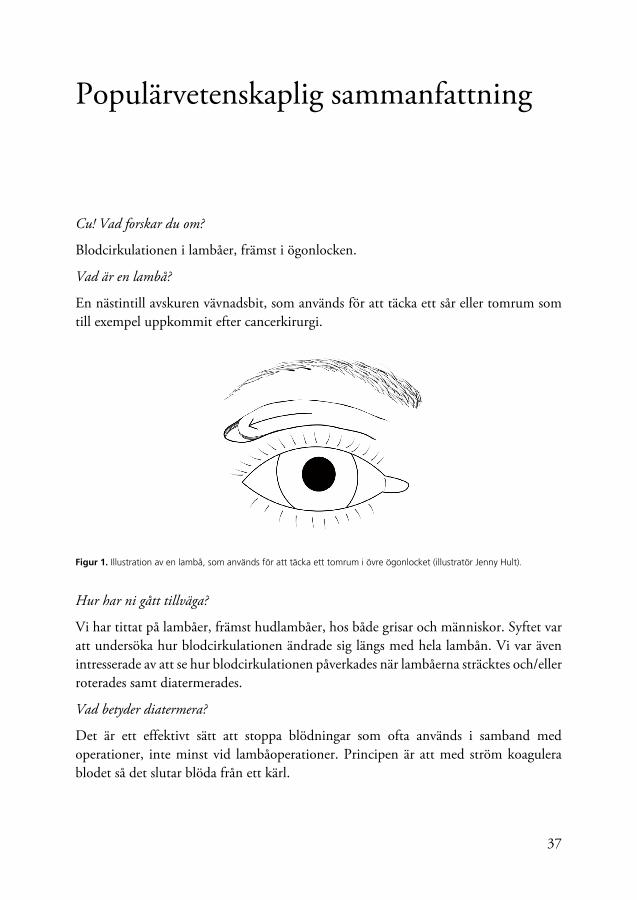
En nästintill avskuren vävnadsbit, som används för att täcka ett sår eller tomrum som till exempel uppkommit efter cancerkirurgi.
Figur 1. Illustration av en lambå, som används för att täcka ett tomrum i övre ögonlocket (illustratör Jenny Hult).
Hur har ni gått tillväga?
Vi har tittat på lambåer, främst hudlambåer, hos både grisar och människor. Syftet var att undersöka hur blodcirkulationen ändrade sig längs med hela lambån. Vi var även intresserade av att se hur blodcirkulationen påverkades när lambåerna sträcktes och/eller roterades samt diatermerades.
Vad betyder diatermera?
Det är ett effektivt sätt att stoppa blödningar som ofta används i samband med operationer, inte minst vid lambåoperationer. Principen är att med ström koagulera blodet så det slutar blöda från ett kärl.
38
Hur gör ni för att mäta blodcirkulationen i en lambå?
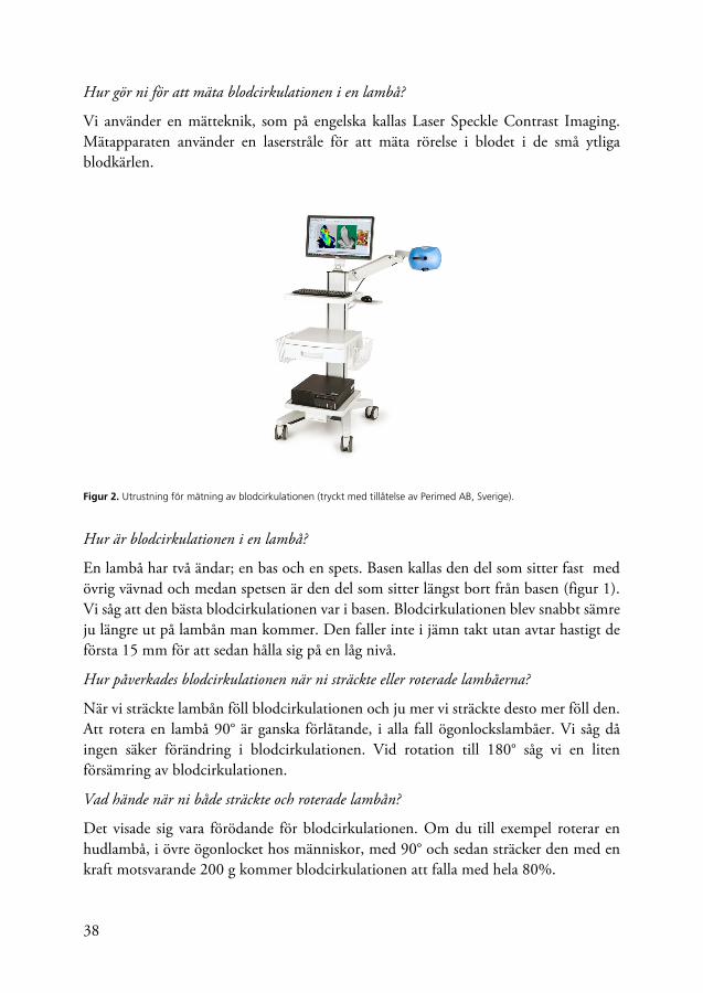
Vi använder en mätteknik, som på engelska kallas Laser Speckle Contrast Imaging. Mätapparaten använder en laserstråle för att mäta rörelse i blodet i de små ytliga blodkärlen.
Figur 2. Utrustning för mätning av blodcirkulationen (tryckt med tillåtelse av Perimed AB, Sverige).
Hur är blodcirkulationen i en lambå?
En lambå har två ändar; en bas och en spets. Basen kallas den del som sitter fast med övrig vävnad och medan spetsen är den del som sitter längst bort från basen (figur 1). Vi såg att den bästa blodcirkulationen var i basen. Blodcirkulationen blev snabbt sämre ju längre ut på lambån man kommer. Den faller inte i jämn takt utan avtar hastigt de första 15 mm för att sedan hålla sig på en låg nivå.
Hur påverkades blodcirkulationen när ni sträckte eller roterade lambåerna?
När vi sträckte lambån föll blodcirkulationen och ju mer vi sträckte desto mer föll den. Att rotera en lambå 90° är ganska förlåtande, i alla fall ögonlockslambåer. Vi såg då ingen säker förändring i blodcirkulationen. Vid rotation till 180° såg vi en liten försämring av blodcirkulationen.
Vad hände när ni både sträckte och roterade lambån?
Det visade sig vara förödande för blodcirkulationen. Om du till exempel roterar en hudlambå, i övre ögonlocket hos människor, med 90° och sedan sträcker den med en kraft motsvarande 200 g kommer blodcirkulationen att falla med hela 80%.

39
Intressant. Vad hände när ni diatermerade lambåerna?
Vi såg att blodcirkulationen till lambån snabbt tog mycket stryk. Inte överraskande blev blodcirkulationen sämre och sämre ju flera gånger vi diatermerade och efter tre gånger med diatermi var det i stort sett ingen blodcirkulation i lambån.
Vad är slutsatsen av dina studier?
Ett: blodcirkulationen faller mycket snabbt från basen av lambån till dess spets. Två: blodcirkulationen faller dramatiskt när vi sträcker eller när vi både sträcker och roterar en lambå. Tre: blodcirkulationen försämras avsevärt när vi diatermerar en lambå.
Hur kommer de här kunskaperna att kunna vara till nytta för patienterna?
Det här är begynnande steg för att öka vår förståelse om blodcirkulationen i lambåer. Förhoppningsvis kan det så småningom mynna ut i förbättringar av operationstekniker för lambåer. Vilket jag hoppas kommer att hända i närliggande framtid.
Då har vi något att se framemot. Tack för att du ställde upp!
Tack själv!

40
41
Acknowledgements
När jag nu sätter punkt för min avhandling ser jag tillbaka på fyra händelserika år. Så många sena kvällar, så många svettiga timmar, men inte minst så många roliga minnen. Jag har haft turen att komma in i en frodande forskningsmiljö med fantastiska handledare och forskarkollegor. Jag hade aldrig klarat det här ensamt. Min stora tacksamhet går ut till alla som har bidragit och möjliggjort genomförandet av denna avhandling, och särskilt vill jag tacka:
Malin Malmsjö, huvudhandledare och professor. Denna avhandling hade inte funnits till utan hennes engagemang, outtömliga positivitet och genomföringskraft. Hon är makalöst energisk med en otrolig förmåga att ta snabba beslut och hitta alternativa lösningar. Med henne som katalysator är avståndet mellan idé och verklighet förvånansvärt kort. En lysande förhandlare som man vill ha på rätt sida av bordet. Hennes snabbhet och effektivitet får min puls att stiga bara av att vara i hennes närhet. En sann inspirationskälla för mig och många kollegor.
Karl Engelsberg, bihandledare och ögonplastikkirurg. Introducerade mig till ögonplastikkirurgin. Mycket hjälpsam och kunnig handledare. Levererar alltid ett gediget arbete både som kirurg och föreläsare. Har gett mig många kloka livsråd. Ett motto som jag tar med mig: familjen framför allt.
Sandra Lindstedt Ingemansson, bihandledare, thoraxkirurg och docent. Har funnits som stöd i bakgrunden och ryckt in närhelst det behövts.
Rafi Sheikh, katarakt- och vitreoretinalkirurg, forskarkollega. Föregångsfigur och fantastisk stöd i forskargruppen. Tycker om att ha många järn i elden, frodas med nya utmaningar och förmår att prestera även under enorm tidspress. Känns som han hinner med mer trots de gemensamma 24 timmarna i dygnet. Rak på sak, har inget problem att ta en konflikt till frukost. Stort hjärta och mycket hjälpsam.
Ulf Dahlstrand, av vem jag har ärvt många förtroendeposter. Ett ovärderligt stöd för mig och många kollegor. Forskargruppens ”Dumbledore”. Skarpsinnig med förmågan att vara ett steg före de flesta av oss. Obestridlig sköterskefavorit, inte ens jag har en chans. Smyger fyndigt in humor i alla möjliga samtal, oavsett ämne. Kommer att skapa sig ett namn inom ögonplastik- och skelningskirurgin.
42
Johanna Berggren, stigande stjärna inom ögonplastikkirurgi och forskning. Har en beundransvärd arbetskapacitet och fokus, verkar kunna ta på sig hur mycket som helst. Alltid nära till ett skratt. Kommer att gå hur långt som helst i sin karriär.
Kajsa Tenland, forskarkollega, som kan konkurrera med Nils Holgersson när det gäller att resa riket runt. Har en fantastisk känsla för det väsentliga och dessutom det estetiska. Tar otroligt fina bilder med oanade talang för inredning.
Jenny Hult, forskarkollega med fantastisk arbetsmoral. Effektiv, noggrann och målinriktad. Är pennan bakom många av forskargruppens snygga publikationsfigurer.
Josefine Bunke, forskarkollega med bas i Växjö. För alltid med sig uppiggande energi. Otrolig pålitlig med en beundransvärd förmåga att genomföra saker.
Khashayar Memarzadeh, tidigare forskarkollega. Fantastiskt duktig kirurg och kliniker. Rik informationskälla, som ständigt bjuder på ett gott samtal.
Magdalena Naumovska, skarpsinnig forskarkollega med ett stort hjärta. Avundsvärd vilja att hjälpa andra framåt. Det är en fröjd att läsa hennes klartänkta journalanteckningar.
Björn Hammar, topprankad neurooftalmolog med en fantastisk iver att leverera undervisning i världsklass. Höjer ribban och sätter standarden för pedagogik inom svensk ögonsjukvård.
Jonas Blohmé, skelningskirurg av tionde graden. Konst skapas när han arbetar med sina händer. Jag har inte sett nån som är bättre på att suturera.
Bodil Gesslein, forskargruppens obestridliga administratör med både PhD och postdoc i bagaget. Gör ett otroligt arbete i bakgrunden för att vi andra ska kunna fokusera på att forska.
John Albinsson, forskarkollega med ingenjörsbakgrund. Har en mycket lugnande effekt på sin omgivning. Återkommande källa för kunskap när det gäller statistikfrågor och annat tekniskt.
Aboma Medasa, forskarkollega med ingenjörsbakgrund. Har ett mycket vänligt sinne med hjälpsam inställning. Det ryktas om oanade talanger när det gäller skapande av 3D-bilder.
Helen Sheppard, språkgranskaren, med en otrolig förmåga att destillera fram essensen i text och skapa ”flow” och precision i språket.
43
Ola Rauer, min mentor och tidigare ST-handledare, vars underskrift garanterar uveitvård i världsklass. Har ett hjälpande hjärta med patientens bästa i fokus. Oerhört vis med ett fantastiskt minne. Mångfacetterade intresseområden av imponerande djup. En dyrbar källa för kloka råd och ovärderlig kunskap. Har lärt mig mer än de flesta, både om ögonen och om livet.
Anders Bergström, min kirurgiska huvudhandledare. En inspirerande läromästare med många strängar på sin lyra. Det är svårt att hitta en som är mer villig att ställa upp i alla tänkbara sammanhang. Sällsynt mentor med stort tålamod. Har ryckt in och räddat mig många gånger om. Förstklassig kirurg, som ständigt är på jakt efter förbättringspotential. Lämnar mig på perrongen när gäller att adaptera ny teknik.
Jesper Hougaard, min glaukomkirurgiske handledare, som är mycket omtyckt av patienterna. Det är sällan gott för ens självförtroende när patienterna säger att de hellre vill träffa dr. Hougaard vid nästa återbesök. En mycket noggrann handledare, som alltid bjuder på ett gott skratt.
Sten Kjellström, en framsynt chef med många visioner och höga ambitioner. Arbetar tålmodigt med stenkoll på talen. En dålig hårdag har jag ännu inte sett.
Kristina Johansson & Lena Rung, cheferna, som förmår att skapa god arbetsmiljö och prioritera personalens välmående. Är en av de bidragande krafterna till att kliniken nu upplever de sju goda åren.
Vesna Ponjavic, tangoälskande studierektor med ett häpnadsväckande sinne för ordning och reda (titta på hennes datorskrivbord). Jag och många kollegor har henne att tacka för våra välstrukturerade ST-förlopp.
Personalen på operation och avdelning 40 i Lund, för ert tålamod och stöd till vår forskningsverksamhet. Eva och Sussie för deras glada lynne och den omtänksamhet de har för patienterna och mig.
Personalen på operation och dagkirurgen i Malmö, för ert bidrag till vår forskning och det tålamod ni har med mig som ung kirurg och nybliven specialist. För de ständiga skratten som bjuds. Jag har aldrig skrattat så mycket på en arbetsplats.
Operationskoordinatorer och sekreterare, som har varit beundransvärd flexibla när det gäller operationsbokningar och schemaläggning.
Alla kollegor och personal, som jag inte nämnt vid namn men som förgyller min dag. En hjälpande hand i nödens stund. Ett vänligt ord. Ett uppiggande samtal i korridoren. Ett gemensamt skratt. Ni gör det roligt att komma till jobbet.
44
Min far, ba An, som skapade förutsättningar för en bättre framtid för mig och hela familjen. Intelligent och framsynt, som har lärt mig så mycket om livet genom de timslånga samtalen under min uppväxt.
Min mor, me Huong, som trots många motgångar alltid behöll livsglädjen. Mitt hjärta har jag ärvt från henne och i mitt hjärta bär jag henne med mig.
Lac, Nghiep och Phu, mina syskon, som är skyldiga mig många timmars läxhjälp. Ert sällskap och närvaro är som en växande skatt. Den blir mer och mer dyrbar med åren.
Sverige, som tog emot mig och min familj. Har gett mig möjligheter som jag förmodligen aldrig hade fått annars.
Hans och Ruth Ström, som hjälpt och följt mig sedan mina begynnande steg i Sverige. Ruth, svenskläraren, som lärde mig svenska och introducerade mig till det svenska samhället. Deras kärlek till varandra och tron på Herren är en källa till inspiration.
Yngve och Gitte Larsen med familj, min danska familj, som stöttade mig och öppnade sitt hem för mig under min tid i Danmark. Har ett beundransvärt sätt att leva livet på. Här finns familjen, här finns värmen och här finns glädjen. Deras kärleksrelation är min inspiration.
Min älskade Alma, som fångade mitt hjärta och uppmärksamhet en kall januaridag. Din skönhet förtrollade mig och ditt driv lockade mig. Jag trodde hoppet var ute när jag svarade fel om din ögonfärg. De är gröna! Och jag ska kalla mig ögonläkare. Det till trots finns du som ett orubbligt stöd vid min sida genom denna avhandling och andra svårigheter genom livet. Du har lärt mig att ta nya vägar och uppskatta det vackra i livet samtidigt som du har tillfört så mycket vackert i mitt liv. Ditt stöd, de uppmuntrande orden, en liten spark när helst det behövs och din ständigt flödande kärlek är en gudagåva. Fudge på det! Det blir oslagbart!
45
References
1. Shew M, Kriet JD, Humphrey CD. Flap Basics II: Advancement Flaps. Facial Plast Surg Clin North Am. 2017;25(3):323-35.
2. Patrinely JR, Marines HM, Anderson RL. Skin flaps in periorbital reconstruction. Surv Ophthalmol. 1987;31(4):249-61.
3. McGregor IA, Morgan G. Axial and random pattern flaps. Br J Plast Surg. 1973;26(3):202-13.
4. Borbely L, Kovacs A. Axial and random skin flaps. Acta Chir Hung. 1986;27(3):185-94. 5. Stell PM. The viability of skin flaps. Ann R Coll Surg Engl. 1977;59(3):236-41. 6. Starkman SJ, Williams CT, Sherris DA. Flap Basics I: Rotation and Transposition
Flaps. Facial Plast Surg Clin North Am. 2017;25(3):313-21. 7. Tenzel RR. Reconstruction of the central one half of an eyelid. Arch Ophthalmol.
1975;93(2):125-6. 8. Mustarde JC. The use of flaps in the orbital region. Plast Reconstr Surg.
1970;45(2):146-50. 9. Arda O, Goksugur N, Tuzun Y. Basic histological structure and functions of facial skin.
Clin Dermatol. 2014;32(1):3-13. 10. Park J-s, Ha S-W, Lew H. Histopathologic Properties of Eyelid Skin and Conjunctiva in
Patients with Dermatochalasis. Journal of the Korean Ophthalmological Society. 2011;52:582.
11. Braverman IM. The cutaneous microcirculation. J Investig Dermatol Symp Proc. 2000;5(1):3-9.
12. Forrester JVa, Dick ADa, McMenamin PGa, et al. The eye : basic sciences in practice. 4th edition. ed.
13. Lopez R, Lauwers F, Paoli JR, et al. The vascular system of the upper eyelid. Anatomical study and clinical interest. Surg Radiol Anat. 2008;30(3):265-9.
14. Codner MA, McCord CD, Mejia JD, Lalonde D. Upper and lower eyelid reconstruction. Plast Reconstr Surg. 2010;126(5):231e-45e.
15. Yamamoto Y, Ohura T, Nohira K, et al. Laserflowgraphy: a new visual blood flow meter utilizing a dynamic laser speckle effect. Plast Reconstr Surg. 1993;91(5):884-94.
16. Allen J, Howell K. Microvascular imaging: techniques and opportunities for clinical physiological measurements. Physiol Meas. 2014;35(7):R91-R141.
46
17. Davis MA, Kazmi SM, Dunn AK. Imaging depth and multiple scattering in laser speckle contrast imaging. J Biomed Opt. 2014;19(8):086001.
18. Tian P, Devor A, Sakadzic S, et al. Monte Carlo simulation of the spatial resolution and depth sensitivity of two-dimensional optical imaging of the brain. J Biomed Opt. 2011;16(1):016006.
19. Heeman W, Steenbergen W, van Dam G, Boerma EC. Clinical applications of laser speckle contrast imaging: a review. J Biomed Opt. 2019;24(8):1-11.
20. Boas DA, Dunn AK. Laser speckle contrast imaging in biomedical optics. J Biomed Opt. 2010;15(1):011109.
21. Zotterman J, Bergkvist M, Iredahl F, et al. Monitoring of partial and full venous outflow obstruction in a porcine flap model using laser speckle contrast imaging. J Plast Reconstr Aesthet Surg. 2016;69(7):936-43.
22. McGuire PG, Howdieshell TR. The importance of engraftment in flap revascularization: confirmation by laser speckle perfusion imaging. J Surg Res. 2010;164(1):e201-12.
23. Stewart CJ, Frank R, Forrester KR, et al. A comparison of two laser-based methods for determination of burn scar perfusion: laser Doppler versus laser speckle imaging. Burns. 2005;31(6):744-52.
24. Hecht N, Muller MM, Sandow N, et al. Infarct prediction by intraoperative laser speckle imaging in patients with malignant hemispheric stroke. J Cereb Blood Flow Metab. 2016;36(6):1022-32.
25. Klijn E, Hulscher HC, Balvers RK, et al. Laser speckle imaging identification of increases in cortical microcirculatory blood flow induced by motor activity during awake craniotomy. J Neurosurg. 2013;118(2):280-6.
26. Shiba C, Shiba T, Takahashi M, et al. Relationship between glycosylated hemoglobin A1c and ocular circulation by laser speckle flowgraphy in patients with/without diabetes mellitus. Graefes Arch Clin Exp Ophthalmol. 2016;254(9):1801-9.
27. Hashimoto K, Kunikata H, Yasuda M, et al. The relationship between advanced glycation end products and ocular circulation in type 2 diabetes. J Diabetes Complications. 2016;30(7):1371-7.
28. Briers JD, Webster S. Laser speckle contrast analysis (LASCA): a nonscanning, full-field technique for monitoring capillary blood flow. J Biomed Opt. 1996;1(2):174-9.
29. Forrester KR, Tulip J, Leonard C, et al. A laser speckle imaging technique for measuring tissue perfusion. IEEE Trans Biomed Eng. 2004;51(11):2074-84.
30. Sharif SA, Taydas E, Mazhar A, et al. Noninvasive clinical assessment of port-wine stain birthmarks using current and future optical imaging technology: a review. Br J Dermatol. 2012;167(6):1215-23.
31. Huang YC, Tran N, Shumaker PR, et al. Blood flow dynamics after laser therapy of port wine stain birthmarks. Lasers Surg Med. 2009;41(8):563-71.
47
32. Huang YC, Ringold TL, Nelson JS, Choi B. Noninvasive blood flow imaging for real-time feedback during laser therapy of port wine stain birthmarks. Lasers Surg Med. 2008;40(3):167-73.
33. Ren J, Li P, Zhao H, et al. Assessment of tissue perfusion changes in port wine stains after vascular targeted photodynamic therapy: a short-term follow-up study. Lasers Med Sci. 2014;29(2):781-8.
34. Qiu H, Zhou Y, Gu Y, et al. Monitoring microcirculation changes in port wine stains during vascular targeted photodynamic therapy by laser speckle imaging. Photochem Photobiol. 2012;88(4):978-84.
35. Tenland K, Memarzadeh K, Berggren J, et al. Perfusion Monitoring Shows Minimal Blood Flow From the Flap Pedicle to the Tarsoconjunctival Flap. Ophthalmic Plast Reconstr Surg. 2019;35(4):346-9.
36. Berggren JV, Sheikh R, Hult J, et al. Laser Speckle Contrast Imaging of a Rotational Full-Thickness Lower Eyelid Flap Shows Satisfactory Blood Perfusion. Ophthalmic Plast Reconstr Surg. 2020;Publish Ahead of Print.
37. Sheikh R, Memarzadeh K, Torbrand C, et al. Blood Perfusion in a Full-Thickness Eyelid Flap, Investigated by Laser Doppler Velocimetry, Laser Speckle Contrast Imaging, and Thermography. Eplasty. 2018;18:e9.
38. Tenland K, Berggren JV, Dybelius Ansson C, et al. Blood Perfusion in Rotational Full-Thickness Lower Eyelid Flaps Measured by Laser Speckle Contrast Imaging. Ophthalmic Plast Reconstr Surg. 2020;36(2):148-51.
39. Rauh A, Henn D, Nagel SS, et al. Continuous Video-Rate Laser Speckle Imaging for Intra- and Postoperative Cutaneous Perfusion Imaging of Free Flaps. J Reconstr Microsurg. 2019;35(7):489-98.
40. Zotterman J, Opsomer D, Farnebo S, et al. Intraoperative Laser Speckle Contrast Imaging in DIEP Breast Reconstruction: A Prospective Case Series Study. Plast Reconstr Surg Glob Open. 2020;8(1):e2529.
41. Zotterman J, Tesselaar E, Farnebo S. The use of laser speckle contrast imaging to predict flap necrosis: An experimental study in a porcine flap model. J Plast Reconstr Aesthet Surg. 2019;72(5):771-7.
42. AB P. PeriCam PSI System; LSCI features https://www.perimed-instruments.com/content/pericam-psi-nr/: Perimed AB; 2021 [
43. Memarzadeh K, Sheikh R, Blohme J, et al. Perfusion and Oxygenation of Random Advancement Skin Flaps Depend More on the Length and Thickness of the Flap Than on the Width to Length Ratio. Eplasty. 2016;16:e12.
44. Stranc MF, Sowa MG, Abdulrauf B, Mantsch HH. Assessment of tissue viability using near-infrared spectroscopy. Br J Plast Surg. 1998;51(3):210-7.
45. Milton SH. Pedicled skin-flaps: the fallacy of the length: width ratio. Br J Surg. 1970;57(7):502-8.
48
46. Stell PM. The effects of varying degrees of tension on the viability of skin flaps in pigs. Br J Plast Surg. 1980;33(3):371-6.
47. Berggren J, Castelo N, Tenland K, et al. Revascularization After H-plasty Reconstructive Surgery in the Periorbital Region Monitored With Laser Speckle Contrast Imaging. Ophthalmic Plast Reconstr Surg. 2020.
48. Larrabee WF, Jr., Holloway GA, Jr., Sutton D. Wound tension and blood flow in skin flaps. Ann Otol Rhinol Laryngol. 1984;93(2 Pt 1):112-5.
49. Burkhardt R, Lang NP. Role of flap tension in primary wound closure of mucoperiosteal flaps: a prospective cohort study. Clin Oral Implants Res. 2010;21(1):50-4.
50. Larrabee WF, Jr., Sutton D. The biomechanics of advancement and rotation flaps. Laryngoscope. 1981;91(5):726-34.
51. Tyers AG, JRO. C. Colour Atlas of Ophthalmic Plastic Surgery E-book. 4th ed. Philadelphia, PA: Elsevier Inc; 2017:60.
52. Memarzadeh K, Gustafsson L, Blohme J, Malmsjo M. Evaluation of the Microvascular Blood Flow, Oxygenation, and Survival of Tarsoconjunctival Flaps Following the Modified Hughes Procedure. Ophthal Plast Reconstr Surg. 2016;32(6):468-72.
53. Memarzadeh K, Engelsberg K, Sheikh R, Malmsjo M. Large Eyelid Defect Repair Using a Free Full-Thickness Eyelid Graft. Plast Reconstr Surg Glob Open. 2017;5(7):e1413.
54. van Overdam KA, Kilic E, Verdijk RM, Manning S. Intra-ocular diathermy forceps. Acta Ophthalmol. 2018;96(4):420-2.
55. Converse JM, Rapaport FT. The vascularization of skin autografts and homografts; an experimental study in man. Ann Surg. 1956;143(3):306-15.
56. Clemmesen T, Ronhovde DA. Restoration of the blood-supply to human skin autografts. Scand J Plast Reconstr Surg. 1968;2(1):44-6.
57. Lindenblatt N, Calcagni M, Contaldo C, et al. A new model for studying the revascularization of skin grafts in vivo: the role of angiogenesis. Plast Reconstr Surg. 2008;122(6):1669-80.
58. Mirdell R, Farnebo S, Sjoberg F, Tesselaar E. Interobserver reliability of laser speckle contrast imaging in the assessment of burns. Burns. 2019;45(6):1325-35.
59. Zotterman J, Mirdell R, Horsten S, et al. Methodological concerns with laser speckle contrast imaging in clinical evaluation of microcirculation. PLoS One. 2017;12(3):e0174703.
60. Mahe G, Rousseau P, Durand S, et al. Laser speckle contrast imaging accurately measures blood flow over moving skin surfaces. Microvasc Res. 2011;81(2):183-8.
61. Richards LM, Towle EL, Fox DJ, Jr., Dunn AK. Intraoperative laser speckle contrast imaging with retrospective motion correction for quantitative assessment of cerebral blood flow. Neurophotonics. 2014;1(1):015006.

49
62. Parthasarathy AB, Tom WJ, Gopal A, et al. Robust flow measurement with multi-exposure speckle imaging. Opt Express. 2008;16(3):1975-89.


CU
DY
BELIU
S AN
SSON
Perfusion M
onitoring of Advancem
ent and Rotational Flaps
2021:38
Department of Clinical Sciences and Ophthalmology
Lund University, Faculty of Medicine Doctoral Dissertation Series 2021:38
ISBN 978-91-8021-044-7ISSN 1652-8220
Perfusion Monitoring of Advancement and Rotational FlapsOculoplastic Surgery and Laser Speckle Contrast Imaging
CU DYBELIUS ANSSON
DEPARTMENT OF CLINICAL SCIENCES AND OPHTHALMOLOGY | LUND UNIVERSITY
Perfusion Monitoring of Advancement and Rotational Flaps
Cu Dybelius Ansson was born in Vietnam, 1985. He moved to Sweden in 1991 and later to Denmark to study medicine at the University of Copenhagen. He received his medical degree in 2012. He is an ophthalmologist special ized in glaucoma and cataract surgery.
9789180
210447
NO
RDIC
SW
AN
EC
OLA
BEL
3041
090
3Pr
inte
d by
Med
ia-T
ryck
, Lun
d 20
21


