Embed Size (px)
Citation preview
diagnostics
Case Report
Prenatally Diagnosed Infantile Myofibroma of SartoriusMuscle—A Differential for Soft Tissue Masses in Early Infancy
S, tefan Popa 1,2, Dan Apostol 2, Ovidiu Bîcă 2,3, Diana Benchia 2,4, Ioan Sârbu 2,4,* and Carmen Iulia Ciongradi 2,4
�����������������
Citation: Popa, S, .; Apostol, D.;
Bîca, O.; Benchia, D.; Sârbu, I.;
Ciongradi, C.I. Prenatally Diagnosed
Infantile Myofibroma of Sartorius
Muscle—A Differential for Soft Tissue
Masses in Early Infancy. Diagnostics
2022, 11, 2389. https://doi.org/
10.3390/diagnostics11122389
Academic Editor: Edward J. Pavlik
Received: 5 November 2021
Accepted: 11 December 2021
Published: 18 December 2021
Publisher’s Note: MDPI stays neutral
with regard to jurisdictional claims in
published maps and institutional affil-
iations.
Copyright: © 2021 by the authors.
Licensee MDPI, Basel, Switzerland.
This article is an open access article
distributed under the terms and
conditions of the Creative Commons
Attribution (CC BY) license (https://
creativecommons.org/licenses/by/
4.0/).
1 3rd Department of Medical Specialities–Legal Medicine, “Grigore T. Popa” University of Medicine andPharmacy Ias, i, 700115 Ias, i, Romania; [email protected]
2 “Sfânta Maria” Emergency Children Hospital Ias, i, 700309 Ias, i, Romania; [email protected] (D.A.);[email protected] (O.B.); [email protected] (D.B.); [email protected] (C.I.C.)
3 2nd Department of Morphofunctional Sciences–Cell and Molecular Biology,“Grigore T. Popa” University of Medicine and Pharmacy Ias, i, 700115 Ias, i, Romania
4 2nd Department of Surgery–Pediatric Surgery and Orthopedics,“Grigore T. Popa” University of Medicine and Pharmacy Ias, i, 700115 Ias, i, Romania
* Correspondence: [email protected]; Tel.: +40-745-760-716
Abstract: Background: Infantile myofibromatosis (IM) is a soft tissue disease with solitary or multiplebenign tumors, and an etiology still unknown. IM is a mesenchymal disorder of early infancyand is more frequent in males. IM may present as a solitary lesion of the skin, bone, muscle,subcutaneous tissue, located at the head, neck, and trunk, with good prognosis; or, as a multicentricform, with or without visceral involvement (heart, lung, gastrointestinal tract, kidney), with a poorprognosis. The definitive diagnosis of IM is confirmed by pathology. Treatment may be conservative,surgical, or chemotherapeutical. Case presentation: A two months old female patient, prenatallydiagnosed at 30 weeks, presenting with a tumor on the antero-internal aspect of the left thigh. Shewas admitted due to rapid postnatal evolution, and the patient required surgery for tumor resection.Previously, clinically, biological and imaging investigations were performed, but the final diagnosiswas histological and by immunostaining. The patient had a favorable postoperative outcome.Conclusions: Despite its low frequency, IM should be considered in the differential diagnosis ofsoft tissue masses at an early age. The clinical form (solitary or multicentric), location, and visceralinvolvement will dictate the treatment and prognosis.
Keywords: infantile myofibromatosis; lower limb tumor; thigh tumor; prenatal diagnose
1. Introduction
Infantile myofibromatosis (IM) manifests as a single or multicentric lesion of benignnature [1]. The incidence of the disease is low, although it is considered the most com-mon type of mesenchymal tumor during infancy and early childhood, with an unknownetiology [2]. Sporadic presentation is more common in children < 2 years.
This article describes a case of a 2 months old girl who was detected with a leftthigh mass on the prenatal ultrasound at 30 weeks of gestation; she was presented forconsultation due to the presence and rapid growth of the tumor. The aim of this article isto present the clinical case, its management and to review the literature on this disease.
2. Case Report
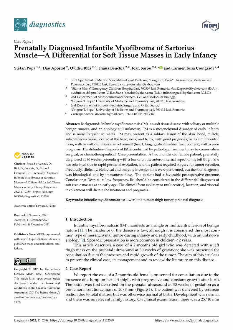
We report the case of a 2 months old female, presented for consultation due to thepresence of a lump on her left thigh, with progressive and constant growth after birth.The lesion was first described on the prenatal ultrasound at 30 weeks of gestation as apre-femoral soft tissue mass of 20/7 mm (Figure 1). The patient was delivered by cesareansection due to fetal distress but was otherwise normal at birth. Development was normal,and there was no relevant family history. On clinical examination, there was a 25/10 mm
Diagnostics 2022, 11, 2389. https://doi.org/10.3390/diagnostics11122389 https://www.mdpi.com/journal/diagnostics
Diagnostics 2022, 11, 2389 2 of 11
nodule on the antero-intern side of the left thigh that was firm, mobile and within thedeep layers. The overlying skin was normal. There were no other lesions elsewhere on thepatient’s body.
Diagnostics 2021, 11, x FOR PEER REVIEW 2 of 11
and there was no relevant family history. On clinical examination, there was a 25/10 mm nodule on the antero-intern side of the left thigh that was firm, mobile and within the deep layers. The overlying skin was normal. There were no other lesions elsewhere on the patient’s body.
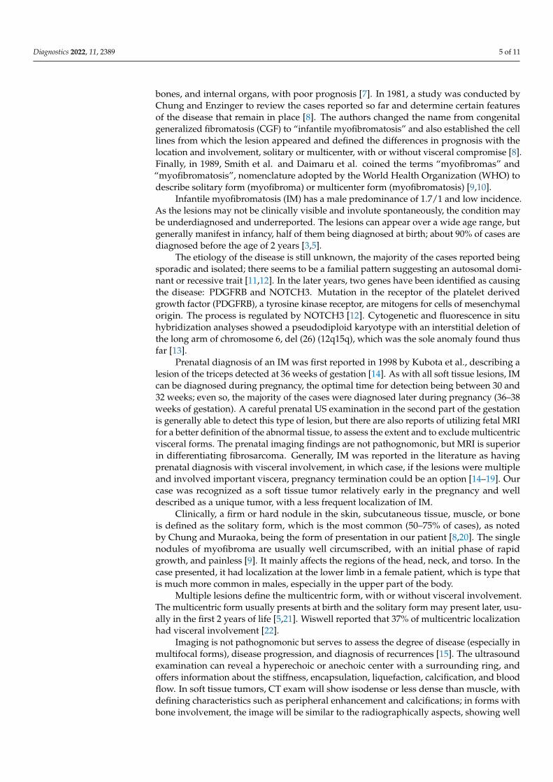
Figure 1. Prenatal ultrasound images at 30 weeks of gestation, showing an oval mass, hypoechoic with calcification on the thigh, with no evidence of bone involvement.
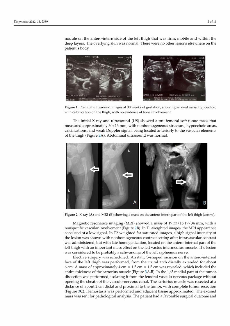
The initial X-ray and ultrasound (US) showed a pre-femoral soft tissue mass that measured approximately 30/13 mm, with nonhomogeneous structure, hypoechoic areas, calcifications, and weak Doppler signal, being located anteriorly to the vascular elements of the thigh (Figure 2A). Abdominal ultrasound was normal.
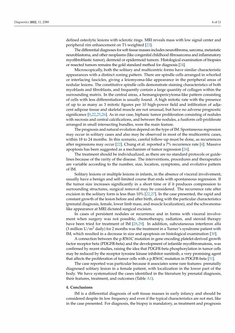
Figure 2. X-ray (A) and MRI (B) showing a mass on the antero-intern part of the left thigh (arrow).
Magnetic resonance imaging (MRI) showed a mass of 19,33/15,19/34 mm, with a nonspecific vascular involvement (Figure 2B). In T1-weighted images, the MRI appearance consisted of a low signal. In T2-weighted fat-saturated images, a high signal intensity of the lesion was shown with nonhomogeneous contrast setting after intravascular contrast was administered, but with late homogenization, located on the antero-internal part of the left thigh with an important mass effect on the left vastus intermedius muscle. The lesion was considered to be probably a schwanoma of the left saphenous nerve.
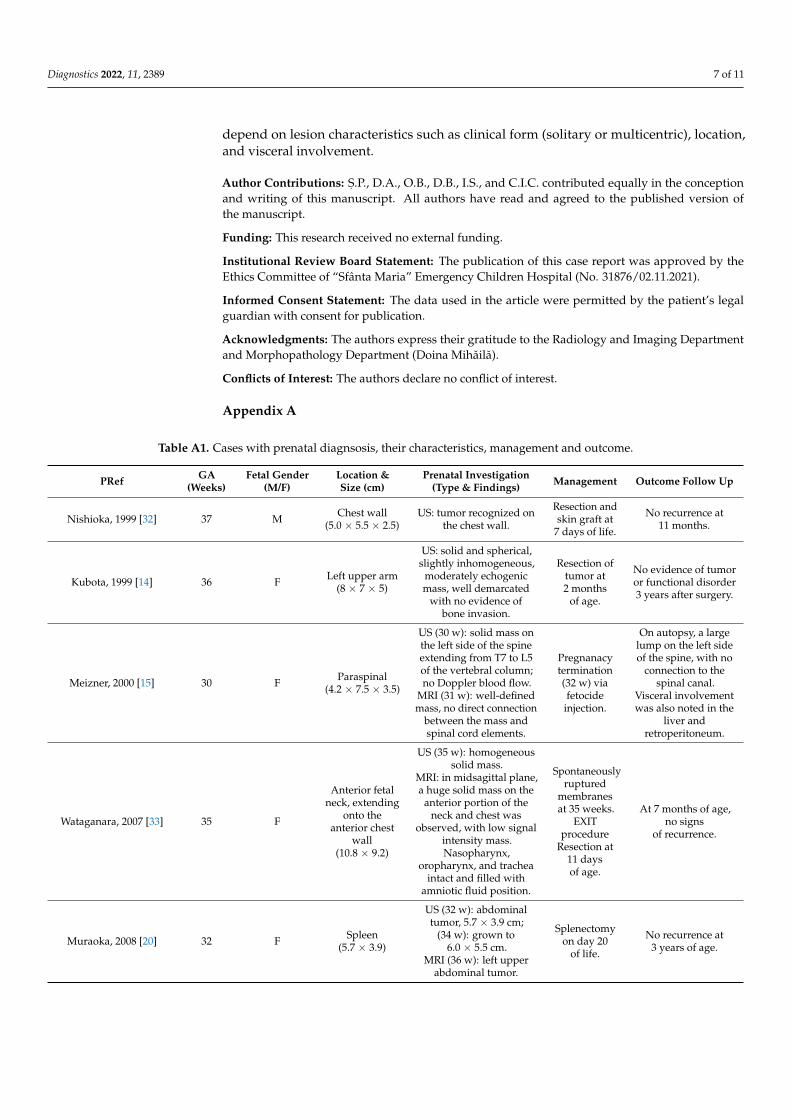
Elective surgery was scheduled. An italic S-shaped incision on the antero-internal face of the left thigh was performed, from the crural arch distally extended for about 6 cm. A mass of approximately 4 cm × 1.5 cm × 1.5 cm was revealed, which included the entire thickness of the sartorius muscle (Figure 3A,B). In the 1/3 medial part of the tumor, dissection was performed, isolating it from the femoral vasculo-nervous package without
Figure 1. Prenatal ultrasound images at 30 weeks of gestation, showing an oval mass, hypoechoicwith calcification on the thigh, with no evidence of bone involvement.
The initial X-ray and ultrasound (US) showed a pre-femoral soft tissue mass thatmeasured approximately 30/13 mm, with nonhomogeneous structure, hypoechoic areas,calcifications, and weak Doppler signal, being located anteriorly to the vascular elementsof the thigh (Figure 2A). Abdominal ultrasound was normal.
Diagnostics 2021, 11, x FOR PEER REVIEW 2 of 11
and there was no relevant family history. On clinical examination, there was a 25/10 mm nodule on the antero-intern side of the left thigh that was firm, mobile and within the deep layers. The overlying skin was normal. There were no other lesions elsewhere on the patient’s body.
Figure 1. Prenatal ultrasound images at 30 weeks of gestation, showing an oval mass, hypoechoic with calcification on the thigh, with no evidence of bone involvement.
The initial X-ray and ultrasound (US) showed a pre-femoral soft tissue mass that measured approximately 30/13 mm, with nonhomogeneous structure, hypoechoic areas, calcifications, and weak Doppler signal, being located anteriorly to the vascular elements of the thigh (Figure 2A). Abdominal ultrasound was normal.
Figure 2. X-ray (A) and MRI (B) showing a mass on the antero-intern part of the left thigh (arrow).
Magnetic resonance imaging (MRI) showed a mass of 19,33/15,19/34 mm, with a nonspecific vascular involvement (Figure 2B). In T1-weighted images, the MRI appearance consisted of a low signal. In T2-weighted fat-saturated images, a high signal intensity of the lesion was shown with nonhomogeneous contrast setting after intravascular contrast was administered, but with late homogenization, located on the antero-internal part of the left thigh with an important mass effect on the left vastus intermedius muscle. The lesion was considered to be probably a schwanoma of the left saphenous nerve.
Elective surgery was scheduled. An italic S-shaped incision on the antero-internal face of the left thigh was performed, from the crural arch distally extended for about 6 cm. A mass of approximately 4 cm × 1.5 cm × 1.5 cm was revealed, which included the entire thickness of the sartorius muscle (Figure 3A,B). In the 1/3 medial part of the tumor, dissection was performed, isolating it from the femoral vasculo-nervous package without
Figure 2. X-ray (A) and MRI (B) showing a mass on the antero-intern part of the left thigh (arrow).
Magnetic resonance imaging (MRI) showed a mass of 19.33/15.19/34 mm, with anonspecific vascular involvement (Figure 2B). In T1-weighted images, the MRI appearanceconsisted of a low signal. In T2-weighted fat-saturated images, a high signal intensity ofthe lesion was shown with nonhomogeneous contrast setting after intravascular contrastwas administered, but with late homogenization, located on the antero-internal part of theleft thigh with an important mass effect on the left vastus intermedius muscle. The lesionwas considered to be probably a schwanoma of the left saphenous nerve.
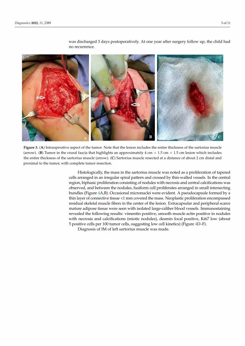
Elective surgery was scheduled. An italic S-shaped incision on the antero-internalface of the left thigh was performed, from the crural arch distally extended for about6 cm. A mass of approximately 4 cm × 1.5 cm × 1.5 cm was revealed, which included theentire thickness of the sartorius muscle (Figure 3A,B). In the 1/3 medial part of the tumor,dissection was performed, isolating it from the femoral vasculo-nervous package withoutopening the sheath of the vasculo-nervous canal. The sartorius muscle was resected at adistance of about 2 cm distal and proximal to the tumor, with complete tumor resection(Figure 3C). Hemostasis was performed and adjacent tissue approximated. The excisedmass was sent for pathological analysis. The patient had a favorable surgical outcome and
Diagnostics 2022, 11, 2389 3 of 11
was discharged 3 days postoperatively. At one year after surgery follow up, the child hadno recurrence.
Diagnostics 2021, 11, x FOR PEER REVIEW 3 of 11
opening the sheath of the vasculo-nervous canal. The sartorius muscle was resected at a distance of about 2 cm distal and proximal to the tumor, with complete tumor resection (Figure 3C). Hemostasis was performed and adjacent tissue approximated. The excised mass was sent for pathological analysis. The patient had a favorable surgical outcome and was discharged 3 days postoperatively. At one year after surgery follow up, the child had no recurrence.
Figure 3. (A) Intraoperative aspect of the tumor. Note that the lesion includes the entire thickness of the sartorius muscle (arrow). (B) Tumor in the crural fascia that highlights an approximately 4 cm × 1.5 cm × 1.5 cm lesion which includes the entire thickness of the sartorius muscle (arrow). (C) Sartorius muscle resected at a distance of about 2 cm distal and proximal to the tumor, with complete tumor resection.
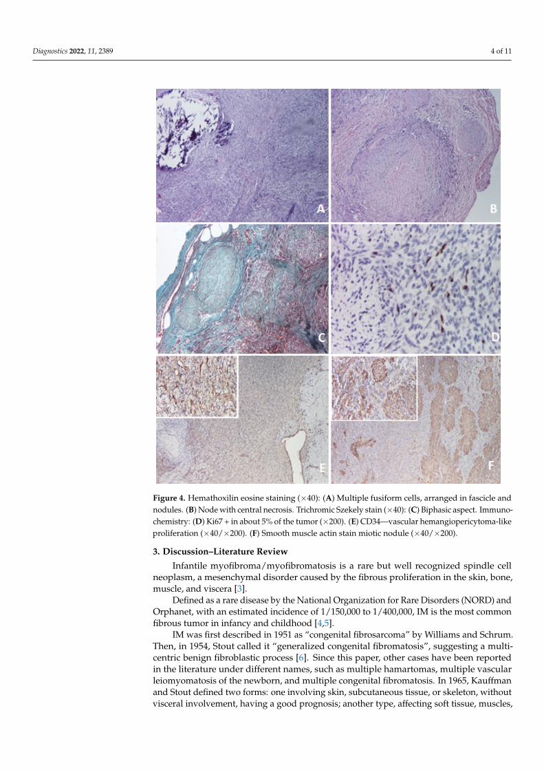
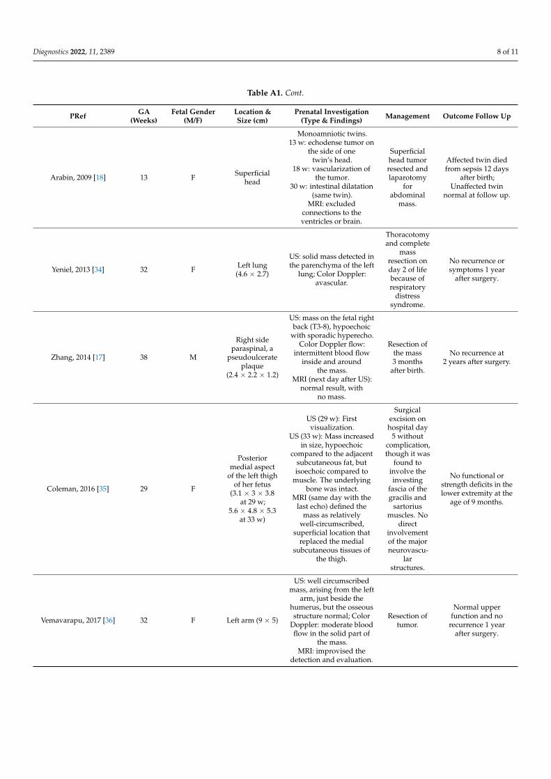
Histologically, the mass in the sartorius muscle was noted as a proliferation of tapered cells arranged in an irregular spiral pattern and crossed by thin-walled vessels. In the central region, biphasic proliferation consisting of nodules with necrosis and central calcifications was observed, and between the nodules, fusiform cell proliferates arranged in small intersecting bundles (Figure 4A,B). Occasional micronuclei were evident. A pseudocapsule formed by a thin layer of connective tissue <1 mm covered the mass. Neoplastic proliferation encompassed residual skeletal muscle fibers in the center of the lesion. Extracapsular and peripheral scarce mature adipose tissue were seen with isolated large-caliber blood vessels. Immunostaining revealed the following results: vimentin positive, smooth muscle actin positive in nodules with necrosis and calcifications (miotic nodules), desmin focal positive, Ki67 low (about 5 positive cells per 100 tumor cells, suggesting low cell kinetics) (Figure 4D–F).
Diagnosis of IM of left sartorius muscle was made.
Figure 3. (A) Intraoperative aspect of the tumor. Note that the lesion includes the entire thickness of the sartorius muscle(arrow). (B) Tumor in the crural fascia that highlights an approximately 4 cm × 1.5 cm × 1.5 cm lesion which includesthe entire thickness of the sartorius muscle (arrow). (C) Sartorius muscle resected at a distance of about 2 cm distal andproximal to the tumor, with complete tumor resection.
Histologically, the mass in the sartorius muscle was noted as a proliferation of taperedcells arranged in an irregular spiral pattern and crossed by thin-walled vessels. In the centralregion, biphasic proliferation consisting of nodules with necrosis and central calcifications wasobserved, and between the nodules, fusiform cell proliferates arranged in small intersectingbundles (Figure 4A,B). Occasional micronuclei were evident. A pseudocapsule formed by athin layer of connective tissue <1 mm covered the mass. Neoplastic proliferation encompassedresidual skeletal muscle fibers in the center of the lesion. Extracapsular and peripheral scarcemature adipose tissue were seen with isolated large-caliber blood vessels. Immunostainingrevealed the following results: vimentin positive, smooth muscle actin positive in noduleswith necrosis and calcifications (miotic nodules), desmin focal positive, Ki67 low (about5 positive cells per 100 tumor cells, suggesting low cell kinetics) (Figure 4D–F).
Diagnosis of IM of left sartorius muscle was made.
Diagnostics 2022, 11, 2389 4 of 11Diagnostics 2021, 11, x FOR PEER REVIEW 4 of 11
Figure 4. Hemathoxilin eosine staining (×40): (A) Multiple fusiform cells, arranged in fascicle and nodules. (B) Node with central necrosis. Trichromic Szekely stain (×40): (C) Biphasic aspect. Immunochemistry: (D) Ki67 + in about 5% of the tumor (×200). (E) CD34—vascular hemangiopericytoma-like proliferation (×40/×200). (F) Smooth muscle actin stain miotic nodule (×40/×200).
3. Discussions–Literature Review Infantile myofibroma/myofibromatosis is a rare but well recognized spindle cell
neoplasm, a mesenchymal disorder caused by the fibrous proliferation in the skin, bone, muscle, and viscera [3].
Defined as a rare disease by the National Organization for Rare Disorders (NORD) and Orphanet, with an estimated incidence of 1/150.000 to 1/400.000, IM is the most common fibrous tumor in infancy and childhood [4,5].
IM was first described in 1951 as “congenital fibrosarcoma” by Williams and Schrum. Then, in 1954, Stout called it “generalized congenital fibromatosis”, suggesting a multicentric benign fibroblastic process [6]. Since this paper, other cases have been reported in the literature under different names, such as multiple hamartomas, multiple vascular leiomyomatosis of the newborn, and multiple congenital fibromatosis. In 1965, Kauffman and Stout defined two forms: one involving skin, subcutaneous tissue, or skeleton, without visceral involvement, having a good prognosis; another type, affecting
Figure 4. Hemathoxilin eosine staining (×40): (A) Multiple fusiform cells, arranged in fascicle andnodules. (B) Node with central necrosis. Trichromic Szekely stain (×40): (C) Biphasic aspect. Immuno-chemistry: (D) Ki67 + in about 5% of the tumor (×200). (E) CD34—vascular hemangiopericytoma-likeproliferation (×40/×200). (F) Smooth muscle actin stain miotic nodule (×40/×200).
3. Discussion–Literature Review
Infantile myofibroma/myofibromatosis is a rare but well recognized spindle cellneoplasm, a mesenchymal disorder caused by the fibrous proliferation in the skin, bone,muscle, and viscera [3].
Defined as a rare disease by the National Organization for Rare Disorders (NORD) andOrphanet, with an estimated incidence of 1/150,000 to 1/400,000, IM is the most commonfibrous tumor in infancy and childhood [4,5].
IM was first described in 1951 as “congenital fibrosarcoma” by Williams and Schrum.Then, in 1954, Stout called it “generalized congenital fibromatosis”, suggesting a multi-centric benign fibroblastic process [6]. Since this paper, other cases have been reportedin the literature under different names, such as multiple hamartomas, multiple vascularleiomyomatosis of the newborn, and multiple congenital fibromatosis. In 1965, Kauffmanand Stout defined two forms: one involving skin, subcutaneous tissue, or skeleton, withoutvisceral involvement, having a good prognosis; another type, affecting soft tissue, muscles,
Diagnostics 2022, 11, 2389 5 of 11
bones, and internal organs, with poor prognosis [7]. In 1981, a study was conducted byChung and Enzinger to review the cases reported so far and determine certain featuresof the disease that remain in place [8]. The authors changed the name from congenitalgeneralized fibromatosis (CGF) to “infantile myofibromatosis” and also established the celllines from which the lesion appeared and defined the differences in prognosis with thelocation and involvement, solitary or multicenter, with or without visceral compromise [8].Finally, in 1989, Smith et al. and Daimaru et al. coined the terms “myofibromas” and“myofibromatosis”, nomenclature adopted by the World Health Organization (WHO) todescribe solitary form (myofibroma) or multicenter form (myofibromatosis) [9,10].
Infantile myofibromatosis (IM) has a male predominance of 1.7/1 and low incidence.As the lesions may not be clinically visible and involute spontaneously, the condition maybe underdiagnosed and underreported. The lesions can appear over a wide age range, butgenerally manifest in infancy, half of them being diagnosed at birth; about 90% of cases arediagnosed before the age of 2 years [3,5].
The etiology of the disease is still unknown, the majority of the cases reported beingsporadic and isolated; there seems to be a familial pattern suggesting an autosomal domi-nant or recessive trait [11,12]. In the later years, two genes have been identified as causingthe disease: PDGFRB and NOTCH3. Mutation in the receptor of the platelet derivedgrowth factor (PDGFRB), a tyrosine kinase receptor, are mitogens for cells of mesenchymalorigin. The process is regulated by NOTCH3 [12]. Cytogenetic and fluorescence in situhybridization analyses showed a pseudodiploid karyotype with an interstitial deletion ofthe long arm of chromosome 6, del (26) (12q15q), which was the sole anomaly found thusfar [13].
Prenatal diagnosis of an IM was first reported in 1998 by Kubota et al., describing alesion of the triceps detected at 36 weeks of gestation [14]. As with all soft tissue lesions, IMcan be diagnosed during pregnancy, the optimal time for detection being between 30 and32 weeks; even so, the majority of the cases were diagnosed later during pregnancy (36–38weeks of gestation). A careful prenatal US examination in the second part of the gestationis generally able to detect this type of lesion, but there are also reports of utilizing fetal MRIfor a better definition of the abnormal tissue, to assess the extent and to exclude multicentricvisceral forms. The prenatal imaging findings are not pathognomonic, but MRI is superiorin differentiating fibrosarcoma. Generally, IM was reported in the literature as havingprenatal diagnosis with visceral involvement, in which case, if the lesions were multipleand involved important viscera, pregnancy termination could be an option [14–19]. Ourcase was recognized as a soft tissue tumor relatively early in the pregnancy and welldescribed as a unique tumor, with a less frequent localization of IM.
Clinically, a firm or hard nodule in the skin, subcutaneous tissue, muscle, or boneis defined as the solitary form, which is the most common (50–75% of cases), as notedby Chung and Muraoka, being the form of presentation in our patient [8,20]. The singlenodules of myofibroma are usually well circumscribed, with an initial phase of rapidgrowth, and painless [9]. It mainly affects the regions of the head, neck, and torso. In thecase presented, it had localization at the lower limb in a female patient, which is type thatis much more common in males, especially in the upper part of the body.
Multiple lesions define the multicentric form, with or without visceral involvement.The multicentric form usually presents at birth and the solitary form may present later, usu-ally in the first 2 years of life [5,21]. Wiswell reported that 37% of multicentric localizationhad visceral involvement [22].
Imaging is not pathognomonic but serves to assess the degree of disease (especially inmultifocal forms), disease progression, and diagnosis of recurrences [15]. The ultrasoundexamination can reveal a hyperechoic or anechoic center with a surrounding ring, andoffers information about the stiffness, encapsulation, liquefaction, calcification, and bloodflow. In soft tissue tumors, CT exam will show isodense or less dense than muscle, withdefining characteristics such as peripheral enhancement and calcifications; in forms withbone involvement, the image will be similar to the radiographically aspects, showing well
Diagnostics 2022, 11, 2389 6 of 11
defined osteolytic lesions with sclerotic rings. MRI reveals mass with low signal center andperipheral rim enhancement on T1-weighted [23].
The differential diagnoses for soft tissue masses includes neurofibroma, sarcoma, metastaticneuroblastoma, and other neoplasms (like congenital childhood fibrosarcoma and inflammatorymyofibroblastic tumor), dermoid or epidermoid tumors. Histological examination of biopsiesor resected tumors remains the gold standard method for diagnosis [24].
Microscopically, both the solitary and multicentric forms have similar characteristicappearances with a distinct zoning pattern. There are spindle cells arranged in whorledor interlacing fascicles, giving a leiomyoma-like appearance in the peripheral areas ofnodular lesions. The constitutive spindle cells demonstrate staining characteristics of bothmyoblasts and fibroblasts, and frequently contain a large quantity of collagen within thesurrounding matrix. In the central areas, a hemangiopericytoma-like pattern consistingof cells with less differentiation is usually found. A high mitotic rate with the presenceof up to as many as 3 mitotic figures per 10 high-power field and infiltration of adja-cent adipose tissue and skeletal muscle are not unusual, but have no adverse prognosticsignificance [8,22,25,26]. As in our case, biphasic tumor proliferation consisting of noduleswith necrosis and central calcifications, and between the nodules, a fusiform cell-proliferatearranged in small intersecting bundles, were the main feature.
The prognosis and natural evolution depend on the type of IM. Spontaneous regressionmay occur in solitary cases and also may be observed in most of the multicentric cases,within 18 to 24 months. In this scenario, careful follow-up must be done, as recurrencesafter regressions may occur [22]. Chung et al. reported a 7% recurrence rate [4]. Massiveapoptosis has been suggested as a mechanism of tumor regression [26].
The treatment should be individualized, as there are no standard protocols or guide-lines because of the rarity of the disease. The interventions, procedures and therapeuticsare variable according to the number, size, location, symptoms, and evolutive patternof IM.
Solitary lesions or multiple lesions in infants, in the absence of visceral involvement,usually have a benign and self-limited course that ends with spontaneous regression. Ifthe tumor size increases significantly in a short time or if it produces compression tosurrounding structures, surgical removal may be considered. The recurrence rate afterexcision in the solitary form is less than 10% [22,27]. In the case presented, the rapid andconstant growth of the lesion before and after birth, along with the particular characteristics(prenatal diagnosis, female, lower limb mass, and muscle localization), and the schwanoma-like appearance at MRI dictated surgical excision.
In cases of persistent nodules or recurrence and in forms with visceral involve-ment when surgery was not possible, chemotherapy, radiation, and steroid therapyhave been tried for treatment of IM [28,29]. In addition, subcutaneous interferon alfa(3 million U/m2 daily) for 2 months was the treatment in a Turner’s syndrome patient withIM, which resulted in a decrease in size and apoptosis on histological examination [30].
A connection between the p.R561C mutation in gene encoding platelet-derived growthfactor receptor beta (PDGFR-beta) and the development of infantile myofibromatosis, wasconfirmed by recent studies, raising the idea that PDGFR-beta phosphorylation in tumor cellsmay be reduced by the receptor tyrosine kinase inhibitor sunitinib, a very promising agentthat affects the proliferation of tumor cells with a p.R561C mutation in PDGFR-beta [31].
The case reported was particular because it associates some rare features: prenatallydiagnosed solitary lesion in a female patient, with localization in the lower part of thebody. We have systematized the cases identified in the literature by prenatal diagnosis,their features, treatment, and outcomes (Table A1).
4. Conclusions
IM is a differential diagnosis of soft tissue masses in early infancy and should beconsidered despite its low frequency and even if the typical characteristics are not met, likein the case presented. For diagnosis, the biopsy is mandatory, as treatment and prognosis
Diagnostics 2022, 11, 2389 7 of 11
depend on lesion characteristics such as clinical form (solitary or multicentric), location,and visceral involvement.
Author Contributions: S, .P., D.A., O.B., D.B., I.S., and C.I.C. contributed equally in the conceptionand writing of this manuscript. All authors have read and agreed to the published version ofthe manuscript.
Funding: This research received no external funding.
Institutional Review Board Statement: The publication of this case report was approved by theEthics Committee of “Sfânta Maria” Emergency Children Hospital (No. 31876/02.11.2021).
Informed Consent Statement: The data used in the article were permitted by the patient’s legalguardian with consent for publication.
Acknowledgments: The authors express their gratitude to the Radiology and Imaging Departmentand Morphopathology Department (Doina Mihăilă).
Conflicts of Interest: The authors declare no conflict of interest.
Appendix A
Table A1. Cases with prenatal diagnsosis, their characteristics, management and outcome.
PRef GA(Weeks)
Fetal Gender(M/F)
Location &Size (cm)
Prenatal Investigation(Type & Findings) Management Outcome Follow Up
Nishioka, 1999 [32] 37 M Chest wall(5.0 × 5.5 × 2.5)
US: tumor recognized onthe chest wall.
Resection andskin graft at
7 days of life.
No recurrence at11 months.
Kubota, 1999 [14] 36 F Left upper arm(8 × 7 × 5)
US: solid and spherical,slightly inhomogeneous,
moderately echogenicmass, well demarcated
with no evidence ofbone invasion.
Resection oftumor at2 months
of age.
No evidence of tumoror functional disorder3 years after surgery.
Meizner, 2000 [15] 30 F Paraspinal(4.2 × 7.5 × 3.5)
US (30 w): solid mass onthe left side of the spineextending from T7 to L5of the vertebral column;no Doppler blood flow.
MRI (31 w): well-definedmass, no direct connection
between the mass andspinal cord elements.
Pregnanacytermination(32 w) viafetocide
injection.
On autopsy, a largelump on the left sideof the spine, with no
connection to thespinal canal.
Visceral involvementwas also noted in the
liver andretroperitoneum.
Wataganara, 2007 [33] 35 F
Anterior fetalneck, extending
onto theanterior chest
wall(10.8 × 9.2)
US (35 w): homogeneoussolid mass.
MRI: in midsagittal plane,a huge solid mass on the
anterior portion of theneck and chest was
observed, with low signalintensity mass.Nasopharynx,
oropharynx, and tracheaintact and filled with
amniotic fluid position.
Spontaneouslyruptured
membranesat 35 weeks.
EXITprocedure
Resection at11 daysof age.
At 7 months of age,no signs
of recurrence.
Muraoka, 2008 [20] 32 F Spleen(5.7 × 3.9)
US (32 w): abdominaltumor, 5.7 × 3.9 cm;
(34 w): grown to6.0 × 5.5 cm.
MRI (36 w): left upperabdominal tumor.
Splenectomyon day 20
of life.
No recurrence at3 years of age.
Diagnostics 2022, 11, 2389 8 of 11
Table A1. Cont.
PRef GA(Weeks)
Fetal Gender(M/F)
Location &Size (cm)
Prenatal Investigation(Type & Findings) Management Outcome Follow Up
Arabin, 2009 [18] 13 F Superficialhead
Monoamniotic twins.13 w: echodense tumor on
the side of onetwin’s head.
18 w: vascularization ofthe tumor.
30 w: intestinal dilatation(same twin).
MRI: excludedconnections to theventricles or brain.
Superficialhead tumorresected andlaparotomy
forabdominal
mass.
Affected twin diedfrom sepsis 12 days
after birth;Unaffected twin
normal at follow up.
Yeniel, 2013 [34] 32 F Left lung(4.6 × 2.7)
US: solid mass detected inthe parenchyma of the left
lung; Color Doppler:avascular.
Thoracotomyand complete
massresection onday 2 of lifebecause ofrespiratory
distresssyndrome.
No recurrence orsymptoms 1 year
after surgery.
Zhang, 2014 [17] 38 M
Right sideparaspinal, a
pseudoulcerateplaque
(2.4 × 2.2 × 1.2)
US: mass on the fetal rightback (T3-8), hypoechoic
with sporadic hyperecho.Color Doppler flow:
intermittent blood flowinside and around
the mass.MRI (next day after US):
normal result, withno mass.
Resection ofthe mass3 months
after birth.
No recurrence at2 years after surgery.
Coleman, 2016 [35] 29 F
Posteriormedial aspect
of the left thighof her fetus
(3.1 × 3 × 3.8at 29 w;
5.6 × 4.8 × 5.3at 33 w)
US (29 w): Firstvisualization.
US (33 w): Mass increasedin size, hypoechoic
compared to the adjacentsubcutaneous fat, butisoechoic compared to
muscle. The underlyingbone was intact.
MRI (same day with thelast echo) defined the
mass as relativelywell-circumscribed,
superficial location thatreplaced the medial
subcutaneous tissues ofthe thigh.
Surgicalexcision onhospital day
5 withoutcomplication,though it was
found toinvolve theinvesting
fascia of thegracilis and
sartoriusmuscles. No
directinvolvementof the majorneurovascu-
larstructures.
No functional orstrength deficits in thelower extremity at the
age of 9 months.
Vemavarapu, 2017 [36] 32 F Left arm (9 × 5)
US: well circumscribedmass, arising from the left
arm, just beside thehumerus, but the osseousstructure normal; Color
Doppler: moderate bloodflow in the solid part of
the mass.MRI: improvised the
detection and evaluation.
Resection oftumor.
Normal upperfunction and no
recurrence 1 yearafter surgery.
Diagnostics 2022, 11, 2389 9 of 11
Table A1. Cont.
PRef GA(Weeks)
Fetal Gender(M/F)
Location &Size (cm)
Prenatal Investigation(Type & Findings) Management Outcome Follow Up
Pekar-Zlotin, 2018 [37] 34 - Multipletumors
US: multiorganinvolvement, with masses
predominantly in thelower extremities, heart,abdominal cavity, andneck. Normal Doppler
studies in the major fetalarterial and venous
circulations, as well asnormal amniotic fluid
volume and unremarkableappearance ofthe placenta.
Inoperabletumors.
The infant died ofcardiac failure 30 days
after birth.
Rekawek, 2019 [16] 36 MLower thoracic
spine(3.5 × 2.3)
US: avascular,heterogeneous mass of
soft tissues, without cordinvolvement.
MRI: thick-walled partlycystic lesion in the right
paraspinal muscles of thelower thorax.
Biopsy onday 7 of life.
MRI at one year ofage: multiple soft
masses decreased insize compared to
prior imaging, andthree additional
lesions: crus of theleft diaphragam, leftposterior chest wall,
and left shoulder.
Evens, 2021 [38] 33 -Visceral organinvolvementLung masses
US: dilated bowel loop.MRI: suggested the
dilated loop of bowel wascolon, given the internal
high T1 signal compatiblewith meconium.
Additionally, multiple,solid-appearing lung
masses were noted, whichhad not been previouslyvisualized on ultrasound.After thorough review of
the MRI images of theentire fetus, no primarylesion was found. No
masses were visualized inthe extremities.
The baby hadnot passedmeconium
after 1 day oflife, therefor
anexploratorylaparotomy:nodules onthe bowel
serosa,resulting in a
bowelobstruction.Owing the
visceral organinvolvement,chemother-
apy wasconsidered;however, it
wasultimatelydecided toobserve thepatient andtreat only ifthere wasradiologic
progressionand/orclinical
deterioration.
References1. Scheper, M.A.; DiFabio, V.E.; Sauk, J.J.; Nikitakis, N.G. Myofibromatosis: A case report with a unique clinical presentation. Oral
Surg. Oral Med. Oral Pathol. Oral Radiol. Endod. 2005, 99, 325–330. [CrossRef] [PubMed]2. Pelluard-Nehmé, F.; Coatleven, F.; Carles, D.; Alberti, E.M.; Briex, M.; Dallay, D. Multicentric infantile myofibromatosis: Two
perinatal cases. Eur. J. Pediatr. 2007, 166, 997–1001. [CrossRef]3. Venkatesh, V.; Kumar, B.P.; Kumar, K.A.; Mohan, A.P. Myofibroma—A rare entity with unique clinical presentation. J. Maxillofac.
Oral Surg. 2015, 14 (Suppl. 1), 64–68. [CrossRef]
Diagnostics 2022, 11, 2389 10 of 11
4. NORD Rare Disease Database. Available online: https://rarediseases.org/rare-diseases/infantile-myofibromatosis/ (accessedon 1 November 2021).
5. Hausbrandt, P.A.; Leithner, A.; Beham, A.; Bodo, K.; Raith, J.; Windhager, R. A rare case of infantile myofibromatosis and reviewof literature. J. Pediatr. Orthop. B 2010, 19, 122–126. [CrossRef]
6. Stout, A.P. Juvenile fibromatoses. Cancer 1954, 7, 953–978. [CrossRef]7. Kauffman, S.L.; Stout, A.P. Congenital mesenchymal tumours. Cancer 1965, 18, 460–476. [CrossRef]8. Chung, E.B.; Enzinger, F.M. Infantile myofibromatosis. Cancer 1981, 48, 1807–1818. [CrossRef]9. Smith, K.J.; Skelton, H.G.; Barrett, T.L.; Lupton, G.P.; Graham, J.H. Cutaneous myofibroma. Mod. Pathol. 1989, 2, 603–609.10. Daimaru, Y.; Hashimoto, H.; Enjoji, M. Myofibromatosis in adults (adult counterpart of infantile myofibromatosis). Am. J. Surg.
Pathol. 1989, 13, 859–865. [CrossRef]11. Wu, W.; Chen, J.; Cao, X.; Yang, M.; Zhu, J.; Zhao, G. Solitary infantile myofibromatosis in the bones of the upper extremities: Two
rare cases and a review of the literature. Oncol. Lett. 2013, 6, 1406–1408. [CrossRef]12. Martignetti, J.A.; Tian, L.; Li, D.; Ramirez, M.C.; Camacho-Vanegas, O.; Camacho, S.C.; Guo, Y.; Zand, D.J.; Bernstein, A.M.;
Masur, S.K.; et al. Mutations in PDGFRB Cause Autosomal-Dominant Infantile Myofibromatosis. Am. J. Hum. Genet. 2013, 92,1001–1007. [CrossRef]
13. Stenman, G.; Nadal, N.; Persson, S.; Gunterberg, B.; Angervall, L. del(6)(q12q15) as the sole cytogenetic anomaly in a case ofsolitary infantile myofibromatosis. Oncol. Rep. 1999, 6, 1101–1104. [CrossRef]
14. Kubota, A.; Imano, M.; Yonekura, T.; Hirooka, S.; Nose, K.; Oyanagi, H.; Nakayama, M. Infantile myofibromatosis of the tricepsdetected by prenatal sonography. J. Clin. Ultrasound 1999, 27, 147–150. [CrossRef]
15. Meizner, I.; Shalev, J.; Mashiach, R.; Vardimon, D.; Ben-Raphael, Z. Prenatal ultrasound diagnosis of infantile myofibromatosis—A case report. Ultrasound Obstet. Gynecol. 2000, 16, 84–86. [CrossRef] [PubMed]
16. Rekawek, P.; Coleman, B.G.; Kamath, A.; Stone, J.L. Prenatal sonography of multicentric infantile myofibromatosis: Case reportand review of the literature. J. Clin. Ultrasound 2019, 47, 490–493. [CrossRef] [PubMed]
17. Zhang, F.; Cheng, D.; Wu, M.; Ge, L.; Ma, X. Diagnosis of infantile myofibromatosis with pseudo-ulcerated plaque using prenatalultrasound: A case report. Exp. Ther. Med. 2014, 8, 1769–1771. [CrossRef]
18. Arabin, B.; Hack, K.; Nooij, L.; Nikkels, P. Dis-crepant findings in a monoamniotic twin pregnancy affected by infantilemyofibromatosis. Ultrasound Obstet. Gynecol. 2009, 33, 488–490. [CrossRef]
19. Alamo, L.; Beck-Popovic, M.; Gudinchet, F.; Meuli, R. Congenital tumors: Imaging when life just begins. Insights Imaging 2011, 2,297–308. [CrossRef]
20. Muraoka, I.; Ohno, Y.; Kamitamari, A.; Okada, M.; Moriuchi, H.; Kanematsu, T. Congenital occurrence of solitary infantilemyofibromatosis of the spleen. J. Pediatr. Surg. 2008, 43, 227–230. [CrossRef]
21. Thunnissen, B.T.; Bax, N.M.; Rövekamp, M.H.; Beek, F.J.; van Gorp, J. Infantile myofibromatosis: An unusual presentation and areview of the literature. Eur. J. Pediatr. Surg. 1993, 3, 179–181. [CrossRef]
22. Wiswell, T.E.; Davis, J.; Cunnigham, B.E.; Solenberger, R.; Thomas, P.J. Infantile myofibromatosis: The most common fibroustumor of infancy. J. Pediatr. Surg. 1988, 23, 315–318. [CrossRef]
23. Koujok, K.; Ruiz, R.E.; Hernandez, R.J. Myofibromatosis: Imaging characteristics. Pediatr. Radiol. 2005, 35, 374–380. [CrossRef]24. Fraitag, S.; Boccara, O. What to Look Out for in a Newborn with Multiple Papulonodular Skin Lesions at Birth. Dermatopathology
2021, 8, 390–417. [CrossRef]25. Allen, P.W. The fibromatosis: A clinicopathologic classification based on 140 cases. Review articles, part 2. Am. J. Surg. Pathol.
1977, 1, 305–321. [CrossRef]26. Fukasawa, Y.; Ishikura, H.; Takada, A.; Yokoyama, S.; Imamura, M.; Yoshiki, T.; Sato, H. Massive apoptosis in infantile
myofibromatosis: A putative mechanism of tumor regression. J. Pathol. 1994, 144, 480–485.27. Schmidt, D. Fibrous tumors and tumor-like lesions of childhood: Diagnosis, differential diagnosis, and prognosis. Curr. Top.
Pathol. 1995, 89, 175–191. [PubMed]28. Burton, J.L.; Lovell, C.R. Disorders of connective tissue. In Textbook of Dermatology, 6th ed.; Champion, R.H., Burton, J.L.,
Burns, D.A., Breathnach, S.M., Eds.; Blackwell Science: Malden, MA, USA, 1998; Volume 3, pp. 2048–2049.29. Kiel, K.D.; Suit, H.D. Radiation therapy in the treatment of aggressive fibromatosis (desmoid tumors). Cancer 1984, 54, 2051–2055.
[CrossRef]30. Savasan, S.; Fulgenzi, L.A.; Rabah, R.; Mohamed, A.N.; Ravindranath, Y. Generalized infantile myofibromatosis in a patient with
Turner’s syndrome: A trial of interferon-α. J. Pediatr. 1998, 133, 694–696. [CrossRef]31. Sramek, M.; Neradil, J.; Macigova, P.; Mudry, P.; Polaskova, K.; Slaby, O.; Noskova, H.; Sterba, J.; Veselska, R. Effects of Sunitinib
and Other Kinase Inhibitors on Cells Harboring a PDGFRB Mutation Associated with Infantile Myofibromatosis. Int. J. Mol. Sci.2018, 19, 2599. [CrossRef] [PubMed]
32. Nishioka, K.; Seguchi, T.; Yamamura, Y.; Tatsumura, M.; Sou, H.; Gondo, T.; Hoshii, Y.; Iwata, T. Infantile myofibromatosisidentified by fetal ultrasound. Br. J. Dermatol. 1999, 140, 539–541. [CrossRef]
33. Wataganara, T.; Ngerncham, S.; Kitsommart, R.; Fuangtharnthip, P. Fetal neck myofibroma. J. Med. Assoc. Thai. 2007, 90, 376–380.[PubMed]
34. Yeniel, A.O.; Ergenoglu, A.M.; Zeybek, B.; Kazandi, M.; Akercan, F.; Ozcan, C.; Veral, A. Prenatal diagnosis of infantilemyofibromatosis of the lung: A case report and review of the literature. J. Clin. Ultrasound 2013, 41, 38–41. [CrossRef] [PubMed]

Diagnostics 2022, 11, 2389 11 of 11
35. Coleman, A.M.; Calvo-Garcia, M.A.; Zbojniewicz, A.M.; Alonso, M.; Lim, F.Y. Prenatal Diagnosis of Infantile Myofibroma withPostnatal Imaging Correlation. Fetal Diagn Ther. 2016, 40, 73–78. [CrossRef] [PubMed]
36. Vemavarapu, G.; Reddy, P.; Panigrahi, N.; Jayaram, H. Case Report of Prenatal Diagnosis of Fetal Left Arm Tumor (InfantileMyofibromatosis); The Fetal Medicine Foundation, 2017. Available online: https://fetalmedicine.org/abstracts/2017/var/pdf/abstracts/2017/2057.pdf (accessed on 4 November 2021).
37. Pekar-Zlotin, M.; Levinsohn-Tavor, O.; Livneh, A.; Sher, O.; Melcer, Y.; Maymon, R. Gynecology and Oncology Fetal Myofibro-matosis: A Challenge for Prenatal Diagnosis Mini Review of the English Literature. Obstet. Gynecol. Surv. 2019, 74, 607–610.[CrossRef] [PubMed]
38. Evens, A.; Gonzalez-Gomez, I.; Towbin, R.B.; Towbin, A.J.; Kucera, J.N. Infantile myofibromatosis: Prenatal and postnatal imagingfeatures. Appl. Radiol. 2021, 50, 44–46.


