Embed Size (px)
Citation preview
Borneo Journal of Medical Sciences (2016) volume 10, issue 1, pp: 1-13
Preferred Modalities for learning Anatomy: Medical
students’ opinion
Aye Mya Thidar1*, Tin Tin Myint1, Daw Khin Saw Naing2,
Zainal Arifin Mustapha1
1Department of Biomedical Sciences and Therapeutics, Faculty of Medicine & Health Sciences,
Universiti Malaysia Sabah, Jalan UMS, 88400 Kota Kinabalu, Sabah, Malaysia
2Department of Community & Family Medicine, Faculty of Medicine & Health Sciences,
Universiti Malaysia Sabah, Jalan UMS, 88400 Kota Kinabalu, Sabah, Malaysia
*Corresponding author’s email: [email protected]
(Received: October 29, 2014; Accepted: Jun 15, 2015)
ABSTRACT
Learning anatomy is the basic and essential component of medical study when students start to learn in
medical career. Since five hundred years ago, the human cadaver has been used as the silent mentor for
students in learning anatomy. Later, pre-dissected specimens were used in addition to hands-on
dissection of human cadaver. Current advances promote the use of anatomical models as well as
plastinated specimens. This study focused on analyzing the preference of students towards different
learning modalities available for anatomy teaching. It was conducted on first year medical students at
the Faculty of Medicine and Health Sciences, University Malaysia Sabah (FPSK, UMS). A total of 76
students (27 males and 49 females) participated in this study. Out of 76 students, 57 (75%) students
preferred using human cadaver for anatomy learning. Four students (5.3%) opted for plastinated
specimen while 15 students (19.7%) chose the plastic model. Knowledge gained in learning Anatomy
was said to be easier from cadaver (67.1%), followed by plastinated specimen (35.5%) and plastic
models (52.6%). In the present study, 97.4% responded that plastic model was easier to apply their
knowledge in objective structured practical examinations. The present study found that using cadaver
was still favoured by medical students. Further studies are required to determine the preference between
hands-on cadaveric dissections versus pre-dissected specimens.
Keywords: anatomy, plastic model, plastinated specimen, cadaver
Borneo Journal of Medical Sciences (2016) volume 10, issue 1, pp: 1- 13
2
INTRODUCTION
The study of structure of human body, gross anatomy, is the basic and important part when medical
students start to learn in medical career1-2. In medical practice, patients present with a problem in
relation to a body part or an Anatomical site on the body. Identifying the anatomical site of lesion is the
key to effective problem solving in medical practice. The required Anatomical knowledge can be
achieved by exposing and examining of the structures inside the body through systematic dissection of
human cadaver. Thus, thorough knowledge of anatomy is essential from the very beginning of medical
year3. As such, the human cadaver has been used as a major learning tool in anatomy teaching for more
than five hundred years4.
Cadaver dissection has been a regular feature since the Renaissance2. Before entering the
dissection rooms, students may experience considerable stress and anxiety because the first patient that
he/she will face is a dead one 5. Hands-on dissection of cadaver can provide experience on the structure
of the body, especially three-dimensional aspect of human anatomy and anatomical variations as there
are no individuals who are identical anatomically3, 6.
In dissection room, small group teaching around cadaver can create an atmosphere forself-
directed learning, integration of knowledge from text books and lectures with practice, respect for
human body and develop team working spirit. It can also initiate bonding with colleagues while
experiencing the tactile appreciation for fabric of human body which cannot be achieved by
computerized learning aids and prosected specimens7-10.
Prakash et al (2007)10 stated that cadavers are teachers in medical education and mention
dissection as a precious experience that should not be missed. So cadavers are labelled as silent mentors
and cadaver dissection puts the undergraduate students at the “sharp end of medical education”11-12.
More than 75% of pre-clinical students in Nigeria still agreed that cadaver dissection enhanced their
thinking ability. It is the best method and essential for learning anatomy13-15.
Over the past decade, there are many changes in undergraduate anatomy teaching as a result of
advances in science and technology. The traditional anatomy education was based on topographical
structural anatomy taught by didactic lectures and complete dissection of the body. Reduction in
cadaveric donation and reduce availability of resources have forced the medical educators to adopt
Borneo Journal of Medical Sciences (2016) volume 10, issue 1, pp: 1- 13
3
newer and more advanced methods of teaching. For example, use of new preservative technique such
as plastination16-17, use of plastic models, prosection-based methods and multimedia–based learning
packages2,4,18.
The reasons behind these changes include extremely expensive dissection room, difficulties in
obtaining enough cadavers for teaching, time consuming, potentially hazardous and shortage of
qualified anatomists2. However Pawlina pointed that students who viewed only plastic models were
likely to get superficial orientation towards human body, misinformation and less appreciation to
anatomical variations19.
Computer assisted learning are useful tools in enhancing learning Anatomy but it cannot totally
replace the emotional and educational experience gained from cadaver dissection20. Stephen et al
(2013)21 concluded that traditional method of anatomy teaching as cadaveric dissectionis still perceived
to be highly suitable for achieving learning objectives in undergraduate anatomy course.
Azu et al (2012)22 stated that plastinated prosected parts should be used in early stages of
undergraduate training but opportunities for learning with wet cadaver specimens may further enhance
the achievement of learning outcomes. In Singapore, 76.7% of medical students (from all five years of
medical course) felt that gross anatomy is clinically important and 88.7% agreed that the cadaveric
dissection deepened their understanding of gross anatomy23. Researchers have stated that students from
the University of Melbourne favoured dissection method in learning gross anatomy and it would not be
replaced by other teaching methods such as computer assisted learning24-26.
MATERIALS AND METHODS
A crosssectional study involving the first year medical students during 2013-2014 academic year was
conducted during Anatomy session at the University Malaysia Sabah, Malaysia. Out of 90 students, 76
students (27 males and 49 females) participated in this study. The self-administered questionnaires were
used to determine the preference of year 1 medical students in learning of Anatomy; cadaver or
Borneo Journal of Medical Sciences (2016) volume 10, issue 1, pp: 1- 13
4
plastinated specimen or plastic model. The objective of the study was clearly explained to each student
and questionnaires were distributed after obtaining their consent. As the medium of instruction in the
medical school is English, all the questionnaires were developed in simple English.
The questionnaires focused on the following aspects.
Demographic details of the students including age and gender
User Friendliness: Students are required to rate whether the specimen in question is easy to
handle and easy to explore. The rating scale ranged from poor, average to good.
Ability to facilitate understanding through observation: Students need to choose poor,
average or good for each type of specimen.
Ability to facilitate knowledge gain: Students are to respond whether they gain new
knowledge by examining each type of specimen.
Applicability in practical examination (OSPE): Students have to answer whether the specific
type of Anatomy specimen is applicable in their OSPE (Objective Structured Practical
Examination) examinations.
General Preference: Students are to choose one most favoured Anatomy specimen out of three
types and state the reasons why they favoured the specific type.
Opinion Rating : Students are asked to give their opinion on a Likert scale of 1 (lowest rank)
to 5 (highest rank) regarding the following aspects :-
easy to handle
easy to observe
easy for knowledge acquisition
easy to recognize and
easy to understand the important anatomical relations
Data analysis: The results were reviewed by the authors and the students’ responses were verified
whenever required. The analysis was done by the use of Statistical Package for Social Sciences (SPSS
version 16) and Microsoft Excel software.
Borneo Journal of Medical Sciences (2016) volume 10, issue 1, pp: 1- 13
5
RESULTS
A total of 76 medical students (27 male and 49 female) who had undergone Anatomy teaching within
the past one month were included in the study. The students were between 19 to 21 years of age with
the mean age of 20 years. Although all students under study were of Malaysian nationality, they
represented diverse ethnicity like Malays, Chinese, Indian, Bajau, and Sabah ethnic groups like
Kadazan, Dusun and Rungus.
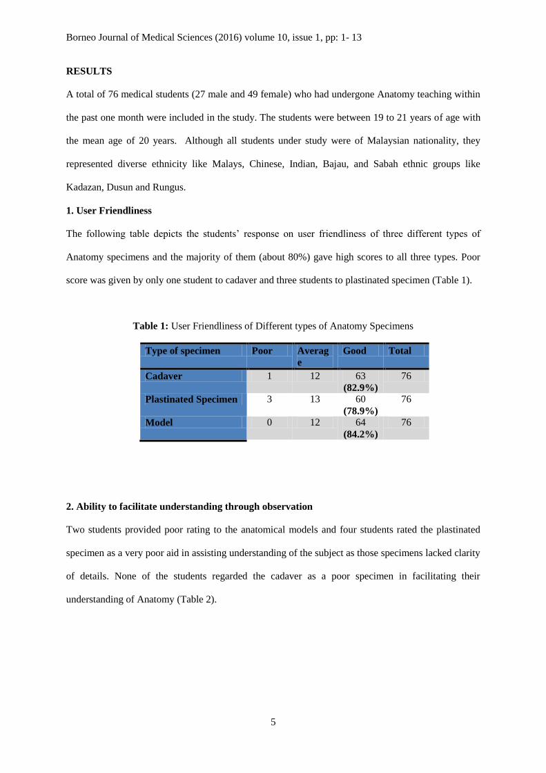
1. User Friendliness
The following table depicts the students’ response on user friendliness of three different types of
Anatomy specimens and the majority of them (about 80%) gave high scores to all three types. Poor
score was given by only one student to cadaver and three students to plastinated specimen (Table 1).
Table 1: User Friendliness of Different types of Anatomy Specimens
Type of specimen Poor Averag
e
Good Total
Cadaver 1 12 63
(82.9%)
76
Plastinated Specimen 3 13 60
(78.9%)
76
Model 0 12 64
(84.2%)
76
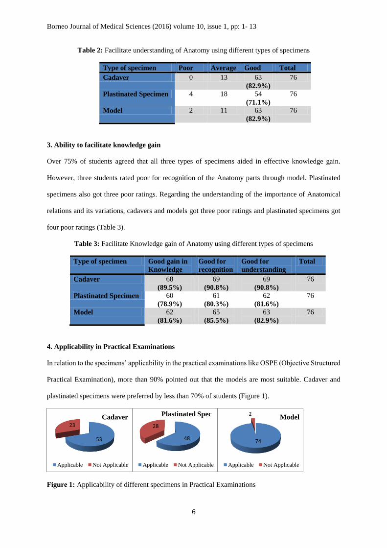
2. Ability to facilitate understanding through observation
Two students provided poor rating to the anatomical models and four students rated the plastinated
specimen as a very poor aid in assisting understanding of the subject as those specimens lacked clarity
of details. None of the students regarded the cadaver as a poor specimen in facilitating their
understanding of Anatomy (Table 2).
Borneo Journal of Medical Sciences (2016) volume 10, issue 1, pp: 1- 13
6
Table 2: Facilitate understanding of Anatomy using different types of specimens
Type of specimen Poor Average Good Total
Cadaver 0 13 63
(82.9%)
76
Plastinated Specimen 4 18 54
(71.1%)
76
Model 2 11 63
(82.9%)
76
3. Ability to facilitate knowledge gain
Over 75% of students agreed that all three types of specimens aided in effective knowledge gain.
However, three students rated poor for recognition of the Anatomy parts through model. Plastinated
specimens also got three poor ratings. Regarding the understanding of the importance of Anatomical
relations and its variations, cadavers and models got three poor ratings and plastinated specimens got
four poor ratings (Table 3).
Table 3: Facilitate Knowledge gain of Anatomy using different types of specimens
Type of specimen Good gain in
Knowledge
Good for
recognition
Good for
understanding
Total
Cadaver 68
(89.5%)
69
(90.8%)
69
(90.8%)
76
Plastinated Specimen 60
(78.9%)
61
(80.3%)
62
(81.6%)
76
Model 62
(81.6%)
65
(85.5%)
63
(82.9%)
76
4. Applicability in Practical Examinations
In relation to the specimens’ applicability in the practical examinations like OSPE (Objective Structured
Practical Examination), more than 90% pointed out that the models are most suitable. Cadaver and
plastinated specimens were preferred by less than 70% of students (Figure 1).
Figure 1: Applicability of different specimens in Practical Examinations
53
23
Cadaver
Applicable Not Applicable
48
28
Plastinated Spec
Applicable Not Applicable
74
2Model
Applicable Not Applicable
Borneo Journal of Medical Sciences (2016) volume 10, issue 1, pp: 1- 13
7
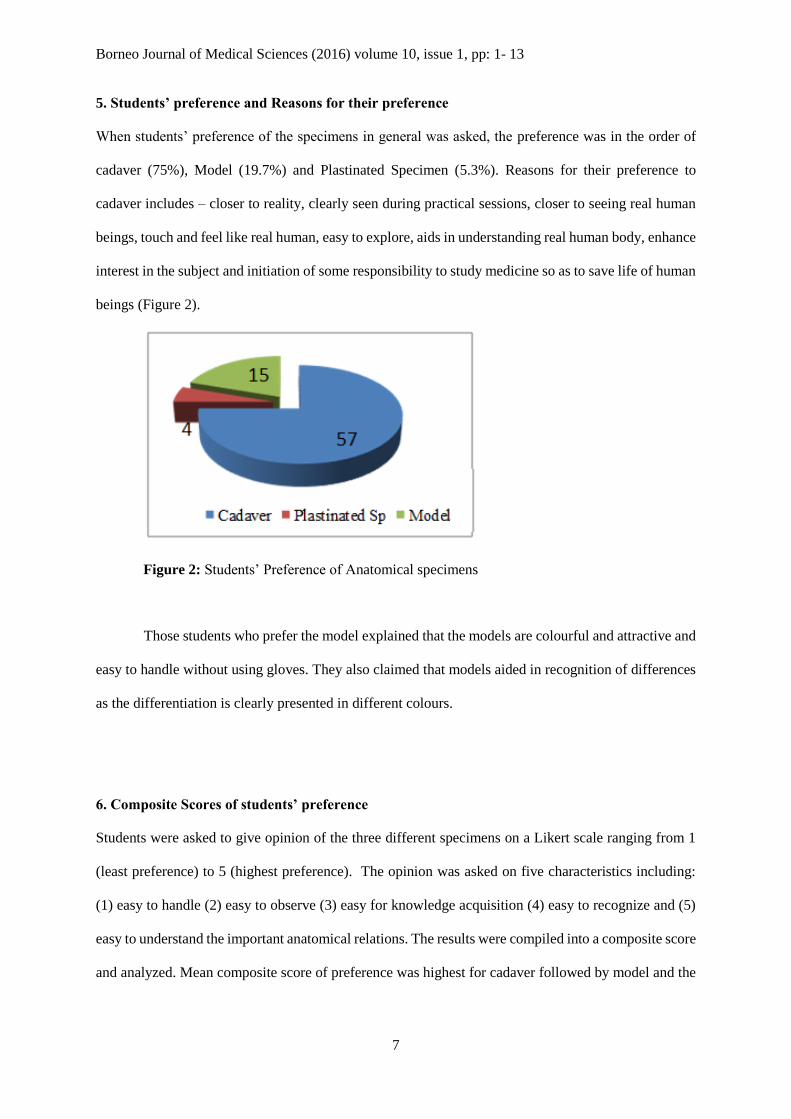
5. Students’ preference and Reasons for their preference
When students’ preference of the specimens in general was asked, the preference was in the order of
cadaver (75%), Model (19.7%) and Plastinated Specimen (5.3%). Reasons for their preference to
cadaver includes – closer to reality, clearly seen during practical sessions, closer to seeing real human
beings, touch and feel like real human, easy to explore, aids in understanding real human body, enhance
interest in the subject and initiation of some responsibility to study medicine so as to save life of human
beings (Figure 2).
Figure 2: Students’ Preference of Anatomical specimens
Those students who prefer the model explained that the models are colourful and attractive and
easy to handle without using gloves. They also claimed that models aided in recognition of differences
as the differentiation is clearly presented in different colours.
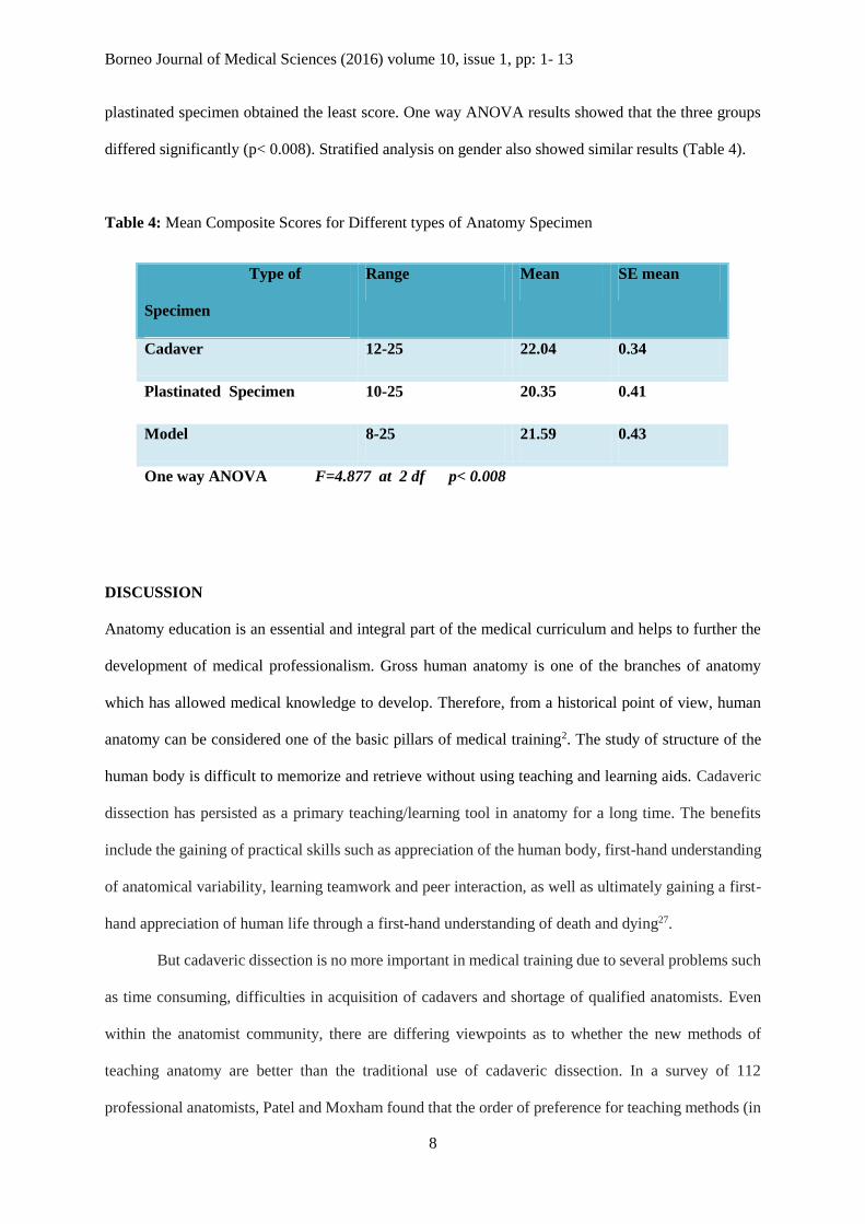
6. Composite Scores of students’ preference
Students were asked to give opinion of the three different specimens on a Likert scale ranging from 1
(least preference) to 5 (highest preference). The opinion was asked on five characteristics including:
(1) easy to handle (2) easy to observe (3) easy for knowledge acquisition (4) easy to recognize and (5)
easy to understand the important anatomical relations. The results were compiled into a composite score
and analyzed. Mean composite score of preference was highest for cadaver followed by model and the
Borneo Journal of Medical Sciences (2016) volume 10, issue 1, pp: 1- 13
8
plastinated specimen obtained the least score. One way ANOVA results showed that the three groups
differed significantly (p< 0.008). Stratified analysis on gender also showed similar results (Table 4).
Table 4: Mean Composite Scores for Different types of Anatomy Specimen
DISCUSSION
Anatomy education is an essential and integral part of the medical curriculum and helps to further the
development of medical professionalism. Gross human anatomy is one of the branches of anatomy
which has allowed medical knowledge to develop. Therefore, from a historical point of view, human
anatomy can be considered one of the basic pillars of medical training2. The study of structure of the
human body is difficult to memorize and retrieve without using teaching and learning aids. Cadaveric
dissection has persisted as a primary teaching/learning tool in anatomy for a long time. The benefits
include the gaining of practical skills such as appreciation of the human body, first-hand understanding
of anatomical variability, learning teamwork and peer interaction, as well as ultimately gaining a first-
hand appreciation of human life through a first-hand understanding of death and dying27.
But cadaveric dissection is no more important in medical training due to several problems such
as time consuming, difficulties in acquisition of cadavers and shortage of qualified anatomists. Even
within the anatomist community, there are differing viewpoints as to whether the new methods of
teaching anatomy are better than the traditional use of cadaveric dissection. In a survey of 112
professional anatomists, Patel and Moxham found that the order of preference for teaching methods (in
Type of
Specimen
Range Mean SE mean
Cadaver 12-25 22.04 0.34
Plastinated Specimen 10-25 20.35 0.41
Model 8-25 21.59 0.43
One way ANOVA F=4.877 at 2 df p< 0.008
Borneo Journal of Medical Sciences (2016) volume 10, issue 1, pp: 1- 13
9
descending order) was cadaveric dissection by students, prosection, living and radiological anatomy,
computer-aided learning (CAL), didactic lectures alone, and the use of models. In most of the anatomy
department in medical universities the role of cadaveric dissection as the primary mode of anatomy
teaching has been reduced or replaced by more innovative approaches such as prosection, plastinated
specimens, plastic models and mutimedia - based learning packages28.
Many studies reported the effectiveness of cadaveric dissection in anatomy teaching and
learning. Leong reported that 60.7% of the students of National University of Singapore found
dissection helpful and 28% of them found very helpful in their understanding of gross anatomy. When
asked whether dissection should be replaced completely by demonstrations on prosected specimens,
86.7% gave a resounding no23. In our present study no cardaveric dissection by students was done and
the cadaver, plastinated specimen and plastic models are demonstrated by clinical anatomists in
practical session.
Plastination is a relatively new advancement in cadaveric science; an effective technique of
tissue preservation of entireorgans or cross-sectional body slices. Using polymers such as resin, silicone,
and polyester give differing mechanical properties that ultimately result in robust, dry, odourless, and
life-like specimens, which can be used well in an educational capacity in gross anatomy. Student
satisfaction and acceptance has also been recorded using plastinated models as well as a significant
difference between control and experimental groups observed in assessment scores29.
Many institutions have overcome problems surrounding dissection with plastic models. Plastic
specimens are modelled to perfectionand possess a longer shelf-life than cadavers but they will
eventually pose problems. No human body is ever modelled to perfection where all organs are colour
coordinated and impeccably shaped30.
There are no studies on comparison on effectiveness of plastination and plastic models using
as anatomy teaching tools. However the present study was conducted to determine the preference of
Anatomy specimen (cadaver or plastinated specimen or plastic model) among the first year medical
students for their effective learning of Anatomy.
Borneo Journal of Medical Sciences (2016) volume 10, issue 1, pp: 1- 13
10
According to the results, in the students’ response on user friendliness of three different
Anatomy specimens the majority of them (about 80%) gave high scores to all three types. Poor score
was given by only one student to cadaver and three students to plastinated specimen. Most of first year
medical students are affability to three different types of materials in anatomy learning.
In facilitating the student' understanding of anatomy cadaver specimen and models recorded
more than 80%. Over 75% of students agreed that all three types of materials aided in effective
knowledge gain. Cadaver specimen gave better for recognition and understanding than other two.
In the present study 75% of students agreed that cadaveric specimen was the most preference
in learning anatomy. Reasons for their preference to cadaver includes – closer to reality, clearly seen
during practical session, closer to seeing real human beings, touch and feel like real human, easy to
explore, aids in understanding real human body. This finding is in line with the study of Izunya et al
(2010) and Oyeyipo (2012), that majority of the students (90%) considered cadaver dissection as
important and indispensable in the study of human anatomy and still remain best method for learning
anatomy13, 14. Similar findings were reported elsewhere by Rajkumari et al (2008), Abay et al (2012),
Weerasuriya (2014)7, 15, 31. This is contrary to the report by Rehman, et al (2012)32.
The different colour presentation of the model can be made easier identification and
differentiation in practical examination. We observed that, in relation to the specimens’ applicability in
the practical examinations like OSPE (Objective Structured Practical Examination), more than 90%
pointed out that the models are most applicable. This is contrary with the results by Godson (2010)33.
CONCLUSION
The study strongly indicated the medical students’ preference to cadaver during their Anatomy learning
sessions. The students rated cadaver as the most preferred specimen for better acquisition of knowledge,
easy recognition and deeper understanding of the subject. In addition, they preferred cadaver as it is
closer to reality and more relevant to the human being. It is recommended that these findings should be
taken into consideration in future curriculum development of medical schools.
Borneo Journal of Medical Sciences (2016) volume 10, issue 1, pp: 1- 13
11
ACKNOWLEDGEMENTS
The authors would like to thank the all year 1 medical students of Faculty of Medicine & Health
Sciences, Universiti Malaysia Sabah of academic session 2013-2014 for their kind cooperation and
contribution of their valuable opinions and answers for this study.
CONFLICT OF INTEREST: None.
REFERENCES
1. Rajkumari AB and Singh YI. (2007). Body donation and its relevance in anatomy learning: A
review. Jour Anat. Soc. India 56(1):1-6.
2. McLachlan JC, Patten D. (2006). Anatomy teaching: ghosts of the past, present and future. Med
Educ 40:243-253.
3. Older J. (2004). Anatomy: A must for teaching the next generation. The surgeon 2(2): 79-90.
4. Mclachian J, Bradley P, Searle J, Bligh J. (2004).Teaching anatomy without cadavers. Med.
Edu 38: 418-424.
5. Bertman, SL, Marks Jnr SC. (1985). Humanities in medical education Rationale and resources
for the dissection laboratory. Med. Educ 19: 374-381.
6. Percac S, McArdle PJ. (1997). Anatomy teaching: Students' perceptions. Surg Radiol Anat 19:
315-317.
7. Rajkumari A, Kumar Das B, Gracelee Tempy N Sangma, Singh YI. (2008). Attitudes and views
of first year medical students toward cadaver dissection in anatomy learning. Calicut Medical
Journal 6(4): e2.
8. Arraez-Aybar LA, Castano-Collado G, Casado-Morales MI. (2004). Dissection from the
Spanish anatomist's perspective: aims, attitudes, and related aspects. Anatomical Record Part
B: New Anatomist 281(1): 15-20.
9. Bay BH, Ling EA. (2007). Teaching of Anatomy in the new millennium. Singapore Med J 48
(3): 182-183.
10. Prakash, Prabhu LV, Rai R, D’ Costa S, Jiji PJ, Singh G. (2007). Cadavers as teachers in
medical education: knowledge is the ultimate gift of body donors. Singapore Med J 48(3): 186-
90.
11. Maguire P. Barriers to psychological care of the dying. (1985). British Medical Journal 291:
1711-1713.
Borneo Journal of Medical Sciences (2016) volume 10, issue 1, pp: 1- 13
12
12. Winkelmann A, Guldner FH. (2004). Cadavers as teachers: the dissecting room experience in
Thailand. BMJ 329: 1455- 1457.
13. Izunya AM, Oaikhena GA, Nwaopara AO. (2010). Attitudes to Cadaver Dissection in a
Nigerian Medical School. Asian Journal of Medical Sciences 2(3): 89-94.
14. Oyeyipo IP, Falana BA. (2012). Attitude of preclinical students to cadaver dissection in a South
West Nigeria Medical School. International J Trop Med 7(1):1-5.
15. Abay M, Desalegn T. (2012). Medical student’s attitudinal changes towards cadaver dissection:
A longitudinal study. Ethiop Health Sci 22(1): 51-58.
16. Von Hagens G, Tiedemann K, Kriz W. (1987). The current potential of plastination. Anat.
Embryol 175: 411–421.
17. Dhingra R, Taranikanti V, Kumar R. (2006). Plastination: teaching aids in anatomy revisited.
Natl. Med. J. India 19: 171.
18. Sugand K, Abrahams P, Khurana A. (2010). The anatomy of anatomy: a review for its
modernization. Anat Sci Edu 3(2): 83-93.
19. Pawlina W, Lachman N. (2004). Dissection in learning and teaching gross anatomy.
Anatomical record 281(1): 9-11.
20. Paalman MH. (2000). Why teach anatomy? Anatomists respond. Anat Rec 261: 1-2.
21. Stephen J, Chapman, Abdul R. Hakeem, Gabriele Marangoni, Prasad KR. (2013). Anatomy in
medical education: Perceptions of undergraduate medical students. Annals of Anatomy 195:
409– 441.
22. Azu OO, Peter AI, Etuknwa BT, Ekandem GJ. (2012). The Awareness of Medical Students in
Nigerian Universities about the Use of Plastinated Specimens for Anatomical Studies. Maced
J Med Sci 5(1): 5-9.
23. Leong SK. Back to basic. (1999). Clinical Anat 12(6): 422-426.
24. Azer SA, Eizenberg N.(2002). Do we need dissection in an integrated problem-based learning
medical course? Perceptions of first- and second-year students. Surg Radiol Anat 9: 173–180.
25. Hasan T, Ageely H, Hasan D. (2010). The role of traditional dissection in medical education.
Education in Med J 2(1): 30-34.
26. McLachlan J. (2004). New Path for Teaching Anatomy: Living anatomy and medical imaging
vs. dissection. Anat Rec B New Anat 281(1): 4–5.
27. Granger NA. (2004). Dissection laboratory is vital to medical gross anatomy education. Anat
Rec (Part B: New Anat) 281: 6–8.
28. Patel KM, Moxham BJ. (2006). Attitudes of professional anatomists to curricular change. Clin
Anat 19: 132-14.
29. Latorre RM, Garcı´a-Sanz MP, Moreno M, et al. (2007). How useful is plastination in learning
anatomy? J Vet Med Educ 34:172-176.
Borneo Journal of Medical Sciences (2016) volume 10, issue 1, pp: 1- 13
13
30. Kapil, Peter A, Ashish K. (2010). The Anatomy of Anatomy: A Review for Its Modernization,
Anat Sci Edu 3: 83–93.
31. Weerasuriya T, Chan F, Yasawardene S, Pinto N. (2014). Preference of Medical Students of
Formaldehyde Preserved Cadaveric Dissection Versus Pre-Dissected Specimens as a Teaching
Tool in Human Anatomy. JMED Research: 1-6.
32. Rehman FU, Khan SN, Yunus SM. (2012). Students, perception of computer assisted teaching
and learning of anatomy in a scenario where cadavers are lacking. Biomedical Research 23 (2):
215-218.
33. Godson EA, Anthony IU. (2010). Impact of the use of cadaver on student’s ability to pass
anatomy examination. Int J Exp Clin Anat 4: 28-34.
34. Arráez-Aybar LA, Sánchez-Montesinos I, Mirapeix RM. (2010). Relevance of human anatomy
in daily clinical practice. Annals of Anatomy 192: 341-348.
35. Azu OO, Osinubi AA. (2011). A survey of problem-based learning and traditional methods of
teaching anatomy to 200 level pharmacy students of the University of Lagos, Nigeria. African
J Pharmacy and Pharmacology 5(2): 219-224.


