Embed Size (px)
Citation preview
OSPECARDIOVASCULAR BLOCK
Color Code
Nerves
Arteries
Veins
Muscles
Lymphatics
Important Points
1. Read the questions carefully.
2. Make sure your write the FULL name of the structures with the correct spelling.
Example: SVC ✕ Superior Vena Cava ✓
Thoracic aorta ✕ Descending thoracic aorta ✓
3. There is NO guarantee whether or not the exam will go out of this file.
Good luck!
ذا شئت سهل ال ما جعلته سهل وأ نت جتعل احلزن ا اللهم ال سهل ا
a
b
c
d
e
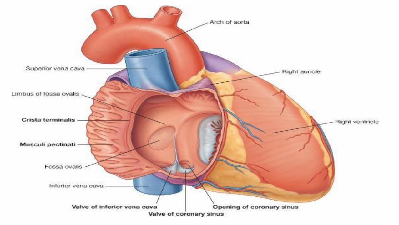
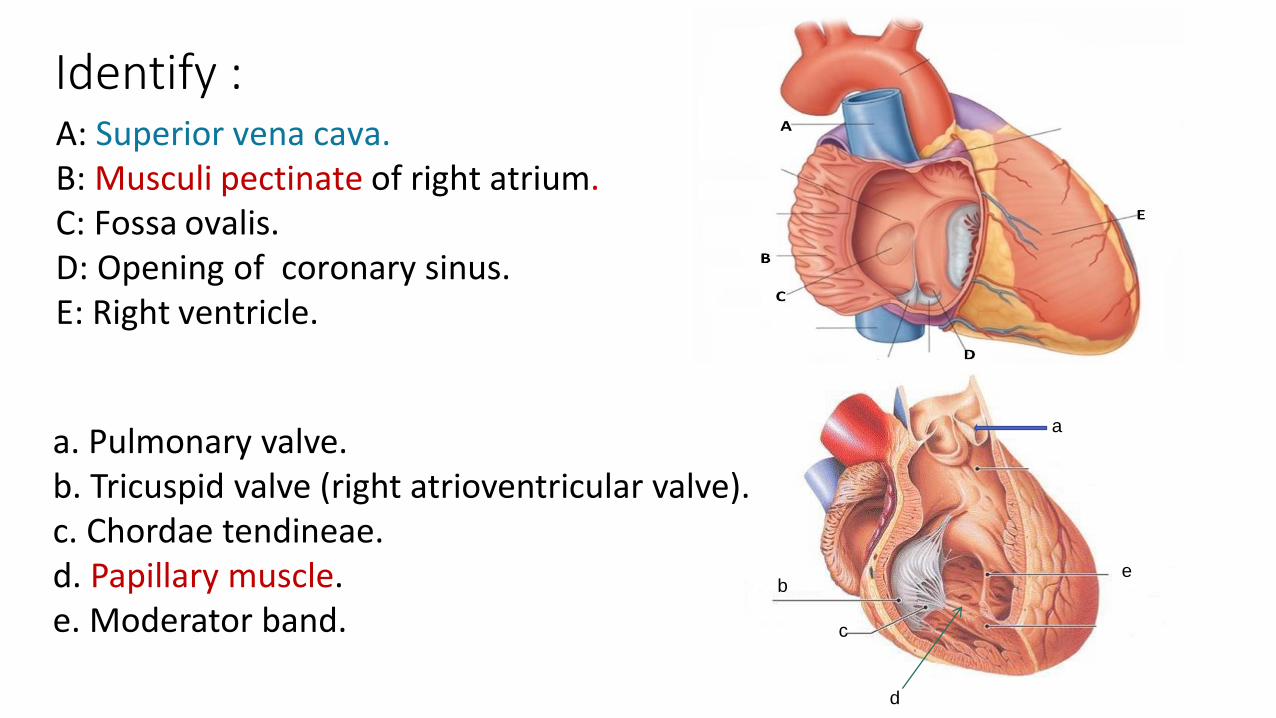
Identify :A: Superior vena cava.B: Musculi pectinate of right atrium.C: Fossa ovalis.D: Opening of coronary sinus. E: Right ventricle.
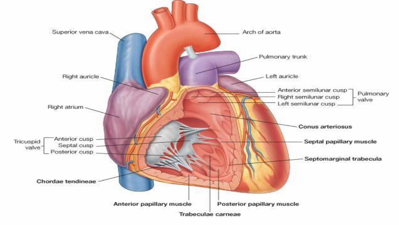
a. Pulmonary valve. b. Tricuspid valve (right atrioventricular valve).c. Chordae tendineae.d. Papillary muscle.e. Moderator band.
a
b
c d
e
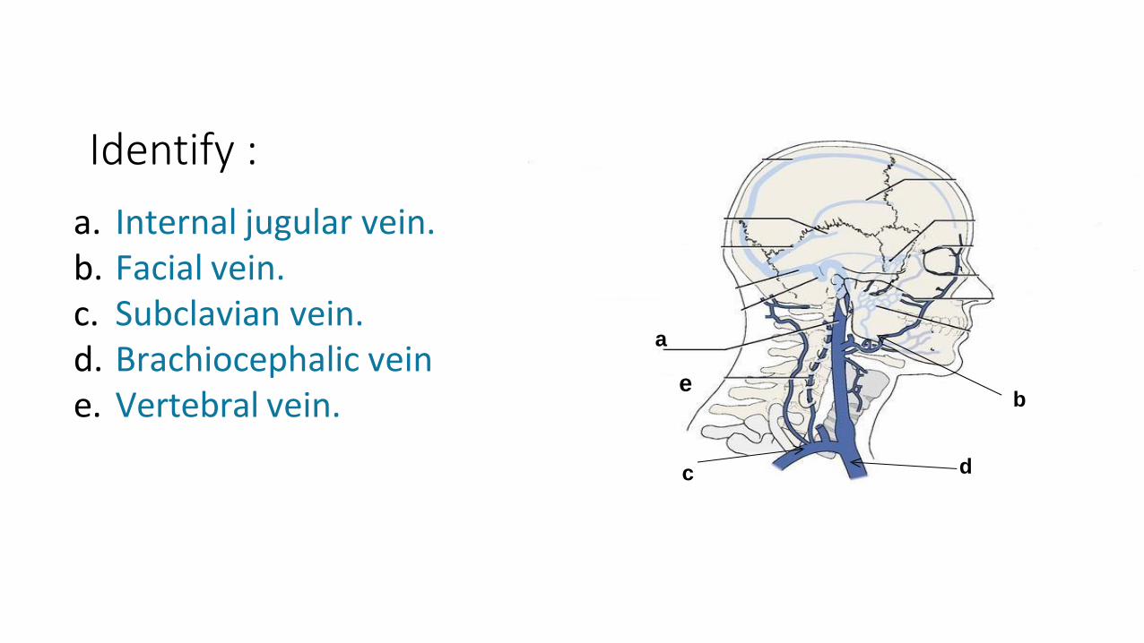
a. Internal jugular vein.b. Facial vein.c. Subclavian vein.d. Brachiocephalic veine. Vertebral vein.
Identify :
a
b cd
e
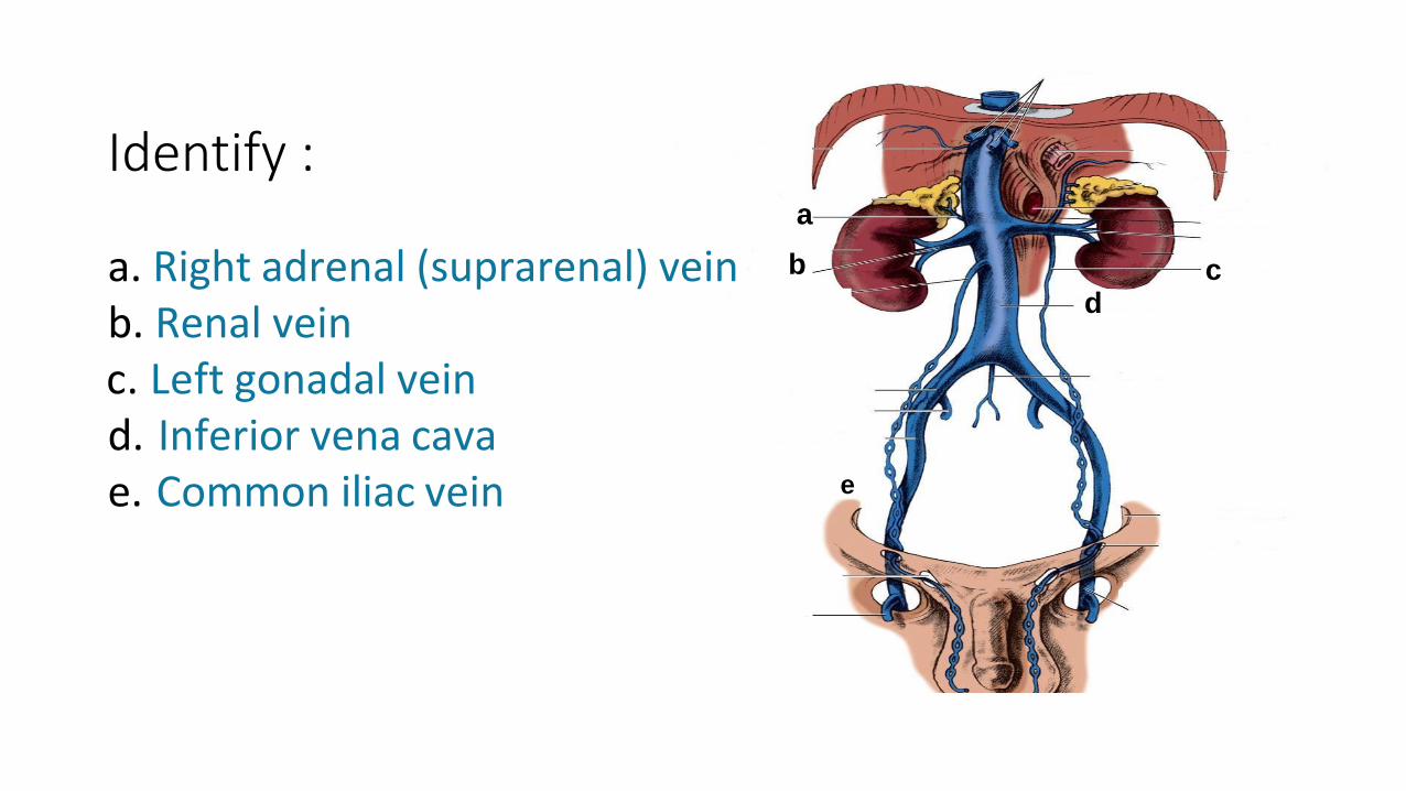
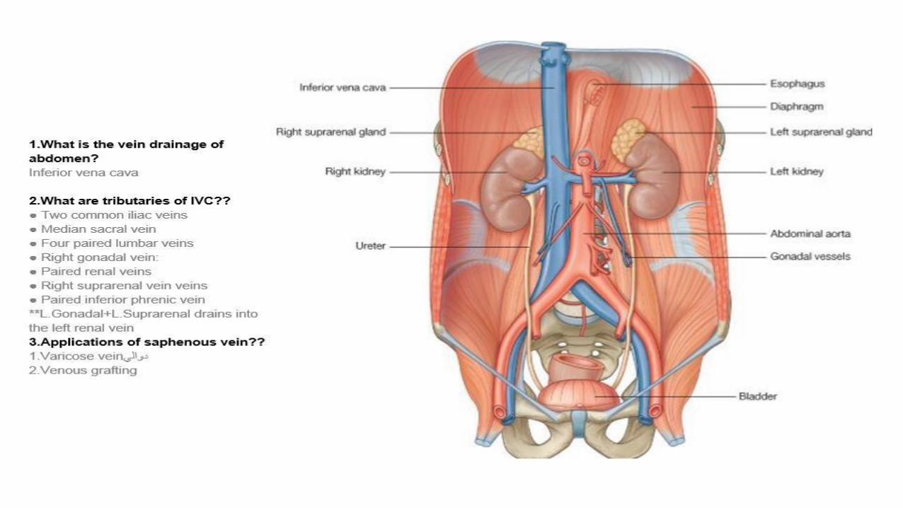
a. Right adrenal (suprarenal) veinb. Renal veinc. Left gonadal veind. Inferior vena cavae. Common iliac vein
Identify :
A
B
C
A
B
C
D
E
F
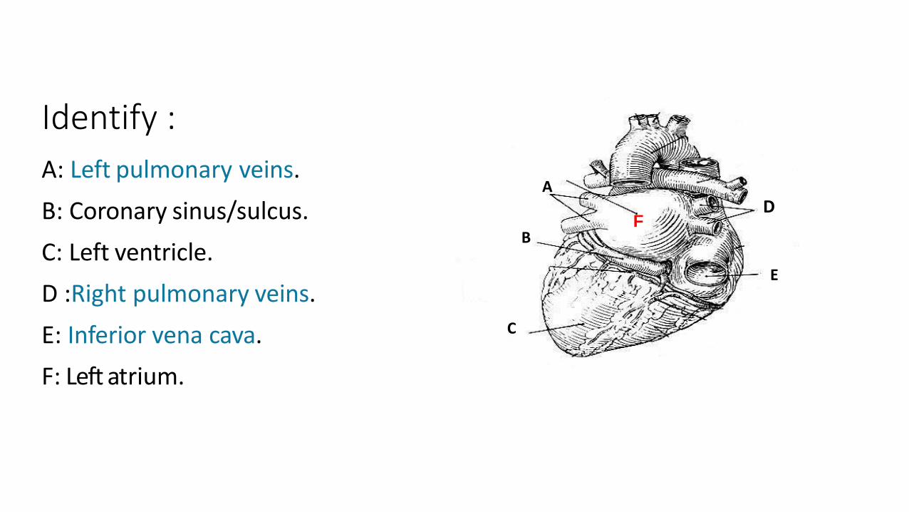
A: Left pulmonary veins.
B: Coronary sinus/sulcus.
C: Left ventricle.
D :Right pulmonary veins.
E: Inferior vena cava.
F: Left atrium.
Identify :
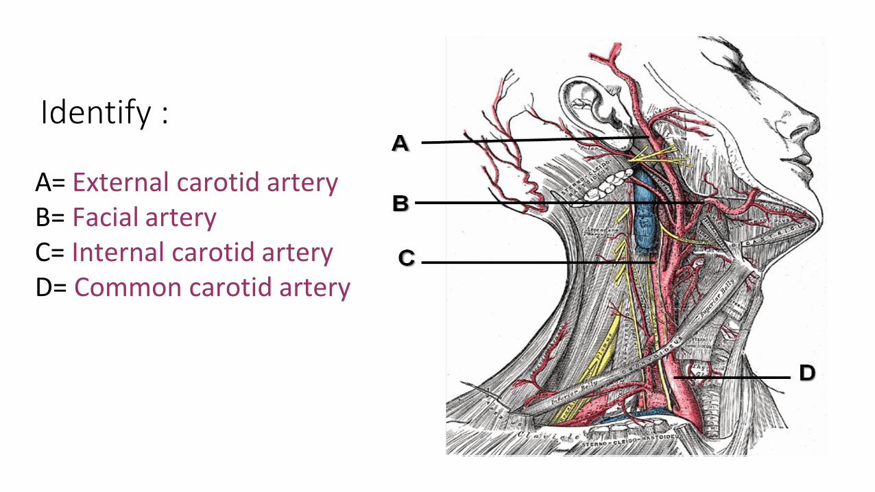
A= External carotid arteryB= Facial arteryC= Internal carotid artery D= Common carotid artery
Identify :
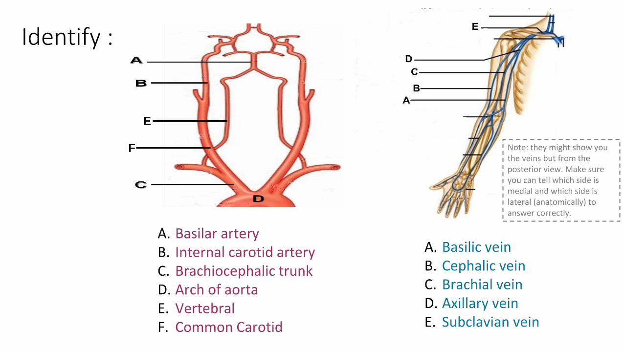
A. Basilar arteryB. Internal carotid arteryC. Brachiocephalic trunkD. Arch of aortaE. VertebralF. Common Carotid
A. Basilic veinB. Cephalic veinC. Brachial veinD. Axillary veinE. Subclavian vein
Identify :
E
F Note: they might show you the veins but from the posterior view. Make sure you can tell which side is medial and which side is lateral (anatomically) to answer correctly.
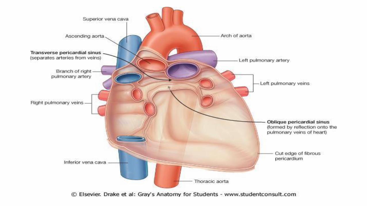
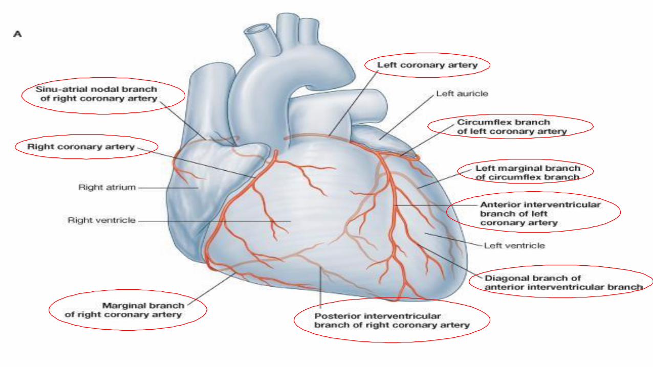
Ascending aortao Originates from left ventricle.o Continues as the arch of aortao Has three dilatations at its base, called aortic sinuseso Branches :
Right & Left coronary arteries (supplying heart), arise from aortic sinuses.
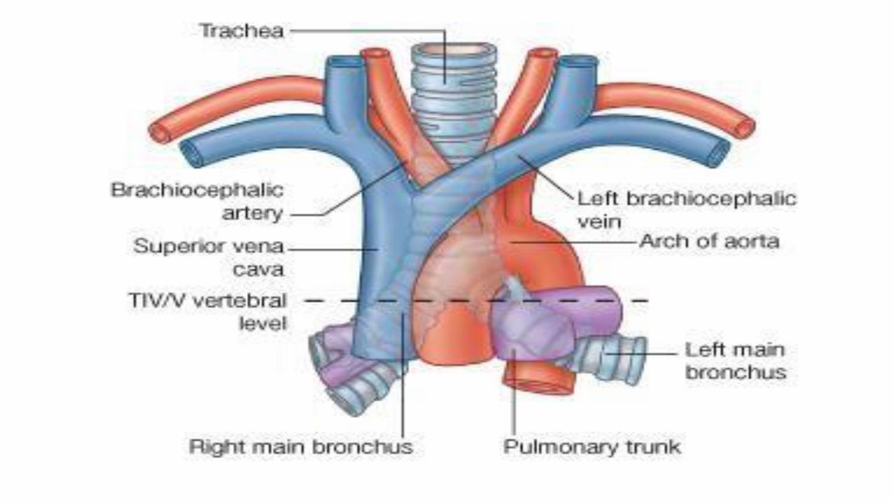
Arch of Aortao Continuation of the ascending aorta.o Leads to descending aorta. o Located behind the lower part of manubrium sterni and on the left side of trachea.o Branches :
1. Brachiocephalic trunk.2. Left common carotid artery. 3. Left subclavian artery.
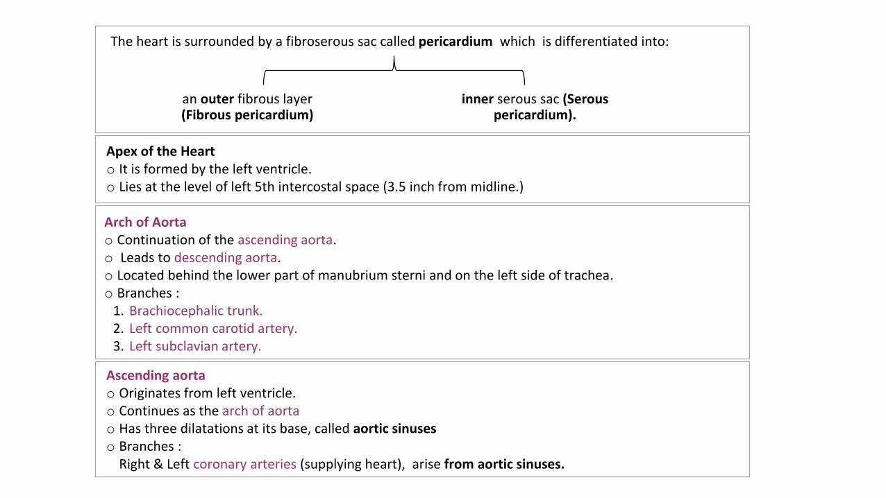
an outer fibrous layer (Fibrous pericardium)
inner serous sac (Serous pericardium).
The heart is surrounded by a fibroserous sac called pericardium which is differentiated into:
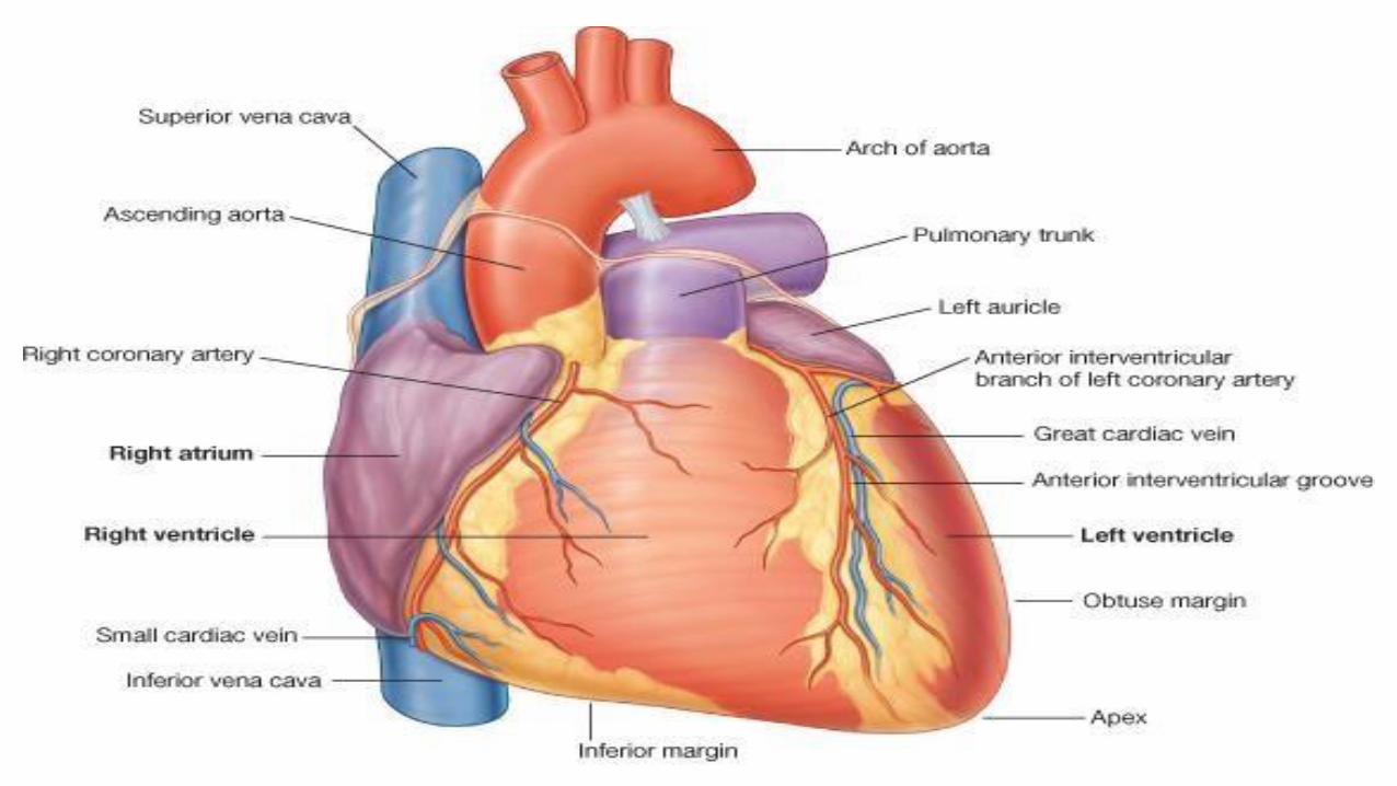
Apex of the Hearto It is formed by the left ventricle. o Lies at the level of left 5th intercostal space (3.5 inch from midline.)
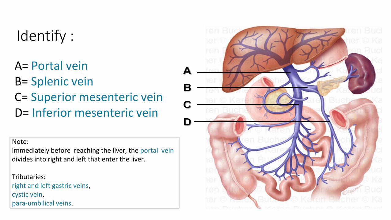
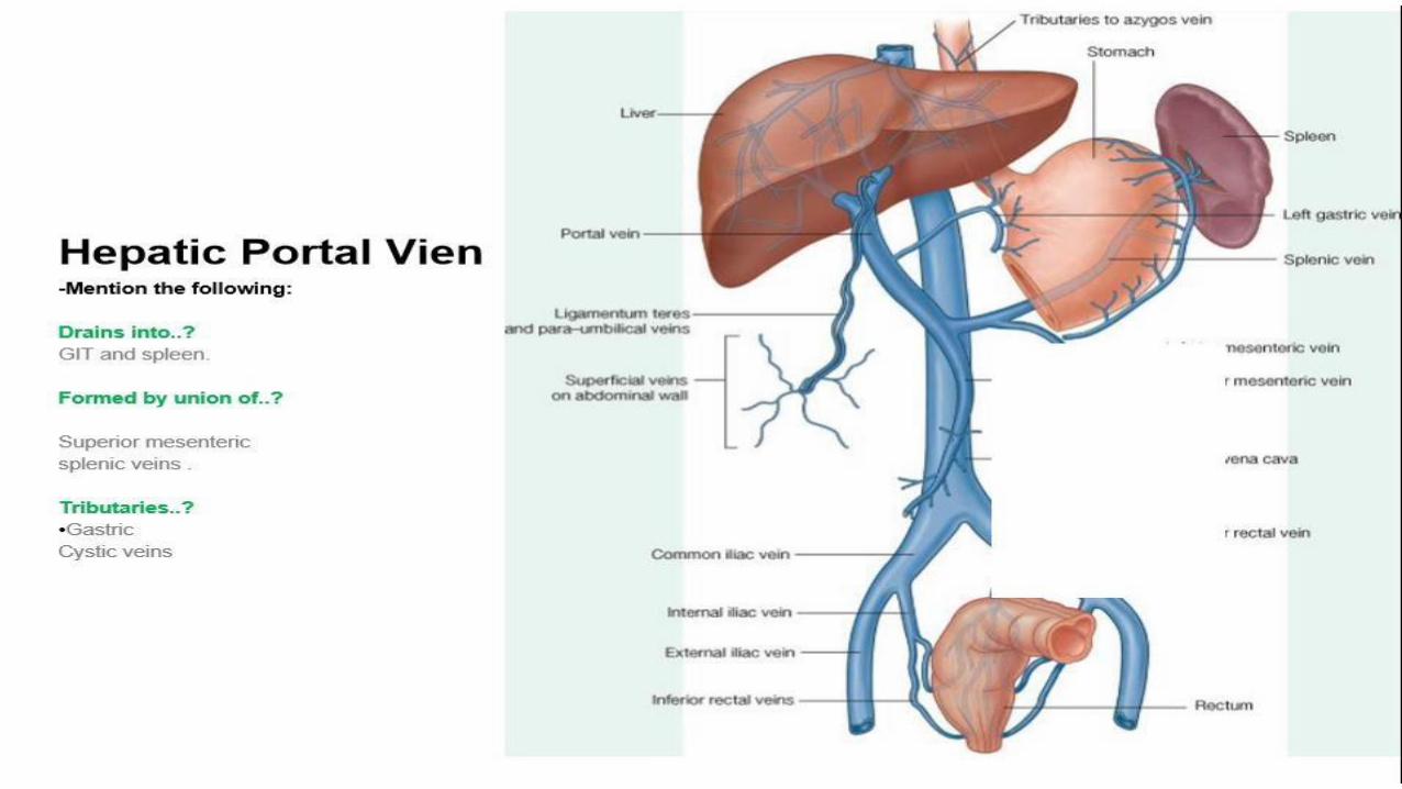
Note: Immediately before reaching the liver, the portal vein divides into right and left that enter the liver.
Tributaries:right and left gastric veins,cystic vein,para-umbilical veins.
A= Portal vein B= Splenic veinC= Superior mesenteric veinD= Inferior mesenteric vein
Identify :
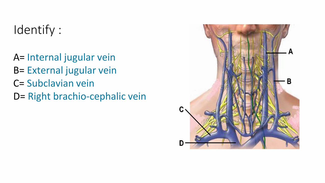
A= Internal jugular vein B= External jugular vein C= Subclavian veinD= Right brachio-cephalic vein
Identify :
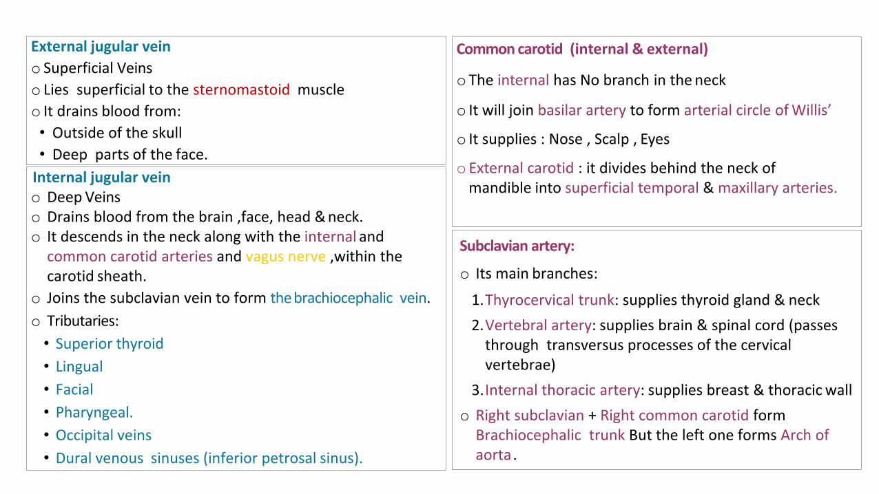
Internal jugular vein o Deep Veinso Drains blood from the brain ,face, head & neck.o It descends in the neck along with the internal and
common carotid arteries and vagus nerve ,within the carotid sheath.
o Joins the subclavian vein to form thebrachiocephalic vein.
o Tributaries:
• Superior thyroid
• Lingual
• Facial
• Pharyngeal.
• Occipital veins
• Dural venous sinuses (inferior petrosal sinus).
External jugular vein
o Superficial Veins
o Lies superficial to the sternomastoid muscle
o It drains blood from:
• Outside of the skull
• Deep parts of the face.
Common carotid (internal & external)
oThe internal has No branch in the neck
o It will join basilar artery to form arterial circle ofWillis’
o It supplies : Nose , Scalp , Eyes
o External carotid : it divides behind the neck of mandible into superficial temporal & maxillary arteries.
Subclavian artery:
o Its main branches:
1.Thyrocervical trunk: supplies thyroid gland & neck
2.Vertebral artery: supplies brain & spinal cord (passesthrough transversus processes of the cervicalvertebrae)
3.Internal thoracic artery: supplies breast & thoracic wall
o Right subclavian + Right common carotid formBrachiocephalic trunk But the left one forms Arch of aorta.
Done by:
Mohammed Ghandour
Mohammed Alyousef
Jawaher Abanumy
@anatomy436
Special Thanks to TEAM 434


