Embed Size (px)
Citation preview

Pre-existence and clonal selection of MET amplification in EGFRmutant NSCLC
Alexa B. Turke1,2,*, Kreshnik Zejnullahu3,4,*, Yi-Long Wu5, Youngchul Song1, Dora Dias-Santagata1, Eugene Lifshits1, Luca Toschi3,4, Andrew Rogers3,4, Tony Mok6, LeciaSequist1, Neal I. Lindeman7, Carly Murphy7, Sara Akhavanfard1, Beow Y. Yeap1,2, YunXiao7,4, Marzia Capelletti3,4, A. John Iafrate1, Charles Lee7, James G. Christensen8, JeffreyA. Engelman1,2,#,¥, and Pasi A. Jänne2,3,4,9,#,¥1Massachusetts General Hospital Cancer Center, Boston, MA 02129, USA2Department of Medicine, Harvard Medical School, Boston, MA 02115, USA3Lowe Center for Thoracic Oncology, Boston, MA 02115, USA4Department of Medical Oncology, Dana-Farber Cancer Institute, Boston, MA 02115, USA5Guangdong Lung Cancer Institute and Cancer Center, Guangdong General Hospital, Guangzhou,China6The Chinese University of Hong Kong, Hong Kong, China7Department of Pathology, Brigham and Women’s Hospital, Boston, MA 02115, USA8Pfizer Global Research and Development, Department of Research Pharmacology, La Jolla Labs,La Jolla, CA 92121, USA9Department of Medicine, Brigham and Women’s Hospital, Boston, MA 02115, USA.
SummaryMET amplification activates ERBB3/PI3K/AKT signaling in EGFR mutant lung cancers, and causesresistance to EGFR kinase inhibitors. We demonstrate that MET activation by its ligand, HGF, alsoinduces drug resistance, but through GAB1 signaling. Using high-throughput FISH analyses in bothcell lines and in lung cancer patients, we identify subpopulations of cells with MET amplificationprior to drug exposure. Surprisingly, HGF accelerates the development of MET amplification bothin vitro and in vivo. EGFR kinase inhibitor resistance, due to either MET amplification or autocrineHGF production, was cured in vivo by combined EGFR and MET inhibition. These findings highlightthe potential to prospectively identify treatment naïve EGFR mutant lung cancer patients who willbenefit from initial combination therapy.
© 2009 Elsevier Inc. All rights reserved.#Address Correspondence to: Jeffrey A. Engelman, M.D., Ph.D. Massachusetts General Hospital Cancer Center Building 149, 13th St,Boston, MA 02129 Phone: 617-724-7298 Fax: 617-724-9648 [email protected] Pasi A. Jänne, M.D., Ph.D. Lowe Center forThoracic Oncology Dana-Farber Cancer Institute, D820 44 Binney St, Boston, MA 02115 Phone: (617) 632-6076 Fax: (617) [email protected].*These authors contributed equally to this work.¥These labs contributed equally to this work.Publisher's Disclaimer: This is a PDF file of an unedited manuscript that has been accepted for publication. As a service to our customerswe are providing this early version of the manuscript. The manuscript will undergo copyediting, typesetting, and review of the resultingproof before it is published in its final citable form. Please note that during the production process errors may be discovered which couldaffect the content, and all legal disclaimers that apply to the journal pertain.
NIH Public AccessAuthor ManuscriptCancer Cell. Author manuscript; available in PMC 2011 January 19.
Published in final edited form as:Cancer Cell. 2010 January 19; 17(1): 77–88. doi:10.1016/j.ccr.2009.11.022.
NIH
-PA Author Manuscript
NIH
-PA Author Manuscript
NIH
-PA Author Manuscript
IntroductionEpidermal growth factor receptor (EGFR) tyrosine kinase inhibitors (TKIs) gefitinib anderlotinib are effective clinical therapies for advanced non-small cell lung cancer (NSCLC)patients with EGFR activating mutations (Asahina et al., 2006; Inoue et al., 2006; Paz-Ares etal., 2006; Sequist et al., 2008; Tamura et al., 2008). A recent phase III clinical trial demonstratedthat patients with EGFR mutant NSCLC had superior outcomes with gefitinib treatmentcompared to standard first line cytotoxic chemotherapy (Mok et al., 2008). However, despitethese dramatic benefits from EGFR TKIs in this genetically defined cohort, all of these patientsultimately develop resistance (referred to as acquired resistance herein) to gefitinib anderlotinib. Two mechanisms of acquired resistance have been validated in patients. Secondarymutations in EGFR itself, including the EGFR T790M “gatekeeper” mutation is observed in50% of resistance cases, and amplification of the MET oncogene is observed in 20% ofresistance cases (Balak et al., 2006; Bean et al., 2007; Engelman et al., 2007b; Kobayashi etal., 2005; Kosaka et al., 2006; Pao et al., 2005). Both resistance mechanisms lead tomaintenance of ERBB3/PI3K/AKT signaling in the presence of gefitinib (reviewed in(Engelman and Janne, 2008)).
In addition to these genetic alterations, activation of IGF-1Rβ/IRS-1 signaling through loss ofIGF binding proteins also drives gefitinib resistance in EGFR wild-type cancer cell lines (Guixet al., 2008). Additionally, a recent study suggested that the MET ligand, HGF, can promoteshort-term resistance in two EGFR mutated cancer cell lines (Yano et al., 2008). Both ligand-dependent resistance mechanisms maintain PI3K/AKT activation despite EGFR inhibition.However, differences between IGF and HGF driven resistance in terms of potency andactivation of downstream signaling pathways have yet to be thoroughly examined.Furthermore, the contribution of HGF, if any, to gefitinib resistance mediated by METamplification is unknown.
Strategies for overcoming acquired resistance to gefitinib are now undergoing clinicalevaluation. In preclinical studies, the EGFR T790M mutation can be overcome by second-generation, irreversible EGFR inhibitors (Engelman et al., 2007a; Kobayashi et al., 2005; Riely,2008). In addition, the growth of EGFR mutant cancers with MET amplification can beinhibited by combined treatment with EGFR and MET kinase inhibitors (Bean et al., 2007;Engelman et al., 2007b). Indeed, there are now clinical trials assessing both irreversible EGFRinhibitors and a combination of MET and EGFR inhibitors in patients with acquired resistanceto gefitinib/erlotinib. Further, clinical activity of the irreversible EGFR inhibitor, PF00299804,has been observed in NSCLC patients that have developed acquired resistance to gefitinib/erlotinib (Janne et al., 2008). As an alternative strategy, to delay or avoid the emergence ofresistance, there is increased enthusiasm to utilize agents effective against specific resistancemechanisms as initial systemic therapies. For example, PF00299804 is now being assessed ina phase II clinical trial of EGFR TKI naïve patients. However, there are currently no methodsto predict the specific resistance mechanism that a cancer will develop.
In the current study, we modeled in vitro resistance to PF00299804 in the TKI sensitive EGFRmutant NSCLC cell line HCC827 (Engelman et al., 2006; Engelman et al., 2007b; Ogino etal., 2007) . In addition, we evaluated the potency of the MET ligand, HGF, to promote resistanceto EGFR TKIs and determined whether MET amplification pre-exists in a subpopulation ofcells prior to treatment with a TKI.
Turke et al. Page 2
Cancer Cell. Author manuscript; available in PMC 2011 January 19.
NIH
-PA Author Manuscript
NIH
-PA Author Manuscript
NIH
-PA Author Manuscript

ResultsMET amplification causes resistance to the irreversible EGFR inhibitor PF00299804 byactivating ERBB3 signaling
We generated in vitro resistant clones of HCC827 cells to the irreversible pan-ERBB kinaseinhibitor, PF00299804, using previously described methods (Engelman et al., 2006; Engelmanet al., 2007b). HCC827 cells were exposed to increasing concentrations of PF00299804,starting with 1nM, until they were able to proliferate freely in 1 M PF00299804, which occurredafter 6 months of drug selection. This concentration was chosen because it is ~ 1000 fold greaterthan the IC50 for growth inhibition of HCC827 cells and approximately 5 times greater thanthe serum concentration of PF00299804 observed in NSCLC patients in the phase I clinicaltrial (Janne et al., 2008; Schellens et al., 2007). Five independent clones were isolated andexpanded for further studies. All five HCC827 PF00299804 resistant (PFR) clones wereresistant to PF00299804 in vitro (Figure 1A and data not shown). No secondary EGFRmutations (e.g. T790M) were detected in any of the clones (data not shown).
We next examined the effects of PF00299804 on EGFR, ERBB3, AKT and ERKphosphorylation in the HCC827 PFR clones. Unlike in parental HCC827 cells, ERBB3activation as well as downstream PI3K/AKT and ERK signaling is maintained in the presenceof PF00299804 in HCC827 PFR cells (Figure 1B). We also observed increased total METprotein in the HCC827 PFR cells, and combined MET and EGFR inhibition down-regulatedERBB3, AKT and ERK phosphorylation as well as the modest EGFR phosphorylation thatwas maintained in the presence of PF00299804 alone (Figure 1B). This behavior followingtreatment with PF00299804 alone or in combination with a MET inhibitor is similar to thatobserved in gefitinib resistant HCC827 cells (HCC827 GR cells), which were generated in ananalogous manner and contained a focal amplification in chromosome 7 harboring the METoncogene (Engelman et al., 2007b).
Given the similarities in the HCC827 PFR and GR cells following treatment with eitherPF00299804 or gefitinib, respectively, we determined whether the addition of a MET inhibitorwould overcome resistance to PF00299804. We used both a tool compound PHA-665,752 andthe MET inhibitor PF2341066 currently undergoing clinical development (Figure 1C, upperand data not shown) (Zou et al., 2007). The combination of PF00299804 and a MET inhibitoreffectively inhibited the growth of HCC827 PFR cells while neither agent alone led to growthinhibition (Figure 1C, upper and data not shown). In addition, the combination of gefitinib andPF2341066 also effectively inhibited the growth of HCC827 PFR cells (Figure 1C, lower).These findings further suggest that the resistance mechanism in the HCC827 PFR cells is notunique or dependent on the differences between reversible (gefitinib) or irreversible(PF00299804) EGFR inhibitors but rather due solely to MET amplification. We also evaluatedthe effects of the irreversible EGFR inhibitor PF00299804 and the MET inhibitor PF-2341066in an HCC827 PFR xenograft model. Treatment with PF00299804 alone was modestly moreeffective than treatment with PF2341066 alone, but the tumors demonstrated resistance toPF00299804. However, combined MET and EGFR inhibition completely inhibited tumorgrowth and produced complete responses (p<0.0001; Figure 1D). In fact, the combinationtreatment was discontinued after 56 days (Figure 1D; arrow) and no tumor re-growth has beenobserved to date in any of the xenografts (after more than 35 weeks off therapy) (Figure 1D),suggesting that the mice have been cured.
We next determined whether the increase in MET protein expression was due to METamplification in the HCC827 PFR cells (Figure 2A). All of the PFR clones contained at leasta four fold amplification of MET, similar to the amplification previously observed in thegefitinib resistant HCC827 (HCC827 GR) cells ((Engelman et al., 2007b) and Figure 2A). Allof the PFR clones also had higher levels of MET protein expression (Figure 2B). Genome-
Turke et al. Page 3
Cancer Cell. Author manuscript; available in PMC 2011 January 19.
NIH
-PA Author Manuscript
NIH
-PA Author Manuscript
NIH
-PA Author Manuscript
wide SNP analysis revealed that the only area of significant copy number gain in HCC827 PFRcells is on distal chromosome 7, similar to that observed in HCC827 GR cells, and containsthe MET oncogene (Figure 2C, D). Furthermore, HCC827 PFR and GR cells share single copylosses of 4p, 5q, 14p, 14q and 19p, but only HCC827 PFR cells have a single copy loss of 16q.Intriguingly, further examination of the region of MET amplification on distal chromosome 7in both set of clones showed that, although the copy number changes within the amplicons arenot identical in the HCC827 GR and PFR cells, the size and the proximal borders of theamplicons are very similar (Figure 2D). Together these findings, along with the multiple sharedregions of single copy genomic loss between the HCC827 PFR and GR cells, suggest that theresistant clones may have arisen from a common origin.
HGF activates PI3K/AKT signaling through GAB1 and leads to gefitinib resistanceMET amplification was previously shown to cause gefitinib resistance in HCC827 GR cells(Engelman et al., 2007b). We investigated whether activation of MET signaling by its ligand,HGF, could also cause resistance to gefitinib and other ERBB-targeted therapies. In a 72 hoursurvival assay, HGF induced substantial gefitinib resistance in HCC827 cells that wasabolished by the addition of PHA-665,752 (Figure 3A). Furthermore, HGF maintained PI3K/AKT, mTORC1 and ERK activation in the presence of gefitinib in a dose-dependent mannerthat mirrored its capacity to maintain cell viability (Figures 3B, C).
We also determined the capacity for HGF to maintain downstream signaling and cell viabilityin other EGFR and HER2 addicted cancers. In cell lines with EGFR exon 19 deletions (HCC827and PC-9), and an EGFR-driven lung cancer cell line carrying the T790M resistance mutation(H1975), HGF restored PI3K/AKT, mTORC1 and ERK signaling, despite continued EGFRinhibition in the presence of 1μM gefitinib or PF00299804 (Figure 3D). HGF also rescued eachof these cell lines from TKI-induced cell death after 72 hours (Figure 4A and Figure S1A-E).In contrast to the EGFR addicted cancers, HGF did not rescue HER2 amplified breast cancercell lines from the effects of lapatinib (Figure 4A and Figure S1F, G), nor did it rescue AKTor mTORC1 signaling in either HER2 driven cell line (Figure 3D). Thus, the capacity to rescuecell viability appears to strongly correlate with capacity to restore downstream signaling,especially along the PI3K/AKT pathway. We suspect that HGF had a minimal effect in BT-474and SKBR3 cells because these cell lines have lower levels of MET expression compared tothe other EGFR-driven cell lines that were tested.
To confirm the ability of HGF to induce resistance to EGFR TKIs, we introduced the humanHGF gene into HCC827 cells (HCC827-HGF). Parental HCC827 cells secrete undetectablelevels of HGF; however, HCC827-HGF cells express HGF protein (Figure S2A) and secreteapproximately 70ng/mL HGF into the culture medium (data not shown). Further, HCC827-HGF cells are gefitinib resistant (Figure S2B) and maintain PI3K/AKT, ERK and mTORsignaling in the presence of gefitinib (Figure S2A); however gefitinib sensitivity is restoredwith the addition of a MET inhibitor (Figure S2B). We also evaluated the capacity of HGF toinduce gefitinib resistance in vivo using an HCC827-HGF xenograft model. We havepreviously shown that parental HCC827 cells demonstrate complete responses to gefitinib invivo (Engelman et al., 2006; Engelman et al., 2007a). However, the HCC827-HGF xenograftsdemonstrated resistance (Figure 3E). Treatment with gefitinib alone was slightly more effectivethan no treatment or treatment with PF2341066 alone, but only the combination of gefitiniband PF2341066 completely inhibited tumor growth (p < 0.001; gefitinib vs. gefitinib/PF2341066; Figure 3E). Indeed, 3 out of 4 mice were cured after 70 days of combined treatmentwith no evidence of re-growth 70 days after stopping treatment.
Since HGF ligand appeared to be a potent inducer of resistance to RTK inhibitors, we comparedits efficacy to that of IGF ligand, which we had previously found to cause gefitinib resistancein A431 cells (Guix et al., 2008). Although IGF exposure led to significant rescue from
Turke et al. Page 4
Cancer Cell. Author manuscript; available in PMC 2011 January 19.
NIH
-PA Author Manuscript
NIH
-PA Author Manuscript
NIH
-PA Author Manuscript

gefitinib-induced cell death in A431 cells, and partial rescue in HN11 EGFR wild-type cells,the other five cell lines tested remained sensitive to ERBB inhibition despite the presence ofIGF (Figure 4A and Figure S1). Interestingly, in three of those cell lines (BT-474, HCC827and H1975), IGF was unable to maintain PI3K/AKT signaling despite potent activation ofIGR-1Rβ (Figure 4B and Table S1). Of note, IGF did not restore ERK phosphorylation in anyof the six cell lines examined, including those in which it induced IGF-1Rβ and/or PI3K/AKTactivation (Figure 4B). Thus, unlike IGF, HGF may be more potent at promoting resistancebecause it leads to activation of both the PI3K/AKT and ERK pathways. Unexpectedly, IGFrestored PI3K/AKT signaling in PC-9 cells, but these cells still remained highly sensitive toEGFR-inhibition after 72 hours (Figure 4A and Figure S1C). This disconnect betweenmaintenance of PI3K/AKT signaling and lack of an effect on cell viability is not due to a brief,transient restoration of downstream signaling, as we observed that IGF maintained PI3Ksignaling in PC-9 cells for at least 24 hours in the presence of gefitinib (data not shown).
MET amplified gefitinib resistant HCC827 GR cells utilize ERBB3 as the primary adaptor toactivate PI3K/AKT signaling (Engelman et al., 2007b). Although HGF treatment was sufficientto rescue AKT phosphorylation in several EGFR-driven cell lines in the presence of TKIs,ERBB3 phosphorylation was not restored (Figure 3D). This suggests that HGF-induced METactivation utilizes an adaptor other than ERBB3 to activate PI3K signaling. To determine whichPI3K adaptors were being utilized to maintain HGF-mediated PI3K signaling, weimmunoprecipitated the p85 regulatory subunit of PI3K and examined co-precipitatingphosphotyrosine proteins (Engelman et al., 2005; Engelman et al., 2007b; Guix et al., 2008).As expected, treatment with a TKI disrupted the association of ERBB3 (and otherphosphotyrosine proteins) with p85, and the addition of HGF did not restore the interaction(Figure 5A). However, we observed that HGF potently induced the association between p85and Grb2 associated binder 1 (GAB1), which runs as a broad, highly tyrosine-phophorylatedband at approximately 110kDa.
To more directly assess if GAB1 mediates HGF-mediated activation of PI3K/AKT signalingand cell viability, we used small interfering RNA (siRNA) to knockdown GAB1 expressionin the HCC827 cells. Knockdown of GAB1 reduced HGF-mediated rescue of PI3K/AKTsignaling (Figure 5B), and inhibited the ability of HGF to rescue HCC827 cells from gefitinibinduced cell death (Figure 5C). Of note, although the addition of HGF leads to substantial lossof GAB1 protein (Figure 5B), the amount of tyrosine phosphorylated GAB1 is dramaticallyincreased (Figure S3), and this facilitates the efficient coupling to PI3K (Figure 5A). Thus,activation of HGF/MET signaling can lead to gefitinib resistance in EGFR mutant cancers byactivating PI3K/AKT signaling through two different adaptors: ERBB3 when MET is activatedby genomic amplification or GAB1 when MET is activated by HGF.
Transient HGF exposure leads to stable ligand-independent gefitinib resistance inHCC827-50GR cells through selection of a pre-exisiting MET amplified clone
Because HGF-induced resistance to EGFR TKIs appears intimately linked to ligand-inducedactivation of downstream signaling, we hypothesized that long-term resistance would requirecontinuous exposure to HGF. We observed that by replenishing cells with HGF in combinationwith the EGFR TKI every 3 days, cells continue to be highly resistant indefinitely (data notshown). Thus, we treated each cell line with HGF in the presence of an EGFR inhibitor for 14days, and then removed HGF, but maintained the cells in the EGFR TKI. Surprisingly, HCC827cells treated transiently with HGF remained permanently resistant to gefitinib after HGFwithdrawal (Figure 6A, B). These stably resistant cells were termed HCC827-50GR (50ngHGF Gefitinib Resistant) cells (Figure 6A). In contrast, HCC827 cells that are not pretreatedwith HGF, develop gefitinib resistance only after 6 months of gradually increasingconcentrations of drug exposure (Engelman et al., 2007b). In addition, when HCC827-50GR
Turke et al. Page 5
Cancer Cell. Author manuscript; available in PMC 2011 January 19.
NIH
-PA Author Manuscript
NIH
-PA Author Manuscript
NIH
-PA Author Manuscript
cells were grown in media alone (without gefitinib) for eight weeks, these cells (HCC827-50GR(8wksR5)) maintained their resistance (Figure S4A). Treatment with HGF alone (withoutgefitinib) for 14 days did not yield stably resistant cells (Figure S4C and Table S2). Thus,lasting resistance conferred by transient HGF requires the selective pressure of gefitinib duringligand exposure.
Stably resistant HCC827-50GR cells maintained PI3K/AKT, mTORC1 and ERK activationin the presence of gefitinib. Surprisingly, ERBB3 also remained phosphorylated inHCC827-50GR cells treated with gefitinib (Figure S4B), which suggests that although initialHGF-mediated resistance mechanisms utilized GAB1 to activate PI3K/AKT signaling, theligand-independent HCC827-50GR cells utilize ERBB3 to activate PI3K/AKT signaling. Thisobservation suggests that short-term exposure to HGF may lead HCC827 cells to develop orselect the same mechanism of stable resistance, through activation of ERBB3/PI3K signaling,as was observed in MET amplified HCC827 GR cells (Engelman et al., 2007b). Unlike theHCC827 cells, several other EGFR-driven cancer cell lines that were made resistant to EGFRTKIs by HGF treatment did not maintain stable ligand-independent resistance after thewithdrawal of HGF (Figure S4D-F and Table S3). These findings suggest that HCC827 cellsare uniquely poised to develop stable ligand-independent resistance.
Stably-resistant HCC827-50GR cells had increased total MET protein levels compared toparental cells and maintained MET phosphorylation in the presence of gefitinib (Figure S4B),mimicking MET amplified HCC827 GR cells. Therefore, we examined MET copy numberusing fluorescent in situ hybridization (FISH), and found significant MET copy number gainsin HCC827-50GR cells compared to parental cells (Figure 6C). Quantitative PCRdemonstrated a three to four fold amplification of MET, similar to the HCC827 GR and PFRcells (data not shown). These results suggest that MET amplification may be driving ERBB3/PI3K/AKT signaling and gefitinib resistance in HCC827-50GR cells.
To examine this hypothesis, we exposed HCC827-50GR cells to PHA-665,752 alone or incombination with gefitinib. Only the combination of gefitinib and PHA-665,752 resulted in asubstantial reduction in the number of viable cells (Figure 6D, upper). In addition, theHCC827-50GR (8wks R5) cells (grown in media without gefitinib for eight weeks) alsoremained sensitive only to the combination of MET and EGFR inhibition (Figure 6D, lower).Further, treatment with gefitinib in combination with PHA-665,752 completely blockedERBB3 phosphorylation as well as downstream PI3K/AKT, mTORC1 and ERK signaling inHCC827-50GR and HCC827-50GR(8wks R5) cells (Figure 6E). Taken together, these resultssuggest that MET inhibition restores EGFR dependence and gefitinib sensitivity inHCC827-50GR cells.
These results led us to examine tissue sections from HCC827-HGF xenograft models treatedwith gefitinib (Figure 3E). Of three tumors that developed gefitinib resistance, one exhibitedsignificant MET amplification (Figure 7A). Thus, MET amplification is also facilitated by HGFin vivo.
Because HCC827 GR, PFR and 50GR cells all eventually develop focal MET amplification asa resistance mechanism, we hypothesized that parental HCC827 cells may harbor a pre-existingMET amplified clone. We analyzed 4237 individual HCC827 cell nuclei using high-throughputfluorescence in situ hybridization (FISH) (Experimental Procedures) and identified 6 cells(0.14%; 6/4237) that harbored significant MET copy number gains (Figure 7B, C). Theseresults were confirmed in an independent experiment using a second gefitinib sensitive parentalHCC827 cell line (HCC827 N1; Figure 7C). We also generated two subclones derived fromsingle cells from the gefitinib sensitive parental HCC827 cell line (HCC827 C1 and C2). Bothsubclones were sensitive to gefitinib in vitro (data not shown), and each also contained a low
Turke et al. Page 6
Cancer Cell. Author manuscript; available in PMC 2011 January 19.
NIH
-PA Author Manuscript
NIH
-PA Author Manuscript
NIH
-PA Author Manuscript
frequency population of MET amplified cells (Figure 7C). We further examined the gefitinibsensitive H3255 and PC-9 cells using FISH. Gefitinib resistant clones of both H3255 and PC-9have been isolated and reported to contain the EGFR secondary resistance mutation T790Mbut not MET amplification (Engelman et al., 2006; Ogino et al., 2007). We did not detect asubpopulation of MET amplified cells in the H3255 or the PC-9 cells (Figure 7C).
We hypothesized that the mechanism by which transient treatment with HGF and gefitinibleads to the generation of MET amplified HCC827-50GR cells is by selecting out this smallpopulation of pre-existing MET amplified cells from the parental HCC827 cell population. Totest this hypothesis, we spiked unlabeled HCC827 parental cells with 0.1% of either GFPlabeled HCC827 cells or GFP labeled MET amplified HCC827 GR6 cells. We treated thesetwo populations with either media alone (no selection) or with gefitinib in combination withHGF. Media was changed and fresh HGF was added every 72 hours, and cells were collectedafter 19 days for FACS to quantify the percent of cells with GFP expression (Figure S5A). Asexpected, there was no significant change in the percentage of GFP labeled HCC827 cells atthe end of 19 days. However, the percentage of GFP labeled MET amplified HCC827 GR6cells increased over 300 fold to almost 33% in just over two weeks (Figure 7D). Taken together,these results suggest that HGF exposure in the presence of an EGFR inhibitor leads to the rapidselection of a pre-existing MET amplified clone in the HCC827 cells (Figure S5B).
Analyses of tumors with acquired resistance to gefitinib/erlotinib reveal evidence of pre-treatment MET amplification and increased HGF expression in resistant cancers
To determine the clinical implications of these in vitro and in vivo observations, we examinedtumor specimens from gefitinib or erlotinib treated EGFR mutant NSCLC patients (Figure 8).All patients had a clinical partial tumor response to gefitinib or erlotinib treatment andsubsequently developed clinical drug resistance. We evaluated 27 patients, 16 with paired preand post geftinib/erlotinib treatment specimens and 11 with drug resistance specimens alone.All specimens, when feasible, were evaluated for MET amplification, HGF expression byimmunohistochemistry (IHC), and presence of EGFR T790M (Figure 8 and Figure S6). Weobserved EGFR T790M in 55 % (15/27) and MET amplification in 4/27 (15%) of resistanttumor specimens. In patients with paired tumor specimens, HGF expression was higher in thedrug resistant specimens compared to pre-treatment specimens (p = 0.025; Wilcoxon signed-rank test). In patients with drug resistant specimens alone, HGF expression was similar to thatof drug resistant specimens in patients with paired tumor specimens. Together these findingssupport our in vitro and in vivo studies on HGF mediating resistance to EGFR TKIs.
We further evaluated the pre-treatment specimens for evidence of MET amplification. In all 4patients with MET amplification in the drug resistant specimens, we observed rare (< 1%)tumor cells with MET amplification from the corresponding pre-treatment specimens (Figure8A, B). In contrast, of 8 cases that had resistant cancers without MET amplification, weobserved rare MET amplified tumor cells in only 1 of the corresponding pre-treatment tumorspecimen. These findings are consistent with cell line data (Figure 7B, C) where we observedevidence of pre-existing MET amplification only in the cell line that subsequently developsMET amplification as its resistance mechanism.
DiscussionKinase inhibitors have emerged as effective clinical therapies for cancers that exhibit oncogeneaddiction to a particular kinase. (Demetri et al., 2002; Druker et al., 2001; Inoue et al., 2006;Mok et al., 2008; Sequist et al., 2008). However, the clinical success of treatment with kinaseinhibitors is uniformly limited by the development of drug resistance. To date, resistancemechanisms have predominately involved secondary genomic alterations in the target kinasethat alter either the physical (such as steric hindrance) or biochemical (change in ATP affinity)
Turke et al. Page 7
Cancer Cell. Author manuscript; available in PMC 2011 January 19.
NIH
-PA Author Manuscript
NIH
-PA Author Manuscript
NIH
-PA Author Manuscript
properties of the receptor and result in drug resistance (Gorre et al., 2001; Shah et al., 2002;Yun et al., 2008). We have previously described MET amplification as a mechanism of gefitinibresistance in EGFR mutant cancers (Engelman et al., 2007b), leading to persistent activationof both PI3K/AKT and ERK signaling in the presence of the EGFR TKI (Engelman et al.,2007b).
A critical question for all resistance mechanisms to kinase inhibitors is whether they occur asa result of treatment or whether they pre-exist prior to treatment and are selected out duringthe course of therapy. At least some imatinib resistant CML clones are thought to be presentat low levels prior to treatment and undergo clonal selection during imatinib exposure(Hofmann et al., 2003; Roche-Lestienne et al., 2003; Roche-Lestienne et al., 2002; Shah et al.,2002). Similarly, EGFR T790M can be detected at low levels in EGFR mutant NSCLC patientsprior to gefitinib or erlotinib treatment (Maheswaran et al., 2008). Our current findings providesupport that this may also be the case for MET amplification both in HCC827 cells (FigureS5B) and in NSCLC patients that subsequently develop MET amplification at the time ofclinical gefitinib or erlotinib resistance (Figure 8). The identification of a drug resistancemechanism from a pre-treatment tumor specimen provides the opportunity to specifically targetthat resistance mechanism prior to its emergence. This approach is clinically appealing ascombined treatment with an EGFR and MET inhibitor, specifically in patients with evidenceof MET amplification at baseline, may lead to a longer time to progression than is currentlyobserved with gefitinib or erlotinib alone (Asahina et al., 2006; Inoue et al., 2006; Mok et al.,2008; Paz-Ares et al., 2006; Sequist et al., 2008; Tamura et al., 2008). In fact, combined EGFRand MET inhibition in HCC827 cells extinguishes the emergence of MET amplified drugresistant clones (data not shown). However, it will be critical to learn whether upfront treatmentwith combination therapy is tolerable (toxicity) and/or will provide more clinical benefit thantreatment at the time of relapse.
Intriguingly, HCC827 cells appear to be pre-disposed to the development of low level METamplification as subclones of cells expanded from single cell clones derived from parentalHCC827 cells (HCC827 N1 and N2) also are found to contain low levels of MET amplification(Figure 7C). MET is located at a fragile site in chromosome 7, which facilitates itsamplification, and subsequently a selection for clones harboring MET amplification can occurunder drug pressure (Hellman et al., 2002). Why this occurs only in the HCC827 cells and asubset of lung cancers, and not in other EGFR mutant cell lines and cancers, is currentlyunknown. Collectively, these studies suggest, but do not prove that the specific mechanismsof resistance that will develop as a result of drug exposure may be pre-determined and occuras a result of drug selection. Understanding why some EGFR mutant cancers are pre-disposedto develop MET amplification will help further refine the clinical development of EGFR andMET inhibitor combinations.
In this study, we also demonstrate two different and distinct roles for HGF in mediating EGFRTKI resistance. First, HGF can independently rescue both PI3K/AKT and ERK signaling inthe presence of gefitinib and lead to drug resistance both in vitro and in vivo. Unlike in METamplified resistant cancers, HGF mediated resistance occurs through GAB1, not ERBB3,signaling. Higher levels HGF can be detected in tumor specimens from NSCLC patients thatare clinically resistant to gefitinib or erlotinib compared to pre-treatment tumor specimens(Figure 8A). Notably in some patients without evidence of EGFR T790M or METamplification, HGF expression is greater in the resistant specimen (patients 1 (Figure S6C) and14) than in the pre-treatment specimen, supporting a role for HGF alone in promoting drugresistance. This is consistent with prior observations (Yano et al., 2008). Ligand mediated drugresistance is unique to HGF as IGF does not rescue TKI-induced cell death in the majority ofcell lines tested. Surprisingly, IGF did not restore P13K/AKT signaling in most EGFR mutantcancers, despite substantial levels of IGF-1Rβ expression and tyrosine phosphorylation.
Turke et al. Page 8
Cancer Cell. Author manuscript; available in PMC 2011 January 19.
NIH
-PA Author Manuscript
NIH
-PA Author Manuscript
NIH
-PA Author Manuscript

Furthermore, unlike HGF, IGF did not restore ERK signaling even in cell lines in which itrestored PI3K/AKT signaling in the presence of a TKI. These signaling differences betweenHGF and IGF may underlie the lack of drug resistance induced by IGF. In its second role, HGFaccelerates the emergence of MET amplification in HCC827 cells both in vitro and in vivo.Intriguingly, this process requires concomitant EGFR inhibition, as HGF exposure alone doesnot lead to emergence of MET amplified clones. It is possible that in the presence of EGFRinhibition, HGF provides a unique proliferative advantage to a subset of cells with high METexpression (those with amplification) thus facilitating their rapid clonal expansion. Activationof MET signaling is a unique resistance mechanism to kinase inhibitors as it can occur throughmultiple independent mechanisms, amplification and/or ligand mediated, and when combinedcan lead to rapid evolution of drug resistance.
Our current findings provide insight into future therapeutic strategies for the treatment ofEGFR mutant NSCLC. Although MET amplification has been detected in up to 20% ofEGFR mutant patients that develop acquired resistance to gefitinib or erlotinib, activation ofMET signaling (by both amplification and mediated by HGF) may in fact account for a largerfraction of gefitinib/erlotinib resistant tumors. It is tempting to speculate that HGF productionby the stroma may also partially explain why clinical resistance emerges discordantly in sometissues like the liver, bone and brain, while pulmonary disease continues to respond to erlotinibtreatment (personal observation). Our study further implies that the therapeutic combinationof an irreversible EGFR inhibitor (effective against EGFR T790M) and a MET inhibitor is anattractive treatment combination for a significant portion of gefitinib/erlotinib resistantEGFR mutant NSCLC patients. In addition, these findings highlight the potential toprospectively identify treatment naïve EGFR mutant lung cancer patients who are likely todevelop MET amplification and may benefit from initial combination therapy with a METinhibitor.
Experimental ProceduresCell culture reagents, viability studies and Western analyses
Cell lines and growth conditions are described in Supplemental Experimental Procedures.Gefitinib and lapatinib were obtained from commercial sources (American Custom ChemicalCorporation and LC Laboratories Woburn, MA). PF00299804, PHA-665,752 and PF2341066were provided by Pfizer (La Jolla, CA). Cell viability was assessed 72 hours following drugexposure by Syto60 staining (Invitrogen) or by MTS assay (Promega). Cells were lysed in anNP-40 containing lysis buffer, separated by SDS/PAGE electrophoresis and transferred toPVDF membranes. Immunoblotting was performed according to the antibody manufacturer’srecommendations. Antibody binding was detected using enhanced chemiluminescence(PerkinElmer, Waltham, MA).
Generation of in vitro drug resistant HCC827 cellsTo generate a resistant cell line, HCC827 cells were exposed to increasing concentrations ofPF00299804 similar to our previously described methods (Engelman et al., 2006; Engelmanet al., 2007b). PF00299804 concentrations were increased stepwise from 1 nM to 1 μM whenthe cells resumed growth kinetics similar to untreated parental cells. To confirm the emergenceof a resistant clone, MTS assays were performed following growth at each concentration.
In vivo treatment studiesAll xenograft studies were performed in accordance with the standards of the InstitutionalAnimal Care and Use Committee (IACUC) under a protocol approved by the Animal Care andUse Committee of Massachusetts General Hospital. Generation and treatment of xenograft
Turke et al. Page 9
Cancer Cell. Author manuscript; available in PMC 2011 January 19.
NIH
-PA Author Manuscript
NIH
-PA Author Manuscript
NIH
-PA Author Manuscript
models were performed as previously described and detailed in Supplementary ExperimentalProcedures (Engelman et al., 2007a).
SNP analysesSNP analyses to evaluate genome wide copy number changes were performed as previouslydescribed (Engelman et al., 2007b). Comparison of gene copy number between HCC827 andthe PFR clones was performed using dChip software according to previously establishedmethods (Engelman et al., 2007b; Zhao and Vogt, 2008). SNP data is available from the ncbigene expression omnibus database (accession number: GSE18797).
FISH probes and hybridizationBacterial artificial chromosome (BAC) clones CTD-2257H21 (EGFR (7p11.2 )) andRP11-95I20 (MET (7q31.2)) were purchased from Children’s Hospital Oakland ResearchInstitute (CHORI; Oakland, CA). DNA was extracted using a Qiagen kit (Valencia, CA) andlabeled with Spectrum Green- or Spectrum Orange-conjugated dUTP by nick translation(Vysis/Abbott Molecular, Des Plaines, IL). The CEP7 probe (Vysis/Abbott Molecular, DesPlaines, Il) was used according to manufacturer’s instructions. Chromosomal mapping andhybridization efficiency for each probe set were verified in normal metaphase spreads (datanot shown). Three color FISH assays were performed as previously described (Engelman etal., 2007b).
High throughput fluorescence in situ hybridizationA Bioview work station with Duet™ software (Bioview Ltd, Rehovot, Israel) was used toscreen for rare MET amplified cells. Automatic scans were performed according tomanufacturer’s suggested guidelines after setting classification criteria for each FISH probe.Images were captured and classified in an automated fashion and manually reviewed to ensureaccuracy. Any unclassified images were manually reviewed and scored. Any cells that couldnot be scored were excluded from the analysis. Paraffin embedded specimens derived fromNSCLC patients or from xenografts were manually scanned for evidence of MET amplification.
NSCLC patientsTumor specimens from gefitinib or erlotinib treated patients were obtained from the DanaFarber Cancer Institute/Brigham and Women’s Hospital (Boston, MA), Massachusetts GeneralHospital (Boston, MA), the Chinese University (Hong Kong, China) and from GuangdongProvincial People’s Hospital (Guangzhou, China) under Institutional Review Board approvedstudies. All patients provided written informed consent. The presence of an EGFR mutationin each specimen was confirmed by exonspecific amplification (exons 18-21), followed bydirect sequencing, or using the Surveyor™ endonuclease coupled with denaturing HPLC(DHPLC), fractionation and sequencing (Janne et al., 2006). The EGFR T790M mutation wasdetected using Surveyor™ endonuclease coupled with DHPLC or an allele specific PCR (Janneet al., 2006; Maheswaran et al., 2008). Both methods are capable of detecting the EGFR T790Mmutation at an allele frequency of 1-5%. HGF immunohistochemistry was performed as usingan anti-HGF 7.2 antibody kindly provided by Dr. George Vander Woude at the Van AndelInstitute (see Supplemental Experimental Procedures).
Highlights
• Rare MET amplified cells exist in some EGFR mutant lung cancers prior totreatment.
• HGF induces resistance to tyrosine kinase inhibitors in EGFR addicted cancers.
Turke et al. Page 10
Cancer Cell. Author manuscript; available in PMC 2011 January 19.
NIH
-PA Author Manuscript
NIH
-PA Author Manuscript
NIH
-PA Author Manuscript
• HGF accelerates MET amplification by expanding pre-existing MET amplifiedcells.
• Analysis of pre-treatment cancers identifies those poised to become METamplified.
Significance
The therapeutic success of EGFR tyrosine kinase inhibitors (TKIs) in EGFR mutant lungcancers is limited by the development of drug resistance, mediated by MET amplificationin a subset of patients. Here we observe that MET amplification is present in a small fractionof cells prior to drug exposure and its emergence is dramatically accelerated by its ligand,HGF. These findings provide insight into the origins of drug resistance in EGFR mutantcancers, and support a rationale for combination treatment strategies as initial therapies,specifically in a molecularly defined cohort of patients with evidence of pre-existingMET amplification.
Supplementary MaterialRefer to Web version on PubMed Central for supplementary material.
AcknowledgmentsThis study is supported by grants from the National Institutes of Health RO1CA114465 (P.A.J., B.Y.Y.),R01CA135257 (P.A.J., B.Y.Y., J.A.E.), NIH K08 grant CA120060 (JAE), R01CA137008 (J.A.E., P.A.J.),R01CA140594 (J.A.E), National Cancer Institute Lung SPORE P50CA090578 (P.A.J., B.Y.Y., J.A.E.), DF/HCCGastrointestinal Cancer SPORE P50 CA127003 (J.A.E.), the American Association for Cancer Research (J.A.E.), theV Foundation (J.A.E.), American Cancer Society RSG-06-102-01-CCE (P.A.J., B.Y.Y., J.A.E.), Hazel and SamuelBellin research fund (P.A.J.), and the Ellison Foundation Scholar (J.A.E.). We also thank Mike Warring and AndrewCosgrove (MGH) for FACS analysis, and Tatiana Zolotarev for technical histology assistance.
ReferencesAsahina H, Yamazaki K, Kinoshita I, Sukoh N, Harada M, Yokouchi H, Ishida T, Ogura S, Kojima T,
Okamoto Y, et al. A phase II trial of gefitinib as first-line therapy for advanced non-small cell lungcancer with epidermal growth factor receptor mutations. Br J Cancer 2006;95:998–1004. [PubMed:17047648]
Balak MN, Gong Y, Riely GJ, Somwar R, Li AR, Zakowski MF, Chiang A, Yang G, Ouerfelli O, KrisMG, et al. Novel D761Y and common secondary T790M mutations in epidermal growth factorreceptor-mutant lung adenocarcinomas with acquired resistance to kinase inhibitors. Clin Cancer Res2006;12:6494–6501. [PubMed: 17085664]
Bean J, Brennan C, Shih JY, Riely G, Viale A, Wang L, Chitale D, Motoi N, Szoke J, Broderick S, et al.MET amplification occurs with or without T790M mutations in EGFR mutant lung tumors withacquired resistance to gefitinib or erlotinib. Proc Natl Acad Sci U S A 2007;104:20932–20937.[PubMed: 18093943]
Demetri GD, von Mehren M, Blanke CD, Van den Abbeele AD, Eisenberg B, Roberts PJ, Heinrich MC,Tuveson DA, Singer S, Janicek M, et al. Efficacy and safety of imatinib mesylate in advancedgastrointestinal stromal tumors. N Engl J Med 2002;347:472–480. [PubMed: 12181401]
Druker BJ, Sawyers CL, Kantarjian H, Resta DJ, Reese SF, Ford JM, Capdeville R, Talpaz M. Activityof a specific inhibitor of the BCR-ABL tyrosine kinase in the blast crisis of chronic myeloid leukemiaand acute lymphoblastic leukemia with the Philadelphia chromosome. N Engl J Med 2001;344:1038–1042. [PubMed: 11287973]
Engelman JA, Janne PA. Mechanisms of acquired resistance to epidermal growth factor receptor tyrosinekinase inhibitors in non-small cell lung cancer. Clin Cancer Res 2008;14:2895–2899. [PubMed:18483355]
Turke et al. Page 11
Cancer Cell. Author manuscript; available in PMC 2011 January 19.
NIH
-PA Author Manuscript
NIH
-PA Author Manuscript
NIH
-PA Author Manuscript
Engelman JA, Janne PA, Mermel C, Pearlberg J, Mukohara T, Fleet C, Cichowski K, Johnson BE, CantleyLC. ErbB-3 mediates phosphoinositide 3-kinase activity in gefitinib-sensitive non-small cell lungcancer cell lines. Proc Natl Acad Sci U S A 2005;102:3788–3793. [PubMed: 15731348]
Engelman JA, Mukohara T, Zejnullahu K, Lifshits E, Borras AM, Gale CM, Naumov GN, Yeap BY,Jarrell E, Sun J, et al. Allelic dilution obscures detection of a biologically significant resistancemutation in EGFR-amplified lung cancer. J Clin Invest 2006;116:2695–2706. [PubMed: 16906227]
Engelman JA, Zejnullahu K, Gale CM, Lifshits E, Gonzales AJ, Shimamura T, Zhao F, Vincent PW,Naumov GN, Bradner JE, et al. PF00299804, an irreversible pan-ERBB inhibitor, is effective in lungcancer models with EGFR and ERBB2 mutations that are resistant to gefitinib. Cancer Res 2007a;67:11924–11932. [PubMed: 18089823]
Engelman JA, Zejnullahu K, Mitsudomi T, Song Y, Hyland C, Park JO, Lindeman N, Gale CM, ZhaoX, Christensen J, et al. MET amplification leads to gefitinib resistance in lung cancer by activatingERBB3 signaling. Science 2007b;316:1039–1043. [PubMed: 17463250]
Gorre ME, Mohammed M, Ellwood K, Hsu N, Paquette R, Rao PN, Sawyers CL. Clinical resistance toSTI-571 cancer therapy caused by BCR-ABL gene mutation or amplification. Science2001;293:876–880. [PubMed: 11423618]
Guix M, Faber AC, Wang SE, Olivares MG, Song Y, Qu S, Rinehart C, Seidel B, Yee D, Arteaga CL,Engelman JA. Acquired resistance to EGFR tyrosine kinase inhibitors in cancer cells is mediated byloss of IGF-binding proteins. J Clin Invest 2008;118:2609–2619. [PubMed: 18568074]
Hellman A, Zlotorynski E, Scherer SW, Cheung J, Vincent JB, Smith DI, Trakhtenbrot L, Kerem B. Arole for common fragile site induction in amplification of human oncogenes. Cancer Cell 2002;1:89–97. [PubMed: 12086891]
Hofmann WK, Komor M, Wassmann B, Jones LC, Gschaidmeier H, Hoelzer D, Koeffler HP, OttmannOG. Presence of the BCR-ABL mutation Glu255Lys prior to STI571 (imatinib) treatment in patientswith Ph+ acute lymphoblastic leukemia. Blood 2003;102:659–661. [PubMed: 12663457]
Inoue A, Suzuki T, Fukuhara T, Maemondo M, Kimura Y, Morikawa N, Watanabe H, Saijo Y, NukiwaT. Prospective phase II study of gefitinib for chemotherapy-naive patients with advanced non-small-cell lung cancer with epidermal growth factor receptor gene mutations. J Clin Oncol 2006;24:3340–3346. [PubMed: 16785471]
Janne PA, Borras AM, Kuang Y, Rogers AM, Joshi VA, Liyanage H, Lindeman N, Lee JC, Halmos B,Maher EA, et al. A rapid and sensitive enzymatic method for epidermal growth factor receptormutation screening. Clin Cancer Res 2006;12:751–758. [PubMed: 16467085]
Janne PA, Schellens JH, Engelman JA, Eckhardt SG, Millham R, Denis LJ, Britten CD, Wong SG, BossDS, Camidge DR. Preliminary activity and safety results from a phase I clinical trial of PF-00299804,an irreversible pan-HER inhibitor, in patients (pts) with NSCLC. Journal of Clinical Oncology2008;26 ASCO #8027.
Kobayashi S, Boggon TJ, Dayaram T, Janne PA, Kocher O, Meyerson M, Johnson BE, Eck MJ, TenenDG, Halmos B. EGFR mutation and resistance of non-small-cell lung cancer to gefitinib. N Engl JMed 2005;352:786–792. [PubMed: 15728811]
Kosaka T, Yatabe Y, Endoh H, Yoshida K, Hida T, Tsuboi M, Tada H, Kuwano H, Mitsudomi T. Analysisof epidermal growth factor receptor gene mutation in patients with non-small cell lung cancer andacquired resistance to gefitinib. Clin Cancer Res 2006;12:5764–5769. [PubMed: 17020982]
Maheswaran S, Sequist LV, Nagrath S, Ulkus L, Brannigan B, Collura CV, Inserra E, Diederichs S,Iafrate AJ, Bell DW, et al. Detection of mutations in EGFR in circulating lung-cancer cells. N EnglJ Med 2008;359:366–377. [PubMed: 18596266]
Mok T, Wu Y, Thongprasert S. Phase III, randomized, open label, first-line study of gefitinib vs.carboplatin/paclitaxel in clinically selected patients with advanced non-small cell lung cancer(IPASS). Annals of Oncology 2008;19
Ogino A, Kitao H, Hirano S, Uchida A, Ishiai M, Kozuki T, Takigawa N, Takata M, Kiura K, TanimotoM. Emergence of epidermal growth factor receptor T790M mutation during chronic exposure togefitinib in a non small cell lung cancer cell line. Cancer Res 2007;67:7807–7814. [PubMed:17699786]
Turke et al. Page 12
Cancer Cell. Author manuscript; available in PMC 2011 January 19.
NIH
-PA Author Manuscript
NIH
-PA Author Manuscript
NIH
-PA Author Manuscript

Pao W, Miller VA, Politi KA, Riely GJ, Somwar R, Zakowski MF, Kris MG, Varmus H. Acquiredresistance of lung adenocarcinomas to gefitinib or erlotinib is associated with a second mutation inthe EGFR kinase domain. PLoS Med 2005;2:e73. [PubMed: 15737014]
Paz-Ares L, Sanchez J, Garcia-Velasco A, Massuti B, Lopez-Vivanco G, Provencio M, Montes A, IslaD, Amador ML, Rossel R. A prospective phase II trial of erlotinib in advanced non-small cell lungcancer (NSCLC) patients (p) with mutations in the tyrosine kinase (TK) domain of the epidermalgrowth factor receptor (EGFR). Journal of Clinical Oncology 2006;24 ASCO #7020.
Riely GJ. Second-generation epidermal growth factor receptor tyrosine kinase inhibitors in non-smallcell lung cancer. J Thorac Oncol 2008;3:S146–149. [PubMed: 18520300]
Roche-Lestienne C, Lai JL, Darre S, Facon T, Preudhomme C. A mutation conferring resistance toimatinib at the time of diagnosis of chronic myelogenous leukemia. N Engl J Med 2003;348:2265–2266. [PubMed: 12773665]
Roche-Lestienne C, Soenen-Cornu V, Grardel-Duflos N, Lai JL, Philippe N, Facon T, Fenaux P,Preudhomme C. Several types of mutations of the Abl gene can be found in chronic myeloid leukemiapatients resistant to STI571, and they can pre-exist to the onset of treatment. Blood 2002;100:1014–1018. [PubMed: 12130516]
Schellens JH, Britten CD, Camidge DR, Boss D, Wong S, Diab S, Guo F, Maguire RP, Letrent SP,Eckhardt SG. First-in-human study of the safety, tolerability, pharmacokinetics (PK), andpharmacodynamics (PD) of PF-00299804, a small molecule irreversible pan-HER inhibitor inpatients with advanced cancer. Journal of Clinical Oncology 2007;25 ASCO #3599.
Sequist LV, Martins RG, Spigel D, Grunberg SM, Spira A, Janne PA, Joshi VA, McCollum D, EvansTL, Muzikansky A, et al. First-line gefitinib in patients with advanced non-small-cell lung cancerharboring somatic EGFR mutations. J Clin Oncol 2008;26:2442–2449. [PubMed: 18458038]
Shah NP, Nicoll JM, Nagar B, Gorre ME, Paquette RL, Kuriyan J, Sawyers CL. Multiple BCR-ABLkinase domain mutations confer polyclonal resistance to the tyrosine kinase inhibitor imatinib(STI571) in chronic phase and blast crisis chronic myeloid leukemia. Cancer Cell 2002;2:117–125.[PubMed: 12204532]
Tamura K, Okamoto I, Kashii T, Negoro S, Hirashima T, Kudoh S, Ichinose Y, Ebi N, Shibata K,Nishimura T, et al. Multicentre prospective phase II trial of gefitinib for advanced non-small celllung cancer with epidermal growth factor receptor mutations: results of the West Japan ThoracicOncology Group trial (WJTOG0403). Br J Cancer 2008;98:907–914. [PubMed: 18283321]
Yano S, Wang W, Li Q, Matsumoto K, Sakurama H, Nakamura T, Ogino H, Kakiuchi S, Hanibuchi M,Nishioka Y, et al. Hepatocyte growth factor induces gefitinib resistance of lung adenocarcinoma withepidermal growth factor receptor-activating mutations. Cancer Res 2008;68:9479–9487. [PubMed:19010923]
Yun CH, Mengwasser KE, Toms AV, Woo MS, Greulich H, Wong KK, Meyerson M, Eck MJ. TheT790M mutation in EGFR kinase causes drug resistance by increasing the affinity for ATP. ProcNatl Acad Sci U S A 2008;105:2070–2075. [PubMed: 18227510]
Zhao L, Vogt PK. Helical domain and kinase domain mutations in p110alpha of phosphatidylinositol 3-kinase induce gain of function by different mechanisms. Proc Natl Acad Sci U S A 2008;105:2652–2657. [PubMed: 18268322]
Zou HY, Li Q, Lee JH, Arango ME, McDonnell SR, Yamazaki S, Koudriakova TB, Alton G, Cui JJ,Kung PP, et al. An orally available small-molecule inhibitor of c-Met, PF-2341066, exhibitscytoreductive antitumor efficacy through antiproliferative and antiangiogenic mechanisms. CancerRes 2007;67:4408–4417. [PubMed: 17483355]
Turke et al. Page 13
Cancer Cell. Author manuscript; available in PMC 2011 January 19.
NIH
-PA Author Manuscript
NIH
-PA Author Manuscript
NIH
-PA Author Manuscript

Figure 1. HCC827 PFR cells are resistant to PF00299804, but combined MET and EGFR inhibitionblocks PI3K/AKT and ERK signaling and restores sensitivity in vitro and in vivo(A) Parental and resistant HCC827 PFR5 cells treated with increasing concentrations ofPF00299804. Cell viability relative to untreated controls measured after 72 hours. Each datapoint represents the mean ±SD of 6 wells. (B) HCC827 and HCC827 PFR5 and PFR6 cellswere treated for 6 hours with 1 μM PF00299804 or gefitinib, PHA-665,752, or theircombination. Cell lysates were immunoblotted to detect indicated proteins. (C) Upper,HCC827 PFR6 cells treated with increasing concentrations of PF00299804, PF2341066, ortheir combination. Lower, HCC827 PFR6 cells treated with increasing concentrations ofgefitinib alone or in combination with PF2341066. Cell viability relative to untreated controlsmeasured after 72 hours. Each data point represents the mean ±SD of 6 wells. (D) HCC827PFR xenogafts in nu/nu mice were treated with PF2341066, PF00299804, or their combination.Tumors measured twice weekly. Only combination treatment led to tumor shrinkage and wasthe most effective treatment in vivo (p < 0.0001). Treatment was stopped after 56 days (arrow)and no tumor re-growth was observed in 35 weeks. Each data point represents the mean ±SDfor 5 mice.
Turke et al. Page 14
Cancer Cell. Author manuscript; available in PMC 2011 January 19.
NIH
-PA Author Manuscript
NIH
-PA Author Manuscript
NIH
-PA Author Manuscript
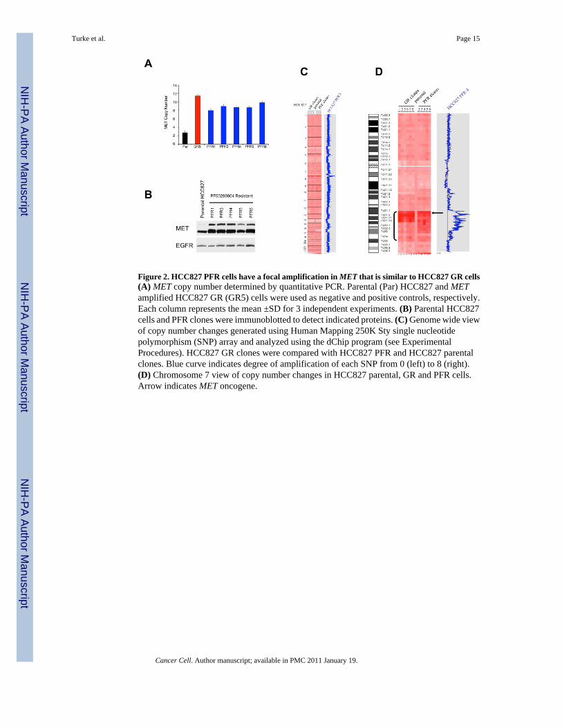
Figure 2. HCC827 PFR cells have a focal amplification in MET that is similar to HCC827 GR cells(A) MET copy number determined by quantitative PCR. Parental (Par) HCC827 and METamplified HCC827 GR (GR5) cells were used as negative and positive controls, respectively.Each column represents the mean ±SD for 3 independent experiments. (B) Parental HCC827cells and PFR clones were immunoblotted to detect indicated proteins. (C) Genome wide viewof copy number changes generated using Human Mapping 250K Sty single nucleotidepolymorphism (SNP) array and analyzed using the dChip program (see ExperimentalProcedures). HCC827 GR clones were compared with HCC827 PFR and HCC827 parentalclones. Blue curve indicates degree of amplification of each SNP from 0 (left) to 8 (right).(D) Chromosome 7 view of copy number changes in HCC827 parental, GR and PFR cells.Arrow indicates MET oncogene.
Turke et al. Page 15
Cancer Cell. Author manuscript; available in PMC 2011 January 19.
NIH
-PA Author Manuscript
NIH
-PA Author Manuscript
NIH
-PA Author Manuscript

Figure 3. HGF induces MET dependent resistance only in cell lines in which it activates PI3K/AKT,ERK and mTORC1 signaling(A, B) HCC827 cells treated with (A) increasing concentrations of gefitinib alone or incombination with PHA-665,752, in the absence or presence of HGF (50ng/mL), or (B)increasing concentrations of gefitinib alone or in combination with the indicated concentrationsof HGF. Cell viability relative to untreated controls measured after 72 hours. Each data pointrepresents the mean ±SD of 6 wells. (C) HCC827 cells were treated for 6 hours with 1μMgefitinib alone or in combination with the indicated concentrations of HGF. Cell lysates wereimmunoblotted to detect indicated proteins. (D) Cells were treated for 6 hours with gefitinib(HCC827, PC-9), PF00299804 (H1975), or lapatinib (BT-474, SKBR3), alone or incombination with HGF (50ng/mL). All drugs were used at 1μM. Cell lysates wereimmunoblotted to detect indicated proteins. *indicates cross-reaction by the p-EGFR antibodyagainst p-HER2. Cell lines in which HGF rescued viability are labeled in blue, and cell linesin which HGF did not rescue viability are labeled in red. (E) HCC827-HGF xenografts in nu/nu mice treated with PF2341066, gefitinib, or their combination and tumors measured twiceweekly. Some growth inhibiton was observed with gefitinib alone, however only combinationtreatment led to complete tumor shrinkage (p = 0.002). Each data point represents the mean±SD for 5 mice. See also Figure S2.
Turke et al. Page 16
Cancer Cell. Author manuscript; available in PMC 2011 January 19.
NIH
-PA Author Manuscript
NIH
-PA Author Manuscript
NIH
-PA Author Manuscript

Figure 4. IGF rescues PI3K/AKT and mTORC1 signaling in some cell lines, but fails to activateERK(A) IC50 values for viability curves (Figure S1) in the presence or absence of HGF and IGF.Cells were treated with increasing concentrations of the appropriate TKI alone (red) or incombination with 50ng/mL HGF (blue) or 75ng/mL IGF (green). (B) Cells were treated for 6hours with gefitinib (HCC827, PC-9, A431, HN11), PF00299804 (H1975) or lapatinib(BT-474, SKBR3) alone or in combination with HGF (50ng/mL) or IGF (75ng/mL). All drugswere used at 1μM. Cell lysates immunoblotted to detect indicated proteins. BT-474 and SKBR3cell lysates were run on the same gel, and no MET or IGF-1Rβ was detected in SKBR3 cellsrelative to BT-474 cells. See also Table S1 for quantification.
Turke et al. Page 17
Cancer Cell. Author manuscript; available in PMC 2011 January 19.
NIH
-PA Author Manuscript
NIH
-PA Author Manuscript
NIH
-PA Author Manuscript
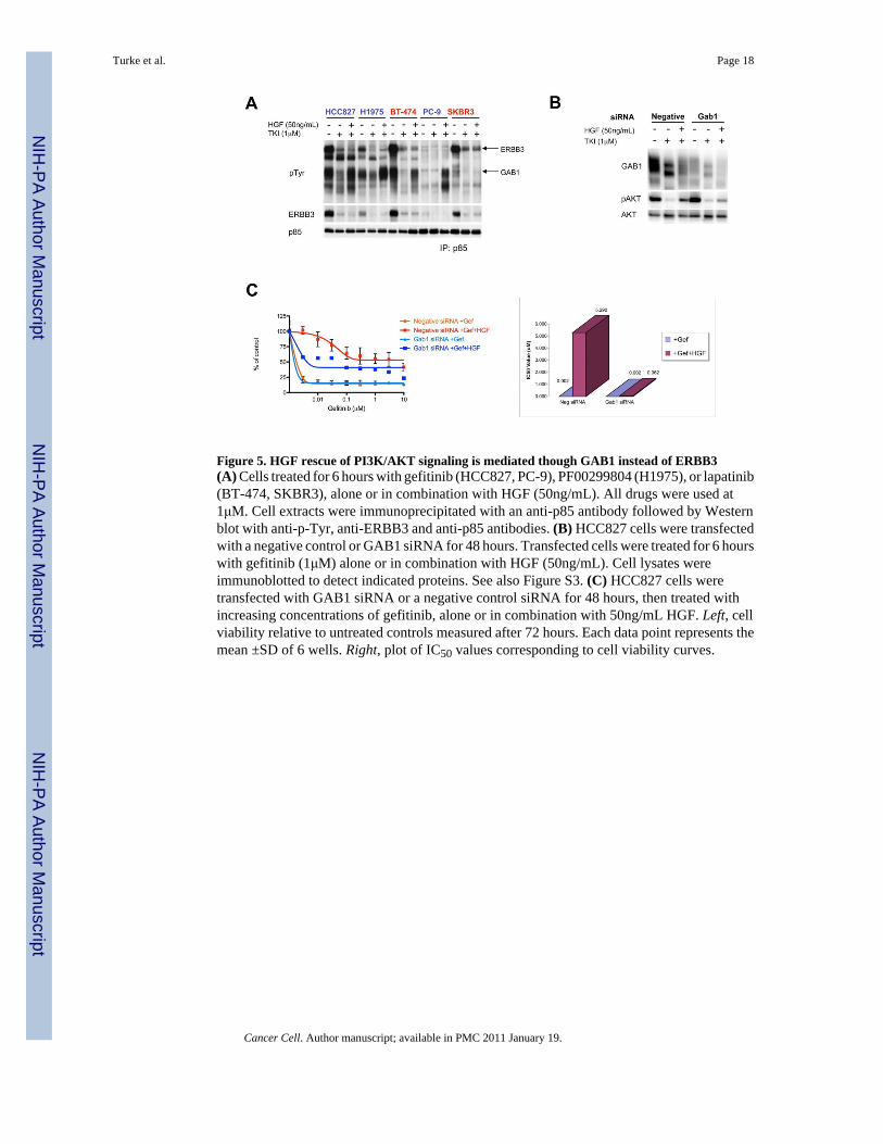
Figure 5. HGF rescue of PI3K/AKT signaling is mediated though GAB1 instead of ERBB3(A) Cells treated for 6 hours with gefitinib (HCC827, PC-9), PF00299804 (H1975), or lapatinib(BT-474, SKBR3), alone or in combination with HGF (50ng/mL). All drugs were used at1μM. Cell extracts were immunoprecipitated with an anti-p85 antibody followed by Westernblot with anti-p-Tyr, anti-ERBB3 and anti-p85 antibodies. (B) HCC827 cells were transfectedwith a negative control or GAB1 siRNA for 48 hours. Transfected cells were treated for 6 hourswith gefitinib (1μM) alone or in combination with HGF (50ng/mL). Cell lysates wereimmunoblotted to detect indicated proteins. See also Figure S3. (C) HCC827 cells weretransfected with GAB1 siRNA or a negative control siRNA for 48 hours, then treated withincreasing concentrations of gefitinib, alone or in combination with 50ng/mL HGF. Left, cellviability relative to untreated controls measured after 72 hours. Each data point represents themean ±SD of 6 wells. Right, plot of IC50 values corresponding to cell viability curves.
Turke et al. Page 18
Cancer Cell. Author manuscript; available in PMC 2011 January 19.
NIH
-PA Author Manuscript
NIH
-PA Author Manuscript
NIH
-PA Author Manuscript

Figure 6. Transient HGF exposure leads to MET amplification and stable ligand-independentgefitinib resistance in HCC827 cells(A) HCC827 cells treated with HGF (50ng/mL) and 1μM gefitinib are resistant to gefitinib(HCC827-50 cells). After the removal of HGF, stably resistant HGF-independentHCC827-50GR cells survive in 1μM gefitinib alone. In contrast, parental HCC827 cells do notsurvive when treated with 1μM gefitinib. (B) Parental HCC827 cells and HCC827-50 cells(pre-treated with gefitinib in combination with HGF (50ng/mL) for 14 days) were grown inmedia alone (No Rx) or media treated with 1μM gefitinib (+Gef) for 7 days. Viable cells werevisualized and quantified using Syto60 staining. (C) Fluorescence in situ hybridization (FISH)of MET/EGFR/CEP7 probe set with HCC827 and HCC827-50GR cells. MET (orange)EGFR (green) CEP7 (aqua). Metaphase spread (bottom) shows multiple copies of EGFR andMET (arrow) on individual chromosomes. Scale bars represent 10μm. (D) HCC827-50GR cells(upper) and HCC827-50GR cells grown in media alone (without gefitinib) for 8 weeks, 50GR8wks R5 (lower), were treated with increasing concentrations of gefitinib or PHA-665,752 ortheir combination for 72 hours. Cell viability was measured relative to untreated controls. Eachdata point represents the mean ±SD of 6 wells. (E) HCC827 cells and stably resistantHCC827-50GR cells were treated for 6 hours with gefitinib, PHA-665,752, or theircombination. All drugs were used at 1μM. Cell lysates were immunoblotted to detect indicatedproteins. See also Figure S4 and Table S2 and S3.
Turke et al. Page 19
Cancer Cell. Author manuscript; available in PMC 2011 January 19.
NIH
-PA Author Manuscript
NIH
-PA Author Manuscript
NIH
-PA Author Manuscript

Figure 7. HGF treatment selects out a small pre-existing population of MET amplified HCC827cells from the parental population in vitro and in vivo(A) Fluorescence in situ hybridization (FISH) of MET/EGFR/CEP7 probe set. MET (red)EGFR (green) CEP7 (aqua). Left, tumor sections from control HCC827 xenograft models thatdo not express HGF showed normal MET copy number. Right, tumor sections from one ofthree HCC827-HGF xenografts treated with gefitinib (Figure 3E) showed significant METamplification (arrow). (B) High-throughput FISH analysis of HCC827 cells identifies asubpopulation harboring MET amplification (arrow). MET (RP-11-95I120; red); 7qter(RP-11-6903; green). All scale bars represent 10μm. (C) Parental HCC827 cells and threeindependent clones harbor a small percentage of MET amplified cells. No pre-existing METamplification was detected in H3255 or PC-9 cell populations. (D) Left, HCC827 cells werespiked with approximately 0.1% of GFP labeled HCC827 cells or GFP labeled MET amplifiedHCC827 GR6 cells. Each population was grown in either media alone or media treated withgefitinib (1μM) with HGF (50ng/mL). Cells were collected after 19 days and GFP levels werequantified using FACS. Each data point for cells treated with gefitinib+HGF represents themean ±SD for 3 independent wells. Fold change is the ratio of Day 19 to Day 0 (%GFP).Right, diagrammatic depiction of results. See also Figure S5.
Turke et al. Page 20
Cancer Cell. Author manuscript; available in PMC 2011 January 19.
NIH
-PA Author Manuscript
NIH
-PA Author Manuscript
NIH
-PA Author Manuscript

Figure 8. HGF expression and pre-existing MET amplification can be detected in tumor specimensfrom NSCLC patients(A) Summary of tumors from geftinib/erlotinib treated patients, including 16 paired, and 11drug resistant samples only. Samples were evaluated for EGFR mutational status, METamplification and HGF expression *Specimen contained less < 30% tumor cells. **METamplification defined by qPCR as previously described (Engelman et al., 2007b). Data onEGFR T790M and MET amplification in resistant specimens only from patients 1-4 and 17-19has been previously published (Engelman et al., 2007b). N/A; not available. (B) FISH analysisof pre-treatment sample from patient 10 shows evidence of a subset of MET amplified cells(arrow) before exposure to an EGFR TKI. MET (RP-11-95I120; orange); CEP 7 (aqua). Scalebars represent 10μm. See also Figure S6.
Turke et al. Page 21
Cancer Cell. Author manuscript; available in PMC 2011 January 19.
NIH
-PA Author Manuscript
NIH
-PA Author Manuscript
NIH
-PA Author Manuscript


