Embed Size (px)
Citation preview
EGFR-targeted immunoliposomes efficiently deliver docetaxel toprostate cancer cells
Eloy, J. O., Ruiz, A., de Lima, F. T., Petrilli, R., Raspantini, G., Nogueira, K. A. B., Santos, E., de Oliveira, C. S.,Borges, J. C., Marchetti, J. M., Al-Jamal, W. T., & Chorilli, M. (2020). EGFR-targeted immunoliposomesefficiently deliver docetaxel to prostate cancer cells. Colloids and Surfaces B: Biointerfaces, 194, [111185].https://doi.org/10.1016/j.colsurfb.2020.111185
Published in:Colloids and Surfaces B: Biointerfaces
Document Version:Peer reviewed version
Queen's University Belfast - Research Portal:Link to publication record in Queen's University Belfast Research Portal
Publisher rightsCopyright 2020 Elsevier.This manuscript is distributed under a Creative Commons Attribution-NonCommercial-NoDerivs License(https://creativecommons.org/licenses/by-nc-nd/4.0/), which permits distribution and reproduction for non-commercial purposes, provided theauthor and source are cited.
General rightsCopyright for the publications made accessible via the Queen's University Belfast Research Portal is retained by the author(s) and / or othercopyright owners and it is a condition of accessing these publications that users recognise and abide by the legal requirements associatedwith these rights.
Take down policyThe Research Portal is Queen's institutional repository that provides access to Queen's research output. Every effort has been made toensure that content in the Research Portal does not infringe any person's rights, or applicable UK laws. If you discover content in theResearch Portal that you believe breaches copyright or violates any law, please contact [email protected].
Download date:11. Feb. 2022
EGFR-targeted immunoliposomes efficiently deliver docetaxel to prostate cancer
cells
Josimar O. Eloy1*, Amalia Ruiz2, Felipe Tita de Lima3, Raquel Petrilli4, Giovanni Raspantini5,
Karina Alexandre Barros Nogueira1, Elias Santos1, Carlos Sabino de Oliveira6, Júlio César
Borges6, Juliana Maldonado Marchetti5, Wafa T. Al-Jamal2, Marlus Chorilli3
¹ Federal University of Ceará, College of Pharmacy, Dentistry and Nursing, Department of Pharmacy,
Fortaleza, Ceará, Brazil
2 School of Pharmacy, Queen’s University Belfast, 97 Lisburn Rd, Belfast BT9 7BL, UK
3 School of Pharmaceutical Sciences of Araraquara – São Paulo State University, UNESP, Department of
Drugs and Medicines, Araraquara, SP, Brazil
4 University for International Integration of the Afro-Brazilian Lusophony, Institute of Health Sciences,
Ceará, Brazil
5 College of Pharmaceutical Sciences of Ribeirão Preto, University of São Paulo, Ribeirão Preto, São Paulo
Brazil.
6 São Carlos Institute of Chemistry, University of São Paulo, São Carlos, São Paulo, Brazil.
Short statistical summary of the article
Total words of manuscript body: 5860
Total Tables: 1
Total Figures: 7
*Corresponding author: Josimar O. Eloy, PhD. Department of pharmacy – Federal University of Ceará.
Rua Pastor Samuel Munguba, 1210, Fortaleza – CE, Brazil. [email protected]
ABSTRACT
Prostate cancer is the second cause of cancer death in men worldwide. Docetaxel (DTX),
an antimitotic drug, is widely used for the treatment of metastatic prostate cancer patients.
Taxotere® is a commercial DTX formulation. It contains a polysorbate 80 surfactant to
improve DTX aqueous solubility, which has been associated with hypersensitivity
reactions in patients. Liposomes have been used as promising delivery systems for a range
of hydrophobic drugs, such as DTX, offering improved drug water solubility and
biocompatibility, without compromising its anticancer activity. Herein, DTX-loaded
liposomes were developed using the Box-Behnken factorial design. The optimized
formulation was nano-sized, homogenous in size (67.47 nm) with high DTX
encapsulation efficiency (99.95%). The encapsulated DTX was in a soluble amorphous
state, which was slowly released. Next, to increase the liposomes selectivity to prostate
cancer cells, cetuximab, an anti-EGFR monoclonal antibody. was successfully conjugated
to the surface of liposomes, without compromising cetuximab protein structure and
stability. As expected, our results showed higher cellular uptake and toxicity of
immunoliposomes, compared to non-targeted liposomes, in DU145 (EGFR-
overxpressing) prostate cancer cells. To the best of our knowledge, this is the first report
of engineering EGFR-targeted liposomes to enhance the selectivity of DTX delivery to
EGFR-positive prostate cancer cells.
Keywords: docetaxel, EGFR, cetuximab, immunoliposomes, prostate cancer.
1. INTRODUCTION
Prostate cancer is the second most common cancer in men worldwide, with a record of
1.276.106 new cases in 2018, as well as 358.989 death (Rawla 2019). There are cases in which
the neoplasm progresses and selective mutations occur that result in metastatic castration-resistant
prostate cancer (mCRPC), in other words, the disease becomes refractory to androgen deprivation
therapy (Tucci et al. 2019; Nelson et al., 2020.).
Docetaxel (DTX) is a first-line drug for the treatment of mCRPC. It is a semi-synthetic
derivative of 10-deacetylbaccatin III, a precursor extracted from the branches of the European
yew (Taxus baccata). This drug belongs to the class of taxanes and its mechanism of action is
based on the binding to beta-tubulin to promote the stabilization of microtubules during the
process of mitosis, thus, the dynamics of microtubules is inhibited and cell division is interrupted.
The formulation containing DTX (Taxotere®, Sanofi-Aventis, Paris, France) has demonstrated
clinical efficacy (Engels, Mathot, and Verweij 2007; Saad and Miller 2014). However, there are
several adverse effects related to drug toxicity. The most significant effect is the occurrence of
neutropenia, anemia, gastrointestinal disorders and peripheral neuropathy (Mager et al. 2019). In
addition, the presence of polysorbate 80 as a surfactant to solubilize the drug, which is highly
lipophilic, can induce hypersensitivity reactions in patients (Engels, Mathot, and Verweij 2007).
To overcome these hurdles, DTX can be loaded into nanocarriers for better therapeutic
outcomes. Among the various nanocarriers, liposomes have been widely studied. Liposomes are
vesicles composed of one or more concentric lipid bilayers, which can encapsulate lipophilic and
hydrophilic substances. They are biodegradable with low immunogenicity. Liposomes allow the
protection of the drug, reducing physiological degradation and thus increasing bioavailability,
controlling release and directing the therapy to have greater tissue selectivity (Li et al. 2019).
Taking advantage of the passive accumulation of nanocarriers at the tumor environment,
it is possible to increase the levels of cell internalization and ensure that the drug interacts with
its site of action, through active targeting of these liposomes, by inserting ligands on their surface.
When an antibody is conjugated to the liposomes, immunoliposomes originate, with high affinity
to surface receptors on tumor cells. The interaction between the ligand and the receptor leads to
ligand-receptor complex formation, which triggers endocytosis, followed by intracellular drug
release (Merino, Zalba, and Garrido 2018).
Epidermal growth factor receptor (EGFR) is highly expressed in some prostate
adenocarcinomas. EGFR is a transmembrane receptor with tyrosine kinase activity that stimulates
tumor cell proliferation, angiogenesis, invasion and metastasis through the activation of several
signaling pathways. Thus, it is possible to target DTX-loaded liposomes to EGFR, using an anti-
EGFR chimeric-murine monoclonal antibody (cetuximab) conjugated to the liposome containing
docetaxel, so that this antibody competitively binds to the EGFR extracellular domain preventing
the activation of the receptor by endogenous ligands and induces the internalization of the
immunoliposome. Previously, docetaxel has been combined as free drug with cetuximab and an
anti-angiogenic drug, sunitinib. Studies conducted in PC3 xenografts revealed that there were
significantly higher pro-apoptotic effects observed for the sunitinib–docetaxel and sunitinib–
docetaxel–cetuximab. Although DTX-loaded liposomes have been previously functionalized
with trastuzumab for breast cancer therapy, to our knowledge this is the first report of anti-EGFR,
DTX-loaded liposomes that would hypothetically assure the selective delivery of DTX to the
prostate cancer cell, preventing a series of adverse effects and increasing the effectiveness of the
treatment (Malmberg, Tolmachev, and Orlova 2011; Joshi et al. 2015; Guardiola, Varese, and Sa
2019; Rodallec et al. 2018; Guerin et al., 2008). We previously developed immunoliposomes, but
for paclitaxel and rapamycin co-delivery, for another target, HER2 with a different monoclonal
antibody, trastuzumab, for breast cancer (Eloy et al., 2017). For docetaxel delivery,
immunoliposomes have been developed, with trastuzumab, for breast cancer (Rodallec et al.,
2018). Regarding anti-EGFR immunoliposomes, we have previously loaded the hydrophilic 5-
fluorouracil for skin cell carcinoma using a different lipid composition, based on DSPC,
cholesterol and DSPE-PEG-maleimide (Petrilli et al., 2018). For docetaxel delivery to EGFR,
the present study is the first report on immunoliposomes, which is very important, considering
that EGFR is highly expressed in some prostate adenocarcinomas, and the clinical relevance of
combining both docetaxel and cetuximab in metastatic castration-resistant prostate cancer has
been previously studied for free drugs in a phase II clinical trial (Cathomas et al., 2012). In terms
of formulation development, for DTX and EGFR, only polymeric nanoparticles have been
developed, and not for prostate, but for lung cancer (Patel et al., 2018). Therefore, we believe that
the present work is innovative and represents an important contribution toward the development
of a selective DTX formulation for aggressive types of prostate cancer.
The present work describes the engineering of anti-EGFR immunoliposomes containing
DTX to treat prostate cancer. To achieve this, the Box-Benken model was used as a tool to
optimize the liposomal formulation, regarding size, polydispersity and DTX encapsulation
efficiency. Solid state physicochemical characterization was employed to determine the state of
encapsulated drug. Next, anti-EGFR antibody was successfully chemically conjugated to the
liposomal surface using maleimide chemistry. The integrity of the antibody following conjugation
was determined using electrophoresis and fluorescence microscopy. Subsequently, the in vitro
release profile and the colloidal stability were determined. Finally, the cytotoxicity and cellular
uptake by confocal microscopy and flow cytometry were studied in low and high EGFR-
expressing prostate cancer cells.
2. MATERIAL AND METHODS
2.1 Material
Lipids soy phosphatidylcholine (SPC) and 1,2-distearoyl-sn-glycero-3-
phosphoethanolamine-N-[amino(polyethylene glycol)-2000] (DSPE-PEG2000) were purchased
from Lipoid (Lipoid GmbH, Germany). Cholesterol (CHOL) and 1,2-distearoyl-sn-glycero-3-
phosphoethanolamine-N [maleimide(polyethylene glycol)-2000] (ammonium salt) (DSPE-PEG-
Mal) were purchased from Sigma Aldrich Co. (St. Louis, MO, USA) and LaySan Bio (Arab, AL,
USA), respectively. Docetaxel (DTX) was supplied by APIChem (China). Erbitux 5 mg/mL
(Cetuximab) was obtained from Merck Serono (Biberach, Germany). Disposable PD-10 desalting
columns and CL-4B were obtained from GE Healthcare (Pittsburgh, PA, USA). 3,3′-
dioctadecyloxacarbocyanine perchlorate (DiO), 1,1'-dioctadecyl-3,3,3',3'-
tetramethylindocarbocyanine perchlorate (DiI), paraformaldehyde, dialysis membrane molecular
weight cut-off 14,000 Da and Traut's reagent were purchased from Sigma Aldrich Co. (St. Louis,
MO, USA). ProLong Diamond with DAPI was purchased from Life Technologies (Carlsbad, CA,
USA). Propidium iodide was purchased from Thermo Scientific (Pittsburgh, PA, USA). Precision
Plus Dual Color Protein was obtained from Biorad.
2.2 Liposomes development and characterization
2.2.1 Liposomes development and factorial design
Liposomes were prepared with soy phosphatidylcholine (SPC), cholesterol (CHOL) and
1,2-distearoyl-sn-glycero-3-phosphoethanolamine-N-[amino(polyethylene glycol)-2000]
(DSPE-PEG2000), with the inclusion of DTX in different concentrations . The preparation of the
liposomes followed the classic hydration technique of the lipid film previously described (Eloy
et al. 2016). The lipids and the drug were dissolved in 2 mL chloroform, which was removed
under reduced pressure in a rotary evaporator so that a thin film remains after 30 min at 65 °C,
and then hydrated for 30 min at 150 rpm using phosphate buffer (pH 7.4) as aqueous phase. Then,
the formulations were subjected to homogenization in a high-pressure homogenizer (Avestin), at
1000 bar, for 30 min. The non-encapsulated drug in suspension was separated by 0.45 µm
filtration and the non-encapsulated drug soluble in the buffer was separated by gel
chromatography on a CL-4B column filtration.
In the present study, a Box-Behnken statistical design with three-factor, three-level was
used to evaluate influence of independent variables (Lipid: drug molar ratio, CHOL lipid molar
ratio and DSPE-PEG:lipid molar ratio) on the physicochemical characteristics of the liposomes.
The experimental design consists of a group of points located at the midpoint of each edge and
one point in the center of a cube. This approach allows the construction of second order
polynomial models for the dependent variables that can provide theoretical value of an output.
The polynomial equation generated by this statistical design (using MiniTab® 16.0 software) is as
follows (Equation 1) (Solanki, Parikh, and Parikh 2007):
𝑌𝑖 = 𝑏0 + 𝑏1𝑋1 + 𝑏2𝑋2 + 𝑏3𝑋3 + 𝑏12𝑋1𝑋2 + 𝑏13𝑋1𝑋3 + 𝑏23𝑋2𝑋3 + 𝑏11𝑋12 + 𝑏22𝑋2
2 +
𝑏33𝑋32
(Equation 1)
Where 𝑌𝑖 is the dependent variable; 𝑏0 is the intercept; 𝑋1, 𝑋2 and 𝑋3 are the independent variables
and 𝑏1 to 𝑏33 are the regression coefficients.
Basically, 15 formulations were selected to evaluate the effects between the independent
variables applied with desirability approach in order to optimize the preparation of liposomes
where the responses such as particle size and polydispersity index value were targeted to the
minimum, and encapsulation efficiency was targeted to the maximum. The desirability function
(D) can vary over the range 0 to 1 for each response. The result of D equal to 0 indicates the
undesirable value of the response and if the maximized value of D is found, the optimal conditions
for the variables are obtained (Das and Mishra 2017).
2.2.2 Nanoparticles characterization
2.2.2.1 Particle size, polydispersity and zeta potential
Nanoparticles size and the polydispersity index (PdI) were determined at 25ºC by
dynamic light scattering measures, using HeNe laser operating at 4 mW and 633 nm wavelength.
The equipment performs non-invasive measurements by "backscatteroptics" (NIBS), made at a
detection angle of 173º and the measurement position of the cuvette is automatically determined
by the equipment's software. The equipment makes an average of 12 determinations for each
analysis. The dispersions were diluted in Milli Q water (10-fold) and placed in cells of 1 cm of
optical path. Zeta potential was determined by the electrophoretic mobility of the scattered
particles subjected to an electric field.
2.2.2.2 Encapsulation efficiency
For the analysis of the encapsulation efficiency, liposome (without non-encapsulated drug
or total fraction) was diluted in acetonitrile, filtered using a PTFE 0.45 µm and analyzed by the
analytical method. Briefly, for the quantification of DTX in nanoparticles, a validated high
performance liquid chromatography (HPLC) method was used, with a mobile phase composed of
acetonitrile: water: methanol, 65:25:15 (v /v), under a flow of 0.8 mL min-1, in a C18 reverse
phase column (4.6 x 250 mm, 5 μm) with injection of 20 µL at 25 ° C and a wavelength of 232
nm. The loading percentage was calculated according to Equation 2.
𝐿𝑜𝑎𝑑𝑖𝑛𝑔 𝑝𝑒𝑟𝑐𝑒𝑛𝑡𝑎𝑔𝑒 = 𝑎𝑚𝑜𝑢𝑛𝑡 𝑜𝑓 𝑑𝑟𝑢𝑔 𝑖𝑛 𝑡ℎ𝑒 𝑙𝑖𝑝𝑠𝑜𝑚𝑎𝑙 𝑓𝑟𝑎𝑐𝑡𝑖𝑜𝑛 (μ𝑔)
𝑎𝑚𝑜𝑢𝑛𝑡 𝑜𝑓 𝑑𝑟𝑢𝑔 𝑖𝑛 𝑡ℎ𝑒 𝑡𝑜𝑡𝑎𝑙 𝑓𝑟𝑎𝑐𝑡𝑖𝑜𝑛 (μ𝑔) 𝑋 100 (Equation 2)
2.2.2.3 Colloidal stability and lyophilization study
In order to assess the nanoparticles stability, liposomes were analyzed for their colloidal
stability. The samples, freeze-dried or not, were kept in a 2-8ºC refrigerator and analyzed at
intervals of up to 60 days. For lyophilized samples, sucrose was used as a cryoprotectant in
different lipid: sucrose molar ratios (1:10; 1:20 and 1:30). These samples were frozen overnight
in an ultra-freezer at -80ºC and then lyophilized for 48 hours. The stability was monitored by
particle size, PdI and zeta potential.
2.2.2.4 Fourier transform infrared spectroscopy (FTIR)
Samples of liposomes previously lyophilized with sucrose (lipid to sugar molar ratio,
1:20) were mixed with potassium bromide and compressed in a hydraulic press. The scans were
obtained at a resolution of 2 cm− 1, from 4000 to 400 cm – 1 on a Shimadzu IR Prestige-21
equipment.
2.2.2.5 Differential Scanning Calorimetry (DSC) analyses
The samples, previously lyophilized (lipid to sugar molar ratio, 1:20) were placed in
aluminum pans and heated from 15 ºC to 250 ºC at a rate of 10 ºC/min, under nitrogen pressure
of 3 kgf/cm² using a Jade Perkin Elmer equipment. Calibration was performed using indium and
n-octadecane as reference materials. In the experiment, it was ensured that the method had enough
sensitivity to detect the amount of loaded drugs.
2.2.3 Immunoliposomes development
The functionalization of liposomes containing docetaxel followed the method previously
described by our group, with modifications (Petrilli et al. 2017; Eloy et al. 2017). Initially,
liposomes of SPC: CHOL: DSPE-PEG-Maleimide compositions were prepared for drug
encapsulation, using the same protocol previously described. In parallel, the cetuximab antibody
was diluted in PBS buffer pH 8.0 containing EDTA (5 mM) and the pH of the solution was
adjusted to 8.0 with NaOH (0.1 N). To the cetuximab solution, 5 mg/mL, Traut's reagent solution
(2 mg/mL) was added, in a 40: 1 molar ratio (Traut´s reagent:antibody). This solution was
incubated for 1 h at 37 °C to allow thiolation. The separation of the thiolated antibody from the
excessive Traut's reagent was carried out on gel filtration chromatography, using a PD-10 column,
with eluent PBS-EDTA pH 8.0. After determining the concentration of cetuximab in each aliquot
by BCA assay (bicinchoninic acid), the fractions containing the antibody were pooled and added
to the liposomes previously prepared. The mixture was incubated overnight and finally the
immunoliposome was purified on a CL-4B column to separate the liposome and free antibody,
eluting using PBS buffer, pH 7.4. The immunoliposomes were further characterized by size, PdI
and DTX encapsulation efficiency.
2.2.3.1 Antibody conjugation efficiency
The immunoliposome was separated from the free antibody by gel filtration
chromatography with Sepharose CL-4B as stationary phase and PBS buffer (pH 7.4) as a mobile
phase. The efficiency of cetuximab conjugation to liposomes was determined by the BCA assay,
following the manufacturer's protocol (ThermoScientific). The conjugation efficiency was
calculated by the percentage of antibody quantified in the immunoliposome in relation to the total
antibody used in the experiment (Petrilli et al. 2017; Eloy et al. 2017).
2.2.3.2 Antibody integrity after conjugation
The integrity of the antibody in the immunoliposomes was assessed both by
electrophoresis in polyacrylamide gel (SDS-PAGE) for its primary structure and for its tertiary
structure using a fluorescence assay with intrinsic probes. Briefly, the SDS-PAGE electrophoresis
was conducted according to the procedure previously described following the manufacturer's
protocol (Eloy et al. 2017). The samples were previously incubated with 2-mercapto-ethanol and
Laemmli sample buffer at 60 °C for 10 min for reduction. Mini-PROTEAN electrophoretic
chamber and running buffer containing Tris / glycine / SDS were used for electrophoresis at 175
V for 30 min. The protein standard used was Precision Plus Dual Color Protein. After staining
with Coomassie blue dye, the gels were discolored with deionized water and photographed.
For local tertiary structure investigation, antibody samples both in buffer (cetuximab) or
immunoliposomes were analyzed by its intrinsic fluorescence emission spectra aiming to
investigate possible interactions and modifications in protein tertiary conformation after
cetuximab linkage to the liposomes. The analyses were carried out using a Hitachi F-4500
spectrophotometer with excitation at 280 nm and emission between 300 and 420 nm. Guanidine
hydrochloride (6 mmol.L-1) was used as a denaturing agent in order to evaluate the suppression
of fluorescence and the displacement of the maximum wavelength (λmax) to less energetic
wavelengths (Eftink 1994).
2.2.4 In vitro drug release
Liposomes and immunoliposomes containing the drug were evaluated for the release in
50 mL of phosphate buffer (PBS) containing 1.25% sodium lauryl sulfate as previously checked
for the sink conditions, with stirring at 150 rpm and 37ºC. For this study, samples were diluted in
1 mL of the buffer, at pH 7.4, and placed in dialysis bags of 12-14 kDa (limit molecular weight)
(Yoon et al. 2017). Samples were collected in the receptor medium at different times, up to 48 h.
Subsequently they were filtered and analyzed by the HPLC method.
2.2.5 Cell culture studies
DU145 cells (high EGFR-expressing cells) were cultured in DMEM medium
supplemented with 10% FBS and 1% antibiotic / antimycotic solution, at 37ºC with 5% CO2,
according to ATCC recommendations. PC-3 cells (low EGFR-expressing cells) (Kharmate et al.,
2016) were cultured using the RPMI medium, supplemented with 10% FBS and 1% antibiotic /
antimycotic solution. Once 90% of confluence was reached, the cells were trypsinized.
2.2.5.1 Cellular uptake
Liposomes and immunoliposomes uptake were assessed by confocal microscopy and
flow cytometry studies. For this purpose, the formulations were prepared without drugs,
containing the fluorescent lipid bilayer marker, DiO, encapsulated at 0.5 mol%. The preparation
proceeded in the same way as previously specified for liposomes and immunoliposomes. The
experiments were carried out with the cell line PC-3 (low EGFR-expressing cell line) and DU145
(EGFR positive cell line). Thus, for confocal studies, 1x10⁶ cells/well were plated in 6-well
microplates, containing sterile 22 mm / 22 mm coverslips and incubated for 24 h at 37 ° C in an
atmosphere containing 5% CO2. Then, the cells were washed with PBS buffer pH 7.4 and
incubated with the samples (100 µL) diluted in incomplete medium for 2h or 4h. After treatment,
the cells were washed with PBS buffer and then 2 mL of 1% paraformaldehyde was added to the
wells. The plates were kept overnight at 4 ° C, sealed with film. Then, the wells were again washed
with PBS buffer and the coverslips were poured into histology slides containing Prolong
Diamond® with DAPI, to preserve fluorescence and core labeling. The slides were visualized in
a Leica TCS SP8 confocal microscope (λexc = 405 nm, λem = 413-472 nm (DAPI) and λexc = 488
nm, λem = 503-588nm (DiO), increase of 63 times with oil immersion objective. For flow
cytometry, the formulations were labelled with Dil. Liposomes were incubated in presence of 1
mol% of Dil at 37 ºC for 1 hour and then purified using a PD-10 column. Cell uptake was
monitored by flow cytometry using the fluorescent signal of Dil (λex = 549 nm, λem = 565 nm)
after incubation with the formulations. Cells were seeded in 12-well plates (1×10⁵ cells/well, 1
mL/well). Next day, liposomes or immunoliposomes, were diluted in 400 µL of media and added
to the cells. Cell uptake was monitored up to 3 hours after exposure to the formulations. The
supernatant was aspirated and discarded, and the cells were washed twice with PBS. Cells were
detached from the plates using a solution of 0.05 % Trypsin/EDTA. Cells were washed with PBS
and immediately acquired on the cytometer (BD FACSCalibur™, Becton, Dickinson and
Company, UK). At least 10000 cells were counted for each sample, and experiments were
performed in triplicates. Flow cytometry data were generated using BD CellQuest Pro software.
The results are expressed as average ± SD (n = 3). Statistical analysis was performed using Two-
way ANOVA followed by Bonferroni post-test.
2.2.5.2 Cytotoxicity
PC-3 and DU145 cells were seeded at a seeding density of 1 × 10⁴ cells/well in
polysterene 96-well plates (Nunclon, Thermo Fisher Scientific, UK) in complete RPMI 1640
media. Next day, DTX, Lip-DTX or Lip-DTX-Ab were diluted in serum-free and antibiotics-free
media and added to the cells. Untreated cells were used as a 100% viability control. After 6 h
incubation, media containing liposomes were removed, cells were washed with PBS and
replenished with fresh media supplements with 10% (v/v) FBS and 1% (v/v) antibiotics. At 24 h
and 48 h post-incubation, a resazurin cell viability assay was performed, which is based on the
mitochondrial metabolic activity of live cells. Resazurin reagent was prepared as previously
described (Walzl et al., 2014). Briefly, cells were incubated with 0.01 mg/mL resazurin solution
for 4 h. After incubation, fluorescence (λex = 544 nm, λem = 590 nm) was read using an
automated FLUOstar Omega (BMG Labtech, UK) plate reader. Six replicates per condition were
used. The results were expressed as the percentage of cell viability (mean ± SD) and normalized
to control untreated cells (Pereira et al., 2016). The results were analyzed by two-way ANOVA,
followed by Bonferroni post-test.
3. RESULTS AND DISCUSSION
Liposomes are lipid vesicles widely used for the encapsulation of drugs, with several
advantages, including protection against degradation, sustained release and improved
pharmacokinetics. It is noteworthy, however, that several factors can affect the encapsulation of
the drug, including the type of lipid, the ratio of drug to lipid, the presence of cholesterol and the
PEGylation strategy for the long circulation time (Eloy et al., 2014b). An effective strategy for
optimizing the composition of the release system is the use of a factorial planning, which
rationalizes the development and allows the optimization of the number of experiments. This tool
is part of the concept of “Quality by Design”, which involves the definition of critical parameters
that influence the attributes of the product, and is recommended by regulatory agencies in the
drug development process (Singh, Sharma, and Robertson 2012; Troiano et al. 2016).
The experimental design matrix and the observed responses for the 15 experiments
proposed by the Box–Behnken design produced ranges with maximum and minimum values for
particle size (Y1) (58.91 to 108.63 nm), PdI (Y2) (0.168 to 0.549), and EE% (Y3) of, (68.56 to
99.45) as shown in Table 1.
Table 1: Box Behnken Design and Experimental responses.
<Table 1>
From Figure 1 it was concluded that the variables studied did not adversely affect the
particle size, considering that it remained below 100 nm, with no statistical significance for the
different observed particle size values. Soema and collaborators, on the other hand, concluded
that the type of lipid (1,2-dioleoyl-sn-glycero-3-phosphoethanolamine (DOPE), 1,2-dioleoyl-3-
trimethylammoniumpropane (DOTAP) and 3ß-[N-(N0,N0-dimethylaminoethane)-
carbamoyl]cholesterol (DC-Chol)), and also the quantity can affect the particle size in liposomes
prepared by extrusion (Soema et al. 2015). In addition, cholesterol, important for membrane
fluidity and better physicochemical stability, has been reported to cause an increase in the
liposome diameter (Shaker, Gardouh, and Ghorab 2017). Furthermore, it is known that the
nanometric particle size influences the biological characteristics of the nanocarrier. For example,
nanoparticles are captured and accumulated inside solid tumors by the EPR effect, due to the
increased permeability of tumor blood vessels and deficient lymphatic clearance (Maeda et al.
2000).
The polydispersity index (PdI) reveals the homogeneity of particle size distribution, and
the closer to 1.0 the PdI value, the more heterogeneous is the population of particles. Previous
works have already reported PdI values close to 0.3, thus in agreement with the values observed
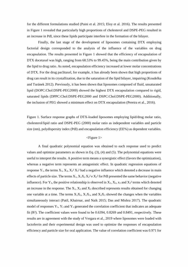
for the different formulations studied (Paini et al. 2015; Eloy et al. 2016). The results presented
in Figure 1 revealed that particularly high proportions of cholesterol and DSPE-PEG resulted in
an increase in PdI, since these lipids participate interfere in the formation of the bilayer.
Finally, the last stage of the development of liposomes containing DTX employing
factorial design corresponded to the analysis of the influence of the variables on drug
encapsulation. The results presented in Figure 1 showed that the efficiency of encapsulation of
DTX docetaxel was high, ranging from 68.53% to 99.45%, being the main contribution given by
the lipid to drug ratio. As noted, encapsulation efficiency increased at lower molar concentrations
of DTX. For the drug paclitaxel, for example, it has already been shown that high proportions of
drug can result in its crystallization, due to the saturation of the lipid bilayer, impairing (Koudelka
and Turánek 2012). Previously, it has been shown that liposomes composed of fluid, unsaturated
lipid (DOPC:Chol:DSPE-PEG2000) showed the highest DTX encapsulation compared to rigid,
saturated lipids (DPPC:Chol:DSPE-PEG2000 and DSPC:Chol:DSPE-PEG2000). Additionally,
the inclusion of PEG showed a minimum effect on DTX encapsulation (Pereira et al., 2016).
Figure 1. Surface response graphs of DTX-loaded liposomes employing lipid/drug molar ratio,
cholesterol/lipid ratio and DSPE-PEG (2000) molar ratio as independent variables and particle
size (nm), polydispersity index (PdI) and encapsulation efficiency (EE%) as dependent variables.
<Figure 1>
A final quadratic polynomial equation was obtained to each response used to predict
values and optimize parameters as shown in Eq. (3), (4) and (5). The polynomial equations were
useful to interpret the results. A positive term means a synergistic effect (favors the optimization),
whereas a negative term represents an antagonistic effect. In quadratic regression equations of
response Y1, the terms X1, X3, X2² X3² had a negative influence which denoted a decrease in main
effects of particle size. The terms X1, X1X2 X2² e X3² for PdI presented the same behavior (negative
influence). For Y3, the positive relationship is observed in X1, X2, x3 and X3² terms which denoted
an increase in the response. The X1, X2 and X3 described represents results obtained for changing
one variable at a time. The terms X1X2, X1X3, and X2X3 showed the changes when the variables
simultaneously interact (Patil, Khairnar, and Naik 2015; Das and Mishra 2017). The quadratic
model of responses Y1, Y2 and Y3 generated the correlation coefficient that indicates an adequate
fit (R²). The coefficient values were found to be 0.6394, 0.8269 and 0.8491, respectively. These
results are in agreement with the study of Vergara et al., 2019 where liposomes were loaded with
lactoferrin and their experimental design was used to optimize the responses of encapsulation
efficiency and particle size for oral application. The value of correlation coefficient was 0.971 for
EE and 0.841 for particle size represented adequate model for prediction from experimental data
(Vergara and Shene 2019).
𝑌1 = 90.7267 − 2.2225𝑋1 + 4,7587𝑋2 − 1.9687𝑋3 + 0.9575𝑋1𝑋2 + 1.4475𝑋1𝑋3 +
9.2350𝑋2𝑋3 + 0.4192𝑋12 − 15.7183𝑋2
2 − 6.3933𝑋33 (3)
𝑌2 = 0.437 − 0.009375𝑋1 + 0.03525𝑋2 + 0.019125𝑋3 − 0.0005𝑋1𝑋2 +
0.07125𝑋1𝑋3 + 0.062𝑋2𝑋3 + 0.000125𝑋12 − 0.110125𝑋2
2 − 0.092375𝑋33 (4)
𝑌3 = 91.9533 + 8.5103𝑋1 + 1.4035𝑋2 + 2.4236𝑋3 − 0.6850𝑋1𝑋2 − 2.3954𝑋1𝑋3 −
1.0033𝑋2𝑋3 − 10.3987𝑋12 − 2.0862𝑋2
2 + 0.8712𝑋33 (5)
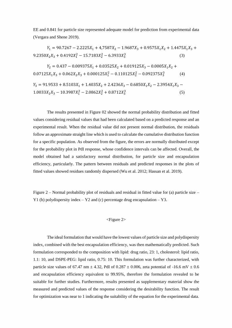
The results presented in Figure 02 showed the normal probability distribution and fitted
values considering residual values that had been calculated based on a predicted response and an
experimental result. When the residual value did not present normal distribution, the residuals
follow an approximate straight line which is used to calculate the cumulative distribution function
for a specific population. As observed from the figure, the errors are normally distributed except
for the probability plot in PdI response, whose confidence intervals can be affected. Overall, the
model obtained had a satisfactory normal distribution, for particle size and encapsulation
efficiency, particularly. The pattern between residuals and predicted responses in the plots of
fitted values showed residues randomly dispersed (Wu et al. 2012; Hassan et al. 2019).
Figure 2 – Normal probability plot of residuals and residual in fitted value for (a) particle size –
Y1 (b) polydispersity index – Y2 and (c) percentage drug encapsulation – Y3.
<Figure 2>
The ideal formulation that would have the lowest values of particle size and polydispersity
index, combined with the best encapsulation efficiency, was then mathematically predicted. Such
formulation corresponded to the composition with lipid: drug ratio, 23: 1, cholesterol: lipid ratio,
1.1: 10, and DSPE-PEG: lipid ratio, 0.75: 10. This formulation was further characterized, with
particle size values of 67.47 nm ± 4.32, PdI of 0.287 ± 0.006, zeta potential of -16.6 mV ± 0.6
and encapsulation efficiency equivalent to 99.95%, therefore the formulation revealed to be
suitable for further studies. Furthermore, results presented as supplementary material show the
measured and predicted values of the response considering the desirability function. The result
for optimization was near to 1 indicating the suitability of the equation for the experimental data.
Overall, the measured values obtained were satisfactory with the values calculated from the Box
Behnken design. The percentage of relative error was calculated for each response. The EE% (Y3)
showed the most adequate adjustment for the model obtained (Sudhakar, Krishna, and Murthy
2016; Vergara and Shene 2019).
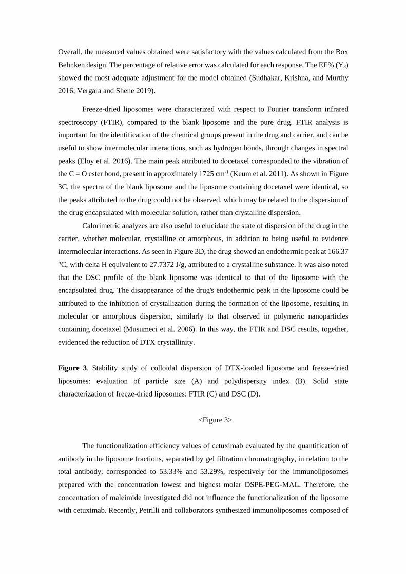
Freeze-dried liposomes were characterized with respect to Fourier transform infrared
spectroscopy (FTIR), compared to the blank liposome and the pure drug. FTIR analysis is
important for the identification of the chemical groups present in the drug and carrier, and can be
useful to show intermolecular interactions, such as hydrogen bonds, through changes in spectral
peaks (Eloy et al. 2016). The main peak attributed to docetaxel corresponded to the vibration of
the C = O ester bond, present in approximately 1725 cm-1 (Keum et al. 2011). As shown in Figure
3C, the spectra of the blank liposome and the liposome containing docetaxel were identical, so
the peaks attributed to the drug could not be observed, which may be related to the dispersion of
the drug encapsulated with molecular solution, rather than crystalline dispersion.
Calorimetric analyzes are also useful to elucidate the state of dispersion of the drug in the
carrier, whether molecular, crystalline or amorphous, in addition to being useful to evidence
intermolecular interactions. As seen in Figure 3D, the drug showed an endothermic peak at 166.37
°C, with delta H equivalent to 27.7372 J/g, attributed to a crystalline substance. It was also noted
that the DSC profile of the blank liposome was identical to that of the liposome with the
encapsulated drug. The disappearance of the drug's endothermic peak in the liposome could be
attributed to the inhibition of crystallization during the formation of the liposome, resulting in
molecular or amorphous dispersion, similarly to that observed in polymeric nanoparticles
containing docetaxel (Musumeci et al. 2006). In this way, the FTIR and DSC results, together,
evidenced the reduction of DTX crystallinity.
Figure 3. Stability study of colloidal dispersion of DTX-loaded liposome and freeze-dried
liposomes: evaluation of particle size (A) and polydispersity index (B). Solid state
characterization of freeze-dried liposomes: FTIR (C) and DSC (D).
<Figure 3>
The functionalization efficiency values of cetuximab evaluated by the quantification of
antibody in the liposome fractions, separated by gel filtration chromatography, in relation to the
total antibody, corresponded to 53.33% and 53.29%, respectively for the immunoliposomes
prepared with the concentration lowest and highest molar DSPE-PEG-MAL. Therefore, the
concentration of maleimide investigated did not influence the functionalization of the liposome
with cetuximab. Recently, Petrilli and collaborators synthesized immunoliposomes composed of
dysteroylphosphatidylcholine (DSPC) and CHOL and functionalized by the tioeter bond with
cetuximab, observing 59.4% efficiency in the functionalization of the blank immunoliposome,
similar to the result obtained in the present study (Petrilli et al. 2017).
The physicochemical characterization of the immunoliposome showed particle size and
PdI corresponding to 128.8 ± 2.35 nm and 0.279 ± 0.002, respectively, while the zeta potential
was -7.51 ± 0.50. It is noteworthy that these values were similar to those found in previous work
of immunoliposome of similar composition, based on SPC: Chol: DSPE-MAL, containing the
antibody trastuzumab (Eloy et al. 2017). Finally, the DTX encapsulation efficiency corresponded
to 93.5 ± 2.3% in the immunoliposome, showing that the functionalization reaction did not
compromise the drug encapsulation.
As seen in Figure 4A, cetuximab exhibited two bands on the electrophoresis gel under
reducing conditions: approximately 55 kDa and 25 kDa, corresponding to the immunoglobulin
heavy and light chains respectively. The electrophoretic profile of the immunoliposome was
identical to that of cetuximab, so it is concluded that there were no damages in the protein and
impairment of its primary structural integrity, as previously observed in liposomes functionalized
with cetuximab (Petrilli et al. 2017)
In order to investigate the integrity of the tertiary structure of the antibody after
functionalization, fluorescence assay was performed using intrinsic probes, that are, the aromatic
amino acid residues present in the cetuximab structure. The tests were carried out at 280 nm, in
which the tryptophans and tyrosines were excited, and, thus, presenting considerable fluorescence
intensity in that wavelength (Eftink, M. R., 1994). Figure 4B depicts the fluorescence emission
spectra at four tested conditions: cetuximab thiol, cetuximab control and the two previous
situations in the presence of 6 mmol.L-1 of GdnHCl (Guanidine hydrochloride), a denaturant
agent. Cetuximab presented the maximum emission wavelength (λmax) centered at 336 ± 1 nm
indicating that the tryptophans, the main fluorophore, are mostly protected from the solvent. The
chemically denaturation led cetuximab to suffer a red shift (λmax at 350 nm) and fluorescence
suppression indicating the tryptophans are now exposed to the solvent. Thiolated cetuximab,
presented λmax at around 340 ± 1 nm indicating that the functionalization reaction caused a slightly
red shift effect and a fluorescence suppression as well. These data could be an indicative that
functionalization caused some change in the cetuximab local tertiary structure but did not cause
denaturation. Thus, biological studies are paramount the investigate the maintenance of
cetuximab biological role.
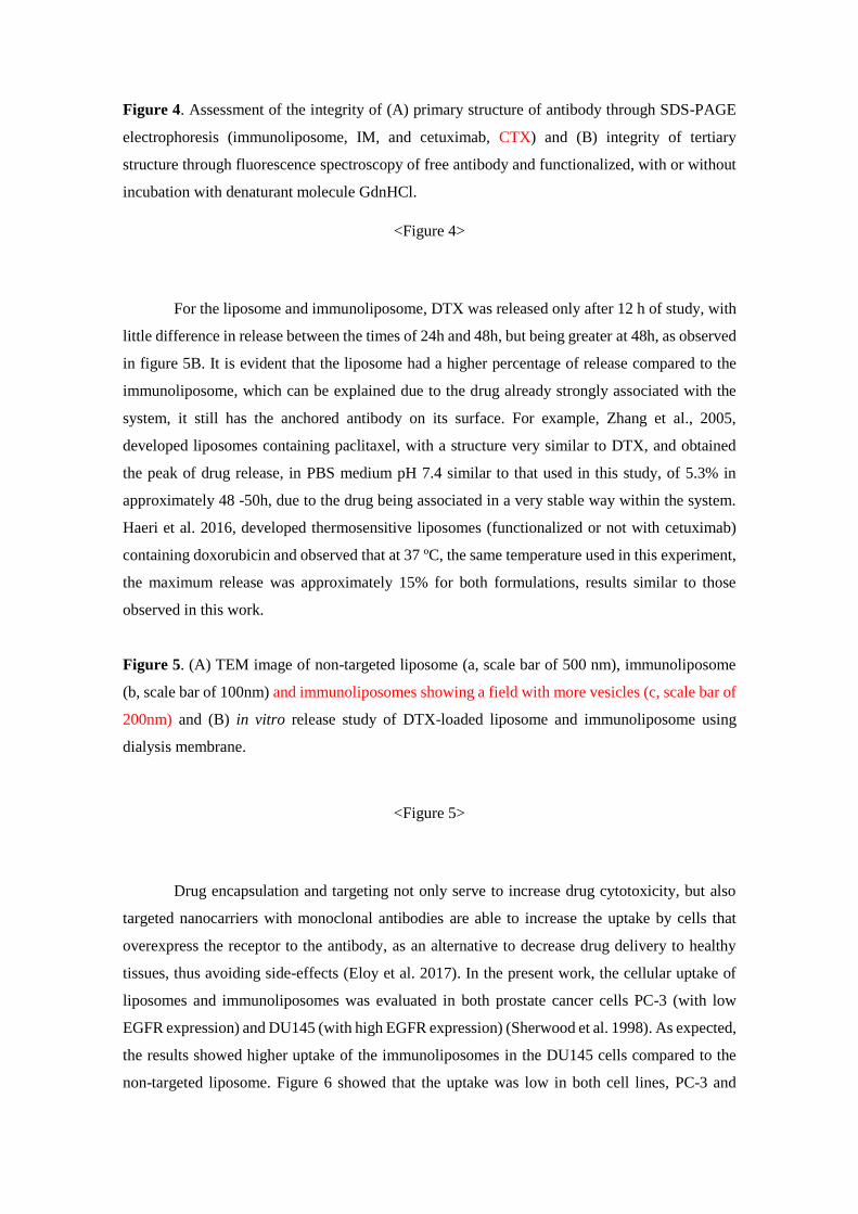
Figure 4. Assessment of the integrity of (A) primary structure of antibody through SDS-PAGE
electrophoresis (immunoliposome, IM, and cetuximab, CTX) and (B) integrity of tertiary
structure through fluorescence spectroscopy of free antibody and functionalized, with or without
incubation with denaturant molecule GdnHCl.
<Figure 4>
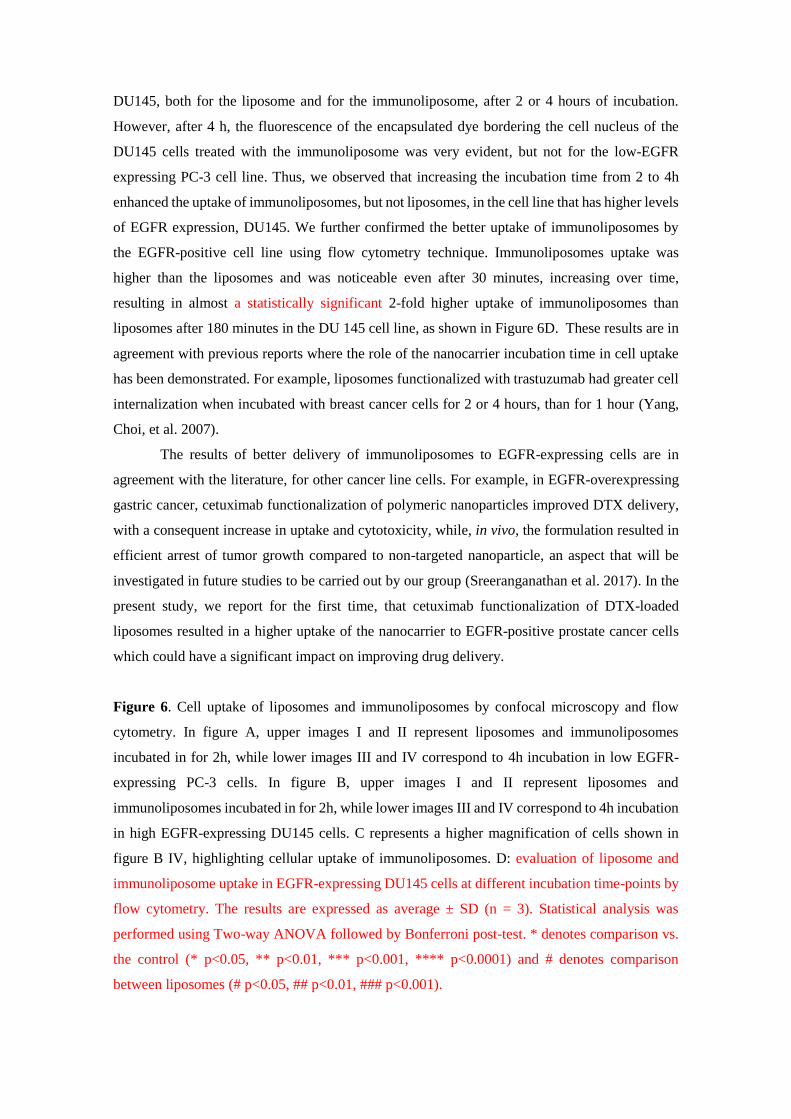
For the liposome and immunoliposome, DTX was released only after 12 h of study, with
little difference in release between the times of 24h and 48h, but being greater at 48h, as observed
in figure 5B. It is evident that the liposome had a higher percentage of release compared to the
immunoliposome, which can be explained due to the drug already strongly associated with the
system, it still has the anchored antibody on its surface. For example, Zhang et al., 2005,
developed liposomes containing paclitaxel, with a structure very similar to DTX, and obtained
the peak of drug release, in PBS medium pH 7.4 similar to that used in this study, of 5.3% in
approximately 48 -50h, due to the drug being associated in a very stable way within the system.
Haeri et al. 2016, developed thermosensitive liposomes (functionalized or not with cetuximab)
containing doxorubicin and observed that at 37 ºC, the same temperature used in this experiment,
the maximum release was approximately 15% for both formulations, results similar to those
observed in this work.
Figure 5. (A) TEM image of non-targeted liposome (a, scale bar of 500 nm), immunoliposome
(b, scale bar of 100nm) and immunoliposomes showing a field with more vesicles (c, scale bar of
200nm) and (B) in vitro release study of DTX-loaded liposome and immunoliposome using
dialysis membrane.
<Figure 5>
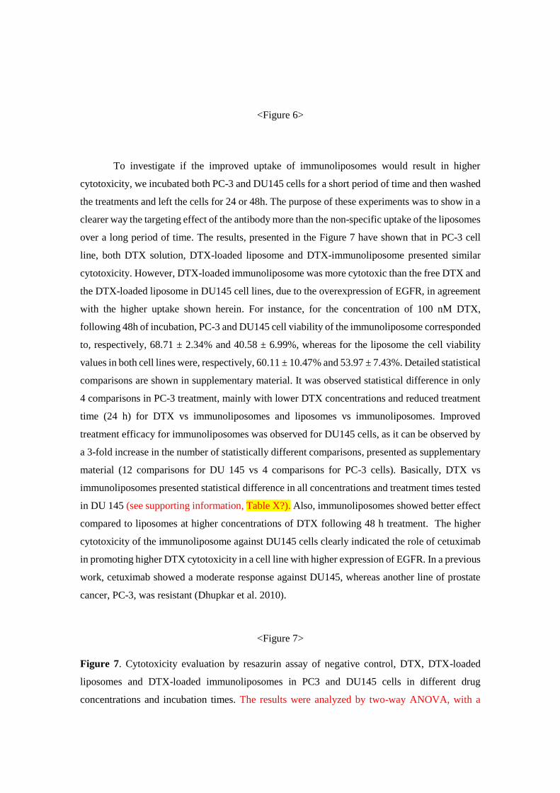
Drug encapsulation and targeting not only serve to increase drug cytotoxicity, but also
targeted nanocarriers with monoclonal antibodies are able to increase the uptake by cells that
overexpress the receptor to the antibody, as an alternative to decrease drug delivery to healthy
tissues, thus avoiding side-effects (Eloy et al. 2017). In the present work, the cellular uptake of
liposomes and immunoliposomes was evaluated in both prostate cancer cells PC-3 (with low
EGFR expression) and DU145 (with high EGFR expression) (Sherwood et al. 1998). As expected,
the results showed higher uptake of the immunoliposomes in the DU145 cells compared to the
non-targeted liposome. Figure 6 showed that the uptake was low in both cell lines, PC-3 and
DU145, both for the liposome and for the immunoliposome, after 2 or 4 hours of incubation.
However, after 4 h, the fluorescence of the encapsulated dye bordering the cell nucleus of the
DU145 cells treated with the immunoliposome was very evident, but not for the low-EGFR
expressing PC-3 cell line. Thus, we observed that increasing the incubation time from 2 to 4h
enhanced the uptake of immunoliposomes, but not liposomes, in the cell line that has higher levels
of EGFR expression, DU145. We further confirmed the better uptake of immunoliposomes by
the EGFR-positive cell line using flow cytometry technique. Immunoliposomes uptake was
higher than the liposomes and was noticeable even after 30 minutes, increasing over time,
resulting in almost a statistically significant 2-fold higher uptake of immunoliposomes than
liposomes after 180 minutes in the DU 145 cell line, as shown in Figure 6D. These results are in
agreement with previous reports where the role of the nanocarrier incubation time in cell uptake
has been demonstrated. For example, liposomes functionalized with trastuzumab had greater cell
internalization when incubated with breast cancer cells for 2 or 4 hours, than for 1 hour (Yang,
Choi, et al. 2007).
The results of better delivery of immunoliposomes to EGFR-expressing cells are in
agreement with the literature, for other cancer line cells. For example, in EGFR-overexpressing
gastric cancer, cetuximab functionalization of polymeric nanoparticles improved DTX delivery,
with a consequent increase in uptake and cytotoxicity, while, in vivo, the formulation resulted in
efficient arrest of tumor growth compared to non-targeted nanoparticle, an aspect that will be
investigated in future studies to be carried out by our group (Sreeranganathan et al. 2017). In the
present study, we report for the first time, that cetuximab functionalization of DTX-loaded
liposomes resulted in a higher uptake of the nanocarrier to EGFR-positive prostate cancer cells
which could have a significant impact on improving drug delivery.
Figure 6. Cell uptake of liposomes and immunoliposomes by confocal microscopy and flow
cytometry. In figure A, upper images I and II represent liposomes and immunoliposomes
incubated in for 2h, while lower images III and IV correspond to 4h incubation in low EGFR-
expressing PC-3 cells. In figure B, upper images I and II represent liposomes and
immunoliposomes incubated in for 2h, while lower images III and IV correspond to 4h incubation
in high EGFR-expressing DU145 cells. C represents a higher magnification of cells shown in
figure B IV, highlighting cellular uptake of immunoliposomes. D: evaluation of liposome and
immunoliposome uptake in EGFR-expressing DU145 cells at different incubation time-points by
flow cytometry. The results are expressed as average ± SD (n = 3). Statistical analysis was
performed using Two-way ANOVA followed by Bonferroni post-test. * denotes comparison vs.
the control (* p<0.05, ** p<0.01, *** p<0.001, **** p<0.0001) and # denotes comparison
between liposomes (# p<0.05, ## p<0.01, ### p<0.001).
<Figure 6>
To investigate if the improved uptake of immunoliposomes would result in higher
cytotoxicity, we incubated both PC-3 and DU145 cells for a short period of time and then washed
the treatments and left the cells for 24 or 48h. The purpose of these experiments was to show in a
clearer way the targeting effect of the antibody more than the non-specific uptake of the liposomes
over a long period of time. The results, presented in the Figure 7 have shown that in PC-3 cell
line, both DTX solution, DTX-loaded liposome and DTX-immunoliposome presented similar
cytotoxicity. However, DTX-loaded immunoliposome was more cytotoxic than the free DTX and
the DTX-loaded liposome in DU145 cell lines, due to the overexpression of EGFR, in agreement
with the higher uptake shown herein. For instance, for the concentration of 100 nM DTX,
following 48h of incubation, PC-3 and DU145 cell viability of the immunoliposome corresponded
to, respectively, 68.71 ± 2.34% and 40.58 ± 6.99%, whereas for the liposome the cell viability
values in both cell lines were, respectively, 60.11 ± 10.47% and 53.97 ± 7.43%. Detailed statistical
comparisons are shown in supplementary material. It was observed statistical difference in only
4 comparisons in PC-3 treatment, mainly with lower DTX concentrations and reduced treatment
time (24 h) for DTX vs immunoliposomes and liposomes vs immunoliposomes. Improved
treatment efficacy for immunoliposomes was observed for DU145 cells, as it can be observed by
a 3-fold increase in the number of statistically different comparisons, presented as supplementary
material (12 comparisons for DU 145 vs 4 comparisons for PC-3 cells). Basically, DTX vs
immunoliposomes presented statistical difference in all concentrations and treatment times tested
in DU 145 (see supporting information, Table X?). Also, immunoliposomes showed better effect
compared to liposomes at higher concentrations of DTX following 48 h treatment. The higher
cytotoxicity of the immunoliposome against DU145 cells clearly indicated the role of cetuximab
in promoting higher DTX cytotoxicity in a cell line with higher expression of EGFR. In a previous
work, cetuximab showed a moderate response against DU145, whereas another line of prostate
cancer, PC-3, was resistant (Dhupkar et al. 2010).
<Figure 7>
Figure 7. Cytotoxicity evaluation by resazurin assay of negative control, DTX, DTX-loaded
liposomes and DTX-loaded immunoliposomes in PC3 and DU145 cells in different drug
concentrations and incubation times. The results were analyzed by two-way ANOVA, with a
Bonferroni test (p ˂ 0.05 was considered the minimum value of significance) and the complete
analysis was supplied as a supplementary material (Table X?).
4. CONCLUSION
Herein, docetaxel liposomes were developed using Box–Behnken factorial design. The developed
liposomes were nano-sized with high loading efficiency and colloidal stability. Furthermore, the
lyophilization process preserved liposomal physicochemical characteristics and solid-state
characterizations were conducted showing that docetaxel was encapsulated with reduced
crystallinity. Finally, the chosen formulation (with lipid: drug molar ratio, 23: 1, cholesterol: lipid
molar ratio, 1.1:10, and DSPE-PEG:lipid molar ratio, 0.75:10) was conjugated with anti-EGFR
antibody, cetuximab, with high efficiency, preserving the liposomal nanometric size and DTX
high encapsulation. The integrity of anti-EGFR antibodies after the covalent linkage via
maleimide chemistry to the liposomal surface was assessed and demonstrated primary and tertiary
structure integrity. Both liposomes and immunoliposomes demonstrated DTX sustained release,
in vitro. Also, biological in vitro cell culture studies were conducted in prostate cancer cell lines,
with overexpression or not of the EGF receptor. Improved uptake was demonstrated due to
receptor-ligand interactions in DU145 cell line compared with PC-3 cells. Besides, it was
demonstrated that anti-EGFR liposomes were more effective than non-targeted nanoparticles in
the EGFR-overexpressing cell line in terms of cytotoxicity. Thus, the developed
immunoliposomes can be considered a potential platform for drug delivery of docetaxel to
prostate cancer cells, with improved tumor cell selectivity and efficacy compared to non-targeted
liposomes.
ACKNOWLEDGEMENTS
We thank FAPESP (Fundação do Amparo à Pesquisa do Estado de São Paulo) (projects
# 2016/02723-8, 2017/04091-1, 2018/00814-1 and 2018/21104-2), CNPQ (Conselho Nacional de
Desenvolvimento Científico e Tecnológico (Project # 409362/2018-2) and CAPES
(Coordenação de Aperfeiçoamento de Pessoal de Nível Superior – Brasil) – Finance Code 001
for financial support. This work is part of the National Institute of Science and Technology in
Pharmaceutical Nanotechnology: a transdisciplinary approach INCT-NANOFARMA. Dr Al-
Jamal would like to thank Prostate Cancer UK (CDF12-002) for funding.
REFERENCES
Cathomas, Richard, Christian Rothermundt, Dirk Klingbiel, Lukas Bubendorf, Rolf
Jaggi, Daniel C. Betticher, Peter Brauchli, Denise Cotting, Cornelia Droege, Ralph
Winterhalder, Daniele Siciliano, Dominik R. Berthold, Miklos Pless, Ralph Schiess,
Roger von Moos, and Silke Gillessen. 2012. “Efficacy of Cetuximab in Metastatic
Castration-Resistant Prostate Cancer Might Depend on EGFR and PTEN Expression:
Results from a Phase II Trial (SAKK 08/07).” Cancer Therapy: Clinical 12: 6049-6057.
DOI: 10.1158/1078-0432.CCR-12-2219.
Das, Shilpi, and Susmita Mishra. 2017. “Box-Behnken Statistical Design to Optimize
Preparation of Activated Carbon from Limonia Acidissima Shell with Desirability
Approach.” Journal of Environmental Chemical Engineering 5 (1): 588–600.
https://doi.org/10.1016/j.jece.2016.12.034.
Dhupkar, Pooja, Melissa Dowling, Keith Cengel, and Bin Chen. 2010. “Effects of Anti-
EGFR Antibody Cetuximab on Androgen-Independent Prostate Cancer Cells.”
Anticancer Research 30 (6): 1905–10.
Eftink, M. R. 1994. “The Use of Fluorescence Methods to Monitor Unfolding Transitions
in Proteins.” Biochemistry (Moscow) 63 (3): 276–84.
Eloy, Josimar O., Raquel Petrilli, Deise L. Chesca, Fabiano P. Saggioro, Robert J. Lee,
and Juliana Maldonado Marchetti. 2017. “Anti-HER2 Immunoliposomes for Co-
Delivery of Paclitaxel and Rapamycin for Breast Cancer Therapy.” European
Journal of Pharmaceutics and Biopharmaceutics 115 (4): 159–67.
https://doi.org/10.1016/j.ejpb.2017.02.020.
Eloy, Josimar O., Raquel Petrilli, José Fernando Topan, Heriton Marcelo Ribeiro
Antonio, Juliana Palma Abriata Barcellos, Deise L. Chesca, Luciano Neder Serafini,
Daniel G. Tiezzi, Robert J. Lee, and Juliana Maldonado Marchetti. 2016. “Co-
Loaded Paclitaxel/Rapamycin Liposomes: Development, Characterization and in
Vitro and in Vivo Evaluation for Breast Cancer Therapy.” Colloids and Surfaces B:
Biointerfaces 141: 74–82. https://doi.org/10.1016/j.colsurfb.2016.01.032.
Engels, Frederike K., Ron A.A. Mathot, and Jaap Verweij. 2007. “Alternative Drug
Formulations of Docetaxel: A Review.” Anti-Cancer Drugs 18 (2): 95–103.
https://doi.org/10.1097/CAD.0b013e3280113338.
Guardiola, Salvador, Monica Varese, and Macarena Sa. 2019. “Review A Third Shot at
EGFR : New Opportunities in Cancer Therapy,” 1–15.
https://doi.org/10.1016/j.tips.2019.10.004
Guérin, O, P. Formento, C, Lo Nigro, P. Hoffman, J.L. Fischel, M.C. Etienne-Grimaldi,
M. Merlano, J.M. Ferrero, G. Milano. "Supra-additive antitumor effect of sunitinib
malate (SU11248, Sutent®) combined with docetaxel. A new therapeutic
perspective in hormone refractory prostate cancer". Journal of Cancer Research and
Clinical Oncology 134: 51-57, 10.1007/s00432-007-0247-4.
Haeri, Azadeh, Sara Zalba, Timo L.M. ten Hagen, Simin Dadashzadeh, and Gerben A.
Koning. 2016. “EGFR Targeted Thermosensitive Liposomes: A Novel
Multifunctional Platform for Simultaneous Tumor Targeted and Stimulus
Responsive Drug Delivery.” Colloids and Surfaces B: Biointerfaces 146: 657–69.
https://doi.org/10.1016/j.colsurfb.2016.06.012.
Hassan, Mohamad Zaki, S. M. Sapuan, Siti Amni Roslan, Sa’Ardin Abdul Aziz, and
Shamsul Sarip. 2019. “Optimization of Tensile Behavior of Banana Pseudo-Stem
(Musa Acuminate) Fiber Reinforced Epoxy Composites Using Response Surface
Methodology.” Journal of Materials Research and Technology 8 (4): 3517–28.
https://doi.org/10.1016/j.jmrt.2019.06.026.
Immordino, Maria Laura, Franco Dosio, and Luigi Cattel. 2006. “Stealth Liposomes:
Review of the Basic Science, Rationale, and Clinical Applications, Existing and
Potential.” International Journal of Nanomedicine 1 (3): 297–315.
Joshi, Gaurav, Pankaj Kumar Singh, Arvind Negi, Anil Rana, Sandeep Singh, and Raj
Kumar. 2015. “Growth Factors Mediated Cell Signalling in Prostate Cancer
Progression: Implications in Discovery of Anti-Prostate Cancer Agents.” Chemico-
Biological Interactions 240: 120–33. https://doi.org/10.1016/j.cbi.2015.08.009.
Kharmate, Geetanjali, Elham Hosseini-Beheshti, Josselin Caradec, Mei Yieng Chin,
Emma S. Tomlinson Guns, 2015. "Epidermal Growth Factor Receptor in Prostate
Cancer Derived Exosomes". PLOS ONE 11(5):e0154967. doi:10.1371/
journal.pone.0154967.
Keum, Chang Gu, Young Wook Noh, Jong Suep Baek, Ji Ho Lim, Chan Ju Hwang,
Young Guk Na, Sang Chul Shin, and Cheong Weon Cho. 2011. “Practical
Preparation Procedures for Docetaxel-Loaded Nanoparticles Using Polylactic Acid-
Co-Glycolic Acid.” International Journal of Nanomedicine 6: 2225–34.
https://doi.org/10.2147/ijn.s24547.
Koudelka, Štěpán, and Jaroslav Turánek. 2012. “Liposomal Paclitaxel Formulations.”
Journal of Controlled Release 163 (3): 322–34.
https://doi.org/10.1016/j.jconrel.2012.09.006.
Li, Mingyuan, Chunyang Du, Na Guo, Yuou Teng, Xin Meng, Hua Sun, Shuangshuang
Li, Peng Yu, and Hervé Galons. 2019. “Composition Design and Medical
Application of Liposomes.” European Journal of Medicinal Chemistry 164: 640–
53. https://doi.org/10.1016/j.ejmech.2019.01.007.
Maeda, H., J. WU, T. Sawa, Y Matsumura, and K Hori. 2000. “Tumor Vascular
Permeability and the EPR Effect in Macromolecular Therapeutics: A Review.”
Journal of Controlled Release 65: 271–284.
Mager, Rene, Olga Savko, Katharina Böhm, Anita Thomas, Robert Dotzauer, Hendrik
Borgmann, Wolfgang Jäger, et al. 2019. “Comparative Assessment of Docetaxel for
Safety and Efficacy between Hormone-Sensitive and Castration-Resistant
Metastatic Prostate Cancer.” Urologic Oncology: Seminars and Original
Investigations 37 (12): 999–1005. https://doi.org/10.1016/j.urolonc.2019.07.005.
Malmberg, Jennie, Vladimir Tolmachev, and Anna Orlova. 2011. “Imaging Agents for in
Vivo Molecular Profiling of Disseminated Prostate Cancer - Targeting EGFR
Receptors in Prostate Cancer: Comparison of Cellular Processing of [111In]-
Labeled Affibody Molecule ZEGFR:2377 and Cetuximab.” International Journal of
Oncology 38 (4): 1137–43. https://doi.org/10.3892/ijo.2011.915.
Martins, Susana, Bruno Sarmento, Domingos C. Ferreira, and Eliana B. Souto. 2007.
“Lipid-Based Colloidal Carriers for Peptide and Protein Delivery - Liposomes
versus Lipid Nanoparticles.” International Journal of Nanomedicine 2 (4): 595–607.
Merino, María, Sara Zalba, and María J. Garrido. 2018. “Immunoliposomes in Clinical
Oncology: State of the Art and Future Perspectives.” Journal of Controlled Release
275 (2017): 162–76. https://doi.org/10.1016/j.jconrel.2018.02.015.
Moraes, Carolina M., Eneida De Paula, André H. Rosa, and Leonardo F. Fraceto. 2010.
“Physicochemical Stability of Poly(Lactide-Co-Glycolide) Nanocapsules
Containing the Local Anesthetic Bupivacaine.” Journal of the Brazilian Chemical
Society 21 (6): 995–1000. https://doi.org/10.1590/S0103-50532010000600008.
Musumeci, T., C. A. Ventura, I. Giannone, B. Ruozi, L. Montenegro, R. Pignatello, and
G. Puglisi. 2006. “PLA/PLGA Nanoparticles for Sustained Release of Docetaxel.”
International Journal of Pharmaceutics 325 (1–2): 172–79.
https://doi.org/10.1016/j.ijpharm.2006.06.023.
Nabila, Morshed, Nishat Jahan, and Diandra Elizabeth Penheiro. 2018. “Polymeric
Nanoparticles for Targeted Delivery in Cancer Treatment: An Overview.”
International Journal of Pharmaceutical Sciences Review and Research 52 (1): 101–
11. http://globalresearchonline.net/journalcontents/v52-
1/19.pdf%0Ahttp://ovidsp.ovid.com/ovidweb.cgi?T=JS&PAGE=reference&D=em
exa&NEWS=N&AN=624202638.
Nelson, William G, Emmanuel S Antonarakis, H Ballentine Carter, Angelo M De Marzo,
and Theodore L Deweese. n.d. Prostate Cancer. Abeloff’s Clinical Oncology. Sixth
Edit. Elsevier Inc. https://doi.org/10.1016/B978-0-323-47674-4.00081-5.
Paini, Marco, Sean Ryan Daly, Bahar Aliakbarian, Ali Fathi, Elmira Arab Tehrany,
Patrizia Perego, Fariba Dehghani, and Peter Valtchev. 2015. “An Efficient Liposome
Based Method for Antioxidants Encapsulation.” Colloids and Surfaces B:
Biointerfaces 136: 1067–72. https://doi.org/10.1016/j.colsurfb.2015.10.038.
Patel, Jitendrakumar, Jitendra Amrutiya, Priyanka Bhatt, Ankit Javia, Mukul Jain &
Ambikanandan Misra. 2018. Journal of microencapsulation 35, 204-217.
https://doi.org/10.1080/02652048.2018.1453560.
Patil, Pritam, Gokul Khairnar, and Jitendra Naik. 2015. “Preparation and Statistical
Optimization of Losartan Potassium Loaded Nanoparticles Using Box Behnken
Factorial Design: Microreactor Precipitation.” Chemical Engineering Research and
Design 104: 98–109. https://doi.org/10.1016/j.cherd.2015.07.021.
Pereira, Sara, Rapahel Egbu, Gemma Jannati, and Wafa' T. Al-Jamal. 2016. "Docetaxel-
loaded liposomes: The effect of lipid composition and purification on drug
encapsulation and in vitro toxicity." International Journal of Pharmaceutics
514:150-159. https://doi.org/10.1016/j.ijpharm.2016.06.057.
Petrilli, Raquel, Josimar Eloy, Renata Lopez, and Robert Lee. 2017. “Cetuximab
Immunoliposomes Enhance Delivery of 5-FU to Skin Squamous Carcinoma Cells.”
Anti-Cancer Agents in Medicinal Chemistry 17: 301–8.
https://doi.org/10.2174/1871520616666160526110913.
Petrilli, Raquel, Josimar O. Eloy, Fabiano P. Saggioro, Deise L. Chesca, Marina Claro de
Souza,Marcos V.S. Dias, LuisL.P. da Silva, Robert J. Lee, Renata F.V.Lopez. 2018.
" Skin cancer treatment effectiveness is improved by iontophoresis of EGFRtargeted
liposomes containing 5-FU compared with subcutaneous injection." Journal of
Controlled Release 283:151-162. https://doi.org/10.1016/j.jconrel.2018.05.038
Rawla, Prashanth. 2019. “Epidemiology of Prostate Cancer.” World Journal of Oncology
10 (2): 63–89. https://doi.org/10.1159/000423644.
Rodallec, Anne, Jean Michel Brunel, Sarah Giacometti, Helene Maccario, Florian
Correard, Eric Mas, Caroline Orneto, et al. 2018. “Docetaxel–Trastuzumab Stealth
Immunoliposome: Development and in Vitro Proof of Concept Studies in Breast
Cancer.” International Journal of Nanomedicine 13: 3451–65.
https://doi.org/10.2147/IJN.S162454.
Saad, Fred, and Kurt Miller. 2014. “Treatment Options in Castration-Resistant Prostate
Cancer: Current Therapies and Emerging Docetaxel-Based Regimens.” Urologic
Oncology: Seminars and Original Investigations 32 (2): 70–79.
https://doi.org/10.1016/j.urolonc.2013.01.005.
Shaker, Sherif, Ahmed Gardouh, and Mamdouh Ghorab. 2017. “Factors Affecting
Liposomes Particle Size Prepared by Ethanol Injection Method.” Research in
Pharmaceutical Sciences 12 (5): 346–52. https://doi.org/10.4103/1735-
5362.213979.
Sherwood, E. R., J. L. Van Dongen, C. G. Wood, S. Liao, J. M. Kozlowski, and C. Lee.
1998. “Epidermal Growth Factor Receptor Activation in Androgen-Independent but
Not Androgen-Stimulated Growth of Human Prostatic Carcinoma Cells.” British
Journal of Cancer 77 (6): 855–61. https://doi.org/10.1038/bjc.1998.142.
Singh, Sanjay, Arati Sharma, and Gavin P. Robertson. 2012. “Realizing the Clinical
Potential of Cancer Nanotechnology by Minimizing Toxicological and Targeted
Delivery Concerns.” Cancer Res. 72 (22): 5663–68. https://doi.org/10.1158/0008-
5472.CAN-12-1527.
Soema, Peter C., Geert Jan Willems, Wim Jiskoot, Jean Pierre Amorij, and Gideon F.
Kersten. 2015. “Predicting the Influence of Liposomal Lipid Composition on
Liposome Size, Zeta Potential and Liposome-Induced Dendritic Cell Maturation
Using a Design of Experiments Approach.” European Journal of Pharmaceutics and
Biopharmaceutics 94: 427–35. https://doi.org/10.1016/j.ejpb.2015.06.026.
Solanki, Ajay B., Jolly R. Parikh, and Rajesh H. Parikh. 2007. “Formulation and
Optimization of Piroxicam Proniosomes by 3-Factor, 3-Level Box-Behnken
Design.” AAPS PharmSciTech 8 (4). https://doi.org/10.1208/pt0804086.
Sreeranganathan, Maya, Saji Uthaman, Bruno Sarmento, Chethampadi Gopi Mohan, In
Kyu Park, and Rangasamy Jayakumar. 2017. “In Vivo Evaluation of Cetuximab-
Conjugated Poly(γ-Glutamic Acid)-Docetaxel Nanomedicines in EGFR-
Overexpressing Gastric Cancer Xenografts.” International Journal of Nanomedicine
12: 7167–82. https://doi.org/10.2147/IJN.S143529.
Sudhakar, Beeravelli, Mylangam Chaitanya Krishna, and Kolapalli Venkata Ramana
Murthy. 2016. “Factorial Design Studies of Antiretroviral Drug-Loaded Stealth
Liposomal Injectable: PEGylation, Lyophilization and Pharmacokinetic Studies.”
Applied Nanoscience (Switzerland) 6 (1): 43–60. https://doi.org/10.1007/s13204-
015-0408-8.
Troiano, Greg, Jim Nolan, Donald Parsons, Christina Van Geen Hoven, and Stephen Zale.
2016. “A Quality by Design Approach to Developing and Manufacturing Polymeric
Nanoparticle Drug Products.” AAPS Journal 18 (6): 1354–65.
https://doi.org/10.1208/s12248-016-9969-z.
Tucci, Marcello, Orazio Caffo, Consuelo Buttigliero, Carla Cavaliere, Carmine D’aniello,
Massimo Di Maio, Stefania Kinspergher, et al. 2019. “Therapeutic Options for First-
Line Metastatic Castration-Resistant Prostate Cancer: Suggestions for Clinical
Practise in the CHAARTED and LATITUDE Era.” Cancer Treatment Reviews 74
(January): 35–42. https://doi.org/10.1016/j.ctrv.2019.01.002.
Vergara, Daniela, and Carolina Shene. 2019. “Encapsulation of Lactoferrin into Rapeseed
Phospholipids Based Liposomes: Optimization and Physicochemical
Characterization.” Journal of Food Engineering 262 (February): 29–38.
https://doi.org/10.1016/j.jfoodeng.2019.05.012.
Wu, Long, Kit Lun Yick, Sun Pui Ng, and Joanne Yip. 2012. “Application of the Box-
Behnken Design to the Optimization of Process Parameters in Foam Cup Molding.”
Expert Systems with Applications 39 (9): 8059–65.
https://doi.org/10.1016/j.eswa.2012.01.137.
Yang, Tao, Min Koo Choi, Fu De Cui, Jung Sun Kim, Suk Jae Chung, Chang Koo Shim,
and Dae Duk Kim. 2007. “Preparation and Evaluation of Paclitaxel-Loaded
PEGylated Immunoliposome.” Journal of Controlled Release 120 (3): 169–77.
https://doi.org/10.1016/j.jconrel.2007.05.011.
Yang, Tao, Fu De Cui, Min Koo Choi, Jei Won Cho, Suk Jae Chung, Chang Koo Shim,
and Dae Duk Kim. 2007. “Enhanced Solubility and Stability of PEGylated
Liposomal Paclitaxel: In Vitro and in Vivo Evaluation.” International Journal of
Pharmaceutics 338 (1–2): 317–26. https://doi.org/10.1016/j.ijpharm.2007.02.011.
Yoon, Ho Yub, Seong Shin Kwak, Moon Ho Jang, Min Hyung Kang, Si Woo Sung,
Chang Hyun Kim, Sung Rae Kim, Dong Woo Yeom, Myung Joo Kang, and Young
Wook Choi. 2017. “Docetaxel-Loaded RIPL Peptide (IPLVVPLRRRRRRRRC)-
Conjugated Liposomes: Drug Release, Cytotoxicity, and Antitumor Efficacy.”
International Journal of Pharmaceutics 523 (1): 229–37.
https://doi.org/10.1016/j.ijpharm.2017.03.045.
Zalba, Sara, Ana M. Contreras, Azadeh Haeri, Timo L.M. Ten Hagen, Iñigo Navarro,
Gerben Koning, and María J. Garrido. 2015. “Cetuximab-Oxaliplatin-Liposomes for
Epidermal Growth Factor Receptor Targeted Chemotherapy of Colorectal Cancer.”
Journal of Controlled Release 210: 26–38.
https://doi.org/10.1016/j.jconrel.2015.05.271.

![[Randomized clinical case-control trial for the comparison of docetaxel plus thiotepa versus docetaxel plus capecitabine in patients with metastatic breast cancer]](https://img.dokumen.tips/doc/110x75/63536a1d6ff1b55f420e545b/randomized-clinical-case-control-trial-for-the-comparison-of-docetaxel-plus-thiotepa.jpg)


